
Submitted:
01 August 2024
Posted:
01 August 2024
You are already at the latest version
Abstract
Tilapia, the second most extensively farmed fish in the Philippines, generates significant amounts of waste that are often underutilized, leading to environmental concerns. One specific waste material is tilapia heads, which contain a valuable extracellular matrix (ECM) and an abundant source of bioactive materials that have a potential application in various fields, including biomaterials for wound healing and nutraceuticals. This study aims to evaluate the effects of sonication as a viable decellularization method for extracting ECM from tilapia fish heads. Histological analyses, employing H&E staining, revealed a higher reduction in basophilic components in the sonication-assisted (dWS) samples compared to those without sonication (dNS), as confirmed by DNA quantification. Notably, dWS samples subjected to 1% SDS sonication for 10 minutes exhibited the lowest DNA content with a removal rate of 93.7%. However, the same dWS sample also manifested the highest protein removal with a retained protein of 33.86%. The SDS-PAGE analysis of both dWS and dNS samples exhibited a uniform pattern, featuring identical four subunit bands—namely, the β-dimer (225-227 kDa), γ bands (228-230 kDa), and two alpha chains (α1 and α2 chains) (116-120 kDa)—indicating that sonication did not introduce discernible alterations in the helical structure of collagen. ATR-FTIR spectra further confirmed the presence of collagen as indicated by the detection of collagen type I indicators, including amides A, B, I, II, and III, in all decellularized samples. The above findings highlight the potential of sonication to aid in the decellularization of waste tilapia heads with future research targeting practical applications in wound healing, nutraceuticals, and sustainable environmental preservation through waste utilization.
Keywords:
waste valorization
; decellularization
; tilapia heads
; extracellular matrix extraction
1. Introduction
The Philippines, ranking 8th globally in fish production, places significant emphasis on its aquaculture industry. In 2018, tilapia, the second-highest produced fish, generated substantial waste, including skin, scales, bones, viscera, and heads [1]. However, this waste if not properly disposed can lead to environmental concerns [1]. Current waste utilization focuses on low-value products like fishmeal and fertilizers, but efforts are underway to explore higher-value applications [2]. Extracting valuable products, especially from tilapia heads, has promising potential for industries like food, pharmaceuticals, and cosmetics [3].
Fish heads, constituting 29-35% of the fish's mass, serve as a significant source of marine-based extracellular matrix (ECM). Characterized by its richness in diverse proteins (collagens, elastin, fibrillin, fibronectin, laminin), proteoglycans (heparin sulfate, chondroitin sulfate, keratin sulfate, GAGs), and growth factors, the ECM serves as a reservoir for the molecular elements and growth factors present in the native tissue [4]. The predominant utilization of ECM revolves around its role in constructing tissue scaffolds and presents a significant avenue in tissue engineering and regenerative medicine [5,6].
Demineralization and decellularization are essential methods employed to extract ECM from fish heads. A common demineralizing agent is hydrochloric acid, specifically at a concentration of 0.5 N for a duration of 1 h [7,8]. Important factors in an effective demineralization method include efficient mineral removal, collagen preservation, and the assessment of micro-porosity [9]. Concurrently, decellularization involves the removal native cells from tissue while retaining a three-dimensional ECM structure, preserving bioactivity and mechanical properties [10]. This approach offers a distinctive top-down method for creating natural scaffolds in tissue engineering [10]. However, careful selection of effective decellularization agents is imperative to minimize disruptions and ensure thorough cell removal while preserving the structural and functional proteins within the three-dimensional structure, referred to as decellularized extracellular matrix (dECM). As each decellularization agent induces distinct effects on ECM proteins, the choice of method should be tailored to the tissue biomechanics essential for optimal functionality.
In tissue decellularization, various chemical detergents are commonly used. A study by Li et al. (2021) introduced an optimized dextrose/sodium lauryl ester sulfate (SLES)/Triton X-100 (TX-100) cocktail method for porcine whole lungs decellularization to generate a clinical-scale bioengineered scaffold [11]. This investigation, alongside with previous study, highlighted Triton X-100’s potential disruption on lipid–lipid and lipid–protein interactions, despite its effectiveness in solubilizing cell membranes, disengaging cytoskeletal proteins, and detaching DNA and DNA remnants from proteins [11,12]. Another study recommended the utilization of SLES over sodium dodecyl sulfate (SDS) due to SDS's prolonged tissue treatment, causing significant ECM degradation [13]. Other studies also highlighted the capability of SDS to remove cells and DNA components while potentially damaging collagen and glycosaminoglycans during prolonged treatment [14,15]. However, Kawasaki et al. (2015) identified SDS as a promising decellularization agent due to its accessibility and efficient cell removal despite the issue of causing pronounced ECM damage [16]. Nevertheless, TX-100 and SDS are recognized for meeting the rigorous criteria for effective decellularization [15,17]. The effectiveness of TX-100 on decellularization is contingent upon the inherent characteristics of the tissue undergoing the procedure and the integration of other decellularization methods [17]. Prior assessments of SDS’s affirm its capability for removal of cellular components and achieving a minimum 90% reduction in host DNA content [15]. Moreover, SDS is a viable decellularizing agent which exhibited low likelihood of inducing toxic effects on cells, provided if thoroughly removed [15].
Several studies, including those conducted by Keshvari et al. (2023) on kidney tissue with SLES and SDS, Yaghoubi et al. (2022) and Hassanpour et al. (2018) employing SLES for rat liver and human ovarian tissue respectively, and Miranda et al. (2021) using TX-100 for murine skeletal muscles, have explored diverse decellularization methodologies [18,19,20,21]. However, these methods have their limitations, including extended treatment duration, alterations in mechanical properties, and potential residual toxicity [22]. To address the constraints of existing decellularization protocols, researchers are actively developing new methods with the goal of reducing treatment duration, minimizing exposure to chemical or organic agents, and diminishing tissue damage [23]. Sonication, recognized as an alternative for decellularization, involves the application of ultrasonic energy to induce cavitation phenomena, leading to the physical dissociation of molecules by causing the implosion of air bubbles in a liquid [23,24]. Notably, Shen et al. (2020) introduced a method incorporating freezing-thawing, sectioning, and sonication in deionized water for the decellularization of cartilage [25]. Azhim et al. (2013) proposed a sonication protocol using SDS for decellularizing aortic tissues [26]. These studies highlight the potential of sonication in achieving complete decellularization of the ECM, contributing valuable insights to decellularization methods.
At present, a standardized methodology for the decellularization of tilapia heads is yet to be established. This study explores the viability of tilapia fish heads as a promising source for the production of dECM. The primary aim is to evaluate the impact of integrating sonication in decellularization, utilizing 1% TX-100, 1% SDS, and deionized water, into established methods. This integration is designed to reduce treatment duration, limit exposure to decellularizing agents, and minimize tissue damage in the resulting dECM. To assess the effectiveness of sonication-assisted decellularization of tilapia heads, specific criteria established by Kawecki et al. (2018) are employed [27]. These criteria include the absence of visible nuclear remnants, a DNA content of less than 50 ng/mg of dried tissue, and the preservation of the ECM structure [28]. These benchmarks serve as critical indicators to assess the success of decellularization while ensuring the integrity of the ECM [27]. This research represents a significant endeavor to enhance and optimize the decellularization process for tilapia heads, providing valuable insights for the development of robust protocols in the broader context of tissue engineering and regenerative medicine.
2. Materials and Methods
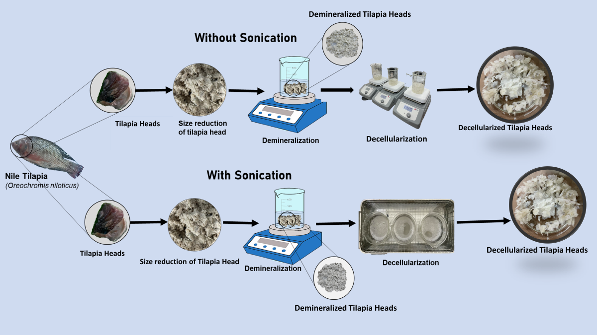
2.1. Sample Preparation and Demineralization
The methods employed for sample preparation and demineralization were adapted from the techniques utilized in the studies conducted by Shen et al. (2020) and Nisperos et al. (2023) [7,8]. Briefly, 30 kg of tilapia, weighing approximately 400-600 grams each, were purchased from a local wet market in Tambacan, Iligan City, Philippines, and stored in an ice-cooled container. The tilapia heads were manually removed from the purchased fish, and washed with distilled water after removal of soft tissues. The samples were ground using an electric grinder and pre-washed with phosphate-buffered saline (PBS) solution for 2 h to remove impurities.
Subsequently, the ground samples underwent demineralization through immersion and agitation in a 0.5 N hydrochloric acid (HCl) solution for 1 h with a mass-to-volume ratio of 1:10. After demineralization, the samples were washed and neutralized with distilled water, and were subsequently referred to as demineralized tilapia heads (dmTH) samples.
2.2. Decellularization Process
The dmTH samples were then decellularized under two treatments. The first treatment employed was adapted from the study conducted by Lin et al. (2021) using sonication-assisted (dWS) decellularization through a water bath sonicator (YTK-YASON, Shenzhen, China) [23]. The second method involved the same decellularization method but without sonication (dNS).
In both treatments, the decellularization was conducted at room temperature using three different decellularizing reagents. These reagents were 1% Sodium Dodecyl Sulfate (SDS) (Loba Chemie, Mumbai, India), 1% Triton X-100 (TX-100) (Loba Chemie, Mumbai, India), and deionized water (DEW) with a mass-to-solvent ratio of 1:10. The summary of the decellularization conditions is presented in Table 1.
After decellularization, the samples were washed thrice for 15 mins through agitation with distilled water. The washed samples were then referred as dWS and dNS corresponding to sonication-assisted and without sonication decellularization treatments, respectively. In addition, all decellularized samples are referred to as dcTH.
The moisture from the dWS and dNS were removed through lyophilization by initially freezing in an ultralow-temperature refrigerator (Haier, Qiangdao, China) at a temperature of -80 °C for at least 24 h. Subsequently, the frozen samples underwent lyophilization using a freeze dryer (HyperCOOL 3055 Gyrozen, Gimpo, Republic of Korea) operating under a vacuum at -55°C for a period of 24 h.
2.3. Evaluation of the dECM
2.3.1. Hematoxylin and Eosin (H&E) Staining
The methodology for histological staining was adapted from a previous study conducted by Oliveira et al. (2013) with some modifications [29]. Approximately 4 × 4 mm² dcTH samples were fixed in 10% buffered formalin for a duration of 72 h. Following fixation, the dcTH samples were dehydrated using ethanol. The samples were treated with xylene and embedded in paraffin wax. The embedded samples were then sliced into thin sections using a microtome (SLEE medical GmbH, Nieder-Olm, Germany). These sections were stained using hematoxylin and eosin (H&E) (Biognost®, Zagreb, Croatia), as per the method described by Ijima et al. (2019) [30]. Finally, the stained samples were examined and photographed using a phase-contrast microscope (Olympus CX43RF, Tokyo, Japan) to assess the presence of intact nuclei.
2.3.2. DNA Quantification
A 25 mg of the dcTH samples were weighed and transferred into individual 1.5 mL DNA-free microtubes. For DNA extraction, the DNeasy Blood and Tissue Kit (Qiagen®, California, USA) was used. The quantification of residual DNA content was performed using the Qubit™ 1X dsDNA HS Assay Kits (Thermo Fisher Scientific, Massachusetts, USA). The methods were conducted in accordance with the manufacturer's instructions.
2.3.3. Protein Quantification
To determine the protein content, the dcTH samples were digested in a 0.5 M acetic acid containing 0.01% pepsin. The mixtures were stirred for 48 h to ensure complete breakdown of proteins. After digestion, the solutions underwent centrifugation at 1670 rcf for 30 mins to separate the supernatant from the solid residue. The collected supernatants were analyzed according to the method of Steinhilber (2018) using a Qubit Protein Assay Kit (Thermo Fischer Scientific, Massachusetts, USA) and the protein concentrations were quantified with a Qubit Fluorometer (Thermo Fischer Scientific, Massachusetts, USA) [31].
2.3.4. Attenuated Total Reflectance- Fourier Transform Infrared Spectroscopy (ATR-FTIR) Analysis
The ATR-FTIR (Shimadzu IRTracer-100, Kyoto, Japan) was used to analyze and identify chemical bonds and functional groups present in the tissue. It allows for the characterization and identification of organic and inorganic compounds based on their unique infrared absorption spectra. The functional groups present in dmTH and dcTH were identified by scanning samples within the wave number range of 400 cm-1 to 4000 cm-1.
2.3.5. Sodium Dodecyl-Sulfate Polyacrylamide Gel Electrophoresis (SDS-PAGE)
SDS-PAGE was conducted according to the method of Laemmli (1970) with slight modification [32,33]. Briefly, dcTH samples were solubilized using 0.5 M acetic acid with a sample-solvent ratio of 1:100 and added with 0.01% pepsin (Merck, St Louis, MO, USA) which was then stirred continuously for 48 h. Electrophoresis of the solubilized samples was conducted using a mini vertical protein electrophoresis system (omniPAGE CVS10DSYS-CU, Cleaver Scientific, Warwickshire, United Kingdom) at 200V and 20mA for 1 h. After electrophoresis, the gels were stained using Coomassie Brilliant Blue R-25 (Abcam, Massachusetts, USA).
2.3.6. Differential Scanning Calorimetry (DSC)
The denaturation temperature of the dcTH samples was performed using a Differential Scanning Calorimeter (DSC 4000, Perkin Elmer, Waltham, MA, USA). The analysis was carried out under a nitrogen-purged atmosphere within a temperature range of 30°C to 300°C, with a heating rate of 10 °C/min.
2.3.7. Residual Detergent Determination
The concentration of residual detergents in the dcTH samples was determined using UV-VIS spectrophotometer (Thermo Fisher Scientific Genesys 10s, Massachusetts, USA). To quantify the residual TX-100 in the samples, the method outlined by Pavlović et al. (2016) was employed by modifying the concentration of standards used [34]. For residual SDS, the methylene blue examination method described by Alizadeh et al. (2019) was followed [35].
2.3.8. Statistical Analysis
The quantitative data obtained from the experiment, based on three replicates, were presented using the mean and the standard deviation (mean ± SD). To assess the significance of differences among multiple groups, one-way analysis of variance (ANOVA) was employed. This test allows comparison among several groups simultaneously. Subsequently, to identify specific group differences, a post hoc Tukey test was conducted. In this analysis, a result was considered significant when the probability value (p-value) associated with the comparison was less than 0.05.
3. Results
3.1. Hematoxylin and Eosin (H&E) Staining
The H&E staining of the raw sample, as shown in Figure 1A revealed the presence of basophilic components, evident by their characteristic blue staining pattern. Notably, after decellularization, a significant decrease in the basophilic components were observed for all dcTH samples, indicating the removal of cellular materials in the tissue. Moreover, the dWS samples, as shown in Figure 1 (H-M), exhibited a more pronounced reduction in the blue stains as compared to the dNS samples (B-G). In addition, the samples treated with 1% SDS (Figure 1B, C, H, I) and TX-100 (Figure 1D, E, J, K) exhibited minimal blue stains when compared to the samples treated DEW (Figure 1F, G, L, M). Furthermore, dcTH samples treated at higher contact time was observed to have a significant reduction of basophilic components. Overall, the use of 1% SDS for 10 mins with sonication resulted to the least basophilic components observed in the tissue among the dcTH samples.
3.2. DNA Quantification
The extent of cell removal through decellularization was assessed by analyzing the residual DNA in the dcTH samples, as illustrated in Figure 2. All decellularization treatments resulted to a significant reduction of DNA content (p<0.05). Notably, all dWS samples exhibited significant removal of DNA compared to dNS samples (p<0.05). The DEW for 5 mins showed the highest DNA residual with 84.21 ng/mg. Interestingly, the utilization of sonication assisted with 1% SDS for 10 mins revealed to have the lowest residual DNA having 7.67 ng/mg, with around 93.7% removal. Additionally, the quantity of residual DNA is generally lower at longer decellularization time, noting that contact time has a significant impact on the DNA removal of the tilapia heads (p<0.05).
3.3. Protein Quantification
The protein content of the dcTH samples is shown in Figure 3. A significant reduction of residual protein was observed in all dcTH samples as compared to the raw samples, which initially contained 218.25 ± 3.63 µg/mL of protein. The samples treated with DEW for 5 mins in both dNS and dWS displayed the highest protein content. Specifically, the DEW-treated with sonication showed lower protein content (126.67 ± 2.75 µg/mL) as compared to the DEW without sonication samples (147.08 ± 2.00 µg/mL). In contrast, 1% SDS for 10 mins with sonication yielded the lowest protein content of 73.92 ± 3.13 µg/mL. Samples treated with sonication illustrated lower protein content compared to those without sonication (p<0.05). Furthermore, statistical analysis shows that both treatments and duration significantly affect the protein content of the ECM (p<0.05).
3.4. Attenuated Total Reflectance-Fourier Transform Infrared Spectroscopy (ATR-FTIR) Analysis
The findings depicted in Figures 4A and 4B provide evidence of the preservation of collagen type I markers after decellularization. FTIR spectra obtained from the dNS samples (Figure 4A) revealed distinct band positions indicative of key components. Specifically, the amides I, II, and III were observed at 1645 cm-1, 1552 cm-1, and 1242 cm-1, respectively. The amide A band (3324 cm-1) was attributed to CH2 stretching, while the presence of collagen was signaled by the amide B band (3093 cm-1) [1] . Additionally, intensities of CH2 symmetric (2840 cm-1) and antisymmetric stretching bands (2920 cm-1) were more pronounced in samples treated with 1% TX-100. This aligns with previous reported studies confirming that the observed bands in the ATR-FTIR spectra effectively maintains the collagen structure after decellularization [9].
Conversely, in dWS treatments, a more pronounced intensities for CH2 symmetric and antisymmetric bands were observed for samples treated with 1% SDS as shown in Figure 4B. However, despite the variations in intensities, the characteristic bands associated with key biomolecules were still evident. Specifically, the presence of prominent bands such as Amides A (3315 cm-1), B (3097 cm-1), I (1633 cm-1), II (1552 cm-1), and III (1244 cm-1) was observed in the dWS samples.
3.5. Sodium Dodecyl Sulfate-Polycrylamide Gel Electrophoresis (SDS-PAGE)
The SDS-PAGE results, as shown in Figure 5, revealed distinct molecular weight bands of the dcTH samples ranging from approximately 50 to 225 kDa. Notably, the dNS samples did not exhibit distinct bands corresponding to the γ bands in contrast to the dWS samples. Specifically, the dWS samples demonstrated the presence of noteworthy protein markers, including the γ band (228-230 kDa), β-dimer (225-227 kDa), and α chains (116-120 kDa).
3.5. Differential Scanning Calorimetry (DSC)
DSC thermograms presented in Figure 6 provide valuable insights into the thermal characteristics of dcTH samples. The first endothermic peak spanning the temperature range of 60–90 °C was evident on all samples for both decellularization treatments. This peak is attributed to the release of free and bound moisture [36]. The second endothermic peak, ranging from 210-230 °C was also observed in both raw and dcTH samples for all decellularization conditions. Interestingly, among all samples, the use of SDS for 10 mins showed the highest denaturation peak temperature of 89.5 °C and 95.7 °C for dNS and dWS, respectively. Additionally, it was found that the dWS samples exhibited higher peak intensities compared to those subjected to dNS. Specifically, the employment of sonication in conjunction with deionized water for 5 mins resulted in the highest denaturation peak temperature, followed by TX-100 and SDS for 10 mins.
3.7. Residual Detergent Determination
The quantification of residual detergent was performed on the dcTH as shown in Figure 7. It was observed that dNS has higher residual detergent compared to dWS. With an increase in decellularization time, a higher concentration of residual detergent was observed in the dcTH samples. The use of SDS for decellularization resulted in significantly higher residual detergent concentrations as compared to the use of TX-100 (p<0.05). This finding is consistent with previous research, that indicates the challenging nature of removing SDS compared to other anionic detergents such as TX-100 [36]. Notably, a significant reduction of approximately 99% in residual detergent content was observed for the samples treated with 1% TX-100 at 5 mins.
4. Discussion
The utilization of fish waste, particularly tilapia heads, presents significant environmental and economic benefits by enabling the production of high-value products. Treated fish waste finds diverse applications, including the extraction of collagen and antioxidants for cosmetics, generating biogas/biodiesel, creating fertilizers, producing chitosan for dietary purposes, utilizing gelatine and chitosan in food packaging, and isolating enzymes like proteases. These applications showcase the versatility and value derived from fish waste processing across multiple industries [37]. The collagen-rich ECM of tilapia heads makes it an alternative source of collagen which is distinct from traditional sources such as bovine and pig skin [32,38]. However, it is important to note that there is still no decellularization method specifically tailored for tilapia heads. Therefore, research is needed to explore and establish effective decellularization methods specifically for tilapia heads. These efforts would significantly contribute to the utilization of tilapia heads in the development of ECM-based biomaterials for various applications.
Top of Form
The effects of sonication-assisted decellularization of tilapia heads with different reagents and varying contact time were investigated. Histological analysis revealed that sonication enhanced cell removal. Cell removal rate also increased with longer decellularization times. These histological findings were supported by the DNA quantification, which exhibited a similar trend of increased DNA removal with sonication and longer decellularization time. The decellularization was able to achieve the residual DNA levels below 50 ng/mg of dry tissue weight which is the criteria to ensure minimal potential immunogenicity and promote compatibility when utilizing the decellularized ECM for various applications [6].
The enhanced cell removal achieved through sonication can be attributed to its bioeffects on cell cytoplasm, inducing physical and chemical disruptions such as cavitation and emulsion [23]. Moreover, Azhim et al. (2013) highlighted that sonication can enhance the efficiency of decellularization agents like SDS by reducing dissolved oxygen levels thus maximizing the cavitation effect during the decellularization process. Additionally, in a separate study by Syazwani et al. (2014), sonication was successfully employed to achieve complete decellularization of an aorta, providing further evidence of the significant enhancement that sonication can bring to the removal of cellular components [24].
The analysis of protein content in the decellularized samples revealed a significant decrease, particularly in the samples treated with sonication using SDS. This decrease was expected as it is known that SDS is disruptive to ECM causing denaturation and unfolding of the protein structure [35]. Moreover, the decrease in protein could also be caused by the demineralization process involved in the dcTH samples, which is primarily concerned with the removal of minerals such as calcium that is bound to bind with collagen [8]. The depletion of non-collagenous proteins, therefore, could be the primary cause of the observed decrease in protein content while the collagen structure remained intact on TX-100 and DEW-treated samples. However, several studies have showed that sonication could be beneficial in dissociating cells from ECMs while preserving the major proteins and biomolecules in the ECM [23]. The difference on the results could be attributed the power or duration of sonication which could disrupt the main structural fibers of the ECM [23,39].
The preservation of essential collagen markers and their structural integrity in the dcTH samples was successfully confirmed through ATR-FTIR and SDS-PAGE analysis. These analytical techniques allowed for the identification and verification of collagen preservation despite the observed decrease in protein content. ATR-FTIR spectra exhibited similar peaks and bands for various functional groups in both dNS and dWS samples, indicating the effective preservation of collagen, the main component of the ECM. However, the dWS samples displayed more intense bands compared to the dNS samples. This finding suggests that the combined demineralization and sonication-assisted decellularization effectively removed hydroxyapatite while retaining important collagen markers such as amide I, II, III, and amides A and B [8]. Notably, the sonication treatment demonstrated a noticeable decrease in the V4PO peak compared to the without sonication, indicating the protonation of hydroxyapatite as a possible contributing factor to this observation.
The molecular weight bands on SDS-PAGE were predominantly observed in the range of 50-225 kDa for the dcTH samples. However, a more emphasized molecular weight bands were observed in dWS samples, especially with the use of SDS. Additionally, the analysis of dcTH samples revealed higher quantities of β dimer and molecular cross-linked components [32]. The method employed pepsin solubilization, which resulted in a prominent proportion of α chains, in accordance with the findings of Iijima et al. (2018) [30]. Furthermore, it was discovered that these collagens exhibited a chain composition of two α1 chains and a single α2 chain, as reported by Barajan et al. (2013) [32].
Thermal analysis of the dcTH samples revealed significant shifts in temperature peaks. The observed shifts indicate changes in the protein structure and composition due to the decellularization process, as well as the influence of sonication. In the case of the dcTH samples, it was observed that the denaturation peak temperatures were higher in the dWS compared to the dNS. This may suggests that the dWS samples contained proteins with higher thermal stability, possibly due to differences in tissue composition or the preservation of specific protein structures.
The presence of residual materials (detergent) on the ECM can lead to significant and potentially harmful inflammatory reactions [23]. In order to ensure thorough removal of nuclear materials (detergent), it is necessary to implement rigorous washing procedures. The use of SDS requires extensive washing process to avoid cytotoxic effect on the cell [35,40]. Moreover, future research should assess the biocompatibility and cytotoxicity of the generated decellularized extracellular matrix (dECM) to determine its suitability for tissue applications.
5. Conclusion
The decellularization of fish heads constitutes a crucial step in extracting the extracellular matrix (ECM) for diverse applications. However, limited research has been conducted on the decellularization of tilapia heads with a focus on preserving the ECM structure and other essential components. This study reveals that sonication-assisted decellularization presents notable advantages, including significantly enhanced cell and DNA removal, preservation of the ECM structure, and elevated denaturation temperatures. The preservation of ECM integrity is substantiated through the identification of collagen markers, absence of cellular components, analysis of protein content, and evaluation of thermal properties. Notably, the utilization of 1% SDS for 10 minutes with sonication emerges as the most effective approach for achieving decellularization and ECM preservation among the tested methods. Future investigations could delve into optimizing sonication power and frequency, as well as experimenting with varying concentrations of conventional reagents combined with sonication. Furthermore, complementary analyses, such as amino acid analysis and scanning electron microscopy (SEM), could provide additional insights into the effectiveness of optimized protocols. This study underscores the potential of sonication as a valuable tool in the decellularization of discarded tilapia heads. It not only contributes to the development of efficient protocols for biomaterial extraction but also presents a sustainable approach for transforming waste materials into valuable resources, thereby advancing the prospects of creating environmentally friendly and clinically relevant biomaterials.
Author Contributions
Conceptualization R.B.; methodology, L.B., R.B. and H.B.; software, L.B., K.D.D.V., and J.A.; formal analysis, L.B., M.J.N., F.A., G.L., and J.A.; investigation, L.B. and R.B.; data curation, R.B., L.B., and M.L.; writing—original draft preparation, L.B.; writing—review and editing, R.B., L.B., J.P., K.D.D.V., and M.L.; supervision, R.B. and H.B. All authors have read and agreed to the published version of the manuscript.
Funding
This research was funded by the Department of Science and Technology Philippine Council for Industry, Energy and Emerging Technology Research and Development (DOST-PCIEERD) under the Niche Centers in the Regions (NICER)—Science for Change Program (S4CP), grant number and DPMIS number 2021-03-A2-NICER-3209.
Institutional Review Board Statement
Not applicable.
Data Availability Statement
Not applicable.
Acknowledgments
The authors would like to thank Princess Grace Ducao, Zesreal Cain Bantilan and Johnel Alimasag for their valuable contributions and suggestions that helped to improve the quality of this manuscript.
Conflicts of Interest
The authors declare no conflict of interest. The funders had no role in the design of the study; in the collection, analyses, or interpretation of data; in the writing of the manuscript; or in the decision to publish the results.
References
- P. Gao, L. Li, W. Xia, Y. Xu, and S. Liu, “LWT - Food Science and Technology Valorization of Nile tilapia ( Oreochromis niloticus ) fish head for a novel fish sauce by fermentation with selected lactic acid bacteria,” LWT - Food Sci. Technol., vol. 129, no. April, p. 109539, 2020. [CrossRef]
- D. Coppola, C. Lauritano, F. P. Esposito, G. Riccio, C. Rizzo, and D. de Pascale, “Fish Waste: From Problem to Valuable Resource,” Marine Drugs, vol. 19, no. 2. MDPI, Feb. 01, 2021. [CrossRef]
- Y. Sun, L. Yan, S. Chen, and M. Pei, “Functionality of decellularized matrix in cartilage regeneration: A comparison of tissue versus cell sources,” Acta Biomaterialia, vol. 74. Acta Materialia Inc, pp. 56–73, Jul. 01, 2018. [CrossRef]
- C. Frantz, K. M. Stewart, V. M. Weaver, C. Frantz, K. M. Stewart, and V. M. Weaver, “The extracellular matrix at a glance The Extracellular Matrix at a Glance,” vol. 2010, pp. 4195–4200, 2010. [CrossRef]
- G. Agmon and K. L. Christman, “Controlling stem cell behavior with decellularized extracellular matrix scaffolds,” Curr. Opin. Solid State Mater. Sci., vol. 20, no. 4, pp. 193–201, 2016. [CrossRef]
- E. V. Isaeva, E. E. Beketov, N. V. Arguchinskaya, S. A. Ivanov, P. V. Shegay, and A. D. Kaprin, “Decellularized Extracellular Matrix for Tissue Engineering (Review),” Sovrem. Tehnol. v Med., vol. 14, no. 3, pp. 57–69, 2022. [CrossRef]
- S. F. Badylak, D. O. Freytes, and T. W. Gilbert, “Extracellular matrix as a biological scaffold material: Structure and function,” Acta Biomater., vol. 5, no. 1, pp. 1–13, 2009. [CrossRef]
- M. J. Nisperos et al., “Time-Dependent Demineralization of Tilapia ( Oreochromis niloticus ) Bones Using Hydrochloric Acid for Extracellular Matrix Extraction,” 2023.
- S. Pang, F. Y. Su, A. Green, J. Salim, J. McKittrick, and I. Jasiuk, “Comparison of different protocols for demineralization of cortical bone,” Sci. Rep., vol. 11, no. 1, pp. 1–10, 2021. [CrossRef]
- S. K. Gupta and N. C. Mishra, “Decellularization Methods for Scaffold Fabrication,” 2017. [CrossRef]
- Y. Li, Q. Wu, L. Li, F. Chen, J. Bao, and W. Li, “Decellularization of porcine whole lung to obtain a clinical-scale bioengineered scaffold,” J. Biomed. Mater. Res. - Part A, vol. 109, no. 9, pp. 1623–1632, 2021. [CrossRef]
- J. L. Balestrini et al., “Production of decellularized porcine lung scaffolds for use in tissue engineering,” Integr. Biol. (United Kingdom), vol. 7, no. 12, pp. 1598–1610, 2015. [CrossRef]
- S. Alaee, R. Asadollahpour, A. Hosseinzadeh Colagar, and T. Talaei-Khozani, “The decellularized ovary as a potential scaffold for maturation of preantral ovarian follicles of prepubertal mice,” Syst. Biol. Reprod. Med., vol. 67, no. 6, pp. 413–427, 2021. [CrossRef]
- T. Prebeg, D. Omerčić, V. Erceg, and G. Matijašić, “Comparison of Sodium Lauryl Sulfate and Sodium Lauryl Ether Sulfate Detergents for Decellularization of Porcine Liver for Tissue Engineering Applications,” Chem. Eng. Trans., vol. 100, no. April, pp. 745–750, 2023. [CrossRef]
- C. A. M. Bondi, J. L. Marks, L. B. Wroblewski, H. S. Raatikainen, S. R. Lenox, and K. E. Gebhardt, “Human and Environmental Toxicity of Sodium Lauryl Sulfate ( SLS ): Evidence for Safe Use in Household Cleaning Products,” pp. 27–32, 2015. [CrossRef]
- T. Wang et al., “This article has been accepted for publication and undergone full peer review but has not been through the copyediting, typesetting, pagination and proofreading process which may lead to differences between this version and the Version of Record. Please c,” Laryngoscope, vol. 44, no. 0, pp. 2–31, 2016.
- T. W. Gilbert, T. L. Sellaro, and S. F. Badylak, “Decellularization of tissues and organs,” vol. 27, pp. 3675–3683, 2006. [CrossRef]
- M. A. Keshvari et al., “Decellularization of kidney tissue: comparison of sodium lauryl ether sulfate and sodium dodecyl sulfate for allotransplantation in rat,” Cell Tissue Res., vol. 386, no. 2, pp. 365–378, 2021. [CrossRef]
- Yaghoubi, N. Azarpira, S. Karbalay-Doust, S. Daneshi, Z. Vojdani, and T. Talaei-Khozani, “Prednisolone and mesenchymal stem cell preloading protect liver cell migration and mitigate extracellular matrix modification in transplanted decellularized rat liver,” Stem Cell Res. Ther., vol. 13, no. 1, pp. 1–19, 2022. [CrossRef]
- Hassanpour, T. Talaei-khozani, E. Kargar-abarghouei, and V. Razban, “Decellularized human ovarian scaffold based on a sodium lauryl ester sulfate ( SLES ) -treated protocol , as a natural three- dimensional scaffold for construction of bioengineered ovaries,” pp. 1–13, 2018.
- M. F. C. Miranda et al., “Effects of chemical and physical methods on decellularization of murine skeletal muscles,” An. Acad. Bras. Cienc., vol. 93, no. 2, pp. 1–11, 2021. [CrossRef]
- K. Sawada, D. Terada, T. Yamaoka, S. Kitamura, and T. Fujisato, “Cell removal with supercritical carbon dioxide for acellular artificial tissue,” J. Chem. Technol. Biotechnol., vol. 83, no. 6, pp. 943–949, Jun. 2008. [CrossRef]
- Lin et al., “Sonication-Assisted Method for Decellularization of Human Umbilical Artery for Small-Caliber Vascular Tissue Engineering,” 2021.
- Azhim, N. Syazwani, Y. Morimoto, K. S. Furukawa, and T. Ushida, “The use of sonication treatment to decellularize aortic tissues for preparation of bioscaffolds,” vol. 29, no. 1, pp. 130–141, 2015. [CrossRef]
- W. Shen, K. Berning, S. W. Tang, and Y. W. Lam, “Rapid and detergent-free decellularization of cartilage,” Tissue Eng. - Part C Methods, vol. 26, no. 4, pp. 201–206, Apr. 2020. [CrossRef]
- Azhim et al., “The use of sonication treatment to completely decellularize aorta tissue,” IFMBE Proc., vol. 39 IFMBE, pp. 1987–1990, 2013. [CrossRef]
- M. Kawecki et al., “A review of decellurization methods caused by an urgent need for quality control of cell-free extracellular matrix’ scaffolds and their role in regenerative medicine,” J. Biomed. Mater. Res. - Part B Appl. Biomater., vol. 106, no. 2, pp. 909–923, 2018. [CrossRef]
- T. W. Gilbert, T. L. Sellaro, and S. F. Badylak, “Decellularization of tissues and organs,” Biomaterials, vol. 27, no. 19. pp. 3675–3683, Jul. 2006. [CrossRef]
- C. Oliveira et al., “Evaluation of Small Intestine Grafts Decellularization Methods for Corneal Tissue Engineering,” PLoS One, vol. 8, no. 6, Jun. 2013. [CrossRef]
- H. Ijima, S. Nakamura, R. Bual, N. Shirakigawa, and S. Tanoue, “Physical Properties of the Extracellular Matrix of Decellularized Porcine Liver,” pp. 1–15, 2018. [CrossRef]
- M. S. Immunoassay et al., “Omics Technologies Applied to Agriculture and Food Species Differentiation and Quantification of Processed Animal Proteins and Blood Products in Fish Feed using Article Species Differentiation and Quantification of Processed Animal Proteins and Blood Prod,” 2018. [CrossRef]
- P. Barajan, S. Sujithra, N. Kiruthiga, M. J. Prabhu, and R. Kumeresan, “Isolation and Determination of Type I Collagen from Tilapia (Oreochromis niloticus) Waste”.
- laemmli, U. Cleavage of Structural Proteins during the Assembly of the Head of Bacteriophage T4. Nature 227, 680–685 (1970). [CrossRef]
- B. Pavlović, N. Cvijetić, L. Dragačević, B. Ivković, Z. Vujić, and V. Kuntić, “Direct UV Spectrophotometry and HPLC determination of triton X-100 in split virus influenza vaccine,” J. AOAC Int., vol. 99, no. 2, pp. 396–400, 2016. [CrossRef]
- M. Alizadeh, L. Rezakhani, M. Soleimannejad, E. Sharifi, M. Anjomshoa, and A. Alizadeh, “Evaluation of vacuum washing in the removal of SDS from decellularized bovine pericardium: method and device description,” Heliyon, vol. 5, no. 8, 2019. [CrossRef]
- R. Bual et al., “Characterization of Decellularized Extracellular Matrix from Milkfish (Chanos chanos) Skin,” Biomimetics, vol. 7, no. 4, pp. 1–13, 2022. [CrossRef]
- S. Arvanitoyannis and A. Kassaveti, “Fish industry waste: Treatments, environmental impacts, current and potential uses,” Int. J. Food Sci. Technol., vol. 43, no. 4, pp. 726–745, Apr. 2008. [CrossRef]
- Sockalingam and H. Z. Abdullah, “Extraction of high value added gelatin biopolymer from black tilapia (Oreochromis mossambicus) head bones,” in AIP Conference Proceedings, Jul. 2015, vol. 1669. [CrossRef]
- Suslick, K.S. (2000). Sonochemistry. In Kirk-Othmer Encyclopedia of Chemical Technology, (Ed.). [CrossRef]
- Gilpin and Y. Yang, “Decellularization Strategies for Regenerative Medicine : From Processing Techniques to Applications,” vol. 2017, 2017.
Figure 1.
H&E Staining images of raw, dWS and dNS samples at different time durations with a magnification of 20x and scale bar of 100μm.
Figure 1.
H&E Staining images of raw, dWS and dNS samples at different time durations with a magnification of 20x and scale bar of 100μm.
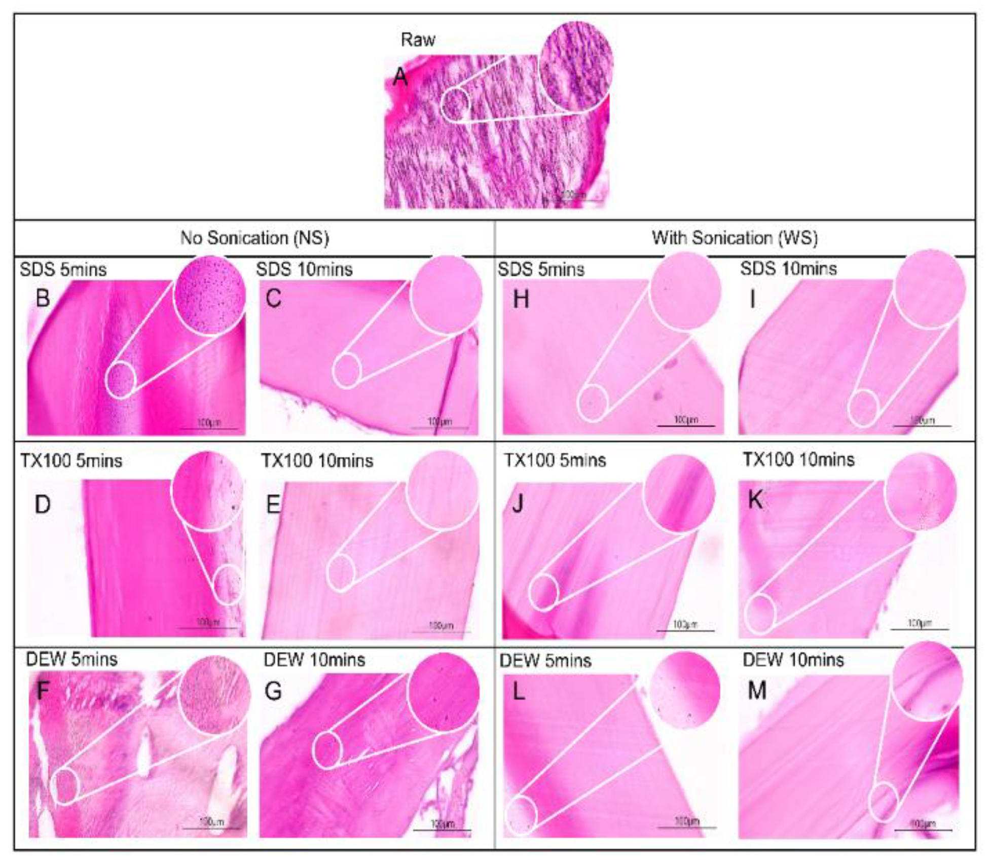
Figure 2.
DNA Quantification of raw and dcTH samples at different time durations. Bars represent standard deviation (n=3). * p<0.05.
Figure 2.
DNA Quantification of raw and dcTH samples at different time durations. Bars represent standard deviation (n=3). * p<0.05.
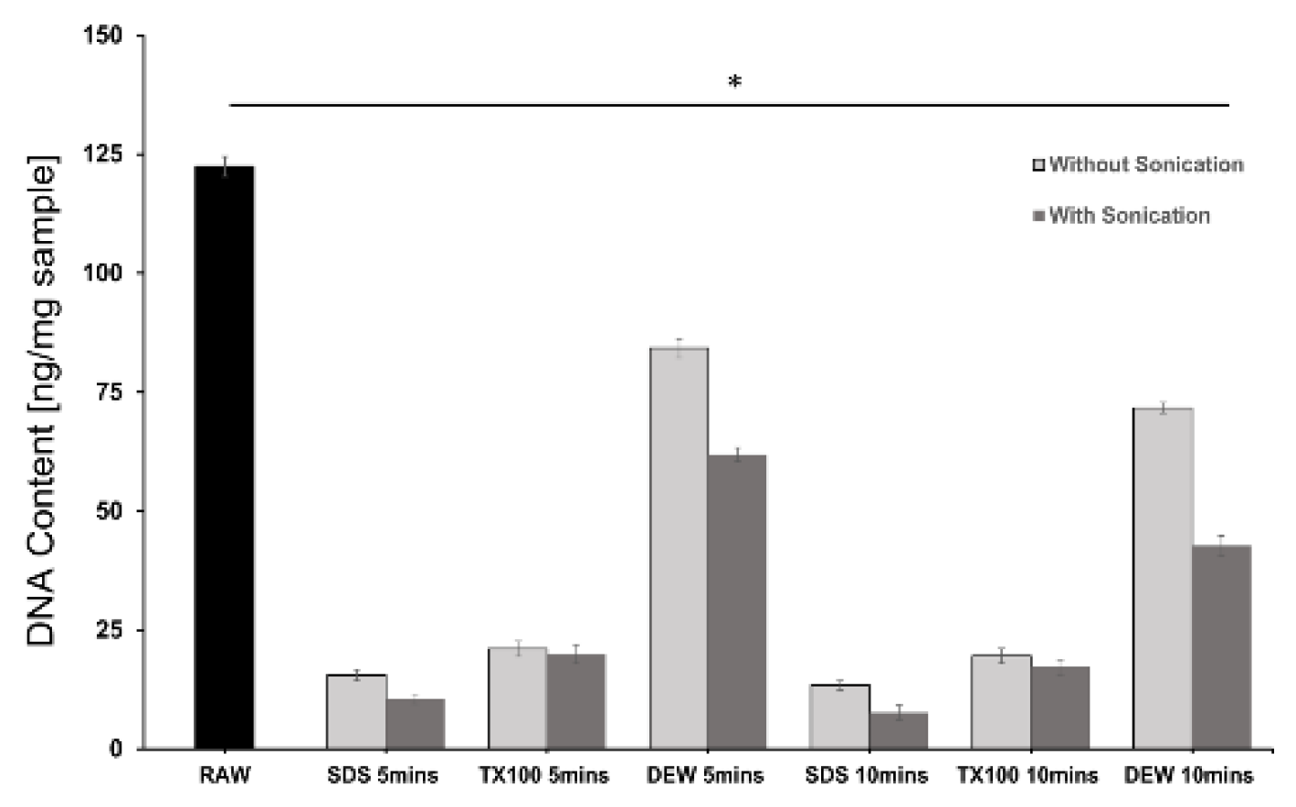
Figure 3.
Protein quantification of the raw and dcTH samples. Bars represent standard deviation (n=3). * p<0.05 .
Figure 3.
Protein quantification of the raw and dcTH samples. Bars represent standard deviation (n=3). * p<0.05 .
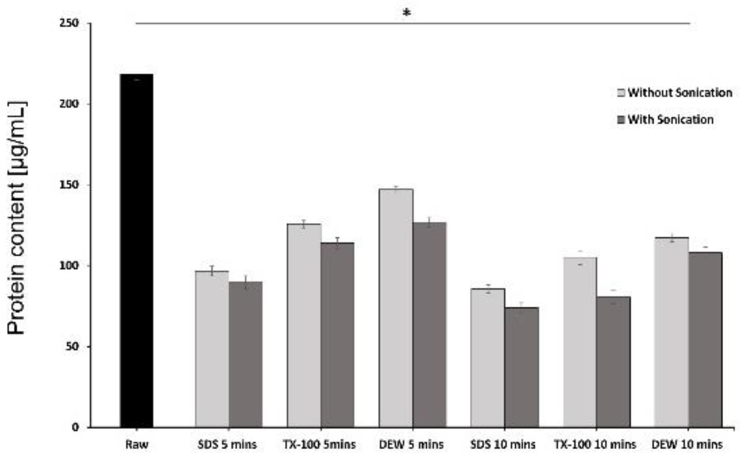
Figure 4.
(A) ATR-FTIR spectra of raw, dmTH, and dNS samples. (B)ATR-FTIR spectra of the raw, dmTH, and dWS samples.
Figure 4.
(A) ATR-FTIR spectra of raw, dmTH, and dNS samples. (B)ATR-FTIR spectra of the raw, dmTH, and dWS samples.
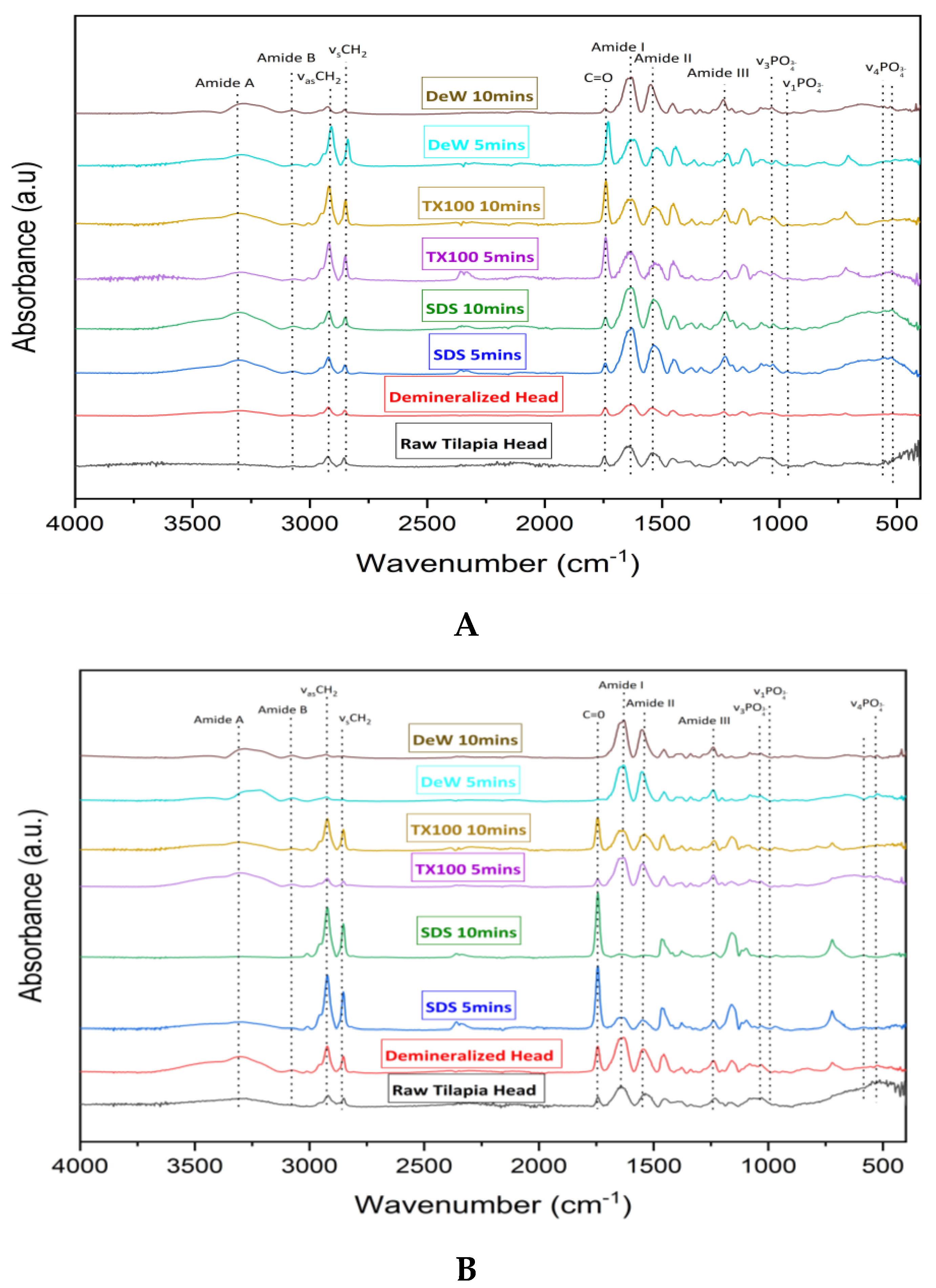
Figure 5.
SDS-PAGE analysis of the raw and dcTH samples.
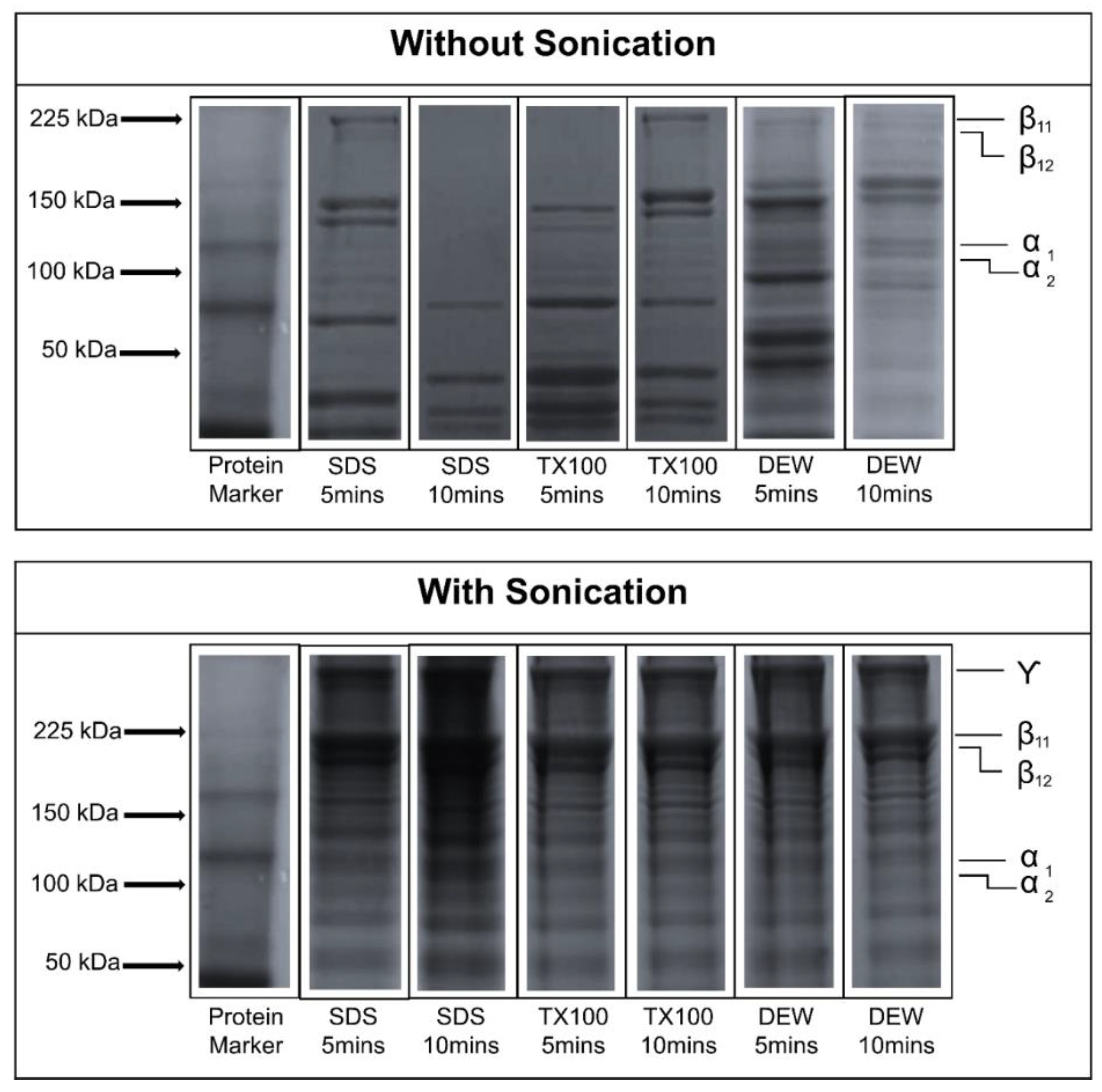
Figure 6.
Differential Scanning Calorimetry (DSC) thermograms of dNS (left) and dWS (right).
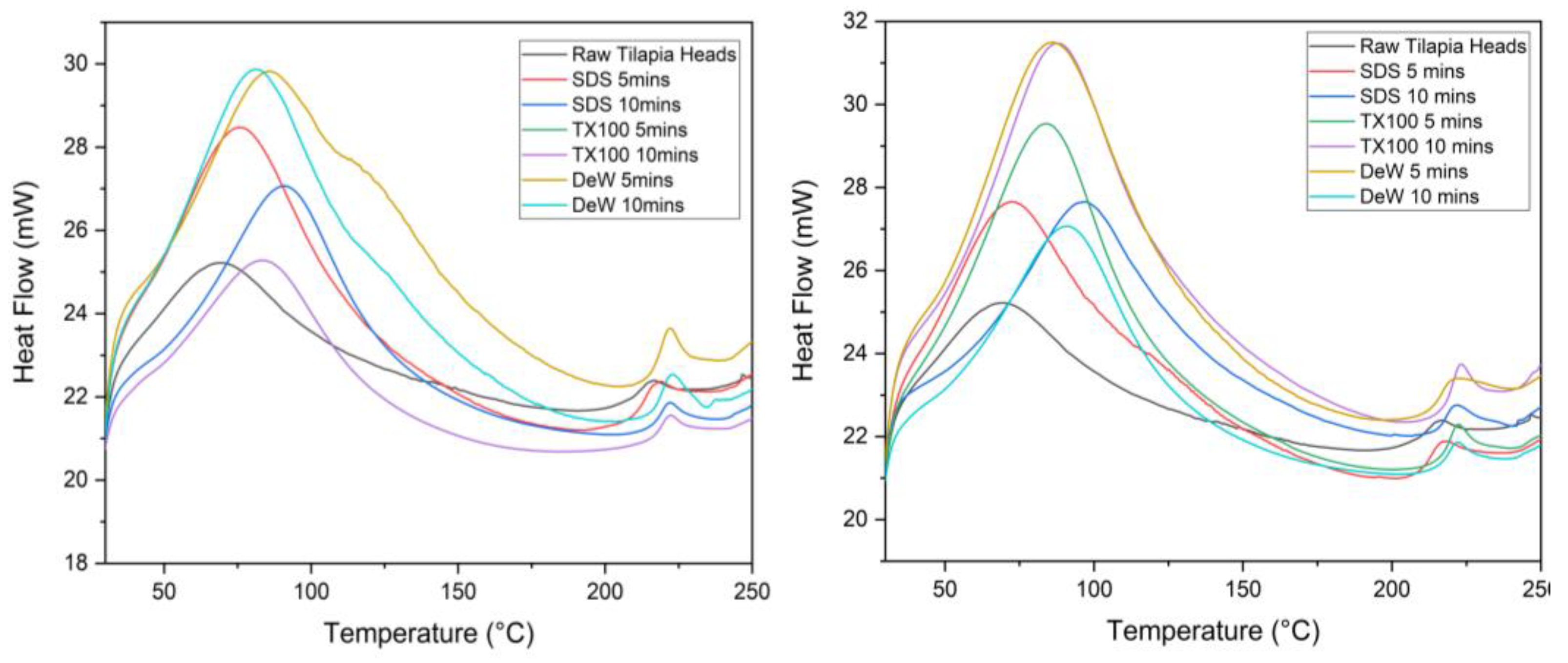
Figure 7.
Residual detergent test for 1% TX-100 and 1% SDS in the dcTH samples. Bars represent standard deviation (n=3). * p< 0.05.
Figure 7.
Residual detergent test for 1% TX-100 and 1% SDS in the dcTH samples. Bars represent standard deviation (n=3). * p< 0.05.
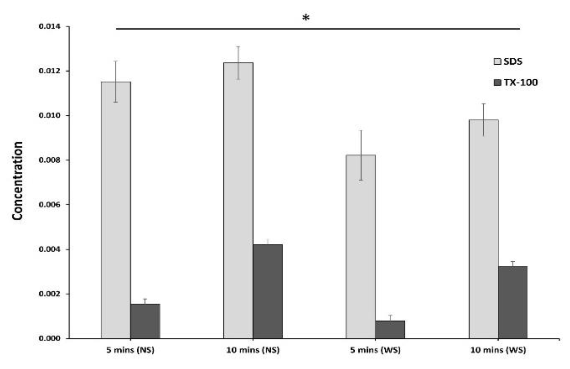

Table 1.
Overview of the decellularization conditions.
| Decellularizing Agent | Treatment | Contact time |
|---|---|---|
| 1% SDS, 1% TX-100, Deionized water (DEW) |
Sonication assisted (WS), Without sonication (NS) | 5 mins, 10 mins |
Disclaimer/Publisher’s Note: The statements, opinions and data contained in all publications are solely those of the individual author(s) and contributor(s) and not of MDPI and/or the editor(s). MDPI and/or the editor(s) disclaim responsibility for any injury to people or property resulting from any ideas, methods, instructions or products referred to in the content. |
© 2024 by the authors. Licensee MDPI, Basel, Switzerland. This article is an open access article distributed under the terms and conditions of the Creative Commons Attribution (CC BY) license (http://creativecommons.org/licenses/by/4.0/).
Copyright: This open access article is published under a Creative Commons CC BY 4.0 license, which permit the free download, distribution, and reuse, provided that the author and preprint are cited in any reuse.


