
Submitted:
19 July 2024
Posted:
23 July 2024
You are already at the latest version
Abstract
Background: Rectal prolapse is usually self-limiting in early childhood. Laparoscopic rectopexy has become a safe and effective method for treating children with rectal prolapse recurrence, especially those older than 4 years of age. We present the clinical case of an infant with recurrent rectal prolapse and secondary complications. The infant required surgical intervention with laparoscopic rectopexy after the failure of conservative management. Case presentation: A 2-year- and 7-month-old infant with a 15-month history of rectal prolapse who did not respond to conservative management developed bloody diarrhea, intestinal infection and electrolyte imbalance. Laparoscopic surgical correction was performed via anterior ventral rectopexy, and the results were satisfactory. Conclusion: Infants with recurrent rectal prolapse and associated complications who do not respond to conservative measures may benefit from laparoscopic rectopexy.
Keywords:
Rectal Prolapse
; Laparoscopic ventral rectopexy
; infant
INTRODUCTION
Rectal prolapse (RP) is defined as the protrusion of the rectum beyond the anus [1]. In pediatric patients, RP occurs more frequently at approximately three years of age and with an equal sex distribution. RP is called complete or true RP when all the layers of the rectal wall protrude outside the anus, mucous RP when only the mucous layer is prolapsed, and internal or hidden RP when the prolapsed mucosa does not extend beyond the anal canal. RP that recurs despite treatment is called recurrent RP, and one of the safest correction techniques is laparoscopic rectopexy (LR) [2]. We present the case of a female infant with recurrent RP, with complications including infections and electrolyte imbalance, who underwent LR. To our knowledge, this is the first reported infant case of recurrent RP treated with LR in Cartagena, Colombia, in the literature.
CASE PRESENTATION
A female patient aged 2 years and 7 months born in Lima, Peru, and permanently residing in Cartagena, Colombia, was evaluated in the emergency department of the Napoleón Franco Pareja Children’s Hospital in Cartagena, Colombia, because of a 6-day history of moderate-quantity watery stools with mucus and traces of bright red blood (~20 such bowel movements a day for the first four days, decreasing to 6 a day the last two days), accompanied by unquantified fever. Physical examination revealed a weight (W) of 10.7 kg (z score: 0.94), a height (H) of 85 cm (z score: 0.73), a W/ H:z ratio of 0.71 and a head circumference of 49 cm, with a normal nutrition status patient classification according to resolution 2465 of Colombia (https://www.minsalud.gov.co/sites/rid/Lists/BibliotecaDigital/RIDE/DE/DIJ/resolucion-2465-2016.pdf). In the perineal region, a reddish mass was observed protruding from the anal sphincter corresponding to the lower section of the rectum. The mass was not manually reducible and was considered a Grade V RP according to the Oxford classification [3]. The rest of the physical examination results were within normal limits.
Previously, the mother had noticed a red mass that protruded from the anorectal region at 16 months of age (Figure 1), with a normal result in the barium enema study. Since then, the patient had been evaluated for recurrent episodes of dysenteric diarrhea that were managed with electrolyte solutions, antibiotic therapy, zinc sulfate, dietary measures and manual reduction of the RP.
Complete rectal prolapse of the patient involving full-thickness extrusion of the rectal wall (Oxford Grade V)
Initial laboratory examination results showed a leukocyte count of 10-15 cells/field in a stool sample, elevated C-reactive protein (CRP) levels and moderate hypokalemia (Table 1). In consideration of gastrointestinal infection of presumed bacterial origin, ceftriaxone was administered at 100 mg/kg/day and hypokalemia was corrected by administering 40 mEq/L potassium chloride solution at 0.2 mEq/kg/hour. Conservative management was indicated, with dietary pattern modification, lidocaine with topical hydrocortisone and attempted manual reduction of the RP. Multiculture results were negative (Table 1).
Due to the persistence of symptoms and signs of complete RP (Grade III), laparoscopic surgical correction by anterior ventral rectopexy was indicated. Briefly, for the operation, a 5 mm trocar was inserted at the umbilical level via the open technique, the pneumoperitoneum was insufflated, and two 5 mm trocars were placed in the right and left flanks. The intestinal loops were displaced in the cephalic direction, leaving the hollow pelvis and rectum in the visual area, and by quadrant exploratory laparoscopy, a lax rectum and rectosigmoid junction were observed, which led to RP. After perirectal infiltration in the four quadrants with a hypertonic saline solution, the peritoneum was sectioned, and the mesorectum was dissected with Enseal TM X1 forceps (Ethicon. Ohio, USA), identifying and avoiding vascular structures and the right ureter. The rectopexy was performed with multiple polypropylene points (Prolene 4-0 JOHNSON & JOHNSON MEDTECH, Colombia) between the posterior wall of the rectum pulled cephalad and the sacral promontory, verifying the correction of the prolapse (Video S1). After hemostatic revision, the trocars were removed under laparoscopic view, and the pneumoperitoneum was evacuated for the spatial closure of the ports. In the immediate postoperative period, a complete perineum was observed, and there was no active bleeding and only slight inflammation. The patient was discharged via outpatient management with polyethylene glycol at 1 gram/kg/day orally for 3 days and nutritional recommendations with high-fiber foods. The patient was evaluated in the late postoperative period, and the perineum was complete and without signs of inflammation, there was no RP, and significant improvement in fecal incontinence was reported by the mother, with an absence of constipation.
Outpatient follow-up of the patient included interdisciplinary support from clinicians in the departments of nutrition, gastroenterology, psychology, pediatrics, and pediatric surgery.
Discussion
The case describes a female patient aged 2 years/7 months with intractable full-thickness RP for 15 months treated with laparoscopic rectopexy by reason of the persistence of her prolapse despite conservative measures.
RP is a protrusion of one or more layers of the rectum through the anal sphincter [4]. Symptoms and complaints related to RP differ according to age [5]. In the pediatric population, RP occurs more commonly in children under four years of age and is related to anatomical aspects such as the vertical course of the rectum along the straight surface of the sacrum and coccyx, a low position of the rectum in relation to other pelvic organs, increased mobility of the sigmoid colon, a relative lack of support from the levator ani muscle, a laxity of the junction of the redundant rectal mucosa to the underlying muscle, and the absence of Houston valves in approximately 75% of infants under 1 year old. In children, prolonged straining during toilet training, constipation, acute and chronic diarrhea, intestinal parasites, and malnutrition are common RP etiologies [6]. Connective tissue dysfunction has been described as a genetic factor, as a novel heterozygous mutation, c.1406G> T; p.G469V, was found in exon 11 of the COLGALT2 gene (collagen beta (1-O) galactosyltranferase 2) encoding the enzyme GLT25D2 (glycosyltransferase 25 domain containing 2) involved in collagen glycosylation in an 11-year-old patient and her family with RP [7]. In adults, a Cleveland Clinic/Wexner Fecal Incontinence score greater than 9 was more common in patients older than 65 years of age (87% versus 60%, p = 0.002) (6). Older patients were more likely than younger patients to report pain (38% vs. 19%, p = 0.021) and bleeding (12% vs. 2%, p = 0.046). Most of the patients reported rectal mucus discharge (older patients, 72%; younger patients, 66%; p = 0.54). Older patients had more severe prolapse exteriorization than younger patients did (at rest, 33% versus 11%; during activity, 26% versus 19%; only with defecation, 40% versus 64%, p = 0.006) [5].
Although RP is usually a self-limiting disorder in children under four years of age and most patients respond to conservative treatment within a year [8,9,10], surgical management decisions in clinical cases are based on the recurrence of RP without a response to manual reduction and the appearance of complications such as dysenteric diarrhea with signs of bacterial infection and serum electrolyte disorders.
The surgical approach for treating RP in pediatric patients is reserved for cases of ulceration of the rectal mucosa, irreducible prolapse and recurrence before conservative management under bowel control programs that include laxatives, nutritional adjustment and bowl habit training [9]. Nonsurgical options include perirectal sclerotherapy (ethyl alcohol and hypertonic saline), local procedures such as anal cerclage, excision with resection and rectosacropexy, and transabdominal surgical approaches include open mesh rectopexy and LR [8,11]. The use of the LR surgical technique has been promulgated throughout the world [12,13,14]. The selection of patients for LR correction is supported by the fact that it is a procedure that has the advantages of good accessibility, optimal visualization of the anatomy of the narrow pelvic space during surgery, less postoperative pain, a shorter hospital stay and earlier recovery than laparotomy [13,15]. In this patient, ventral LR was selected, in which direct pexy of the rectum to the sacral promontory was performed, and successful recovery was achieved. Other types of LR have been described. For example, sutureless LR was performed in 62 patients (median age, 80 years (IQR 10-91). The procedure involved rectal mobilization (together with the bilateral peritoneum) and fixation to the sacral promontory by applying 2 to 3 bilaterally fixed tacks with a fixation device (Fixation Device AbsorbaTack; Covidien Japan, Inc., Japan or Fixation Device ProTack; Covidien Japan, Inc.). There were no serious intraoperative complications, and conversion to open surgery was required for only one patient [16].
Another alternative is the mesh placement technique, as described in a study carried out in Al-Azhar, Cairo, Egypt, where mesh LR was compared with suture LR in seventy-eight children (: 8 years, standard deviation: 5-12) with persistent complete RP. The authors reported that the mean operative time in the suture LR group was shorter than that in the mesh LR group. No early postoperative complications occurred. There were also no cases of recurrence in the mesh LR group, while in the suture LR group, there were 4 cases of recurrence (14.2%). Postoperative constipation occurred in one patient (3.57%) in the suture LR group and in one patient (3.3%) in the mesh LR group. Fecal incontinence resolved in 26/28 patients (92.8%) in the suture LR group and in 30/30 patients (100%) in the mesh LR group. The authors found that both mesh LR and suture LR are feasible and reliable methods for the treatment of complete RP in children. However, in the mesh LR group, there was no recurrence, and there was a lower incidence of postoperative constipation and an improvement in incontinence; for this reason, the authors recommended mesh LR as an option for the treatment of complete RP [13]. Other authors prefer a meshless method, finding that postoperative constipation was the only side effect of the suture LR method [17].
To reduce the recurrence and postoperative constipation rates, modifications of the classic method have been proposed in which the rectum is mobilized only from the right side and the lateral ligaments or the peritoneal fold are not excised; the rectum is fixed only with a suture in the promontory of the sacrum [15].
In this case, the rectum was mobilized only from the right to reduce the possibility of constipation after surgery. The cut of the peritoneal fold allowed us to expose the posterior aspect of the rectum, and suturing toward the sacral promontory allowed the rectum to be fixed more to the sacrum with healing, with no recurrence of RP.
Surgical recourse in the correction of RP in children under 4 years of age is infrequent, and in some situations, affected children may have contraindications for surgical correction. For example, in a study of 15 children with persistent complete RP, two were 2 months old, and one had severe splenomegaly due to Gaucher disease; these patients were excluded from the study. The remaining 12 patients, with an average age of 3.32 ± 2.70 years, underwent rectopexy and laparoscopic sigmoidopexy [18]. In this case, the patient underwent surgery, and the results were successful, with improvement in her general condition and nutritional and psychomotor development and good acceptance by family members. Therefore, we consider this method an alternative for RP correction in infants with complete RP and complications secondary to this clinical condition, as few postoperative complications occur, and the recurrence rate is low.
A limitation of this case report is that no genetic tests were performed for the patient and therefore no genetic explanation of the etiology of the persistent RP can be provided.
Conclusion
LR can be an effective and safe alternative for the correction of recurrent complete RP in infants, as it can reduce the recurrence rate and improve patient quality of life, which allows improved psychomotor development in infants in optimal conditions.
Supplementary Materials
The following supporting information can be downloaded at the website of this paper posted on Preprints.org. The surgical procedure can be seen in the video S1: Correction of rectal prolapse of the clinical case through anterior ventral rectopexy.
Author Contributions
Conceptualization: P.T.O., L.V.U., H.P.R., D.F.R. Methodology: P.T.O., L.V.U., D.F.R. Formal analysis: P.T.O, L.V.U., H.P.R., D.F.R. Investigation: P.T.O., L.V.U, D.F.R. Writing-Original Draft preparation: L.V.U., D.F.R. Writing-Review and Editing: D.F.R. Visualization: P.T.O., L.V.U., H.P.R., D.F.R. Supervision: P.T.O., H.P.R., D.F.R.
Funding
This research received no external funding.
Institutional Review Board Statement
Not applicable.
Informed Consent Statement
Informed consent was obtained from the mother of the patient involved in the manuscript.
Conflicts of Interest
The authors declare no conflict of interest.
References
- Segal J, McKeown DG, Tavarez MM. Pediatric Rectal Prolapse. StatPearls. Treasure Island (FL)2024.
- Cares K, Klein M, Thomas R, El-Baba M. Rectal Prolapse in Children: An Update to Causes, Clinical Presentation, and Management. J Pediatr Gastroenterol Nutr. 2020;70(2):243-6. [CrossRef]
- Park BS, Cho SH, Son GM, Kim HS, Cho YH, Ryu DG, et al. Absent or impaired rectoanal inhibitory reflex as a diagnostic factor for high-grade (grade III-V) rectal prolapse: a retrospective study. BMC Gastroenterol. 2021;21(1):157. [CrossRef]
- Oruc M, Erol T. Current diagnostic tools and treatment modalities for rectal prolapse. World J Clin Cases. 2023;11(16):3680-93. [CrossRef]
- Neshatian L, Lee A, Trickey AW, Arnow KD, Gurland BH. Rectal Prolapse: Age-Related Differences in Clinical Presentation and What Bothers Women Most. Dis Colon Rectum. 2021;64(5):609-16. [CrossRef]
- Pfefferkorn MD, Fitzgerald JF. 48 - Disorders of the Anorectum: Fissures, Fistulas, Prolapse, Hemorrhoids, Tags. In: Wyllie R, Hyams JS, editors. Pediatric Gastrointestinal and Liver Disease (Fourth Edition). Saint Louis: W.B. Saunders; 2011. p. 521-7.e1.
- Sadakierska-Chudy A, Szymanowski P, Lebioda A, Ploski R. Identification and In Silico Characterization of a Novel COLGALT2 Gene Variant in a Child with Mucosal Rectal Prolapse. Int J Mol Sci. 2022;23(7). [CrossRef]
- Rentea RM, St Peter SD. Pediatric Rectal Prolapse. Clin Colon Rectal Surg. 2018;31(2):108-16. [CrossRef]
- Short SS, Wynne EK, Zobell S, Gaddis K, Rollins MD. Most children experience resolution of idiopathic pediatric rectal prolapse with bowel management alone. J Pediatr Surg. 2022;57(10):354-8. [CrossRef]
- Morrison ZD, LaPlant M, Hess D, Segura B, Saltzman D. A systematic review of management options in pediatric rectal prolapse. J Pediatr Surg. 2019;54(9):1782-7. [CrossRef]
- al PVe. Rectosacropexia de Ekehorn en el manejo del prolapso rectal en pacientes pediátricos. A propósito de cuatro casos. Canarias Pediátrica. 2022;46:26-8.
- Ismail M, Gabr K, Shalaby R. Laparoscopic management of persistent complete rectal prolapse in children. J Pediatr Surg. 2010;45(3):533-9. [CrossRef]
- al IMe. Laparoscopic Rectopexy; Is It Useful for Persistent Rectal Prolapse in Children?. Surgical Science. 2014;5:128-33.
- Tsunoda A. Surgical Treatment of Rectal Prolapse in the Laparoscopic Era; A Review of the Literature. J Anus Rectum Colon. 2020;4(3):89-99. https://doi.org/10.23922/jarc.2019-035. [CrossRef]
- Pandey V, Khanna AK, Srivastava V, Kumar R, Panigrahi P, Sharma SP, et al. Simplified Laparoscopic Suture Rectopexy for Idiopathic Rectal Prolapse In Children: Technique and Results. J Pediatr Surg. 2020;55(5):972-6. [CrossRef]
- Tomochika S, Suzuki N, Yoshida S, Fujii T, Tokumitsu Y, Shindo Y, et al. Laparoscopic Sutureless Rectopexy Using a Fixation Device for Complete Rectal Prolapse. Surg Laparosc Endosc Percutan Tech. 2021;31(5):608-12. [CrossRef]
- Koivusalo A, Pakarinen M, Rintala R. Laparoscopic suture rectopexy in the treatment of persisting rectal prolapse in children: a preliminary report. Surg Endosc. 2006;20(6):960-3.
- A M, M A, S S. Evaluation of laparoscopic rectosigmoidopexy for the treatment of complete rectal prolapse in children. Mini-invasive Surg. 2017;1:24-30.
Figure 1.
Rectal prolapse.
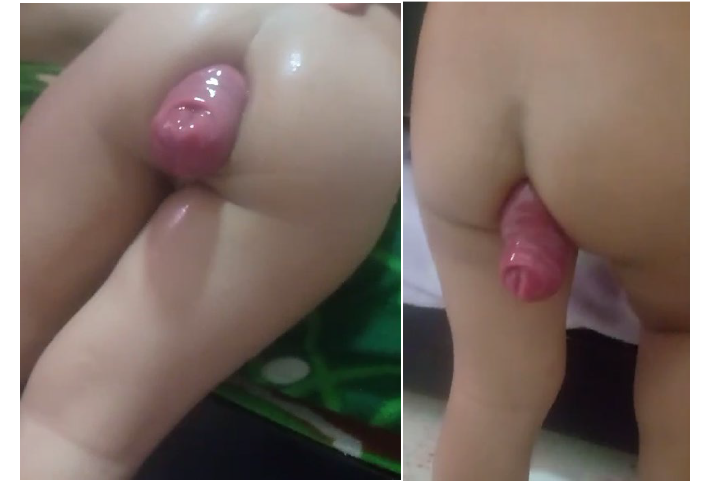

Table 1.
Laboratory examination results.
| Study | Result | Reference values |
|---|---|---|
|
Blood count |
Hemoglobin (Hb): 11.2 grams/deciliter (g/dL) Hematocrit (Hct): 31.5% Erythrocytes: 3.9 million cells/microliter (µL) Leukocytes (L): 24.720 target/µ/L Neutrophils (N): 43.4% Lymphocytes (L): 38.8% Monocytes (M): 11% Eosinophils (E): 6.8% Platelets (P): 410.500 target/µ/L |
13.5±3 g/dL 39±2% 3.8-5.5 1 400 (5.0-19.0) 41-68% 26-36% 1-7% 0.4-3% 140 000-470 000 |
|
Serum electrolytes |
Login: Sodium (Na++): 136.5 milliequivalents/liter (mEq/L) Potassium (K+): 2.81 mEq/L Calcium (Ca++): 8.88 milligrams/deciliters (mg/dL) Chlorine (Cl): 102 mEq/L Control after correction of hypokalemia: Na++: 137.5 mEq/L K+: 4.33 mEq/L Cl: 106.2 mEq/L Ca++: 9.02 mg/dL |
135-145 mEq/L 3.5-5.5 mEq/L 8.5-10.2 mg/dL 96-106 mEq/L |
| Blood glucose | 115.2 mg/dL | 80-105 |
| C-reactive protein | 39.65 mg/L | 3-10 |
| Stool sample analysis | Soft appearance. Blood: absent. Bacterial flora. Normal Leukocytes: 10-15 cells/field (c) Erythrocytes: 0-2 cells/field Intestinal parasites: absent |
Leukocytes: up to 5 cells/c |
| Cultures | Stool culture, urine culture and blood culture: negative | |
| Serum urea nitrogen | 5.27 mg/dL | 3-12 |
| Serum creatinine | 0.3 mg/dL | 0.2-0.6 |
| Urinalysis | Clear appearance. pH: 6.0 Density: 1022 Proteins, glucose, ketone bodies: negative Bilirubin, hemoglobin: negative Nitrites, leukocyte esterase: negative Epithelial cells: negative or scarce Leukocytes: 1-2/c Kidney cells: 0-2/c |
Clear appearance. pH: 5.5-6.0 Density: 1010-1025 Proteins, glucose, ketone Bodies: negative Bilirubin, hemoglobin: negative Nitrites, leukocyte esterase: negative Epithelial cells: negative or scarce Leukocytes: 0-2/c Kidney cells: 0-2/c |
| Pyruvic transaminase/alanine amino transferase (ALAT) | 12.42 units/liter (U/L) | 5-45 |
| Oxaloacetic transaminase/aspartate amino transferase (AST) | 31.62 U/L | 15-45 |
Disclaimer/Publisher’s Note: The statements, opinions and data contained in all publications are solely those of the individual author(s) and contributor(s) and not of MDPI and/or the editor(s). MDPI and/or the editor(s) disclaim responsibility for any injury to people or property resulting from any ideas, methods, instructions or products referred to in the content. |
© 2024 by the authors. Licensee MDPI, Basel, Switzerland. This article is an open access article distributed under the terms and conditions of the Creative Commons Attribution (CC BY) license (http://creativecommons.org/licenses/by/4.0/).
Copyright: This open access article is published under a Creative Commons CC BY 4.0 license, which permit the free download, distribution, and reuse, provided that the author and preprint are cited in any reuse.


