Submitted:
06 June 2024
Posted:
07 June 2024
You are already at the latest version
Abstract
By using a two-stage physiologically based extraction test (PBET) and (W-PBET), the authors of this paper have estimated the bioaccessibility of metals (Pb and Fe) and their compounds in the GI tract of waterfowl and mammals. It has been shown that the metals (Pb and Fe) have low bioaccessibility; combined with the short time of shot exposition at the GI tract, this eliminates the risk of toxic effects in the case of shot ingestion by waterfowl. It has also been discovered that there is a significant difference in the bioaccessibility of metals and their compounds. A model was proposed and tested to predict the toxic effects of lead on biological objects depending on the threshold mass of consumption. This model takes into account data on the bioavailability of lead and regulatory values indicating dangerous concentrations of lead.
Keywords:
lead shot
; steel shot
; soil
; Pb and Fe bioaccessibility
; PBET
; waterfowl
; risk assessment
; shooting activity
1. Introduction
According to some community groups, the use of lead ammunition in sports and hunting activities is associated with the risks of environmental pollution and toxic effects on wildlife and humans (Pain et al., 2015, Arnemo et al., 2016; Andersen et al., 2018; Pain et al., 2019; Andersen et al., 2020; Treu et al., 2020; Allen et al., 2023). Since February 2023, the use of lead shot in wetlands has been banned throughout the EU/EEA. In 2021, the European Chemicals Agency (ECHA) proposed to restrict the use of lead ammunition for outdoor sports shooting and fishing. The proposal was supported by the Committee for Socio-economic Analysis (SEAC) and the Committee for Risk Assessment (RAC)[1]. The key argument in favour of the restrictions is that ingestion of lead causes poisoning of wildlife[2].
The principal routes by which the gastrointestinal (GI) tract of wildlife can be exposed to lead are (Haig et al., 2014):
- -
- primary ingestion of spent lead ammunition (shot) and fishing tackle as gastroliths by waterfowl; ECHA estimates2 that at least 135 million birds are currently at risk of lead poisoning each year from primary ingestion of lead (including 7 million from the ingestion of lead fishing tackle);
- -
- secondary ingestion where predators eat animals that have been wounded with lead ammunition; ECHA estimates2 that 14 million birds are at risk of poisoning from secondary ingestion each year after inadvertently eating fragments of lead that are in the tissues of their food.
The authors of this paper believe that the ingestion of lead ammunition by waterfowl is probabilistic in nature, which is reflected by the varied patterns of shot in the stomachs of birds. The results presented in the paper by Liljebäck et al. (2023) indicate that no lead was present in the stomach of three species of sea ducks within the Baltic/Wadden Sea flyway (a total of 205 birds). Anderson et al. (2000) studied the content of the gizzards in ducks harvested in the Mississippi Flyway during two hunting seasons. On average, spent shot was present in the gizzards of 4.3–12.7% of the total amount of birds (16 651 birds). According to Pain et al. (2019), the prevalence of lead shot ingestion in waterfowl species is:
- -
- 1–12% in North America, with a maximum of 17,5% (the total number of birds — 171 697);
- -
- 2.5–12% in Europe, with a maximum of 23–31% (the total number of birds — 75 761).
Therefore, the average probability of primary ingestion of lead shot by waterfowl does not exceed 10–15%, even on hunting grounds.
For other animals and humans, direct ingestion of lead ammunition and its fragments is a random nature (Laidlaw et al., 2017). The risks of the introduction of lead into the GI tract with such objects are associated with the consumption of food (for people), grass (for herbivorous animals), and with the soiling of hands with soil dust containing lead compounds (for children)[3] (Oomen et al., 2003; Laidlaw et al., 2017; Green and Pain, 2019). After being deposited into the soil, lead ammunition oxidizes under the influence of the environment and is encapsulated by the poorly soluble lead compounds (hydroxide, carbonate, sulphate) within two or three weeks. The transformation of the lead ammunition surface in the soil is accompanied with the fixation of lead in the upper part of the soil profile, as part of the soil adsorption complex (Lisin et al., 2020). The formation of soil dust containing lead compounds may result from the wind erosion of the surface soil which should also be considered a probabilistic process.
33 countries have banned the use of lead ammunition partially or completely (Thomas et al., 2019), including 23 European countries (Mateo and Kanstrup, 2019). This has made it necessary to use non-lead ammunition, primarily, made of steel (Thomas et al., 2019). Like lead ammunition, steel ammunition accumulates on the soil surface and may serve as gastroliths for birds. According to authors earlier paper (Lisin et al., 2022), as a result of steel shot transformation in natural conditions, corrosive iron particles (rust) do not only become irreversibly assimilated by the soils and become part of the soil dust but also form dispersed dust on the soil surface. Moreover, unlike the cyclical process of lead shot corrosion, the corrosion of steel shot is a continuous process which results in its complete assimilation in the environment, leading to a longer presence of an iron dust source in shooting areas.
Traditionally, the risks of wildlife poisoning are equated to the ingestion of lead (steel) ammunition and introduction of dust particles (soil dust) into the GI tract2. However, the probability of toxic effects associated with the presence of ammunition and dust particles in the GI tract is primarily determined by the bioaccessibility of metallic Pb, Fe and their compounds, which requires a separate estimation and should be taken into the account.
Methodological approaches for determining the bioaccessibility of metals in the GI tract have been suggested over the past several decades (Ruby et al., 1993; Oomen et al., 2002; Wragg and Cave, 2003; Marschner et al., 2006; Bruce et al., 2007; Bannon et al., 2009; Pelfrêne et al., 2010; Zia et al., 2011; Beyer et al., 2016; Zingaretti et al., 2021). After analysing these papers, it is possible to conclude that the main approaches to estimating the bioaccessibility of elements in the GI tract are the following:
- -
- determining the ratio of the concentration of contaminants (accessible for assimilation) dissolved in the gastric juice to their content in the solid phase (metal/soil) according to physiologically based tests of bioaccessibility[4]in vitro, imitating the conditions of the GI tract in laboratory conditions;
- -
- determining the ratio of the concentration of contaminants in blood to their concentration in the GI tract according to an assessment of bioavailability[5] in vivo, where the contaminants are introduced into the body of tested animals directly in natural form, as part of a liquid, or as part of a food matrix.
For soil lead compounds, in most cases, both Pb bioavailability and bioaccessibility are estimated; the above-mentioned summarising papers indicate a high consistency of the obtained results. There are known papers on estimating the bioavailability of metallic Pb. They include forced introduction of lead shot into the GI tract of waterfowl with further measurement of lead concentration in their blood, kidneys, liver, and bones (Finley and Dieter, 1978; Brewer et al., 2003; Krone et al., 2019).
It should be noted that there is no information on systematized Fe bioaccessibility assessments both in the metallic phase and in the form of oxidized compounds and compounds with soil matrix components.
The lack of research into the bioaccessibility of Pb and Fe and their compounds in relation to the various characteristics of the GI tract of birds (and wildlife in general) in combination with the toxic effect risk assessment for such biological objects have determined the objectives of this paper: 1) to estimate the Pb and Fe bioaccessibility in the case of the entering of shot and polluted soil into the GItract of humans; 2) to study the metals bioaccessibility in the case of the entering of lead and steel shot into the GI tract of waterfowl; 3) to analyse the mutual effect of the joint presence of Pb and Fe in shooting areas; 4) to assess the risks associated with the exposure of wildlife (waterfowl and mammals) to Pb and Fe in shooting areas.
The researchers suggest the following criteria for the risk assessment tools:
- -
- the bioaccessibility value which gives a general indication of the percentage of metal (Pb and Fe) that can be assimilated by a biological object (the recipient);
- -
- thresholds of the element concentration in the GI tract (according to the absence of toxic effects) obtained using the model calculations based on the bioaccessibility value and reference values for waterfowl.
2. Materials and Methods
2.1. Shot Collection
The study used commercial steel shot (2.5 mm diameter) and lead shot #7.5 (2.4 mm diameter) for sports shooting. According to production specifications, the following residual elements are present in steel shot: C (0.6–1.0%), Si (0.7–1.2%), Mn (0.5–1.0%), S (up to 0.07%), P (up to 0.09%). The main residual elements in lead shot are Sb (4–6%), Sn (0.1–0.2%), As (0.2%), Cu (0.02%), Ag (0.002%), and Zn (0.001%).
To conduct the experiments and study the metals bioaccessibility for the shot entering into the GI tract of mammals, the shot samples were grouped in the following manner: 1) lead shot exposed alone (0.4 g); 2) steel shot exposed alone (0.4 g); 3) a 1:1 mixture of lead and steel shot (0.4 g each); 4) a 2:1 mixture of lead and steel shot (0.26 and 0.15 g respectively); 5) a 1:2 mixture of lead and steel shot (0.13 and 0.31 g respectively); 6) a 1:6 mixture of lead and steel shot (0.06 and 0.38 g respectively).
To conduct the experiments and study the metals bioaccessibility for the shot entering into the GI tract of waterfowl, three groups of samples were formed: 1) lead shot exposed alone (0,36 g); 2) steel shot exposed alone (0,36 g); 3) a 1:1 mixture of lead and steel shot (0.18 g each).
2.2. Soil
When conducting the experiments to study Pb and Fe bioaccessibility in the case of the entering of soil dust into the GI tract of mammals, authors took into the account the conclusions of their earlier paper (Lisin et al., 2022) showed that the Pb in soil is forming specifically sorbed, carbonate, and sulphate species and is bound with iron hydroxides and organic matter, and iron forms part of the soil matrix and is assimilated in the form of corroded iron hydroxide particles.
The following testing samples were used: 1 – the soils of shooting areas where only lead shot was used; 2 – the soils of shooting areas (only lead ammunition was used), with the adding of steel shot; 3 – the joint entering of steel shot and lead shot into the soils of areas with no prior shooting activities. Shot was deposited into soils of the second and third types for four months under the conditions of moderate humidification (the methodology is described in the paper by Lisin et al. (2022)). After the exposure, all soils were air dried and sieved (<1 mm) and pulverization (74 µm). The soil characteristics are described in Table 1.
The pH value and electrical conductivity of the aqueous soil extract (distilled water extraction, a solid-to-liquid ratio of 1:5) were determined using the potentiometry (by “Expert 001” (Econix Expert Ltd., Russia) with measuring glass electrode (type ESL-43-07)) and conductometry (by HM Digital COM80 (HM Digital, Inc., South Korea)) respectively. Total metal concentrations of the soil were analysed using a Niton FXL 950 energy dispersive X-ray fluorescence spectrometer (ED-XRF, Thermo Fisher Scientific Inc.). Mineral composition of soils was examined by X-ray diffraction (ULTIMA-IV, Rigaku, Japan). The soil texture was determined using the sieve analysis method (GOST 12536-2014) before the pulverization of samples.
2.3. Bioaccessibility Tests
For mammals, taking into the account the probable sources of element ingestion, bioaccessible Pb and Fe were determined both in pulverized soils and in shot. The experiments were conducted using the two-stage physiologically based extraction test (PBET, Ruby et al.,1993, 1996) with changes introduced in the paper by Bosso and Enzweiler (2007): to imitate the peristalsis, the flasks were regularly shaken, and instead of dialysis bags, titration with oversaturated sodium bicarbonate solution was used in the procedure for increasing the solution рН.
The PBET was based on the solubility of Pb compounds in the solutions modelling the conditions of the human stomach and intestine, that is, in the solutions containing organic acids, enzymes, and bile salts. A model gastric solution is prepared using a mix of 50 mg of pepsin (Servicebio) and organic acids — 0.5 g of citric acid (Servicebio), 0.5 g of malic acid (Panreac), 0.42 ml of lactic acid (Servicebio), and 0.5 ml of glacial acetic acid (Panreac) — dissolved in 1 litre of DI water, with the рН modified to 1.5 using hydrochloric acid (in a 1:1 ratio). The shot/soil was combined with model gastric solution (solid-to-liquid ratio of 1:10) and incubated at a temperature of 37°С, with regular shaking; one and a half hours for soil, and two hours for shot. Each 30 minutes, the рН in the reaction flask was controlled, and samples (2 mL each) were collected for analysis. At the same time, 2 ml of model gastric solution was added to the reaction flask in order to maintain the solid-to-liquid ratio. Prior to the analysis, the sample aliquot was filtered using a 0.45-µm membrane filter.
After the end of the exposure, an oversaturated solution of NaHCO3 was added to the reaction flask until the рН reached 7.0; after that, 70 mg of bile salts (Pronadisa Conda) and 20 mg of pancreatin (AppliChem) were mixed in. In this way, the passing from the stomach to the intestine was modelled. The resulting mixture was incubated at a temperature of 37°С, with regular shaking, for two hours. After the end of the exposure, the solution was filtered using a 0.45-µm membrane filter.
Pb and Fe bioaccessibility when shot is in the GI tract of birds was estimated using the two-stage PBET procedure adapted for the GI tract of waterfowl (W-PBET, Furman et al., 2003). The model gastric juice was a mixture of 1M saline solution and 10 g of pepsin (Servicebio) in 1 litre of distilled water with the рН adjusted to 2.5 using hydrochloric acid. The shot was deposited into the model gastric juice with the solid-to-liquid ratio of 1:12; the reaction flask was sealed and incubated in a water bath at a temperature of 42°С with continuous stirring. After one hour, the sample (2 mL) was filtered using a 0.45-µm membrane for further analysis. The remaining solution in the reaction flask was adjusted to the pH of 6.2 using a saturated NaHCO3 solution; bile salts (CONDA Pronadisa) and pancreatin (AppliChem) were added in the amount of 0.35% and 0.035% respectively. The resulting mixture, imitating the intestine conditions, was incubated for 2 hours using a water bath at a temperature of 42°С, with continuous stirring. After the incubation, the solution was filtered using a 0.45-µm membrane, and the Pb and Fe concentrations were determined.
Pb content in the filtered gastric and intestine model solutions was measured by the anodic stripping voltammetry (the analyser is AKV07 MK with the POLAR 4.1 software). Element detection threshold — 0.001 mg/l. Relative error in the interval of detectable concentration up to 8% (p=0.95). Concentration of Fe was measured by the sulfosalicylic method with a spectrophotometry (Bhavna V. Mohite, 2011), the wavelength of 420 nm (Portlab 501). The calibration based on the model gastric (pH 1.5 and 2.5) and intestine (pH 7.0 and 6.2) solutions. The element detection threshold in acid solution was 0.03 mg/l, in weakly alkaline solutions 0.05 mg/l. Relative error up to 12% (p=0.95) in the interval of detectable concentration. The obtained results accuracy was controlled using the atomic absorption spectrometry method (ContrAA®700, Analytik Jena).
All the tests were repeated triple, the relative standard deviation of each series not exceeding 15%; all the chemicals used were of analytical grade. The results of the measurement of Pb and Fe content in the model gastric and intestine solutions of biological objects are indicated in Table 2 and Table 3. For each metal, its average content in the solution (mg/l) is indicated, as well as the respective value of standard deviation based on three measurements. The amount of dissolved metal per one gram of shot (mg/g), calculated based on the average metal content in the solution.
Pb and Fe bioaccessibility was calculated as the ratio of metal concentration in the extractable phase (on a solid substance basis) to the total Pb and Fe content in shot (1) ore soil (2). The calculations were made using the average metal contents in the model solutions of the GI tract.
– element bioaccessibility in case of shot ingestion, %; – element bioaccessibility in case of soil particle ingestion, %; ― element concentration in the solution, mg/l; ― solution volume, l; ― shot mass, mg; ― soil mass, g; ― total element in the soil, mg/kg.
3. Results
Shot in the gastrointestinal tract of waterfowl. The metals bioaccessibility when the shot is in the GI tract of waterfowl is shown on Figure 1.
Pb bioaccessibility:
- -
- when lead shot is exposed alone the bioaccessible Pb in the stomach is 0.05% of the shot mass; in the intestine is 0.011 % of the shot mass, it is 5 times lower.
- -
- when lead and steel shot are exposed together Pb bioaccessibility decreases by 1.3 times in the stomach, while the percentage of bioaccessible Pb in the intestine is negligible.
Fe bioaccessibility:
- -
- when steel shot is exposed alone the bioaccessible Fe in the stomach is 0.07% of the shot mass; in the intestine is 0.025% of the shot mass, it is 3 times lower;
- -
- when lead and steel shot are exposed together Fe bioaccessibility remains almost the same in the stomach, while in the intestine, it increases by 1.7 times.
The numbers indicate the presence of the following materials in GI tract: 1 – Pb shot; 2 – a mix of Pb and Fe shot (1:1);3 – Fe shot.
Shot in the gastrointestinal tract of mammals. The metals bioaccessibility when the shot is exposed in the GI tract of waterfowl is shown on Figure 2.
Pb bioaccessibility (Figure 2a):
- -
- when lead shot is exposed alone in the GI tract, Pb bioaccessibility reaches 0.08% of the mass of metal in the stomach and decreases to 0.04% of the metal mass in the intestine;
- -
- when lead and steel shot are exposed together in the stomach and intestine solutions, the percentage of bioaccessible Pb decreases and remains within 0.03% and 0.02% of the metal mass respectively.
- -
- for the variable mixtures of lead and steel shot in the GI tract (Figure 2c), regardless the ratio of shot mass, the Pb bioaccessibility is permanently and stably lower than when lead shot is exposed alone in the GI tract. As the percentage of steel shot in the mix increases, Pb bioaccessibility decreases by 2 or 3 times and is around 0.02% of the mass of metal in both the stomach and the intestine.
Fe bioaccessibility (Figure 2b):
- -
- when steel shot is alone in the GI tract, the percentage of bioaccessible Fe is 0.08% of the mass of shot in all sections of the GI tract;
- -
- when lead and steel shot are together, the Fe bioaccessibility at the early stage of interaction with the gastric solution is two times lower, but by the end of the stomach stage bioaccessible Fe increases and stabilizes in the intestine at the 0.08% of the metal mass.
In general, Fe bioaccessibility (Figure2d) is not much influenced by the lead-to-steel-shot ratio. It is constant throughout the stomach stage, and by its end it remains the same in the intestine. It is similar to the only steel shot is in the GI.
The numbers indicate the presence of the following materials in GI tract:
1– Pb or Fe shot;
2 – mix of Pb and Fe shot, 1:1;
3 – mix of Pb and Fe shot, 2:1;
4 – mix of Pb and Fe shot, 1:2;
5 – mix of Pb and Fe shot, 1:6.
The soil of shooting areas in the GI tract of mammals.
When soil 1 particles are in an aggressive stomach solution, Pb bioaccessibility (Figure3a) is around 40% of total at the beginning, increasing to 79–80% by the end of the stomach stage. In the intestine, where the pH reaches 7 due to the secretion of an alkaline bicarbonate, Pb bioaccessibility of soil 1 drops to 3% of total. In soil 2 and soil 3, bioaccessible Pb is 75–79 % of total at the beginning and remains stable until the end of the stomach phase; in the intestine, it drops to 8–12% of total.
Fe bioaccessibility (Figure 3b) of soil 1 in the GI tract does not exceed 1% of total in the stomach and 0.2% in the intestine. When steel shot is deposited into the soil of a shooting area, as well as when lead and steel shot are deposited into the soil (soil 2 and soil 3), Fe bioaccessibility in the stomach is 1.5% of total at the beginning and increases to 3.5–4%; after passing into the intestine, the bioaccessibility drops to 1–1.5% of total.
soil 1 – the soils of shooting areas where only lead ammunition was used
(total Pb – 4 500 mg/kg, total Fe – 15 000 mg/kg);
soil 2 – the soils of shooting areas (only lead ammunition was used), with the adding of steel shot
(total Pb – 3 300 mg/kg, the steel shot load of 0.02 kg/0.1 m2, total Fe – 26300 mg/kg);
soil 3 – the soils of areas with no prior shooting activities where lead and steel shot are deposited at the same time
(total Pb – 3 200 mg/kg, total Fe – 25 000 mg/kg).
4. Discussion
Pb and Fe bioaccessibility when shot in the GI tract of wildlife does not exceed 0.1% of the shot mass according to the data obtained by the authors.
When lead or steel shot is alone in the GI tract of mammals, the bioaccessibility of both Pb and Fe in the stomach is 0.08% of the metal mass. When the shot pass into the intestine neutral conditions, Pb precipitates and its bioaccessible drops twice. The percentage of bioaccessible Fe remains the same, supposedly, due to the formation of stable organoferric compounds.
The joint presence of lead and steel shot into the GI tract of mammals results in the limited Pb bioaccessibility, more pronounced if there is more steel shot in the mixture, due to the lead ion sorption from the solution on the corrosion layer surface of steel shot and coprecipitation with the formed Fe (hydr)oxides; (Lisin et al., 2022). Fe bioaccessibility in the GI tract is not influenced by the presence of lead shot; it remains stable regardless of the lead-and-steel-shot ratio in the soil.
In general, the identified effects on mammals are also observed in the cases of the presence of lead and steel shot in the GI tract of waterfowl. However, the lower acidity of the gastric solution of birds determines a lower Pb bioaccessibility (0.05% of total) compared with mammals, with a similar Fe bioaccessibility (0.07% of total), while the lack of an organic acid complex determines almost a complete precipitation of Pb and partial precipitation of Fe when the stomach contents pass into the intestine. It should be noted that if lead and steel shot are equal in the GI tract of waterfowl, Fe bioaccessibility in the intestine is 1.6 times higher than when steel shot is alone.
Pb and Fe bioaccessibility in soil depends on the solubility of their solid phases in the specific conditions of the GI tract (Bosso and Enzweiler, 2007; Zia et al., 2011; Beyer et al., 2016; Walraven et al., 2015; Fayiga and Saha, 2016; Zingaretti et al., 2021). When particles of the shooting areas soils enter the stomach of mammals, Pb bioaccessibility (80% of total) is significantly higher than Fe bioaccessibility (1% of total) due to the instability of Pb compounds in the aggressive stomach conditions (рН 1.5, hydrochloric acid, pepsin), while Fe is bound in stable and well-crystalised minerals of the oxide and hydroxide group and as part of the silicate soil matrix. In the neutral intestine conditions, the bioaccessibility of elements decreases by an order of magnitude.
The presence of steel shot corrosion products in the soil does not only determine an increased Fe bioaccessibility in the GI tract but also makes Pb bioaccessibility increase in the intestine by 2.5–4 times compared to the soils that do not contain iron corrosion products. Supposedly, this happens due to the stabilisation of dissolved Pb in the neutral conditions as part of the colloidal ferroorganic compounds.
The comparative assessment of the Pb and Fe bioaccessibility when shot and soil dust particles are exposed in the GI tract of wildlife has shown that the bioaccessibility of metals is significantly lower than the bioaccessibility of elements compounds with soil components: for Pb – 1000 times lower (0.08% and 80% respectively), for Fe – 50 times lower (0.08 and 4% respectively)[6].
It is known that the toxic effects of Pb compounds are determined by the Pb2+, regardless of whether metallic lead or a lead compound is the source. However, the difference in the bioaccessibility of metallic Pb and various forms of Pb compounds for biological objects makes it obvious that it would be incorrect to equate the risks of toxic effects for metallic Pb and its compounds. Unfortunately, the opposite often happens in practice. For example, the water Pb concentration standard applies to both “lead and its compounds”[7], and in the comments to the specified Environmental Quality Standards, there is a note that they apply to bioaccessible concentrations of substances.
Assessment of toxic effect risks It is rather difficult to prepare a relevant assessment of toxic effect risks for the presence of Pb and Fe in wildlife since there is no standardized approach and regulatory framework.
Iron is considered to be non-toxic for humans and wildlife (ECHA (Annex C), 2021). However, in the International Statistical Classification of Diseases and Related Health Problems (ICD-10 Version: 2019), there is a group of human diseases associated with an excessive Fe accumulation in organs and tissues (ICD-10: E83.1. Disorders of iron metabolism).
The absence of thresholds for Fe in biological objects has prevented the researchers from analysing the risks. At the same time, the authors of this paper have obtained data on the constant bioaccessibility of metallic Fe in all chambers of the GI tract of wildlife which makes it possible to claim that there are no limits of the absorption of iron unlike in the case of lead.
Lead is more widely discussed in the context of its impact on humans and health effects based on the measurement of blood lead concentrations[8]. It is believed that the toxic effects of lead are numerous and largely irreversible (Laidlaw et al., 2017; Pain et al., 2022). According to the World Health Organization Guideline (WHO Guideline, 2022), the toxic effects of lead on humans are associated mainly with the absorption of lead from the GI tract and affect almost all body systems. The threshold blood Pb concentration for humans is believed to be 0.05 mg/l (WHO, 2022), while the Pb in other organs and tissues does not have a standard threshold value, as well as that of the GI tract. At the same time, according to the WHO[9] and the European Food Safety Authority (EFSA)[10], there is no safe level of lead exposure for humans.
No critical, threshold, and permitted levels of Pb in the GI tract and blood have been determined for wildlife. For Anseriformes (including ducks), Franson and Pain (2011) suggest the following categories of blood lead concentrations (mg/l):
<0.2 – no Pb source;
0.2<0.5 – subclinical poisoning, pathological manifestations of physiological effects are insufficient, and negative effects are reversible if Pb source is terminated;
0.5–1.0 – clinical poisoning, the initiation of pathological manifestations of physiological effects[11], leading to probable death if Pb source were to continue;
>1.0 – severe clinical poisoning which may be directly life threatening.
To assess the risks of the Pb effects on waterfowl, the authors of this paper have suggested a model based on the calculation of thresholds Pb mass introduced into the GI tract (according to the absence of toxic effects). The model is based on the data on Pb bioaccessibility in the GI tract of mammals and waterfowl obtained by the authors of this paper and includes the following parameters:
- Blood Pb threshold (mg/l): for mammals (humans) – 0.05, for waterfowl – 0.2 (Franson and Pain, 2011) (1)
- Reference average duck mass (kg) – 1.3 (according to Finley et al., 1976; Rocke&Samuel, 1991; Plouzeau et al., 2011; Lewis et al., 2021) (2)
- Reference average blood volume (L) in humans – 5.012, in waterfowl – 0.13 (10% of duck mass (Portman et al., 1952)). (3)
- It is known that 3–10% of the ingested lead is absorbed in the GI tract; once absorbed, lead is initially bound to the blood erythrocytes (WHO, 2022). The authors of this paper have accepted the value of 10% for calculation. (4)
- Pb bioaccessibility in the stomach (hereinafter referred to as the maximum bioaccessibility) defines the maximum Pb amount soluble in the GI tract (Bosso and Enzweiler, 2007), while Pb bioaccessibility in the intestine (hereinafter referred to as the actual bioaccessibility) defines the lead amount that can be assimilated as it is known that the majority of GI content, including Pb, is absorbed by the small intestine (Denbow, 2000), after the food is processed in the stomach chambers (Figure 4). (5)
Based on parameters (1), (2), and (3), the total content of Pb in human blood should not exceed 0.25 mg lead, and in waterfowl blood – 0.03 mg. The results of model calculations of Pb mass thresholds in the ingested substance, based on the condition of absence of toxic effects, are presented in Table 4. Parameter (4) determines the Pb threshold in the GI tract. Taking parameter (5) into the account makes it possible to define the Pb mass threshold of and of Pb-containing substance depending on the conditions in various chambers of the GI tract.
When shot is into the GI tract of humans, the calculated Pb mass threshold, as determined based on maximum bioaccessibility, is 2.5 g which is equivalent to the mass of 36 shot pellets. Taking into the account the actual Pb bioaccessibility, the estimated Pb mass threshold is 6.3 g, which is equivalent to 90 shot pellets being present in the GI tract.
The Pb mass threshold into the GI tract of humans with soil dust particles, with the maximum Pb bioaccessibility, should not exceed 3.0 mg. With the average Pb total in the soil of shooting areas of 4.5 mg/g, the corresponding mass of soil particles in the GI tract is 0.7 g. According to the actual Pb bioaccessibility, Pb mass threshold is no more than 21–83 mg, which is equivalent to 5–19 g of soil particles being present in the GI tract.
The obtained mass of ingested soil dust particles and shot seems unlikely to be ingested at once or be present at once in the GI tract of humans, even with maximum bioaccessibility.
When shot is into the GI tract of waterfowl, Pb mass threshold (with maximum bioaccessibility), should not exceed 0.5 g, which is equivalent to the presence of 7 shot pellets in the GI tract at the same time. For the signs of subclinical poisoning to appear, there should be 19 shot pellets in the GI tract at the same time, for a severe form of clinical poisoning – 36 shot pellets. However, there are data on the absence of toxic effects on ducks even when the mass of lead in the GI tract is two times the modelled threshold mass: according to Krone et al. (2019), there are no clear clinical signs of lead poisoning or change in the behaviour of Pekin ducks (40 test birds) which received 6 lead shot pellets (with a mass of 0.191 g each) throughout a four-week study.
Pb mass threshold, modelled for the actual Pb bioaccessibility in the GI tract of waterfowl, was 25 g. This is equivalent to 357 shot which is impossible to ingest.
At the same time, using the model with the maximum bioaccessibility (for the stomach) suggested by the authors, it can be demonstrated that there are varied opinions on the threshold values of Pb content in the GI tract of birds (Table 5): for example, birds are considered to be in good health after ingesting from 1 to 286 shot pellets (it is obvious that the upper limit is extremely unlikely). Moreover, the initial Pb in blood (control group) vary significantly (from 0.05 to 0.71 mg/l) which reflects not only the specifics of their habitats, species, nutritional conditions, and Pb sensitivity but also the adaptability effect. These factors significantly decrease the risks, increasing the safe exposure range.
According to the data obtained by Pain et al., which were used in the model, only a Pb blood concentration of < 0.2 mg/l in birds indicates the absence of a Pb source and is absolutely safe, while the Pb blood concentration of <0.5 mg/l is associated with minimal risks. However, according to the information from Table 5, even with the concentrations which significantly exceed the risk level (1 mg/l and above), birds do not experience prolonged sings of suppression or poisoning. Only in individual cases, the increase of the subcritical concentration level by approximately 8 times indicates the probability of a risk event. However, to achieve such a Pb blood concentration, a bird needs to ingest around 300 shot pellets (of the #7.5 shot) which is impossible.
Haig et al. (2014) studied over 150 sources and indicated that the Pb sensitivity may vary not only between different species but also within one species which causes the discrepancies in the evaluation of its effects on the population, makes the risk assessment more difficult, and results in incorrect conclusions regarding the risk levels.
On average, shot remains in the GI tract of birds for a period between several hours and several days depending on the eating habits, size and quantity of gastroliths ingested by various bird species (French et al., 2020). Moreover, the stomach of a duck contains two chambers with various conditions (Figure4). The interaction between the shot and the aggressive gastric juice does not last longer than 30–40 minutes (in the proventriculus of a bird), after that the shot passes into the gizzard together with food and then into the intestine (Denbow, 2000) where Pb bioaccessibility drops to 0.001% of the metal mass which almost completely eliminates any toxic effects even if excretion of the shot from the GI tract takes longer than usual.
Figure 4.
The digestive tract of waterfowl and bioaccessibility of Pb in its parts.1 – esophagus; 2 – crop (expanded section of the esophagus); 3 – proventriculus (pars glandularis); 4 – gizzard (pars muscularis); 5 – small intestine; 6 – large intestine.
Figure 4.
The digestive tract of waterfowl and bioaccessibility of Pb in its parts.1 – esophagus; 2 – crop (expanded section of the esophagus); 3 – proventriculus (pars glandularis); 4 – gizzard (pars muscularis); 5 – small intestine; 6 – large intestine.
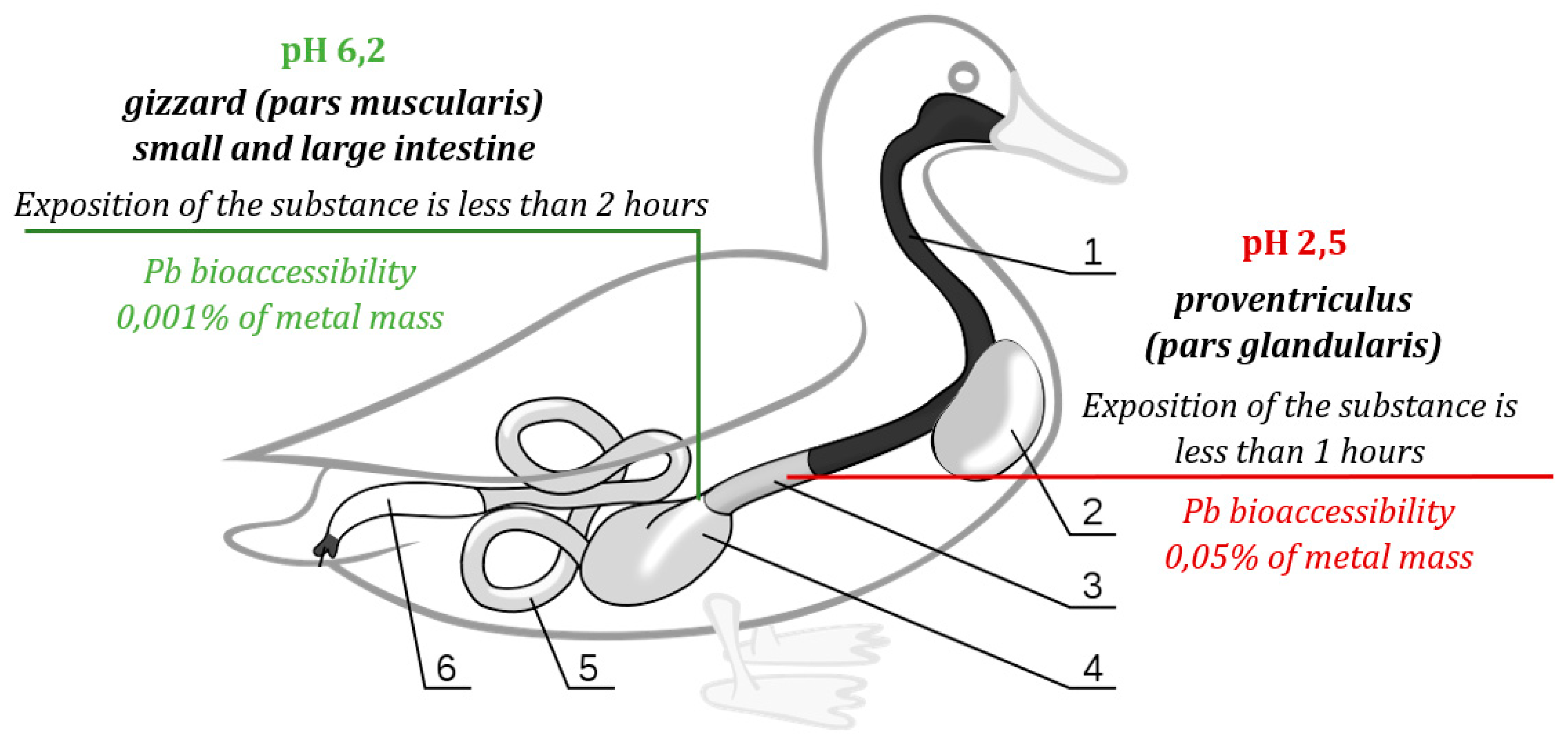
The duck stomach consists of two chambers, the proventriculus (pars glandularis) and the gizzard (pars muscularis). The proventriculus glands secrete a gastric juice with a рН 2.5, consisting of hydrochloric acid and enzymes (pepsin). The gizzard has strong muscular walls, the interior surface is lined with a hard membrane called the cuticle. Bile secretion and pancreatic secretion (enriched with hydrocarbonates) enter the gizzard, forming a neutral environment (рН 6.2), there gastroliths are present also (small stones, shells, and other solid particles). Food enters the proventriculus, softens due to the effect of gastric juice and passes into the gizzard where, thanks to strong muscular contractions, it is ground between the gastroliths and the cuticle. After that, the finely ground and softened food passes into the small intestine where most of its components are absorbed, including the vitamins and minerals, which is promoted by the large surface of its inner lining, the fleecy mucous membrane (according to Denbow, 2000).
As a result, when talking about the possible risks for birds associated with the use of lead (steel) ammunition, it should be understood that the risks are based on a combination of consecutive probabilistic processes: ingestion-exposure-assimilation.
The probabilistic nature of ingestion in natural conditions is determined by the dispersion (scattering) of shot throughout the hunting areas, recreation areas, and sports complexes, and is confirmed by the presence of shot among the gastroliths in the stomach of only 4.3–12.7% of birds. The second probabilistic factor of lead ingestion is the eating habits of various bird species which determine the differences in the required gastrolith consumption and therefore the difference in the number of shot pellets in the GI tract.
The risk of toxic effects may be manifested exclusively as a result of the dissolution of shot by the aggressive gastric juice and the absorption of the solution in the GI tract of birds. The probability of toxic effects is limited by the short period of the shot exposure to the GI tract birds which is determined by the structure of their GI tract.
Lead (iron) bioaccessibility reflects the existence of a soluble metal form but does not indicate the pattern and volume of its assimilation. The transfer of dissolved metal from the GI tract to other body systems is a specific biological process. This is why the discussion of the toxic effects associated with the presence of shot in the GI tract is ungrounded as they are based not on established standard or concentration values but instead on the suggestion that “ingestion equals poisoning”.
5. Conclusions
The results of the paper bring close attention to a variety of circumstances associated with the assessment of the toxic effect risks in the case of lead shot ingestion by wildlife:
- -
- it is incorrect to equate the fact of metallic shot ingestion with toxic effects; the analysis of a considerable number of publications on the effects of lead on waterfowl over the past 20 years shows that birds can feel well at various blood lead concentrations, including high levels (up to 1 mg/l);
- -
- Pb as metal has been shown to have low bioaccessibility in the GI tract of waterfowl (and mammals) (even in the presence of metallic Fe);
- -
- using the model suggested by the authors, it has been shown that there is low probability of toxic effects on waterfowl due to the fact that is impossible for a bird to have around 300 shot in its stomach.
Taking these points into account, we suppose that the risks for biological objects associated with the use of lead ammunition, as suggested by the ECHA, are substantially exaggerated, and the ECHA proposal to restrict the use of lead ammunition in outdoor areas seems poorly grounded.
References
- Arnemo J.M., Andersen O., Stokke S.et al. (2016) Health and Environmental Risks from Lead-based Ammunition: Science Versus Socio-Politics. EcoHealth 13, 618–622. [CrossRef]
- Allen C., Andersen O., Andreotti A., et al (2023) European scientists’ open letter on the risks of lead ammunition. http://www.europeanscientists.eu/open-letter-2023/.
- Andersen O, Andreotti A, Arnemo JM et al (2018) European scientists’ open letter on the risks of lead ammunition. http://www.europeanscientists.eu/open-letter-2018/.
- Andersen O, Arnemo JM, Bellinger DC et al (2020) European scientists’ open letter on the risks of lead ammunition. http://www.europeanscientists.eu/open-letter-2020/.
- Anderson WL, Havera SP, Zercher BW (2000) Ingestion of lead and nontoxic shotgun pellets by ducks in the Mississippi flyway. J Wildl Manage 64, 848–857. [CrossRef]
- Bannon, D. I., Drexler, J. W., Fent, G. M., Casteel, S. W., Hunter, P. J., Brattin, W. J., & Major, M. a. (2009). Evaluation of Small Arms Range Soils for Metal Contamination and Lead Bioavailability. Environmental Science & Technology, 43(24), 9071–9076. [CrossRef]
- Beyer, W. Nelson; Basta, Nicholas T.; Chaney, Rufus L.; Henry, Paula F.P.; Mosby, David E.; Rattner, Barnett A.; Scheckel, Kirk G.;Sprague, Daniel T.; and Weber, John S., "Bioaccessibility Tests Accurately Estimate Bioavailability Of Lead To Quail" (2016). USGS/ Staff -- Published Research. 968.http://digitalcommons.unl.edu/usgsstaffpub/968 [accessed September 16, 2023].
- Bhavna V. Mohite. Iron Determination - A Review of Analytical Methods. Asian J. Research Chem. 4(3): March 2011; Page 348-361.
- Bosso, S. T., & Enzweiler, J. (2007). Bioaccessible lead in soils, slag, and mine wastes from an abandoned mining district in Brazil. Environmental Geochemistry and Health, 30(3), 219–229. [CrossRef]
- Brewer, L.; Fairbrother, A.; Clark, J.; Amick, D. Acute toxicity of lead, steel, and an iron-tungsten-nickel shot to mallard ducks (Anas platyrhynchos). Journal of Wildlife Diseases 2003, 39, 638–648. [Google Scholar] [CrossRef]
- Bruce, S., Noller, B., Matanitobua, V., & Ng, J. (2007). In Vitro Physiologically Based Extraction Test (PBET) and Bioaccessibility of Arsenic and Lead from Various Mine Waste Materials. Journal of Toxicology and Environmental Health, Part A, 70(19), 1700–1711. [CrossRef]
- Denbow D.M. Chapter 12: Gastrointestinal Anatomy and Physiology. In: Whittow GC, editor. Sturkies Avian Physiology. 5th ed. San Diego, California: Academic Press; 2000. p. 299–325.
- ECHA (2021) Annex XVRestriction Report – Lead in outdoor shooting and fishing. https://echa.europa.eu/documents/10162/da9bf395-e6c3-b48e-396f-afc8dcef0b21.
- Fayiga, A. O., Saha, U. K. (2016). Soil pollution at outdoor shooting ranges: Health effects, bioavailability and best management practices. Environmental Pollution, 216, 135–145. [CrossRef]
- Finley M.T., Dieter M.P., Locke L.N. (1976) δ-Aminolevulinic acid dehydratase: Inhibition in ducks dosed with lead shot, Environmental Research,Volume 12, Issue 2, рages 243-249. [CrossRef]
- Finley M.T., DieterM.P. Toxicity of experimental lead–iron shot versus commercial lead shot in mallards. Journal of Wildlife Management 42, 32–39. [CrossRef]
- Franson J.C., and Pain D.J. (2011). Lead in birds. In Environmental contaminants in biota: Interpreting tissue concentrations, ed. W.N. Beyer and J.P. Meador, 563–593. Boca Raton: Taylor and Francis Group.
- French, A. D., Shaw, K., Barnes, M., Cañas-Carrell, J. E., Conway, W. C., & Klein, D. M. (2020). Bioaccessibility of antimony and other trace elements from lead shot pellets in a simulated avian gizzard environment. PLOS ONE, 15(2), e0229037. [CrossRef]
- Furman, O., Strawn, D. G., Heinz, G. H., & Williams, B. (2006). Risk Assessment Test for Lead Bioaccessibility to Waterfowl in Mine-Impacted Soils. Journal of Environment Quality, 35(2), 450. [CrossRef]
- GOST 12536-2014. Soils. Methods of laboratory granulometric (grain-size) and microaggregate distribution.
- Green, R.E., Pain, D.J. (2019)Risks to human health from ammunition-derived lead in Europe. Ambio 48, 954–968. [CrossRef]
- Haig, S. M., D’Elia, J., Eagles-Smith, C., Fair, J. M., Gervais, J., Herring, G., Rivers J.M., Schulz, J. H. (2014). The persistent problem of lead poisoning in birds from ammunition and fishing tackle. The Condor, 116(3), 408–428. [CrossRef]
- ICD-10 Version:2019. The International Statistical Classification of Diseases and Related Health Problems. https://icd.who.int/browse10/2019/en.
- ICD-10:E83.1 Disorders of iron metabolism. ICD-10 Version:2019 https://icd.who.int/browse10/2019/en#/E83.1.
- Krone O, Kenntner N, Ebner N, Szentiks CA, Dänicke S. Comparing erosion and organ accumulation rates of lead and alternative lead-free ammunition fed to captive domestic ducks. Ambio. 2019 Sep;48(9):1065-1071. [CrossRef]
- Laidlaw M.A.S., Filippelli G., Mielke H., Gulson B., BallА.S. (2017)Lead exposure at firing ranges—a review. Environ Health 16, 34. [CrossRef]
- Lewis N.L, Nichols T.C, Lilley C, Roscoe D.E, Lovy J. 2021. Blood lead declines in wintering American black ducksin New Jersey following the lead shot ban. Journal of Fish and Wildlife Management 12(1):174–182; e1944-687X. [CrossRef]
- Liljebäck, N., Sørensen, I.H., Sterup, J. et al. Prevalence of imbedded and ingested shot gun pellets in breeding sea ducks in the Baltic Sea—possible implications for future conservation efforts. Eur J Wildl Res 69, 73 (2023). [CrossRef]
- Lisin V., Chizhikova V., Lubkova T., Yablonskaya D. (2020) Management of Environmental Risks Related to the Use of Lead Ammunition at Outdoor Sports Facilities (Shooting Ranges) Guidelines on the Best Available Practices.
- Lisin V., Chizhikova V., Lubkova T., Yablonskaya D. Steel Shot as a Risk Factor for Soils at the Area of Shooting Activity. Preprints 2022, 202205.0309. [CrossRef]
- Lisin, V.,Chizhikova, V., Lubkova, T.,Yablonskaya, D. Experimental Study of Steel Shot and Lead Shot Transformation Under the Environmental Factors. Preprints 2022, 2022010077. [CrossRef]
- Marschner, B., Welge, P., Hack, A., Wittsiepe, J., & Wilhelm, M. (2006). Comparison of Soil Pb in Vitro Bioaccessibility and in Vivo Bioavailability with Pb Pools from a Sequential Soil Extraction. Environmental Science & Technology, 40(8), 2812–2818. [CrossRef]
- Mateo R., Kanstrup N. (2019) Regulations on lead ammunition adopted in Europe and evidence of compliance. Ambio 48(9): 989-998. [CrossRef]
- Oomen, A. G., Hack, A., Minekus, M., Zeijdner, E., Cornelis, C., Schoeters, G., … Van Wijnen, J. H. (2002). Comparison of Five In Vitro Digestion Models To Study the Bioaccessibility of Soil Contaminants. Environmental Science & Technology, 36(15), 3326–3334. [CrossRef]
- Oomen, A. G., Tolls, J., Sips, A. J. A. M., &Groten, J. P. (2003). In Vitro Intestinal Lead Uptake and Transport in Relation to Speciation. Archives of Environmental Contamination and Toxicology, 44(1), 116–124. [CrossRef]
- Pain DJ, Cromie R, Green RE (2015) Poisoning of birds and other wildlife from ammunition-derived lead in the UK. In: Proceedings of the Oxford Lead Symposium: Lead Ammunition: Understanding and Minimizing the Risks to Human and Environmental Health, Delahay RJ, Spray CJ (editors), Oxford University: Edward Grey Institute, pp 58-84.
- Pain, D.J., Mateo, R. & Green, R.E. Effects of lead from ammunition on birds and other wildlife: A review and update. Ambio 48, 935–953 (2019). [CrossRef]
- Pain, D.J., Green, R.E., Taggart, M.A. et al. How contaminated with ammunition-derived lead is meat from European small game animals? Assessing and reducing risks to human health. Ambio 51, 1772–1785 (2022). [CrossRef]
- Pelfrêne, A., Waterlot, C., Mazzuca, M., Nisse, C., Bidar, G., & Douay, F. (2010). Assessing Cd, Pb, Zn human bioaccessibility in smelter-contaminated agricultural topsoils (northern France). Environmental Geochemistry and Health, 33(5), 477–493. [CrossRef]
- Plouzeau E, Guillard O, Pineau A, Billiald P, Berny P. Will leaded young mallards take wing? Effects of a single lead shot ingestion on growth of juvenile game-farm Mallard ducks Anas platyrhynchos. Sci Total Environ. 2011 May 15;409(12):2379-83. [CrossRef]
- Portman, O. W., Mcconnell, K. P., & Rigdon, R. H. (1952). Blood Volumes of Ducks Using Human Serum Albumin Labeled with Radioiodine. Experimental Biology and Medicine, 81(3), 599–601. [CrossRef]
- Rocke T. E., & Samuel M. D. (1991). Effects of lead shot ingestion on selected cells of the mallard immune system. Journal of Wildlife Diseases, 27(1), 1–9. [CrossRef]
- Ruby, M.V.; Davis, A.; Link, T.E.; Schoof, R.; Chaney, R.L.; Freeman, G.B.; Bergstrom, P. Ruby, M.V.; Davis, A.; Link, T.E.; Schoof, R.; Chaney, R.L.; Freeman, G.B.; Bergstrom, P. Development of an in vitro screening test to evaluate the in vivo bioaccessibility of ingested mine-waste lead. Environ. Sci. Technol. 1993, 27, 2870–2877.
- Ruby, M. V., Davis, A., Schoof, R., Eberle, S., &Sellstone, C. M. (1996). Estimation of Lead and Arsenic Bioavailability Using a Physiologically Based Extraction Test. Environmental Science & Technology, 30(2), 422–430. [CrossRef]
- Treu, G., Drost, W. & Stock, F. An evaluation of the proposal to regulate lead in hunting ammunition through the European Union’s REACH regulation. Environ Sci Eur 32, 68 (2020). [CrossRef]
- Thomas, V.G., Kanstrup, N. & Fox, A.D. The transition to non-lead sporting ammunition and fishing weights: Review of progress and barriers to implementation. Ambio 48, 925–934 (2019). [CrossRef]
- Walraven, N., Bakker, M., van Os, B. J. H., Klaver, G. T., Middelburg, J. J., & Davies, G. R. (2015). Factors controlling the oral bioaccessibility of anthropogenic Pb in polluted soils. Science of The Total Environment, 506-507, 149–163. [CrossRef]
- WHO. (2022) Guideline for the clinical management of exposure to lead.World Health Organization. Geneva. License: CC BY-NC-SA 3.0 IGO.
- Wragg J. and Cave M.R. (2003) In-vitro Methods for the Measurement of the Oral Bioaccessibility of Selected Metals and Metalloids in Soils: A Critical Review https://assets.publishing.service.gov.uk/media/5a7c379340f0b674ed20f9c5/sp5-062-tr-1-e-e.pdf [accessed August 10, 2023].
- Zia, M. H., Codling, E. E., Scheckel, K. G., & Chaney, R. L. (2011). In vitro and in vivo approaches for the measurement of oral bioavailability of lead (Pb) in contaminated soils: A review. Environmental Pollution, 159(10), 2320–2327. [CrossRef]
- Zingaretti, D.; Baciocchi, R. Different Approaches for Incorporating Bioaccessibility of Inorganics in Human Health Risk Assessment of Contaminated Soils. Appl. Sci. 2021, 11, 3005. [CrossRef]
| 1 | The relevant documents are available at echa.europa.eu (https://echa.europa.eu/hot-topics/lead-in-shot-bullets-and-fishing-weights) |
| 2 |
https://echa.europa.eu/hot-topics/lead-in-shot-bullets-and-fishing-weights, accessed on September 4, 2023 |
| 3 | EFSA. Scientific opinion on lead in food. http://www.efsa.europa.eu/sites/default/files/scientific_output/files/main_documents/1570.pdf [accessed September 27, 2023] |
| 4 | the percentage of metal that enters the gastrointestinal tract when the substrate dissolved in gastrointestinal solutions. |
| 5 | the percentage of metal that enters the blood circulatory system from the gastrointestinal tract. |
| 6 | indicated for the bioaccessibility levels of elements obtained by the authors of this paper. |
| 7 |
https://echa.europa.eu/environmental-quality-standards/-/legislationlist/details/EU-EQS_WATER-ANX_I-100.239.187-VSK-G076O8; https://echa.europa.eu/environmental-quality-standards/-/legislationlist/details/EU-EQS_WATER-ANX_I-100.239.187-VSK-G076O8 [accessed December 11, 2023] |
| 8 | |
| 9 | WHO. Lead Poisoning and Health. http://www.who.int/mediacentre/factsheets/fs379/en/ [accessed September 15, 2023] |
| 10 | EFSA. Scientific opinion on lead in food. [accessed September 27, 2023] |
| 11 | anaemia, weight loss, muscular incoordination, green diarrhoea, anorexia, microscopic lesions in tissues. |
Figure 1.
Pb and Fe bioaccessibility (% of shot mass) when the shot is in the gastrointestinal tract of waterfowl.
Figure 1.
Pb and Fe bioaccessibility (% of shot mass) when the shot is in the gastrointestinal tract of waterfowl.
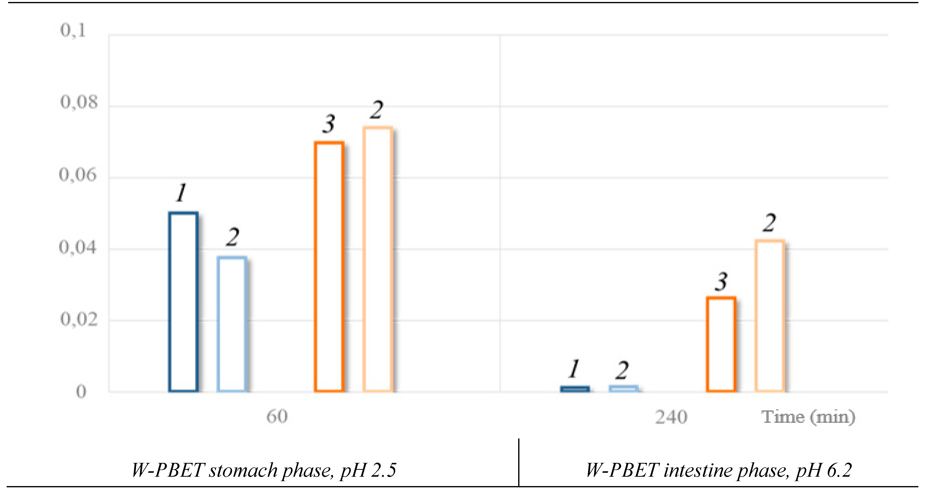
Figure 2.
Pb and Fe bioaccessibility (% of shot mass) when the shot is in the gastrointestinal tract of mammals.
Figure 2.
Pb and Fe bioaccessibility (% of shot mass) when the shot is in the gastrointestinal tract of mammals.
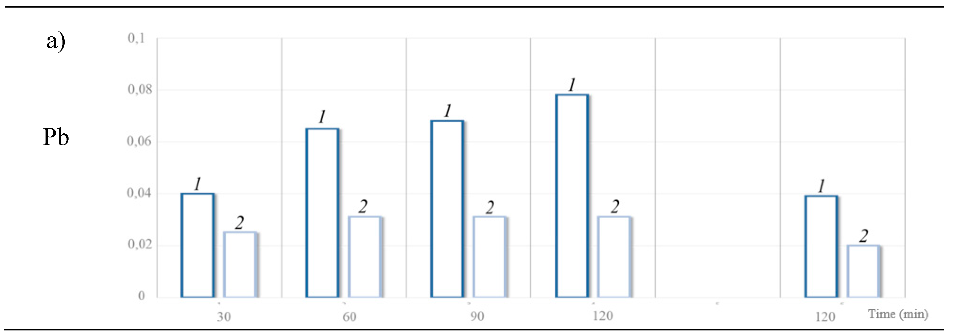
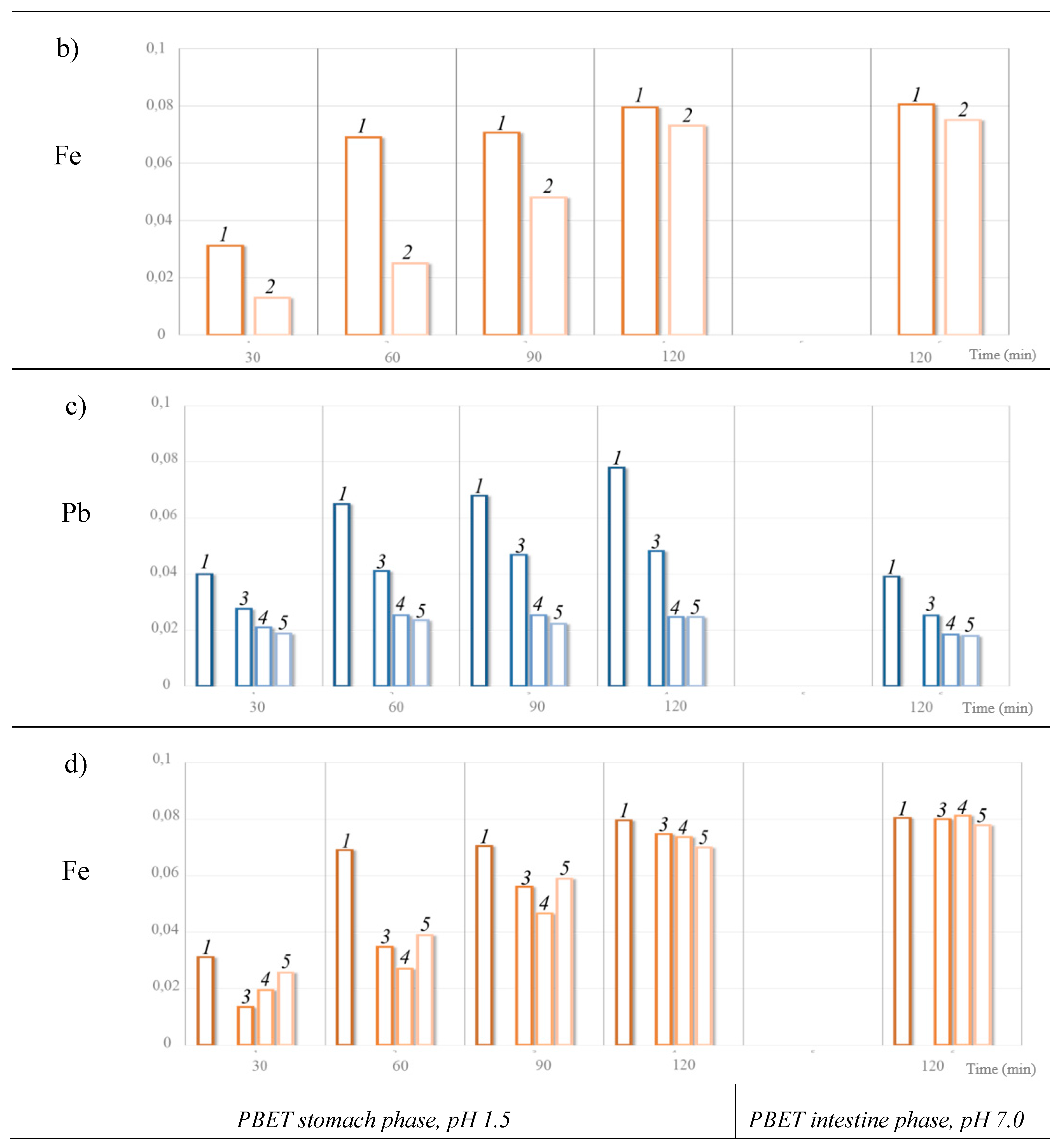
Figure 3.
Pb and Fe bioaccessibility (% of total) when soil particles is in the gastrointestinal tract of mammals and humans.
Figure 3.
Pb and Fe bioaccessibility (% of total) when soil particles is in the gastrointestinal tract of mammals and humans.
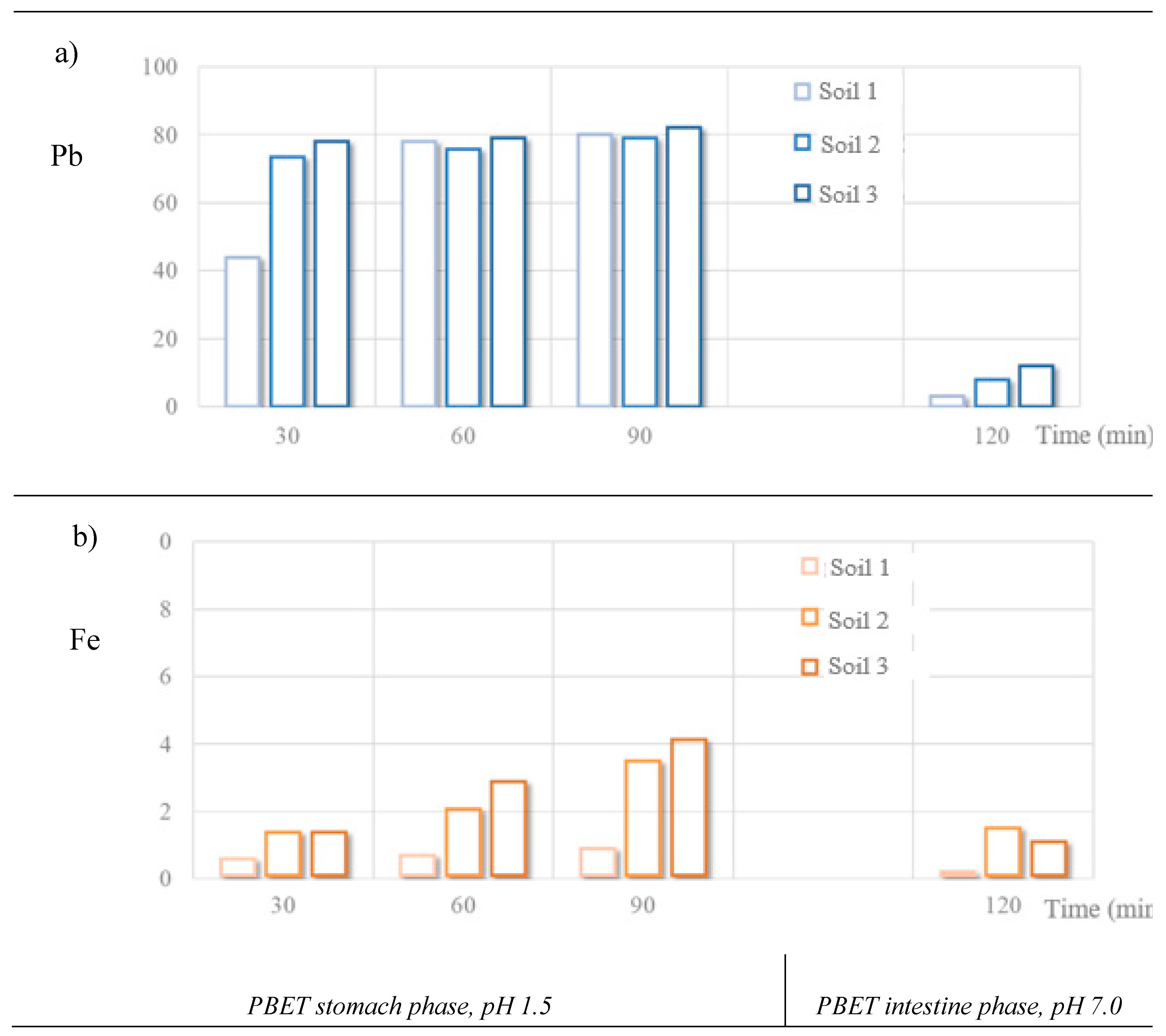

Table 1.
Total metal content and soil properties.
| Soil | Total metal, mg/kg | pH | EC | The main phases, % (identified by the XRD) | Soil texture (%) | Type | ||
|---|---|---|---|---|---|---|---|---|
| Pb | Fe | >250 μm | <250 μm | |||||
| 1 | 4500 | 15000 | 7.9 | 140 | Quartz (64), albite (13), microcline (10), carbonate (7), clay minerals (6) |
65 | 35 | Sandy loam |
| 2 | 3300 | 26300 | 7.8 | 190 | 64 | 36 | Sandy loam | |
| 3 | 3200 | 25000 | 7.6 | 200 | Quartz (69), albite (10), microcline (10), clay minerals (8) carbonate (3) | 56 | 44 | Sandy loam |
Soil 1 – the soils of shooting areas where only lead shot was used; Soil 2 – the soils of shooting areas (only lead ammunition was used), with the adding of steel shot; Soil 3 – the joint entering of steel shot and lead shot into the soils of areas with no prior shooting activities. EC – electrical conductivity, µS/cm.
Table 2.
The metal in the solution when the ammunition is in the digestive system.
| Shot | GI tract | Time, min | Pb, mg/l | Pb, mg/g | Fe, mg/l | Fe, mg/g |
|---|---|---|---|---|---|---|
| (mean ± SE) | (mean ± SE) | |||||
| The gastrointestinal tract of waterfowl | ||||||
| Lead | Stomach, рН 2.5 | 60 | 5.84 ± 0.14 | 0.50 | ||
| Intestine, рН 6.2 | 240 | 0.12 ± 0.02 | 0.01 | |||
| Steel | Stomach, рН 2.5 | 60 | 8.27 ± 0.85 | 0.70 | ||
| Intestine, рН 6.2 | 240 | 3.10 ± 0.47 | 0.26 | |||
| Lead:steel 1:1 |
Stomach, рН 2.5 | 60 | 2.25 ± 0.33 | 0.38 | 4.52 ± 0.49 | 0.74 |
| Intestine, рН 6.2 | 240 | 0.08 ± 0.01 | 0.01 | 2.58 ± 0.38 | 0.42 | |
| The gastrointestinal tract of mammals and humans | ||||||
| Lead | Stomach рН 1.5 |
30 | 4.00 ± 0.57 | 0.40 | ||
| 60 | 6.53 ± 1.00 | 0.65 | ||||
| 90 | 6.80 ± 0.42 | 0.68 | ||||
| 120 | 7.80 ± 0.42 | 0.78 | ||||
| Intestine, рН 7 | 120 | 3.89 ± 0.57 | 0.39 | |||
| Steel | Stomach рН 1.5 |
30 | 3.10 ± 0.42 | 0.31 | ||
| 60 | 7.13 ± 0.81 | 0.71 | ||||
| 90 | 7.05 ± 0.21 | 0.71 | ||||
| 120 | 7.95 ± 0.21 | 0.80 | ||||
| Intestine, рН 7 | 120 | 8.05 ± 0.07 | 0.81 | |||
| Lead:steel 1:1 |
Stomach, рН 1.5 |
30 | 2.51 ± 0.04 | 0.25 | 1.31 ± 0.20 | 0.13 |
| 60 | 3.06 ± 0.11 | 0.31 | 2.50 ± 0.05 | 0.25 | ||
| 90 | 3.11 ± 0.21 | 0.31 | 4.79 ± 0.10 | 0.48 | ||
| 120 | 3.07 ± 0.10 | 0.31 | 7.30 ± 0.31 | 0.73 | ||
| Intestine, рН 7 | 120 | 2.04 ± 0.19 | 0.20 | 7.47 ± 0.47 | 0.75 | |
| Lead:steel 2:1 |
Stomach рН 1.5 |
30 | 1.91 ± 0.22 | 0.28 | 0.49 ± 0.04 | 0.13 |
| 60 | 2.71 ± 0.17 | 0.41 | 1.30 ± 0.03 | 0.35 | ||
| 90 | 3.14 ± 0.28 | 0.47 | 2.11 ± 0.31 | 0.56 | ||
| 120 | 3.28 ± 0.05 | 0.48 | 2.81 ± 0.07 | 0.75 | ||
| Intestine, рН 7 | 120 | 1.73 ± 0.15 | 0.25 | 3.01 ± 0.09 | 0.80 | |
| Lead:steel 1:2 |
Stomach рН 1.5 |
30 | 0.68 ± 0.06 | 0.21 | 1.50 ± 0.08 | 0.19 |
| 60 | 0.82 ± 0.10 | 0.25 | 2.08 ± 0.06 | 0.27 | ||
| 90 | 0.82 ± 0.02 | 0.25 | 3.62 ± 0.23 | 0.46 | ||
| 120 | 0.80 ± 0.10 | 0.25 | 5.67 ± 0.12 | 0.74 | ||
| Intestine, рН 7 | 120 | 0.60 ± 0.06 | 0.18 | 6.31 ± 0.02 | 0.81 | |
| Lead:steel 1:6 |
Stomach рН 1.5 |
30 | 0.31 ± 0.05 | 0.20 | 2.56± 0.06 | 0.26 |
| 60 | 0.42 ± 0.06 | 0.25 | 3.50 ± 0.13 | 0.39 | ||
| 90 | 0.40 ± 0.03 | 0.24 | 5.27 ± 0.30 | 0.59 | ||
| 120 | 0.43 ± 0.02 | 0.26 | 6.34 ± 0.36 | 0.70 | ||
| Intestine, рН 7 | 120 | 0.45 ± 0.01 | 0.20 | 7.34± 0.49 | 0.76 | |
For each metal, its average content in the solution (mg/l) is indicated, as well as the respective standard deviation based on three measurements. The amount of dissolved metal per one gram of shot (mg/g) calculated by the average metal content in the solution.
Table 3.
The metal in the solution when soil particles is in the digestive system of the mammal’s and human’s.
Table 3.
The metal in the solution when soil particles is in the digestive system of the mammal’s and human’s.
| Soil | GI tract | Time, min | Pb, mg/l | Pb, mg/g | Fe, mg/l | Fe, mg/g |
|---|---|---|---|---|---|---|
| (mean ± SE) | (mean ± SE) | |||||
| 1 | Stomach, рН 1.5 | 30 | 20.4 ± 3 | 2.0 | 1.1 ± 0.4 | 0.1 |
| 60 | 35.0 ± 3 | 3.5 | 1.0 ± 0.2 | 0.1 | ||
| 90 | 36.0 ± 5 | 3.6 | 2.0 ± 0.6 | 0.2 | ||
| Intestine, рН 7 | 120 | 1.4 ± 0.4 | 0.14 | 0.3 ± 0.2 | 0.03 | |
| 2 | Stomach, рН 1.5 | 30 | 25.2 ± 1.4 | 2.5 | 3.7 ± 0.5 | 0.4 |
| 60 | 26.1 ± 1.2 | 2.6 | 5.4 ± 1.7 | 0.5 | ||
| 90 | 27.1 ± 1.2 | 2.7 | 9.2 ± 4.8 | 0.9 | ||
| Intestine, рН 7 | 120 | 2.8 ± 0.1 | 0.3 | 2.9 ± 1.3 | 0.3 | |
| 3 | Stomach, рН 1.5 | 30 | 25.1 ± 2.1 | 2.5 | 3.4 ± 1.1 | 0.3 |
| 60 | 25.3 ± 0.7 | 2.5 | 7.2 ± 2.4 | 0.7 | ||
| 90 | 26.3 ± 1.9 | 2.6 | 10.3 ± 4.9 | 1.0 | ||
| Intestine, рН 7 | 120 | 3.3 ± 0.6 | 0.3 | 2.9 ± 1.5 | 0.4 |
soil 1 – the soils of shooting areas where only lead ammunition was used (the total content of Pb – 4 500 mg/kg, Fe – 15 000 mg/kg);.soil 2 – the soils of shooting areas (only lead ammunition was used), with the adding of steel shot (the steel shot load of 0.02 kg/0.1 m2, the total content of Pb – 3 300 mg/kg, Fe – 26 300 mg/kg); soil 3 – the soils of areas with no prior shooting activities where lead and steel shot are deposited at the same time (the shot load of 0.02 kg/0.1 m2 each, the total content of Pb – 3 200 mg/kg, Fe – 25 000 mg/kg).
Table 4.
Model calculations of Pb mass thresholds in the case of the introduction of shot/soil into the GI tract.
Table 4.
Model calculations of Pb mass thresholds in the case of the introduction of shot/soil into the GI tract.
| Site | Pb threshold level in the GI tract, mg | GI tract organ | Pb bioaccessibility | Pb mass threshold (mg) in the substance entering the GI tract | ||
|---|---|---|---|---|---|---|
| shot, % of metal mass | soil, % of total | shot* | soil | |||
| Human | 2.5 | stomach | 0.08 | 82 | 2,500 | 3.0 |
| intestine | 0.04 | 3/12** | 6300 | 83/21 | ||
| Waterfowl | 0.25 | stomach | 0.05 | - | 500 | - |
| intestine | 0.001 | - | 25,000 | - | ||
*- lead shot #7.5 for sports shooting, pellet weight 0.07 g; **- actual/maximum value of Pb bioaccessibility in soil
Table 5.
The calculated amount of ingested shot (model).
| Blood lead concentration at the end of the experiment, mg/l | Calculated amount of shot based on the model, pcs | Note | |
| Control group | Test group | ||
| 0.69–0.71 | 1.64–3.08 | 59–110* (21–40)** |
12 ducks were intubated with one #4 lead shot (0.193 g) by gavage; at the end of one week, no signs of poisoning were observed (Finley et al., 1976). |
| 0.5–0.78 | 18–28* (6–10)** |
12 ducks were intubated with one #4 lead shot (0.193 g) by gavage; at the end of four weeks, no signs of poisoning were observed, the ducks had put on weight (Finley et al., 1976). | |
| 0.05–0.17 | 0.15–8.01 | 5–286* | 55 mallards placed in a 4-acre enclosure on a hunting wetland (with a lead shot density of 931±67 pellets per acre in the top 10 cm of sediment); at the end of 80 days, no clinical signs of lead poisoning were observed (Rocke&Samuel, 1991) |
| 0.65–8.71 | 23–311 *(8–109)** |
16 mallards were intubated with two #4 lead shot (0.2 g each) by gavage; by the end of 80 days, three of the birds had died while the others exhibited clinical signs of lead poisoning (Rocke&Samuel, 1991) | |
| 0.11–0.29 | 1.05 | 37 *(8)** |
14 ducks were intubated with one #2 lead shot (0.325 g) by gavage; at the end of 30 days, no signs of poisoning were observed (Plouzeau et al., 2011) |
| 0.67 | 24 *(9)** |
14 ducks were intubated with one #4 lead shot (0.177 g) by gavage; at the end of 30 days, no signs of poisoning were observed (Plouzeau et al., 2011) | |
| 0.90 | 33 *(19)** |
13 ducks were intubated with one #6 lead shot (0.125 g) by gavage; at the end of 30 days, no signs of poisoning were observed (Plouzeau et al., 2011) | |
| - | 0.03 | 1* | 277 ducks, minimum blood lead concentration in the natural habitat in shooting areas (New Jersey), no signs of poisoning were observed (Lewis et al., 2021) |
| 0.98 | 35* | 277 ducks, maximum blood lead concentration in the natural habitat in shooting areas (New Jersey), no signs of poisoning were observed (Lewis et al., 2021) | |
*the calculation is based on #7.5 lead shot (0.07 g); **the calculation is based on hunting shot, with the number and mass according to the comment.
Disclaimer/Publisher’s Note: The statements, opinions and data contained in all publications are solely those of the individual author(s) and contributor(s) and not of MDPI and/or the editor(s). MDPI and/or the editor(s) disclaim responsibility for any injury to people or property resulting from any ideas, methods, instructions or products referred to in the content. |
© 2024 by the authors. Licensee MDPI, Basel, Switzerland. This article is an open access article distributed under the terms and conditions of the Creative Commons Attribution (CC BY) license (http://creativecommons.org/licenses/by/4.0/).
Copyright: This open access article is published under a Creative Commons CC BY 4.0 license, which permit the free download, distribution, and reuse, provided that the author and preprint are cited in any reuse.


