
Submitted:
27 May 2024
Posted:
28 May 2024
You are already at the latest version
Abstract
Objectives: This study aims to explore the use of murine models in peripheral nerve transection research, evaluating and synthesizing key methods for analyzing nerve regeneration to guide future research and clinical interventions.
Methodology: A systematic review was conducted in February 2024, adhering to Cochrane and PRISMA 2020 guidelines, using Medline, Scielo, and Lilacs databases. The focus was on experimental studies of nerve regeneration in animal models post-transection. Only experimental clinical trials reporting nerve regeneration outcomes were included; studies lacking a comparator group or functional evaluation methods were excluded.
Results: Out of 273 studies initially identified from MEDLINE, 19 were selected for detailed analysis. The average study included 32.5 subjects, with about 10.16 subjects per intervention subgroup. The predominant model was the sciatic nerve injury with a 10mm gap. The most common intervention involved unprocessed adipose-derived stem cells, utilized in 14 articles.
Conclusions: The review underscores the significant potential of current methodologies in peripheral nerve regeneration, particularly highlighting the use of murine models and thorough evaluation techniques. These studies significantly contribute to our understanding of nerve regeneration processes and inform directions for future research.
Keywords:
Microsurgery
; Nerve Repair
; Nerve Regeneration
; Functional Analysis
; Fat Graft
1. Introduction
Peripheral nerve injury (PNI) has been garnering increased attention due to its rising prevalence alongside rapid societal developments[1]. The main causes of PNI, such as diabetes mellitus, autoimmune diseases, infections, medications, and traumatic injuries, exhibit diverse clinical manifestations. Literature reviews indicate a prevalence of 8% for non-traumatic cases[2] and 5% for traumatic instances[3]. Despite advancements in microsurgical techniques, patients with PNI still face unsatisfactory recovery prognosis[4].
Injuries are categorized into neuropraxia, axonotmesis, and neurotmesis, each with distinct implications for treatment and functional recovery[5,6].This scenario underscores the need for innovative therapeutic approaches that could offer more promising outcomes.
In this context, adipose-derived stem cells (ADSCs) emerge as a promising source for regenerative therapies due to their ability to differentiate into various cell types, such as Schwann-like cells, and to release regenerative factors through paracrine mechanisms[7,8].
Scientific literature highlights the potential of these cells in treating peripheral nerve injuries in animal models, with evidence of their role in promoting regeneration and improving nerve function. Experimental studies have been primarily conducted in rodent models, as their nerves share similar size, fascicular organization, and morphology to humans[9]. Regarding the use of ADSCs in treatment, the potential to contribute to the recovery of damaged nerves makes them promising candidates for future clinical applications[10].
Additionally, complementary techniques such as the stromal vascular fraction (SVF) and nanofat are explored to optimize the regenerative potential of ADSCs with paracrine and immunomodulatory actions to induce tissue regeneration. These approaches combine the cellular richness of the SVF with the benefits of the extracellular matrix and paracrine signals provided by nanofat, enabling the development of more effective treatments against nerve injuries[11,12].
In this context, this systematic review aims to present the state of the art in the current literature regarding the use of murine models subjected to peripheral nerve injuries. Subsequently, it intends to synthesize the main methods of analyzing nerve regeneration and their respective results in the selected articles.
2. Materials and Methods
2.1. General Information
This is a systematic search study following Cochrane standards, and this manuscript was conducted according to the PRISMA (Preferred Reporting Items for Systematic Reviews and Meta-Analyses) 2020 guidelines[13], with data research conducted in February 2024. As this is a systematic literature review, this study was exempt from institutional review board approval and did not utilize external funding sources. The databases MEDLINE, Scielo, and LILACS were used to search for articles addressing peripheral nerve regeneration in animals subjected to nerve transection. The following keywords were used in MEDLINE: ("rat"[All Fields] OR "mice"[All Fields] OR "rabbits"[All Fields] OR "macaca"[All Fields] OR "swine"[All Fields] OR "muridae"[All Fields]) AND ("peripheral nerve injuries"[All Fields] OR "denervation"[All Fields]) AND ("microsurgery"[All Fields] OR "neurosurgery"[All Fields] OR "mesenchymal stem cells"[All Fields] OR "adipose tissue"[All Fields]) AND "nerve regeneration"[All Fields]. The same terms were used in Embase, Scielo, and LILACS with necessary modifications according to the rules of these databases.
2.2. Data Selection and Extraction
Following data research, article selection was conducted by three independent reviewers in two stages: analysis of article titles and abstracts, followed by full-text review. Any conflicts were resolved by a third author. The literature was restricted to articles in English, Portuguese, and Spanish without date limitations. Inclusion criteria were limited to articles assessing peripheral nerve regeneration as the primary outcome and considering the section of the peripheral nerve studied. Moreover, only experimental clinical trials were included since they are the primary studies with the highest impact, thus providing more reliability for the data the authors aimed to analyze. Exclusion criteria were defined as follows: articles that did not address the presence of a comparator group among the studied subgroups; those that did not include methods for the functional evaluation of the studied nerve regeneration; and those that did not use adipose derivatives in at least one subgroup. After applying the inclusion and exclusion criteria, 19 studies were included for full-text analysis. It is important to note that the choice of inclusion and exclusion criteria was made a priori.
Regarding data extraction, it was performed by three independent reviewers who collected the following information from the articles: author, publication year, intervention, objective, conclusion, study location, animal follow-up, population, number of animals per subgroup, breed of animal, functional analyses, histological analyses, and other parameters assessing nerve regeneration. Data on funding sources were also collected.
3. Results
The literature search yielded 273 records, mainly from the MEDLINE database. Abstract screening removed the majority of the articles. The main causes for removal were wrong study design and studies that did not use adipose tissue byproducts.
Figure 1.
Flowchart representing the literature search.
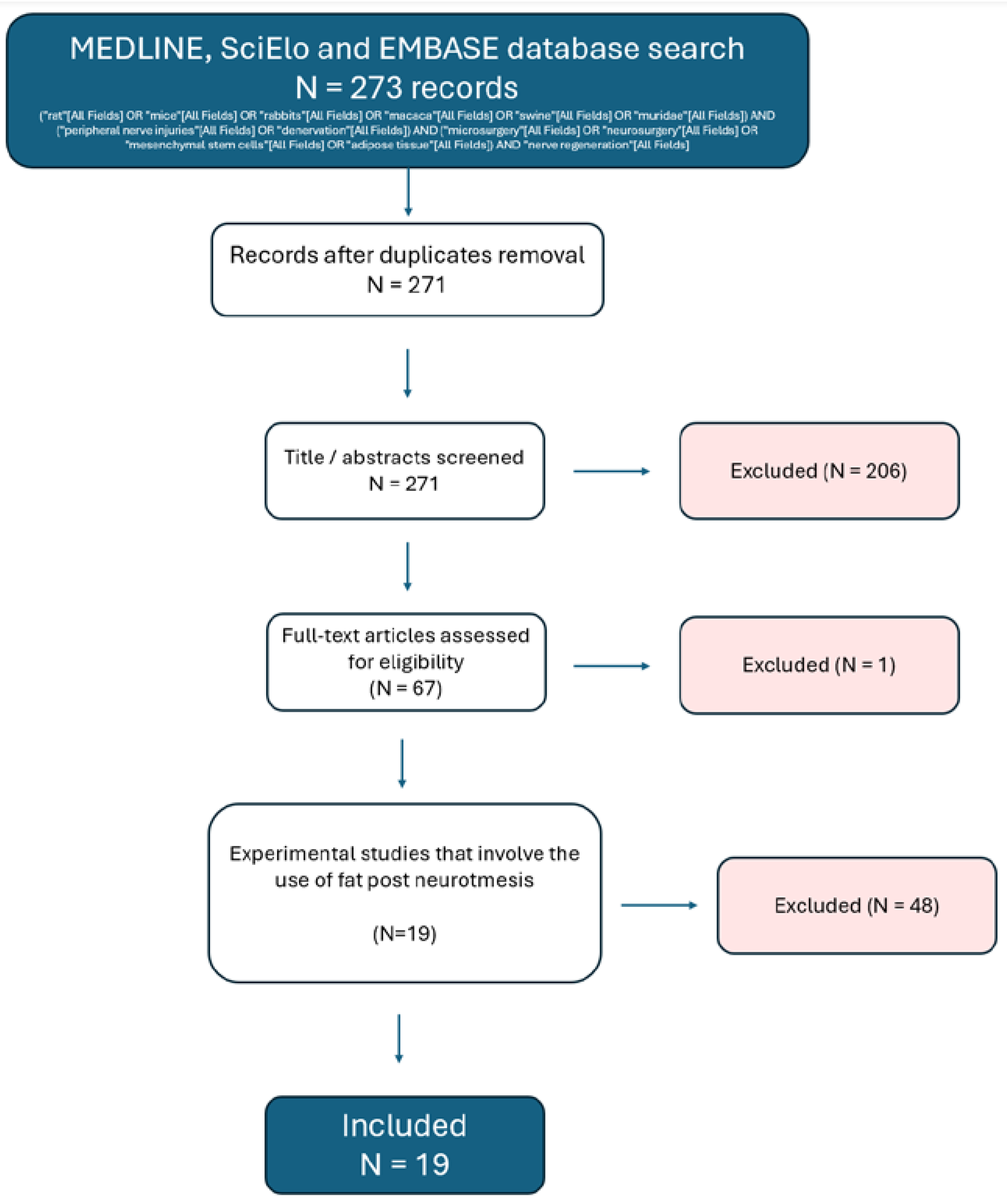
Figure 2.
Country of the institution to which the first author is affiliated.
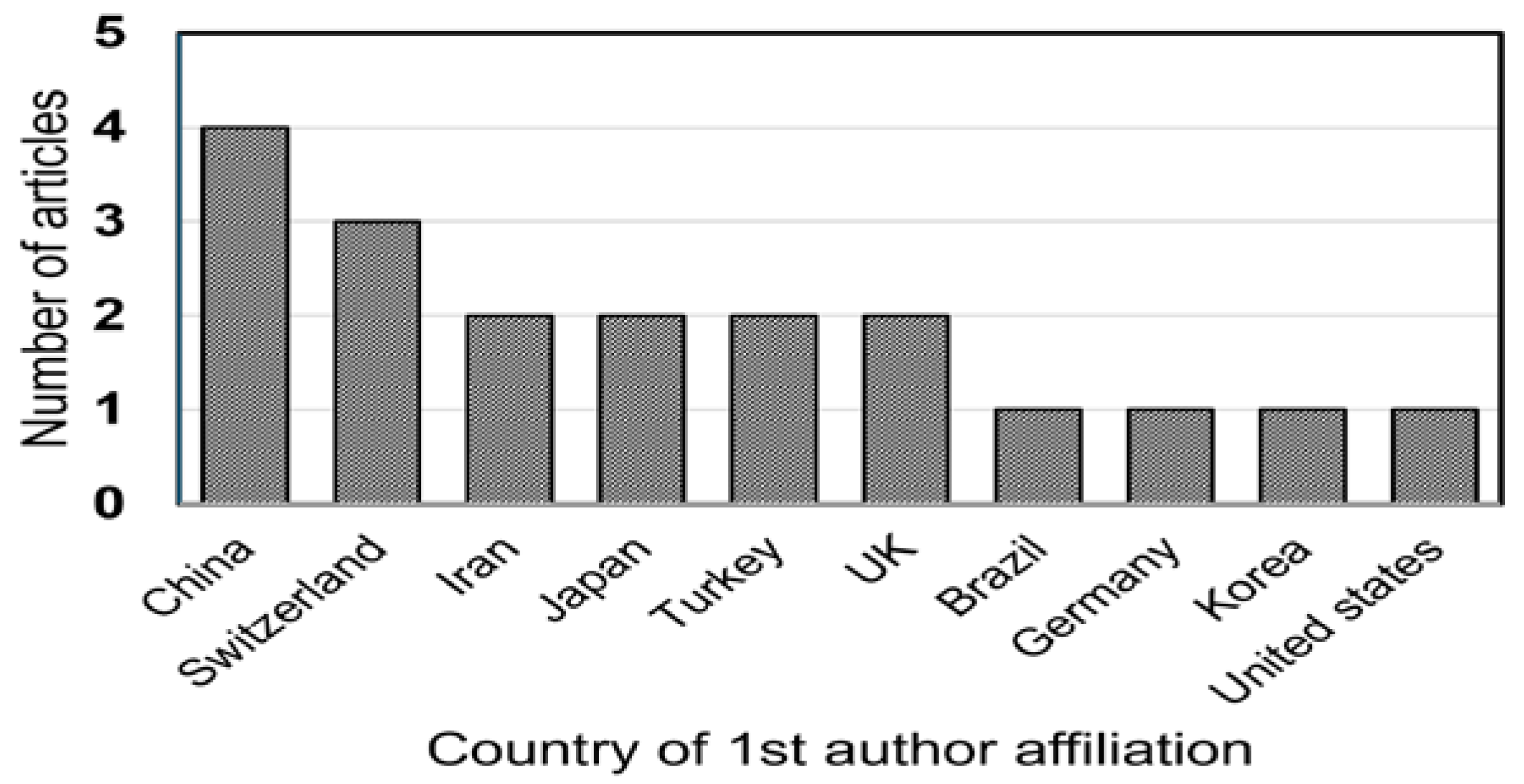
Figure 3.
Analytic view of publishing countries.
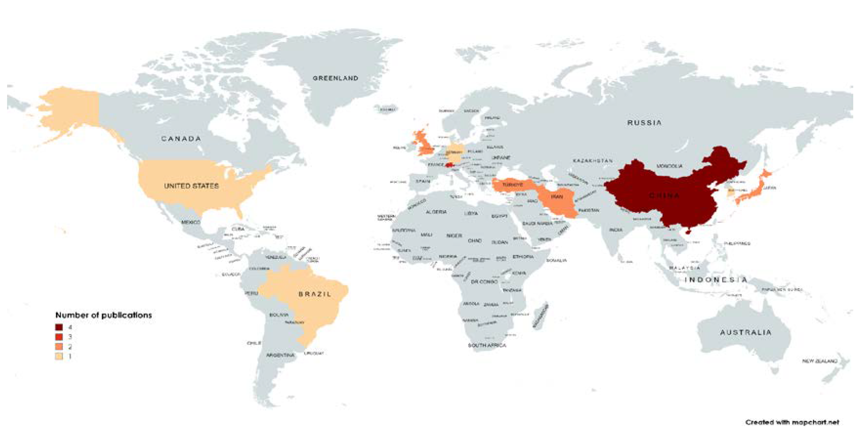
The country that produced the most results was China (N=4), followed by Switzerland.
Figure 4.
Animal species used in the experiments retrieved.
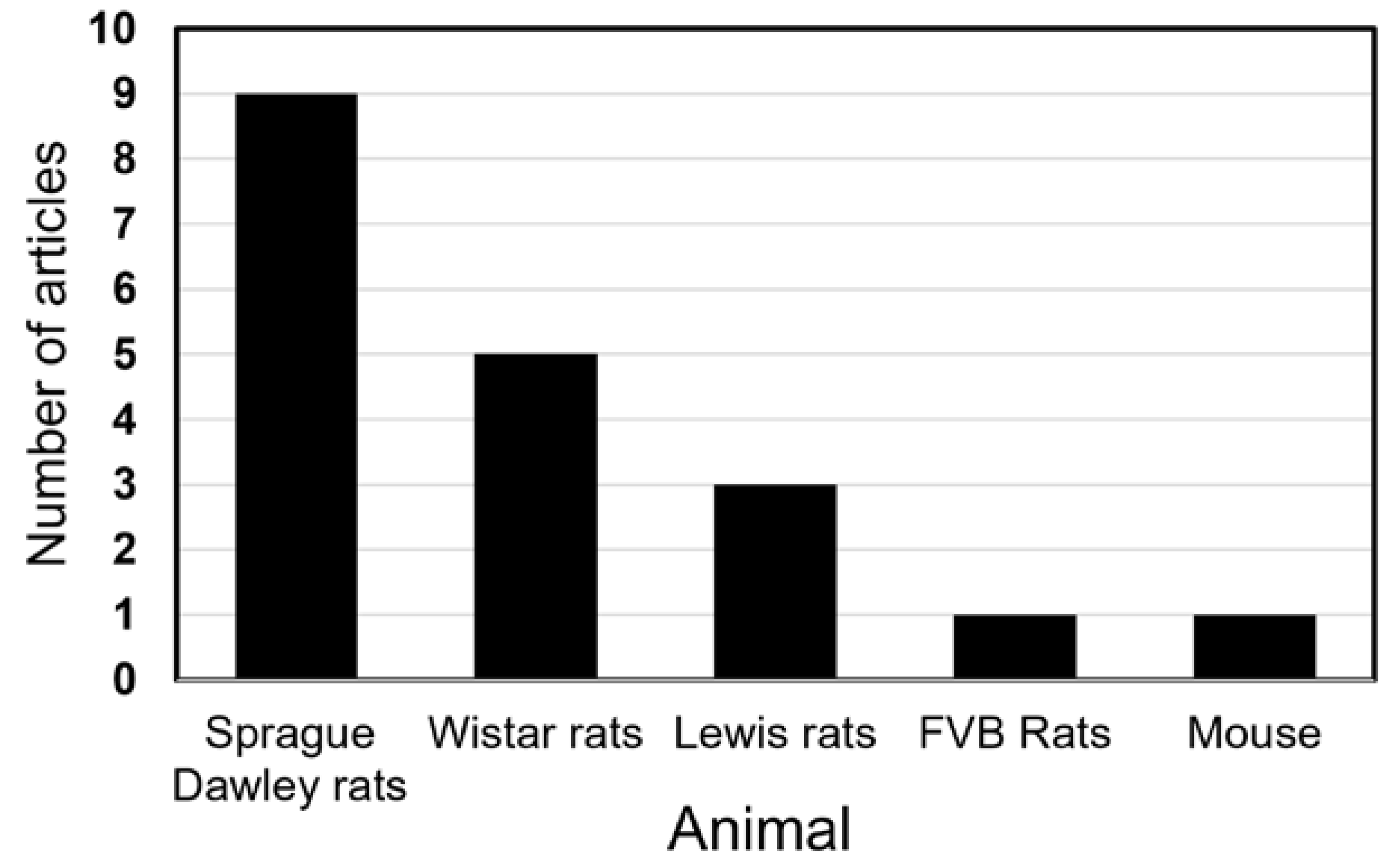
Despite including several animal species in our search terms, we only located studies conducted in muridae. The most commonly used species was the Sprague Dawley rat, followed by Wistar and Lewis rats. FVB rats and the mouse were used in a single study each. The average number of subjects was 32.5, being the average number of subjects per intervention group 10.16.
Figure 5.
Observation time of the subjects.
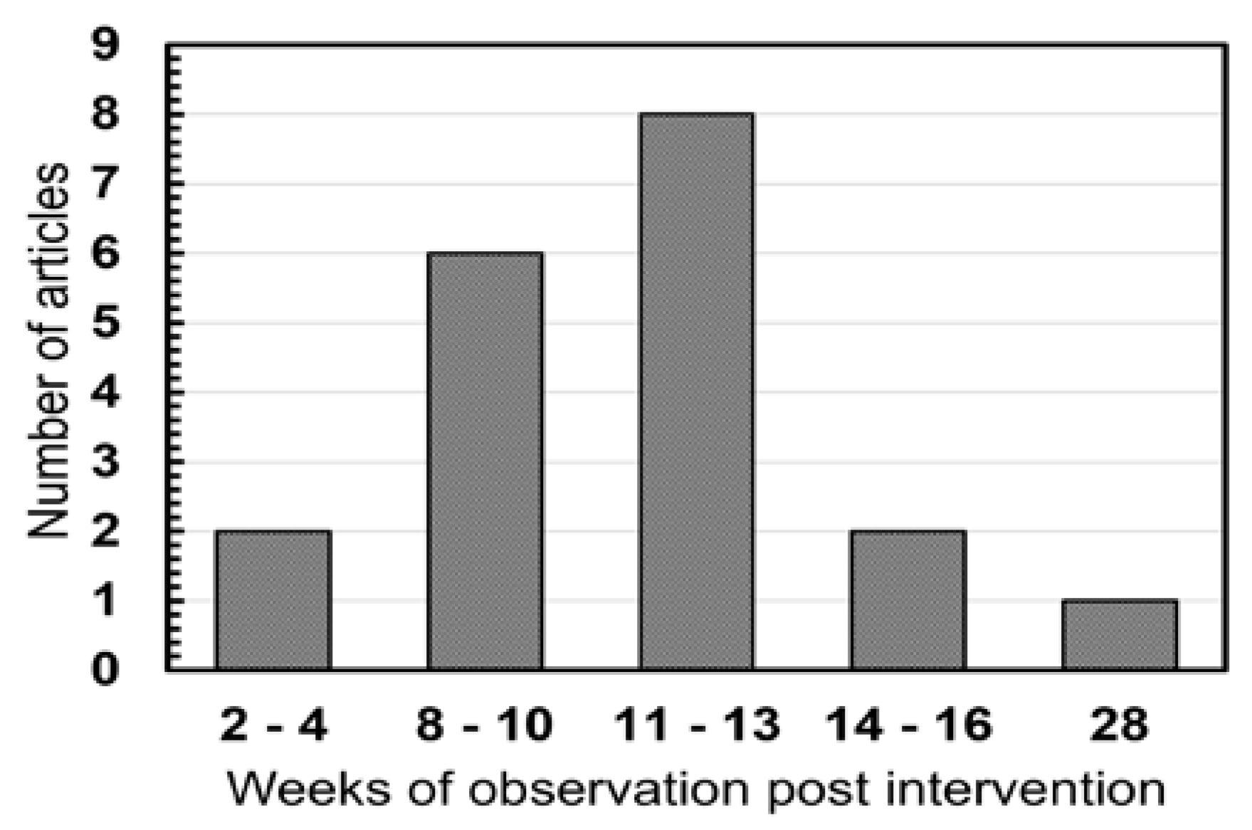
Most articles performed their intervention immediately after neurotmesis, and then observed the rat for a mean number of 11.26 weeks.
Figure 6.
Nerve injury models utilized.
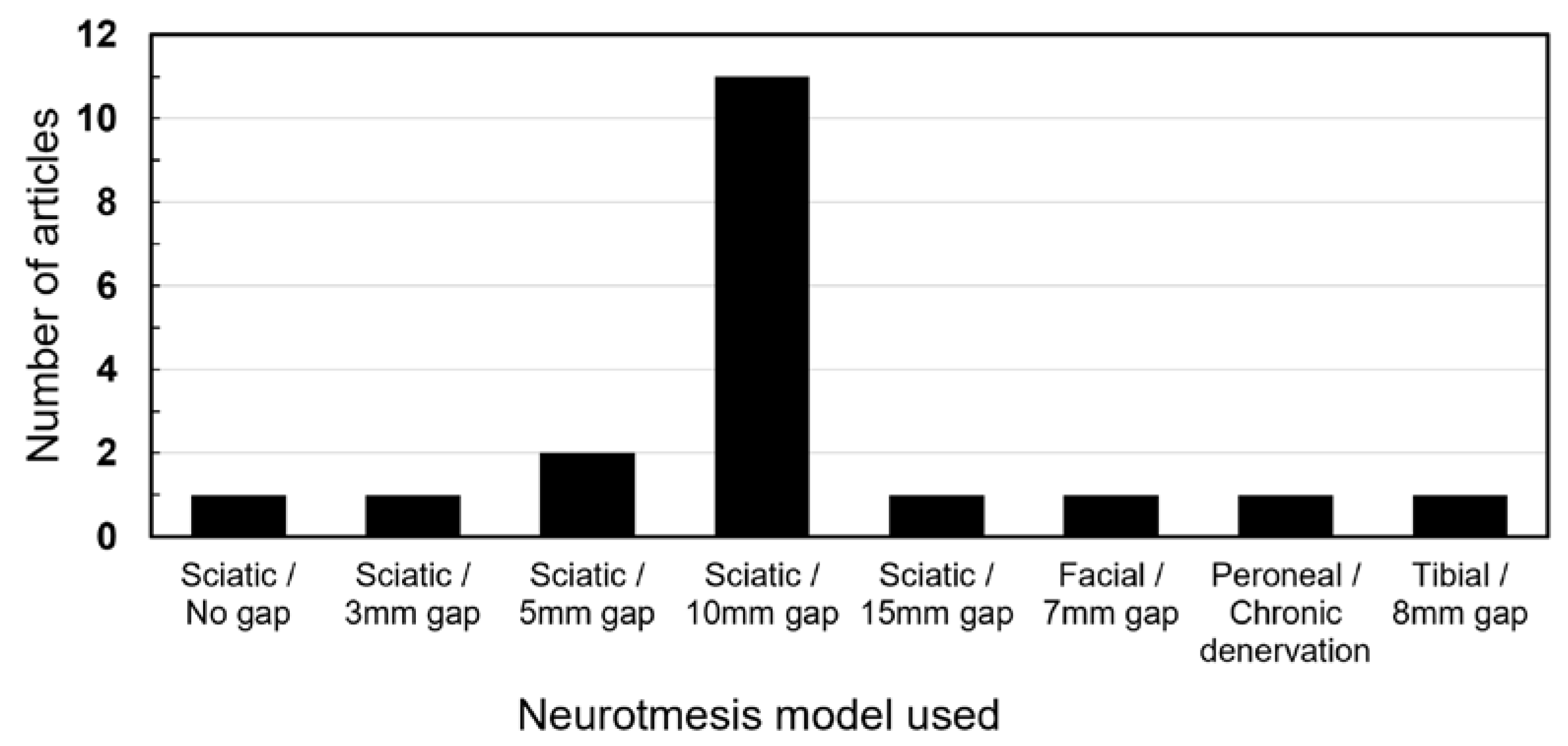
The sciatic nerve injury with a gap of 10mm was the overall most commonly utlized model by a far margin (N = 11). Other gaps were utilized in smaller nerves or animals. One study utilized a chronic denervation model by severing the peroneal nerve and suturing the ends away from each other.
Figure 7.
Interventions of control groups.
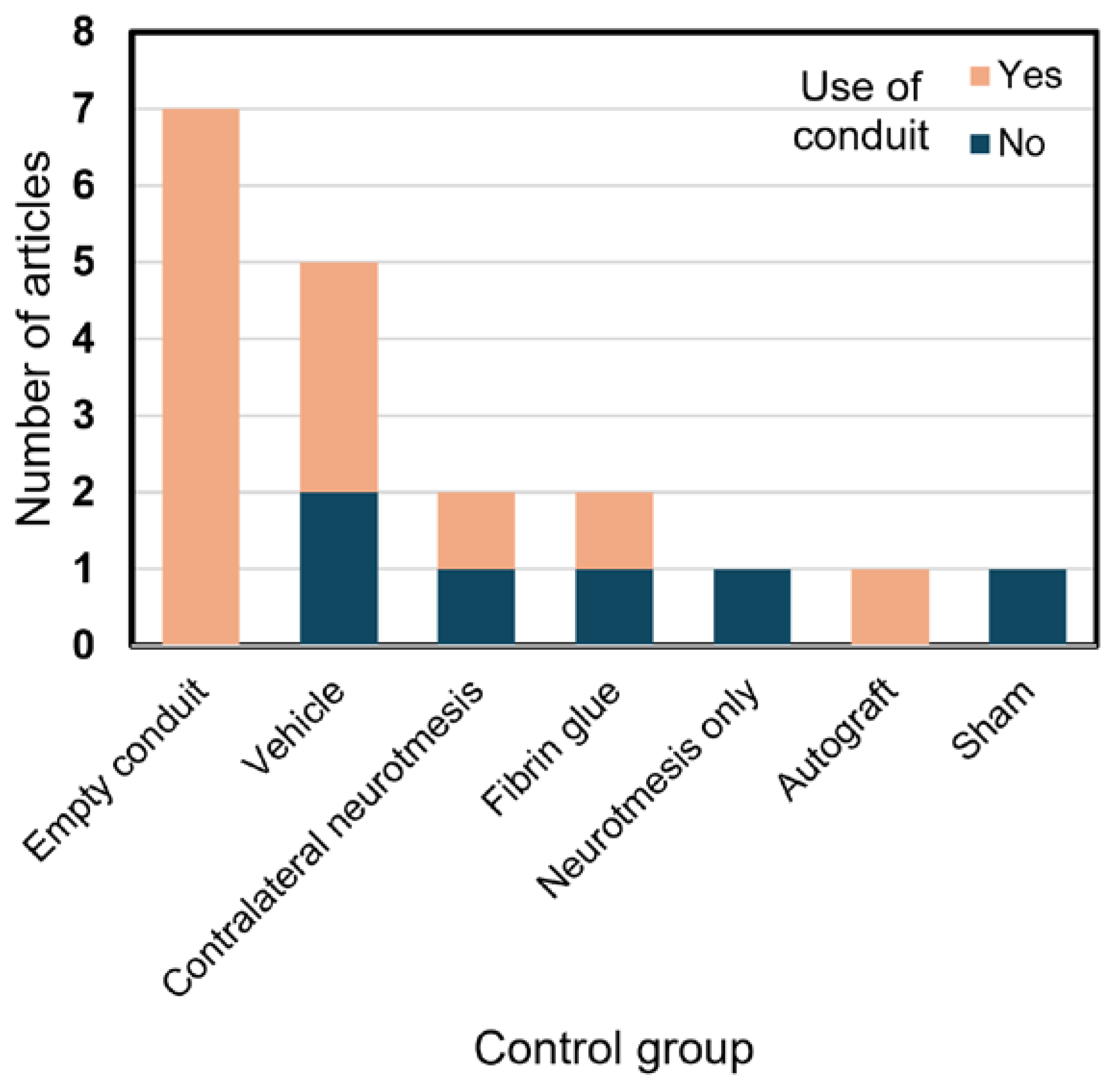
38.8% of control groups utilized empty conduits. All of the articles utilized a control group in accordance to the intervention.
Figure 8.
Materials utilized in the nerve during interventions by number of articles.
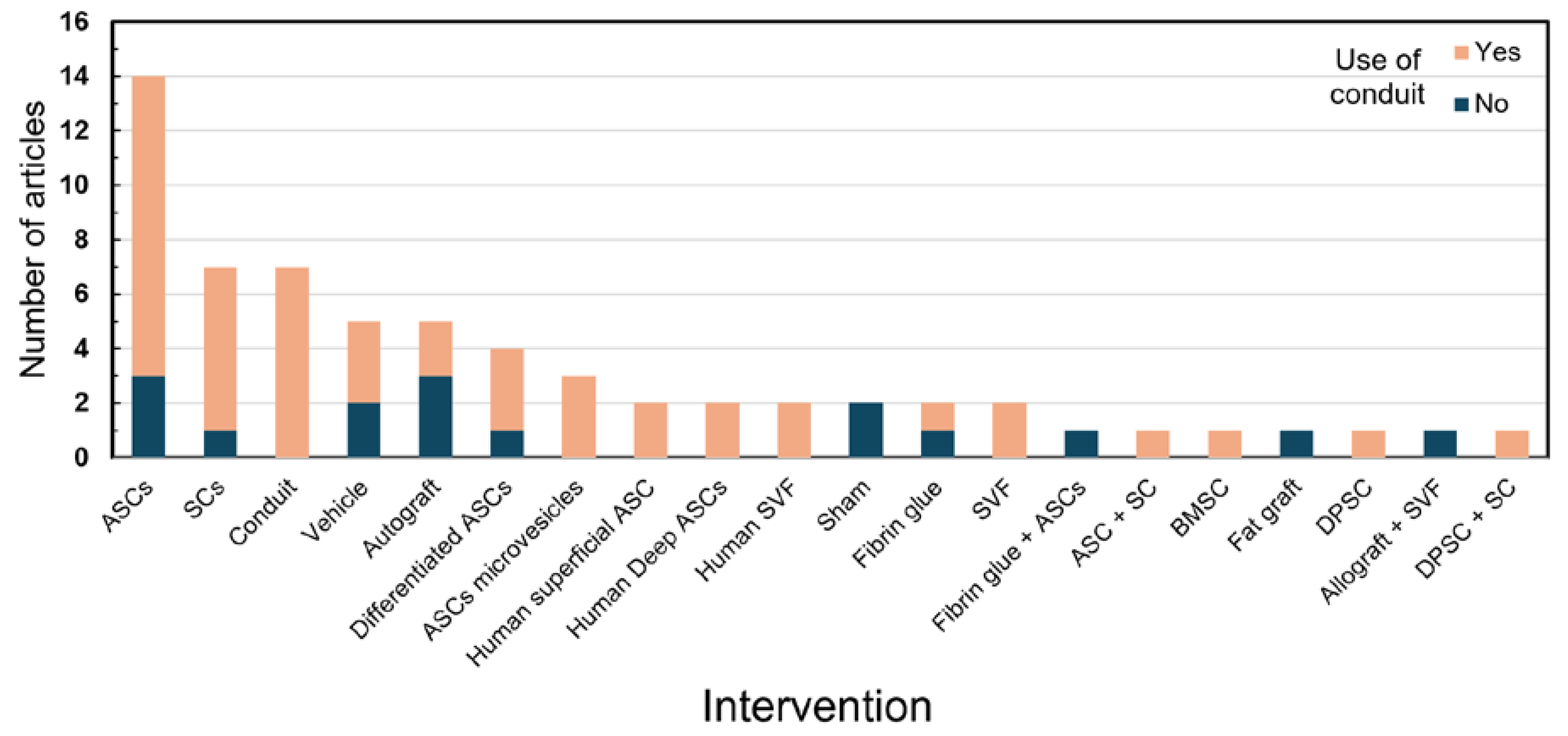
Figure 9.
Materials utilized in the nerve during interventions by total number of nerves severed.
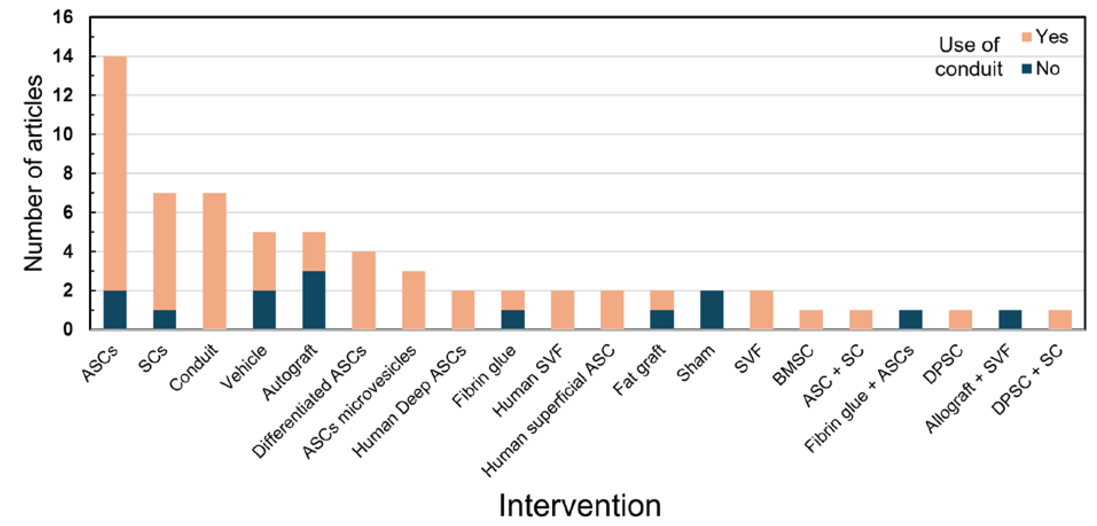
ASCs with no further processing were the most common intervention, utilized in a total of 14 articles and 122 nerves. 68/4% of the articles utilized conduits to bridge the nerve gaps. Aside from ASCs, other adipose tissue byproducts utilized were SVF and fat grafts.
4. Discussion
Based on current research, tissue bioengineering and microsurgery emerge as some of the fundamental pillars of regenerative medicine. As intervention studies on vital structures progress, the techniques described increasingly approach practical applicability.
4.1. General Aspects
Among the selected studies, China contributed the highest number (n=5), followed by Switzerland (n=3). This find is compatible with the recent advances that China has been making in the academic publishing scene[33]. Remarkably, Chinese articles had the second-highest average journal impact factor, surpassed only by Korea. This indicates that China has had an increase not only in quantity, but in quality of their publications.
4.2. Fat Grafting and Stem Cells
In accordance with our inclusion criteria, all articles involved ASC-based interventions in at least one group, totaling 269 nerves undergoing sectioning and ASC interventions. The use of ASCs has been gaining popularity within the literature since its discovery as a potential material for tissue regeneration[34]. A search on PubMed for 'Adipose-derived stem cells' shows an increasing trend, indicating growing interest in this topic. Several uses of ASCs are currently under investigation, with the main ones including wound healing, bone healing, immunomodulation, and nerve regeneration[35]. ASCs have gained special attention in the field of stem cell research because they are widely accessible throughout the body, easy to process, and lack the ethical controversies associated with other stem cell sources[36].
The first article proposing the use of ASCs for neuronal regeneration was published in 2005, followed by numerous animal and human research models exploring its therapeutic potential[37]. Their applications across various specialties, including ophthalmology (optic nerve regeneration), urology (treatment of nerve-induced erectile dysfunction), and reconstructive plastic surgery, have been extensively studied[38,39,40].
4.3. Trauma Mechanism
Studies on ASCs in nerve regeneration remain predominantly confined to animal models, notably involving the sciatic nerve neurotmesis with a 10mm gap. Among the analyzed articles, 11 studies employed this specific model, while others explored neurotmesis involving different gaps or other nerves, such as the brachial plexus and the facial nerve. Additionally, several studies investigated crush injury models[41].
The selected studies exhibited a broad spectrum of methodologies and approaches, leading to considerable variability in the types of functional and histological analyses performed. This diversity hindered the feasibility of conducting a systematic review, which generally demands a more standardized approach to data synthesis and analysis. Consequently, we adopted a narrative review methodology to provide a comprehensive and interpretive synthesis of the existing literature[42].
4.4. Functional Analysis
In this study, eight articles[16,19,20,21,22,25,26,31] utilized the "Sciatic Functional Index" (SFI) for evaluating nerve function. Based on Medicaneli et al. (1982)[43], the method is reliable for assessing muscle strength index and tissue electrophysiology, thus evaluating the performance of nerve function in the injured lower limb. For instance, the regeneration of the sciatic nerve was assessed by Dai et al. (2013)[19] with recovery rates ranging from 20.4% to 23.4% over a three-month period. Similarly, Orbay et al. (2012)[44] reported a functional recovery rate of 24.9% within two months in mice that received the intervention, demonstrating the method's effectiveness in analyzing this variable[45,46,47,48]. Conversely, other studies also used alternative scales. Mohammadi et al. (2015)[22], in addition to the SFI, employed the "Basso, Beattie, and Bresnahan for limb motor function" (BBB) and "Static Sciatic Index" (SSI) for assessing the same parameter. Although less used in the reviewed articles, both have validation in the scientific literature for their use[49,50]. Only one article used the "Peroneal Functional Index" (PFI), although the literature equally validates its use compared to the SFI[51].
Regarding methods of nerve electrostimulation, the parameter Compound Muscle Action Potential (CMAP) was used in five articles[15,17,18,28,30], Compound Nerve Action Potential (CNAP) in one article[25], and Nerve Conduction Velocity (NCV) in four articles [16,19,20,25].
The CMAP is intended to measure the sum of action potentials of the concerned muscle fibers, focusing on the response of the innervated muscle[52]. Notably, the experiment by Di Summa et al. (2011)[12] assessed nerve regeneration through significant increases in axonal diameter and CMAP using either autograft or fibrin seeded with ASCs on the PNI. The validation of this method is also present in the literature[53].
The use of CNAP also proved to be a satisfactory parameter in the study by He et al. (2016)[25], being effective in comparing the proposed regeneration in the Lv-ADSCS group versus the 34a-ADSCS group, which proved more effective in the latter.
The measurement of nerve conduction velocity is also an important factor for evaluating innervation of the site, and depends on factors such as axon diameter, myelin sheath thickness, and the length of the "internodes"[54,55]. According to Raisi et al. (2014)[20], NCV was effective in ascertaining poorer performance in nerve regeneration in the pro-inflammatory MSC group, with a lower conduction velocity rate (72%). This aspect was crucial in questioning which factors were triggering, such as: smaller diameter of regenerating axons, thinner myelin sheaths, or even an immaturity of myelinated nerve fibers in that group.
Regarding the Muscle Weight Ratio (MWR), nine articles used this method[17,19,21,22,23,25,26,29,31]. This parameter appears to be used due to the ease of its application in studies, often being the only type of functional evaluation performed[23,29]. In the experimental trial conducted by Pollari et al. (2018)[56], the method proved effective in comparing nerve regeneration in the transection versus crushing of the peripheral nerve group.
4.5. Histological Analysis
The use of optical microscopy alongside software for image analysis was highlighted in all selected articles. Based on Kappos et al. (2015)[21], this combined method was crucial for measuring axonal diameter and fiber diameter. Thus, the thickness of myelin and other important average parameters for evaluating local tissue regeneration can be calculated and compared across groups[57].
The light microscope method was enhanced by complementary analyses in several studies. Notably, the use of toluidine blue in five articles[22,23,25,27,29] emphasized its role in staining myelinated structures that facilitate neuronal visualization[58,59,60]. According to Ozkan et al. (2016)[27], this method proved effective for verifying regular myelinated pre-neuronal bodies with increased diameter in the study group, demonstrating better organization of nerve tissue regeneration.
The scientific literature indicates the efficiency of immunofluorescence methods for analyzing the regeneration of Schwann cells and peripheral remyelination[61,62]. In agreement, Kastamoni et al. (2023)[32] found that the expression of EphA4 was associated with a negative effect on the axonal repair of the sciatic nerve, consistent with previous studies [63,64,65].
Despite its requirement for considerable time and specialized training, transmission electron microscopy is widely mentioned in the literature as a method for obtaining detailed, high-quality images of nervous structures[66,67,68,69]. In this regard, several studies have used this method to obtain highly specific images of structures[16,18,20,25,28,29,30].
4.6. Other Analysis
Tremp et al. (2015)[24] identified a correlation between axonal length measured by MRI and axonal length measured by immunohistochemistry, reaffirming the nerve damage between the sciatic nerve of the intervention versus control. Thus, it is understood that the use of this method can add comparative value between the analyses performed.
Flow cytometry is an effective tool in studies of axonal nerve regeneration, due to its ability to quantitatively analyze individual cells in large populations[70,71,72]. One study used this method to evaluate specific cellular groups and their tissue behavior in regeneration[29].
RNL is a prospective method for evaluating nerve regeneration that compares neuronal expansion through its connections[73,74,75]. Di Summa et al. (2011)[15], evaluated using this method and electrostimulation that the autograft group and the ASCs group showed greater motoneuron regeneration compared to the control, which is in dialogue with current literature[76,77,78].
4.7. Strengths and Limitations
This review fulfills its role of assessing the current state of the scientific literature regarding the use of murine models for the experimentation of peripheral nerve regeneration, as well as elucidating and analyzing the main functional assessment models used in such experimental trials. Another point analyzed was the global distribution of scientific production for this article model, with an emphasis on comparing the use of different fat derivatives, such as ADSCs, SVF, and NanoFat. However, although it has a well-defined study population and outcomes, the breadth of types of peripheral nerve injury makes the execution of a meta-analysis unfeasible, due to the heterogeneity of the selected articles concerning the type of intervention proposed in each study. Lastly, the casuistry of the subgroups is not equivalent, which complicates a more complex biostatistical analysis.
5. Conclusion
The techniques and procedures described in this review hold significant potential for future practical application in peripheral nerve regeneration, as further studies continue to advance the field. Thus, this work serves its purpose by elucidating the current evidence on the topic, evaluating the principal models and formats of experimental studies that permeate the literature. Additionally, it enhances the clarity of the main types of fat derivatives and the methods for evaluating nerve regeneration in the most methodologically rigorous articles reviewed.
Acknowledgements
Nothing to declare.
Formatting of funding sources
This research had no funding sources.
Declarations of interest
None.
References
- England JD, Asbury AK. Peripheral neuropathy. The Lancet. 2004 Jun;363(9427):2151–61.
- Martyn CN, Hughes RA. Epidemiology of peripheral neuropathy. Journal of Neurology, Neurosurgery & Psychiatry [Internet]. 1997 Apr 1;62(4):310–8. Available from:https://www.ncbi.nlm.nih.gov/pmc/articles/PMC1074084/pdf/jnnpsyc00004-0006.pdf.
- Robinson LR. Traumatic injury to peripheral nerves. Muscle & Nerve. 2022 Sep 7;66(6):661–70.
- Scholz TD, Krichevsky A, Sumarto A, Jaffurs D, Wirth GA, Paydar KZ, et al. Peripheral Nerve Injuries: An International Survey of Current Treatments and Future Perspectives. 2009 Mar 19;25(06):339–44.
- Hems T. Nerve injury: Classification, clinical assessment, investigation, and management. [cited 2022 Mar 25]; Available from: https://series.publisso.de/de/publisso_gold/export/chapter/43/pdf/lhhs000030.pdf.
- Robinson LR. Electrodiagnosis and Rehabilitation of Peripheral Nerve Injuries. The Japanese Journal of Rehabilitation Medicine. 2004;41(Supplement):S71–1.
- Walsh S, _ _, Midha R. Practical considerations concerning the use of stem cells for peripheral nerve repair. Neurosurgical Focus. 2009 Feb;26(2):E2.
- Leberfinger AN, Ravnic DJ, Payne R, Rizk E, Koduru SV, Hazard SW. Adipose-Derived Stem Cells in Peripheral Nerve Regeneration. Current Surgery Reports. 2017 Feb;5(2).
- Nichols CM, Myckatyn TM, Rickman SR, Fox IK, Hadlock T, Mackinnon SE. Choosing the correct functional assay: A comprehensive assessment of functional tests in the rat. Behavioural Brain Research. 2005 Sep;163(2):143–58.
- Erba P, Mantovani C, Kalbermatten DF, Gerhard Pierer, Giorgio Terenghi, Kingham PJ. Regeneration potential and survival of transplanted undifferentiated adipose tissue-derived stem cells in peripheral nerve conduits. Journal of Plastic Reconstructive and Aesthetic Surgery. 2010 Dec 1;63(12):e811–7.
- Gentile P, Calabrese C, De Angelis B, Pizzicannella J, Kothari A, Garcovich S. Impact of the Different Preparation Methods to Obtain Human Adipose-Derived Stromal Vascular Fraction Cells (AD-SVFs) and Human Adipose-Derived Mesenchymal Stem Cells (AD-MSCs): Enzymatic Digestion Versus Mechanical Centrifugation. International Journal of Molecular Sciences. 2019 Nov 2;20(21):5471.
- Verpaele A, Tonnard P. Discussion: Nanofat Cell Aggregates: A Nearly Constitutive Stromal Cell Inoculum for Regenerative Site-Specific Therapies. Plastic and reconstructive surgery/PSEF CD journals. 2019 Nov 1;144(5):1089–90.
- Page MJ, McKenzie JE, Bossuyt PM, Boutron I, Hoffmann TC, Mulrow CD, et al. A declaração PRISMA 2020: diretriz atualizada para relatar revisões sistemáticas. Revista Panamericana de Salud Pública [Internet]. 2022 Dec 30;46:1. Available from: https://iris.paho.org/bitstream/handle/10665.2/56882/v46e1122022.pdf?sequence=5.
- Reid AJ, Sun M, Wiberg M, Downes S, Terenghi G, Kingham PJ. Nerve repair with adipose-derived stem cells protects dorsal root ganglia neurons from apoptosis. Neuroscience. 2011 Dec;199:515–22.
- di Summa PG, Kalbermatten DF, Pralong E, Raffoul W, Kingham PJ, Terenghi G. Long-term in vivo regeneration of peripheral nerves through bioengineered nerve grafts. Neuroscience. 2011 May;181:278–91.
- Tong X. Transplantation of adipose-derived stem cells for peripheral nerve repair. International Journal of Molecular Medicine. 2011 Jun 17.
- Tomita K, Madura T, Mantovani C, Terenghi G. Differentiated adipose-derived stem cells promote myelination and enhance functional recovery in a rat model of chronic denervation. Journal of Neuroscience Research. 2012 Mar 15;90(7):1392–402.
- Hea Gu J, Hwa Ji Y, Dhong ES, Hwee Kim D, Yoon ES. Transplantation of Adipose Derived Stem Cells for Peripheral Nerve Regeneration in Sciatic Nerve Defects of the Rat. Current Stem Cell Research & Therapy. 2012 Aug 1;7(5):347–55.
- Dai LG, Huang GS, Hsu SH. Sciatic Nerve Regeneration by Cocultured Schwann Cells and Stem Cells on Microporous Nerve Conduits. Cell Transplantation. 2013 Nov;22(11):2029–39.
- Raisi A, Azizi S, Delirezh N, Heshmatian B, Farshid AA, Amini K. The mesenchymal stem cell–derived microvesicles enhance sciatic nerve regeneration in rat. Journal of Trauma and Acute Care Surgery. 2014 Apr;76(4):991–7.
- Kappos EA, Engels PE, Tremp M, Meyer zu Schwabedissen M, di Summa P, Fischmann A, et al. Peripheral Nerve Repair: Multimodal Comparison of the Long-Term Regenerative Potential of Adipose Tissue-Derived Cells in a Biodegradable Conduit. Stem Cells and Development [Internet]. 2015 Sep 15 [cited 2024 Apr 22];24(18):2127–41. Available from: https://pubmed.ncbi.nlm.nih.gov/26134465/.
- Mohammadi R, Moein Mehrtash, Moeid Mehrtash, Seyedeh-Sepideh Sajjadi. Nonexpanded Adipose Stromal Vascular Fraction Local Therapy on Peripheral Nerve Regeneration Using Allografts. Journal of Investigative Surgery. 2015 Dec 18;29(3):149–56.
- Reichenberger MA, Mueller W, Hartmann J, Diehm Y, Lass U, Koellensperger E, et al. ADSCs in a fibrin matrix enhance nerve regeneration after epineural suturing in a rat model. Microsurgery. 2015 Dec 30;36(6):491–500.
- Tremp M, Meyer Zu Schwabedissen M, Kappos EA, Engels PE, Fischmann A, Scherberich A, et al. The regeneration potential after human and autologous stem cell transplantation in a rat sciatic nerve injury model can be monitored by MRI. Cell Transplantation [Internet]. 2015 [cited 2024 Apr 22];24(2):203–11. Available from: https://pubmed.ncbi.nlm.nih.gov/24380629/.
- He X, Ao Q, Wei Y, Song J. Transplantation of miRNA-34a overexpressing adipose-derived stem cell enhances rat nerve regeneration. Wound Repair and Regeneration: Official Publication of the Wound Healing Society [and] the European Tissue Repair Society [Internet]. 2016 May 1 [cited 2024 Apr 22];24(3):542–50. Available from: https://pubmed.ncbi.nlm.nih.gov/26899299/.
- Sowa Y, Kishida T, Imura T, Numajiri T, Nishino K, Tabata Y, et al. Adipose-Derived Stem Cells Promote Peripheral Nerve Regeneration In Vivo without Differentiation into Schwann-Like Lineage. Plastic and Reconstructive Surgery [Internet]. 2016 Feb 1 [cited 2024 Apr 22];137(2):318e330e. Available from: https://pubmed.ncbi.nlm.nih.gov/26818322/.
- Irkoren S. Repairing peripheral nerve defects by vein grafts filled with adipose tissue derived stromal vascular fraction: an experimental study in rats. Turkish Journal of Trauma and Emergency Surgery. 2015.
- Shimizu M, Matsumine H, Osaki H, Ueta Y, Tsunoda S, Kamei W, et al. Adipose-derived stem cells and the stromal vascular fraction in polyglycolic acid-collagen nerve conduits promote rat facial nerve regeneration. Wound Repair and Regeneration. 2018 Oct 25;26(6):446–55.
- Chen J, Ren S, Duscher D, Kang Y, Liu Y, Wang C, et al. Exosomes from human adipose-derived stem cells promote sciatic nerve regeneration via optimizing Schwann cell function. Journal of Cellular Physiology. 2019 May 23;234(12):23097–110.
- Durço DFPA, Pestana FM, Oliveira JT, Ramalho B dos S, Souza LM, Cardoso FS, et al. Grafts of human adipose-derived stem cells into a biodegradable poly (acid lactic) conduit enhances sciatic nerve regeneration. Brain Research. 2020 Nov;1747:147026.
- Schilling BK, Baker JS, Komatsu C, Turer DM, Bengur FB, Nerone WV, et al. Intramuscular Nanofat Injection Promotes Inflammation-Induced Gastrocnemius Regeneration in a Syngeneic Rat Sciatic Nerve Injury Model. Plastic and Reconstructive Surgery [Internet]. 2023 Jun 1 [cited 2024 Apr 22];151(6):947e958e. Available from: https://pubmed.ncbi.nlm.nih.gov/36728782/.
- Kastamoni M, Yavaş SE, Ozgenel GY, Ersoy S. The effects of fat graft and platelet-rich fibrin combination after epineurectomy in rats. Revista da Associação Médica Brasileira [Internet]. 2023 Mar 3 [cited 2024 Apr 16];69:272–8. Available from: https://www.scielo.br/j/ramb/a/qJvqDHfjC6vzNg3xKZryQNS/?lang=en#.
- Woolston C. What China’s leading position in natural sciences means for global research. Nature [Internet]. 2023 Aug 9 [cited 2023 Sep 13];620(7973):S2–5. Available from: https://www.nature.com/articles/d41586-023-02159-7#:~:text=China%20is%20already%20one%20of.
- Zuk PA, Zhu M, Ashjian P, De Ugarte DA, Huang JI, Mizuno H, et al. Human Adipose Tissue Is a Source of Multipotent Stem Cells. Raff M, editor. Molecular Biology of the Cell. 2002 Dec;13(12):4279–95.
- Si Z, Wang X, Sun C, Kang Y, Xu J, Wang X, et al. Adipose-derived stem cells: Sources, potency, and implications for regenerative therapies. Biomedicine & Pharmacotherapy [Internet]. 2019 Jun 1;114:108765. Available from: https://www.sciencedirect.com/science/article/pii/S0753332219307346.
- Bacakova L, Zarubova J, Travnickova M, Musilkova J, Pajorova J, Slepicka P, et al. Stem cells: their source, potency and use in regenerative therapies with focus on adipose-derived stem cells – a review. Biotechnology Advances. 2018 Jul;36(4):1111–26.
- Kokai LE, Rubin JP, Marra KG. The Potential of Adipose-Derived Adult Stem Cells as a Source of Neuronal Progenitor Cells. Plastic and Reconstructive Surgery. 2005 Oct;116(5):1453–60.
- Li X, Zhao S, Wang L. Therapeutic effect of adipose-derived stem cell transplantation on optic nerve injury in rats. Molecular Medicine Reports. 2017 Nov 20;
- Ahmet Gokce, Peak TC, Abdel-Mageed AB, Hellstrom WJ. Adipose Tissue-Derived Stem Cells for the Treatment of Erectile Dysfunction. Current urology reports. 2016 Jan 13;17(2).
- Kim YJ, Jeong JH. Clinical Application of Adipose Stem Cells in Plastic Surgery. Journal of Korean Medical Science. 2014;29(4):462.
- Bin Bin Wang, Guo C, Sheng Qiao Sun, Xing Nan Zhang, Li Z, Wei Jie Li, et al. Comparison of the Nerve Regeneration Capacity and Characteristics between Sciatic Nerve Crush and Transection Injury Models in Rats. PubMed. 2023 Feb 20;36(2):160–73.
- Martins RS, Siqueira MG, Silva CF da, Plese JPP. Correlation between parameters of electrophysiological, histomorphometric and sciatic functional index evaluations after rat sciatic nerve repair. Arquivos de Neuro-Psiquiatria. 2006 Sep;64(3b):750–6.
- de Medinaceli L, Freed WJ, Wyatt RJ. An index of the functional condition of rat sciatic nerve based on measurements made from walking tracks. Experimental Neurology. 1982 Sep;77(3):634–43.
- Orbay H, Uysal AC, Hyakusoku H, Mizuno H. Differentiated and undifferentiated adipose-derived stem cells improve function in rats with peripheral nerve gaps. Journal of Plastic, Reconstructive & Aesthetic Surgery [Internet]. 2012 May 1 [cited 2022 Aug 23];65(5):657–64. Available from: https://www.jprasurg.com/article/S1748-6815(11)00652-8/fulltext.
- Shen N, Zhu J. Application of sciatic functional index in nerve functional assessment. Microsurgery. 1995;16(8):552–5.
- Shenaq JM, Shenaq SM, Spira M. Reliability of sciatic function index in assessing nerve regeneration across a 1 cm gap. Microsurgery. 1989;10(3):214–9.
- Moattari M, Kouchesfehani HM, Kaka G, Sadraie SH, Naghdi M, Mansouri K. Chitosan-film associated with mesenchymal stem cells enhanced regeneration of peripheral nerves: A rat sciatic nerve model. Journal of Chemical Neuroanatomy. 2018 Mar;88:46–54.
- Guo Q, Liu C, Hai B, Ma T, Zhang W, Tan J, et al. Chitosan conduits filled with simvastatin/Pluronic F-127 hydrogel promote peripheral nerve regeneration in rats. Journal of Biomedical Materials Research Part B: Applied Biomaterials. 2017 Apr 3;106(2):787–99.
- Basso DM, Beattie MS, Bresnahan JC. A sensitive and reliable locomotor rating scale for open field testing in rats. Journal of Neurotrauma [Internet]. 1995 Feb 1;12(1):1–21. Available from: https://pubmed.ncbi.nlm.nih.gov/7783230/.
- Bervar M. An alternative video footprint analysis to assess functional loss following injury to the rat sciatic nerve. Acta Chirurgiae Plasticae [Internet]. 2002 [cited 2024 Apr 22];44(3):86–9. Available from: https://pubmed.ncbi.nlm.nih.gov/12514995/.
- Bain JR, Mackinnon SE, Hunter DA. Functional Evaluation of Complete Sciatic, Peroneal, and Posterior Tibial Nerve Lesions in the Rat. Plastic and Reconstructive Surgery. 1989 Jan;83(1):129–36.
- Pollari E, Prior R, Robberecht W, Van Damme P, Van Den Bosch L. In Vivo Electrophysiological Measurement of Compound Muscle Action Potential from the Forelimbs in Mouse Models of Motor Neuron Degeneration. Journal of Visualized Experiments. 2018 Jun 15;(136).
- Mohammadi R, Sanaei N, Ahsan S, Masoumi-Verki M, Khadir F, Mokarizadeh A. Stromal vascular fraction combined with silicone rubber chamber improves sciatic nerve regeneration in diabetes. Chinese Journal of Traumatology = Zhonghua Chuang Shang Za Zhi [Internet]. 2015 [cited 2024 Apr 22];18(4):212–8. Available from: https://pubmed.ncbi.nlm.nih.gov/26764542/.
- Ao Q, Fung CK, Yat-Ping Tsui A, Cai S, Zuo HC, Chan YS, et al. The regeneration of transected sciatic nerves of adult rats using chitosan nerve conduits seeded with bone marrow stromal cell-derived Schwann cells. Biomaterials. 2011 Jan;32(3):787–96.
- Matsumoto K, Ohnishi K, Kiyotani T, Sekine T, Ueda H, Nakamura T, et al. Peripheral nerve regeneration across an 80-mm gap bridged by a polyglycolic acid (PGA)–collagen tube filled with laminin-coated collagen fibers: a histological and electrophysiological evaluation of regenerated nerves. Brain Research. 2000 Jun;868(2):315–28.
- Bin Bin Wang, Guo C, Sheng Qiao Sun, Xing Nan Zhang, Li Z, Wei Jie Li, et al. Comparison of the Nerve Regeneration Capacity and Characteristics between Sciatic Nerve Crush and Transection Injury Models in Rats. PubMed. 2023 Feb 20;36(2):160–73.
- Wang G, Lu G, Ao Q, Gong Y, Zhang X. Preparation of cross-linked carboxymethyl chitosan for repairing sciatic nerve injury in rats. Biotechnology Letters [Internet]. 2010 Jan 1 [cited 2024 Apr 22];32(1):59–66. Available from: https://pubmed.ncbi.nlm.nih.gov/19760120/.
- Ghnenis AB, Czaikowski RE, Zhang ZJ, Bushman JS. Toluidine Blue Staining of Resin-Embedded Sections for Evaluation of Peripheral Nerve Morphology. Journal of Visualized Experiments. 2018 Jul 3;(137).
- Sridharan G, Shankar A. Toluidine blue: A review of its chemistry and clinical utility. Journal of Oral and Maxillofacial Pathology [Internet]. 2012;16(2):251. Available from: https://www.ncbi.nlm.nih.gov/pmc/articles/PMC3424943/.
- Scipio FD, Raimondo S, Tos P, Geuna S. A simple protocol for paraffin-embedded myelin sheath staining with osmium tetroxide for light microscope observation. Microscopy Research and Technique. 2008 Jul;71(7):497–502.
- Jessen KR, Mirsky R. The origin and development of glial cells in peripheral nerves. Nature Reviews Neuroscience [Internet]. 2005 Sep 1;6(9):671–82. Available from: https://www.nature.com/articles/nrn1746.
- Jessen KR, Mirsky R. The Success and Failure of the Schwann Cell Response to Nerve Injury. Frontiers in Cellular Neuroscience [Internet]. 2019 Feb 11 [cited 2019 Sep 26];13. Available from: https://www.ncbi.nlm.nih.gov/pmc/articles/PMC6378273/.
- Harboe M, Torvund-Jensen J, Kjaer-Sorensen K, Laursen LS. Ephrin-A1-EphA4 signaling negatively regulates myelination in the central nervous system. Glia. 2018 Jan 19;66(5):934–50.
- Wang Y, Zheng Z, Hu D. Inhibition of EphA4 expression promotes Schwann cell migration and peripheral nerve regeneration. Neuroscience Letters. 2013 Aug;548:201–5.
- Chen R, Yang X, Zhang B, Wang S, Bao S, Gu Y, et al. EphA4 Negatively Regulates Myelination by Inhibiting Schwann Cell Differentiation in the Peripheral Nervous System. Frontiers in Neuroscience. 2019 Nov 13;13.
- Wang Y, Sun B, Shibata B, Guo F. Transmission electron microscopic analysis of myelination in the murine central nervous system. STAR Protocols. 2022 Jun;3(2):101304.
- Yu Hwa Nam, Park S, Yum Y, Jeong S, Hyo Eun Park, Ho Jin Kim, et al. Preclinical Efficacy of Peripheral Nerve Regeneration by Schwann Cell-like Cells Differentiated from Human Tonsil-Derived Mesenchymal Stem Cells in C22 Mice. Biomedicines. 2023 Dec 17;11(12):3334–4.
- Rui B, Guo S, Zeng B, Wang J, Chen X. An implantable electrical stimulator used for peripheral nerve rehabilitation in rats. Experimental and Therapeutic Medicine. 2013 May 13;6(1):22–8.
- Ge J, Zhu S, Yang Y, Liu Z, Hu X, Huang L, et al. Experimental immunological demyelination enhances regeneration in autograft-repaired long peripheral nerve gaps. Scientific Reports [Internet]. 2016 Dec 23 [cited 2024 Apr 22];6(1):39828. Available from: https://www.nature.com/articles/srep39828.
- Sulong AF, Hassan NH, Hwei NM, Lokanathan Y, Naicker AS, Abdullah S, et al. Collagen-coated polylactic-glycolic acid (PLGA) seeded with neural-differentiated human mesenchymal stem cells as a potential nerve conduit. Advances in Clinical and Experimental Medicine: Official Organ Wroclaw Medical University [Internet]. 2014 [cited 2024 Apr 22];23(3):353–62. Available from: https://pubmed.ncbi.nlm.nih.gov/24979505/.
- Nadeau S, Filali M, Zhang J, Kerr BJ, Rivest S, Soulet D, et al. Functional recovery after peripheral nerve injury is dependent on the pro-inflammatory cytokines IL-1β and TNF: implications for neuropathic pain. The Journal of Neuroscience: The Official Journal of the Society for Neuroscience [Internet]. 2011 Aug 31 [cited 2020 Jun 15];31(35):12533–42. Available from: https://www.ncbi.nlm.nih.gov/pubmed/21880915.
- McKinnon KM. Flow Cytometry: an Overview. Current Protocols in Immunology [Internet]. 2018 Feb 21;120(1):5.1.1–11. Available from: https://www.ncbi.nlm.nih.gov/pmc/articles/PMC5939936/.
- Hayashi A, Moradzadeh A, Hunter D, Kawamura D, Puppala V, Tung T, et al. Retrograde Labeling in Peripheral Nerve Research: It Is Not All Black and White. Journal of Reconstructive Microsurgery. 2007 Oct;23(7):381–9.
- Catapano J, Willand MP, Zhang JJ, Scholl D, Gordon T, Borschel GH. Retrograde labeling of regenerating motor and sensory neurons using silicone caps. Journal of neuroscience methods. 2016 Feb 1;259:122–8.
- Chen L, Leng C, Ru Q, Xiong Q, Zhou M, Wu Y. Retrograde Labeling of Different Distribution Features of DRG P2X2 and P2X3 Receptors in a Neuropathic Pain Rat Model. BioMed Research International [Internet]. 2020 Jul 22;2020:9861459. Available from: https://www.ncbi.nlm.nih.gov/pmc/articles/PMC7396081/.
- Pettersson J, Kalbermatten DF, McGrath A, Novikova LN. Biodegradable fibrin conduit promotes long-term regeneration after peripheral nerve injury in adult rats. Journal of Plastic Reconstructive and Aesthetic Surgery. 2010 Nov 1;63(11):1893–9.
- Rodrı́guezFJ, Verdú E, Ceballos D, Navarro X. Nerve Guides Seeded with Autologous Schwann Cells Improve Nerve Regeneration. Experimental Neurology. 2000 Feb;161(2):571–84.
- Vleggeert-Lankamp CLAM. The role of evaluation methods in the assessment of peripheral nerve regeneration through synthetic conduits: a systematic review. Journal of Neurosurgery. 2007 Dec;107(6):1168–89.
Disclaimer/Publisher’s Note: The statements, opinions and data contained in all publications are solely those of the individual author(s) and contributor(s) and not of MDPI and/or the editor(s). MDPI and/or the editor(s) disclaim responsibility for any injury to people or property resulting from any ideas, methods, instructions or products referred to in the content. |
© 2024 by the authors. Licensee MDPI, Basel, Switzerland. This article is an open access article distributed under the terms and conditions of the Creative Commons Attribution (CC BY) license (http://creativecommons.org/licenses/by/4.0/).
Copyright: This open access article is published under a Creative Commons CC BY 4.0 license, which permit the free download, distribution, and reuse, provided that the author and preprint are cited in any reuse.


