Submitted:
16 May 2024
Posted:
17 May 2024
You are already at the latest version
Abstract
Corneal diseases are the third leading cause of blindness worldwide. There are numerous causes of corneal blindness, and the common treatment for this condition often involves corneal tissue transplantation, such as Descemet's Membrane Endothelial Keratoplasty (DMEK). DMEK has been established as the preferred surgical technique for the treatment of corneal endothelial disorders. The success of DMEK depends largely on the quality of the donor endothelial cells and the trans-plantation procedure. However, the scarcity of suitable donor tissue and the sensitivity of endo-thelial cells pose a major challenge. In recent years, tissue engineering has attracted attention as potential solutions to these problems. This review offers an outline of the current landscape of DMEK in the context of bioengineering, exploring various methodologies, advancements, and fu-ture prospects.
Keywords:
Descemet Membrane Endothelial Keratoplasty (DMEK)
; biofabrication
; tissue engineering
; corneal endothelium
; regenerative medicine
; Fuchs Endothelial Corneal Dystrophy
; corneal endothelial cells
; Descemet's membrane
1. Introduction
Corneal diseases are the third leading cause of blindness worldwide [1]. The medical relevance of artificial corneas is gaining importance, not at least due to the SARS-CoV-2 pandemic. A multicenter study from 26 European countries with 110 participating cornea banks has highlighted the rapidly growing donor shortage in 2020: it showed an average monthly donor decline of 38%, 68%, and 41% in month-to-month comparisons from February 2018 to May 2020 [2]. This highlights the urgent need to replace the apparent donor shortage with artificial corneal transplants and to intensify and promote research in this field. Worldwide the situation is even more dramatic, especially since there are many countries without regulated access to corneal transplantation.
The reasons for corneal transplantation are manifold. Possible causes are acute and chronic infections, previous trauma, degenerating aging diseases and corneal dystrophies, such as Fuchs endothelial corneal dystrophy (FECD) which affects the corneal endothelium. The endothelium is a monolayer of specialized cells located in the back of the eye and essential for maintaining corneal transparency by regulating the stromal hydration and pumping excess fluid out of the cornea Corneal endothelial dysfunction can lead to visual impairment and blindness, necessitating surgical intervention.
FECD is characterized by a progressive gradual deterioration of the corneal endothelium, leading to vision impairment and discomfort, necessitating surgical intervention. As one of the most common causes of corneal edema and subsequent vision loss, FECD poses significant challenges to affected individuals. FECD is a complex and heterogeneous genetic disease where the interplay between genetic and environmental factors results in oxidative stress, autophagy (including mitophagy), unfolded protein response, mitochondrial dysfunction, and ultimately leads to a progressive decline of corneal endothelial cells (CECs) through cellular apoptosis. [3] FECD shows variations in size (polymegethism) and shape (pleomorphism) of CEC morphology, decreased ECD, and the formation of extracellular matrix (ECM) outgrowths called guttae [4,5].
Family history is a significant risk factor, suggesting a genetic predisposition to the condition. The relevant genes have been mapped and identified and the specificmutations are known [6]. Hereby are two types of FECD: Early-onset FECD, which manifests in the first decade of life, and late-onset FECD, which occurs after the age of 40 and has a higher incidence compared to the early-onset form. Both the early-onset and late-onset forms of FECD are associated with genetic changes. The early-onset form is predominantly linked to alterations in the COL8A gene located on chromosome 1p34.3-32.3. The genetics of the late-onset subtype of FECD appear to be more complex and varied: Typically, there is a mutation in the transcription factor 4 (TCF4) gene on chromosome 18, characterized by an expansion of a CTG triplet repeat. Furthermore, other genes such as transcription factor 8 (TCF8), ATP/GTP binding protein like 1 (AGBL1), lipoxygenase homology domain 1 (LOXHD1), SLC transporter SLC4A11, and transforming growth factor-beta induced (TGFBI) are also implicated [6,7]. Epigenetic factors are also considered in both types of FECD as an additional pathogenetic mechanism [8,9].
The exact cause of FECD remains still elusive, although both genetic and environmental factors such as UV- A light, smoking, obesity, and diabetes are believed to contribute to its development in form of interactive influence on the epigenetic changes of FECD. [10] Smoking has been shown to have a detrimental effect on the development of FECD since it causes an increase in free radicals and a decrease in antioxidants in the blood and subsequently in ocular tissues. Consequently, smokers' eyes are at a higher risk for free radical damage and oxidative stress, which can lead to increased apoptosis of endothelial cells. This is associated with a higher prevalence of corneal guttae and a more severe Krachmer grading in smokers [11,12,13,14,15,16,17,18]. Obesity with a Body Mass Index (BMI) ≥ 30 in FECD patients is also associated with an earlier onset of FECD. The presence the comorbidity diabetes mellitus correlates with the presence of a high Krachmer grade (Figure 1) [19,20,21,22,23,24,25,26].
FECD typically manifests in the fifth or sixth decade of life and is accentuated to occur more frequently in women, in a ratio of 3-4:1 and tends to progress slowly over time [10,27,28,29]. The early stages of FECD may be asymptomatic, with symptoms becoming more pronounced as the condition progresses. Common signs and symptoms of FECD include, blurred or cloudy vision, particularly in the morning upon waking, increased sensitivity to glare, halos, or starbursts around lights, eye discomfort or pain, often exacerbated by bright light or windy conditions, decreased visual acuity, even with corrective lenses as well as corneal swelling (edema), leading to a thickened or hazy appearance of the cornea. Persistent corneal edema causes death of keratocytes in the stroma and subepithelial fibrosis, resulting in an irregular anterior cornea and loss of vision, which shows up as a fibrillar layer on slit lamp microscopy, first described by Matthaei et al. in 2021 [30,31]. The diagnosing of FECD typically involves a comprehensive eye examination by an ophthalmologist. Key diagnostic tests should include the best corrected visual acuity as well as a glare vision test, a slit-lamp examination to evaluate the corneal endothelium and detect characteristic findings such as corneal guttae (tiny excrescences on the inner surface of the cornea), measurement of the corneal pachymetry to detect corneal thickness and assess also signs of edema and in addition a specular microscopy to examine the density and morphology of corneal endothelial cells.
While there is no cure for FECD, several treatment options aim to manage its symptoms and slow disease progression. These may include a conservative therapy with hypertonic saline drops or ointments to reduce corneal edema and alleviate symptoms. However, corneal transplantation is currently the only effective treatment option for FECD patients in form of endothelial lamellar keratoplasty, especially Descemet Membrane Endothelial Keratoplasty (DMEK) to replace the damaged corneal endothelium with healthy donor tissue [32,33,34].
2. Descemet Membrane Endothelial Keratoplasty (DMEK)
DMEK has emerged as a revolutionary surgical technique, offering a good opportunity for patients with conditions like FECD and endothelial failure. It represents a paradigm shift in corneal transplantation, promising superior visual outcomes, faster recovery, and reduced risk of rejection due to the selective lamellar transplantation of the Descemet membrane and endothelial cells from a donor cornea to a recipient eye. [35,36,37,38,39] Unlike traditional full-thickness corneal transplants, DMEK preserves the recipient's healthy corneal layers, leading to better visual outcomes and reduced risk of postoperative complications, such as donor graft rejection. In the field of transplant immunology, both direct and indirect alloantigen recognition play pivotal roles (compare Figure 2).
- Direct alloantigen recognition involves donor antigen-presenting cells (APCs) presenting intact donor major histocompatibility complex (MHC) molecules to recipient T cells. This direct presentation leads to activation of the recipient's T cells against the donor antigens.
- Indirect alloantigen recognition occurs when recipient APCs present processed donor antigens, including donor MHC peptides, on recipient MHC molecules to recipient T cells. This indirect pathway can also trigger an immune response against the donor graft.
In lamellar keratoplasty such as Descemet membrane endothelial keratoplasty (DMEK), the risk of transplant rejection is reduced due to the absence of direct contact with the immune system, attributed to the corneal privilege of avascularity. However, acute or chronic infections can still trigger rejection reactions (compare Figure 2 and Figure 3).
Understanding these mechanisms is crucial for developing strategies to mitigate transplant rejection and improve outcomes in corneal transplantation. Continued research in this area will contribute to advancements in transplant immunology and clinical practice.
2.1. DMEK: Clinical procedure
By replacing only the dysfunctional endothelial layer, DMEK offers an effective minimally invasive yet highly effective solution for corneal endothelial disorders (compare Figure 3 and Figure 4). [35,36,37,38,39] Therefore, it has rapidly gained acceptance as the preferred surgical approach for treating endothelial dysfunction, with expanding indications beyond FECD to include conditions such as pseudophakic bullous keratopathy, corneal decompensation and failed previous grafts. DMEK procedure represents a significant advancement in corneal transplantation techniques, offering several advantages over traditional approaches.
DMEK is a demanding clinical procedure that relies on a precise sequence of surgical steps. Figure 4a-p illustrates in detail the required steps for this purpose.
2.2. DMEK: Advantages
Throughout clinical practice, the DMEK procedure has brought forth numerous advantages, which can be summarized as follows:
- Lower Risk of Rejection: DMEK's targeted approach minimizes antigenic stimulus, reducing the risk of immune-mediated rejection and enhancing graft survival rates [36].
While DMEK offers significant advantages, it is not without challenges (Figure 5). Graft preparation and handling require meticulous surgical skill, and the availability of suitable donor tissue remains a concern [43,44]. However, ongoing advancements in tissue processing techniques, surgical instrumentation, and postoperative management protocols continue to refine and optimize DMEK procedures [43,44,45]. Additionally, research into tissue engineering and regenerative medicine holds promise for generating bioengineered corneal substitutes, further addressing the limitations of traditional donor tissue.
Keratoplasty (DMEK). Created by S. Zwingelberg with Biorender.com.
2.3. DMEK: Requirements for grafts
Nevertheless, DMEK grafts remain critical components in the surgical treatment of corneal endothelial disorders (compare Figure 6) since DMEK grafts do not fit in all situations [46]. These grafts must meet several stringent requirements to ensure successful transplantation and optimal visual outcomes for patients (compare Figure 5) [47].
- Transparency: Unlike many other tissues in the body, the cornea is avascular, meaning it lacks blood vessels. Instead, it receives oxygen and nutrients from the tear film on its outer surface and the aqueous humor within the anterior chamber of the eye. This avascular nature reduces light absorption and maintains corneal transparency.
Additionally, the absence of pigmentation in the cornea further enhances its transparency by minimizing light absorption and scattering [52,53]. The corneal surface is highly smooth and regular, allowing light to pass through without significant distortion or aberration. Even minor irregularities or imperfections on the corneal surface can disrupt the passage of light and compromise visual acuity. The tear film covering the cornea helps maintain its smoothness and optical clarity by providing a smooth refractive surface. The transparency of the cornea is essential for optimal visual function. Any disruption or opacification of the cornea, whether due to injury, disease, or surgical intervention, can lead to visual impairment and decreased quality of life.
- Purity and Viability: The graft should be free from contaminants, debris, or cellular remnants that could provoke an immune response or impair endothelial cell function. Viability of endothelial cells within the graft is essential for their survival and function following transplantation [36].
- Uniformity and Smoothness: The DMEK graft should exhibit uniform thickness and smoothness to facilitate handling and positioning during surgery. Irregularities or unevenness in the graft may lead to difficulties in unfolding and adherence to the recipient cornea [54,55]. Factors, which must be taken into account is the stripping, splitting and rolling behavior as well as the fragility [43,44].
- Sterility and Safety: Sterility of the graft is paramount to prevent the risk of infection or disease transmission. Donor tissue must undergo thorough screening and processing procedures to ensure safety and minimize the risk of adverse events post-transplantation.
- Storage and Transportation Stability: Grafts should maintain stability and viability during storage and transportation from the donor to the recipient surgical site. Proper storage conditions, such as maintenance of temperature and hydration levels, are essential to preserve graft quality.
- Compatibility with Surgical Techniques: The DMEK graft should be compatible with surgical instrumentation and techniques employed during transplantation. Grafts that are too rigid or fragile may pose challenges during surgical manipulation and insertion.
In summary, a successful DMEK graft must fulfill criteria related to structural integrity, endothelial cell density, purity, uniformity, sterility, handling characteristics, storage stability, and compatibility with surgical techniques. Meeting these requirements ensures optimal outcomes for patients undergoing DMEK surgery, leading to improved visual acuity and quality of life.
3. DMEK: Recent developments and future directions
Future directions in DMEK research include personalized medicine approaches using patient-specific grafts, integration of advanced imaging technologies for precise graft sizing and positioning, and the development of adjunctive therapies to enhance endothelial cell survival and function post-transplantation. Ongoing research aims to further refine DMEK techniques and address existing challenges. This includes optimizing donor tissue preparation methods, developing novel cell culture and bioengineering approaches, and exploring adjunctive therapies to enhance graft survival and endothelial cell function.
3.1. Advancements in in vitro cell culture for DMEK
Developing of new cell culture methods of corneal endothelium (CE) has become an emerging research field to overcome the limitations of traditional corneal transplants and the shortage of corneal donors [55].
3.1.1. Selective cell isolation for cell culture in vitro
DMEK has emerged as a highly effective treatment for corneal endothelial dysfunction. However, the success of DMEK relies on the availability of healthy and functional CECs. One of the key challenges in DMEK surgery is obtaining a sufficient number of viable CECs for transplantation. In recent years, significant progress has been made in developing improved cell culture techniques to expand CEC populations ex vivo, providing a promising solution to address this challenge. Traditionally, the isolation of CECs has been challenging due to their delicate nature and the need to maintain cellular integrity. Techniques such as mechanical stripping or enzymatic digestion of DMs have been used but can be associated with reduced cell viability and damage to the endothelial layer. There exist different advanced techniques for selective cell isolation [56,57,58,59,60,61]:
- Descemetorhexis with bubble technique (DEBUT): DEBUT is a novel method that involves creating a small air bubble under DM to detach it from the stroma. This technique allows for precise and controlled removal of DM with attached endothelial cells, minimizing trauma to the tissue.
- Endothelial cell sheet harvesting: In this approach, CECs are harvested is intact cell sheets using a specially designed spatula or forceps. This method preserves cell-cell junctions and reduces cellular damage, leading to higher cell viability post-harvest.
- Femtosecond laser-assisted techniques: Femtosecond lasers can be used to create precise cuts along the DM, facilitating selective removal of the endothelial layer. This technique offers high precision and minimizes collateral damage to adjacent tissues.
- Perfusion and microfluidic devices: Perfusion systems and microfluidic devices have been developed to isolate CECs based on their unique properties, such as cell size or adhesion characteristics. These systems allow for automated and gentle cell isolation, improving yield and purity of harvested cells.
Selective cell isolation techniques minimize trauma and maximize cell viability, ensuring the availability of healthy and functional CECs for transplantation. In addition, a higher-quality CECs obtained through selective isolation contribute to better visual outcomes and reduced risk of post-operative complications.
3.1.2. Cell culture media composition
CECs have limited proliferative capacity in vivo, making it challenging to obtain a sufficient number of cells for transplantation. Moreover, maintaining the characteristic hexagonal morphology and pump function of CECs during ex vivo expansion is essential for successful transplantation outcomes. In this context one of the major issues with primary isolated corneal endothelial cells with the techniques mentioned above, poses the fact that CECs poorly proliferate in vivo, making it difficult to culture them in vitro over long term. On top of that, maintaining the proper phenotype and preventing endothelial-to-mesenchymal transition during culture expansion is problematic. CECs tend to lose their characteristic morphology and markers after a few passages in proliferative media [62,63,64]. Furthermore, the shortage of cornea donors worldwide and the problem that many factors, such as age, impact protocol standardizations necessitate developing efficient culture methods to expand limited starting cells [65]. Therefore, multiple approaches are being explored to circumvent the given obstacles of CEC cell culture, with optimization of cell culture media being the most straight forward. Advanced cell culture media formulations containing growth factors, cytokines, and extracellular matrix components have been designed to support CEC proliferation while preserving cellular morphology and function. Optimized culture media contains the following components [66,67,68,69]:
- Growth factors: Growth factors such as basic fibroblast growth factor (bFGF), epidermal growth factor (EGF), and transforming growth factor-beta (TGF-β) play key roles in promoting CEC proliferation and maintaining cell viability. These factors stimulate cell division and support the growth of healthy CEC populations.
- Serum supplements: Fetal bovine serum (FBS) or human serum albumin (HSA) are commonly used as supplements in culture media to provide essential nutrients, hormones, and growth factors necessary for CEC growth and survival.
- Extracellular Matrix (ECM) components: ECM proteins like collagen, laminin, and fibronectin can be incorporated into culture media to mimic the natural environment of CECs and promote cell adhesion, migration, and differentiation.
- Osmolarity and pH control: Maintaining optimal osmolarity and pH levels in culture media is crucial for CEC health and function. Isotonic solutions and buffering agents are used to regulate osmotic pressure and maintain physiological pH.
- Antibiotics and antimycotics: Additional use of antibiotics (e.g., penicillin-streptomycin) and antimycotics (e.g., amphotericin B) helps prevent microbial contamination and ensures the sterility of culture media.
In addition, advanced culture techniques are a raising key future for selective cell isolation. Perfusion-based culture systems may allow a continuous nutrient supply and waste removal, improving cell growth kinetics and maintaining cellular homeostasis. Moreover, three-Dimensional (3D) culture systems could support a better mimic of the in vivo microenvironment of CECs, promoting cell-cell interactions and enhancing cell survival and function. Recent studies explored the use of xenogeneic free culture medium as effective method for culturing functional corneal endothelial cells [69]: To bypass the original problematic of having to isolate CECs and the low number of usable cells, various approaches of differentiating viable CEC-like cells from other, more deadly available cells like embryonic stem cells, pluripotent stem cells and even mesenchymal stem cells (Joyce 2012) are being explored [70,71,72,73]:
3.1.3. Bioactive substrates for CEC in vitro culture
CEC expansion for DMEK relies on innovative techniques to enhance cell adhesion, growth, and function in culture. One important strategy involves cell coating and substrate engineering, which optimize the culture environment to support CEC proliferation while preserving cellular phenotype and function. The success of CEC expansion in vitro is contingent upon replicating the physiological microenvironment of endothelial cells within the eye. Cell coating and substrate engineering aim to mimic this environment by providing structural and biochemical cues that promote cell adhesion, proliferation, and differentiation. There exist different key techniques in cell coating and substrate engineering:
- Extracellular Matrix (ECM) coatings: ECM proteins such as collagen, fibronectin, laminin, and vitronectin are commonly used to coat culture surfaces. These proteins facilitate cell adhesion by interacting with specific integrin receptors on CECs, promoting cell spreading and survival [74,75,76,77,78,79,80,81,82].
- Synthetic polymers: Synthetic polymers like polyethylene glycol (PEG) and poly(lactic-co-glycolic acid) (PLGA) can be modified to mimic ECM properties and enhance cell-substrate interactions. These materials offer tunable physical and chemical properties for tailored cell culture applications [75,76,77,78,79].
- Nano-topography and surface modification: Surface roughness and topographical features can be engineered at the nanoscale to guide cell behavior. Nanostructured surfaces promote cell adhesion, alignment, and differentiation by influencing cytoskeletal organization and intracellular signaling pathways [77].
Optimized coatings and engineered substrates promote robust cell adhesion, minimizing cell detachment and loss during culture. Improved cell functionality can be received by mimicking the native ECM environment enhances CEC phenotype and function, preserving critical pump function and barrier integrity. Standardized coating protocols and engineered substrates facilitate large-scale CEC expansion for clinical applications, ensuring in addition the reproducibility and quality control.
3.1.3. Mechanical and biophysical stimulation for CEC in vitro culture
The application of mechanical forces or biophysical cues, such as shear stress or substrate stiffness, has also demonstrated additional potential in promoting CEC proliferation and maintaining cellular phenotype. CEC expansion is a crucial aspect of DMEK, necessitating innovative approaches to enhance cell proliferation and preserve cellular function in vitro. Mechanical and biophysical stimuli represent promising strategies to modulate CEC behavior and facilitate cell expansion for transplantation [83,84,85,86,87,88,89]. Given that the corneal endothelium is constantly subjected to mechanical forces within the eye, which influence cellular behavior and function, replicating these physiological cues in culture can enhance CEC growth, morphology, and phenotype, ultimately enhancing transplant success. Key techniques in mechanical and biophysical stimulation include:
- Fluid shear stress: Mimicking aqueous humor flow in the eye, fluid shear stress can be applied to CECs using perfusion bioreactors. This mechanical stimulation promotes cell alignment, elongation, and proliferation by activating mechanosensitive pathways.
- Substrate stiffness: The stiffness of the culture substrate can influence CEC behavior. Soft substrates resembling the native corneal tissue elasticity promote cell spreading and maintain endothelial phenotype, while stiffer substrates can induce cell differentiation.
- Mechanical stretch: Controlled mechanical stretching of cell monolayers can enhance CEC proliferation and extracellular matrix production. Stretch-induced mechano-transduction pathways regulate gene expression and cellular responses.
- Micropatterning and topographical cues: Nanoscale or microscale surface patterns can guide CEC alignment and morphology. Substrate micropatterning influences cytoskeletal organization and focal adhesion dynamics, impacting cell adhesion and function.
Mechanical stimulation promotes uniform CEC alignment, resembling the native tissue architecture critical for corneal transparency. Biophysical cues support the maintenance of endothelial barrier integrity, preserving pump function and preventing corneal edema. Biomechanical cues stimulate CEC proliferation and metabolic activity, facilitating the generation of transplantable cell populations. Further exploration of mechanical and biophysical stimuli in CEC culture aims to optimize culture conditions for DMEK applications. Integrating these techniques into tissue engineering strategies holds promise for enhancing CEC expansion efficiency, improving transplant outcomes, and advancing regenerative therapies for corneal endothelial dysfunction.
3.1.4. Cell reprogramming and expansion in cell culture in vitro
Innovative strategies utilizing cell reprogramming techniques, such as induced pluripotent stem cells (iPSCs) or cell transdifferentiation, have been investigated to generate functional CECs in large quantities. CEC expansion is a critical aspect of DMEK, and recent advancements in cell reprogramming techniques offer promising approaches to generate sufficient CECs for transplantation. Possible cell reprogramming strategies include:
- Induced Pluripotent Stem Cells (iPSCs): iPSC technology involves reprogramming somatic cells, such as skin fibroblasts or peripheral blood cells, into a pluripotent state using defined factors (e.g., Oct4, Sox2, Klf4, c-Myc). These iPSCs can then be differentiated into CEC-like cells through stepwise induction protocols [90,91,92].
- Direct cell conversion (Transdifferentiation): Transdifferentiation strategies aim to directly convert one cell type into another without passing through a pluripotent state. For example, fibroblasts or other cell types can be directly reprogrammed into CEC-like cells using specific transcription factors or small molecules [93,94,95,96].
Optimization of expansion protocols should be focused regarding a defined culture condition and quality control and characterization:
- Culturing reprogrammed or transdifferentiated CECs in specialized media containing growth factors, hormones, and extracellular matrix components that mimic the native corneal microenvironment.
- Comprehensive characterization of expanded CECs to ensure the preservation of endothelial phenotype, morphology, and functional properties essential for corneal transparency and pump function.
The clinical implications and benefits encompass the potential of cell reprogramming techniques to generate patient-specific CECs, thereby reducing the risk of immune rejection, and broadening the donor pool for DMEK procedures. Scalable expansion protocols further enable the production of ample quantities of functional CECs, meeting the demand for transplantable cells in corneal regenerative medicine.
3.2. Tissue engineering of grafts for DMEK
Cell injection therapy and cell sheet technology are two scaffold-free approaches for the regeneration of healthy CE. In cell injection therapy, cultured CECs is directly injected into the posterior corneal surface without any carrier scaffold. The injection of human CECs supplemented with a Rho-associated kinase (ROCK) inhibitor (which will be mentioned later) was implemented to bullous keratopathy patients and the study showed increase in the cell density after 24 weeks in 11 patients. Although promising results in clinical trials of human CEC injection, safety issues are still a concern. In this therapy, cells are not bound to any scaffold and after injection to cornea the route of unattached CECs is not yet fully known. It was speculated that unattached CECs can enter the systemic circulation via adjacent veins and may lead to tumor formation [99]. In cell sheet engineering, cell layers are formed by cultivating human corneal endothelial cells on a culture dish coated with a stimuli-responsive polymers. Then confluent cell sheets are detached from the polymer surface by using a stimulus. Main limitation is that cell sheets have weak mechanical strength and the possibility to shrink or rupture during detachment, therefore it is challenging to preserve the cell–cell, cell-ECM interactions, and the integrity of cell sheets [100].
To circumvent these risks, intensive research has been conducted in the past to develop mechanically stable and simultaneously biofunctional membranes that meet the implant criteria for DMEK as outlined in Chapter 2.3. The major idea driving scaffold-based strategies is to imitate the ECM of the native tissue using biomaterials for creating a suitable microenvironment for effective cell regeneration [101]. Tissue engineering scaffolds basically work as a template and signal support to provide cells adhesion, growth, migration, and proliferation. In tissue engineering, an ideal scaffold is required to have close similarity to the real tissue in terms of permeability, transparency, porosity, biocompatibility, morphology, etc. [101,102]. DM is formed by extracellular matrix components (like collagen IV-VIII, fibronectin, laminin etc.) that is secreted by CECs and the ECM supports the growth and function of the corneal endothelial cells. For this reason, alterations in the composition or structure of DM are the main reason of CE diseases like FECD.
Since DM is a native substrate for the optimum functionality of human endothelial cells and for the regeneration of healthy corneal endothelium, it is essential to replicate the true features of natural DM in terms of biological, mechanical, and structural properties [103,104]. Especially for DMEK, the desired scaffold is preferred to be thin, transparent, permeable and highly flexible to enable its insertion under the cornea by rolling it up [105].
The following discusses various types of grafts, both of natural origin and synthetic strategies to match the high requirements on DM-grafts.
3.2.1. Decellularized natural membranes as grafts for DMEK
One option for using grafts in DMEK is the use of donor material, i.e., allografts or porcine xenografts. The most common natural membranes used as scaffolds in the biofabrication of CE are placental amniotic membrane, corneal DM, and other decellularized corneal membranes [100,105]. Thereby, it is crucial that the required decellularization process does not cause functionally impairing damage to the donor membrane. The physicochemical properties of the DM are essential for its bio-functionality, and characteristics such as biomechanics must be largely preserved during decellularization. For this purpose, the following methodological aspects must be considered [106,107,108,109,110]:
- Decellularization agents: A combination of detergents (e.g., Triton X-100, sodium dodecyl sulfate) and enzymatic treatments (e.g., trypsin, DNase) is used to remove cellular components from DM while minimizing damage to the extracellular matrix (ECM).
- Perfusion and dynamic systems: Perfusing decellularization agents through the DM using dynamic systems (e.g., bioreactors, flow chambers) enhances agent penetration and distribution, improving efficiency and uniformity of decellularization.
- Optimization of parameters: Parameters such as temperature, pH, and duration of decellularization process must be optimized to achieve maximal cell removal while preserving ECM structure and biomechanical properties.
- Biophysical methods: Mechanical agitation or ultrasound-assisted techniques can aid in cell removal and enhance decellularization efficiency without compromising the integrity of DM.
Evaluation of biomechanical properties, such as mechanical testing, must be conducted in the form of tensile strength analysis to assess the resistance of decellularized DM to deformation and rupture, indicating the preservation of structural integrity. Additionally, spectrophotometric analysis evaluates the transparency and light transmission characteristics of decellularized DM, crucial for visual function and clinical outcomes in DMEK. Decellularized DM with preserved biomechanical properties exhibits reduced immunogenicity, promoting biocompatibility and minimizing the host immune response following transplantation. Retention of biomechanical properties facilitates surgical handling and manipulation of decellularized DM grafts during DMEK procedures, thereby enhancing surgical outcomes and patient recovery. Continued research into enhanced decellularization protocols may help optimize the preservation of biomechanical properties in DM for corneal endothelial transplantation. Integration of advanced techniques and comprehensive characterization methods can drive the development of next-generation tissue-engineered constructs, advancing the field of regenerative medicine in ophthalmology. However, they have many limitations such as shortage of donors, low quality of donor tissue, uncertain and variable membrane compositions, contamination or infection risks, the disruption of tissue integrity during sterilization, decellularization and preservation [111,112]. On the other hand, the important signaling molecules for cornea (like GFs, cytokines, and proteins) are high probably denatured or eliminated during the decellularization process of natural membranes.
A reliable and standardized protocol for the decellularization of human corneas is not yet present. Thus, achieving the complete removal of cellular materials and maintaining membrane integrity simultaneously is a big challenge [112].
3.2.2. Synthetic biopolymer membranes as grafts for DMEK
Alternative to natural membranes, synthetic biopolymers such as polycaprolactone (PCL), polyethylene glycol (PEG), or polyvinyl alcohol (PVA), as well as natural biomaterials like gelatin, chitosan, hyaluronic acid, silk, alginate, cellulose, or collagen, are commonly employed in engineering corneal scaffolds [100,105]. The intriguing aspect is that different cell types thrive on different materials, underscoring the importance of selecting support material based on cell types and cell-material interaction [102]. Moreover, for an effective corneal endothelial transplant, proper and consistent attachment to the posterior corneal surface during the healing process is essential. Researchers have proposed that implants with hydrophilic surfaces enhance attachment to the cornea and reduce the need for stitches and additional binding substances. Consequently, hydrophilic scaffolds such as chitosan, gelatin, collagen, and PEG are favored for corneal endothelial tissue engineering [113]. Synthetic materials often offer better control over mechanical strength compared to natural biomaterials but lack cell-recognition signals necessary for triggering cell proliferation, adhesion, and differentiation. Conversely, natural polymers exhibit excellent biocompatibility, biodegradability, and nontoxic properties. Therefore, in many studies, synthetic and natural materials are combined, such as chitosan-PCL scaffolds, to achieve an optimal balance of strength, biocompatibility, and transparency. In addition to the general properties of natural biomaterials, certain biomaterials like chitosan and alginate possess inherent antimicrobial and anti-inflammatory effects, which are crucial for preventing microbial proliferation and expediting wound healing [105,114]. Both chitosan and alginate are polysaccharide-based biomaterials. Although chitosan has poor mechanical properties, it can be easily modified through crosslinking or combination with other biopolymers [105]. On the other hand, alginate lacks cell adhesion sites and therefore cannot be used alone for corneal regeneration studies. It is necessary to modify or combine alginate with other natural biomaterials to support cell proliferation. As mentioned earlier, the combination of different biopolymers is preferred to overcome the inherent limitations of each biomaterial [105,115].
In previous research, chitosan-PCL blends with chitosan nanoparticles were used as a scaffold membrane for cultivating human CECs. The inclusion of PCL aimed to enhance chitosan's molecular properties, while chitosan nanoparticles improved surface properties and cellular attachment.[102] The transparency of the composite membrane increased with reduced PCL content, crucial for corneal engineering. Human CECs, isolated enzymatically from Descemet’s membrane, were cultured with a ROCK inhibitor to boost proliferation and adhesion. After achieving specific polygonal morphology and adequate confluency, they were transferred to the composite membrane scaffolds. Results indicated that specific combinations of chitosan-PCL-chitosan nanoparticles advanced scaffold characteristics, enhancing CEC-surface interactions for better adhesion, survival, and proliferation while maintaining biocompatibility and biodegradability. Another composite approach applied gelatin as the primary constituent for CEC transplantation [117]. Gelatin nanofibers were produced via electrospinning followed by vapor crosslinking using glutaraldehyde. These nanofiber membranes supported CEC culture and were utilized for DMEK in rabbit eyes. Both immortalized human corneal endothelial cells and primary corneal endothelial cells from rabbits were used. In vitro findings demonstrated non-toxicity and functional protein expression, with successful proliferation and migration on the electrospun gelatin nanofibers. Overall, the gelatin nanofiber membrane showed promise as a carrier scaffold for CEC transplantation, exhibiting adequate thickness, tensile strength, permeability, and transparency comparable to native DM.
However, rapid degradation of the gelatin membrane may limit CEC attachment and migration. Rafat et al. combined chitosan and collagen, crosslinked via either EDC/NHS or PEG-dialdehyde/EDC/NHS, for corneal tissue engineering [118]. Thick hybrid membranes (∼500 μm) were tested in vitro with immortalized human corneal epithelial cells and in vivo by implantation onto pig eyes, demonstrating desirable mechanical properties, optical clarity, suturability, and permeability. The integration of chitosan polymer into the composite membrane notably enhanced toughness and elasticity, rendering it suitable for corneal implantation. Future studies may explore a thinner version of this membrane for endothelial cell carrier in DMEK. In other work, hydroxyethyl chitosan, modified to enhance biocompatibility and solubility, was combined with gelatin and chondroitin sulfate for CEC carriers in corneal tissue engineering [119]. Evaluation of the blend membrane highlighted significant permeability, transparency, water content, and degradability. Rabbit CECs seeded onto the blend membrane demonstrated good cytocompatibility, attachment, and proliferation, suggesting potential for rabbit CEC regeneration. However, its impact on human CECs warrants further investigation. Ozcelik et al. fabricated ultrathin chitosan-PEG hydrogel films (∼50 μm) for CEC regeneration and implantation [120]. PEG crosslinked chitosan polymer, exhibiting high optical transmission and permeability, proved favorable for ophthalmic tissue engineering applications. Sheep CECs cultured on these films showed excellent biodegradability, attachment, and proliferation, with ex vivo trials on sheep eyes demonstrating feasibility for CEC transplantation. Moreover, electrospun silk nanofibers, promising for ocular regeneration, possess non-toxicity, transparency, biodegradability, biocompatibility, and permeability [121]. However, they may induce vascularization as shown in in-vivo studies [122]. To overcome this limitation for corneal tissue engineering, silk was combined with other potential polymers or bioactive molecules. Forouzideh et al. incorporated epigallocatechin gallate (EG) into silk fibroin to prevent angiogenesis for proper ocular regeneration [123]. Electrospinning was conducted to fabricate EG-silk nanofibers displaying high surface-to-volume ratio and porosity of the scaffold. The findings on the effect of EG-silk nanofibers on the corneal limbal cells revealed that the electrospun scaffold promotes adhesion and proliferation of corneal limbal cells. The evaluation of angiogenic activity indicated the controlled EG release for 144 h and the dose-dependent inhibition of human umbilical vein endothelial cells. Overall, the EG-silk nanofibers hold promise as an endothelial tissue engineering scaffold for delivering angiogenic materials.
3.2.2. Bio-active additives to promote DM regeneration
It has been noted that human corneal endothelium (CE) has limited regenerative capacity due to its decreased mitotic activity with age. Therefore, stimulating the proliferation of CECs using signaling molecules is crucial for effective regeneration in cases of CE defects. Signaling molecules, including growth factors (GFs), cytokines, small chemical compounds, and coding mRNAs for growth factors, play essential roles in cell adhesion, differentiation, migration, function, and inflammation prevention by reviving cellular signaling pathways between cell-cell and cell-scaffold interactions. These molecules are often employed as drugs either alone or in combination with cells or scaffolds. CE tissue engineering scaffolds are typically composed of water-soluble materials that can encapsulate signaling molecules or facilitate their binding. Ideally, these scaffolds should provide continuous release of signaling molecules to cells after implantation [113]:
- Growth factors to trigger cellular pathways between CECs: Growth factors function basically by binding to specific cell-surface receptors with high-affinity to initiate the activation of signaling paths. Moreover, the combination, concentration, dose, formulation, and release time of signaling molecules are significant parameters for achieving successful healing of CE. In different studies, fibroblast growth factor (FGF), basic fibroblast growth factor (bFGF or FGF2), epidermal growth factor (EGF), hepatocyte growth factor (HGF) and nerve growth factor (NGF) were observed to promote the proliferation of CECs directly or indirectly. Also, some of GFs accelerate the role of each other, for instance insulin-like growth factor (IGF-1) can contribute to the EGF function on the proliferation of CECs [123,124,125].
- Cytokines to prevent inflammation in CE: Previous studies demonstrated that some DMEK cases trigger immune responses in the host cornea after transplantation, and it may eventually result in graft failure. Although there is not much research on specific inflammatory and anti-inflammatory cytokines involved in CE defects, immune response is related to the increased concentrations of some inflammatory cytokines (such as IL-8) and recruited macrophages (like MCP-3) after DMEK surgery. Therefore, to inhibit inflammatory cytokines and prevent inflammation on defective CE, anti-inflammatory cytokines (IL-4 and IL-13) can be applied to biofabricated scaffolds as signaling molecules [126,127].
- Other anti-inflammatory or bioactive compounds to enhance wound healing: In addition to anti-inflammatory cytokines, anti-inflammatory eye drops containing mydriatics, nonsteroidal and steroidal compounds were used for the treatment of CE. And their protective effect with the decline in inflammation and increase in CEC activity was seen via many times daily use. Nevertheless, eye drops are not the useful delivery system for ocular drugs because the need of prolonged and frequent use and the high amount of drug loss during application on site. Especially for corneal endothelial diseases, eye drops are not very effective so that active compounds in eye drops are not able to reach to the posterior part of the eye. To lower the use of eye drops, these compounds can be incorporated into or onto biofabricated scaffold structures and controlled drug release can be maintained in certain time [128,129].
Another compound which is commonly used in eye drops to accelerate endothelial wound healing is ROCK inhibitor. In addition to the clinical trials of CEC injection therapy with ROCK inhibitor, which was mentioned above, the findings of Okumura and colleagues revealed that ROCK inhibitor can promote the regeneration of a corneal endothelial monolayer with an elevated CEC density by supporting cell adhesion and proliferation and suppressing the apoptosis of CECs in vitro and in vivo [130,131]. Alternative to its use in eye drops and combination with cells, ROCK inhibitors can be incorporated to a suitable scaffold for the successful biofabrication of CE.
3.2.3. Biofabrication of in the context of DM grafting
3D printing technology gives opportunity to prints biomaterials with high resolution. Patient-specific products with diverse patterns can be generated layer by layer by utilizing computer-aided designs (CAD) which is a digital model (compare Figure 7). Despite many 3D bioprinting studies for the regeneration of epithelium and stroma, 3D bioprinting for CE regeneration is not a well-studied technique up to date. However, the 3D structure, thickness, shape, topological and mechanical characteristics of the DM are considered as important parameters directly affecting the activity, attachment, proliferation, differentiation, and migration of CECs. On the other hand, CE and DM are thin and avascular layers. Thus, 3D bioprinting keeps high potential to produce 3D scaffolds similar to the natural DM. Finding right material that have high similarity to native tissue, for appropriate 3D printing approach is the biggest challenge since different biomaterials can be used in diverse 3D printing techniques [132,133].
Because of non-proliferative nature of CECs, only two published study focusing on 3D bioprinting of CECs are present in the literature. The first attempt in 3D bioprinting of CECs was based on extrusion-based 3D printing in 2018. In this study, RNase 5 vector-transfected human CECs were cultured and suspended in gelatin-based bioink. Then, they were printed onto the decellularized AM using extrusion-based 3D bioprinting to obtain dense and homogeneous cell distribution. The results showed that the proliferation and functionality of the 3D bioprinted CEs was improved [135]. In a very recent publication, which was published on 14 March 2024, CECs differentiated from human pluripotent stem cells were bioprinted with covalently crosslinked hyaluronic acid bioink by using extrusion-based 3D bioprinter. The shape fidelity, printability, biocompatibility, and integration of bioink was tested. The results demonstrated confirmed biocompatibility, high integration of the bioink with the cells on human DM, improved cell viability, morphology, and function [136].
Since 3D printing of CE is a newly emerging biofabrication approach, up to date there is no clinical trials to the best of our knowledge. Clinical needs for DMEK should be well considered to create an artificial CE and to apply it in clinic, so collaboration between scientists and ophthalmologists is suggested for the translation of artificial scaffolds from the lab to the clinic.
3.3. Improving DMEK graft quality through dynamic storage techniques
In addition to in vitro cell culture approaches, bioreactors offer a promising option for enhancing corneal graft quality, thereby expanding the pool of corneal grafts suitable for DMEK. In vivo, CECs experience mechanical forces, such as intraocular pressure (IOP) or aqueous humor flow, inducing shear stress [137]. Conventional static storage techniques include long-term organ culture (OC) at 31-37°C and short-term hypothermic storage (HS) at 2-6°C [138]. In both cases, the cornea is isolated from the eyeball and immersed in the respective storage medium. However, these passive storage methods fail to accurately replicate the physiological forces experienced by CECs in vivo, contributing to significant endothelial cell loss during storage and limited graft availability for DMEK [139]. The dynamic storage of corneal transplants can mitigate this effect through two approaches:
- Continuous medium flow: The typical media used during OC and HS are Eagle's minimum essential medium (MEM) with 2-8% fetal bovine serum (FBS) and Optisol-GS, respectively. However, corneal grafts stored under these conditions lack regular exposure to fresh medium. In OC, medium change occurs infrequently, either never or every 1-2 weeks [142]. Dynamic cultivation in flow bioreactors ensures continuous medium renewal, enhancing nutrient transport and metabolic waste removal compared to static storage methods. Additionally, separate media circuits allow for the application of epithelium- and endothelium-specific media tailored to increase CEC viability, such as serum-free media [141,143].
These dynamic conditions have been shown to significantly improve CEC survival and function as evidenced by increased Na+/K+ ATPase expression and improved cell morphology [141]. In conclusion, the controlled dynamic environment provided by a bioreactor, characterized by enhanced nutrient supply and mechanical stimulation, represents a promising platform for the cultivation and preservation of DMEK grafts from ex vivo cornea tissue. Furthermore, this platform has the potential to be used for the cultivation of tissue engineered DMEK grafts, thereby also potentially improving their overall quality.
4. Discussion
In recent years, DMEK has emerged as a crucial method in the clinical treatment of corneal defects. As advancements in healthcare continue to improve quality and safety standards, the need for standardized methods in the operative realm of regenerative medicine becomes increasingly evident. Material selection for implants plays a pivotal role, with emphasis placed on their origin and properties. The functionality of the deendothelialized DM hinges on several critical factors, including transparency, thickness, biomechanics, and permeability. Corneal endothelial cells typically synthesize DM through the precise arrangement of components such as collagen IV-VIII, fibronectin, and laminin. However, in cases of defects requiring DMEK, replacement membranes must mimic the properties of natural DM to enable endothelial cells to resume their natural function and reconstruct a functional DM. Traditional approaches using donor-derived membranes have demonstrated promising outcomes, yet addressing the demand for membranes amidst a growing donor shortage and evolving safety standards presents a significant challenge. Given that endothelial transplantation, such as DMEK, is indicated for various conditions beyond FECD—including pseudophakic bullous keratopathy currently under investigation for ROCK inhibitor therapy, as well as cases with glaucomatous or inflammatory origins—the potential applications for artificial corneal endothelial replacement therapy are extensive. Tissue Engineering techniques enabling drug-loading offer prospects for personalized therapies in the future. This could involve the incorporation of ROCK inhibitors, nerve growth factor (NGF), other growth factors, or anti-inflammatory agents. Such approaches have the potential to modulate immunological processes therapeutically within the context of corneal transplantation. Moreover, advancements in storage processes and infrastructure could lead to sustainable improvements in this field, ensuring enhanced availability and efficacy of artificial corneal endothelial replacements. One avenue to address this challenge is the utilization of biotechnological methods to fabricate artificial DM implants, which are standardized and scalable sources for DM replacement membranes.
This overview article summarizes the relevant aspects and challenges associated with fabricating artificial DM implants. Based on the current state of technology and recent developments, future advancements or new technologies are likely to shape the field further. For instance, optimizing the biofabrication process for DMEK grafts to meet stringent requirements poses significant obstacles.
The use of donor cells introduces logistical challenges, necessitating scalable fabrication processes and cost-effective solutions. Additionally, personalized approaches using patient-derived cells offer potential benefits but also present complexities in cultivation procedures, potentially leading to higher failure rates and increased treatment costs. Furthermore, the precise sizing and orientation of grafts are paramount for successful transplantation. While various imaging techniques allow for in vivo monitoring of grafts, comprehensive biometric analytical methods are essential to ensure graft suitability. Moreover, integrating bioengineered corneal substitutes with host tissue remains a significant challenge, with immunological compatibility being a key concern. Advanced imaging and diagnostic tools are required to monitor cell functionality and integration, providing immediate feedback for adjusting therapeutic strategies.
In conclusion, interdisciplinary approaches that combine cellular biology, materials science, and clinical practices are essential to overcoming these challenges and enhancing the success rates of DMEK procedures relying on biofabricated implants. Animal studies, such as those using murine or porcine models, are crucial for validating stability and integration before clinical implementation. Moreover, exploring regenerative medicine strategies to enhance endothelial cell survival and function post-transplantation offers promising avenues for improving outcomes in DMEK procedures. Targeted delivery systems and preconditioning techniques may play pivotal roles in creating a favorable post-transplant environment, ultimately advancing the field of corneal transplantation and regenerative medicine in ophthalmology.
Author Contributions
Conceptualization: S.Z. and G.L.; resources, S.Z. and G.L.; Writing—original draft preparation: S.Z., G.L., G.K. and P.G.; Writing—review and editing: S.Z., P.G., M.v.B., S.E., C.L., F.G.B., D.K., U.J., G.L.; Visualization: S.Z., G.K.; Supervision: S.Z. and G.L.; Project administration: S.Z. and G.L.; Funding acquisition, S.Z. and G.L. All authors have read and agreed to the published version of the manuscript.
Funding
Please add: This research was funded by the German Research Foundation (DFG, Deutsche Forschungsgemeinschaft), Priority Programme SPP 2416, CodeChi, project number 525934737 (PIs: Sarah Zwingelberg and Gregor Lang).
Acknowledgments
The authors express their gratitude to the Graduate School of Life Sciences (GSLS) at the University of Würzburg for their support of our Ph.D. students and Prof. Dr. med. Claus Cursiefen and Prof. Dr. med. Björn Bachmann.
Conflicts of Interest
The authors declare no conflict of interest.
References
- Pascolini, D.; Mariotti, S. P. Global estimates of visual impairment: 2010. Br. J. Ophthalmol. 2012, 96, 614-618. [CrossRef]
- Thuret, G.; Courrier, E.; Poinard, S.; Gain, P.; Baud'Huin, M.; Martinache, I.; Cursiefen, C.; Maier, P.; Hjortdal, J.; Ibanez, J. S.; Ponzin, D.; Ferrari, S.; Jones, G.; Griffoni, C.; Rooney, P.; Bennett, K.; Armitage, W. J.; Figueiredo, F.; Nuijts, R.; Dickman, M. One threat, different answers: the impact of COVID-19 pandemic on cornea donation and donor selection across Europe. Br. J. Ophthalmol. 2022, 106, 312-318. [CrossRef]
- Ong Tone, S.; Kocaba, V.; Böhm, M.; Wylegala, A.; White, T.L.; Jurkunas, U.V. Fuchs endothelial corneal dystrophy: The vicious cycle of Fuchs pathogenesis. Prog Retin Eye Res. 2021, 80, 100863. [CrossRef]
- Krachmer, J.H.; Purcell, J.J.Jr; Young, C.W.; Bucher, K.D. Corneal endothelial dystrophy. A study of 64 families. Archives of ophthalmology (Chicago, Ill.: 1960) 1978, 96, 2036–2039. [CrossRef]
- Vedana, G.; Villarreal, G. Jr; Jun, A.S. Fuchs endothelial corneal dystrophy: current perspectives. Clin Ophthalmol. 2016, 10, 321–330. DOI: 10.2147/OPTH.S83467.
- Weiss JS, Rapuano CJ, Seitz B, Busin M, Kivelä TT, Bouheraoua N, Bredrup C, Nischal KK, Chawla H, Borderie V, Kenyon KR, Kim EK, Møller HU, Munier FL, Berger T, Lisch W. IC3D Classification of Corneal Dystrophies-Edition 3. Cornea. 2024 Apr 1;43(4):466-527. [CrossRef]
- Matthaei, M.; Hu, J.; Kallay, L.; Eberhart, C.G.; Cursiefen, C.; Qian, J.; Lackner, E.M.; Jun, A.S. Endothelial cell microRNA expression in human late-onset Fuchs' dystrophy. Investigat. Ophthalmol. Visual Sci. 2014, 55, 216–225. DOI: 10.1167/iovs.13-12689.
- Khuc, E.; Bainer, R.; Wolf, M.; Clay, S.M.; Weisenberger, J.K.; Weaver, V.M.; Hwang, D.G.; Chan M.F. Comprehensive characterization of DNA methylation changes in Fuchs endothelial corneal dystrophy. PLoS One. 2017, 12, e0175112. [CrossRef]
- Pan, P.; Weisenberger, D.J.; Zheng, S.; Wolf, M.; Hwang, D.G.; Rose-Nussbaumer, J.R.; Jurkunas, U.V.; Chan M.F. Aberrant DNA methylation of miRNAs in Fuchs endothelial corneal dystrophy. Sci Rep. 2019, 9, 16385. [CrossRef]
- Zwingelberg, S.B.; Lautwein, B.; Baar, T.; Heinzel-Gutenbrunner, M.; Brandenstein, M.; Nobacht, S.; Matthei, M.; Cursiefen, C.; Bachmann, B.O. Smoking, Diabetes Mellitus and Obesity as risk factors for the degree of Fuchs Endothelial Corneal Dystrophy (FECD). [CrossRef]
- Cheng, A.C.K.; Pang, C.P.; Leung, A.T.S.; Chua, J.K.H.; Fan, D.S.P.; Lam, D.S.C. The association between cigarette smoking and ocular diseases. Hong Kong Med. J. 2000, 6, 195–202. PMID: 10895144.
- Galor, A.; Lee, D.J.; Effects of smoking on ocular health. Curr. Opin. Ophthalmol. 2011, 22, 477–482. [CrossRef]
- Lois, N.; Abdelkader, E.; Reglitz, K.; Garden, C.; Ayres, J.G. Environmental tobacco smoke exposure and eye disease. Br. J. Ophthalmol. 2008, 92, 1304–1310.. [CrossRef]
- Nita, M.; Grzybowski, A. Smoking and eye pathologies. A systemic review. Part I. Anterior eye segment pathologies. Curr. Pharmaceut. Des. 2017, 23, 629–638. [CrossRef]
- Solberg, Y.; Rosner, M.; Belkin, M. The association between cigarette smoking and ocular diseases. Surv. Ophthalmol. 1998, 42, 535–547. [CrossRef]
- Ye, J.; He, J.; Wang, C.; Wu, H.; Shi, X.; Zhang, H.; Xie, J.; Lee, S.Y. Smoking and risk of age-related cataract: a meta-analysis. Investigat. Ophthalmol. Visual Sci. 2012, 53, 3885–3895. [CrossRef]
- Zhang, X.; Igo, R.P.Jr; Fondran, J.; Mootha, V.V.; Oliva, M.; Hammersmith, K.; Sugar, A.; Lass, J.H.; Iyengar, S.K.; Fuchs’ Genetics Multi-Center Study Group. Association of smoking and other risk factors with Fuchs' endothelial corneal dystrophy severity and corneal thickness. Investigat. Ophthalmol. Visual Sci. 2013, 54, 5829–5835. [CrossRef]
- Larsson, L.I.; Bourne, W.M.; Pach, J.M.; Brubaker, R.F. Structure and function of the corneal endothelium in diabetes mellitus type I and type II. Archives of ophthalmology. 1996, 114, 9–14. [CrossRef]
- Schultz, R.O.; Matsuda, M.; Yee, R.W.; Edelhauser, H.F.; Schultz, K.J. Corneal endothelial changes in type I and type II diabetes mellitus. Am. J. Ophthalmol. 1984, 98, 401–410. [CrossRef]
- Lass, J.H.; Spurney, R.V.; Dutt, R.M.; Anderson, H.; Kochar, H.; Rodman, H.M.; Stern, R.C.; Doershuk, C.F. A morphologic and fluorophotometric analysis of the corneal endothelium in type I diabetes mellitus and cystic fibrosis. Am. J. Ophthalmol. 1985, 100, 783–788. [CrossRef]
- Folli, F.; Corradi, D.; Fanti, P.; Davalli, A.; Paez, A.; Giaccari, A.; Perego, C.; Muscogiuri, G. The role of oxidative stress in the pathogenesis of type 2 diabetes mellitus micro- and macrovascular complications: avenues for a mechanistic-based therapeutic approach. Curr. Diabetes Rev. 2011, 7, 313–324. [CrossRef]
- Rolo, A.P.; Palmeira, C.M. Diabetes and mitochondrial function: role of hyperglycemia and oxidative stress. Toxicol. Appl. Pharmacol. 2006, 212, 167–178. [CrossRef]
- Azizi, B.; Ziaei, A.; Fuchsluger, T.; Schmedt, T.; Chen, Y.; Jurkunas, U.V. p53- regulated increase in oxidative-stress–induced apoptosis in Fuchs endothelial corneal dystrophy: a native tissue model. Investigat. Ophthalmol. Visual Sci. 2011, 52, 9291–9297. DOI: 10.1167/iovs.11-8312.
- Jurkunas, U.V.; Bitar, M.S.; Funaki, T.; Azizi, B. Evidence of oxidative stress in the pathogenesis of fuchs endothelial corneal dystrophy. Am. J. Pathol. 2010, 177, 2278–2289. [CrossRef]
- Tangvarasittichai, O.; Tangvarasittichai, S. Oxidative stress, ocular disease and diabetes retinopathy. Curr. Pharmaceut. Des 2018, 24, 4726–4741. [CrossRef]
- Zoega, G.M.; Fujisawa, A.; Sasaki, H.; Kubota, A.; Sasaki, K.; Kitagawa, K.; Jonasson, F. Prevalence and risk factors for cornea guttata in the Reykjavik Eye Study. Ophthalmology. 2006, 113, 565–569. [CrossRef]
- Afshari, N.A.; Pittard, A.B.; Siddiqui, A.; Klintworth G.K. Clinical study of Fuchs corneal endothelial dystrophy leading to penetrating keratoplasty: a 30-year experience. Archives of ophthalmology (Chicago, Ill.: 1960) 2006, 124, 777–780. [CrossRef]
- Minear, M.A.; Li, Y.J.; Rimmler, J.; Balajonda, E.; Watson, S.; Allingham, R.R.; Hauser, M.A.; Klintworth, G.K.; Afshari, N.A.; Gregory, S.G. Genetic screen of African Americans with Fuchs endothelial corneal dystrophy. Mol Vis 2013,19, 2508–2516. PMID: 24348007.
- Zwingelberg, S.B.; Büscher, F.; Schrittenlocher, S.; Rokohl, A.C.; Loreck, N.; Wawer-Matos, P.; Fassin, A.; Schaub, F.; Roters, S. Matthei, M.; Heindl, L.M.; Bachmann, B.; Cursiefen, C. Long-Term Outcome of Descemet Membrane Endothelial Keratoplasty in Eyes With Fuchs Endothelial Corneal Dystrophy Versus Pseudophakic Bullous Keratopathy. Cornea. 2022, 41, 304-309. [CrossRef]
- Hamill, C.E.; Schmedt, T.; Jurkunas U. Fuchs endothelial cornea dystrophy: a review of the genetics behind disease development. Semin Ophthalmol 2013, 28, 281–286. DOI: 10.3109/08820538.2013.825283.
- Hribek; A.; Clahsen, T.; Horstman, J.; Siebelmann, S.; Loreck, N.; Heindl, L.M.; Bachmann, B.O.; Cursiefen, C.; Matthaei, M. Fibrillar Layer as a Marker for Areas of Pronounced Corneal Endothelial Cell Loss in Advanced Fuchs Endothelial Corneal Dystrophy. Am J Ophthalmol. 2021, 222, 292-301. [CrossRef]
- Ali, M.; Cho, K.; Srikumaran, D. Fuchs Dystrophy and Cataract: Diagnosis, Evaluation and Treatment. Ophthalmol Ther. 2023, 12, 691-704. [CrossRef]
- Storp, J.J.; Lahme, L.; Al-Nawaiseh, S.; Eter, N.; Alnawaiseh, M. Descemet Membrane Endothelial Keratoplasty (DMEK) Reduces the Corneal Epithelial Thickness in Fuchs' Patients. J Clin Med. 2023, 12, 3573. [CrossRef]
- Groeneveld-van Beek, E.A.; Vasanthananthan, K.; Lie, J.T.; Melles, G.; Wees, J.V.; Oellerich, S.; Kocaba V. 32 Corneal guttae after descemet membrane endothelial keratoplasty (DMEK). BMJ Open Ophthalmol. 2022, 7 (Suppl 2), 13-14. [CrossRef]
- Cursiefen, C.; Kruse, F.E. DMEK: posteriore lamelläre Keratoplastiktechnik [DMEK: Descemet membrane endothelial keratoplasty]. Ophthalmologe. 2010, 107, 370-376. [CrossRef]
- Hos, D.; Matthaei, M.; Bock, F.; Maruyama, K.; Notara, M.; Clahsen, T.; Hou, Y.; Hung Le, V.N.; Salabarria, A.C.; Horstmann, J.; Bachmann, B.O.; Cursiefen, C. Immune reactions after modern lamellar (DALK, DSAEK, DMEK) versus conventional penetrating corneal transplantation. Prog Retin Eye Res. 2019, 73, 100768. [CrossRef]
- Augustin, V.A.; Son, H.S.; Yildirim, T.M.; Meis, J.; Łabuz, G.; Auffarth, G.U.; Khoramnia, R. Refractive outcomes after DMEK: meta-analysis. J Cataract Refract Surg. 2023, 49, 982-987. [CrossRef]
- Price, M.O.; Gupta, P.; Lass, J.; Price F.W.Jr. EK (DLEK, DSEK, DMEK): New Frontier in Cornea Surgery. Annu Rev Vis Sci. 2017, 3, 69-90. [CrossRef]
- Romano, V.; Passaro, M.L.; Bachmann, B.; Baydoun, L.; Ni Dhubhghaill, S.; Dickman, M.; Levis, H.J.; Parekh, M.; Rodriguez-Calvo-De-Mora, M.; Costagliola, C.; Virgili, G.; Semeraro F. Combined or sequential DMEK in cases of cataract and Fuchs endothelial corneal dystrophy-A systematic review and meta-analysis. Acta Ophthalmol. 2024, 102, e22-e30. [CrossRef]
- Agha, B.; Ahmad, N.; Dawson, D.G.; Kohnen, T.; Schmack, I. Refractive outcome and tomographic changes after Descemet membrane endothelial keratoplasty in pseudophakic eyes with Fuchs' endothelial dystrophy. Int Ophthalmol. 2021, 41, 2897-2904. [CrossRef]
- Moshirfar, M.; Thomson, A.C.; Ronquillo, Y. Corneal Endothelial Transplantation. StatPearls Publishing: Treasure Island, USA; 2024, PMID: 32965936.
- Seitz, B.; Daas, L.; Flockerzi, E.; Suffo, S. Descemet membrane endothelial keratoplasty DMEK - Donor and recipient step by step. Ophthalmologe. 2020, 117, 811-828. [CrossRef]
- Schrittenlocher, S.; Matthaei, M.; Bachmann, B.; Cursiefen, C. The Cologne-Mecklenburg-Vorpommern DMEK Donor Study (COMEDOS) - design and review of the influence of donor characteristics on Descemet membrane endothelial keratoplasty (DMEK) outcome. Graefes Arch Clin Exp Ophthalmol. 2022, 260, 2417-2426. [CrossRef]
- Schrittenlocher, S.; Weliwitage, J.; Matthaei, M.; Bachmann, B.; Cursiefen, C. Influence of Donor Factors on Descemet Membrane Endothelial Keratoplasty (DMEK) Graft Preparation Outcome. Clin Ophthalmol. 2024, 18, 793-797. [CrossRef]
- Siebelmann, S.; Janetzko, M.; König, P.; Scholz, P.; Matthaei, M.; Händel, A.; Cursiefen, C.; Bachmann, B. Flushing Versus Pushing Technique for Graft Implantation in Descemet Membrane Endothelial Keratoplasty. Cornea. 2020, 39, 605-608. [CrossRef]
- Parekh, M.; Romano, D.; Wongvisavavit, R.; Coco, G.; Giannaccare, G.; Ferrari, S.; Rocha-de-Lossada, C.; Levis, H.J.; Semeraro, F.; Calvo-de-Mora, M.R.; Scorcia, V.; Romano, V. DMEK graft: One size does not fit all. Acta Ophthalmol. 2023, 101, e14-e25. [CrossRef]
- Okumura, N.; Koizumi, N. Regeneration of the Corneal Endothelium. Curr Eye Res. 2020, 45, 303-312. [CrossRef]
- Tausif, H.N.; Johnson, L.; Titus, M.; Mavin, K.; Chandrasekaran, N.; Woodward, M.A.; Shtein, R.M.; Mian, S.I. Corneal donor tissue preparation for Descemet's membrane endothelial keratoplasty. J Vis Exp. 2014, 17, 51919. [CrossRef]
- Singh, N.P.; Said, D.G.; Dua, H.S. Lamellar keratoplasty techniques. Indian J Ophthalmol. 2018, 66, 1239-1250. [CrossRef]
- Miron, A.; Sajet, A.; Groeneveld-van Beek, E.A.; Kok, J.S.; Dedeci, M.; de Jong, M.; Amo-Addae, V.; Melles, G.R.J.; Oellerich, S.; van der Wees, J. Endothelial Cell Viability after DMEK Graft Preparation. Curr Eye Res. 2021, 46, 1621-1630. [CrossRef]
- Hayashi, T.; Schrittenlocher, S.; Siebelmann, S.; Le, V.N.H.; Matthaei, M.; Franklin, J.; Bachmann, B.; Cursiefen, C. Risk factors for endothelial cell loss after Descemet membrane endothelial keratoplasty (DMEK). Sci Rep. 2020, 10, 11086. [CrossRef]
- Godinho, J.V.; Mian, S.I. Update on Descemet membrane endothelial keratoplasty. Curr Opin Ophthalmol. 2019, 30, 271-274. [CrossRef]
- Maghsoudlou P.; Sood G.; Akhondi H. Cornea Transplantation. StatPearls Publishing: Treasure Island, USA, 2022 PMID: 30969512.
- Sun, Y.; Peng, R.; Hong, J. Preparation, preservation, and morphological evaluation of the donor graft for descemet membrane endothelial keratoplasty: an experimental study. Chin Med J (Engl). 2014, 127, 1902-1906. PMID: 24824253.
- Matthaei, M.; Bachmann, B.; Siebelmann, S.; Cursiefen, C. Technique of Descemet membrane endothelial keratoplasty (DMEK): Video article. Ophthalmologe. 2018, 115, 778-784. [CrossRef]
- Khalili, M.; Asadi, M.; Kahroba, H.; Soleyman, M.R.; Andre, H.; Alizadeh, E. Corneal endothelium tissue engineering: An evolution of signaling molecules, cells, and scaffolds toward 3D bioprinting and cell sheets. J Cell Physiol. 2020, 236, 3275-3303. [CrossRef]
- Parekh, M.; Ferrari, S.; Sheridan, C.; Kaye, S.; Ahmad, S. Concise Review: An Update on the Culture of Human Corneal Endothelial Cells for Transplantation. Stem Cells Transl Med. 2016, 5, 258-264. [CrossRef]
- Ignacio, T.S.; Nguyen, T.T.; Sarayba, M.A.; Sweet, P.M.; Piovanetti, O.; Chuck, R.S.; Behrens, A. A technique to harvest Descemet's membrane with viable endothelial cells for selective transplantation. Am J Ophthalmol. 2005, 139, 325-30. [CrossRef]
- Joyce, N.C.; Zhu, C.C. Human corneal endothelial cell proliferation: potential for use in regenerative medicine. Cornea. 2004, 23 (8 Suppl), 8-19. [CrossRef]
- Mimura T.; Yamagami, S.; Yokoo, S.; Usui, T.; Amano S. Selective isolation of young cells from human corneal endothelium by the sphere-forming assay. Tissue Eng Part C Methods. 2010, 16, 803-12. [CrossRef]
- Campos Muñoz A. Artificial cornea, cell culture and tissue engineering. An R Acad Nac Med (Madr) 2005, 122, 619-626; discussion 626-628. Spanish. PMID: 16776319.
- Senoo, T.; Joyce, N.C. Cell cycle kinetics in corneal endothelium from old and young donors. Invest Ophthalmol Vis Sci. 2000, 41, 660-667. PMID: 10711678.
- McGlumphy, E.J.; Margo, J.A.; Haidara, M.; Brown, C.H.; Hoover, C.K.; Munir, W.M. Predictive Value of Corneal Donor Demographics on Endothelial Cell Density. Cornea 2018, 37, 1159-1162. [CrossRef]
- Miyata, K.; Drake, J.; Osakabe, Y.; Hosokawa, Y.; Hwang, D.; Soya, K.; Oshika, T.; Amano, S. Effect of donor age on morphologic variation of cultured human corneal endothelial cells. Cornea. 2001, 20, 59-63. [CrossRef]
- Gain, P.; Jullienne, R.; He, Z.; Aldossary, M.; Acquart, S.; Cognasse, F.; Thuret, G. Global Survey of Corneal Transplantation and Eye Banking. JAMA Ophthalmol. 2016, 134, 167-173. [CrossRef]
- Hyldahl, L. Control of cell proliferation in the human embryonic cornea: an autoradiographic analysis of the effect of growth factors on DNA synthesis in endothelial and stromal cells in organ culture and after explantation in vitro. J Cell Sci. 1986, 83, 1-21. [CrossRef]
- Barisani-Asenbauer, T.; Kaminski, S.; Schuster, E.; Dietrich, A.; Biowski, R.; Lukas, J.; Gosch-Baumgartner, I. Impact of growth factors on morphometric corneal endothelial cell parameters and cell density in culture-preserved human corneas. Cornea. 1997, 16, 537-540. PMID: 9294685.
- Aouimeur, I.; Sagnial, T.; Coulomb, L.; Maurin, C.; Thomas, J.; Forestier, P.; Ninotta, S.; Perrache, C.; Forest, F.; Gain, P.; Thuret, G.; He, Z. Investigating the Role of TGF-β Signaling Pathways in Human Corneal Endothelial Cell Primary Culture. Cells. 2023, 12, 1624. [CrossRef]
- Alonso-Alonso, S.; Vázquez, N.; Chacón, M.; Caballero-Sánchez, N.; Del Olmo-Aguado, S.; Suárez; C.; Alfonso-Bartolozzi, B.; Fernández-Vega-Cueto, L.; Nagy, L.; Merayo-Lloves, J.; Meana, A. An effective method for culturing functional human corneal endothelial cells using a xenogeneic free culture medium. Sci Rep. 2023, 13, 19492. [CrossRef]
- Lee, H.T.; Lee, J.G.; Na, M.; Kay, E.P. FGF-2 induced by interleukin-1 beta through the action of phosphatidylinositol 3-kinase mediates endothelial mesenchymal transformation in corneal endothelial cells. J Biol Chem. 2004, 279, 32325-32332. [CrossRef]
- Parekh, M.; Ferrari, S.; Sheridan, C.; Kaye, S.; Ahmad, S. Concise Review: An Update on the Culture of Human Corneal Endothelial Cells for Transplantation. Stem Cells Transl Med. 2016, 5, 258-264. [CrossRef]
- Ng, X.Y.; Peh, G.S.L.; Yam, G.H.; Tay, H.G.; Mehta, J.S. Corneal Endothelial-like Cells Derived from Induced Pluripotent Stem Cells for Cell Therapy. Int J Mol Sci. 2023, 24, 12433. [CrossRef]
- Chen, P.; Chen, J.Z.; Shao, C.Y.; Li, C.Y.; Zhang, Y.D.; Lu, W.J.; Fu, Y.; Gu, P.; Fan, X. Treatment with retinoic acid and lens epithelial cell-conditioned medium in vitro directed the differentiation of pluripotent stem cells towards corneal endothelial cell-like cells. Exp Ther Med. 2015, 9, 351-360. [CrossRef]
- Hsueh, Y.J.; Ma, D.H.; Ma, K.S.; Wang, T.K.; Chou, C.H.; Lin, C.C.; Huang, M.C.; Luo, L.J.; Lai, J.Y.; Chen, H.C. Extracellular Matrix Protein Coating of Processed Fish Scales Improves Human Corneal Endothelial Cell Adhesion and Proliferation. Transl Vis Sci Technol. 2019, 8, 27. [CrossRef]
- Soh, W.W.M.; Zhu, J.; Song, X.; Jain, D.; Yim, E.K.F.; Li, J. Detachment of bovine corneal endothelial cell sheets by cooling-induced surface hydration of poly[(R)-3-hydroxybutyrate]-based thermoresponsive copolymer coating. J Mater Chem B. 2022, 10, 8407-8418. [CrossRef]
- Koo, S.; Muhammad, R.; Peh, G.S.; Mehta, J.S.; Yim, E.K. Micro- and nanotopography with extracellular matrix coating modulate human corneal endothelial cell behavior. Acta Biomater. 2014, 10, 1975-1984. [CrossRef]
- Mohay, J.; Lange, T.M.; Soltau, J.B.; Wood, T.O.; McLaughlin, B.J. Transplantation of corneal endothelial cells using a cell carrier device. Cornea. 1994, 13, 173-182. [CrossRef]
- Kassumeh, S.A.; Wertheimer, C.M.; von Studnitz, A.; Hillenmayer, A.; Priglinger, C.; Wolf, A.; Mayer, W.J.; Teupser, D.; Holdt, L.M.; Priglinger, S.G.; Eibl-Lindner, K.H. Poly(lactic-co-glycolic) Acid as a Slow-Release Drug-Carrying Matrix for Methotrexate Coated onto Intraocular Lenses to Conquer Posterior Capsule Opacification. Curr Eye Res. 2018, 43, 702-708. [CrossRef]
- Nitschke, M.; Gramm, S.; Götze, T.; Valtink, M.; Drichel, J.; Voit, B.; Engelmann, K.; Werner, C. Thermo-responsive poly(NiPAAm-co-DEGMA) substrates for gentle harvest of human corneal endothelial cell sheets. J Biomed Mater Res A. 2007, 80, 1003-1010. [CrossRef]
- Kennedy, S.; Lace, R.; Carserides, C.; Gallagher, A.G.; Wellings, D.A.; Williams, R.L.; Levis, H.J. Poly-ε-lysine based hydrogels as synthetic substrates for the expansion of corneal endothelial cells for transplantation. J Mater Sci Mater Med. 2019, 30, 102. [CrossRef]
- Kimoto, M.; Shima, N.; Yamaguchi, M.; Hiraoka, Y.; Amano, S.; Yamagami, S. Development of a bioengineered corneal endothelial cell sheet to fit the corneal curvature. Invest Ophthalmol Vis Sci. 2014, 55, 2337-2343. [CrossRef]
- Ross, M.; Amaral, N.; Taiyab, A.; Sheardown, H. Delivery of Cells to the Cornea Using Synthetic Biomaterials. Cornea. 2022, 41, 1325-1336. [CrossRef]
- D'hondt, C.; Himpens, B.; Bultynck G. Mechanical stimulation-induced calcium wave propagation in cell monolayers: the example of bovine corneal endothelial cells. J Vis Exp. 2013, 16, e50443. [CrossRef]
- Gomes, P.; Srinivas, S.P.; Van Driessche, W.; Vereecke, J.; Himpens, B. ATP release through connexin hemichannels in corneal endothelial cells. Invest Ophthalmol Vis Sci. 2005, 46, 1208-1218. [CrossRef]
- Rae, J.L.; Watsky; M.A. Ionic channels in corneal endothelium. Am J Physiol. 1996, 270, 975-989. [CrossRef]
- Weant, J.; Eveleth, D.D.; Subramaniam, A.; Jenkins-Eveleth, J.; Blaber, M.; Li, L.; Ornitz, D.M.; Alimardanov, A.; Broadt, T.; Dong, H.; Vyas, V.; Yang, X.; Bradshaw, R.A. Regenerative responses of rabbit corneal endothelial cells to stimulation by fibroblast growth factor 1 (FGF1) derivatives, TTHX1001 and TTHX1114. Growth Factors. 2021, 39, 14-27. [CrossRef]
- Duan, S.; Li, Y.; Zhang, Y.; Zhu, X.; Mei, Y.; Xu, D.; Huang, G. The Response of Corneal Endothelial Cells to Shear Stress in an In Vitro Flow Model. J Ophthalmol. 2021, 27, 2021:9217866. [CrossRef]
- Navaratnam, J.; Utheim, T.P.; Rajasekhar, V.K.; Shahdadfar, A. Substrates for Expansion of Corneal Endothelial Cells towards Bioengineering of Human Corneal Endothelium. J Funct Biomater. 2015, 6, 917-945. [CrossRef]
- Zhu, Q.; Zhu, Y.; Tighe, S.; Liu, Y.; Hu, M. Engineering of Human Corneal Endothelial Cells In Vitro. Int J Med Sci. 2019, 16, 507-512. [CrossRef]
- Ng, X.Y.; Peh, G.S.L.; Yam, G.H.; Tay, H.G.; Mehta, J.S. Corneal Endothelial-like Cells Derived from Induced Pluripotent Stem Cells for Cell Therapy. Int J Mol Sci. 2023, 24, 12433. [CrossRef]
- So, S.; Park, Y.; Kang, S.S.; Han, J.; Sunwoo, J.H.; Lee, W.; Kim, J.; Ye, E.A.; Kim, J.Y.; Tchah, H.; Kang, E.; Lee, H. Therapeutic Potency of Induced Pluripotent Stem-Cell-Derived Corneal Endothelial-like Cells for Corneal Endothelial Dysfunction. Int J Mol Sci. 2022, 24, 701. [CrossRef]
- Zhou, Q.; Li, Z.; Duan, H. iPSC-Derived Corneal Endothelial Cells. Handb Exp Pharmacol. 2023, 281, 257-276. [CrossRef]
- Bosch, B.M.; Salero, E.; Núñez-Toldrà, R.; Sabater, A.L.; Gil, F.J.; Perez, R.A. Discovering the Potential of Dental Pulp Stem Cells for Corneal Endothelial Cell Production: A Proof of Concept. Front Bioeng Biotechnol. 2021, 9, 617724. [CrossRef]
- Guerrero-Ramirez, G.I.; Valdez-Cordoba, C.M.; Islas-Cisneros, J.F.; Trevino, V. Computational approaches for predicting key transcription factors in targeted cell reprogramming (Review). Mol Med Rep. 2018, 18, 1225-1237. [CrossRef]
- Gruenert, A.K.; Czugala, M.; Mueller, C.; Schmeer, M.; Schleef, M.; Kruse, F.E.; Fuchsluger, T.A. Self-Complementary Adeno-Associated Virus Vectors Improve Transduction Efficiency of Corneal Endothelial Cells. PLoS One. 2016, 11, e0152589. [CrossRef]
- Pan, S.H.; Zhao, N.; Feng, X.; Jie, Y.; Jin, Z.B. Conversion of mouse embryonic fibroblasts into neural crest cells and functional corneal endothelia by defined small molecules. Sci Adv. 2021, 7, eabg5749. [CrossRef]
- Uehara, H.; Zhang, X.; Pereira, F.; Narendran, S.; Choi, S.; Bhuvanagiri, S.; Liu, J.; Ravi Kumar, S.; Bohner, A.; Carroll, L.; Archer, B.; Zhang, Y.; Liu, W.; Gao, G.; Ambati, J.; Jun, A.S.; Ambati, B.K. Start codon disruption with CRISPR/Cas9 prevents murine Fuchs' endothelial corneal dystrophy. Elife. 2021, 8, e55637. [CrossRef]
- Zeng, Z.; Li, S.; Ye, X.; Wang, Y.; Wang, Q.; Chen, Z.; Wang, Z.; Zhang, J.; Wang, Q.; Chen, L.; Zhang, S.; Zou, Z.; Lin, M.; Chen, X.; Zhao, G.; McAlinden, C.; Lei, H.; Zhou, X.; Huang, J. Genome Editing VEGFA Prevents Corneal Neovascularization In Vivo. Adv Sci (Weinh). 2024, 6, e2401710. [CrossRef]
- Kinoshita, S.; Koizumi, N.; Ueno, M.; Okumura, N.; Imai, K.; Tanaka, H.; Yamamoto, Y.; Nakamura, T.; Inatomi, T.; Bush, J.; Toda, M.; Hagiya, M.; Yokota, I.; Teramukai, S.; Sotozono, C.; Hamuro, J. Injection of Cultured Cells with a ROCK Inhibitor for Bullous Keratopathy. N Engl J Med. 2018, 378, 995-1003. [CrossRef]
- Khalili, M.; Asadi, M.; Kahroba, H.; Soleyman, M.R.; Andre, H.; Alizadeh, E. Corneal endothelium tissue engineering: An evolution of signaling molecules, cells, and scaffolds toward 3D bioprinting and cell sheets. J Cell Physiol. 2021, 236, 3275–3303. PMID: 33090510.
- Suamte, L.; Tirkey, A.; Barman, J.; Babu, P.J. Various manufacturing methods and ideal properties of scaffolds for tissue engineering applications. Smart Materials in Manufacturing 2023, 1, e100011. DOI: doi.org/10.1016/j.smmf.2022.100011.
- Tayebi, T.; Baradaran-Rafii A.; Hajifathali, A.; Rahimpour, A.; Zali, H.; Shaabani, A.; Niknejad, H. Biofabrication of chitosan/chitosan nanoparticles/polycaprolactone transparent membrane for corneal endothelial tissue engineering. Sci Rep. 2021, 11, 7060. [CrossRef]
- Chi, M.; Yuan, B.; Xie, Z.; Hong, J. The Innovative Biomaterials and Technologies for Developing Corneal Endothelium Tissue Engineering Scaffolds: A Review and Prospect. Bioengineering (Basel). 2023, 10, 1284. [CrossRef]
- Petrela, R.B.; Patel, S.P. The soil and the seed: The relationship between Descemet's membrane and the corneal endothelium. Exp Eye Res. 2023, 227, e109376. [CrossRef]
- Ahearne, M.; Fernández-Pérez, J.; Masterton, S.; Madden, P. W.; Bhattacharjee, P. Designing Scaffolds for Corneal Regeneration. Adv. Funct. Mater. 2020, 30, 1908996. DOI: doi.org/10.1002/adfm.201908996.
- Du, L.; Wu, X. Development and characterization of a full-thickness acellular porcine cornea matrix for tissue engineering. Artif Organs. 2011, 35, 691-705. [CrossRef]
- Fernández-Pérez, J.; Ahearne, M. The impact of decellularization methods on extracellular matrix derived hydrogels. Sci Rep. 2019, 9, 14933. [CrossRef]
- Wilson, S.L.; Sidney, L.E.; Dunphy, S.E.; Dua, H.S.; Hopkinson, A. Corneal Decellularization: A Method of Recycling Unsuitable Donor Tissue for Clinical Translation? Curr Eye Res. 2016, 41, 769-782. [CrossRef]
- Alio del Barrio, J.L.; Chiesa, M.; Garagorri, N.; Garcia-Urquia, N.; Fernandez-Delgado, J.; Bataille, L.; Rodriguez, A.; Arnalich-Montiel, F.; Zarnowski, T.; Álvarez de Toledo, J.P.; Alio, J.L.; De Miguel, M.P. Acellular human corneal matrix sheets seeded with human adipose-derived mesenchymal stem cells integrate functionally in an experimental animal model. Exp Eye Res. 2015, 132, 91-100. [CrossRef]
- Dong, M.; Zhao, L.; Wang, F.; Hu, X.; Li, H.; Liu, T.; Zhou, Q.; Shi, W. Rapid porcine corneal decellularization through the use of sodium N-lauroyl glutamate and supernuclease. J Tissue Eng. 2019, 10, e2041731419875876. [CrossRef]
- Dua, H.S., Maharajan, V.S., Hopkinson, A. Controversies and Limitations of Amniotic Membrane in Ophthalmic Surgery. Cornea and External Eye Disease, Rhainhard, T.; Larkin, D.F.P.; Springer: Berlin, Germany, 2006, Essentials in Ophthalmology, 21-33. DOI: doi.org/10.1007/3-540-31226-9_2.
- Wilson, S.L.; Sidney, L.E.; Dunphy, S.E.; Rose, J.B.; Hopkinson, A. Keeping an eye on decellularized corneas: a review of methods, characterization and applications. J Funct Biomater. 2013, 4, 114-161. [CrossRef]
- Luo, X.; He, X.; Zhao, H.; Ma, J.; Tao, J.; Zhao, S.; Yan, Y.; Li, Y.; Zhu, S. Research Progress of Polymer Biomaterials as Scaffolds for Corneal Endothelium Tissue Engineering. Nanomaterials (Basel). 2023, 13, 1976. [CrossRef]
- Guarnieri, A.; Triunfo, M.; Scieuzo, C.; Ianniciello, D.; Tafi, E.; Hahn, T.; Zibek, S.; Salvia, R.; De Bonis, A.; Falabella, P. Antimicrobial properties of chitosan from different developmental stages of the bioconverter insect Hermetia illucens. Sci Rep. 2022, 12, 8084. [CrossRef]
- Reddy, M.S.B.; Ponnamma, D.; Choudhary, R.; Sadasivuni, K.K. A Comparative Review of Natural and Synthetic Biopolymer Composite Scaffolds. Polymers (Basel). 2021, 13, 1105. [CrossRef]
- Song, E.; Chen, K.M.; Margolis, M.S.; Wungcharoen, T.; Koh, W.G.; Myung, D. Electrospun Nanofiber Membrane for Cultured Corneal Endothelial Cell Transplantation. Bioengineering 2024, 11, 54. [CrossRef]
- Rafat, M.; Li, F.; Fagerholm, P.; Lagali, N.S.; Watsky, M.A.; Munger, R.; Matsuura, T.; Griffith, M. PEG-stabilized carbodiimide crosslinked collagen–chitosan hydrogels for corneal tissue engineering. Biomaterials 2008, 29, 3960–3972. [CrossRef]
- Liang, Y.; Liu, W.; Han, B., Yang, C.; Ma, Q.; Zhao, W.; Rong, M.; Li, H.; Fabrication and characters of a corneal endothelial cells scaffold based on chitosan. J Mater Sci: Mater Med 2011, 22, 175–183. [CrossRef]
- Ozcelik, B.; Brown, K.D.; Blencowe, A.; Daniell, M.; Stevens, G.W.; Qiao, G.G. Ultrathin chitosan–poly(ethylene glycol) hydrogel films for corneal tissue engineering. Acta Biomater 2013, 9, 6594–6605. [CrossRef]
- Biazar, E., Baradaran-Rafii; A., Heidari-Keshel; S., Tavakolifard, S. Oriented nanofibrous silk as a natural scaffold for ocular epithelial regeneration. J Biomater Sci Polym Ed 2015, 26, 1139–1151. [CrossRef]
- Stoppato, M.; Stevens, H.Y.; Carletti, E.; Migliaresi, C.; Motta, A.; Guldberg, R.E. Effects of silk fibroin fiber incorporation on mechanical properties, endothelial cell colonization and vascularization of PDLLA scaffolds. Biomaterials 2013, 4, 4573-4581. [CrossRef]
- Forouzideh, N.; Nadri, S.; Fattahi, A.; Abdolahinia, E.D.; Habibizadeh, M.; Rostamizadeh, K.; Baradaran-Rafii, A.; Bakhshandeh, H.; Epigallocatechin gallate loaded electrospun silk fibroin scaffold with anti-angiogenic properties for corneal tissue engineering. J Drug Deliv Technol 2020, 56, 101498. [CrossRef]
- Tripathi, B.J.; Kwait, P.S.; Tripathi, RC. Corneal growth factors: a new generation of ophthalmic pharmaceuticals. Cornea. 1990, 9, 2-9. PMID: 2404663.
- Yu, W.Y.; Sheridan, C.; Grierson, I.; Mason, S.; Kearns, V.; Lo, A.C.; Wong, D. Progenitors for the corneal endothelium and trabecular meshwork: a potential source for personalized stem cell therapy in corneal endothelial diseases and glaucoma. J Biomed Biotechnol. 2011, 2011, 412743. [CrossRef]
- Hoppenreijs, V.P.; Pels, E.; Vrensen, G.F.; Treffers, W.F. Corneal endothelium and growth factors. Surv Ophthalmol. 1996, 41, 155-164. [CrossRef]
- Hayashi, T.; Takahashi, H.; Inoda, S.; Shimizu, T.; Kobayashi, A.; Kawashima, H.; Yamaguchi, T.; Yamagami, S. Aqueous humour cytokine profiles after Descemet's membrane endothelial keratoplasty. Sci Rep. 2021, 11, 17064. [CrossRef]
- Kammrath Betancor, P.; Hildebrand, A.; Böhringer, D.; Emmerich, F.; Schlunck, G.; Reinhard, T.; Lapp, T. Activation of human macrophages by human corneal allogen in vitro. PLoS One. 2018, 13, e0194855. [CrossRef]
- Ghiță, A.C.; Ilie, L.; Ghiță, A.M. The effects of inflammation and anti-inflammatory treatment on corneal endothelium in acute anterior uveitis. Rom J Ophthalmol. 2019, 63, 161-165. PMID: 31334395.
- Lynch, C.R.; Kondiah, P.P.D.; Choonara, Y.E.; du Toit, L.C.; Ally, N.; Pillay, V. Hydrogel Biomaterials for Application in Ocular Drug Delivery. Front Bioeng Biotechnol. 2020, 8, 228. [CrossRef]
- Koizumi, N.; Okumura, N.; Ueno, M.; Kinoshita, S. New therapeutic modality for corneal endothelial disease using Rho-associated kinase inhibitor eye drops. Cornea. 2014, 33 (Suppl 11), 25-31. [CrossRef]
- Okumura, N.; Koizumi, N.; Ueno, M.; Sakamoto, Y.; Takahashi, H.; Hirata, K.; Torii, R.; Hamuro, J.; Kinoshita, S. Enhancement of corneal endothelium wound healing by Rho-associated kinase (ROCK) inhibitor eye drops. Br J Ophthalmol. 2011, 95, 1006-1009. [CrossRef]
- Jia, S.; Bu, Y.; Lau, D.A.; Lin, Z.; Sun, T.; Lu, W.W.; Lu, S.; Ruan, C.; Chan, C.J. Advances in 3D bioprinting technology for functional corneal reconstruction and regeneration. Front Bioeng Biotechnol. 2023, 10, 1065460. [CrossRef]
- Lin, N.; Gagnon, M.; Wu, K.Y. The Third Dimension of Eye Care: A Comprehensive Review of 3D Printing in Ophthalmology. Hardware. 2024, 2, 1-32. DOI: doi.org/10.3390/hardware2010001.
- Isaacson, A.; Swioklo, S.; Connon, C.J. 3D bioprinting of a corneal stroma equivalent. Exp Eye Res. 2018, 173, 188-193. [CrossRef]
- Kim, K.W.; Lee, S.J.; Park, S.H.; Kim, J.C. Ex Vivo Functionality of 3D Bioprinted Corneal Endothelium Engineered with Ribonuclease 5-Overexpressing Human Corneal Endothelial Cells. Adv Healthc Mater. 2018, 7, e1800398. [CrossRef]
- Grönroos, P.; Mörö, A.; Puistola, P.; Hopia, K.; Huuskonen, M.; Viheriälä, T.; Ilmarinen, T.; Skottman, H. Bioprinting of human pluripotent stem cell derived corneal endothelial cells with hydrazone crosslinked hyaluronic acid bioink. Stem Cell Res Ther. 2024, 15, 81. [CrossRef]
- Yang, S.; Zhang, J.; Tan, Y.; Wang, Y. Unraveling the mechanobiology of cornea: From bench side to the clinic. Frontiers in bioengineering and biotechnology 2022, 10, 953590. [CrossRef]
- Wójcik, G.; Ferrari, S.; Romano, V.; Ponzin, D.; Ahmad, S.; Parekh, M. Corneal storage methods: considerations and impact on surgical outcomes. Expert Review of Ophthalmology. 2020, 16, 1-9. [CrossRef]
- Kitazawa, K.; Inatomi, T.; Tanioka, H.; Kawasaki, S.; Nakagawa, H.; Hieda, O.; Fukuoka, H.; Okumura, N.; Koizumi, N.; Iliakis, B.; Sotozono, C.; Kinoshita, S. The existence of dead cells in donor corneal endothelium preserved with storage media. Br J Ophthalmol. 2017, 101, 1725–1730. [CrossRef]
- Schmid, R.; Tarau, I.S.; Rossi, A.; Leonhardt, S.; Schwarz, T.; Schuerlein, S.; Lotz, C.; Hansmann, J. In Vivo-Like Culture Conditions in a Bioreactor Facilitate Improved Tissue Quality in Corneal Storage. Biotechnol. J. 2018, 13, 1700344. [CrossRef]
- Garcin, T.; Gauthier, A.S.; Crouzet, E.; Herbepin, P.; He, Z.; Thuret, G.; Gain, P. Corneal Active Storage Machine allows corneal graft delivery for up to 3 months. Invest. Ophthalmol. Vis. Sci. 2019, 60, 3826.
- Armitage, W.J. Preservation of Human Cornea. Transfus Med Hemother. 2011, 38, 143–147. [CrossRef]
- Valtink, M.; Donath, P.; Engelmann, K.; Knels, L. Effect of different culture media and deswelling agents on survival of human corneal endothelial and epithelial cells in vitro. Graefe's archive for clinical and experimental ophthalmology 2016, 254, 285–295. [CrossRef]
Figure 1.
Schematic representation of the multifactorial genesis of Fuchs endothelial corneal dystrophy (FECD). Created by S. Zwingelberg with Biorender.com.
Figure 1.
Schematic representation of the multifactorial genesis of Fuchs endothelial corneal dystrophy (FECD). Created by S. Zwingelberg with Biorender.com.
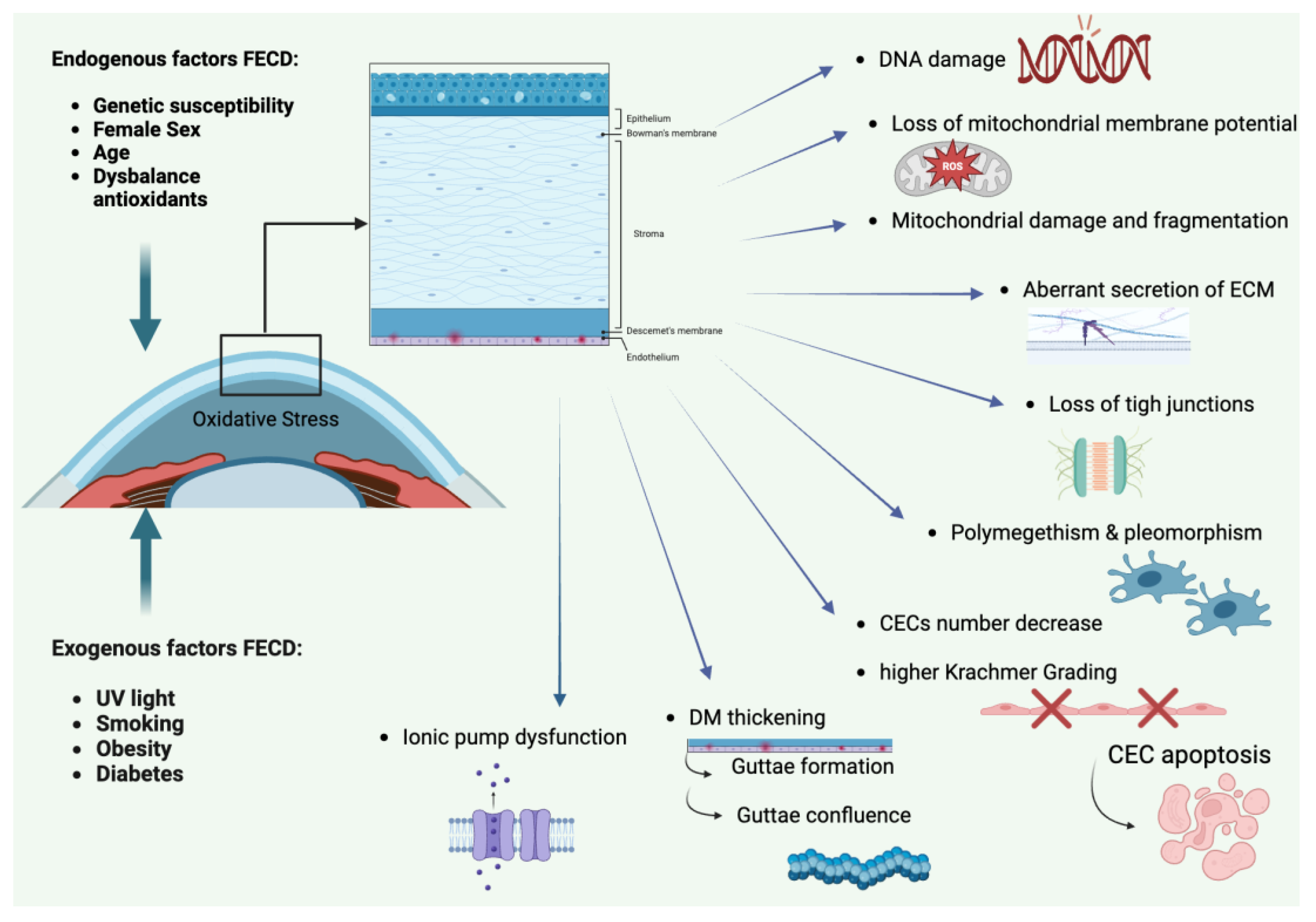
Figure 2.
Schematic overview of direct and indirect alloantigen recognition in corneal transplantation. Created by S. Zwingelberg with Biorender.com.
Figure 2.
Schematic overview of direct and indirect alloantigen recognition in corneal transplantation. Created by S. Zwingelberg with Biorender.com.
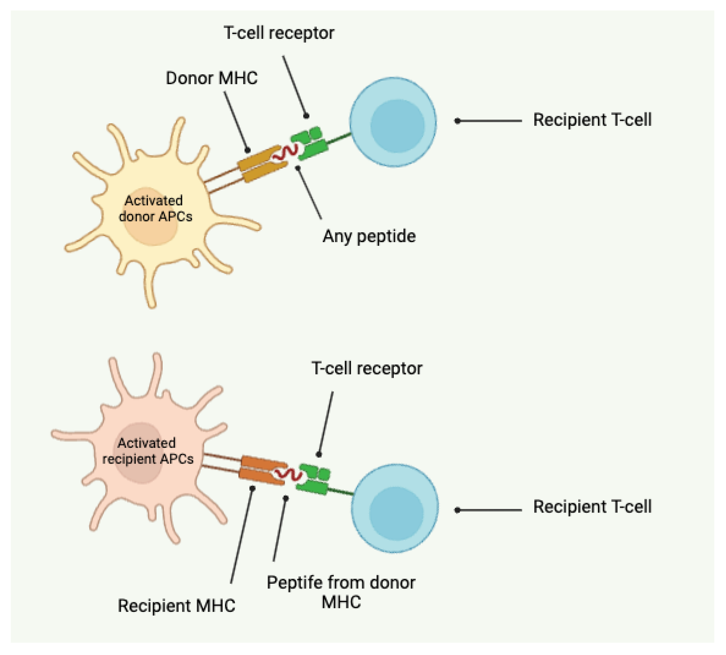
Figure 3.
Schematic overview of Descemet Membrane Endothelial Keratoplasty (DMEK) and the anterior chamber associated immune deviation (ACAID). Created by S. Zwingelberg with Biorender.com.
Figure 3.
Schematic overview of Descemet Membrane Endothelial Keratoplasty (DMEK) and the anterior chamber associated immune deviation (ACAID). Created by S. Zwingelberg with Biorender.com.
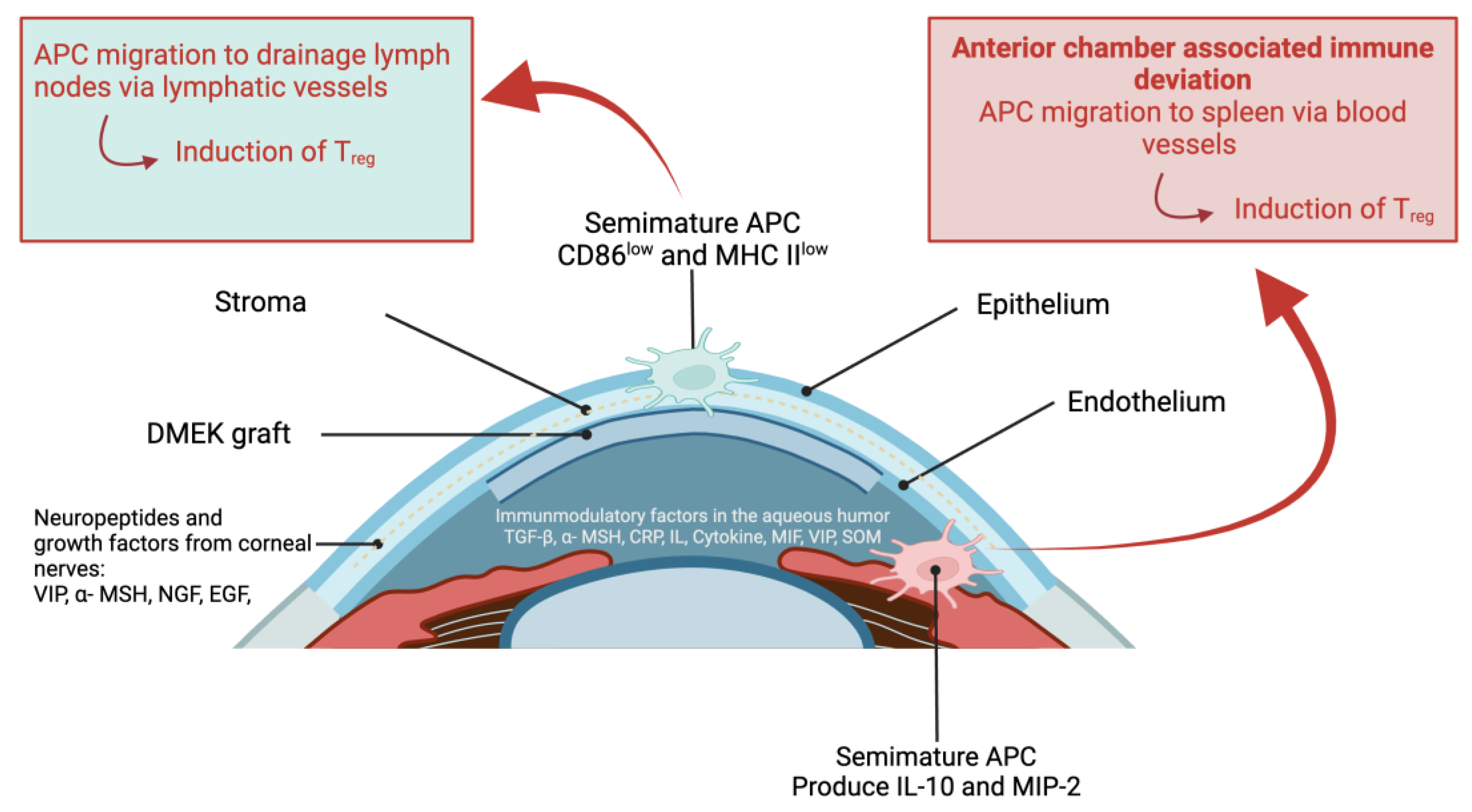
Figure 4.
Step-by-step procedure of DMEK: (a) Circular rhexis of the peripheral Descemet membrane and staining with trypan blue followed by lifting of the Descemet edge. (b) Separation of the Descemet membrane using two forceps. (c) Trephination with an 8-mm trephine. (d,e) Placement of graft markings for intraoperative orientation using a 1-mm trephine (green arrows). (f) Peeling off the graft. (g,h) Loading the graft into the injector cartridge. (i) Making incisions. (j) Spreading the iridotomy with vitreous scissors. (k) Descemetorhexis. (l) Insertion of the graft. (m) Unfurling of the graft. (n) Verification of graft markings (green arrows). (o) Injection of the air bubble. (p) Filling with 20% sulfur hexafluoride (SF6) gas.

Figure 5.
Schematic overview of the advantages of Descemet Membrane Endothelial.
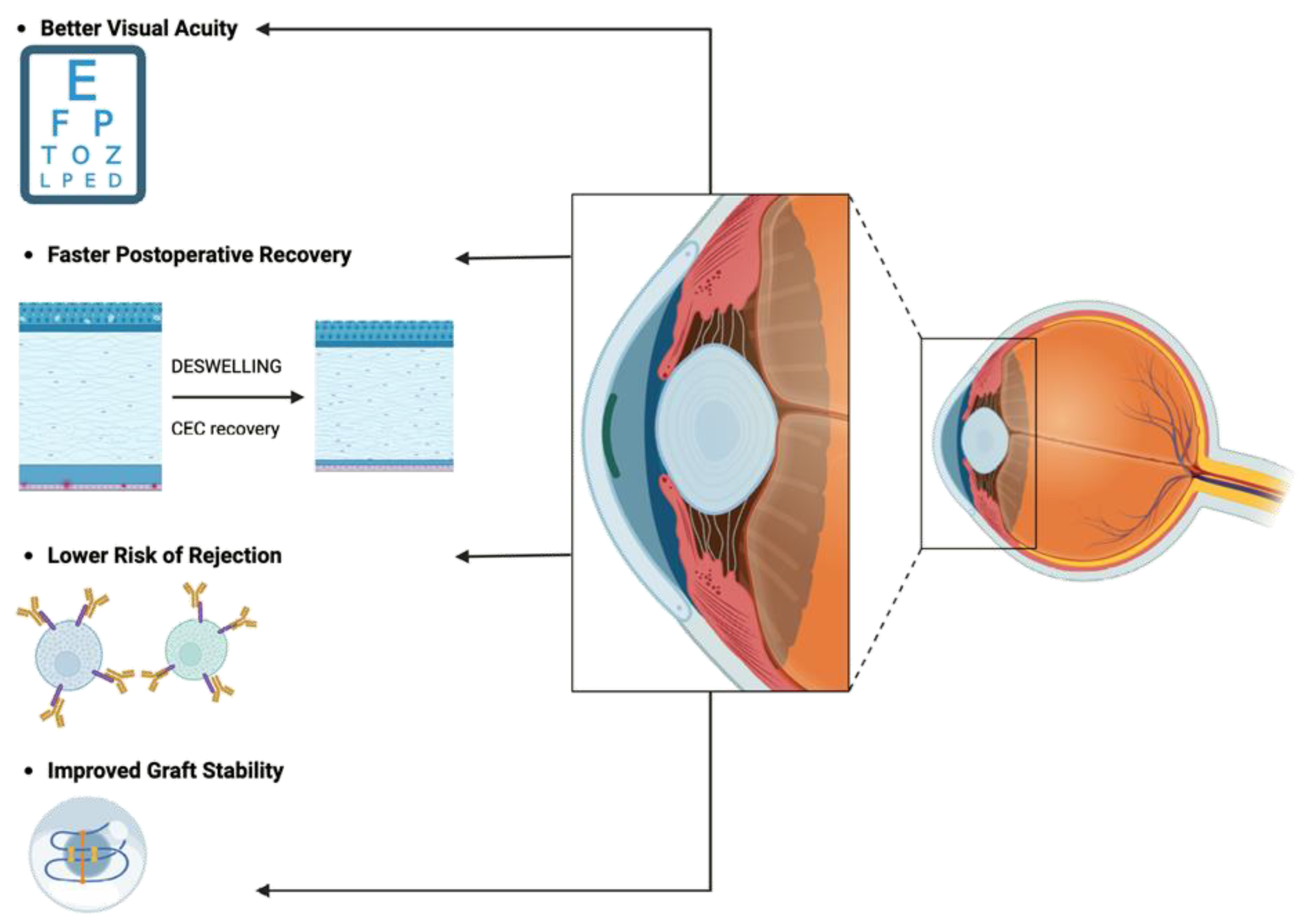
Figure 6.
Schematic overview of the requirements for Descemet Membrane Endothelial Keratoplasty (DMEK). Created by S. Zwingelberg with Biorender.com.
Figure 6.
Schematic overview of the requirements for Descemet Membrane Endothelial Keratoplasty (DMEK). Created by S. Zwingelberg with Biorender.com.
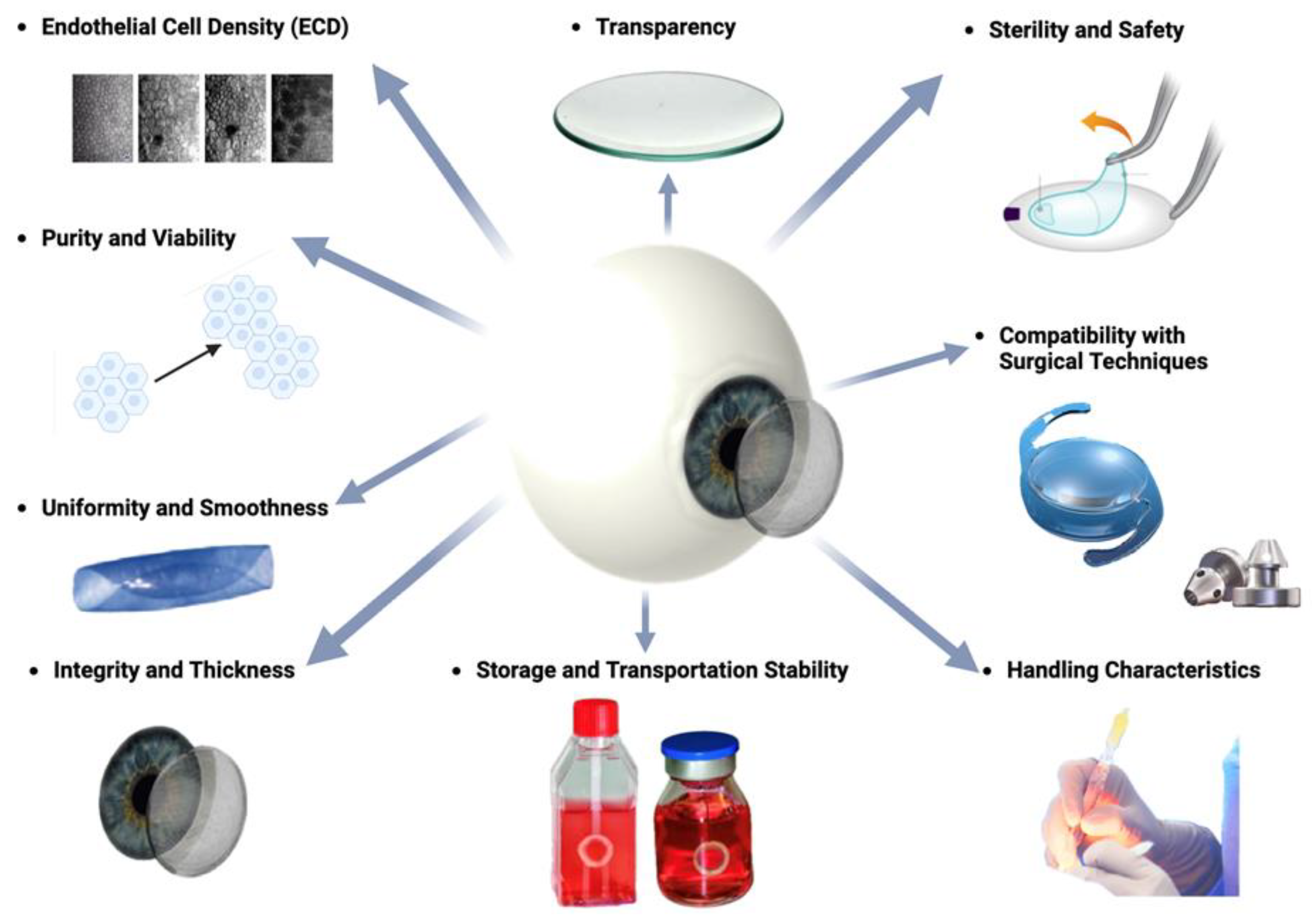
Figure 7.
Model of 3D-bioprinting for engineering of artificial corneal structures (From: Isaacson A, Swioklo S, Connon CJ. 3D bioprinting of a corneal stroma equivalent. [134].
Figure 7.
Model of 3D-bioprinting for engineering of artificial corneal structures (From: Isaacson A, Swioklo S, Connon CJ. 3D bioprinting of a corneal stroma equivalent. [134].
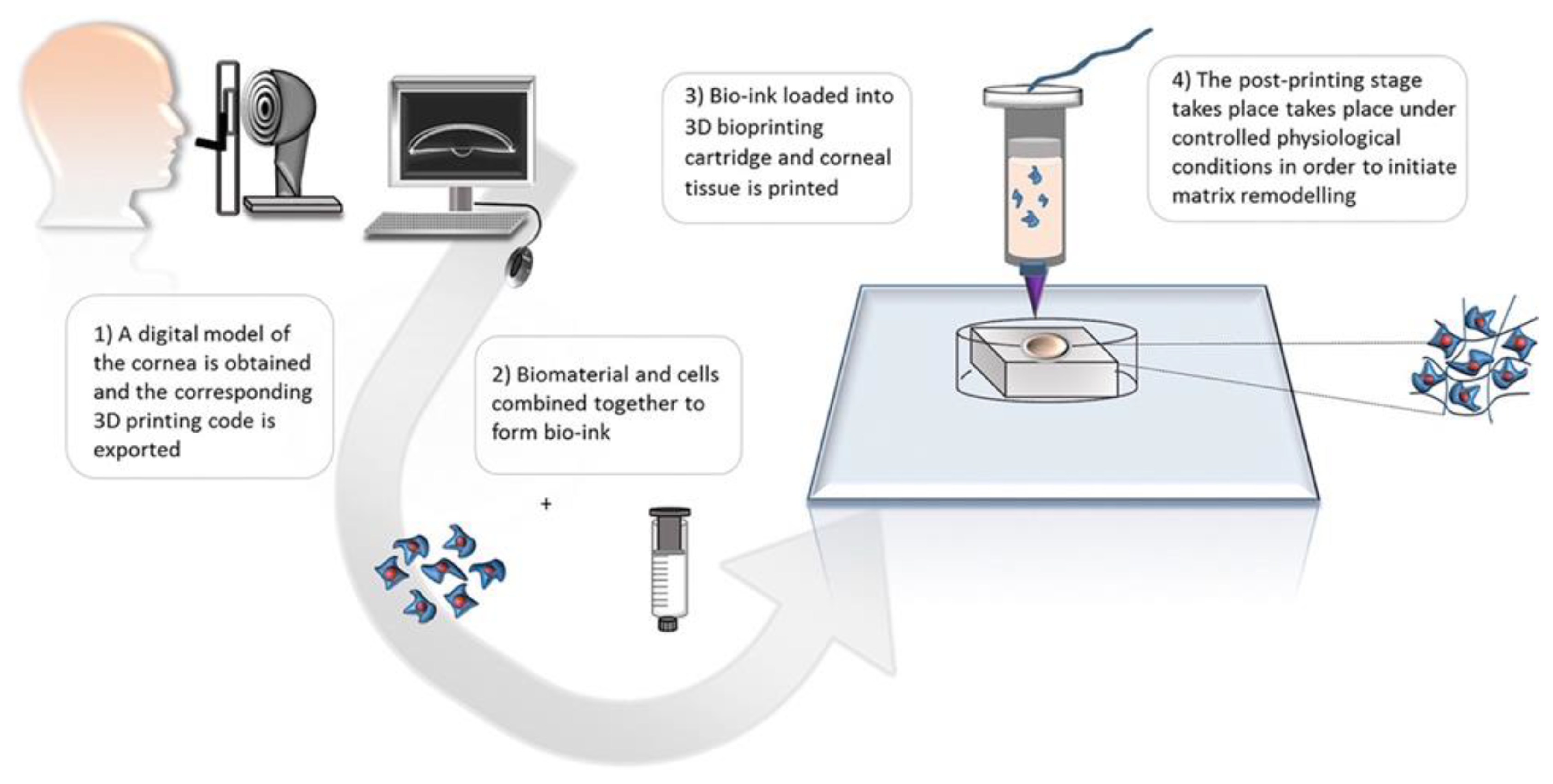
Disclaimer/Publisher’s Note: The statements, opinions and data contained in all publications are solely those of the individual author(s) and contributor(s) and not of MDPI and/or the editor(s). MDPI and/or the editor(s) disclaim responsibility for any injury to people or property resulting from any ideas, methods, instructions or products referred to in the content. |
© 2024 by the authors. Licensee MDPI, Basel, Switzerland. This article is an open access article distributed under the terms and conditions of the Creative Commons Attribution (CC BY) license (http://creativecommons.org/licenses/by/4.0/).
Copyright: This open access article is published under a Creative Commons CC BY 4.0 license, which permit the free download, distribution, and reuse, provided that the author and preprint are cited in any reuse.


