Submitted:
15 January 2024
Posted:
15 January 2024
You are already at the latest version
Abstract
This study aimed to evaluate potential posttreatment changes in ADC values within the tissue surrounding the enhancing lesion, particularly in areas not exhibiting MRI characteristics of in-volvement. Additionally, the objective was to investigate the correlation between ADC values, treatment response, and survival outcomes in individuals diagnosed with gliomas.
This retrospective study included a total of 49 patients that underwent either stereotactic biopsy or maximal surgical resection. Histologically confirmed as Grade III or IV gliomas, all cases adhered to the 2016 and 2021 WHO classifications, with subsequent radio-chemotherapy administered post-surgery. Patients were divided into two groups: short and long survival group. Baseline and follow-up MRI scans were obtained on a 1.5T MRI scanner. Two ROI circles were positioned near the enhancing area, one ROI in the NAWM ipsilateral to the neoplasm and another symmetrically in the contralateral hemisphere on ADC maps.
At follow-up there was a significant difference in both ipsilateral and contralateral NAWM be-tween the two groups, -0.0857 (p=0.004) and -0.0607 (p=0.037), respectively. There was a weak negative correlation between survival and ADC values in ipsilateral and contralateral NAWM at the baseline with the correlation coefficient -0.328 (p=0.02) and -0.302 (p=0.04), respectively. The correlation was stronger at the follow-up.
The findings indicate that ADC values in normal appearing white matter (NAWM) may function as a prognostic biomarker in patients with diffuse gliomas.
Keywords:
glioma
; magnetic resonance imaging
; apparent diffusion coefficient
; biomarkers
1. Introduction
Gliomas are the most common primary brain neoplasms, with glioblastomas (GBM) accounting for 49,1% of malignant brain tumors [1]. GBM, the most aggressive primary brain neoplasm has a poor prognosis with a mean survival of 12-14 months [2,3,4]. The current standard of care includes maximum surgical resection followed by radiotherapy combined with chemotherapy (concurrent chemo-radiation) [2,4]. The scope of surgery is based on preoperative imaging and whether the tumor is located near an eloquent region of the brain [5]. The extent of resection is an important prognostic factor, as patients with a larger residual tumor volume have a shorter survival time. When surgery cannot be safely done (because of the location or the patient's clinical condition) a stereotactic biopsy is performed [4,5,6]. Radiotherapy allows for improved local control and increased survival [4,7]. Most patients with diffuse gliomas recieve concurrent chemotherapy with an alkylating agent, primarily temozolomide, as it has a better safety profile than other alkylating agents [4,8].
Gliomas have specific growth patterns, spreading perineuronaly, perivascularly and perifascicularly [9]. Because of their invasive growth, it is essentially impossible to completely remove diffuse gliomas. Residual neoplastic cells will likely be the source of disease progression, but these areas of infiltrative growth cannot be reliably visualized on MRI [10]. Various biomarkers that might guide the treatment strategy and predict the response to therapy have been investigated, however, none were sufficiently reliable.
Brain MRI is the standard of reference for detection and evaluation of brain tumors [4,11], based on which a biopsy or a surgical resection may be performed to provide a tissue sample for the definitive pathohistological diagnosis [4]. Functional imaging methods are performed on a regular basis to better characterize neoplastic brain lesions. Diffusion MR imaging is used to evaluate the molecular function and micro-architecture of the tissues, by probing water diffusion over distances that correspond to typical cell sizes. Apparent diffusion coefficient (ADC) maps are calculated to remove the inherent T2-weighting of diffusion-weighted imaging (DWI) and represent a measure of average diffusion of water molecules within each voxel. As cell membranes are one of the factors that impede the diffusion, an increase in cell density, and therefore membrane density, leads to a decrease in diffusivity. ADC maps are therefore able to differentiate highly cellular from acellular regions. Tissues with high cellularity have low ADC values (low diffusivity) as the mobility of water protons is impeded [3,12,13,14]. It has been shown that ADC values in the regions surrounding the neoplastic signal abnormality allow for differentiation of low grade from high grade gliomas [15]. Additionally, significantly elevated diffusivity was found in the contralateral normal appearing white matter (NAWM) of glioma patients [16].
The diffusivity of the neoplastic tissue, as can be identified on MRI, has been extensively investigated and reported in the literature. We decided to analyze ADC values in the tissue surrounding the enhancing gliomas in order look for potential associations with aggressiveness of the lesions, treatment response and survival of patients. The goal of this research was to assess whether there are posttreatment changes of ADC values in the tissue surrounding the preoperative enhancing lesion, which does not appear involved by MRI characteristics, and in the contralateral brain. The second goal was to establish whether ADC values correlate with treatment response and survival in patients with gliomas.
2. Materials and Methods
The study adhered to the principles outlined in the Helsinki Declaration, as approved by the ethical committee.
A total of 49 patients were included in this retrospective study. All patients were diagnosed and treated from 2016 to 2021 in a single centre. Patients diagnosed and treated before 2016 were excluded as the imaging studies were not available in PACS. Other exclusion factors were lack of baseline or follow-up MRI, lack of diffusion imaging, and prominent artifacts rendering MR images unsuitable for measurement of ADC values.
The included patients had either a stereotactic biopsy or a maximum surgical resection following the baseline MRI. Surgical resection was performed whenever possible (tumor in a non-eloquent region and favorable clinical status of the patient). A total of 42 patients underwent surgical resection and 7 patients had stereotactic biopsy. Grade III or IV gliomas were histologically confirmed in all cases, according to the 2016 WHO classification and corresponding to adult type diffuse gliomas by the most recent 2021 WHO classification. Following surgery, all patients received concomitant radio-chemotherapy with temozolomide and a total dose of 60 Gy.
For the purpose of this investigation, the overall survival was considered to be from the date of the initial diagnosis to the date of the last clinical follow up. The patients were divided into two groups based on overall survival, as we had noticed a bimodal distribution with a large central gap and divided the patients accordingly into short and long survival groups. The short survival group (n=39) had an overall survival ≤596 days and the long survival group (n=10) ≥924 days.
Patients’ age ranged from 28 to 78 years. The baseline MRI was performed before any treatment and the follow-up study 1-2 months after completed treatment.
All scans were obtained with a 1,5T MR scanner (Magnetom Avanto; Siemens; Munich, Germany). The imaging included axial spin-echo (TR 550 ms, TE 8.7 ms, FOV 230 mm) or 3D gradient-echo (TR 1910 ms, TE 3.53 ms, FOV 256 mm) T1-weighted sequences pre- and post-contrast, axial T2-weighted sequence (TR 5000 ms, TE 96 ms, FOV 230 mm), axial FLAIR (TR 8000 ms, TE 92 ms, FOV 230 mm) and DWI/ADC maps. Echo-planar diffusion imaging was performed in the axial plane before contrast administration. Diffusion-weighted images were acquired using b values = 0 and 1000 s/mm2 applied in the X, Y, and Z directions. ADC maps were calculated on a voxel-by-voxel basis with the software incorporated in the MRI unit.
All scans were analyzed with the software provided by the scanner manufacturer (Syngo.via; Siemens; Munich, Germany). Each MRI study was evaluated by a radiologist. Using freehand volume of interest tool we outlined the margins of the enhancing lesion. Two region of interest (ROI) circles were placed adjacent to the enhancing area (approximately 2 mm away from the enhancing margin, within the signal abnormality on FLAIR images), one ROI at least 4 cm away from the enhancement in the ipsilateral normal appearing white matter (NAWM), and an additional ROI in the contralateral NAWM in a symmetric fashion (Figure 1). The surface area of ROI circles was 0,4-0,5 cm2 (Figure 1.). The software calculated mean ADC values of the selected ROIs.
A: T1 axial spin-echo showing the enhancing lesion; B: Axial FLAIR showing white matter hyperintensity adjacent to the enhancing lesion; C: Axial ADC map showing ROI placement adjacent to the enhancing lesion; D: Axial FLAIR showing normal appearing white matter; E: Axial ADC at the same level as “D” showing ROI placement.
All statistical analyses were performed using IBM SPSS Statistics software. The differences between groups in ADC values were determined using t-test. A p value <0.05 was considered statistically significant. Spearman's rank correlation test was performed for assessment of correlation between ADC values and survival.
3. Results
The median overall survival was 228 days.
Mean ADC values at baseline and follow-up were higher adjacent to the enhancing lesion, as shown in Table 1.
The short survival group patients had lower mean ADC values adjacent to the enhancing lesion compared to the patients in the long survival group, both at baseline and the follow-up. (Figure 2 and Figure 3). There was a statistically significant difference in one of the two measurements adjacent to the enhancing lesion between the two groups: 0.3348 (p=0.013) (Table 2). There were no statistically significant differences in ADC values between ipsilateral and contralateral NAWM at baseline, -0.0404 (p=0.14) and -0.0268 (p=0.279), respectively. At the follow-up MRI there was a statistically significant difference in both ipsilateral and contralateral NAWM between the two groups, -0.0857 (p=0.004) and -0.0607 (p=0.037),respectively (Table 3).
The results showed no correlation between survival and ADC values adjacent to the enhancing lesion at the baseline or the follow-up. However, there was a weak negative correlation between survival and ADC values in ipsilateral and contralateral NAWM at the baseline with the correlation coefficient -0.328 (p=0.02) and -0.302 (p=0.04), respectively (Table 4). The correlation was stronger at the follow-up with the correlation coefficients -0.575 (p=0) and -0.605 (p=0) for ADC values in the ipsilateral and contralateral NAWM, respectively (Table 5).
4. Discussion
In our retrospective study, we wanted to test the hypothesis that the infiltrative growth pattern of gliomas leads to changes in the ADC values of white matter outside of the enhancing tumor mass and that these changes could serve as a prognostic biomarker for patient survival.
The results of our study showed that there was a statistically significant difference in ADC values adjacent to the enhancing mass between the patients stratified according to the survival times, which corresponds to the findings of Yazdani et al. [15]. Several other studies have also shown that lower diffusivity suggests a high-grade glioma and higher ADC values are indicative of a low-grade glioma, which is in accordance with the more cellular areas exhibiting lower water mobility [13,17,18].
Additionally, our study found that the ADC values were lower both at baseline and follow-up in the regions immediately adjacent to the enhancing portion of the neoplasm in patients with poor survival times, which could be explained by higher cellularity and disrupted water proton mobility in these regions [13,17]. While previous studies have shown that perilesional ADC values can differentiate high-grade gliomas from normal tissue, they were not able to distinguish neoplastic tissue from the adjacent edema [19,20]. This represents a possible limitation of diffusivity measurements in the peritumoral region, as vasogenic edema is present in many cases. Our study is unique in that it sought to determine whether patients with shorter survival times had changes in ADC values that could be indicative of more aggressive neoplasms infiltrating normal brain tissue at a microscopic level.
Horvath et al. found that ADC values of contralateral NAWM were higher in high grade gliomas than in low grade gliomas [16]. All of our patients had high grade gliomas, but we also found that the patients with longer survival times had relatively lower diffusivity in both contralateral and ipsilateral NAWM, which could be due to different intrinsic neoplastic potential or less aggressive growth [17]. Another study using biexponential diffusion analysis showed that the diffusion patterns in NAWM of patients with gliomas are similar to those in the clearly neoplastic tissue, suggesting infiltrative growth [21].
We found a weak negative correlation between survival and ADC values in NAWM both in the vicinity of the enhancing mass and on the contralateral side. This correlation was even stronger at the follow-up MRI. The observed changes in ADC values in NAWM may be due to vasogenic edema, as suggested by Horvath et al. [21]. Vasogenic edema refers to accumulation of fluid in the extracellular spaces of the brain, occuring as a result of various pathological processes, including growth and infiltration of gliomas. The edema can alter the water proton mobility in the affected tissue and hence result in changes of ADC values, as seen in our study. The observed changes in ADC values in NAWM might suggest the presence of infiltrating glioma cells in otherwise apparently normal brain tissue on imaging studies.
It is important to note several limitations of our study, including a relatively small sample size and lack of molecular characterization of the gliomas, as well as the fact that not all patients underwent a maximum surgical resection. An increased number of measurements with a wider variety of ROI sizes could also provide a more accurate diffusivity information. Additionally, various genetic mutations and epigenetic changes can alter the patterns of glioma growth and hence affect ADC values.
5. Conclusion
In conclusion, our study provides a valuable insight into the correlation between NAWM diffusivity and survival in patients with gliomas. Although more research is needed, our results suggest that ADC values in NAWM could serve as a prognostic biomarker in these patients. Further studies with larger sample size and molecular characterization of the neoplasms are needed to confirm these findings and improve our understanding of glioma growth, ultimately providing better patient care.
Author Contributions
Conceptualization, Dolić, K.; Methodology, Bušić, M. and Dolić, K.; Formal Analysis, Bušić, Ž.; Investigation Bušić, M., Čerina D and Dolić, K.; Writing - original draft, Bušić M.; Writing – Review & Editing, Rumboldt, Z, Dolic K.
Funding
This research received no external funding.
Institutional Review Board Statement
The study was conducted in accordance with the Declaration of Helsinki, and approved by the Ethics Committee of University hospital Split, Croatia, (protocol code: 520-03/23-01/02, approval date January 4th 2023).
Conflicts of Interest
The authors declare no conflict of interest.
References
- Ostrom QT, Cioffi G, Waite K, Kruchko C, Barnholtz-Sloan JS. CBTRUS statistical report: Primary Brain and other central nervous system tumors diagnosed in the United States in 2014–2018. Neuro-Oncology. 2021;23(Supplement_3):iii1–105. [CrossRef]
- Ellingson BM, Sahebjam S, Kim HJ, Pope WB, Harris RJ, Woodworth DC, et al. Pretreatment ADC Histogram Analysis is a predictive imaging biomarker for bevacizumab treatment but not chemotherapy in recurrent glioblastoma. American Journal of Neuroradiology. 2013;35(4):673–9. [CrossRef]
- Zulfiqar M, Yousem DM, Lai H. ADC values and prognosis of malignant astrocytomas: Does lower ADC predict a worse prognosis independent of grade of tumor?—a meta-analysis. American Journal of Roentgenology. 2013;200(3):624–9. [CrossRef]
- Weller M, van den Bent M, Preusser M, Le Rhun E, Tonn JC, Minniti G, et al. EANO guidelines on the diagnosis and treatment of diffuse gliomas of adulthood. Nature Reviews Clinical Oncology. 2020;18(3):170–86. [CrossRef]
- Grabowski MM, Recinos PF, Nowacki AS, Schroeder JL, Angelov L, Barnett GH, et al. Residual tumor volume versus extent of resection: Predictors of survival after surgery for glioblastoma. Journal of Neurosurgery. 2014;121(5):1115–23. [CrossRef]
- Molinaro AM, Hervey-Jumper S, Morshed RA, Young J, Han SJ, Chunduru P, et al. Association of maximal extent of resection of contrast-enhanced and non–contrast-enhanced tumor with survival within molecular subgroups of patients with newly diagnosed glioblastoma. JAMA Oncology. 2020;6(4):495. [CrossRef]
- Walker MD, Alexander E, Hunt WE, MacCarty CS, Mahaley MS, Mealey J, et al. Evaluation of BCNU and/or radiotherapy in the treatment of anaplastic gliomas. Journal of Neurosurgery. 1978;49(3):333–43. [CrossRef]
- Stupp R, Mason WP, van den Bent MJ, Weller M, Fisher B, Taphoorn MJB, et al. Radiotherapy plus concomitant and adjuvant temozolomide for glioblastoma. New England Journal of Medicine. 2005;352(10):987–96. [CrossRef]
- Claes A, Idema AJ, Wesseling P. Diffuse glioma growth: A guerilla war. Acta Neuropathologica. 2007;114(5):443–58. [CrossRef]
- Xiong L, Wang F, Qi Xie X. Advanced treatment in high-grade gliomas. JBUON. 2019;24(2):424–30.
- Ellingson BM, Wen PY, Cloughesy TF. Modified criteria for radiographic response assessment in glioblastoma clinical trials. Neurotherapeutics. 2017;14(2):307–20. [CrossRef]
- Maier SE, Sun Y, Mulkern RV. Diffusion Imaging of brain tumors. NMR in Biomedicine. 2010;23(7):849–64. [CrossRef]
- Murakami R, Sugahara T, Nakamura H, Hirai T, Kitajima M, Hayashida Y, et al. Malignant supratentorial astrocytoma treated with postoperative radiation therapy: Prognostic value of pretreatment quantitative diffusion-weighted mr imaging. Radiology. 2007;243(2):493–9. [CrossRef]
- Baliyan V, Das CJ, Sharma R, Gupta AK. Diffusion Weighted Imaging: Technique and Applications. World Journal of Radiology. 2016;8(9):785. [CrossRef]
- Yazdani M, Rumboldt Z, Tabesh A, Giglio P, Schiarelli C, Morgan PS, et al. Perilesional apparent diffusion coefficient in the preoperative evaluation of glioma grade. Clinical Imaging. 2018;52:88–94. [CrossRef]
- Horváth A, Perlaki G, Tóth A, Orsi G, Nagy S, Dóczi T, et al. Increased diffusion in the normal appearing white matter of brain tumor patients: Is this just tumor infiltration? Journal of Neuro-Oncology. 2015;127(1):83–90. [CrossRef]
- Guo AC, Cummings TJ, Dash RC, Provenzale JM. Lymphomas and high-grade astrocytomas: Comparison of water diffusibility and histologic characteristics. Radiology. 2002;224(1):177–83. [CrossRef]
- Server A, Kulle B, Gadmar ØB, Josefsen R, Kumar T, Nakstad PH. Measurements of diagnostic examination performance using quantitative apparent diffusion coefficient and Proton MR spectroscopic imaging in the preoperative evaluation of tumor grade in cerebral gliomas. European Journal of Radiology. 2011;80(2):462–70. [CrossRef]
- Catalaa I, Henry R, Dillon WP, Graves EE, McKnight TR, Lu Y, et al. Perfusion, diffusion and spectroscopy values in newly diagnosed cerebral gliomas. NMR in Biomedicine. 2006;19(4):463–75. [CrossRef]
- Castillo M, Smith JK, Kwock L, Wilber K. Apparent diffusion coefficients in the evaluation of high-grade cerebral gliomas. American Journal of Neuroradiology. 2001;22(1):60–4.
- Horváth A, Perlaki G, Tóth A, Orsi G, Nagy S, Dóczi T, et al. Biexponential diffusion alterations in the normal-appearing white matter of glioma patients might indicate the presence of global vasogenic edema. Journal of Magnetic Resonance Imaging. 2016;44(3). [CrossRef]
Figure 1.
ROI placement.
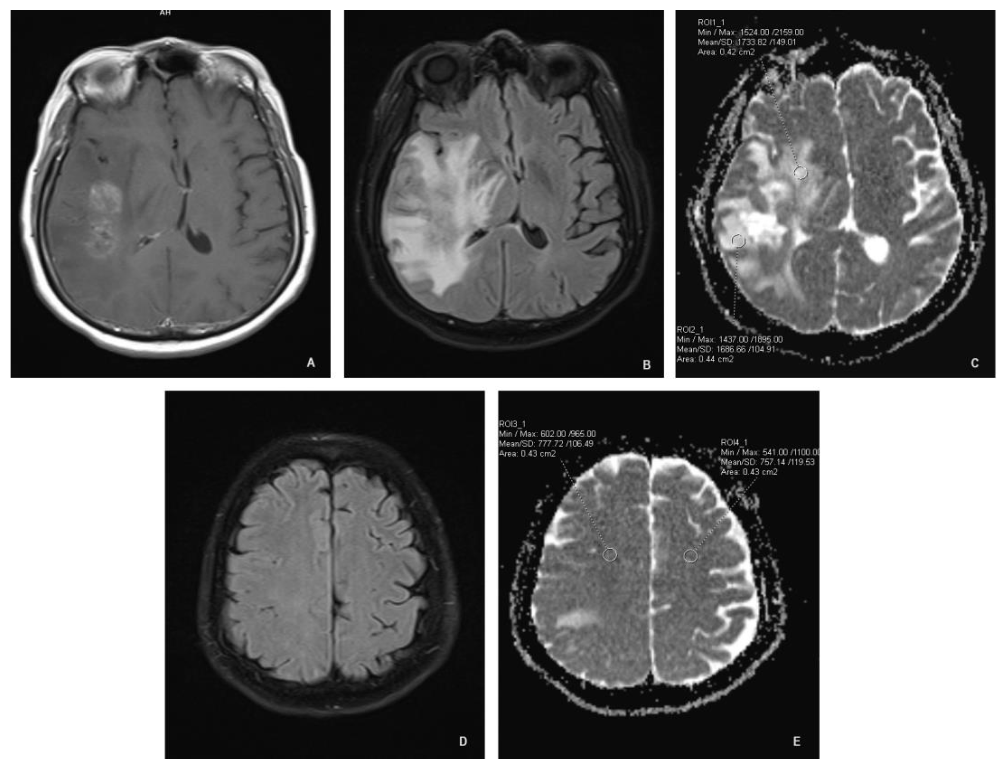
Figure 2.
Mean ADC values for both groups at baseline.
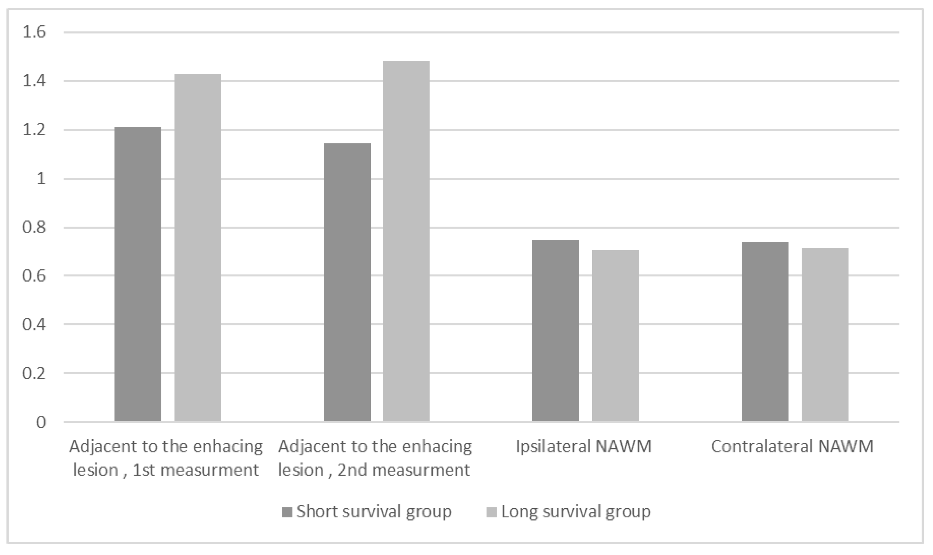
Figure 3.
Mean ADC values for both groups at follow-up.
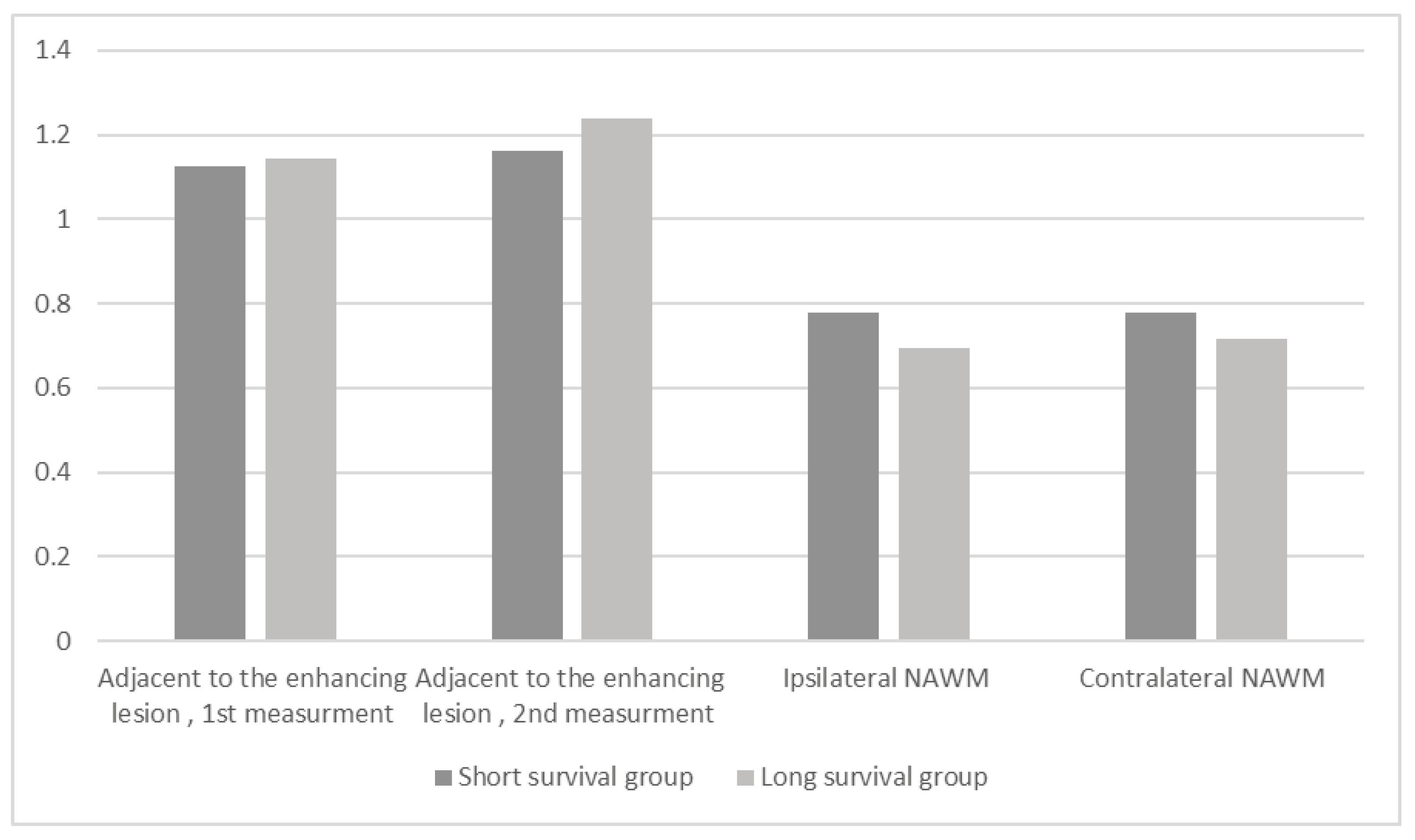

Table 1.
Mean apparent diffusion coefficient (ADC) values of all patients (n=49) at baseline and follow-up.
Table 1.
Mean apparent diffusion coefficient (ADC) values of all patients (n=49) at baseline and follow-up.
| Baseline MRI (Mean ± SD, 10-3 mm2/s) |
Follow-up MRI (Mean ± SD, 10-3 mm2/s) |
|
|---|---|---|
| Adjacent to the enhacing lesion, 1st measurement | 1.2504 ± 0.3843 | 1.1292 ± 0.3001 |
| Adjacent to the enhacing lesion, 2nd measurement | 1.2089 ± 0.3720 | 1.1781 ± 0.3242 |
| Ipsilateral NAWM | 0.7407 ± 0.0738 | 0.7621 ± 0.0854 |
| Contralateral NAWM | 0.7358 ± 0.0663 | 0.7662 ± 0.0827 |
Table 2.
Differences in mean ADC values between the groups at baseline.
| Short survival group (Mean, 10-3 mm2/s) | Long survival group (Mean, 10-3 mm2/s) | p-value | |
|---|---|---|---|
| Adjacent to the enhacing lesion, 1st measurement | 1.2095 | 1.4726 | 0.1260 |
| Adjacent to the enhacing lesion, 2nd measurement | 1.1461 | 1.4809 | 0.0130 |
| Ipsilateral NAWM | 0.7482 | 0.7078 | 0.1400 |
| Contralateral NAWM | 0.7408 | 0.7140 | 0.2790 |
Table 3.
Differences in mean ADC values between the groups at follow up.
| Short survival group (Mean, 10-3 mm2/s) | Long survival group (Mean, 10-3 mm2/s) | p-value | |
|---|---|---|---|
| Adjacent to the enhacing lesion, 1st measurement | 1.1258 | 1.1422 | 0.8800 |
| Adjacent to the enhacing lesion, 2nd measurement | 1.6252 | 1.2388 | 0.5130 |
| Ipsilateral NAWM | 0.7796 | 0.6938 | 0.0040 |
| Contralateral NAWM | 0.7786 | 0.7179 | 0.0370 |
Table 4.
Correlation between overall survival and ADC values at baseline.
| Correlation coefficient | p-value | |
|---|---|---|
| Adjacent to the enhacing lesion, 1st measurement | 0.1740 | 0.2360 |
| Adjacent to the enhacing lesion, 2nd measurement | 0.1690 | 0.2500 |
| Ipsilateral NAWM | -0.328 | 0.0230 |
| Contralateral NAWM | -0.302 | 0.0370 |
Table 5.
Correlation between overall survival and ADC values at follow-up.
| Correlation coefficient | p-value | |
|---|---|---|
| Adjacent to the enhacing lesion, 1st measurement | -0.0030 | 0.9860 |
| Adjacent to the enhacing lesion, 2nd measurement | 0.0800 | 0.5840 |
| Ipsilateral NAWM | -0.5750 | 0.0000 |
| Contralateral NAWM | -0.6050 | 0.0000 |
Disclaimer/Publisher’s Note: The statements, opinions and data contained in all publications are solely those of the individual author(s) and contributor(s) and not of MDPI and/or the editor(s). MDPI and/or the editor(s) disclaim responsibility for any injury to people or property resulting from any ideas, methods, instructions or products referred to in the content. |
© 2024 by the authors. Licensee MDPI, Basel, Switzerland. This article is an open access article distributed under the terms and conditions of the Creative Commons Attribution (CC BY) license (https://creativecommons.org/licenses/by/4.0/).
Copyright: This open access article is published under a Creative Commons CC BY 4.0 license, which permit the free download, distribution, and reuse, provided that the author and preprint are cited in any reuse.


