
Submitted:
30 August 2023
Posted:
31 August 2023
You are already at the latest version
Abstract
Marine soft corals are prolific sources of various natural products that have served as a wealthy reservoir of diverse chemical scaffolds for new drug leads. The genus Litophyton contains almost 100 species but only a small part of them had been classified and chemically investigated, which calls for more attentions from the global researchers. In the current work, 109 secondary metabolites have been discussed based on published data from 1975 to July, 2023, covering a period of near five decades. The studied species of the genus Litophyton inhabited various tropical and temperate regions and afforded a variety of biologically active natural products such as terpenes, steroids, alkaloids, and lipids. A wide spectrum of pharmacological effects of these compounds had been evaluated, including cytotoxic, anti-HIV, antibacterial, antifungal, anti-malarial, antifeedant, molluscicidal, PTP1B inhibitory, and insect growth inhibitory activities. This review aims to provide an up-to-date literature survey and comprehensive insight into chemical structures, taxonomical distributions, and biological activities of the reported metabolites from the title genus whenever available.
Keywords:
soft coral
; Litophyton
; secondary metabolites
; terpenes
; bioactivities
; cytotoxicity
1. Introduction
More than two-thirds of the Earth’s surface is covered by oceans, which harbor a vast array of creatures, including plants, animals and microbes. Since the ancient times, marine organisms had been served as the sources of foods [1], cosmetic ingredients [2], and drugs [3], which are hotspots for global researchers nowadays [4]. Continuous studies focused on the secondary metabolites derived from marine environments, resulting a rapid expansion of marine natural products [5]. These substances displayed a wide spectrum of potential pharmacological effects, including antivirus [6], anti-osteoclastogenesis [7], antimicrobial [8], antitumor [9]. To date, almost 20 drugs from marine sources are in clinical use [10].
The marine soft coral genus Litophyton belongs to the family Nephtheidae, order Alcyonacea, subclass Octocorallia. The Litophyton genus consists of near 100 species, according to the Word Register of Marine Species (WoRMS) [11]. They are widely distributed throughout tropical and temperate waters, such as South China Sea [12], Red Sea [13], as well as other waters of Indo-Pacific Ocean [14,15,16]. However, only a few specimens collected from South China Sea [12], Red Sea [17,18], Indonesian [15,19] and Japanese [16] waters have been chemically investigated, which calls for more attentions from the global researchers.
In was observed that the alcyonarian Litophyton viridis provided chemical protection of a fish Abudefduf leucogaster [20]. The extracts of animals of the genus Litophyton was biologically screened and showed a variety of potent bioactivities, such as antioxidant [21], genotoxic [18], cytotoxic [21,22], and HIV-1 enzyme inhibitory [22] activities. Inspired by these bioassays, chemical investigations on the Litophyton soft corals were carried out by the researchers worldwide. These reported studies revealed that the members of the genus Litophyton are one of the most prolific producers of bioactive secondary metabolites (Table S1). However, there was no specific review of substances from the soft corals of the title genus. On the basis of a extensive literature search using SciFinder, this work specifically summarized all metabolites from the genus Litophyton for the first time, covering a period of near five decades (between 1975 and July, 2023).
2. Classification of Secondary Metabolites from the Genus Litophyton
Since the first report of a novel cembrane diterpene named 2-hydroxynephtenol from the soft coral L. viridis in 1975 [19], many research groups around the world have carried out chemical investigation on the genus Litophyton, resulting in fruitful achievements. A total of 109 secondary metabolites have been isolated and characterized in the Litophyton corals during 49 years of research (Table S1). These chemical compositions can be classified structurally as sesquiterpene, diterpene, tetraterpene, polyhydroxylated steroid, ceramide, nucleotide, prostaglandin, γ-lactone, fatty acid and glycerol ether. Intriguingly, one uncommon bis-sesquiterpene was encountered during the research of the alcyonarian Litophyton nigrum [23]. In the following subsections, these compounds were further grouped under different catalogs based on structural features. Among them, sesquiterpene and bis-sesquiterpene-type metabolites were grouped as ‘sesquiterpenes and a related dimer’ according to the corresponding structural relationship. The ceramide and nucleotide-type compounds were put under one category ‘alkaloids’. The pack of ‘lipids’ comprise prostaglandin, γ-lactone, fatty acid and glycerol ether. Herein, the chemical structures, taxonomical distributions, and biological activities of the reported metabolites from the title genus whenever available were described.
3. Sesquiterpenes and A Related Dimer
This was largest cluster of terpenes obtained from the genus Litophyton. Since the first sesquiterpene (–)-bicyclogermacrene [24] was found, more and more efforts were made to search sesquiterpenes from different species of this genus, resulting in an account of 37 compounds. These substances possessed a variety of carbon frameworks, which could be further classified into 15 categories: bicyclogermacrane, sec-germacrane, guaiane, pseudoguaiane, himachalene, eudesmane, seco-eudesmane, tri-nor-eudesmane, eremophilane, nardosinane, nornardosinane, eremophilane, neolemnane, seconeolemnane, and eremophilane-nardosinane dimmer (Figure 1). These different types of sesquiterpenes distributed in three species L. arboreum, L. nigrum and Litophyton setoensis, which were inhabited in different marine environments including Red Sea, South China Sea and Indonesian water (Table S1).
Figure 1.
The reported carbon frameworks of sesquiterpenes from the genus Litophyton.
3.1. Bicyclogermacrane Sesquiterpene
Chemical probing of the soft coral L. arboreum, which was collected near Bali, Indonesia, yielded the sesquiterpene (–)-bicyclogermacrene (1) [24] (Figure 2). This compound exhibited low antiproliferative activities against the cell lines L-929 and K-562 with GI50 values of 186 and 200 μM, respectively, and low cytotoxic effect against the HeLa cell line with CC50 of 182 μM.
Figure 2.
The chemical structure of bicyclogermacrane sesquiterpene from the genus Litophyton.
3.2. Sec-germacrane Sesquiterpene
Very recently, Ahmed et al. [17] carried out the chemical investigation of the Red Sea specimen L. arboreum, which was collected at Neweba, Egypt. The acyclic sesquiterpene (2E,6E)-3-isopropyl-6-methyl-10-oxoundeca-2,6-dienal (2) was found from this sample, which possessed a sec-germacrane nucleus (Figure 3). Anti-malarial bioassays disclosed the isolate 2 was active against chloroquine-sensitive (D6) and chloroquine-resistant (W2) strains of Plasmodium falciparum with IC50 values of 3.7 and 2.2 mg/mL, respectively. In addition, the metabolite 2 was non-toxic to the Vero cell line at the concentration of 4.76 mg/mL. These findings demonstrated that sesquiterpene 2 could be developed as a anti-malarial lead compound with highly safe in the range of tested concentrations.
Figure 3.
The chemical structure of sec-germacrane sesquiterpene from the genus Litophyton.
3.3. Guaiane Sesquiterpenes
Interestingly, the guaiane sesquiterpenes were frequently encountered in the Red Sea soft coral L. arboreum.
Bioassay-guided fractionation of the Red Sea alcyonarian L. arboreum by Ellithey et al., which was collected at Sharm El-Sheikh, Egypt, yielded three guaiane sesquiterpenes alismol (3), 10-O-methyl alismoxide (4) and alismoxide (5) [25] (Figure 4). Compound 3 showed potent inhibitory activity against HIV-1 protease receptor with IC50 of 7.2 µM, compared to the positive control, which had IC50 of 8.5 μM. Molecular docking study disclosed the hydrogen bond between 3 and the amino acid residue of Asp 25 in the hydrophobic receptor pocket with a score of −11.14. Meanwhile, sesquiterpenes 3 and 4 showed moderate cytotoxic activities against the cell lines HeLa (IC50 30 and 38 μM, respectively) and Vero (IC50 49 and 49.8 μM, respectively). Morevoer, 4 exhibited moderate cytotoxicity against the U937 cell line with IC50 of 50 µM. However, 5 was judged as inactive against the above-mentioned cell lines (all IC50 > 100 µM). In the further study, compounds 2 and 5 demonstrated cytostatic action in HeLa cells, revealing potential use in virostatic cocktails. In Ellithey’s continual study [26], alismol (3) showed promising cytotoxic effects against the cancer cell lines HepG2, MDA and A549 (IC50 4.52, 7.02, and 9.23 μg/mL, respectively).
Hawas’s group reported the presence of alismol (3) in Red Sea specimen L. arboreum collected off the coast of Jeddah, Saudi Arabia, together with another guaiane sesquiterpene alismorientol B (6) [27] (Figure 4). These two secondary metabolites were subjected to antimicrobial and cytotoxic bioassays. As a result, metabolites 3 and 6 showed weak to strong antibacterial activities against Escherichia coli ATCC 10536, Pseudomonas aeruginosa NTCC 6750, Bacillus cereus ATCC 9634, Bacillus subtilis ATCC6633, Staphylococcus aureus ATCC5141 with MIC values ranging from 10.4 to 1.3 μg/mL. Of which, 6 had significant activity against B. cereus ATCC 9634 with MIC of 1.3 μg/mL. And they exhibited weak to moderate antifungal activities against Candida albicans and Aspergillus niger with MIC values ranging from 10.1 to 6.0 μg/mL. Moreover, they displayed potent cytotoxic effects against the cell lines MCF-7, HCT-116 and HepG2 with IC50 ranging from 4.32 to 44.52 μM. Of which, 6 showed the most potent cytotoxic effect against MCF-7 cells with IC50 of 4.32 μM. Additionally, Hawas’s group evaluated the methanolic extract of the above-mentioned soft coral for its in vivo genotoxicity and antigenotoxicity against the mutagenicity induced by the anticancer drug cyclophosphamide [18]. The extract was found to be safe and nongenotoxic at 100 mg/kg b. wt. Moreover, the mice group of cyclophosphamide pretreated with the extract (100 mg/kg b. wt.) showed significant reduction in the percentage of chromosomal aberrations induced in bone marrow and mouse spermatocytes.
The existence of alismoxide (5) was shown in the Egyptian Red Sea L. arboreum collection from Hurghada by Mahmoud et al. [28]. In the anticancer bioassays, sesquiterpene 5 displayed no cytotoxic activities against the cell lines A549, MCF-7 and HepG2 (all IC50 > 100 µmol/mL). The co-existence of alismol (3) and alismoxide (5) as well as an undescribed sesquiterpene, litoarbolide A (7), and three known analogues 4α,7β,10α-trihydroxyguai-5-ene (8), leptocladol B (9) and nephthetetraol (10) (Figure 4) in another Egyptian Red Sea L. arboreum specimen from Neweba, was revealed by Ahmed et al.’s work [17]. Viewing from the perspective of their structures, litoarbolide A (7) was supposed to be the biosynthetic precursor to other sesquiterpenes, which could be generated via further post-translational modifications. The anti-malarial properties of substances 7–10 were evaluated. However, nly compounds 9 and 10 exhibited anti-malarial activities against chloroquine-resistant Plasmodium falciparum W2 with IC50 values of 4.3 and 3.2 mg/mL, respectively.
Figure 4.
The chemical structures of guaiane sesquiterpenes from the genus Litophyton.
3.4. Pseudoguaiane Sesquiterpenes
A new pseudoguaiane-type sesquiterpene named litopharbol (11) (Figure 5) was isolated from the methanolic extract of the Saudi Arabian Red Sea soft coral L. arboreum by Hawas’s group [27]. Its structure was determined through the elucidation of NMR data. Compound 11 exhibited a wide spectrum of antibacterial activities against Gram-negative bacteria E. coli ATCC 10536 and P. aeruginosa NTCC 6750, as well as Gram-positive bacteria B. cereus ATCC 9634, B. subtilis ATCC6633 and S. aureus ATCC5141 with MIC values ranging from 1.8 to 9.6 μg/mL. Among these bacteria, 11 showed significant activity against B. cereus ATCC 9634 with MIC of 1.8 μg/mL. And this sesquiterpene exhibited weak antifungal activities against C. albicans and A. niger with MIC values of 12.5 and 12.9 μg/mL, respectively. Moreover, it displayed potent cytotoxic effects against cell lines MCF-7, HCT-116 and HepG2 with IC50 values of 9.42, 26.21 and 38.92 μM. In Hawas’s continual study, litopharbdiol (12) was identified, which shared the same carbon framework with 11 [18] (Figure 5). However, no bioassay for this compound was reported in the article.
Figure 5.
The chemical structures of pseudoguaiane sesquiterpenes from the genus Litophyton.

3.5. Himachalene Sesquiterpenes
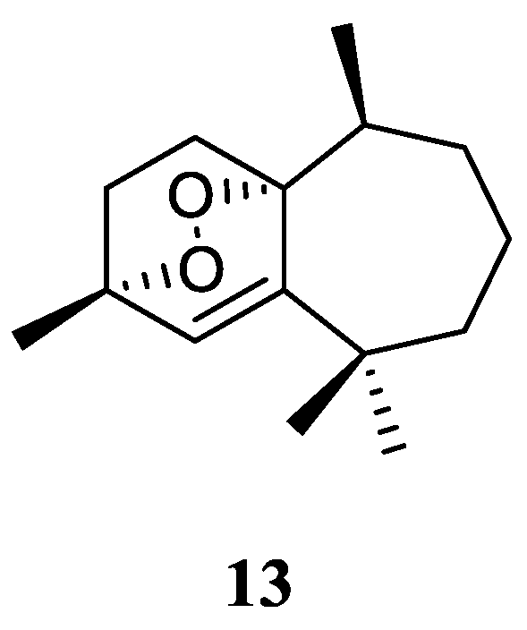
Purification of the CH2Cl2/MeOH extract of Saudi Arabian Red Sea alcyonarian L. arboreum yielded a new himachalene-type sesquiterpene 3α,6α-epidioxyhimachal-1-ene (13) (Figure 6), which showed antiproliferative effects toward three different cancer cell lines MCF-7, HCT116 and HepG-2 [29]. (It might be worth to point out that no specific data of the bioassay results was provided in this article.)
Figure 6.
The chemical structure of himachalene sesquiterpene from the genus Litophyton.
3.6. Eudesmane Sesquiterpene
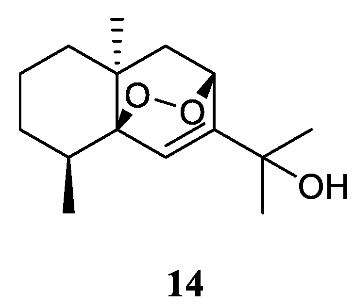
The n-hexane-chloroform (1:1) fraction of the Egyptian Red Sea L. arboreum sample exhibited noticeable cytotoxicity towards A549 cell line (IC50 22.6 mg/mL) [28]. The subsequent bioassay-guided isolation yielded a eudesmane sesquiterpene 5β,8β-epidioxy-11-hydroxy-6-eudesmene (14) (Figure 7). Compound 14 exerted noticeable activity against A549 cell line (IC50 67.3 µmol/mL) compared to etoposide as standard cytotoxic agent (IC50 48.3 µmol/mL). However, this compound did not show cytotoxic effects against cell lines MCF-7 and HepG2 (both IC50 > 100 µmol/mL).
Figure 7.
The chemical structure of eudesmane sesquiterpene from the genus Litophyton.
3.7. Seco-eudesmane Sesquiterpene
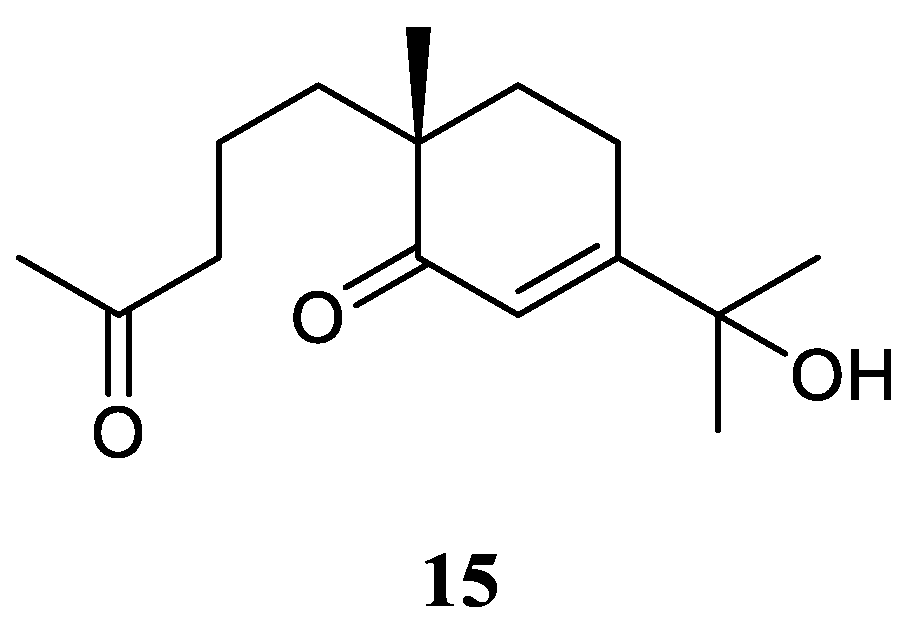
In the above-mentioned study [28], a seco-eudesmane sesquiterpene chabrolidione B (15) (Figure 8) was co-isolated. However, compound 15 were judged as inactive against the cell lines A549, MCF-7 and HepG2 (all IC50 > 100 µmol/mL).
Figure 8.
The chemical structure of seco-eudesmane sesquiterpene from the genus Litophyton.
3.8. Tri-nor-eudesmane Sesquiterpenes
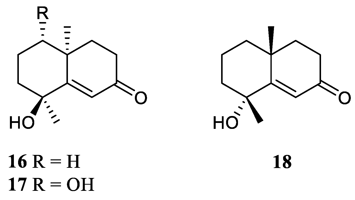
The methanolic extract of the Saudi Arabia Red Sea L. arboreum collection harbored two tri-nor-eudesmane sesquiterpenes teuhetenone A (16) and calamusin I (17) [27] (Figure 9). Intrestingly, these two nor-sesquiterpenes 16 and 17 displayed a wide spectrum of bioactivities. In the antibacterial bioassays, they showed moderate to strong activities against E. coli ATCC 10536, P. aeruginosa NTCC 6750, B. cereus ATCC 9634, B. subtilis ATCC6633, S. aureus ATCC5141 with MIC values ranging from 10.9 to 1.2 μg/mL. Of which, 16 exhibited the most potent activity against E. coli ATCC 10536 with MIC of 1.9 μg/mL, and 17 displayed the most potent activity against P. aeruginosa NTCC 6750 with MIC of 1.2 μg/mL. In the antifungal biotests, they exhibited weak to moderate activities against C. albicans and A. niger with MIC values ranging from 7.4 to 3.2 μg/mL. In the cytotoxic experiments, they displayed potent cytotoxic effects against cell lines MCF-7 and HepG2 with IC50 ranging from 6.43 to 39.23 μM. While, the methanolic extract of the Egyptian Red Sea L. arboreum sample yielded another tri-nor-eudesmane sesquiterpene 7-oxo-tri-nor-eudesm-5-en-4β-ol (18) [28] (Figure 9). However, this nor-sesquiterpene 18 did not show cytotoxic activities against the cell lines A549, MCF-7 and HepG2 (all IC50 > 100 µmol/mL).
Figure 9.
The chemical structures of tri-nor-eudesmane sesquiterpenes from the genus Litophyton.
3.9. Eremophilane Sesquiterpene
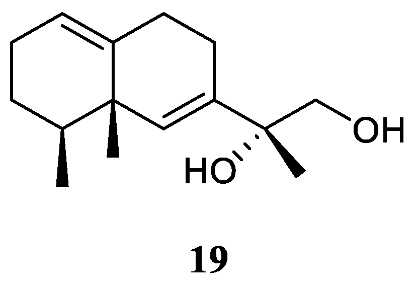
11,12-Dihydroxy-6,10-eremophiladiene (19) (Figure 10) was obtained from the soft coral Litophyton nigrum, using a structure-oriented HR-MS/MS approach [23]. This alcyonarian specimen was collected at Xisha Islands, Hainan, China. However, no bioassays were performed due to its scarcity of amounts.
Figure 10.
The chemical structure of eremophilane sesquiterpene from the genus Litophyton.
3.10. Nardosinane Sesquiterpenes
Interestingly, the South China Sea soft coral L. nigrum is a rich source of nardosinane sesquiterpenes.
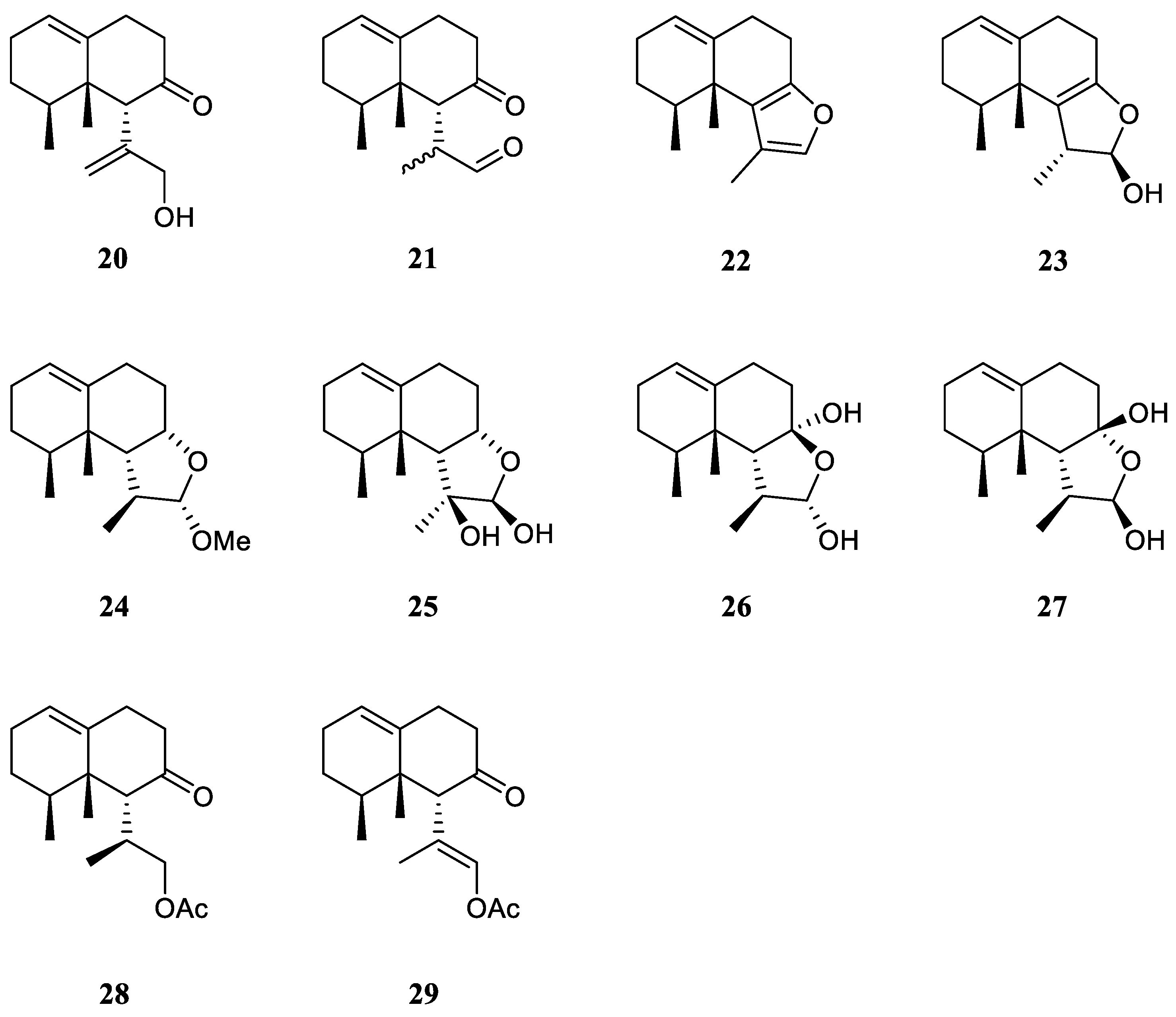
The chemical investigation of the Xisha collection by Yang et al. afforded two new terpenes linardosinenes B (20) and C (21) [12] (Figure 11). These two compounds were evaluated for cytotoxities against different cell lines. Sesquiterpene 20 exhibited cytotoxic effect against the THP-1 cell line with IC50 of 59.49 μM. While compound 21 displayed cytotoxicities against the cell lines SNU-398 and HT-29 with IC50 of 24.3 and 44.7 μM, respectively. In their continual study on the Xisha sample, four additional new secondary metabolites linardosinenes D–G (22–25) (Figure 11) were obtained [30]. All metabolites exhibited weak inhibitory effect against bromodomain-containing protein 4 (BRD4), a promising therapeutic target in various human diseases, at a concentration of 10 μM with inhibitory rates ranging from 15.8% to 18.1%.
Using a structure-oriented HR-MS/MS approach, an undescribed sesquiterpene linardosinene I (26), along with its known 7β,12α-epimer lemnal-l(l0)-ene-7β,12α-diol (27) (Figure 11) were isolated from Xisha alcyonarian L. nigrum [23]. The absolute configuration of terpene 27 was determined to be 4S,5S,6R,7S,11S,12S by single crystal X-ray diffraction analysis with Cu Kα radiation [Flack parameter: 0.13(14)]. Sesquiterpene 26 exhibited a potent PTP1B inhibitory activity (IC50 10.67 μg/mL). It also showed moderate cytotoxic activities against the human tumour cell lines HT-29, Capan-1 and SNU-398 with IC50 values of 35.48, 42.55, and 25.17 μM, respectively. However, co-isolated metabolite 27 was inactive against PTP1B (IC50 >20 μg/mL) or cell lines HT-29, Capan-1 and SNU-398 (all IC50 >50 μM).
Recently, two members of this cluster, paralemnolin J (28) and (lS,8S,8aS)-l-[(E)-2′-acetoxy-l′-methylethenyl]-8,8a-dimethyl-3,4,6,7,8,8a-hexahydronaphthalen-2(1H)-one (29) (Figure 11), were isolated in the chemical investigation of a Balinese soft coral L. setoensis [15]. In terms of biological activity, cytotoxic effects against several solid tumor and leukemia cell lines HT-29, Capan-1, A549, and SNU-398 were assessed for compounds 28 and 29. As a result, both compounds showed weak cytotoxic activities against the test cell lines (all IC50 >20 μM).
Figure 11.
The chemical structures of nardosinane sesquiterpenes from the genus Litophyton.
3.11. Nornardosinane Sesquiterpene
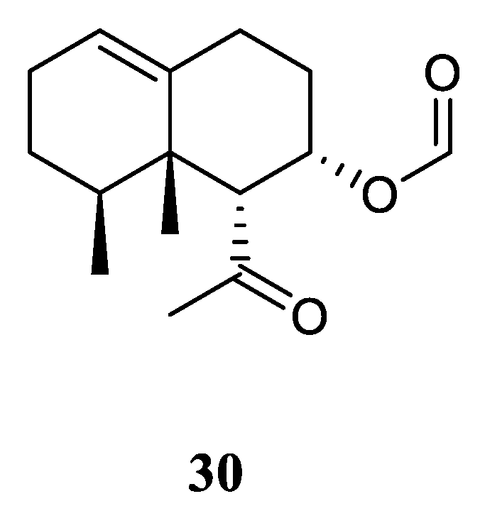
Chemical probing of Xisha alcyonarian L. nigrum afforded an uncommon nornardosinane sesquiterpene linardosinene A (30) [12] (Figure 12). The absolute configuration of 30 was determined by modified Mosher's method and TDDFT ECD approach. This isolate was evaluated for cytotoxicity against the THP-1 cell line and inhibitory activities against the PTP1B, BRD4, HDAC1 and HDAC6 protein kinases. However, it was inactive against the above-mentioned cell line and protein kinases.
Figure 12.
The chemical structure of nornardosinane sesquiterpene from the genus Litophyton.
3.12. Eremophilane-Nardosinane Bis-Sesquiterpene
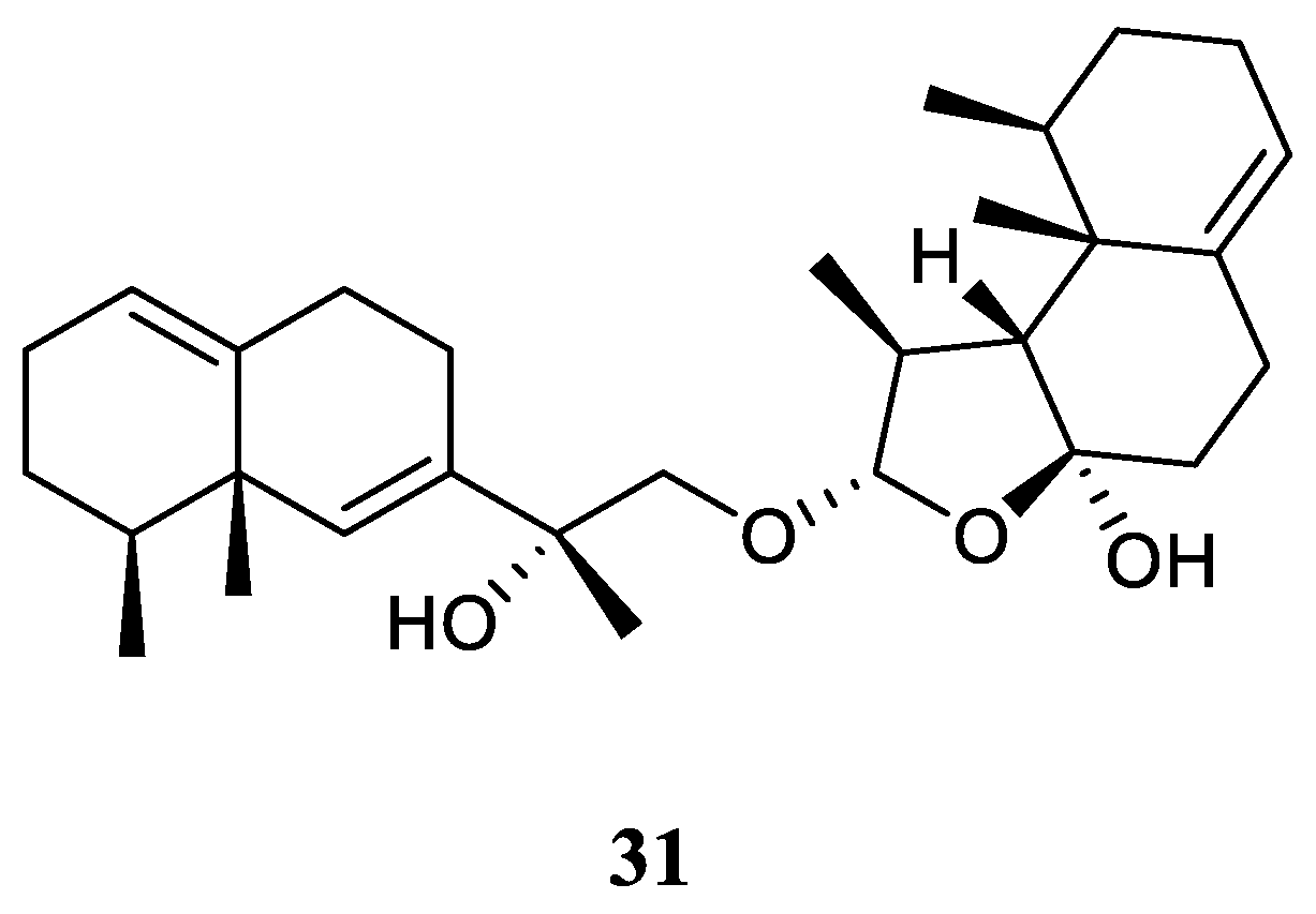
Interestingly, one uncommon sesquiterpe dimer, linardosinene H (31) (Figure 13), was found in the soft coral L. nigrum collected at Xisha Islands, South China Sea, whose structure consisted of a eremophilane sesquiterpene 19 and a nardosinane sesquiterpene 26 [23]. Contrast to its monomer 26, this bis-sesquiterpene 31 did not exhibit inhibitory activity against PTP1B (IC50 >20 μg/mL) or the cell lines HT-29, Capan-1, A549, and SNU-398 (all IC50 >20 μM).
Figure 13.
The chemical structure of eremophilane-nardosinane bis-sesquiterpene from the genus Litophyton.
Figure 13.
The chemical structure of eremophilane-nardosinane bis-sesquiterpene from the genus Litophyton.
3.13. Neolemnane Sesquiterpene
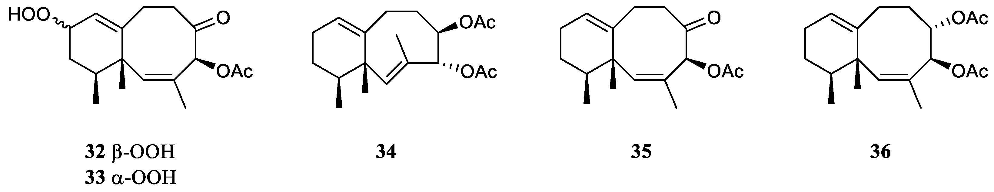
Study on the chemical constituents of the Chinese soft coral L. nigrum yielded three new sesquiterpenes lineolemnenes A–C (32–34) possessed the neolemnane carbon framework, together with the related known compound 4-acetoxy-2,8-neolemnadien-5-one (35) [12] (Figure 14). It might be worth to point out that the absolute configuration of 35 was unambiguously determined to be 1S,4S,12S by X-ray diffraction analysis for the first time. The cytotoxicities of substances 32 and 33 against SNU-398, HT-29, Capan-1, and A549 were evaluated. It revealed that 32 and 33 only exhibited potent cytotoxic activity against SNU-398 with IC50 values of 44.4 and 27.6 μM, respectively. And none of them showed potent inhibitory activities against the PTP1B, BRD4, HDAC1 and HDAC6 protein kinases. Compound 35 was also found in the Indonesian soft coral L. setoensis, together with another sesquiterpene paralemnolin E (36) [15] (Figure 14). They were subjected to cytotoxic bioassays against several solid tumor and leukemia cell lines HT-29, Capan-1, A549, and SNU-398. The results revealed both two compounds had weak cytotoxic activities against the test cell lines (all IC50 >20 μM).
Figure 14.
The chemical structures of neolemnane sesquiterpenes from the genus Litophyton.
3.14. Seconeolemnane Sesquiterpene
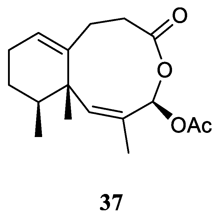
A new sesquiterpene lineolemnene D (37) (Figure 15) was isolated and characterized from the Xisha soft coral L. nigrum [12]. Structurally, this compound possessed an unusual seconeolemnane skeleton. The absolute configuration of 30 was determined to be 1S,4R,12S by TDDFT ECD approach. Bioassays including cytotoxicity against the THP-1 cell line and inhibitory activities against the PTP1B, BRD4, HDAC1 and HDAC6 protein kinases were performed for this isolate. However, it was judged as inactive in these biotests.
Figure 15.
The chemical structure of seconeolemnane sesquiterpene from the genus Litophyton.
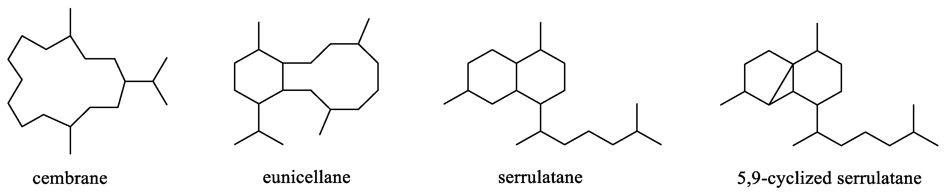
4. Diterpenes
Diterpenes were a second largest cluster of terpenes consisting of 31 members. The first new compound from the genus Litophyton, 2-hydroxynephtenol [19], belonged to this category. Structurally, this pack of secondary metabolites could be divided into four subgroups: cembrane, eunicellane, serrulatane and 5,9-cyclized serrulatane (Figure 16). Analysis of taxonomical distributions revealed they were from L. viridis, L. arboreum, Litophyton viscudium, L. setoensis, and unclearly indentified Litophyton sp., which were collected at Red Sea, Indonesian and Japanese waters (Table S1).
Figure 16.
The reported carbon frameworks of diterpenes from the genus Litophyton.
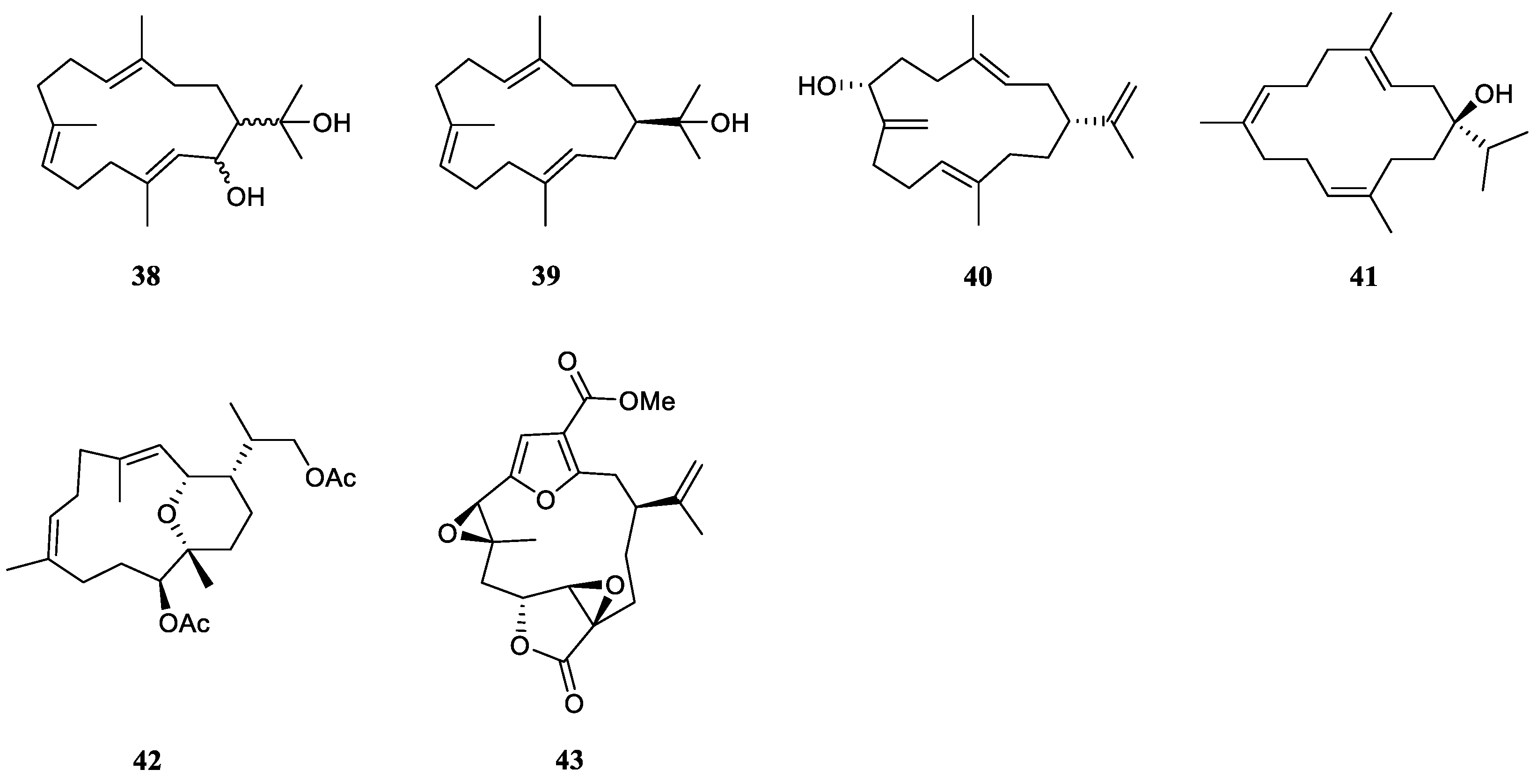
4.1. Cembrane Diterpenes
In 1975, Tursch et al. reported the isolation and structure elucidation of a new compound 2-hydroxynephtenol (38) and its known analogue (–)-nephtenol (39) (Figure 17) from the alcyonarian L. viridis, collected off Serwaru (Leti Island, Maluku Province, Indonesia) [19]. Based on the chemical transformation, the absolute configuration of 39 was determined as 1R. However, the configurations of C-1 and C-2 of 38 were not determined.
A new cembrane diterpene (3E,11E)-cembra-3,8(19),11,15-tetraene-7α-ol (40) (Figure 17), along with the known (–)-nephthenol (39), had been isolated from the Red Sea soft coral L. arboreum, which was collected at Hurghada, Egypt [31]. The relative configuration of 40 was determined as 1R,7R. The (3E)- and (11E)-configurations were determined by comparison of the 13C chemical shifts for C-18 and C-20 methyl signals (<20.0 ppm). The biogenetical pathway of new terpene 40 from structurally related metabolite 39 was proposed in this work. Intrestingly, nephthenol (39) was also found in another Red Sea sample L. arboreum collected from Jeddah coast, Saudi Arabia [18].
Chemical investigation on the chemical compositions of another Egyptian specimen L. arboreum collected at Sharm El-Sheikh led to the discovery of sarcophytol M (41) [25] (Figure 17). Compound 41 displayed a wide spectrum of bioactivities. It showed weak inhibitory activity against HIV-1 protease receptor with IC50 of 15.7 µM, compared to the positive control, which had IC50 of 8.5 μM. Molecular docking study disclosed the hydrogen bond between 41 and the amino acid residue of Asp 25 in the hydrophobic receptor pocket with a score of −14.44. And sesquiterpene 41 showed moderate cytotoxic activities against the cell lines HeLa (IC50 27.5 μM), Vero (IC50 22 μM) and U937 (IC50 31.7 μM).
Sarcophytol M (41) co-existed with a pyrane-based cembranoid 11-acetoxy-15,17-dihydroxy-2,12-epoxy-(3E,7E)-1-cembra-3,7-diene (42) (Figure 17) in the extract of Saudi Arabian alcyonarian L. arboreum [29]. Both compounds displayed antiproliferative effects toward cancer cell lines MCF-7, HCT116 and HepG-2 in comparison with standard anticancer drug (Doxorubicin). Of which, 42 showed significant antiproliferative activities against the cell lines MCF-7, HCT116 and HepG2 (IC50 19.1, 22.0, 24.0 μM, respectively). Further investigation on the possible mechanism of action had been done. The results showed 42 significantly increased the G0/G1 non-proliferating cell fraction from 55.42% to 68.98% with compensatory decrease in cell populations in S-phase and G2/M-phase from 31.99% to 21.99% and from 10.82% to 7.63%, respectively.
Chemical probing of the soft coral L. arboreum, collected near Bali, Indonesia, afforded a furanocembranoid diterpene 11β,12β-epoxypukalide (43) (Figure 17) [24]. This diterpene 43 showed low antiproliferative activities against the cell lines L-929 and K-562 (both GI50 >129 μM), and low cytotoxic effect against the HeLa cell line (CC50 115 μM).
Figure 17.
The chemical structures of cembrane diterpenes from the genus Litophyton.
4.2. Eunicellane Diterpenes
This was the largest cluster of diterpenes found in the genus Litophyton.
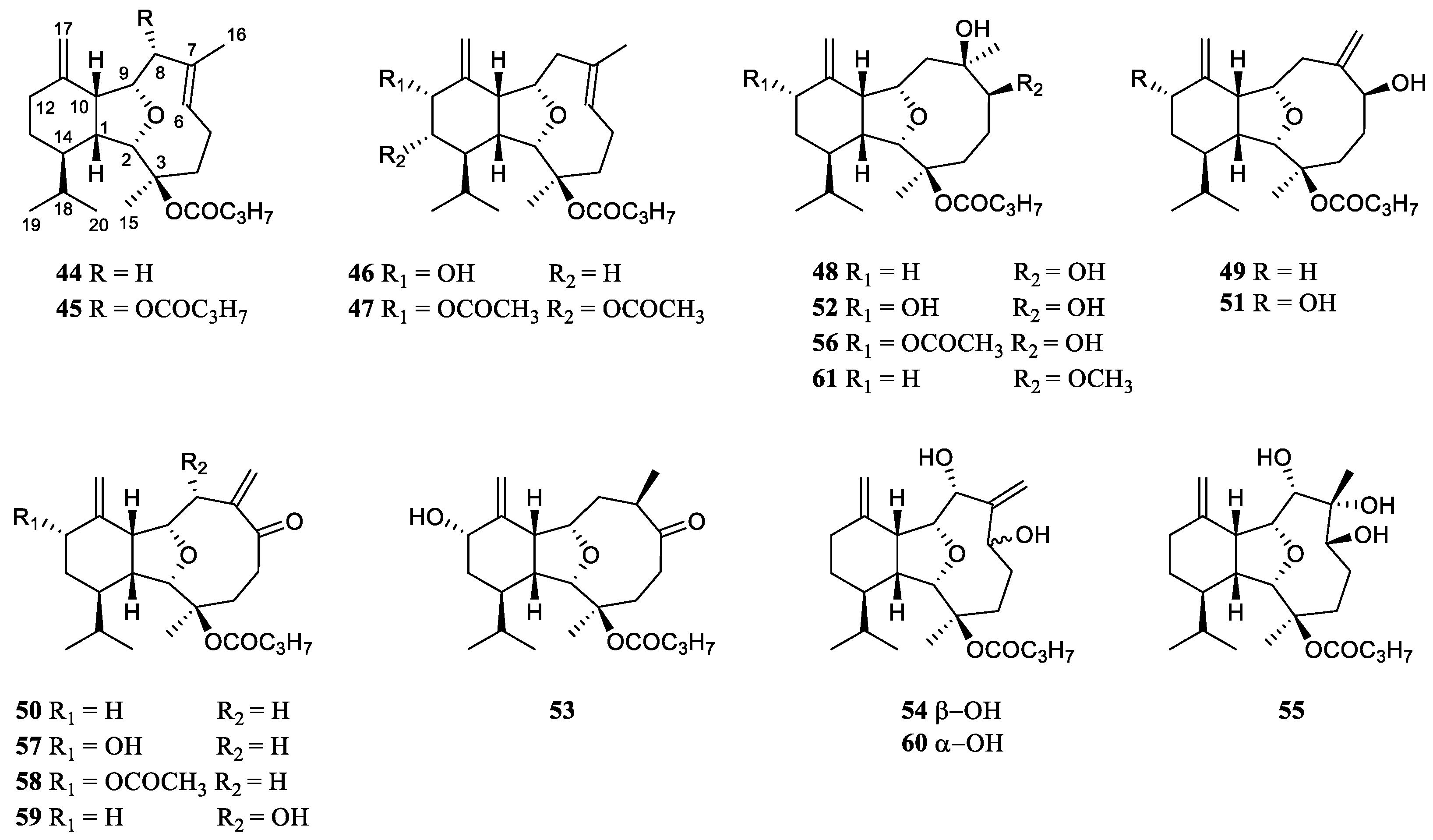
In 1987, Ochi et al. reported the eunicellane diterpenes from the Litophyton animals for the first time. They were litophynins A (44) and B (45) (Figure 18) from the soft coral Litophyton sp., which was collected from a shallow area of Sukumo Bay in Kochi Prefecture, Japan [32]. Their structures had been fully characterized by extensive 2D NMR studies and molecular mechanics calculations. Structurally, 45 was the butyric ester derivative of 44. In the artificial diet feeding bioassay, they exhibited insect growth inhibitory against the silkworm, Bombyx mori L., with ED50 values of 12 and 2.7 ppm, respectively.
Inspired by this work, Ochi et al. performed the continual investigations on the insect growth inhibitory diterpenoids from the previously studied alcyonarian Litophyton sp., leading to the discovery of an array of new eunicellane diterpenes including litophynins C (46) [33], D (47) [34], E (48) [34], F (49) [35], G (50) [35], H (51) [35], I (52) [36], and J (53) [36] (Figure 18). The variations of their structures were mainly at the segment C-6, C-7 and C-16, which usually formed a double bond Δ6 (endo), or Δ7(16) (exo) accompanied with a hydroxlyl or a ketone at C-6. The hydroxylation or acetylation at C-12/C-13 was also observed. The absolute configuration of litophynin C (46) was determined by analysis of CD spectrum of its p-bromobenzoate, based on the exciton chirality method of allylic alcohol benzoate [33]. Similarly, the absolute configuration of litophynin D (47) was determined by an application of the dibenzoate chirality rule [34].
Interestingly, these diterpenes exhibited various bioactivities. Litophynins C (46) and G (50) displayed insect growth inhibitory activity against the second instar larvae of the silkworm Bombyx mori L. (ED50 25 [33] and 42 [35] ppm, respectively). Litophynin D (47) exhibited significant brine shrimp lethalty (LD50 0.9 ppm) [34]. Litophynins I (52) and J (53) possess significant molluscicidal and repellent activities against the muricid gastropod Drupella fragum [36]. At 30 ppm concentration, diterpenes 52 and 53 exhibited 100% mortality to the snail within 24 hours. They were also repellent to the gastropod when impregnated on filterpaper by 45 μg/cm2. These compounds in combination with a wide variety of compounds stored in skin glands of Litophyton sp., appeared to be the foundation of a chemical defense adaptation to survive in predator-rich environments.
Miyamoto et al. investigated the chemical constituents of the mucus secreted by the soft coral Litopbyton sp., which was collected from the rocky coast of Nango-cho, Miyazaki Prefecture, Japan [37]. In this study, two new eunicellin-type diterpenoids, litophynols A (54) and B (55), and three known diterpenoids litophynins E (48), H (51) and I monoacetate (56) (Figure 18) were identified. The absolute configurations of litophynols A (54) and B (55) were determined by application of the CD exciton chirality method, while the absolute configuration of litophynin E (48) was assigned by the Mosher’s method. Additionally, the absolute configurations of litophynin E (48) and litophynol B (55) were furthermore confirmed by the application of the octant rule to their ozonolysis products, respectively. Interestingly, it was found that these five eunicellin-based diterpenoids were also present in the animal bodies of Litopbyton sp. but in low yields compared with the mucus. The performed bioassays revealed these five isolates were positive in a hemolytic reaction test, and crude diterpenoid fractions exhibited ichthyotoxicity (IC100 20 ppm). This suggests that this soft coral holds eunicellin-type diterpenoids in its mucus for the purpose of defense against predators.
Iwagawa et al. found that the CH2Cl2-soluble portion of the MeOH extract of the Japanese alcyonarian L. viscudium showed moderate cytotoxic activity (IC50 = 6.9 μg/mL) against the proliferation of human promyelocytic leukemia cells (HL-60) [16,38]. Study on the chemical compositions of this species yielded five new eunicellin-type diterpenes, 6-oxo litophynin H (57), 6-oxo litophynin H 12-acetate (58), 6-oxo litophynol A (59), 6-epi litophynol A (60), and 6-methyl litophynol E (61), together with a previously reported litophynin F (49) (Figure 18) [16]. These secondary metabolites exhibited different levels of cytotoxicities against HL-60. Diterpenes 57 and 58 having a hydroxyl group or acetoxyl group at C-12 showed moderate cytotoxic activities (both IC50 20 μM), while compound 59 possessing an additional hydroxyl group at C-8 and its reduced derivative 60 exhibited significant cytotoxic activities (IC50 5.7 and 4.2 μM, respectively). The C-6 methoxyl and C-7 hydroxyl groups dramatically reduced the toxicity of diterpene 61 (IC50 50 μM). Compound 49 with the absence of a hydroxyl group at C-8 and the presence of a β-hydroxyl group at C-6 displayed much less cytotoxic activity (IC50 18 μM) than that of 60.
Figure 18.
The chemical structures of eunicellane diterpenes from the genus Litophyton.
4.3. Serrulatane Diterpenes
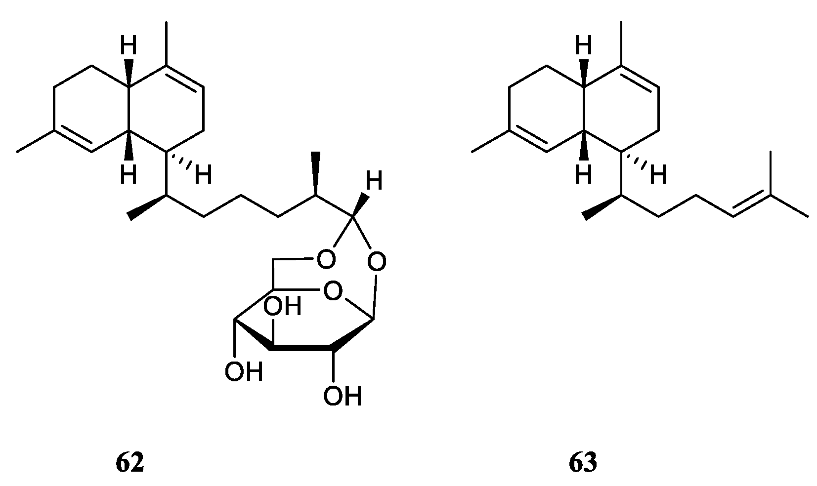
Two secondary metabolites lemnabourside (62) and biflora-4,9,15-triene (63) (Figure 19), which possessed the serrulatane carbon framework, were obtained from the soft coral L. setoensis collected along the coast of Singaraja, Bali Island, Indonesia [15]. In the bioassays, compounds 62 and 63 showed weak cytotoxic activities against the test cell lines HT-29, Capan-1, A549, and SNU-398 (all IC50 >20 μM).
Figure 19.
The chemical structures of serrulatane diterpenes from the genus Litophyton.
4.4. 5,9-Cyclized Serrulatane Diterpenes
Interestingly, five new diterpenes, litosetoenins A–E (64–68) (Figure 20), were isolated from a Balinese alcyonarian L. setoensis [15]. Their structures were elucidated by extensive spectroscopic analysis, quantum mechanical nuclear magnetic resonance approach, and chemical transformations. All of them possessed a rearranged serrulatane-type backbone with an unusual tricyclo[3.0.4]decane core. Moreover, 66–68 displayed intriguing tetracyclic backbones bearing either an additional tetrahydropyran or tetrahydrofuran ring, which were unprecedented and unique. All the isolates were subjected to the cytotoxic bioassays against cell lines HT-29, Capan-1, A549, and SNU-398. As a result, all the metabolites showed weak cytotoxic activities against these celllines with IC50 values >20 μM.
Figure 20.
The chemical structures of 5,9-cyclized serrulatane diterpenes from the genus Litophyton.
Figure 20.
The chemical structures of 5,9-cyclized serrulatane diterpenes from the genus Litophyton.
5. Tetraterpene
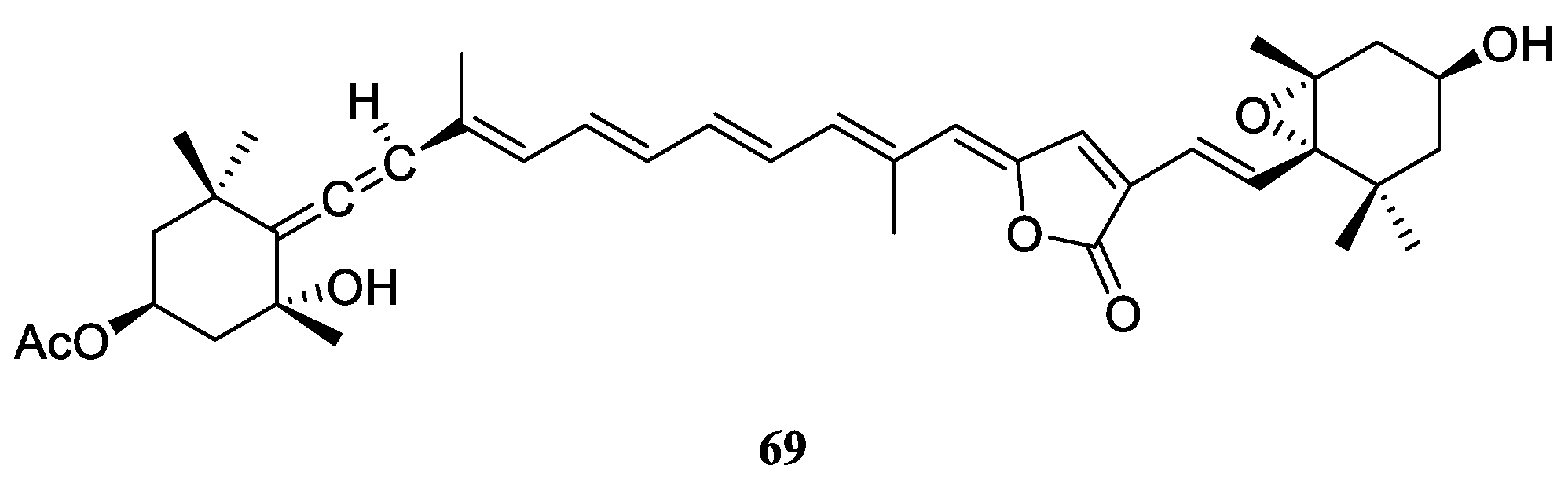
As revealed in literature, there was only one member of tetraterpene found in the genus Litophyton. That was all-trans-peridinin (69) (Figure 21), obtained from the Red Sea soft coral L. arboreum [31]. Terpene 69 showed moderate antiproliferative activities against cell lines HUVEC and K-562 (GI50 48.4 and 53.8 μM, respectively), and moderate cytotoxicity against the HeLa cell line (IC50 51.9 μM).
Figure 21.
The chemical structures of tetraterpene from the genus Litophyton.
6. Steroids
It seemed the documentation of the steroids from the genus Litophyton started in 1976, where two 19-hydroxysterols were reported from L. viridis by Bortolotto et al. [39]. Till now, 23 steroids had been obtained from four species, including L. viridis, Litophyton mollis, L. arboreum and unclearly indentified Litophyton sp. Structurally, ergostanes and 4α-methylated ergostanes dominated the steroidal profiling of this genus, with three exceptions. The exceptional cases include one stigmastane, one 13,14-seco steroid, and one 4α,23-dimethylated ergostane (Table S1). Considering these, the following presentation of steroids was divided into two categories 4α-methylated and other miscellaneous steroids.
6.1. 4α-Methylated Steroids
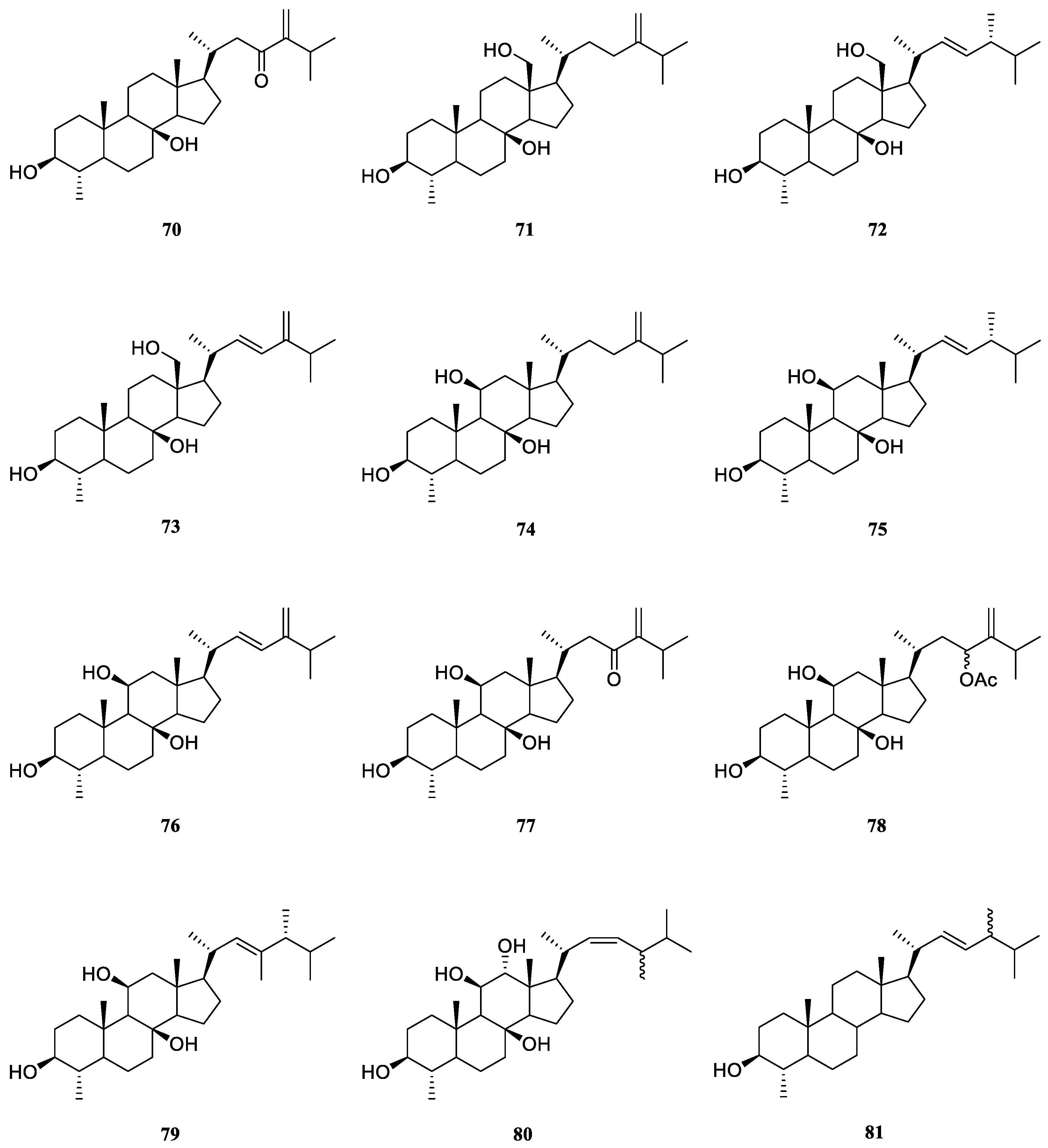
Examination of less polar fractions of the extract of the soft coral L. viridis, which was collected in the Lesser Sunda Islands, led to the isolation of a novel polyoxygenated sterol 4α-methyl-3β,8β-dihydroxy-5α-ergost-24(28)-en-23-one (70) [40] (Figure 22). The structure and relative configuration of 70 were established unambiguously by X-Ray diffraction analysis on its p-bromobenzoate derivative.
Končić et al. conducted the first chemical investigation on the metabolic profile of the Red Sea alcyonarian L. mollis, resulting in the isolation of ten 4α-methylated steroids 71–80 [41] (Figure 22). These steroids differed not only in the substitution of hydroxyl groups at steroidal nucleus but also in diverse oxidation of side chains. The absolute configuration of C-24 in compounds 72, 75 and 79 was assigned as R based on the chemical shift difference between C-26 and C-27 carbon atoms, which was a powerful rule to determine the absolute configuration of steroidal side chains [42,43,44]. The cytotoxic activities of metabolites 71–79 were evaluated against cell lines K562 and A549 [41]. As a result, compounds 71 and 75–78 displayed potent cytotoxicity against K562 cells with IC50 values ranging from 5.6 to 8.9 μM. Meanwhile, these compounds showed low toxicity against healthy PBMCs, thus denoting interesting differential toxicity. Additionally, the tested steroids exhibited moderate levels of toxicity against A549 cells with IC50 values around 20 μM, further underlining their antileukemic activity.
The Red Sea soft coral L. arboreum was frequently encountered by marine natural product chemists. Shaker et al. found that the Egyptian specimen L. arboreum harbored 4α,24-dimethyl-cholest-22Z-en-3β-ol (81) (Figure 22), the complete assignments of 13C NMR data of which was reported for the first time [45]. Interestingly, the presence of nebrosteroid M (74) in another Egyptian sample L. arboreum had been reported by Mahmoud et al., which was collected in front of the National Institute of Oceanography and Fisheries at Hurghada province [28]. It was also found sterol 74 showed cytotoxic effect against A549 cell line (IC50 36.9 μmol/mL). Moreover, this compound exhibited moderate cytotoxicity against MCF-7 (IC50 55.3 μmol/mL), but no activity against HepG2 (IC50 >100 μmol/mL).
Ahmed et al. also made a Egyptian collection of L. arboreum from Neweba. Chemical probing of this sample led to isolation of previously reported 4α-methylated steroids 74, 75 and 79 [17]. Anti-malarial activities against chloroquine-sensitive (D6) and chloroquine-resistant (W2) strains of Plasmodium falciparum, together with the cytotoxic effect against the Vero cell line were evaluated for these three isolates. However, they were judged as inactive at the concentration of 4.76 mg/mL in the above-mentioned bioassays.
Figure 22.
The chemical structures of 4α-methylated steroids from the genus Litophyton.
6.2. Other Miscellaneous Steroids
Two novel polyhydroxylated sterols, 24-methylenecholest-5-en-3β,7β,19-triol (82) and its 7-monoacetate derivative (83) (Figure 23) were isolated from the soft coral L. viridis, collected off Serwaru, Leti Island, Maluku Province, Indonesia [39]. The structure of 82 had been established by X-ray diffraction analysis [46]. It was said these two substances were the first instances of naturally occurring 19-hydroxysterols [39]. More than ten years later, another two new 19-hydroxysterols, litosterol (84) and 5,6-epoxylitosterol (85) (Figure 23), were reported from the Okinawan sample L. viridis [47]. The latter compound showed an antileukemic activity (IC50 0.5 μg/mL) against leukemia cells P388 in vitro.
Interestingly, 19-hydroxysterols 82 and 83 were widely distributed in the species L. arboreum collected at different waters.
Study on the substances of South China Sea alcyonarian L. arboreum, which was collected at Xisha Islands, led to the co-isolation of the previously reported sterol 82 and undescribed (24E)-24-ethyl-5α-cholesta-8,24(28)-diene-3β,12β,19-triol (86) [48] (Figure 23).
Chemical investigation of the Egyptian Red Sea soft coral L. arboreum by Ellithey et al., which was collected at Sharm El-Sheikh, revealed the co-existence of three steroids 82, 83 and 24-methylcholesta-5,24(28)-diene-3β-ol (87) [25] (Figure 23). Compounds 82 and 83 demonstrated strong cytotoxicity against HeLa cells (IC50 8 and 5.3 μM, respectively) and moderate cytotoxicity against U937 cells (IC50 16.4 and 10.6 μM, respectively). Wheares steroid 87 showed weak cytotoxicity against HeLa cells (IC50 48 μM) and no potent cytotoxicity against U937 cells (inhibition rates <80%). Moreover, sterol 83 displayed strong inhibitory activity against HIV-1 protease witht IC50 of 4.85 μM. In Ellithey’s continuous study, sterols 82 and 83 had strong cytotoxic effects against cancer cell lines HepG2 (IC50 8.5 and 6.07 μg/mL, respectively), MDA (IC50 5.5 and 6.3 μg/mL, respectively) and A549 (IC50 9.3 and 5.2 μg/mL, respectively) [26].
Figure 23.
The chemical structures of other miscellaneous steroids from the genus Litophyton.
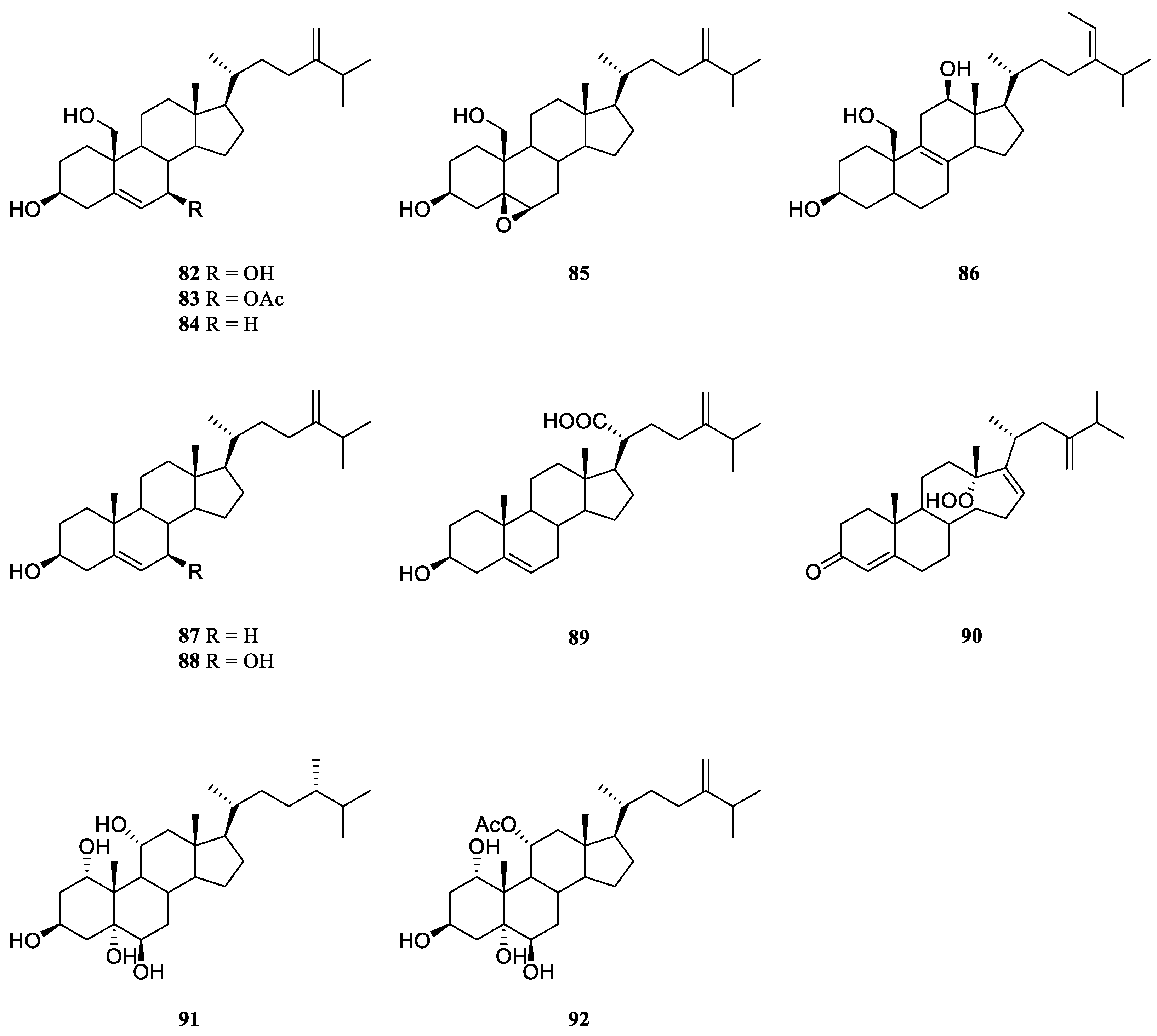
Co-existence of three known secondary metabolites 82, 84 and 87 in the Egyptian Red Sea collection L. arboretum from Hurghada was reported by Shaker et al. [45]. Recently, study on another Egyptian Red Sea alcyonarian L. arboreum collected at the same coast by Mahmoud et al., disclosed the existence of sterol 87, too [28]. In this study, metabolite 87 exhibited noticeable cytotoxicity against A549 cell line (IC50 28.5 μmol/mL) and weak cytotoxic activities against both cell lines MCF-7 and HepG2 (IC50 70.0 and 77.6 μmol/mL, respectively).
Chemical probing of Egyptian Red Sea collection L. arboreum from Neweba afforded steroids 83, 84, 3β,7β-dihydroxy-24-methylenecholesterol (88), and chabrolosteroid I (89) [17] (Figure 23). Anti-malarial bioassays indicated that compound 88 displayed weak activity against chloroquine-resistant strain P. falciparum W2 with IC50 of 4.0 mg/mL, but was inactive against chloroquine-sensitive strain P. falciparum D6 at the concentration of 4.76 mg/mL.
A novel seco-steroid 13,14-seco-22-norergosta-4,24(28)-dien-19-hydroperoxide-3-one (90) (Figure 23) together with the known one 83 were found in the chemical investigation of Saudi Arabian Red Sea specimen L. arboreum by Ghandourah et al., which was collected from the North of Jeddah coast [29]. They showed antiproliferative effects toward three different cancer cell lines MCF-7, HCT116 and HepG-2. (It might be worth to point out no specific data was provided in this article.) In addition, Hawas et al. reported the presence of sterols 82 and 87 in another Saudi Arabian Red Sea sample L. arboreum [18].
Extensive studies indicated the methanolic extract of Egyptian Red Sea alcyonarian Litophyton sp. showed anti-colon cancer therapeutic potential. [21] The following chromatography resulted in the purification of two polyhydroxylated sterols sarcsteroid F (91) and 24-methylenecholestane-1α,3β,5α,6β,11α-pentol-11-monoacetate (92) (Figure 23).
7. Alkaloids
Alkaloids were a small cluster of secondary metabolites from the genus Litophyton. This cluster consisted of six compounds, which could be divided into two subgroups ceramides and nucleotides. All of them were isolated from the species L. arboreum, which lived in different regions of Red Sea (Table S1).
7.1. Ceramides
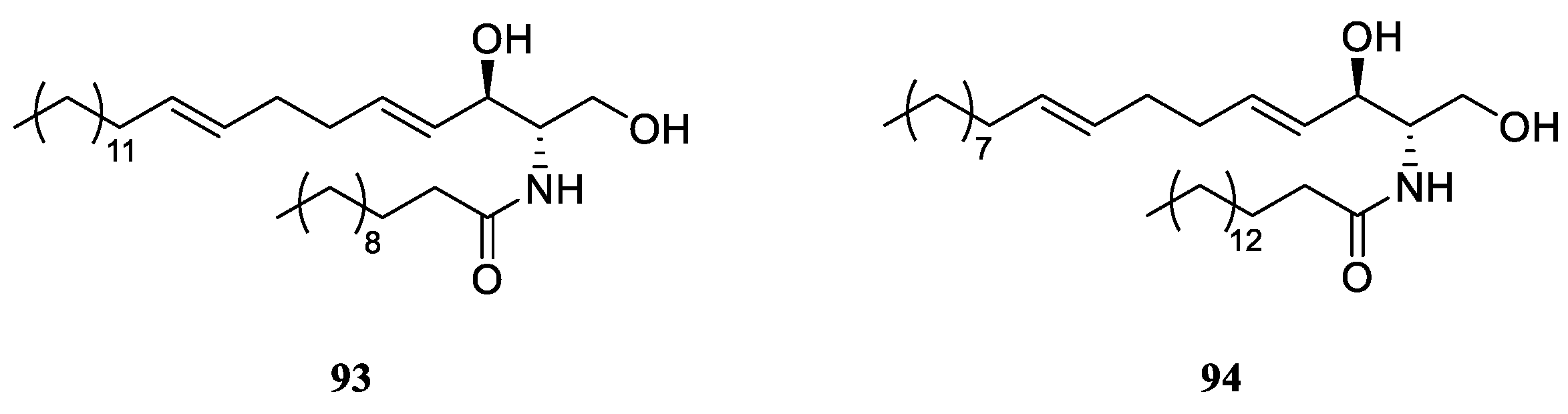
Chemical probing of Red Sea alcyonarian L. arboreum afforded erythro-N-dodecanoyl-docosasphinga-(4E,8E)-dienine (93) (Figure 24), which inhabited in the water of Sharm El-Sheikh, Egypt [25]. This metabolite showed strong inhibitory activity against HIV-1 protease (IC50 4.80 μM), but exhibited weak cytotoxicity against the HeLa cell line (IC50 38.17 μM). Additionally, the wide distribution of ceramide 93 in different collections of L. arboreum was indicated by several studies. The localities of these specimens included Jeddah, Saudi Arabia [27,29] and Neweba, Egypt [17]. However, the chemical investigation of the sample L. arboreum from Hurghada, Egypt, yielded a different ceramide, erythro-N-palmityl-octadecasphinga-4(E),8(E)-dienine (94) [45] (Figure 24).
Figure 24.
The chemical structures of ceramides from the genus Litophyton.
7.2. Nucleotides
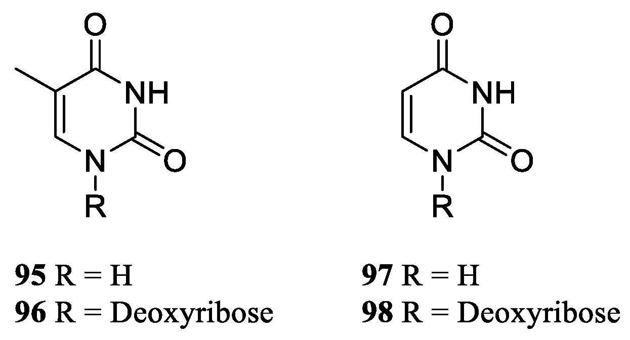
Study on the chemical constituents of Saudi Arabian soft coral L. arboreum led to the isolation and identification of thymine (95) and thymidine (96) [27] (Figure 25). Investigation on the compositions of Egyptian collection L. arboreum revealed the co-isolation of nucleotides 95, uracil (97) and uridine (98) [49] (Figure 25). Metabolites 95, 97 and 98 were in vitro estimated for their cytotoxic activities against three human cancer cell lines A549, MCF-7 and HepG2, and antileishmanial potential against Leishmania major. However, none of them was active in these bioassays.
Figure 25.
The chemical structures of nucleotides from the genus Litophyton.
8. Lipids
This cluster consisted of one prostaglandin, four γ-lactones, four fatty acids, and two glycerol ethers. These secondary metabolites distributed in L. arboretum and unclearly identified Litophyton sp., which were collected in Red Sea and Japanese water (Table S1).
8.1. Prostaglandin
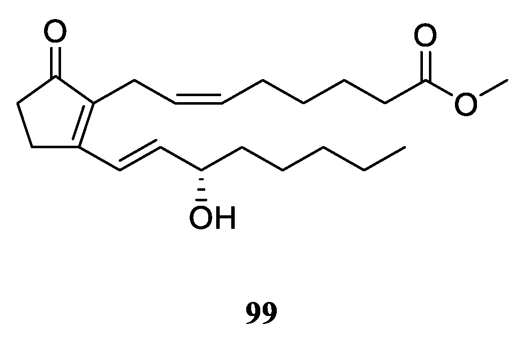
The sole one prostaglandin from the genus Litophyton, PGB2 methyl ester (99) (Figure 26), was characterized in the research of Red Sea alcyonarian L. arboreum, which lived in the gulf of Aqaba, Eilat, Israel [50].
Figure 26.
The chemical structure of prostaglandin from the genus Litophyton.
8.2. γ-Lactones
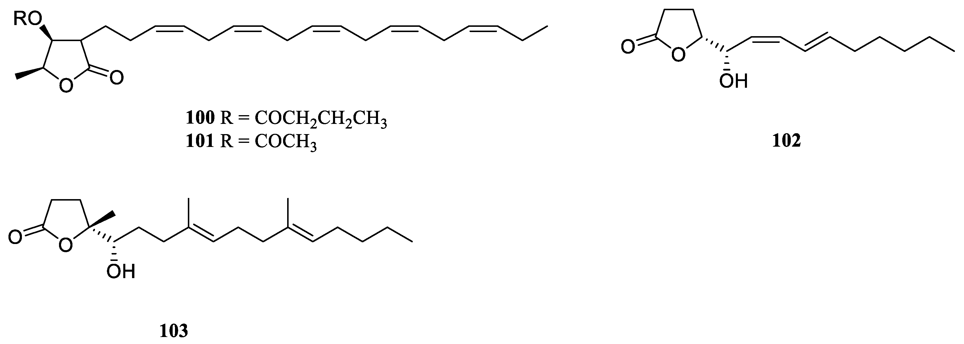
Two new branched-chain lipids containing a γ-lactone ring, which was named litophytolides A (100) and B (101) (Figure 27), was isolated from a Japanese soft coral Litophyton sp. [51]. The difference of their structures was the replacement of the butyryl group in 100 by the acetyl group in 101.
Chemical probing of Israeli Red Sea alcyonarian L. arboreum led to the discovery of two novel γ-lactones with unsaturated chains 102 and 103 [50] (Figure 27). The absolute configuration of C-5 was assigned as S for 102 and 103 by applying the Mosher’s method. In the toxicity bioassay, these two secondary metabolites were toxic to brine shrimp Artemia salina (CC50 15.3 and 21.4 μg/mL, respectively). Antibacterial evaluations indicated the two γ-lactones were active only against Gram-positive bacteria S. aureus and B. subtilis with diameters of inhibition zones ranging from 5.6 to 18.6 mm, but they were inactive against Gram-negative bacterium E. coli and yeast Saccharomyces cerevisiae.
Figure 27.
The chemical structures of γ-lactones from the genus Litophyton.
8.3. Fatty Acids
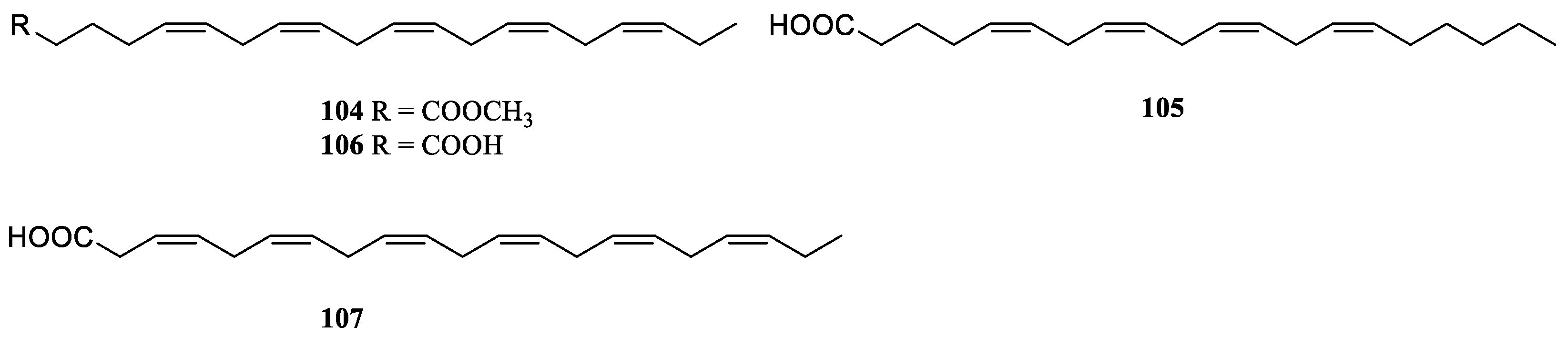
During a search for the chemical constituents of a Japanese soft coral Litophyton sp., methyl (5Z,8Z,11Z,14Z,17Z)-5,8,11,14,17-icosapentaenoate (104) was encountered, together with the above-decribed γ-lactones litophytolides A (100) and B (101) [51] (Figure 28). The co-occurrence of litophytolides 100 and 101 and unsaturated fatty acid 104 in the same animal led to the proposed biogenesis of branched-chain lipids with a γ-lactone ring that involved the condensation of unsaturated fatty acids with pyruvate. GC-MS analysis of the fraction of a Israeli alcyonarian L. arboreum revealed the presence of arachidonic acid (105), eicosapentaenoic acid (106) and docosahexaenoic acid (107) [50] (Figure 28).
Figure 28.
The chemical structures of fatty acids from the genus Litophyton.
8.4. Glycerol ethers
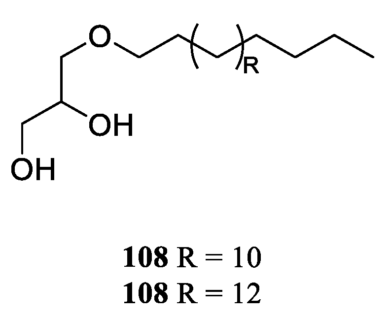
Investigation of the chemical compostions of Red Sea soft coral L. arboreum, which inhabited the coast of Sharm El-Sheikh, Egypt, resulted in the isolation and characterization of chimyl alcohol (108) [25] (Figure 29). This alcohol not only showed cytotoxic effects against the cell lines HeLa and Vero (IC50 23.35 and 60 μM, respectively), but also exhibited inhibitory activity against HIV-1 protease (IC50 26.6 μM). The presence of 108 in the Saudi Arabian Red Sea sample L. arboreum was reported [27].
Chemical probing of Red Sea specimen L. arboreum, which was collected at Hurghada, Egypt, disclosed the co-existence of chimyl alcohol (108) and batyl alcohol (109) [28] (Figure 29). Cytotoxic bioassays were also performed for these two glycerol ethers, but none of them was active against the tested cell lines A549, MCF-7 and HepG2 (all IC50 >100 µmol/mL).
Figure 29.
The chemical structures of glycerol ethers from the genus Litophyton.
9. Conclusions
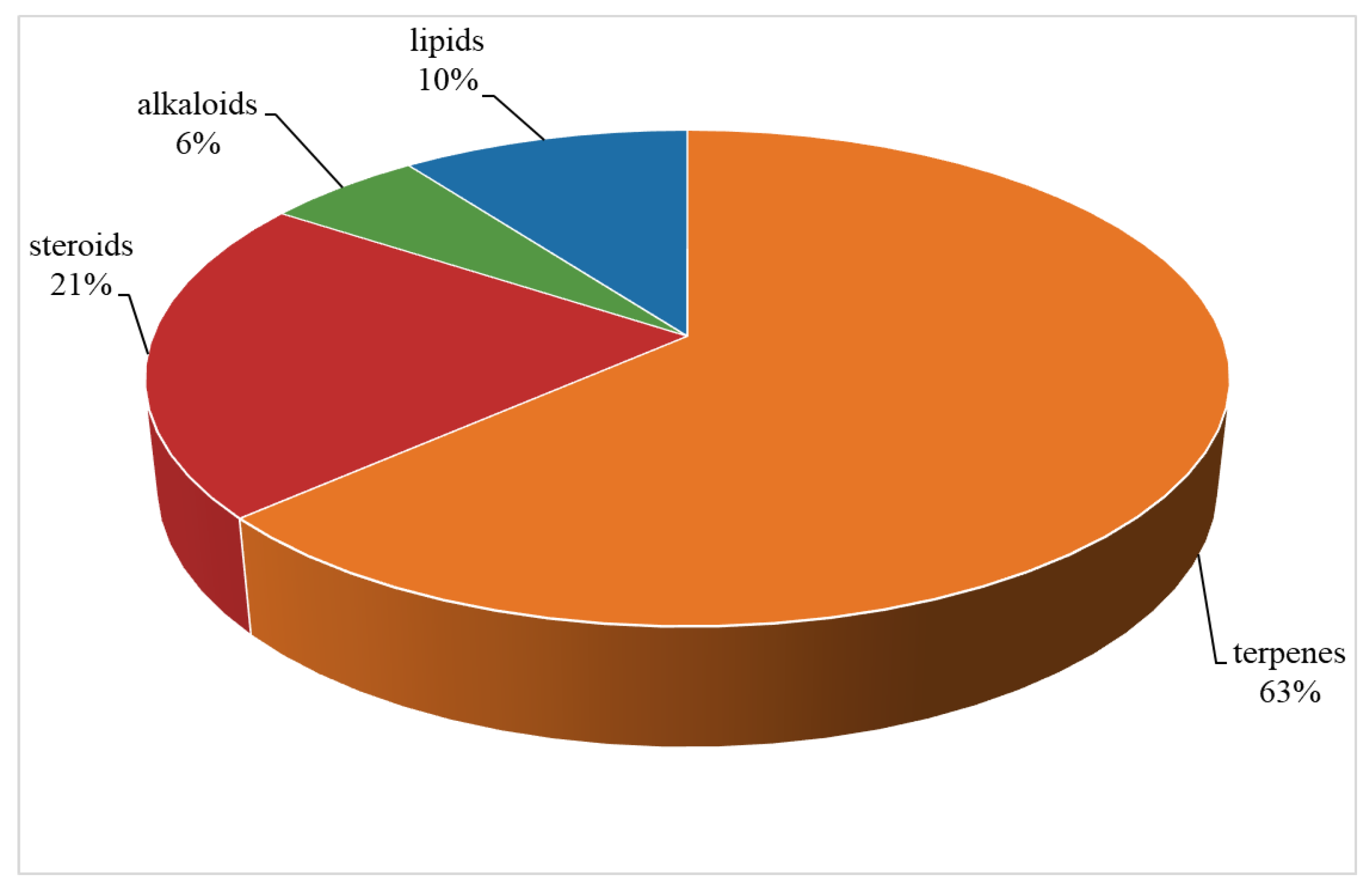
The current work presents an up-to-date documentation of the reported studies on the genus Litophyton with a special focus on their diverse chemical classes of secondary metabolites and their bioactivities. Those investigated soft corals of this genus were inhabited in various marine environments from tropical to temperate regions, especially in the South China Sea, Red Sea, Indonesian and Japanese waters (Table S1). A total of 109 compounds from a variety of species of this genus were reported from 1975 to the July, 2023, covering a period of near five decades. These substances illustrated in this work could be categorized as four major chemical classes: terpenes, alkaloids, steroids and lipids (Figure 30). Among them, terpenes were predominant chemical compositions, which consisted of 36 sesquiterpenes, 31 diterpenes, one bis-sesquiterpene and one tetraterpene (Table S1). Additionally, the very recently reported one sec-germacrane sesquiterpene [17], one himachalene sesquiterpene [29], one nornardosinane sesquiterpene [12], one seconeolemnane sesquiterpene [12], one eremophilane-nardosinane bis-sesquiterpene [23], and five 5,9-cyclized serrulatane diterpenes [15] were quite uncommon marine natural products.
Figure 30.
The chemical profile of secondary metabolites from the genus Litophyton.
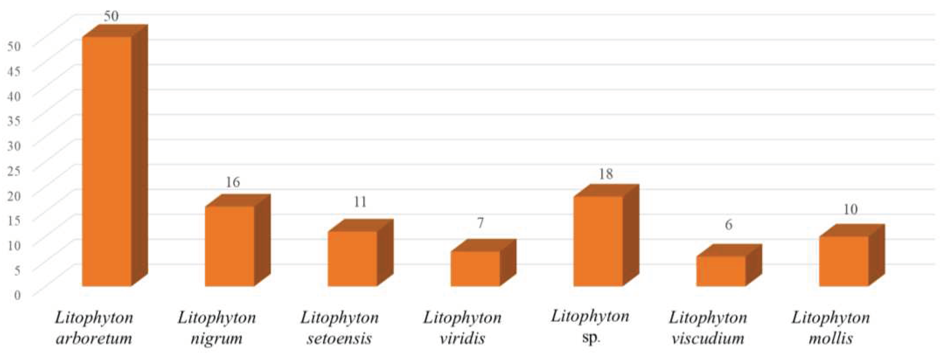
Chemical investigations have been conducted on the species Litophyton arboreum, Litophyton nigrum, Litophyton setoensis, Litophyton viridis, Litophyton viscudium, Litophyton mollis, and unclearly identified Litophyton spp. In terms of the numbers of isolated substances, the animals of L. arboreum were frequently studied members of this genus, yielding 50 compounds (Figure 31). The metabolites of L. arboreum comprised almost structural types of chemical compostions from the title genus, including 18 sesquiterpenes, five diterpenes, one tetraterpene, 12 steroids, two ceramides, four nucleotides, one prostaglandin, two γ-lactones, three fatty acids, and two glycerol ethers (Table S1). Interestingly, bicyclogermacrane, sec-germacrane, guaiane, pseudoguaiane, himachalene, eudesmane, seco-eudesmane, and tri-nor-eudesmane sesquiterpenes were only isolated and characterized from the alcyonarian L. arboreum, which could be regarded as a chemotaxonomic marker for this species (Table S1). Similarly, eremophilane, nornardosinane and seconeolemnane sesquiterpenes, especially a eremophilane-nardosinane bis-sesquiterpene could provide the chemotaxonomic evidence for the species L. nigrum (Table S1). Meanwhile, the chemotaxonomic characters of the species L. setoensis were serrulatane and 5,9-cyclized serrulatane diterpenes (Table S1).
Figure 31.
Number of compounds reported from different species of the genus Litophyton.
These metabolites exhibited a wide spectrum of bioactivities including cytotoxic, anti-malarial, antibacterial, antifungal, anti-HIV, antifeedant, molluscicidal, PTP1B inhibitory, and insect growth inhibitory effects (Table S1). The most frequently evaluated activity for the chemical compositions of the genus Litophyton was cytotoxicity against a panel of human cancer cell lines, such as HeLa, K-562, HepG2, MDA, A549, MCF-7, HCT116, U937, SUN-398, HT-29, Capan-1, THP-1, HL-60, and P388, and quite a large quantity of the substances showed growth inhibitory activity. Although the molluscicidal activity against the muricid gastropod D. fragum for eunicellane diterpenes [36] and toxic activity to brine shrimp A. salina for γ-lactones [50] were performed, more research to better understand the ecological roles of Litophyton metabolites should be conducted.
As presented in this work, the soft corals of the genus Litophyton harbor an array of structurally unique and diversely bioactive secondary metabolites. However, only six clearly indentified species have been investigated, which were a very small portion of the whole genus [11]. It is clear that there is an urgent need for research on exploration of more species of this genus, which are hidden treasure troves of novel marine natural products.
Very recently, coral-encoded terpene cyclase genes that produce the eunicellane and cembrene diterpenes found in soft corals [52,53]. Investigation on the biogenesis of chemical compositions of the genus Litophyton would be another significant and hot research topic in this field. Moreover, the discovery of novel terpene biosynthetic gene clusters could provide potential bioengineering applications for industry.
Supplementary Materials
The following supporting information can be downloaded at the website of this paper posted on Preprints.org, Table S1: Secondary metabolites of the genus Litophyton from 1975 to July, 2023.
Author Contributions
Conceptualization, L.-F.L.; methodology, L.-F.L.; software, Y.-W.G.; formal analysis, L.-F.L.; investigation, X.-Y.Y., L.Z., Q.-B.Y., Z.-Y.G., and L.-F.L.; writing—original draft preparation, L.-F.L.; writing—review and editing, L.-F.L. and Y.-W.G.; funding acquisition, L.-F.L. and Y.-W.G. All authors have read and agreed to the published version of the manuscript.
Funding
This work was financially supported by the National Natural Science Foundation of China (No. 41876194, 81991521).
Institutional Review Board Statement
Not applicable.
Data Availability Statement
Not applicable.
Acknowledgments
Not applicable.
Conflicts of Interest
The authors declare no conflict of interest.
References
- Chakraborty, K.; Joy, M. High-value compounds from the molluscs of marine and estuarine ecosystems as prospective functional food ingredients: an overview. Food Res. Int. 2020, 137, 109637. [Google Scholar] [CrossRef] [PubMed]
- Fonseca, S.; Amaral, M.N.; Reis, C.P.; Custódio, L. Marine natural products as innovative cosmetic ingredients. Mar. Drugs 2023, 21, 170. [Google Scholar] [CrossRef] [PubMed]
- Banerjee, P.; Mandhare, A.; Bagalkote, V. Marine natural products as source of new drugs: an updated patent review (July 2018-July 2021). Expert. Opin. Ther. Pat. 2022, 32, 317–363. [Google Scholar] [CrossRef] [PubMed]
- Liu, M.; Zhang, X.; Li, G. Structural and biological insights into the hot-spot marine natural products reported from 2012 to 2021. Chin. J. Chem. 2022, 40, 1867–1889. [Google Scholar] [CrossRef]
- Carroll, A.R.; Copp, B.R.; Davis, R.A.; Keyzers, R.A.; Prinsep, M.R. Marine natural products. Nat. Prod. Rep. 2023, 40, 275–325. [Google Scholar] [CrossRef] [PubMed]
- Chhetri, B.K.; Tedbury, P.R.; Sweeney-Jones, A.M.; Mani, L.; Soapi, K.; Manfredi, C.; Sorscher, E.; Sarafianos, S.G.; Kubanek, J. Marine natural products as leads against SARS-CoV-2 infection. J. Nat. Prod. 2022, 85, 657–665. [Google Scholar] [CrossRef]
- El-Desoky, A.H.H.; Tsukamoto, S. Marine natural products that inhibit osteoclastogenesis and promote osteoblast differentiation. J. Nat. Med. 2022, 76, 575–583. [Google Scholar] [CrossRef]
- Deng, Y.; Liu, Y.; Li, J.; Wang, X.; He, S.; Yan, X.; Shi, Y.; Zhang, W.; Ding, L. Marine natural products and their synthetic analogs as promising antibiofilm agents for antibiotics discovery and development. Eur. J. Med. Chem. 2022, 239, 114513. [Google Scholar] [CrossRef]
- Ren, X.; Xie, X.; Chen, B.; Liu, L.; Jiang, C.; Qian, Q. Marine natural products: a potential source of anti-hepatocellular carcinoma drugs. J. Med. Chem. 2021, 64, 7879–7899. [Google Scholar] [CrossRef]
- Haque, N.; Parveen, S.; Tang, T.; Wei, J.; Huang, Z. Marine natural products in clinical use. Mar. Drugs 2022, 20, 528. [Google Scholar] [CrossRef]
- World Register of Marine Species. Available online: https://www.marinespecies.org/aphia.php?p=taxdetails&id=204523 (accessed on 2023-08-28).
- Yang, F.; Li, S.-W.; Zhang, J.; Liang, L.-F.; Lu, Y.-H.; Guo, Y.-W. Uncommon nornardosinane, seconeolemnane and related sesquiterpenoids from Xisha soft coral Litophyton nigrum. Bioorg. Chem. 2020, 96, 103636. [Google Scholar] [CrossRef] [PubMed]
- van Ofwegen, L.P. The genus Litophyton Forskål, 1775 (Octocorallia, Alcyonacea, Nephtheidae) in the Red Sea and the western Indian Ocean. Zookeys 2016, 567, 1–128. [Google Scholar] [CrossRef] [PubMed]
- Van Ofwegen, L.P. The genus Litophyton Forskål, 1775 (Octocorallia: Alcyonacea: Nephtheidae) from Australia. Zootaxa 2020, 4764, 1–131. [Google Scholar] [CrossRef]
- Li, S.-W.; Mudianta, I.W.; Cuadrado, C.; Li, G.; Yudasmara, G.A.; Setiabudi, G.I.; Daranas, A.H.; Guo, Y.-W. Litosetoenins A–E, diterpenoids from the soft coral Litophyton setoensis, backbone-rearranged through divergent cyclization achieved by epoxide reactivity inversion. J. Org. Chem. 2021, 86, 11771–11781. [Google Scholar] [CrossRef] [PubMed]
- Iwagawa, T.; Kusatsu, D.; Tsuha, K.; Hamada, T.; Okamura, H.; Furukawa, T.; Akiyama, S.-i.; Doe, M.; Morimoto, Y.; Iwase, F.; et al. Cytotoxic eunicellin-type diterpenes from the soft coral Litophyton viscudium. Heterocycles 2011, 83, 2149–2155. [Google Scholar] [CrossRef]
- Ahmed, M.M.A.; Ragab, E.A.; Zayed, A.; El-Ghaly, E.M.; Ismail, S.K.; Khan, S.I.; Ali, Z.; Chittiboyina, A.G.; Khan, I.A. Litoarbolide A: an undescribed sesquiterpenoid from the Red Sea soft coral Litophyton arboreum with an in vitro anti-malarial activity evaluation. Nat. Prod. Res. 2023, 37, 542–550. [Google Scholar] [CrossRef]
- Hawas, U.W.; Abou El-Kassem, L.T.; Fahmy, M.A.; Farghaly, A.A.; Hassan, Z.M. A new pseudoguaiane-type sesquiterpene and potential genotoxicity and antigenotoxicity effect of the soft coral Litophyton arboreum extract. Lett. Org. Chem. 2018, 15, 1060–1064. [Google Scholar] [CrossRef]
- Tursch, B.; Braekman, J.C.; Daloze, D. Chemical studies of marine invertebrates - XIII 2-Hydroxynephtenol, a novel cembrane diterpene from the soft coral Litophyton viridis (Coelenterata, Octocorallia, Alcyonacea). Bull. Soc. Chim. Belg. 1975, 84, 767–774. [Google Scholar] [CrossRef]
- Tursch, B. Chemical protection of a fish (Abudefduf leucogaster Bleeker) by a soft coral (Litophyton viridis May). J. Chem. Ecol. 1982, 8, 1421–1428. [Google Scholar] [CrossRef]
- Ashry, M.; Askar, H.; Alian, A.; Zidan, S.A.H.; El-Sahra, D.G.; Abdel-Wahhab, K.G.; Lamlom, S.F.; Abdelsalam, N.R.; Abd El-Hack, M.E.; Gomaa, H.F. The antioxidant and antitumor efficiency of Litophyton sp. extract in DMH-induced colon cancer in male rats. Life 2022, 12, 1470. [Google Scholar] [CrossRef]
- Ellithey, M.S.; Lall, N.; Hussein, A.A.; Meyer, D. Cytotoxic and HIV-1 enzyme inhibitory activities of Red Sea marine organisms. BMC Complem. Altern. M. 2014, 14, 77. [Google Scholar] [CrossRef] [PubMed]
- Yang, F.; Hua, Q.; Yao, L.-G.; Liang, L.-F.; Lou, Y.-X.; Lu, Y.-H.; An, F.-L.; Guo, Y.-W. One uncommon bis-sesquiterpenoid from Xisha soft coral Litophyton nigrum. Tetrahedron Lett. 2022, 88, 153571. [Google Scholar] [CrossRef]
- Grote, D.; Dahse, H.-M.; Seifert, K. Furanocembranoids from the soft corals Sinularia asterolobata and Litophyton arboreum. Chem. Biodivers. 2008, 5, 2449–2456. [Google Scholar] [CrossRef] [PubMed]
- Ellithey, M.S.; Lall, N.; Hussein, A.A.; Meyer, D. Cytotoxic, cytostatic and HIV-1 PR inhibitory activities of the soft coral Litophyton arboreum. Mar. Drugs 2013, 11, 4917–4936. [Google Scholar] [CrossRef] [PubMed]
- Ellithey, M.S.; Ahmed, H.H. Bioactive marine-derived compounds as potential anticancer candidates. Asian J. Pharm. Clin. Res. 2018, 11, 464–466. [Google Scholar] [CrossRef]
- Abou El-Kassem, L.T.; Hawas, U.W.; El-Desouky, S.K.; Al-Farawati, R. Sesquiterpenes from the Saudi Red Sea: Litophyton arboreum with their cytotoxic and antimicrobial activities. Z. Naturforsch. 2018, 73, 9–14. [Google Scholar] [CrossRef]
- Mahmoud, A.H.; Zidan, S.A.H.; Samy, M.N.; Alian, A.; Abdelmohsen, U.R.; Fouad, M.A.; Kamel, M.S.; Matsunami, K. Cytotoxicity and chemical profiling of the Red Sea soft corals Litophyton arboreum. Nat. Prod. Res. 2022, 36, 4261–4265. [Google Scholar] [CrossRef]
- Ghandourah, M.A.; Alarif, W.M.; Abdel-Lateff, A.; Al-Lihaibi, S.S.; Ayyad, S.-E.N.; Basaif, S.A.; Badria, F.A. Two new terpenoidal derivatives: a himachalene-type sesquiterpene and 13,14-secosteroid from the soft coral Litophyton arboreum. Med. Chem. Res. 2015, 24, 4070–4077. [Google Scholar] [CrossRef]
- Yang, F.; Hua, Q.; Yao, L.-G.; Liang, L.-F.; Lu, Y.-H.; An, F.-L.; Guo, Y.-W. Further new nardosinane-type sesquiterpenoids from the Xisha soft coral Litophyton nigrum. Fitoterapia 2021, 151, 104906. [Google Scholar] [CrossRef]
- Shaker, K.H.; Müller, M.; Ghani, M.A.; Dahse, H.-M.; Seifert, K. Terpenes from the soft corals Litophyton arboreum and Sarcophyton ehrenbergi. Chem. Biodivers. 2010, 7, 2007–2015. [Google Scholar] [CrossRef]
- Ochi, M.; Futatsugi, K.; Kotsuki, H.; Ishii, M.; Shibata, K. Litophynin A and B, two new insect growth inhibitory diterpenoids from the soft coral Litophyton sp. Chem. Lett. 1987, 16, 2207–2210. [Google Scholar] [CrossRef]
- Ochi, M.; Futatsugi, K.; Kume, Y.; Kotsuki, H.; Asao, K.; Shibata, K. Litophynin C, a new insect growth inhibitory diterpenoid from a soft coral Litophyton sp. Chem. Lett. 1988, 17, 1661–1662. [Google Scholar] [CrossRef]
- Ochi, M.; Yamada, K.; Futatsugi, K.; Kotsuki, H.; Shibata, K. Litophynin D and E, two new diterpenoids from a soft coral Litophyton sp. Chem. Lett. 1990, 19, 2183–2186. [Google Scholar] [CrossRef]
- Ochi, M.; Yamada, K.; Futatsugi, K.; Kotsuki, H.; Shibata, K. Litophynins F, G, and H, three new diterpenoids from a soft coral Litophyton sp. Heterocycles 1991, 32, 29–32. [Google Scholar] [CrossRef]
- Ochi, M.; Yamada, K.; Kataoka, K.; Kotsuki, H.; Shibata, K. Litophynins I and J, two new biologically active diterpenoids from the soft coral Litophyton sp. Chem. Lett. 1992, 21, 155–158. [Google Scholar] [CrossRef]
- Miyamoto, T.; Yamada, K.; Ikeda, N.; Komori, T.; Higuchi, R. Bioactive terpenoids from Octocorallia, I. Bioactive diterpenoids: litophynols A and B from the mucus of the soft coral Litophyton sp. J. Nat. Prod. 1994, 57, 1212–1219. [Google Scholar] [CrossRef]
- Iwagawa, T.; Kusatsu, D.; Tsuha, K.; Hamada, T.; Okamura, H.; Furukawa, T.; Akiyama, S.-i.; Doe, M.; Morimoto, Y.; Iwase, F.; et al. Errata "Cytotoxic eunicellin-type diterpenes from the soft coral Liophyton viscudium": Heterocycles, 2011, 83, 2149. Heterocycles 2012, 85, 2615–2615. [Google Scholar] [CrossRef]
- Bortolotto, M.; Braekman, J.C.; Daloze, D.; Losman, D.; Tursch, B. Chemical studies of marine invertebrates. XXIII. A novel polyhydroxylated sterol from the soft coral Litophyton viridis (coelenterata, octocorallia, alcyonacea). Steroids 1976, 28, 461–466. [Google Scholar] [CrossRef]
- Bortolotto, M.; Braekman, J.C.; Daloze, D.; Tursch, B.; Karlsson, R. Chemical studies of marine invertebrates. XXIX : 4α-methyl-3β,8β-dihydroxy-5α-ergost-24(28)-en-23-one, a novel polyoxygenated sterol from the soft coral Litophyton viridis, (Coelenterata, Octocorallia, Alcyonacea). Steroids 1977, 30, 159–164. [Google Scholar] [CrossRef]
- Končić, M.Z.; Ioannou, E.; Sawadogo, W.R.; Abdel-Razik, A.F.; Vagias, C.; Diederich, M.; Roussis, V. 4α-Methylated steroids with cytotoxic activity from the soft coral Litophyton mollis. Steroids 2016, 115, 130–135. [Google Scholar] [CrossRef]
- Wright, J.L.C.; Mcinnes, A.G.; Shimizu, S.; Smith, D.G.; Walter, J.A.; Idler, D.; Khalil, W. Identification of C-24 alkyl epimers of marine sterols by 13C nuclear magnetic resonance spectroscopy. Can. J. Chem. 1978, 56, 1898–1903. [Google Scholar] [CrossRef]
- Gong, J.; Sun, P.; Jiang, N.; Riccio, R.; Lauro, G.; Bifulco, G.; Li, T.-J.; Gerwick, W.H.; Zhang, W. New steroids with a rearranged skeleton as (h)P300 inhibitors from the sponge Theonella swinhoei. Org. Lett. 2014, 16, 2224–2227. [Google Scholar] [CrossRef] [PubMed]
- Cao, V.A.; Kwon, J.-H.; Kang, J.S.; Lee, H.-S.; Heo, C.-S.; Shin, H.J. Aspersterols A–D, ergostane-type sterols with an unusual unsaturated side chain from the deep-sea-derived fungus Aspergillus unguis. J. Nat. Prod. 2022, 85, 2177–2183. [Google Scholar] [CrossRef] [PubMed]
- Shaker, K.H.; Al-Wahaibi, L.H. 13C-NMR of steroids from the soft coral Litophyton arboretum. Int. J. Pharm. Sci. Rev. Res. 2016, 36, 149–152. [Google Scholar]
- Losman, D.; Karlsson, R. 24-Methylenecholest-5-ene-3β,7β,19-triol. A case of pseudotranslation. Calculation of structure invariants from partial structure information. Acta Crystallogr. 1978, 34B, 2586–2589. [Google Scholar] [CrossRef]
- Iguchi, K.; Saitoh, S.; Yamada, Y. Novel 19-oxygenated sterols from the Okinawan soft coral Litophyton viridis. Chem. Pharm. Bull. 1989, 37, 2553–2554. [Google Scholar] [CrossRef]
- Li, R.; Wang, S.; Tan, G.; Long, K. Two polyhydroxylated steroids from the Chinese soft coral Litophyton arboreum. Steroids 1994, 59, 503–505. [Google Scholar] [CrossRef]
- Mahmoud, A.H.; Zidan, S.A.H.; Samy, M.N.; Alian, A.; Fouad, M.A.; Kamel, M.S.; Matsunami, K. Phytochemical and biological investigation of Litophyton arboreum. J. Pharmacogn. Phytochem. 2022, 11, 12–15. [Google Scholar] [CrossRef]
- Řezanka, T.; Dembitsky, V.M. γ-Lactones from the soft corals Sarcophyton trocheliophorum and Lithophyton arboreum. Tetrahedron 2001, 57, 8743–8749. [Google Scholar] [CrossRef]
- Ochi, M.; Futatsugi, K.; Kume, Y.; Kotsuki, H.; Asao, K.; Shibata, K. Litophytolides A and B, two new lipid metabolites of a soft coral Litophyton sp. Heterocycles 1989, 29, 39–41. [Google Scholar] [CrossRef]
- Scesa, P.D.; Lin, Z.; Schmidt, E.W. Ancient defensive terpene biosynthetic gene clusters in the soft corals. Nat. Chem. Biol. 2022, 18, 659–663. [Google Scholar] [CrossRef] [PubMed]
- Burkhardt, I.; de Rond, T.; Chen, P.Y.-T.; Moore, B.S. Ancient plant-like terpene biosynthesis in corals. Nat. Chem. Biol. 2022, 18, 664–669. [Google Scholar] [CrossRef] [PubMed]
Disclaimer/Publisher’s Note: The statements, opinions and data contained in all publications are solely those of the individual author(s) and contributor(s) and not of MDPI and/or the editor(s). MDPI and/or the editor(s) disclaim responsibility for any injury to people or property resulting from any ideas, methods, instructions or products referred to in the content. |
© 2023 by the authors. Licensee MDPI, Basel, Switzerland. This article is an open access article distributed under the terms and conditions of the Creative Commons Attribution (CC BY) license (http://creativecommons.org/licenses/by/4.0/).
Copyright: This open access article is published under a Creative Commons CC BY 4.0 license, which permit the free download, distribution, and reuse, provided that the author and preprint are cited in any reuse.


