Submitted:
23 August 2023
Posted:
28 August 2023
You are already at the latest version
Abstract
The definition of artificial intelligence (AI) is the capacity of a computer or machine to mimic or simulate human intelligence. AI has been applied in different areas, including medicine. In cardiology, AI techniques have revolutionized the field of cardiac diagnosis, enabling the detection and prediction of cardiovascular diseases to be more accurate and efficient. This article provides an overview of machine learning, deep learning, and neural networks as they pertain to cardiac diagnosis. We will investigate how these techniques harness the power of data and algorithms to analyze complex patterns in cardiac data, thereby facilitating early detection, risk assessment, and treatment decision-making.
Keywords:
Artificial intelligence
; cardiology
; deep learning
; diagnosis
; machine learning
; neural networks
1. Introduction
Artificial intelligence (AI) is the capacity of a computer or machine to imitate or simulate human intelligence [1,2,3,4,5,6,7,8]. Its origins trace back to the 1950s, when scientists began exploring how an intelligent machine could be constructed. The mathematical foundations of AI can be traced to the 1950s work of Alan Turing. The “Universal Turing Machine,” [9] as proposed by Turing, is a mathematical abstraction capable of conducting any computational calculation. Turing brought up the question of whether or not computers are capable of reasoning and developed the now-famous “Turing Test” to evaluate the amount of intelligence exhibited by a piece of software. He was the first person to do so. In the 1950s, scientists began devising algorithms [10,11] and computer programs that mimicked human reasoning and thought. The chess program created by Claude Shannon in 1950 was one of the earliest examples of AI solving complex problems.
As advances in computer hardware proceeded, parallel computing or “High Performance Computing (HPC)” became a crucial element in the evolution of AI. HPC is the ability to execute multiple calculations or tasks simultaneously, allowing for increased processing speed and power. The development of supercomputers and the adoption of parallel architectures resulted in a significant increase in parallel computing capability in the 1980s. This allowed scientists to perform complex calculations and process large amounts of data more rapidly, which accelerated the development of more sophisticated AI algorithms. HPC remains fundamental in contemporary AI [12]. To efficiently train and execute models, machine learning techniques such as artificial neural networks require intensive data processing and parallel calculations. In addition, HPC is used to process large amounts of data in real time, such as voice and image recognition. As stated the origins of AI can be traced back to the work done by Alan Turing in the 1950s.
As computer hardware evolved, HPC became a key factor in the development of artificial intelligence, allowing for faster and more effective processing.
2. Neural network
The term “neural network” refers to a type of computer model that replicates the method in which information is processed by the human brain. Neurons make up its structure, and these neurons are arranged in layers and linked to one another through weighted connections. The weights of these connections determine the amount of influence that one neuron has on another neuron. The fundamental mathematical concept underlying neural networks is the concept of example-based learning. Adjusting the weights of the connections during the learning process enables the neural network to perform specific tasks, including the classification of images, recognition of speech, and data forecasting. The concept of neural networks traces back to the 1940s, but significant progress was made in the 1980s with the development of the backpropagation algorithm [13], which made deep neural network training more efficient. Since then, neural networks have experienced exponential growth and have proven to be extremely effective across a vast array of applications. The application of HPC has been a significant factor in the development of neural networks. Neural networks are highly parallelizable, so the computations required to process large datasets can be executed concurrently by multiple processing units. This substantially accelerates neural network training and inference times. In recent years, the development of specialized hardware for neural network processing, such as graphics processing units (GPUs) and Application-Specific Integrated Circuits (ASICs), has accelerated the use of neural networks in a variety of applications. These devices are intended to conduct parallel computations in an efficient manner, enabling the training and execution of larger and more complex neural networks.
3. Recent advances in artificial intelligence in the field of therapeutics
Since Turing’s proposal in 1950, AI has had a major impact on medicine. The advances of AI in medicine are shown in Figure 1A and Figure 1B.
Three large areas of medicine have been impacted by the use of AI: medical diagnosis, treatment, and drug design (Figure 2).
3.1. Medical diagnosis
In medical diagnosis, AI has shown important advances. AI can generate results quickly and thus improve times in medical diagnoses. Tools like computed tomography, MRI, and X-rays have all benefited from the use of AI. By analyzing images of skin lesions, an AI algorithm outperformed dermatologists in detecting skin cancer. According to a study published by McKinney et al. [14], an AI system outperformed radiologists using mammography in the detection of breast cancer. The application of AI in pathology has shown important perspectives. The use of an AI algorithm has allowed the segmentation of pathological images allowing the detection of metastasis in a short time. The use of AI has enhanced the endoscopic detection of lesions, colorectal polyps, esophageal and gastric cancer. Using unstructured clinical notes, it was demonstrated that an AI model could predict the risk of hospital readmission.
3.2. Medical treatment
One of the most important innovations of AI in recent times has been the Da Vinci surgical AI system. With this system is possible to do complex surgical operations with minimal invasive methods. Among the surgeries that have benefited from this system are: maxillary, gastric, nephritic, prostate and lung cancer surgery. AI technology is used in three-dimensional printing (3DP) systems, which allow model building using digital models created from CT or MRI. These models are very useful for the surgeon, since it allows him to select the regions of interest on which he should concentrate during the surgery.
3.3. Drug design
The traditional model of drug production is complex and requires a lot of time and investment. However, the use of AI has allowed the design of drugs for different pathologies. Using AI algorithms, some pharmaceutical companies are analyzing enormous databases and designing molecules with specific therapeutic properties. Deep learning technology has enabled the design and production of anti-cancer drugs by optimizing therapeutic performance. In this area, 3DP technology has made important contributions. With these technologies, it has been possible to select the size of the drugs, as well as their shape, percentage of the active principle, release patterns and possible combinations, improving the therapeutic effects.
4. AI Applications in Cardiac Diagnosis
AI techniques have revolutionized the field of cardiac diagnosis, enabling the detection and prediction of cardiovascular diseases to be more accurate and efficient. Deep learning, machine learning, and neural networks have all been used in the diagnosis of cardiovascular diseases. These techniques harness the power of data and algorithms to analyze complex patterns in cardiac data, thereby facilitating early detection, risk assessment, and treatment decision-making [15,16].
4.1. Machine Learning
Is branch of AI focusing on teaching computer programs how to learn from experience and make decisions or predictions without being given any explicit instructions. In the context of cardiac diagnosis, machine learning algorithms analyze large datasets comprised of patient data, medical images, electrocardiograms (ECGs), and other pertinent data sources. These algorithms extract patterns, correlations, and characteristics from the data, allowing for the prediction of cardiac conditions including arrhythmias, heart failure, and coronary artery disease. Decision trees, random forests, support vector machines, and logistic regression are commonly used in cardiac diagnosis.
4.2. Deep Learning
Is branch of machine learning where artificial neural networks are used to discover hierarchical data representations. These networks, using the human brain as inspiration, consist of multiple layers of interconnected elements, or “neurons.” Deep learning algorithms can process significant amounts of cardiac data, such as ECG signals, echocardiograms, and cardiac MRI images, in the context of cardiac diagnosis. By leveraging these complex neural networks, deep learning models can automatically extract intricate features and patterns from raw data, allowing for accurate identification of cardiac abnormalities, risk stratification, and outcome prediction.
4.3. Neural Networks
Also referred to as artificial neural networks or “neural nets,” these are a fundamental component of both machine learning and deep learning techniques [17,18,19,20,21,22,23,24,25,26,27,28,29,30,31,32,33,34,35]. These networks are composed of artificial neurons or nodes that are interconnected and organized in layers. Each node receives input signals, conducts computations, and generates output signals, which are then transmitted throughout the network. Neural networks [19] are trained on large datasets to understand the complex relationships between input data (e.g., ECG waveforms) and corresponding cardiac conditions in the context of cardiac diagnosis. During training, the network modifies the weights and biases of its connections to optimize its ability to accurately classify and diagnose cardiac diseases.
5. AI in cardiac diagnosis based on physiological signs
During recent years, the interest in deploying AI approaches for cardiac diagnostics has grown. In particular, this interest has been driven by the need to improve diagnostic precision and efficacy by utilizing physiological signals. One of the primary motivations for this shift in mindset can be attributed to the fact that AI has been shown to improve patient outcomes. This new programmatic approach has the potential to revolutionize cardiovascular care.
AI-based cardiac diagnosis entails the development and implementation of algorithms and models capable of analyzing physiological indications such as electrocardiograms (ECGs), heart rate variability, blood pressure, and other pertinent parameters. These signs provide vital information about the functioning of the heart and can aid in the early detection and diagnosis of cardiac disorders.
AI algorithms can discover patterns and relationships within large datasets of physiological signals by utilizing sophisticated machine learning techniques. This allows the algorithms to detect subtle deviations from normal patterns, which may be indicative of a variety of cardiac abnormalities, such as arrhythmias, ischemia, heart failure, and other cardiovascular diseases.
Integrating AI into cardiac diagnosis offers numerous benefits. First, it has the potential to improve precision and dependability by reducing human error and subjectivity in the interpretation of physiological signals. They are able to deliver diagnoses that are more accurate and consistent because of the capacity of AI algorithms to handle vast quantities of data rapidly and effectively. In addition, AI can assist with the early detection and forecasting of cardiac events, which is a considerable advantage that can be realized. By perpetually monitoring and analyzing physiological signs, AI algorithms can detect changes and abnormalities early on, allowing for proactive interventions and prevention.
Several studies have demonstrated that AI-based cardiac diagnosis is effective. Using ECG signals to diagnose arrhythmias, [11] developed a deep learning model that attained high accuracy. Similarly [36], developed an AI-enabled ECG algorithm for the detection of patients with atrial fibrillation during the heart’s sinus rhythm. The risk of atrial fibrillation using ubiquitous devices by using a deep neural network could be predicted with impressive results.
Despite its enormous potential, however, the implementation of AI in cardiac diagnosis confronts obstacles. To ensure patient confidentiality and confidence in AI-based systems, privacy, security, and ethical concerns must be meticulously addressed. It is necessary to establish regulatory frameworks and guidelines to regulate the development, verification, and deployment of artificial intelligence algorithms in clinical practice.
AI emerges as a promising tool for analyzing and extracting information from physiological signs. In the field of health and medicine, AI has shown its potential to transform the way physiological data is analyzed and utilized. Gathering physiological signs including blood pressure, heart rate, body temperature, and sleep patterns is a common practice in clinical and research settings. These signs provide valuable information about an individual’s health status and can be used for diagnosis, monitoring, and disease prediction. However, manual analysis of large volumes of physiological data can be time-consuming and prone to errors. This is where AI comes into play. AI algorithms can quickly process and analyze large datasets of physiological data, identify complex patterns, and extract relevant information. These algorithms can automatically learn from the data and improve over time, allowing them to adapt to the unique characteristics of each individual and provide more accurate results. An example of AI application in the analysis of physiological signs is its use in the early detection of disease. By using patient data to train machine learning algorithms, specific patterns associated with diseases such as cardiac arrhythmias or sleep disorders can be detected. AI offers the opportunity to improve patient outcomes by assisting medical professionals in diagnosing and treating diseases at earlier stages, thanks to its efficient pattern recognition capabilities. In addition to diagnosis, AI can also be used to predict the risk of diseases or adverse events. By continuously analyzing an individual’s physiological signs, AI algorithms [37] can identify subtle changes that may indicate a higher risk of disease or complications. This enables early and personalized intervention, which can improve health management and prevent future problems.
6. Advantages and Challenges of New Programmatic AI Applications in Cardiac Diagnosis
AI has been shown to be a useful tool in the field of cardiac diagnostics, and it may offer the following possible advantages:
6.1. Improved accuracy and speed of diagnosis
AI algorithms can analyze vast quantities of medical data, such as cardiac images, electrocardiograms, and clinical data, with greater precision and speed than conventional methods, as they operate under high-performance computing schemes. In addition, they can identify data patterns that physicians may overlook, resulting in a more accurate detection of cardiac disease and an earlier diagnosis.
6.2. Early disease detection
AI has the potential to detect early heart disease symptoms before they manifest clinically. AI can analyze multiple variables and risk factors using machine learning algorithms to identify patterns that indicate an increased risk of cardiac disease. This enables clinicians to intervene proactively and administer early treatments, which can improve clinical outcomes and lessen the disease’s impact.
6.3. Supplying personalized recommendations
AI can also help improve cardiac treatments. AI can identify specific patterns in patient responses to various treatments and medications by analyzing large data sets. This enables clinicians to make more informed decisions and tailor treatment plans to the unique characteristics of each patient [38,39].
These examples illustrate some of the potential advantages that could be gained from employing AI in cardiac diagnostics. It is essential to point out, however, that the application of AI in clinical practice still faces challenges and calls for stringent validation before it can be widely adopted. To guarantee that new technologies will enhance patient care in an ethical and effective manner, collaboration among AI specialists and medical professionals is essential.
7. Significant challenges and constraints
7.1. Need for vast data sets:
To train and enhance the performance of AI algorithms, particularly those based on machine learning, massive amounts of data are required. Due to privacy and confidentiality concerns in medical records, it can be challenging to access sufficiently large and representative data sets in the medical field. In addition, medical data is frequently complex and heterogeneous, making it more challenging to acquire sufficient data. These obstacles can hinder the ability of AI models to generalize and generate trustworthy and precise results.
7.2. Interpretability of results
In clinical settings, interpreting results generated by AI models can be challenging. Frequently, AI algorithms function as black boxes, meaning they can generate results without a clear explanation of how that conclusion was reached. This can result in a lack of trust in physicians and make it challenging to adopt the technology. In medicine, it is essential to comprehend the factors and traits that influence clinical decisions in order to provide appropriate and safe care. For AI to be effectively integrated into clinical practice, the interpretability of AI models is a significant obstacle that must be overcome. The interpretation of AI results is extremely difficult, as decisions made by AI algorithms are frequently difficult to comprehend and explain. This is especially important in critical fields such as medicine, where it is essential to comprehend the reasoning behind a particular recommendation or diagnosis. Utilizing interpretable machine learning techniques and data visualization, researchers are devising methods and techniques to interpret and explain AI results.
7.3. Clinical practice integration
Integration of AI into clinical practice poses challenges in terms of infrastructure, workflow, and acceptance by healthcare professionals. It is crucial to ensure the accessibility and availability of AI technology in clinical settings and to integrate it seamlessly into existing workflows. In addition, clinicians must be willing to accept and rely on AI decisions as supplementary tools, rather than as replacements for their clinical expertise. Lack of acceptance and resistance to change can inhibit the successful implementation of artificial intelligence in clinical practice.
7.4. Data privacy and security
The protection of users’ privacy and the safety of their data are likewise of the utmost significance in the field of AI. Training AI algorithms requires vast quantities of data, which frequently includes sensitive and personal information. It is crucial to ensure that AI data is adequately protected and that security measures are in place to prevent unauthorized access or misuse. Researchers are developing privacy and security approaches, such as homomorphic cryptography and federated learning, that make it possible to process data without disclosing sensitive information.
7.5. Clinical validation
In the medical field, clinical validation of AI algorithms is another significant obstacle. Before AI algorithms can be used in clinical settings, rigorous studies need to be conducted to assess their performance, accuracy, and safety. This requires the collection of suitable clinical datasets, comparison with established clinical standards and practices, and validation in various populations and settings. Appropriate clinical validation is essential for guaranteeing confidence and the widespread adoption of AI algorithms in medical practice.
These sources provide a firm foundation for understanding the challenges and limitations of implementing AI in the clinical context, such as the need for large datasets, the interpretability of results, and the incorporation of AI into clinical practice.
8. Future Applications of the AI
The use of AI within the framework of clinical practice involves a number of important challenges and constraints. Among these applications are remote patient monitoring, wearable technology, and multimodal data integration. Using AI for remote patient monitoring, physicians can perpetually monitor patients’ heart health from any location. Cardiac data collected by ubiquitous devices, such as smartwatches or electrocardiogram sensors, can be transmitted to healthcare professionals in real time. This data can be analyzed by AI for aberrant patterns and early warnings can be generated if anomalies are detected, allowing for swift and accurate intervention.
Integrating multimodal data into cardiac diagnostics is also a promising field. This requires integrating information from various sources, including medical images, electrocardiogram records, and genetic data. This vast quantity of information can be efficiently processed and analyzed by AI, revealing correlations and patterns that may be invisible to individual clinicians in a practical time. This multimodal data integration can enhance diagnostic precision and provide a more complete picture of an individual’s cardiac health.
9. Relevance of the ongoing research as well as the development of AI approaches
The continuing study and development of AI approaches has important implications for the enhancement of diagnostic procedures and therapy options for cardiovascular diseases. In every region of the world, the leading causes of death are cardiovascular illnesses. These involve coronary artery disease, congestive cardiac failure, and arrhythmias. A quick and accurate diagnosis, in addition to treatment techniques that are successful, are both essential components for improving patient outcomes and lowering the burden of cardiovascular illnesses on both people and healthcare systems.
In the detection and treatment of heart illness, AI approaches such as deep learning and machine learning algorithms have demonstrated a great deal of promise. These techniques derive meaningful patterns, correlations, and insights from complex cardiac data using the power of large datasets and advanced computational capabilities. AI could help specialists make more accurate and personalized results by looking at a wide range of data sources. Some examples of these data sources are electrocardiograms, echocardiograms, medical imaging, electronic health records, and genetic data.
One of the primary benefits of AI in cardiac disease diagnosis is its ability to recognize subtle patterns and characteristics in medical data that may be difficult for human experts to detect. The ability of machine learning algorithms to learn from vast quantities of data enables them to identify complex relationships and cardiac disease indicators. This can result in earlier detection of conditions, enabling opportune interventions and possibly preventing adverse events.
The therapeutic options that are utilized for heart diseases may also benefit from the application of AI approaches. For example, predictive models can aid in risk stratification, allowing physicians to identify patients with a higher risk of developing cardiac complications. This data can help direct treatment decisions and resource allocation. In addition, AI can optimize treatment strategies by customizing interventions to the unique characteristics, co-morbidities, and genetic profiles of individual patients. This individualized approach has the potential to enhance treatment outcomes and decrease adverse events.
The imaging and analysis of the heart might be completely revolutionized by AI. By autonomously identifying and quantifying abnormalities, advanced image recognition algorithms can aid in the interpretation of cardiac imaging studies, such as cardiac magnetic resonance imaging or computerized tomography scans, by identifying and quantifying abnormalities. This can facilitate a more accurate and efficient diagnosis and provide valuable insights for monitoring disease progression and treatment planning.
10. Conclusions
On the basis of physiological indicators, the potential of new programmatic AI applications for cardiac diagnosis is substantial. In numerous medical fields, including cardiology, AI techniques, such as machine learning and deep learning, have demonstrated promising results. By analyzing physiological signals like electrocardiograms, heart rate variability, and blood pressure data, AI algorithms can identify patterns and anomalies that may indicate cardiac abnormalities or diseases [19,40]. These AI-powered systems are able to handle enormous volumes of data quickly and accurately, giving medical personnel useful insights that can lead to more accurate and efficient diagnosis. Moreover, AI can aid in risk stratification, the prediction of the likelihood of cardiovascular events, and the personalization of treatment. Utilizing programmatic AI applications for cardiac diagnosis has the potential to improve patient outcomes, improve healthcare delivery, and reduce healthcare costs. To ensure their reliability and generalizability, it is necessary to validate and refine these AI models using large-scale clinical trials and real-world data. AI experts, cardiologists, and healthcare professionals must continue their research and work together to realize the complete potential of programmatic AI applications in cardiac diagnosis.
It is essential to note, however, that the development and implementation of AI techniques in the management of cardiac disease are still in their infancy. Steps such as thorough validation, receiving regulatory permission, and integrating AI into clinical processes are essential to guarantee the dependability, safety, and ethical usage of artificial intelligence in healthcare settings. It is very necessary for researchers, doctors, and policymakers to work together in order to effectively solve the practical, ethical, and legal difficulties that are brought about by the implementation of AI in cardiac care.
A list of significant references on AI in cardiovascular diagnostics (Table 1) is provided as the predominant work reference in the field, with the understanding that this list will change rapidly over the next few years.
Author Contributions
Conceptualization, C.P. and G.V.-A.; investigation, R.P.-S. and C.P.; writing-original draft preparation, C.P., R.P.-S. and G.V.-A.; writing-review and editing, C.P., R.P.-S. and G.V.-A. All authors have read and agreed to the published version of the manuscript.
Funding
Open Access funding for this article was supported by Instituto Nacional de Cardiología Ignacio Chávez.
Institutional Review Board Statement
Not applicable.
Informed Consent Statement
Not applicable.
Data Availability Statement
Data are contained within the article.
Conflicts of Interest
The authors declare no conflict of interest.
References
- Amisha; Malik, P.; Pathania, M.; Rathaur, V.K. Overview of artificial intelligence in medicine. J. Fam. Med. Prim. Care 2019, 8, 2328–2331. [Google Scholar] [CrossRef] [PubMed]
- Razavian, N.; Knoll, F.; Geras, K.J. Artificial Intelligence Explained for Nonexperts. Semin. Musculoskelet. Radiol. 2020, 24, 3–11. [Google Scholar] [CrossRef] [PubMed]
- Jiang, F.; Jiang, Y.; Zhi, H.; Dong, Y.; Li, H.; Ma, S.; Wang, Y.; Dong, Q.; Shen, H.; Wang, Y. Artificial intelligence in healthcare: past, present and future. Stroke Vasc. Neurol. 2017, 2, 230–243. [Google Scholar] [CrossRef] [PubMed]
- Bogojeski, M.; Vogt-Maranto, L.; Tuckerman, M.E.; Müller, K.-R.; Burke, K. Quantum chemical accuracy from density functional approximations via machine learning. Nat. Commun. 2020, 11, 5223. [Google Scholar] [CrossRef] [PubMed]
- Johnson, K.W.; Soto, J.T.; Glicksberg, B.S.; Shameer, K.; Miotto, R.; Ali, M.; Ashley, E.; Dudley, J.T. Artificial Intelligence in Cardiology. J. Am. Coll. Cardiol. 2018, 71, 2668–2679. [Google Scholar] [CrossRef]
- Haq, I.U.; Chhatwal, K.; Sanaka, K.; Xu, B. Artificial Intelligence in Cardiovascular Medicine: Current Insights and Future Prospects. Vasc. Heal. Risk Manag. 2022, ume 18, 517–528. [Google Scholar] [CrossRef]
- Namikawa, K.; Hirasawa, T.; Yoshio, T.; Fujisaki, J.; Ozawa, T.; Ishihara, S.; Aoki, T.; Yamada, A.; Koike, K.; Suzuki, H.; et al. Utilizing artificial intelligence in endoscopy: a clinician’s guide. Expert Rev. Gastroenterol. Hepatol. 2020, 14, 689–706. [Google Scholar] [CrossRef]
- Liang, G.; Fan, W.; Luo, H.; Zhu, X. The emerging roles of artificial intelligence in cancer drug development and precision therapy. BioMedicine 2020, 128, 110255. [Google Scholar] [CrossRef]
- Turing, A.M. Computing machinery and intelligence. Mind 1950, 49, 433–460. [Google Scholar] [CrossRef]
- Wang, S.; Yang, D.M.; Rong, R.; Zhan, X.; Xiao, G. Pathology Image Analysis Using Segmentation Deep Learning Algorithms. Am. J. Pathol. 2019, 189, 1686–1698. [Google Scholar] [CrossRef]
- Attia, Z.I.; A Noseworthy, P.; Lopez-Jimenez, F.; Asirvatham, S.J.; Deshmukh, A.J.; Gersh, B.J.; E Carter, R.; Yao, X.; A Rabinstein, A.; Erickson, B.J.; et al. An artificial intelligence-enabled ECG algorithm for the identification of patients with atrial fibrillation during sinus rhythm: a retrospective analysis of outcome prediction. Lancet 2019, 394, 861–867. [Google Scholar] [CrossRef] [PubMed]
- Yu, K.; Zhang, M.; Cui, T.; Hauskrecht, M. Monitoring ICU Mortality Risk with A Long Short-Term Memory Recurrent Neural Network. Pacific Symposium on Biocomputing. Pacific Symposium on Biocomputing 2020, 25, 103–114. [Google Scholar] [PubMed]
- Whittington JC, R.; Bogacz, R. Theories of Error Back-Propagation in the Brain. Trends in cognitive sciences 2019, 23, 235–250. [Google Scholar] [CrossRef] [PubMed]
- McKinney, S.M.; Sieniek, M.; Godbole, V.; Godwin, J.; Antropova, N.; Ashrafian, H.; Back, T.; Chesus, M.; Corrado, G.S.; Darzi, A.; et al. International evaluation of an AI system for breast cancer screening. Nature 2020, 577, 89–94. [Google Scholar] [CrossRef] [PubMed]
- Topol, E.J. High-performance medicine: the convergence of human and artificial intelligence. Nat. Med. 2019, 25, 44–56. [Google Scholar] [CrossRef] [PubMed]
- Han, S.-H.; Kim, K.W.; Kim, S.; Youn, Y.C. Artificial Neural Network: Understanding the Basic Concepts without Mathematics. Dement. Neurocognitive Disord. 2018, 17, 83–89. [Google Scholar] [CrossRef]
- McCulloch, W.S.; Pitts, W. A logical calculus of the ideas immanent in nervous activity. 1943. Bulletin of mathematical biology 1990, 52, 97–99. [Google Scholar]
- LeCun, Y.; Bengio, Y.; Hinton, G. Deep learning. Nature 2015, 521, 436–444. [Google Scholar] [CrossRef]
- Esteva, A.; Kuprel, B.; Novoa, R.A.; Ko, J.; Swetter, S.M.; Blau, H.M.; Thrun, S. Dermatologist-level classification of skin cancer with deep neural networks. Nature 2017, 542, 115–118. [Google Scholar] [CrossRef]
- Chicco, D.; Jurman, G. Machine learning can predict survival of patients with heart failure from serum creatinine and ejection fraction alone. BMC Med Informatics Decis. Mak. 2020, 20, 16. [Google Scholar] [CrossRef]
- Mortazavi, B.J.; Downing, N.S.; Bucholz, E.M.; Dharmarajan, K.; Manhapra, A.; Li, S.-X.; Negahban, S.N.; Krumholz, H.M.; D, H.; K, K.; et al. Analysis of Machine Learning Techniques for Heart Failure Readmissions. Circ. Cardiovasc. Qual. Outcomes 2016, 9, 629–640. [Google Scholar] [CrossRef] [PubMed]
- Mortazavi, B.J.; Downing, N.S.; Bucholz, E.M.; Dharmarajan, K.; Manhapra, A.; Li, S.-X.; Negahban, S.N.; Krumholz, H.M.; D, H.; K, K.; et al. Analysis of Machine Learning Techniques for Heart Failure Readmissions. Circ. Cardiovasc. Qual. Outcomes 2016, 9, 629–640. [Google Scholar] [CrossRef] [PubMed]
- Petmezas, G.; Stefanopoulos, L.; Kilintzis, V.; Tzavelis, A.; A Rogers, J.; Katsaggelos, A.K.; Maglaveras, N. State-of-the-Art Deep Learning Methods on Electrocardiogram Data: Systematic Review. Psychopharmacol. 2022, 10, e38454. [Google Scholar] [CrossRef] [PubMed]
- Nemati, S.; Ghassemi, M.M.; Clifford, G.D. Optimal medication dosing from suboptimal clinical examples: a deep reinforcement learning approach. Annual International Conference of the IEEE Engineering in Medicine and Biology Society IEEE Engineering in Medicine and Biology Society Annual International Conference 2016, 2016, 2978–2981. [Google Scholar] [PubMed]
- Chartrand, G.; Cheng, P.M.; Vorontsov, E.; Drozdzal, M.; Turcotte, S.; Pal, C.J.; Kadoury, S.; Tang, A. Deep Learning: A Primer for Radiologists. Radiographics 2017, 37, 2113–2131. [Google Scholar] [CrossRef] [PubMed]
- Seetharam, K.; Balla, S.; Bianco, C.; Cheung, J.; Pachulski, R.; Asti, D.; Nalluri, N.; Tejpal, A.; Mir, P.; Shah, J.; et al. Applications of Machine Learning in Cardiology. Cardiol. Ther. 2022, 11, 355–368. [Google Scholar] [CrossRef] [PubMed]
- Poplin, R.; Varadarajan, A.V.; Blumer, K.; Liu, Y.; McConnell, M.V.; Corrado, G.S.; Peng, L.; Webster, D.R. Prediction of cardiovascular risk factors from retinal fundus photographs via deep learning. Nat. Biomed. Eng. 2018, 2, 158–164. [Google Scholar] [CrossRef]
- Iniesta, R.; Stahl, D.; McGuffin, P. Machine learning, statistical learning and the future of biological research in psychiatry. Psychol. Med. 2016, 46, 2455–2465. [Google Scholar] [CrossRef]
- Goodfellow, I.; Bengio, Y.; Courville, A. Deep learning. MIT Press. LeCun, Y., Bengio, Y., Hinton, G. (2015). Deep learning. Nature 2016, 521, 436–444. [Google Scholar]
- Seetharam, K.; Balla, S.; Bianco, C.; Cheung, J.; Pachulski, R.; Asti, D.; Nalluri, N.; Tejpal, A.; Mir, P.; Shah, J.; et al. Applications of Machine Learning in Cardiology. Cardiol. Ther. 2022, 11, 355–368. [Google Scholar] [CrossRef]
- Poplin, R.; Varadarajan, A.V.; Blumer, K.; Liu, Y.; McConnell, M.V.; Corrado, G.S.; Peng, L.; Webster, D.R. Prediction of cardiovascular risk factors from retinal fundus photographs via deep learning. Nat. Biomed. Eng. 2018, 2, 158–164. [Google Scholar] [CrossRef] [PubMed]
- Iniesta, R.; Stahl, D.; McGuffin, P. Machine learning, statistical learning and the future of biological research in psychiatry. Psychol. Med. 2016, 46, 2455–2465. [Google Scholar] [CrossRef] [PubMed]
- Goodfellow, I.; Bengio, Y.; Courville, A. Deep learning. MIT Press. LeCun, Y., Bengio, Y., Hinton, G. (2015). Deep learning. Nature 2016, 521, 43–444. [Google Scholar]
- Remedios, S.M.; Armstrong, D.P.; Graham, R.B.; Fischer, S.L. Exploring the Application of Pattern Recognition and Machine Learning for Identifying Movement Phenotypes During Deep Squat and Hurdle Step Movements. Front. Bioeng. Biotechnol. 2020, 8, 364. [Google Scholar] [CrossRef] [PubMed]
- Ghorbani, A.; Ouyang, D.; Abid, A.; He, B.; Chen, J.H.; Harrington, R.A.; Liang, D.H.; Asley, E.A.; Zou, J.Y. Deep learning interpretation of echocardiograms. NPJ. Digit. Med. 2020, 3, 10. [Google Scholar] [CrossRef] [PubMed]
- Wang, C.; Zhang, L.; Qin, T.; Xi, Z.; Sun, L.; Wu, H.; Li, D. 3D printing in adult cardiovascular surgery and interventions: a systematic review. J. Thorac. Dis. 2020, 12, 3227–3237. [Google Scholar] [CrossRef]
- Holzinger, A.; Langs, G.; Denk, H.; Zatloukal, K.; Müller, H. Causability and explainability of artificial intelligence in medicine. WIREs Data Min. Knowl. Discov. 2019, 9. [Google Scholar] [CrossRef]
- Ozdemir, M.A.; Ozdemir, G.D.; Guren, O. Classification of COVID-19 electrocardiograms by using hexaxial feature mapping and deep learning. BMC Med Informatics Decis. Mak. 2021, 21, 170. [Google Scholar] [CrossRef]
- Roshanov, P.S.; You, J.J.; Dhaliwal, J.; Koff, D.; Mackay, J.A.; Weise-Kelly, L.; Navarro, T.; Wilczynski, N.L.; Haynes, R.B.; CCDSS Systematic Review Team. Can computerized clinical decision support systems improve practitioners’ diagnostic test ordering behavior? A decision-maker-researcher partnership systematic review. Implement. Sci. 2011, 6, 88. [Google Scholar] [CrossRef]
- Davis, A.; Billick, K.; Horton, K.; Jankowski, M.; Knoll, P.; Marshall, J.E.; Paloma, A.; Palma, R.; Adams, D.B. Artificial Intelligence and Echocardiography: A Primer for Cardiac Sonographers. Journal of the American Society of Echocardiography official publication of the American Society of Echocardiography 2020, 33, 1061–106. [Google Scholar] [CrossRef]
Figure 1A.
The advances of AI in medicine.
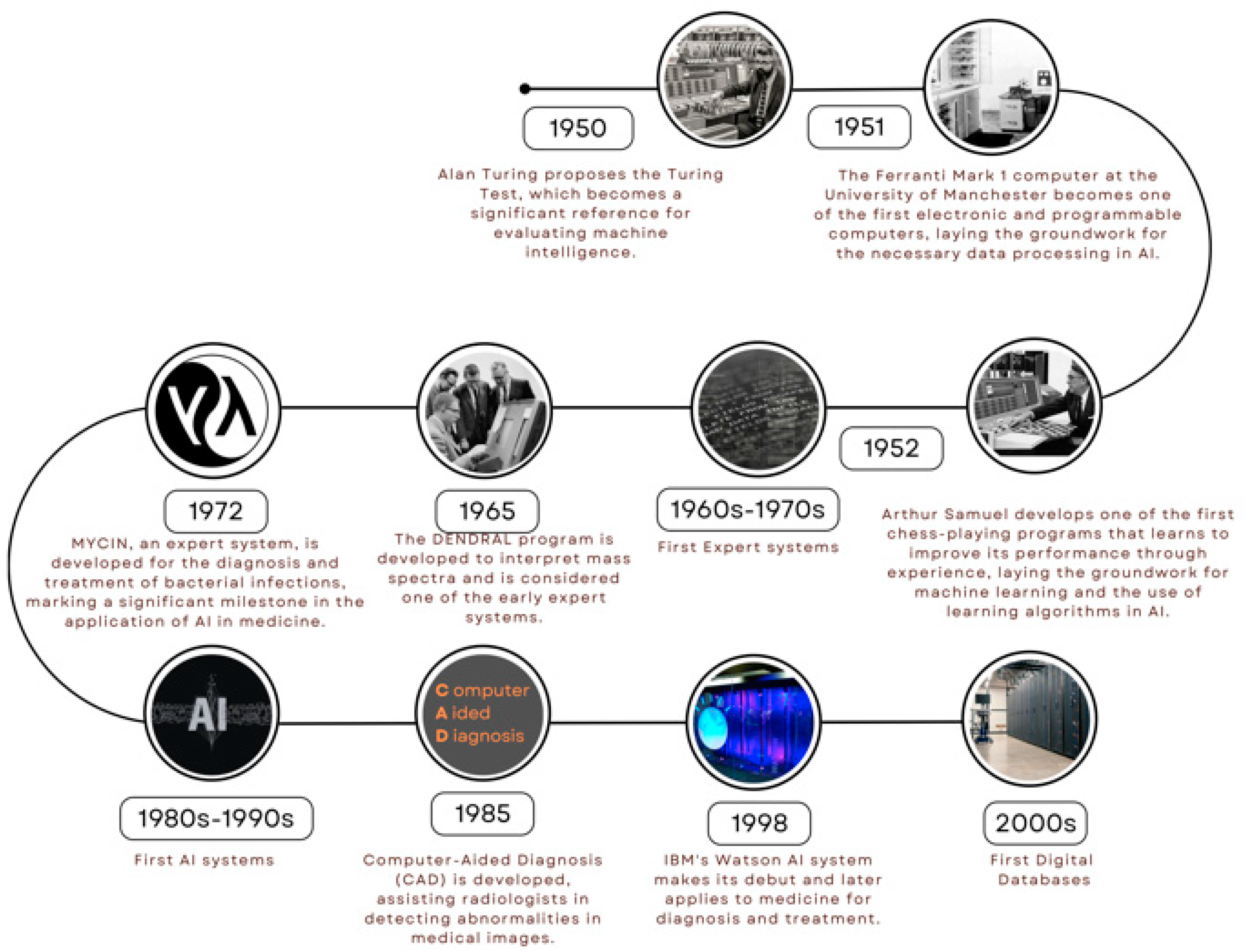
Figure 1B.
The advances of AI in medicine (Cont).
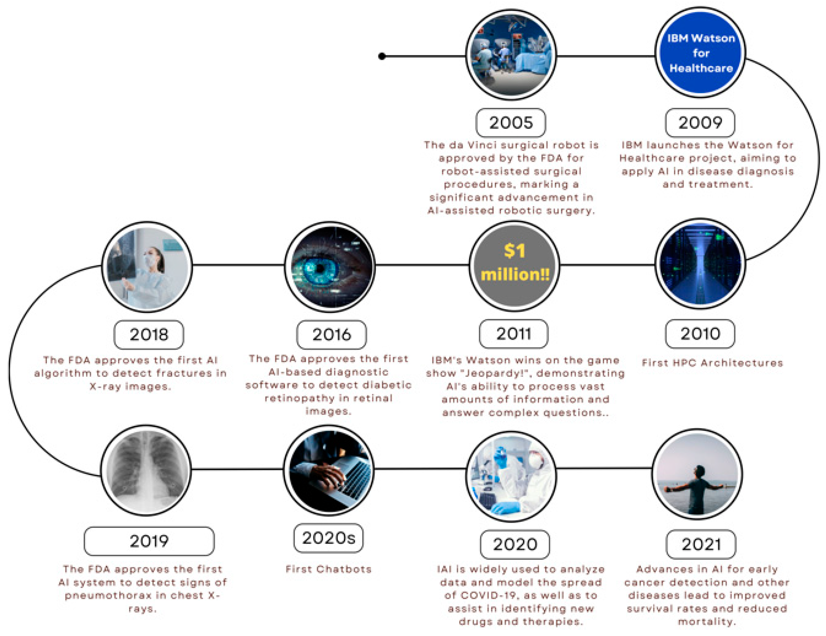
Figure 2.
Areas of medicine that have been impacted by the use of AI.
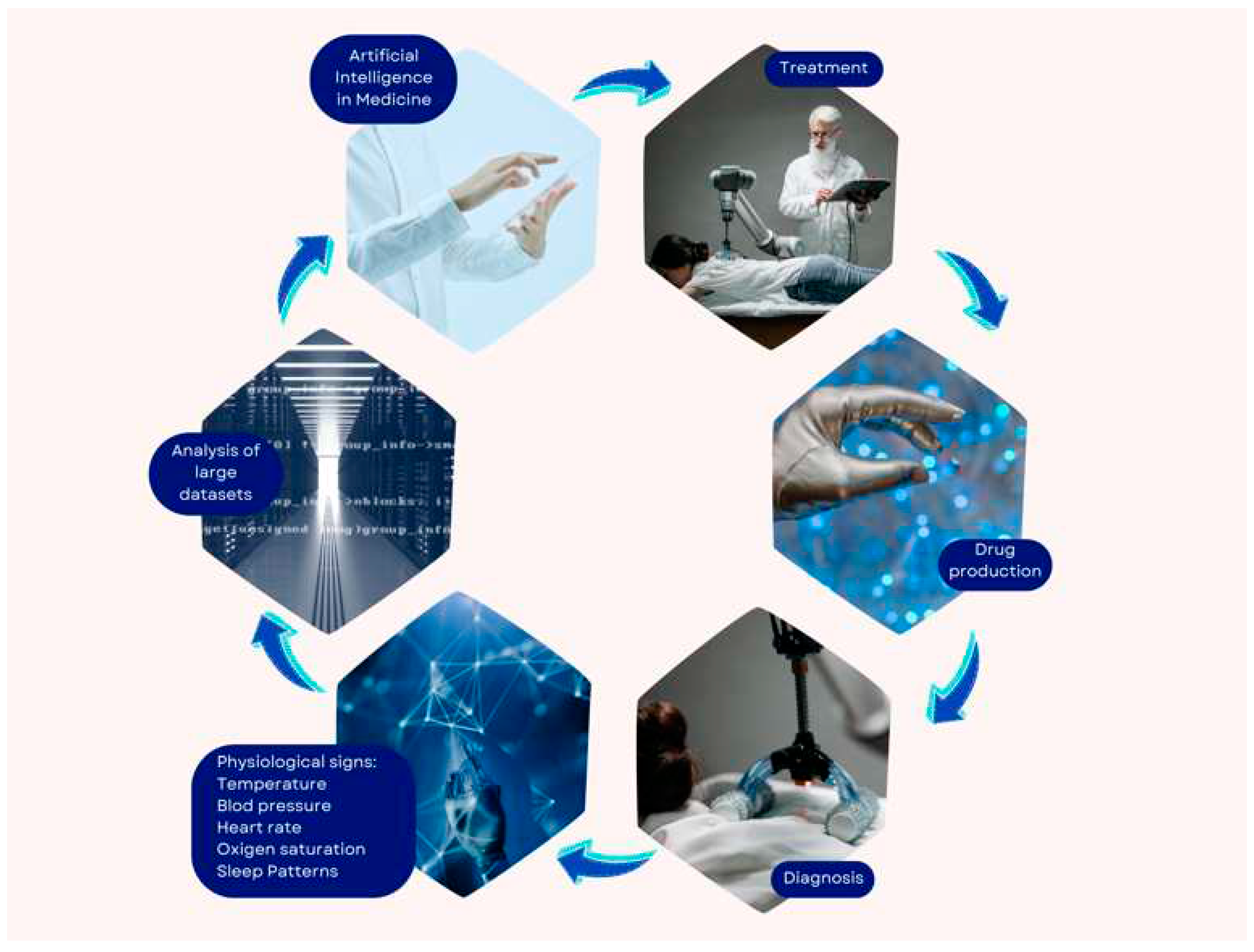

Table 1.
Bibliography relevant to artificial intelligence in Cardiology.
| Bibliography |
| da Silva, H. P., Lourenço, A., Fred, A., & Martins, R. (2014). BIT: Biosignal Igniter Toolkit. Computer methods and programs in biomedicine, 115(1), 20–32. |
| Choi, A., Chung, K., Chung, S. P., Lee, K., Hyun, H., & Kim, J. H. (2022). Advantage of Vital Sign Monitoring Using a Wireless Wearable Device for Predicting Septic Shock in Febrile Patients in the Emergency Department: A Machine Learning-Based Analysis. Sensors (Basel, Switzerland), 22(18), 7054. |
| Alqaraawi, A., Alwosheel, A., & Alasaad, A. (2016). Heart rate variability estimation in photoplethysmography signals using Bayesian learning approach. Healthcare technology letters, 3(2), 136–142. |
| Tzevelekakis, K., Stefanidi, Z., & Margetis, G. (2021). Real-Time Stress Level Feedback from Raw Ecg Signals for Personalised, Context-Aware Applications Using Lightweight Convolutional Neural Network Architectures. Sensors (Basel, Switzerland), 21(23), 7802. |
| Tzevelekakis, K., Stefanidi, Z., & Margetis, G. (2021). Real-Time Stress Level Feedback from Raw Ecg Signals for Personalised, Context-Aware Applications Using Lightweight Convolutional Neural Network Architectures. Sensors (Basel, Switzerland), 21(23), 7802. |
| Sabry, F., Eltaras, T., Labda, W., Alzoubi, K., & Malluhi, Q. (2022). Machine Learning for Healthcare Wearable Devices: The Big Picture. Journal of healthcare engineering, 2022, 4653923. |
| Kwon, K., Kwon, S., & Yeo, W. H. (2022). Automatic and Accurate Sleep Stage Classification via a Convolutional Deep Neural Network and Nanomembrane Electrodes. Biosensors, 12(3), 155. |
| Wang, T., Lu, C., & Shen, G. (2019). Detection of Sleep Apnea from Single-Lead ECG Signal Using a Time Window Artificial Neural Network. BioMed research international, 2019, 9768072. |
| Song, J., Li, J., Zhao, R., & Chu, X. (2023). Developing predictive models for surgical outcomes in patients with degenerative cervical myelopathy: a comparison of statistical and machine learning approaches. The spine journal: official journal of the North American Spine Society, S1529-9430(23)03315-6. |
| Wijsman, J., Grundlehner, B., Liu, H., Hermens, H., & Penders, J. (2011). Towards mental stress detection using wearable physiological sensors. Annual International Conference of the IEEE Engineering in Medicine and Biology Society. IEEE Engineering in Medicine and Biology Society. Annual International Conference, 2011, 1798–1801. |
| Frey, L., Menon, C., & Elgendi, M. (2022). Blood pressure measurement using only a smartphone. NPJ digital medicine, 5(1), 86. |
| Baig, M. M., GholamHosseini, H., Moqeem, A. A., Mirza, F., & Lindén, M. (2017). A Systematic Review of Wearable Patient Monitoring Systems - Current Challenges and Opportunities for Clinical Adoption. Journal of medical systems, 41(7), 115. |
| Chowdhury, M. H., Shuzan, M. N. I., Chowdhury, M. E. H., Mahbub, Z. B., Uddin, M. M., Khandakar, A., & Reaz, M. B. I. (2020). Estimating Blood Pressure from the Photoplethysmogram Signal and Demographic Features Using Machine Learning Techniques. Sensors (Basel, Switzerland), 20(11), 3127. |
| Yin, J., Xu, J., & Ren, T. L. (2023). Recent Progress in Long-Term Sleep Monitoring Technology. Biosensors, 13(3), 395. |
| Parak, J., & Korhonen, I. (2014). Evaluation of wearable consumer heart rate monitors based on photopletysmography. Annual International Conference of the IEEE Engineering in Medicine and Biology Society. IEEE Engineering in Medicine and Biology Society. Annual International Conference, 2014, 3670–3673. |
| Matsubara, T., Sato, K., Hama, K., Tachibana, R., & Uehara, K. (2022). Deep Generative Model Using Unregularized Score for Anomaly Detection with Heterogeneous Complexity. IEEE transactions on cybernetics, 52(6), 5161–5173. |
| Iqbal, T., Elahi, A., Wijns, W., & Shahzad, A. (2022). Exploring Unsupervised Machine Learning Classification Methods for Physiological Stress Detection. Frontiers in medical technology, 4, 782756. |
| Gonzalez-Abreu, A. D., Osornio-Rios, R. A., Elvira-Ortiz, D. A., Jaen-Cuellar, A. Y., Delgado-Prieto, M., & Antonino-Daviu, J. A. (2023). Power Disturbance Monitoring through Techniques for Novelty Detection on Wind Power and Photovoltaic Generation. Sensors (Basel, Switzerland), 23(6), 2908. |
| Sathyanarayana, A., Joty, S., Fernandez-Luque, L., Ofli, F., Srivastava, J., Elmagarmid, A., Arora, T., & Taheri, S. (2016). Sleep Quality Prediction from Wearable Data Using Deep Learning. JMIR mHealth and uHealth, 4(4), e125. |
| Verkruysse, W., Svaasand, L. O., & Nelson, J. S. (2008). Remote plethysmographic imaging using ambient light. Optics express, 16(26), 21434–21445. |
Disclaimer/Publisher’s Note: The statements, opinions and data contained in all publications are solely those of the individual author(s) and contributor(s) and not of MDPI and/or the editor(s). MDPI and/or the editor(s) disclaim responsibility for any injury to people or property resulting from any ideas, methods, instructions or products referred to in the content. |
© 2023 by the authors. Licensee MDPI, Basel, Switzerland. This article is an open access article distributed under the terms and conditions of the Creative Commons Attribution (CC BY) license (http://creativecommons.org/licenses/by/4.0/).
Copyright: This open access article is published under a Creative Commons CC BY 4.0 license, which permit the free download, distribution, and reuse, provided that the author and preprint are cited in any reuse.


