Submitted:
04 August 2023
Posted:
07 August 2023
You are already at the latest version
Abstract
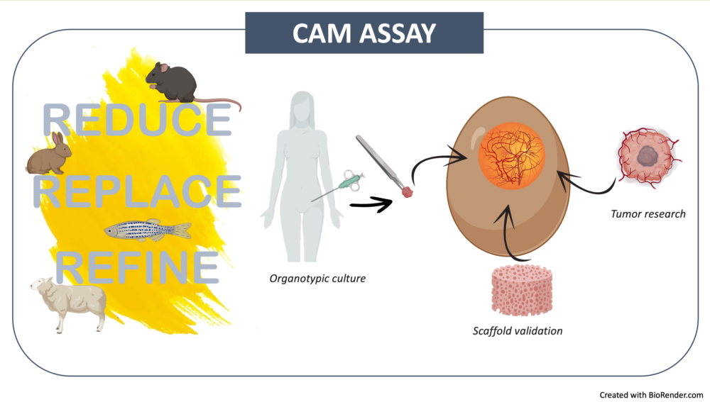
We are witnessing the revival of the CAM model, which was already used in the past by some authors studying angiogenesis and anti-cancer drugs, that now offers a refined model to fill, in the translational meaning, the gap between in vitro-in vivo studies. It can be used for a wide range of purposes, from testing cytotoxicity, pharmacokinetics, tumorigenesis, invasion, to action mechanisms of molecules, and validation of new materials from tissue engineering research. CAM model is easy to use with a fast outcome and makes experimental research more sustainable since allowing to replace, reduce and refine pre-clinical experimentation ("3Rs" rules [1]). This re-view aims to highlight some unique potentials that CAM-assay presents; in particular, the authors intend to use in the next future the CAM model to verify, in a microenvironment comparable to in vivo conditions, albeit simplified, the angiogenic ability of functionalized 3D constructs to be used in regenerative medicine strategies in the recovery of skeletal injuries of critical size (CSD) that do not repair spontaneously. For this purpose, organotypic cultures will be planned on sever-al CAMs set up in temporal sequence: a sort of organ model for assessing CSD conducted in the CAM bioreactor, rather than in vivo.
Keywords:
chorioallantoic Membrane (CAM)
; angiogenesis
; organotypic culture
; engineered 3D scaffold
1. Introduction
The field of clinical research offers significant improvement in the therapeutic strategies which continuously develop to cope with the problems that an aging population imposes. The efficacy and the safety of these strategies need to be evaluated throw an extensive preclinical testing, including animal experimentation, essential for the approval from regulatory office such as Food and Drug Administration (FDA) prior to be applied in clinical approaches. These necessities come in contrast with sustainable research; this is why there is a need for the development of alternative strategies to animal testing [2,3].
In recent years, thanks in part to some funding agencies that are more attentive to animal welfare, there has been much emphasis on projects that include the development of alternatives to animal experimentation, which, to date, appears to be a mandatory and necessary step for bringing new devices/therapies to market, but which, on the other hand, is too expensive and no longer sustainable. Alternatives proposed in the world of basic and clinical research include, for example, tissue cultures [4,5,6], 3D cultures [7,8,9,10], organoids [11,12], microfluidics such as organs-on-a-chip [13,14,15], and also, the use of chicken chorioallantoic membrane [16,17,18,19,20,21,22]. The latter is a technique already developed in the past for angiogenesis assay because it is a richly vascularized, easy-to-use, inexpensive, and of great ethical value. In the last decade, chorioallantoic membrane (CAM) assay has been re-discovered and used not only for the traditional angiogenic assay [23,24] but also, as non-innerved bioreactor and provider of a rapidly growing vascular bed that mimic the blood supply for organ culture [25,26,27,28]. In addition, the embryo is not immunocompetent until 16-17th days of development, therefore cannot sustain rejection reactions [29,30,31]. The versatility of this model has made it widely used in recent years, in fact, this has opened new scenarios to more sustainable, ethical and animal welfare supportive research.
In particular, the field of bone regenerative medicine can benefit, for example, from the use of organ culture models on CAM, inasmuch, a fundamental prerequisite for the formation of new bone is the presence of a well-developed vascular bed that serves as a template for the generation of new bone thanks to the collaboration of the bone forming cells [32,33,34,35,36,37]. The study of proangiogenic potential and associated implications for tissue regeneration require complex in vivo models comprising all steps of the angiogenic process. The CAM model offers a simple, easily accessible and cheap angiogenic screening tool compared to other animal models. In addition, the great ethical value of this in ovo model lies in the application of the 3Rs principle [1]: the possibility of being employed in multiple experiments, for different research fields, replaces the use of in vivo animal models for the experimental phases immediately following the in vitro experimentation (replace); a direct consequence of this is the reduction of the number of animals used during the experimental period (reduce); finally, the use of CAM assay allows to minimize animal suffering since the chick embryo does not exhibit nociception until day 11 of embryonic development (refine) as established by the National Institute of Health [38], as well as the Institutional Animal Care and Use Committee [39]; in addition, pain perception does not fully occur before the 15th day of embryonic development due to immaturity of the portion of the central nervous system devoted to pain perception.
This review provides an overview of the uses of CAM assay in the two last decades and suggests the use of in ovo tests as an alternative to animal testing in the preclinical studies; this could be a good solution to use in the field of regenerative medicine as a model for testing therapies in the resolution of critical bone fractures.
2. The CAM
The chicken embryo’s chorioallantoic membraned (CAM) is a high vascularized extraembryonic structure with function to exchange of gas and nutrients for the embryo during the entire period of its development; further, it is also responsible for calcium mobilization from eggshell to promote embryonic bone mineralization [40,41,42]. It originates by the fusion between the mesodermal layer of the allantois with the adjacent mesodermal layer of the chorion, to form a double layer structure rich in vascular network, connected with the embryonic circulation by two allantoic arteries and one allantoic vein [43]. The mature CAM morphologically resembles a “C” and is permeated by a fluid called allantoic fluid which physiologically provides nutrients and carries waste substances out of the embryo [44,45]. Thanks to its features, CAM is a convenient and versatile biological instrument.
The timing of the embryo development was described by Hamburger and Hamilton in 1951 and it is subdivided in 46 chronological stages (HH stages) by using specific characteristics that occur at each step of the chick’s development regardless of the exact age of the embryo [46]. However, chick developmental time can be difficult to assess because the egg is internally fertilized and goes through a brief period of development; for this, actual incubation days are usually considered for experimental dating assuming that embryonic development begins for all eggs simultaneously at the time of incubation at constant temperature and humidity.
CAM begins its formation at about day 3 of embryonic development (ED3) and reaches completion around ED9, which is precisely why its use requires that tests be set up no earlier than ED 8-9 [47]. All CAM experiments never reach the hatching period of the eggs, which, for chicken, is set at day 21 of embryonic life; however, experimentation can be carried out either in ovo or ex-ovo by latin “inside the egg” or “outside the egg”, respectively. Initially for both methods, fertilized eggs from avian species (i.e. quail, turkey, and duck [48,49,50,51,52,53,54,55]), most commonly chicken, are kept in a humidified incubator at a constant humidity of the 45 % and at a temperature of 37 °C for up to 15 days, by which time experimentation is allowed without the need to seek ethics committee approval. Indeed, as stated earlier, as sanctioned by Institutional Animal Care and Use Committee (IACUC), the chicken embryo is not considered a living organism until the 17th day of embryonic life [38,39].
Within this two-week period, certain operations are performed to make the egg accessible: opening, insertion of materials/molecules to test, final observation and tissue collection [56]. In the case of the ex-ovo technique, at ED3 the eggshell is cracked and its contents placed in a sterile container, several authors have developed various methodologies: eggs in cube [57], use of petri plates [58], use of plastic glass with water [59]; weighing trays [60], etc. In the case of in ovo experimentation, at ED3 the egg can be opened by making a small window access to shell, after aspirating a few ml of albumen from the opposite pole respect to the air chamber, in order to preserve intact the CAM during shell access procedure (Figure 1A). In any methodology chosen, with the opening phase the operator visualizes the viability of the embryo, by verifying the palpitation of the heart and observing the embryonic morphology characterized by the presence of cephalic bud, posterior part, neural tube and the classic "spider" structure of the vessels disposed around the embryo and composed by the main veins and arteries feeding the embryo (Figure 1B); the CAM is planar and can be easily observed as a circled area highly vascularized around the embryo (Figure 1C).
CAM is considered to lack immune-competence until ED18 in fact, the lymphoid cells (i.e., mononuclear phagocytes, T and B cells) are presents from ED11-12 but they are immature, therefore the immune system is not active [62,63,64,65,66,67]. Since the CAM has a very dense capillary network, it is commonly used to study both new vessel formation (angiogenesis versus vasculogenesis) and its inhibition in response to different factors. However, in recent years the use of CAM has been greatly expanded, and there are now many applications.
3. The Application of CAM Up to Now
The vascularized ambient of CAM of avian species offers the possibility to study a variety of molecules and materials; in particular, CAM assay can be used to test pro/anti-angiogenic potential, to perform cancer studies, to test molecules and materials [68,69], to verify transplant reactions, and to test some drug effects.
The clinical research in the field of regenerative medicine typically involves an initial phase of in vitro testing, including test of cytotoxicity, biocompatibility ad others; but later some analyses need to be confirmed in animal model. In this extent, CAM it can be used as an alternative to animal experimentation, since it is comparable to a natural “in ovo“ bioreactor. This makes future research more sustainable and makes it possible to lower the costs of the pre-clinical phase, speed up the preliminary tests needed to get to animal testing (currently mandatory for product entry into the clinical phase).
The following paragraphs provide a brief overview of CAM uses for various experiments conducted in the last decade. Next, we will differentiate the past use of CAM with the perspective uses that can be made of it, such as the set-up, for example, of organotypic culture [70].
3.1. Use of the Cam Assay for Cancer Studies
Jankovic B.D. et al. were among the first to assert that CAM, as a highly vascularized membrane, together with extracellular matrix (ECM) proteins, mimics the physiological environment of cancer [31], so the CAM assay is considered particularly suitable for studying the distinctive aspects of cancer, such as angiogenesis, invasion, metastasis formation, and cancer cell spread [71,72,73,74,75]. Various are the advantages and disadvantages that place the use of CAM versus the use of animal models to study tumors as alternatives.
One advantage that the CAM model has over the animal model in the study of tumor invasion, is the time required for the development of visible microtumors, which in animal models become evident only several weeks after cell transplantation, whereas in the CAM model tumor growth can be observed as early as a few days after cell grafting [76].
The short timing of embryonic development is another benefit of using CAM, because it is possible to speeds up and simplifies data collection in the pre-clinical phase. Indeed, the entire period of embryo development is faster, compared to that of any animal model and this allows to have rapid morphological feedback, for example, on the development of the vascular network in response to different types of grafts. At the same time, however, the difficulty of distinguishing newly formed blood vessels from preexisting ones, is the main cons of using CAM in cancer research. Finally, among the disadvantages of using CAM assay, there is the difficulty of maintaining the system in sterility and avoiding environmental contamination.
For its strengths, which are greater than its advantages, the CAM model is widely used for tumor grafting, which, can be implanted on the membrane in various forms: patient-derived xenografts, solid biopsies, circulating cancer cells in suspension, or most commonly tumor cell lines. Patient-derived xenografts, retains many biological features of primary tumors and, therefore, by grafting them onto CAM it is possible to investigate genetic, protein, morphological and pharmacological patterns, as well as cancer-specific immune evasion mechanisms [76,77,78,79]. By transplanting biopsies of mammalian tumors, it is possible to maintain the main features of primary tumors and to perform studies regarding cell polymorphisms, mechanisms of growth and angiogenesis, interaction with extracellular matrix, and metastasis formation [80,81,82]. Grafting circulating tumor cells is useful to analyze the aggressiveness and proliferation ability of primary tumors, with the aim of performing pre-clinical drugs screening and discovering biomarkers [79].
Table 1 shows some of the several cell lines implanted in CAM, in the last decade.
3.2. Cancer Hallmarks Studied in CAM: Angiogenesis
The CAM model has been widely used in the past to study the hallmarks of cancer, such as angiogenesis, proliferation and tumor invasion, as well as to analyze the conditions underlying cancer therapies [103]. The process of developing a vascular network that supplies nutrients and oxygen to tumor cells is obviously the subject of multiple studies, because growing tumors take advantage of the host’s physiological angiogenesis and promote its exuberant development to secure adequate oxygen and nutrient supply, to dispose of waste products and to facilitate the dissemination of tumor cells to other districts [104].
Tumor onset and progression take place in successive phases, one of which is the avascular phase, during which an "angiogenic switch" can be triggered, resulting in vascular branching and endothelial cell proliferation, allowing the tumor to grow while ensuring a sustained energy supply [105]; in this way, even an initially benign neoplasm can evolve and mutate and vascular proliferation can allow its development beyond its benign dimensions. Deregulation of angiogenesis is also an hallmark of cancer [106] and inhibition of the altered tumor angiogenic process has been introduced as a therapeutic strategy for a long time now strategy [107].
After cell grafting on CAM, tumor become visible within 2-3 days and are readily supplied with CAM-derived blood vessels that penetrate deeply into the tissue. Several qualitative and quantitative approaches have been described to assess the angiogenic response to different types of treatments [72].
Demcisakova Z. et al. have validated angiogenetic potential by immunohistochemistry against embryonic endothelial markers such as WGA (Wheat Germ Agglutinin) and SMA (Smooth Muscle Actin), chicken-specific myofibroblast (α-SMA) and monoclonal antibody specifically recognizes chicken monocytes, macrophages and interdigitating macrophage cells (KUL01); moreover, with RT-PCR it is possible to quantify gene expression of angiogenesis markers such as VEGF (Vascular Endothelial Growth Factor), FGF-2 (Fibroblast Growth Factor-2), ANG-1 (Angiopoietin-1), and HIF-1α (Hypoxia-Inducible Factor 1-alpha) [108].
In other studies, the new vessels formation is quantified through immunohistochemistry to chicken specific CD34 (predominantly regarded as a marker of hematopoietic stem cells and hematopoietic progenitor cells) or using a particular lectin (biotinylated lens culinaris agglutinin) that binds specifically to endothelial cells of chicken veins, arteries, and capillaries. That hybridization was used to assess the angiogenesis that is generated at the intra-tumoral level after grafting osteosarcoma cells onto CAM [86,109]. The tumor supply system has been the object of many studies, in which the use of anti-angiogenic drugs and biomaterials aimed at slowing down the tumor growth process. Some of these anti-angiogenic drugs tested in ovo are molecules that inhibit the VEGF and the Platelet-Derived Growth Factor Receptors (PDGFR) [110,111,112]. Another anti-angiogenic mechanism tested in CAM involves microRNAs (miRNAs) that play a key role in gene expression [113,114]. Among these, microRNA-21 (miR-21) is an oncogenic miRNA, [113] the overexpression of which can downregulate key tumor inhibitory proteins, such as programmed cell death protein [115], TNFα (Tumor Necrosis Factor-α), ERK (Extracellular signal-Regulated Kinase), and VEGF [116]. The miRNA-based therapy can be considered as knockdown of miR-21 expression, induction of tumor cell apoptosis, and suppression of tumor-associated angiogenesis [117,118].
Finally, in a study of Tome Y., another strategy was tested in human osteosarcoma cells transplanted onto CAM, involving the echistatin. That cyclic peptide functions as an anti-angiogenic molecule by bonding to the integrin α v β 3, inhibiting it [119].
3.3. Cancer Hallmarks Studied in CAM: Metastatic Potential
For several years now, the CAM model has been recognized as a viable alternative to animal models for the characterization of tumors and, also for their metastatic potential. [120]. The CAM model also allows for the potential development of metastases in all organs of the chicken embryo [121]. Along with this feature, is also studied the intrinsic aggressiveness of various tumor forms, to have more possible elements in the diagnostic and therapeutic phases. In this context, by supplying the chicken embryo’s circulatory system with blood and nutrients, CAM provides ideal system by recreating the physiological microenvironment for the cell-cell, and cell-matrix interaction studies, that occur during the metastatic cascade [103]. After injection of tumor cells into the circulatory system of the chicken embryo, metastatic potential could be done by tracking of mRNA levels of metastasizing cancer cells in chick embryos and each metastatic site is analyzed from a morphological and invasive potential point of view [122]. Traditional morphological detection techniques used in animal models can also be used in the CAM model. Indeed, to identify cell morphology and their location, tumor samples can be subjected to Hematoxylin & Eosin staining as shown by Shioda and coworkers, who detect colon cancer cells by labeling sections of embryonic organs with the anti-human pan cytokeratin antibody [122,123].
Cell invasiveness, moreover, can be monitored by labeling tumor cells with fluorescent molecules that allow the detection of scattered cell colonies in the various embryonic body districts and, simultaneously, labeling chicken blood vessels with a particular lectin, (biotinylated lens culinaris agglutinin) [124,125]. Ranjan R.A. and his team compared two breast cancer cell lines, MCF-7 and MDA-MB 231, to study growth rates by morphological evaluation, proliferation by immunohistochemistry for Ki-67 protein, aggressiveness by evaluation of mitotic rate and tumor budding, and finally cell spreading by Alu-PCR assay [126].
The latter is a specific in situ hybridization of the repeated sequences in the human genome named Alu, which are present only in humans with a frequency of 5% [127,128]. Tissue sections are subjected to RT-PCR for Alu sequences and CR1 (Chicken Repeat-1), to make a quantitative assessment of the human tumor cells intravasating and disseminating into the chick embryo through the CAM, and distinguish at the same time, chicken cells and vessels [72,86,100,120,121,123,129,130,131]. The looking for human gene sequences by RT-PCR for a determined gene has been a technique used for several years now; in fact, Kobayashi and coworkers, many years ago identified metastatic prostate cancer cells disseminated in the liver and femur of the chicken embryo by amplification of the human beta globin gene [132].
3.4. Tumor Therapy Test in CAM
The CAM model is a versatile, yet also relatively simple, and low-cost model that also allows screening of pharmacological or physical therapies in a short time. Moreover, the use of the CAM model can be considered a precision medicine tool that is being used in the search for tailored cancer drugs [133]. Drugs that inhibit tumor growth have been tested in CAM mainly in two ways: by injecting them into the chicken’s circulatory system or by using them as a treatment of tumor cells, seeded appropriately on CAM, as reported in the protocol developed by Kunz and his team [86].
Therefor the CAM assay is a reference model for several therapeutic approaches, including various chemotherapeutics [89,100], targeted [134,135,136] and checkpoint therapies [137], oncolytic viruses [138], radiotherapy [139], molecules that block the cell cycle and induce apoptosis [140,141] and anti-angiogenesis drugs [142,143].
4. Use of the CAM Assay to Validate Scaffold for Regenerative Purposes
Regenerative medicine, in recent years, is progressing toward new translational approaches based on the formulation and fabrication of Advanced Therapy Medical Products (ATMPs). It is therefore tissue engineering (TE), a branch of research that aims to produce constructs that are the result of a combination of cells, biomaterials, and biologically active molecules, in the form of scaffolds, with the aim of repairing tissues by inducing their regeneration [144,145,146,147,148,149,150,151]. TE can be ex vivo or in situ [152,153]: the first approach requires seeding of donor stem cells onto a scaffold that is inserted into the affected tissue for the purpose of stimulating cell growth and differentiation [154,155,156,157]; the in situ one, on the other hand, avoids the step of seeding cells onto the scaffold and involves the fabrication of scaffolds that can adapt to tissue damage in size and shape. The latter contain biocompatible materials that can be implanted at the site of damaged tissue, where they attract the surrounding host cells necessary for healing to the repair site [150,152,153,158,159]. Specifically, among the components of TE constructs emerge biomaterials which hold many key characteristics for in vivo implantation into host tissues. These include: biocompatibility to avoid the induction of an immune response, sterilizability to be safely incorporated into host tissues, biodegradability to be degraded by tissue cells into easily metabolized molecules from the tissue after performing their function, and bioactivity to stimulate tissue repair. Regardless of biochemical composition and biophysical properties, their most important feature is the interaction with the biological system in which they are embedded [160,161].
Biomaterials can be classified according to their origin. There are those of natural origin such as chitosan, alginate, and cellulose [162,163] or those of synthetic origin such as PLGA poly (lactic-co-glycolic acid), PCL (lactic-co-glycolic acid), PLA (polylactic acid), fibronectine, and polyurethane [163,164].
In this context, once again, the CAM model represents a natural bioreactor to test the main characteristics of biomaterials and the effects they have on the CAM, that represents a viable system. In recent years, the CAM assay become a popular approach in tissue engineering studies, in particular, in the study of different tissue pathologies, such as those related to bone defects [66,165]. The chorioallantoic membrane allows observation of the effect that biomaterials have on the angiogenesis and test their biocompatibility. Considering the central role that angiogenesis plays to tissue regeneration, the evaluation of angiogenic potential of biomaterials has become a priority in TE especially for bone TE [166,167,168,169,170].
Angiogenic potential and biocompatibility of several biomaterials have been already tested in CAM. Many of these biomaterials tested in CAM are of synthetic origin; among them hydrogels, which mimic extracellular matrix materials (ECM) due to their highly hydrated, permeable and porous structures. They enable guided tissue regeneration by facilitating cellular activities, nutrient diffusion, and waste transfer [171]. The swelling and degradation ability of polymer matrix hydrogels makes them suitable for encapsulating and delivering numerous therapeutic agents, such as cells, growth factors, drugs and genes into tissue defects [172,173]. In addition, hydrogels are very often enriched with other molecules such as, for example, acrylamine [174] heparin [175] and hyaluronic acid [176]. The latter designed to treat periodontitis, a chronic biofilm-associated inflammatory disease of the tooth-supporting tissues that causes tooth loss. The scaffold developed by the team, based on controlled oxygen-releasing hyaluronic acid, is useful to avoid a hypoxic environment that would compromise tissue regeneration [177,178]. Other biomaterials tested in CAM include bioplastics, which are eco-friendly materials used in bone tissue regeneration for their biocompatibility and biodegradability; specifically, poly (3-hydroxybutyrate-co-3-hydroxyhexanoate) (PHBH) reinforced witch cellulose nanocrystals (CNCs) is tested in CAM in the form of porous scaffold. The CAM assay enabled the identification of the scaffold pore size, which is more optimal for endothelial cell colonization and blood vessel formation [69]. Bioactive glasses, in the TE field, have also received a lot of interest. The latter enriched with biologically active ions of various kinds, such as boron, are the focus of research by Decker and coworkers. They observe the influence of B-doping of bioactive glasses on the viability, osteogenic differentiation, and expression of osteogenic and angiogenic marker genes of bone marrow-derived mesenchymal stromal cells (BMSCs), in presence of the B-BGs’ ionic dissolution products (IDPs); subsequently they evaluated the influence of IDPs, on chorioallanotic membrane angiogenesis [179]. In the same way as bioglass, synthetic hydroxyapatite (HA), which is a particular type of calcium phosphate, has been widely examined as a regeneration material because of its affinity to the main natural component of bone and its osteoconductivity and bioactivity [180,181,182,183,184]. In this regard, HA formulated with other biomaterials, such as biopolymers, demonstrates remarkable vasculogenesis, as evident from CAM testing, in recent studies focused on finding viable regenerative strategies for the orbital floor [164]. Other biopolymers also fit into this context: Demsisakova et al. have developed a scaffold consisting of the biopolymer polyhydroxybutyrate (PHB) combined with chitosan (CHIT). Also, using the CAM assay, they demonstrate that (PHB)/(CHIT) has a strong endogenous angiogenic potential and could be promising biomaterial for the treatment of hard tissue defects [108]. The most significant advantage of using CAM in studying the properties of biomaterials is that the CAM allows the development and branching of the vascular network on the implanted scaffolds, mimicking what should occur in a tissue in vivo. In this regard, the porosity and pore size of the scaffold, play a key role in vascular infiltration and osteogenic differentiation [185,186,187]; therefore, the challenge for researchers seems to be to formulate ever new constructs that have better and better porosity and efficiency in TE.
5. Use of CAM to Set-Up Organotypic Culture
All the advantages of CAM make it an attractive model also for tissue engineering approaches. The membrane, during the developmental stages of the chick embryo, provides a naturally immunocompromised host, a rapidly growing vascular bed that lacks a nervous system and, therefore, gives a less sentient alternative for animal research, allowing in vivo implantation of organ, and represents a model for xenografts, developing organotypic cultures [73,188]. That gives a solution at the most important limitation of the ex vivo organotypic culture: the lack of blood, immunity cells and bone cells [189,190,191].
Given that, especially in the field of bone tissue engineering, it is possible to use both the embryo and the membrane itself, Blake et al., at the 18th day of embryo development, harvested the femur of the embryo, which they then implanted on the CAM after causing a fracture [189].
As shown by Aldamash A. et al (2017) and Marshall et. Al (2020) the use of the chicken embryo femur also has another application; the works of these two researcher groups had the aim to examine the differentiation potential of specific cells, such as human bone marrow stromal cells (HBMSCs), human neonatal foreskin stromal cells (hNSSCs) alone or in combination with human umbilical vein endothelial cells (HUVECs), under experimental conditions for tissue regeneration. The authors take the chicken embryo femur, cause a fracture, and culture it with different cell types to test their differentiation potential and, in the case of hNSSCs and HUVECs, also their angiogenic potential. [192,193].
Although the CAM model per se allows for short experimental times (from day x to day y), it is also possible to overcome this time limit by performing transplantation from one chorion allantoic membrane to another ex vivo of organotypic cultures or biomaterials to be tested on CAM; of course, in case of the need for increased experimental times one must be careful not to damage the grafted samples/scaffolds in transport from one CAM to the other. In a recent study, Feder et al. set up a protocol whereby it is possible to graft onto the CAM various sections of osteosarcoma tissue, taken by rats and mices; then, they transplanted them on another membrane for five to seven consecutive times, enabling further experimental analyses [188].
Another insight about CAM’s applications in this field is provided by Kanczler and his team, who devise a critically - sized chick femur defect model, which has been used to evaluate different types of molecules engaged in bone tissue engineering. Specifically, CAM is used to test the effects of different factors and proteins involved in the healing bone defect, such as Bone Morphogenetic Protein-2 (BMP-2), vitamin D3, Parathyroid Hormone (PTH) and Parathyroid-Hormone-Related Protein (PTHRP), to assess the potential of osseointegration of scaffolds, and to evaluate their performance, before using them in in vivo studies [189,190,191].
Other studies report how CAM can also be used for transplanting sections of organs or organoids from murine embryos, such as for example the kidney. Embryonic kidneys are taken at ED11.5, transplanted onto CAM at ED8 and then cultivated for 7 days; subsequently, the murine embryonic kidney, grown on CAM, undergoes immunohistochemistry for endothelial growth markers, which highlight the anastomosis between the blood vessels of CAM and those of the murine kidney [192].
6. Discussion and Conclusions
As above described, there is ample evidence that the use of CAM in numerous research areas is effective for research and clinical studies. A positive fact is that CAM model, inspired by the 3R concept, is a viable alternative to the classical animal experimentation, which is no longer sustainable without ethical limitations/controls and which, in the future, will have to be replaced with (or must be accompanied by) alternatives that are more advantageous in economic, ethical and experimental terms. It was already pointed out that research in the field of oncology and that into biomaterials for regenerative medicine are highly developed areas, in which more resources have been invested in the development of techniques and strategies for the use of CAM corresponding to the demands of research. In addition, other applications of CAM are cautiously being developed in order to use this powerful vascularized natural bioreactor for the accomplishment of organ cultures, which could replace the early stages of experimentation, currently performed exclusively on animal models, thus decreasing the negative impact of research on animal welfare.
This review has highlighted the most significant scientific studies from the last decade precisely to underline the current importance of this alternative model to animal experimentation. Finally, we would like to point out that the interest in the use of CAM has recently materialized with the 1st International Conference on CAM held in February 2022, which brought together highly prestigious scholars from all over the world.
7. Future Directions
In the future, it is to be hoped that even more standardized techniques will be developed for setting up CAM testing services for large research macro-areas in order to meet all scientific demands. In addition, more publicity should be done to this alternative model to in vivo testing for all the advantages and properties described above. The possibility of setting up organ cultures on CAM is attractive, useful and innovative. At present, the use of this tool is not widespread and the development of methodologies for the use of CAM for organ cultures is slow. In the coming years, it is hoped that more and more research groups will devote themselves to the implementation of this organ culture model in order to test molecules and clinical strategies in a translational perspective.
Author Contributions
writing—original draft preparation, M.C. and F.S.; review, editing and supervision, C.P. All authors have read and agreed to the published version of the manuscript.”
Funding
this research received no external funding
Acknowledgments
Thanks are due to Prof. Alberto Martelli of the University of Bologna for providing the BioRender software.
Conflicts of Interest
The authors declare no conflict of interest.
References
- Russell, W.M.S.; Burch, R.L. The Principles of Humane Experimental Technique; Methuen, Ed.; London, 1959.
- Hippenstiel, S.; Thöne-Reineke, C.; Kurreck, J. Animal Experiments: EU Is Pushing to Find Substitutes Fast. Nature 2021, 600, 37. [Google Scholar] [CrossRef] [PubMed]
- Fentem, J.; Malcomber, I.; Maxwell, G.; Westmoreland, C. Upholding the EU’s Commitment to ‘Animal Testing as a Last Resort’’ Under REACH Requires a Paradigm Shift in How We Assess Chemical Safety to Close the Gap Between Regulatory Testing and Modern Safety Science. ATLA Alternatives to Laboratory Animals 2021, 49, 122–132. [Google Scholar] [CrossRef]
- Aguilar, A.H.; Smith, L.; Owens, D.; Quelch, R.; Przyborski, S. Recreating Tissue Structures Representative of Teratomas In Vitro Using a Combination of 3D Cell Culture Technology and Human Embryonic Stem Cells. Bioengineering 2022, 9. [Google Scholar] [CrossRef]
- Bédard, P.; Gauvin, S.; Ferland, K.; Caneparo, C.; Pellerin, È.; Chabaud, S.; Bolduc, S. Bioengineering Innovative Human Three-Dimensional Tissue-Engineered Models as an Alternative to Animal Testing. 2020. [Google Scholar] [CrossRef]
- Darling, N.J.; Mobbs, C.L.; González-Hau, A.L.; Freer, M.; Przyborski, S. Bioengineering Novel in Vitro Co-Culture Models That Represent the Human Intestinal Mucosa With Improved Caco-2 Structure and Barrier Function. Front Bioeng Biotechnol 2020, 8. [Google Scholar] [CrossRef] [PubMed]
- Knight, E.; Murray, B.; Carnachan, R.; Przyborski, S. Alvetex®: Polystyrene Scaffold Technology for Routine Three Dimensional Cell Culture. In Methods in Molecular Biology; Humana Press Inc., 2011; Vol. 695, pp. 323–340. [Google Scholar]
- Golinelli, G.; Talami, R.; Frabetti, S.; Candini, O.; Grisendi, G.; Spano, C.; Chiavelli, C.; Arnaud, G.F.; Mari, G.; Dominici, M. A 3D Platform to Investigate Dynamic Cell-to-Cell Interactions Between Tumor Cells and Mesenchymal Progenitors. Front Cell Dev Biol 2022, 9. [Google Scholar] [CrossRef]
- Flagelli, A.; Candini, O.; Frabetti, S.; Dominici, M.; Giardino, L.; Calzà, L.; Baldassarro, V.A. A Novel Three-Dimensional Culture Device Favors a Myelinating Morphology of Neural Stem Cell-Derived Oligodendrocytes. Front Cell Dev Biol 2021, 9. [Google Scholar] [CrossRef]
- Shahin-Shamsabadi, A.; Selvaganapathy, P.R. Tissue-in-a-Tube: Three-Dimensional in Vitro Tissue Constructs with Integrated Multimodal Environmental Stimulation. Mater Today Bio 2020, 7. [Google Scholar] [CrossRef]
- Mukhopadhyay, C.; Paul, M.K. Organoid-Based 3D in Vitro Microphysiological Systems as Alternatives to Animal Experimentation for Preclinical and Clinical Research. Arch Toxicol 2023. [Google Scholar] [CrossRef]
- Puschhof, J.; Pleguezuelos-Manzano, C.; Clevers, H. Organoids and Organs-on-Chips: Insights into Human Gut-Microbe Interactions. Cell Host Microbe 2021, 29, 867–878. [Google Scholar] [CrossRef]
- Low, L.A.; Mummery, C.; Berridge, B.R.; Austin, C.P.; Tagle, D.A. Organs-on-Chips: Into the next Decade. Nat Rev Drug Discov 2021, 20, 345–361. [Google Scholar] [CrossRef]
- Tao, T.; Wang, Y.; Chen, W.; Li, Z.; Su, W.; Guo, Y.; Deng, P.; Qin, J. Engineering Human Islet Organoids from IPSCs Using an Organ-on-Chip Platform. Lab Chip 2019, 19, 948–958. [Google Scholar] [CrossRef]
- Manafi, N.; Shokri, F.; Achberger, K.; Hirayama, M.; Mohammadi, M.H.; Noorizadeh, F.; Hong, J.; Liebau, S.; Tsuji, T.; Quinn, P.M.J.; et al. Organoids and Organ Chips in Ophthalmology. Ocular Surface 2021, 19, 1–15. [Google Scholar] [CrossRef] [PubMed]
- Ribatti, D. Two New Applications in the Study of Angiogenesis the CAM Assay: Acellular Scaffolds and Organoids. Microvasc Res 2022, 140. [Google Scholar] [CrossRef] [PubMed]
- Chen, L.; Wang, S.; Feng, Y.; Zhang, J.; Du, Y.; Zhang, J.; Van Ongeval, C.; Ni, Y.; Li, Y. Cells Utilisation of Chick Embryo Chorioallantoic Membrane as a Model Platform for Imaging-Navigated Biomedical Research. 2021. [Google Scholar] [CrossRef]
- Fonseca, B.B.; da Silva, M.V.; de Morais Ribeiro, L.N. The Chicken Embryo as an in Vivo Experimental Model for Drug Testing: Advantages and Limitations. Lab Anim (NY) 2021, 50, 138–139. [Google Scholar] [CrossRef]
- Dadhich, P.; Das, B.; Pal, P.; Srivas, P.K.; Dutta, J.; Ray, S.; Dhara, S. A Simple Approach for an Eggshell-Based 3D-Printed Osteoinductive Multiphasic Calcium Phosphate Scaffold. ACS Appl Mater Interfaces 2016, 8, 11910–11924. [Google Scholar] [CrossRef]
- Burgio, F.; Rimmer, N.; Pieles, U.; Buschmann, J.; Beaufils-Hugot, M. Characterization and in Ovo Vascularization of a 3D-Printed Hydroxyapatite Scaffold with Different Extracellular Matrix Coatings under Perfusion Culture. Biol Open 2018, 7. [Google Scholar] [CrossRef]
- Baiguera, S.; Macchiarini, P.; Ribatti, D. Chorioallantoic Membrane for in Vivo Investigation of Tissue-Engineered Construct Biocompatibility. J Biomed Mater Res B Appl Biomater 2012, 100 B, 1425–1434. [Google Scholar] [CrossRef]
- Yalcin, H.C.; Shekhar, A.; Rane, A.A.; Butcher, J.T. An Ex-Ovo Chicken Embryo Culture System Suitable for Imaging and Microsurgery Applications. Journal of Visualized Experiments 2010. [Google Scholar] [CrossRef]
- Moreno-Jiménez, I.; Kanczler, J.M.; Hulsart-Billstrom, G.; Inglis, S.; Oreffo, R.O.C. The Chorioallantoic Membrane Assay for Biomaterial Testing in Tissue Engineering: A Short-Term in Vivo Preclinical Model. Tissue Eng Part C Methods 2017, 23, 938–952. [Google Scholar] [CrossRef] [PubMed]
- Ribatti, D.; Nico, B.; Vacca, A.; Roncali, L.; Burri, P.H.; Djonov, V. Chorioallantoic Membrane Capillary Bed: A Useful Target for Studying Angiogenesis and Anti-Angiogenesis in Vivo. Anatomical Record 2001, 264, 317–324. [Google Scholar] [CrossRef] [PubMed]
- Isachenko, V.; Mallmann, P.; Petrunkina, A.M.; Rahimi, G.; Nawroth, F.; Hancke, K.; Felberbaum, R.; Genze, F.; Damjanoski, I.; Isachenko, E. Comparison of in Vitro- and Chorioallantoic Membrane (CAM)-Culture Systems for Cryopreserved Medulla-Contained Human Ovarian Tissue. PLoS One 2012, 7. [Google Scholar] [CrossRef] [PubMed]
- Moreno-Jiménez, I.; Lanham, S.A.; Kanczler, J.M.; Hulsart-Billstrom, G.; Evans, N.D.; Oreffo, R.O.C. Remodelling of Human Bone on the Chorioallantoic Membrane of the Chicken Egg: De Novo Bone Formation and Resorption. J Tissue Eng Regen Med 2018, 12, 1877–1890. [Google Scholar] [CrossRef]
- Fazely, F.; Moses, D.C.; Ledinko, N. Effects of Retinoids on Invasion of Organ Cultures of Chick Chorioallantoic Membrane by Adenovirus Transformed Cells. In Vitro Cellular & Developmental Biology 1985, 21, 409–414. [Google Scholar]
- Martinez-Madrid, B.; Donnez, J.; Van Eyck, A.S.; Veiga-Lopez, A.; Dolmans, M.M.; Van Langendonckt, A. Chick Embryo Chorioallantoic Membrane (CAM) Model: A Useful Tool to Study Short-Term Transplantation of Cryopreserved Human Ovarian Tissue. Fertil Steril 2009, 91, 285–292. [Google Scholar] [CrossRef]
- Leene, W.; Duyzings, M.J.M.; Van Steeg, C. Lymphoid Stem Cell Identification in the Developing Thymus and Bursa of Fabricius of the Chick. Z. Zellforsch 1973, 136, 521–533. [Google Scholar] [CrossRef]
- Ribatti, D. The Chick Embryo Chorioallantoic Membrane in the Study of Angiogenesis and Metastasis; Springer, Ed.; 2010; ISBN 978-90-481-3843-2. [Google Scholar]
- Jankovic, B.D.; Isakovic, K.; Lukic, M.L.; Vujanovic, N.L.; Petrovic, S.; Markovic, B.M. Immunological Capacity of the Chicken Embryo. I. Relationship between the Maturation of Lymphoid Tissues and the Occurrence of Cell-Mediated Immunity in the Developing Chicken Embryo. Immunology 1975, 29, 497–508. [Google Scholar]
- Genova, T.; Petrillo, S.; Zicola, E.; Roato, I.; Ferracini, R.; Tolosano, E.; Altruda, F.; Carossa, S.; Mussano, F.; Munaron, L. The Crosstalk between Osteodifferentiating Stem Cells and Endothelial Cells Promotes Angiogenesis and Bone Formation. Front Physiol 2019, 10, 1–11. [Google Scholar] [CrossRef]
- Kanczler, J.M.; Oreffo, R.O.C. Osteogenesis and Angiogenesis: The Potential for Engineering Bone. Eur Cell Mater 2008, 15, 100–114. [Google Scholar] [CrossRef]
- Portal-Núñez, S.; Lozano, D.; Esbrit, P. Role of Angiogenesis on Bone Formation. Histol Histopathol 2012, 27, 559–566. [Google Scholar] [CrossRef] [PubMed]
- Checchi, M.; Stanzani, V.; Truocchio, S.; Corradini, M.; Ferretti, M.; Palumbo, C. From Morphological Basic Research to Proposals for Regenerative Medicine through a Translational Perspective. Italian Journal of Anatomy and Embryology 2022, 126, 139–145. [Google Scholar] [CrossRef]
- Palumbo, C.; Cavani, F.; Sena, P.; Benincasa, M.; Ferretti, M. Osteocyte Apoptosis and Absence of Bone Remodeling in Human Auditory Ossicles and Scleral Ossicles of Lower Vertebrates: A Mere Coincidence or Linked Processes? Calcif Tissue Int 2012, 90, 211–218. [Google Scholar] [CrossRef] [PubMed]
- Ferretti, M.; Palumbo, C. Static Osteogenesis versus Dynamic Osteogenesis: A Comparison between Two Different Types of Bone Formation. Applied Sciences 2021, 11, 2025. [Google Scholar] [CrossRef]
- National Institutes of Health The Public Health Service Responds to Commonly Asked Questions. ILAR J 1991, 33, 68–70. [CrossRef]
- Institutional Animal Care and Use Committee (IACUC). Policy for Use of Avian Embryos; 2019. [Google Scholar]
- Elaroussi, M.A.; DeLuca, H.F. Calcium Uptake by Chorioallantoic Membrane: Effects of Vitamins D and K. Endocrinol. Metab 1994, 267, E837–E841. [Google Scholar] [CrossRef]
- Tuan, R.; Ono, T. Regulation of Extraembryonic Calcium Mobilization by the Developing Chick Embryo. J Embryol Exp Morphol. 1986, 97, 63–74. [Google Scholar] [PubMed]
- Packard, M.J. Mobilization of Shell Calcium by Chick Chorioallantoic Membrane in Vitro. J. exp. Biol 1994, 190, 141–153. [Google Scholar]
- Maibier, M.; Reglin, B.; Nitzsche, B.; Xiang, W.; Rong, W.W.; Hoffmann, B.; Djonov, V.; Secomb, T.W.; Pries, A.R. Structure and Hemodynamics of Vascular Networks in the Chorioallantoic Membrane of the Chicken. Am J Physiol Heart Circ Physiol 2016, 311, H913–H926. [Google Scholar] [CrossRef]
- Li, Y.; Qu, H.; Ji, J.; Wang, Y.; Liu, T.; He, J.; Wang, J.; Shu, D.; Luo, C. Characterization of the Exosomes in the Allantoic Fluid of the Chicken Embryo. Can J Anim Sci 2021, 101, 307–317. [Google Scholar] [CrossRef]
- Da Silva, M.; Labas, V.; Nys, Y.; Rehault-Godbert, S. Investigating Proteins and Proteases Composing Amniotic and Allantoic Fluids during Chicken Embryonic Development. Poult Sci 2017, 96, 2931–2941. [Google Scholar] [CrossRef]
- Hamburger, V.; Hamilton, H.L. A Series of Normal Stages in the Development of the Chick Embryo. J Morphol 1951, 88, 49–92. [Google Scholar] [CrossRef]
- Crespo, P.; Casar, B. The Chick Embryo Chorioallantoic Membrane as an in Vivo Model to Study Metastasis. Bio Protoc 2016, 6. [Google Scholar] [CrossRef]
- Lazarovici, P.; Lahiani, A.; Gincberg, G.; Haham, D.; Marcinkiewicz, C.; Lelkes, P.I. Nerve Growth Factor-Induced Angiogenesis: 2. The Quail Chorioallantoic Membrane Assay. In Neurotrophic Factors: Methods and Protocols; Skaper, S.D., Ed.; Springer New York: New York, NY, 2018; Vol. 1727, pp. 251–259. ISBN 978-1-4939-7571-6. [Google Scholar]
- Parsons-Wingerter, P.; Lwai, B.; Che Yang, M.; Elliott, K.E.; Milaninia, A.; Redlitz, A.; Clark, J.I.; Helene Sage, E. A Novel Assay of Angiogenesis in the Quail Chorioallantoic Membrane: Stimulation by BFGF and Inhibition by Angiostatin According to Fractal Dimension and Grid Intersection; 1998; Vol. 55. [Google Scholar]
- Kundeková, B.; Máčajová, M.; Meta, M.; Čavarga, I.; Bilčík, B. Chorioallantoic Membrane Models of Various Avian Species: Differences and Applications. Biology (Basel) 2021, 10. [Google Scholar]
- Rasmussen, S. V.; Berlow, N.E.; Price, L.H.; Mansoor, A.; Cairo, S.; Rugonyi, S.; Keller, C. Preclinical Therapeutics Ex Ovo Quail Eggs as a Biomimetic Automation-Ready Xenograft Platform. Sci Rep 2021, 11. [Google Scholar] [CrossRef]
- Lusimbo, W.S.; Leighton, F.A.; Wobeser, G.A. Histology and Ultrastructure of the Chorioallantoic Membrane of the Mallard Duck (Anas Platyrhynchos). Anatomical Record 2000, 259, 25–34. [Google Scholar] [CrossRef]
- Kundeková, B.; Máčajová, M.; Meta, M.; Čavarga, I.; Bilčík, B. Chorioallantoic Membrane Models of Various Avian Species: Differences and Applications. Biology (Basel) 2021, 10. [Google Scholar] [CrossRef]
- Buhr, C.R.; Wiesmann, N.; Tanner, R.C.; Brieger, J.; Eckrich, J. The Chorioallantoic Membrane Assay in Nanotoxicological Research - an Alternative for in Vivo Experimentation. Nanomaterials 2020, 10, 1–16. [Google Scholar] [CrossRef]
- Longenecker, B.M.; Pazderka, F.; Stone, H.S.; Gavora, J.S.; Ruth, R.F. In Ovo Assay for Marek’s Disease Virus and Turkey Herpesvirus. 1975, 11, 922–931. [Google Scholar] [CrossRef]
- Janser, F.; Ney, P.; Pinto, M.; Langer, R.; Tschan, M. The Chick Chorioallantoic Membrane (CAM) Assay as a Three-Dimensional Model to Study Autophagy in Cancer Cells. Bio Protoc 2019, 9. [Google Scholar] [CrossRef]
- Huang, W.; Arai, F.; Kawahara, T. Egg-in-Cube: Design and Fabrication of a Novel Artificial Eggshell with Functionalized Surface. PLoS One 2015, 10. [Google Scholar] [CrossRef] [PubMed]
- Dohle, D.S.; Pasa, S.D.; Gustmann, S.; Laub, M.; Wissler, J.H.; Jennissen, H.P.; Dünker, N. Chick Ex Ovo Culture and Ex Ovo CAM Assay: How It Really Works. Journal of Visualized Experiments 2010. [Google Scholar] [CrossRef]
- García-Gareta, E.; Binkowska, J.; Kohli, N.; Sharma, V. Towards the Development of a Novel Ex Ovo Model of Infection to Pre-Screen Biomaterials Intended for Treating Chronic Wounds. J Funct Biomater 2020, 11. [Google Scholar] [CrossRef] [PubMed]
- Winter, R.; Dungel, P.; Reischies, F.M.J.; Rohringer, S.; Slezak, P.; Smolle, C.; Spendel, S.; Kamolz, L.P.; Ghaffari-Tabrizi-Wizsy, N.; Schicho, K. Photobiomodulation (PBM) Promotes Angiogenesis in-Vitro and in Chick Embryo Chorioallantoic Membrane Model. Sci Rep 2018, 8. [Google Scholar] [CrossRef]
- Consen Egg Anatomy. Available online: https://www.theperfectegg.net/the-onsen-egg-temperature-curve/ (accessed on 30 June 2023).
- Gómez Del Moral, M.; Fonfría, J.; Varas, A.; Jiménez, E.; Moreno, J.; Zapata, A.G. Appearance and Development of Lymphoid Cells in the Chicken (Gallus Gallus) Caecal Tonsil. Anatomical Record 1998, 250, 182–189. [Google Scholar] [CrossRef]
- Kunzi-Rapp, K.; Rück, A.; Kaufmann, R. Characterization of the Chick Chorioallantoic Membrane Model as a Short-Term in Vivo System for Human Skin. Arch Dermatol Res 1999, 291, 290–295. [Google Scholar] [CrossRef] [PubMed]
- Ribatti, D.; Nico, B.; Vacca, A.; Presta, M. The Gelatin Sponge-Chorioallantoic Membrane Assay. Nat Protoc 2006, 1, 85–91. [Google Scholar] [CrossRef] [PubMed]
- Nowak-Sliwinska, P.; Segura, T.; Iruela-Arispe, M.L. The Chicken Chorioallantoic Membrane Model in Biology, Medicine and Bioengineering. Angiogenesis 2014, 17, 779–804. [Google Scholar] [CrossRef]
- Moreno-Jiménez, I.; Hulsart-Billstrom, G.; Lanham, S.A.; Janeczek, A.A.; Kontouli, N.; Kanczler, J.M.; Evans, N.D.; Oreffo, R.O.C. The Chorioallantoic Membrane (CAM) Assay for the Study of Human Bone Regeneration: A Refinement Animal Model for Tissue Engineering. Sci Rep 2016, 6. [Google Scholar] [CrossRef]
- DeBord, L.C.; Pathak, R.R.; Villaneuva, M.; Liu, H.-C.; Harrington, D.A.; Yu, W.; Lewis, M.T.; Sikora, A.G. The Chick Chorioallantoic Membrane (CAM) as a Versatile Patient-Derived Xenograft (PDX) Platform for Precision Medicine and Preclinical Research. Am J Cancer Res 2018, 8, 1642–1660. [Google Scholar]
- Checchi, M.; Bertacchini, J.; Cavani, F.; Magarò, M.S.; Reggiani Bonetti, L.; Pugliese, G.R.; Tamma, R.; Ribatti, D.; Maurel, D.B.; Palumbo, C. Scleral Ossicles: Angiogenic Scaffolds, a Novel Biomaterial for Regenerative Medicine Applications. Biomater Sci 2020, 8, 413–425. [Google Scholar] [CrossRef]
- Stanzani, V.; Giubilini, A.; Checchi, M.; Bondioli, F.; Messori, M.; Palumbo, C. Eco-Sustainable Approaches in Bone Tissue Engineering: Evaluating the Angiogenic Potential of Different Poly(3-Hydroxybutyrate-Co-3-Hydroxyhexanoate)–Nanocellulose Composites with the Chorioallantoic Membrane Assay. Adv Eng Mater 2023, 25. [Google Scholar] [CrossRef]
- Ribatti, D. Two New Applications in the Study of Angiogenesis the CAM Assay: Acellular Scaffolds and Organoids. Microvasc Res 2022, 140. [Google Scholar]
- Ranjan, R.A.; Muenzner, J.K.; Kunze, P.; Geppert, C.I.; Ruebner, M.; Huebner, H.; Fasching, P.A.; Beckmann, M.W.; Bäuerle, T.; Hartmann, A.; et al. The Chorioallantoic Membrane Xenograft Assay as a Reliable Model for Investigating the Biology of Breast Cancer. Cancers (Basel) 2023, 15, 1704. [Google Scholar] [CrossRef] [PubMed]
- Miebach, L.; Berner, J.; Bekeschus, S. In Ovo Model in Cancer Research and Tumor Immunology. Front Immunol 2022, 13. [Google Scholar] [CrossRef]
- Schneider-Stock, R.; Ribatti, D. The CAM Assay as an Alternative In Vivo Model for Drug Testing. In Handbook of Experimental Pharmacology; Springer Science and Business Media Deutschland GmbH, 2020; Vol. 265, pp. 303–323. [Google Scholar]
- Kunz, P.; Schenker, A.; Sähr, H.; Lehner, B.; Fellenberg, J. Optimization of the Chicken Chorioallantoic Membrane Assay as Reliable in Vivo Model for the Analysis of Osteosarcoma. PLoS One 2019, 14. [Google Scholar] [CrossRef]
- Hanahan, D.; Weinberg, R.A. Hallmarks of Cancer: The next Generation. Cell 2011, 144, 646–674. [Google Scholar] [CrossRef]
- Chu, P.Y.; Koh, A.P.F.; Antony, J.; Huang, R.Y.J. Applications of the Chick Chorioallantoic Membrane as an Alternative Model for Cancer Studies. Cells Tissues Organs 2022, 211, 222–237. [Google Scholar] [CrossRef]
- Thelen, M.; Wennhold, K.; Lehmann, J.; Garcia-Marquez, M.; Klein, S.; Kochen, E.; Lohneis, P.; Lechner, A.; Wagener-Ryczek, S.; Plum, P.S.; et al. Cancer-Specific Immune Evasion and Substantial Heterogeneity within Cancer Types Provide Evidence for Personalized Immunotherapy. NPJ Precis Oncol 2021, 5. [Google Scholar] [CrossRef]
- Fischer, D.; Fluegen, G.; Garcia, P.; Ghaffari-Tabrizi-Wizsy, N.; Gribaldo, L.; Huang, R.Y.J.; Rasche, V.; Ribatti, D.; Rousset, X.; Pinto, M.T.; et al. The CAM Model—Q&A with Experts. Cancers (Basel) 2023, 15. [Google Scholar]
- Pizon, M.; Schott, D.; Pachmann, U.; Schobert, R.; Pizon, M.; Wozniak, M.; Bobinski, R.; Pachmann, K. Chick Chorioallantoic Membrane (CAM) Assays as a Model of Patient-Derived Xenografts from Circulating Cancer Stem Cells (CCSCs) in Breast Cancer Patients. Cancers (Basel) 2022, 14. [Google Scholar] [CrossRef]
- Hu, J.; Ishihara, M.; Chin, A.I.; Wu, L. Establishment of Xenografts of Urological Cancers on Chicken Chorioallantoic Membrane (CAM) to Study Metastasis. Precis Clin Med 2019, 2, 140–151. [Google Scholar] [CrossRef] [PubMed]
- Xiao, X.; Zhou, X.; Ming, H.; Zhang, J.; Huang, G.; Zhang, Z.; Li, P. Chick Chorioallantoic Membrane Assay: A 3D Animal Model for Study of Human Nasopharyngeal Carcinoma. PLoS One 2015, 10, 1–13. [Google Scholar] [CrossRef]
- Balčiūnienė, N.; Tamašauskas, A.; Valančiūtė, A.; Deltuva, V.; Vaitiekaitis, G.; Gudinavičienė, I.; Weis, J.; Graf Von Keyserlingk, D.; Balčiūnienė, N. Histology of Human Glioblastoma Transplanted on Chicken Chorioallantoic Membrane EKSPERIMENTINIAI TYRIMAI. Medicina (Kaunas) 2009, 45, 123–131. [Google Scholar] [CrossRef] [PubMed]
- Vézina-Dawod, S.; Perreault, M.; Guay, L.D.; Gerber, N.; Gobeil, S.; Biron, E. Synthesis and Biological Evaluation of Novel 1,4-Benzodiazepin-3-One Derivatives as Potential Antitumor Agents against Prostate Cancer. Bioorg Med Chem 2021, 45. [Google Scholar] [CrossRef]
- Goehringer, N.; Biersack, B.; Peng, Y.; Schobert, R.; Herling, M.; Ma, A.; Nitzsche, B.; Höpfner, M. Anticancer Activity and Mechanisms of Action of New Chimeric EGFR/HDAC-Inhibitors. Int J Mol Sci 2021, 22. [Google Scholar] [CrossRef]
- Miebach, L.; Freund, E.; Horn, S.; Niessner, F.; Sagwal, S.K.; von Woedtke, T.; Emmert, S.; Weltmann, K.D.; Clemen, R.; Schmidt, A.; et al. Tumor Cytotoxicity and Immunogenicity of a Novel V-Jet Neon Plasma Source Compared to the KINPen. Sci Rep 2021, 11. [Google Scholar] [CrossRef]
- Kunz, P.; Schenker, A.; Sähr, H.; Lehner, B.; Fellenberg, J. Optimization of the Chicken Chorioallantoic Membrane Assay as Reliable in Vivo Model for the Analysis of Osteosarcoma. PLoS One 2019, 14. [Google Scholar] [CrossRef] [PubMed]
- Liedtke, K.R.; Freund, E.; Hermes, M.; Oswald, S.; Heidecke, C.D.; Partecke, L.I.; Bekeschus, S. Gas Plasma-Conditioned Ringer’s Lactate Enhances the Cytotoxic Activity of Cisplatin and Gemcitabine in Pancreatic Cancer in Vitro and in Ovo. Cancers (Basel) 2020, 12. [Google Scholar] [CrossRef]
- Privat-Maldonado, A.; Verloy, R.; Cardenas Delahoz, E.; Lin, A.; Vanlanduit, S.; Smits, E.; Bogaerts, A. Cold Atmospheric Plasma Does Not Affect Stellate Cells Phenotype in Pancreatic Cancer Tissue in Ovo. Int J Mol Sci 2022, 23. [Google Scholar] [CrossRef]
- Busch, M.; Papior, D.; Stephan, H.; Dönker, N. Characterization of Etoposide- and Cisplatin-Chemoresistant Retinoblastoma Cell Lines. Oncol Rep 2018, 39, 160–172. [Google Scholar] [CrossRef] [PubMed]
- Khabipov, A.; Freund, E.; Liedtke, K.R.; Käding, A.; Riese, J.; van der Linde, J.; Kersting, S.; Partecke, L.I.; Bekeschus, S. Murine Macrophages Modulate Their Inflammatory Profile in Response to Gas Plasma-Inactivated Pancreatic Cancer Cells. Cancers (Basel) 2021, 13. [Google Scholar] [CrossRef]
- Achkar, I.W.; Kader, S.; Dib, S.S.; Junejo, K.; Al-Bader, S.B.; Hayat, S.; Bhagwat, A.M.; Rousset, X.; Wang, Y.; Viallet, J.; et al. Metabolic Signatures of Tumor Responses to Doxorubicin Elucidated by Metabolic Profiling in Ovo. Metabolites 2020, 10, 1–23. [Google Scholar] [CrossRef] [PubMed]
- Winter, G.; Koch, A.B.F.; Löffler, J.; Jelezko, F.; Lindén, M.; Li, H.; Abaei, A.; Zuo, Z.; Beer, A.J.; Rasche, V. In Vivo PET/MRI Imaging of the Chorioallantoic Membrane. Front Phys 2020, 8. [Google Scholar] [CrossRef]
- Miura, K.; Koyanagi-Aoi, M.; Maniwa, Y.; Aoi, T. Chorioallantoic Membrane Assay Revealed the Role of TIPARP (2,3,7,8-Tetrachlorodibenzo-p-Dioxin-Inducible Poly (ADP-Ribose) Polymerase) in Lung Adenocarcinoma-Induced Angiogenesis. Cancer Cell Int 2023, 23. [Google Scholar] [CrossRef]
- Ribatti, D. The Chick Embryo Chorioallantoic Membrane as an Experimental Model to Study in Vivo Angiogenesis in Glioblastoma Multiforme. Brain Res Bull 2022, 182, 26–29. [Google Scholar] [CrossRef]
- Damanskienė, E.; Balnytė, I.; Valančiūtė, A.; Alonso, M.M.; Preikšaitis, A.; Stakišaitis, D. The Different Temozolomide Effects on Tumorigenesis Mechanisms of Pediatric Glioblastoma PBT24 and SF8628 Cell Tumor in CAM Model and on Cells In Vitro. Int J Mol Sci 2022, 23. [Google Scholar] [CrossRef]
- Kerkhoff, M.; Grunewald, S.; Schaefer, C.; Zöllner, S.K.; Plaumann, P.; Busch, M.; Dünker, N.; Ketzer, J.; Kersting, J.; Bauer, S.; et al. Evaluation of the Effect of Photodynamic Therapy on CAM-Grown Sarcomas. Bioengineering 2023, 10. [Google Scholar] [CrossRef]
- Guder, W.K.; Hartmann, W.; Buhles, C.; Burdack, M.; Busch, M.; Dünker, N.; Hardes, J.; Dirksen, U.; Bauer, S.; Streitbürger, A. 5-ALA-Mediated Fluorescence of Musculoskeletal Tumors in a Chick Chorio-Allantoic Membrane Model: Preclinical in Vivo Qualification Analysis as a Fluorescence-Guided Surgery Agent in Orthopedic Oncology. J Orthop Surg Res 2022, 17. [Google Scholar] [CrossRef]
- Hu, L.; Li, K.; Lin, L.; Qian, F.; Li, P.; Zhu, L.; Cai, H.; You, L.; Song, J.; Kok, S.H.L.; et al. Reversine Suppresses Osteosarcoma Cell Growth through Targeting BMP-Smad1/5/8-Mediated Angiogenesis. Microvasc Res 2021, 135. [Google Scholar] [CrossRef]
- Fialho, S.L.; Silvestrini, B.R.; Vieira, J.; Paiva, M.R.B.; Silva, L.M.; Chahud, F.; Silva-Cunha, A.; Correa, Z.M.; Jorge, R. Successful Growth of Fresh Retinoblastoma Cells in Chorioallantoic Membrane. Int J Retina Vitreous 2020, 6. [Google Scholar] [CrossRef] [PubMed]
- Merlos Rodrigo, M.A.; Casar, B.; Michalkova, H.; Jimenez Jimenez, A.M.; Heger, Z.; Adam, V. Extending the Applicability of In Ovo and Ex Ovo Chicken Chorioallantoic Membrane Assays to Study Cytostatic Activity in Neuroblastoma Cells. Front Oncol 2021, 11. [Google Scholar] [CrossRef]
- Barnett, S.E.; Herrmann, A.; Shaw, L.; Gash, E.N.; Poptani, H.; Sacco, J.J.; Coulson, J.M. The Chick Embryo Xenograft Model for Malignant Pleural Mesothelioma: A Cost and Time Efficient 3Rs Model for Drug Target Evaluation. Cancers (Basel) 2022, 14. [Google Scholar] [CrossRef] [PubMed]
- Vu, B.T.; Shahin, S.A.; Croissant, J.; Fatieiev, Y.; Matsumoto, K.; Le-Hoang Doan, T.; Yik, T.; Simargi, S.; Conteras, A.; Ratliff, L.; et al. Chick Chorioallantoic Membrane Assay as an in Vivo Model to Study the Effect of Nanoparticle-Based Anticancer Drugs in Ovarian Cancer. Sci Rep 2018, 8. [Google Scholar] [CrossRef] [PubMed]
- Schneider-Stock, R.; Flügen, G. Editorial for Special Issue: The Chorioallantoic Membrane (CAM) Model - Traditional and State-of-the Art Applications: The 1st International CAM Conference. Cancers (Basel) 2023, 15. [Google Scholar] [CrossRef]
- Mangieri, D.; Nico, B.; Benagiano, V.; De Giorgis, M.; Vacca, A.; Ribatti, D. Angiogenic Activity of Multiple Myeloma Endothelial Cells in Vivoin the Chick Embryo Chorioallantoic Membrane Assayis Associated to a Down-Regulation in the Expression of Endogenous Endostatin. J. Cell. Mol. Med 2008, 12, 1023–1028. [Google Scholar] [CrossRef]
- Weis, S.M.; Cheresh, D.A. Tumor Angiogenesis: Molecular Pathways and Therapeutic Targets. Nat Med 2011, 17, 1359–1370. [Google Scholar] [CrossRef]
- Hanahan, D.; Weinberg, R.A. The Hallmarks of Cancer Review Evolve Progressively from Normalcy via a Series of Pre. Cell 2000, 100, 57–70. [Google Scholar] [CrossRef]
- Folkman, J. Tumor Angiogenesis: Therapeutic Implications. N Engl J Med. 1971, 285, 1182–1186. [Google Scholar] [CrossRef]
- Demcisakova, Z.; Luptakova, L.; Tirpakova, Z.; Kvasilova, A.; Medvecky, L.; De Spiegelaere, W.; Petrovova, E. Evaluation of Angiogenesis in an Acellular Porous Biomaterial Based on Polyhydroxybutyrate and Chitosan Using the Chicken Ex Ovo Chorioallantoic Membrane Model. Cancers (Basel) 2022, 14. [Google Scholar] [CrossRef]
- Jilani, S.M.; Murphy, T.J.; Thai, S.N.M.; Eichmann, A.; Alva, J.A.; Luisa Iruela-Arispe, M. Selective Binding of Lectins to Embryonic Chicken Vasculature. The Journal of Histochemistry & Cytochemistry 2003, 51, 597–604. [Google Scholar]
- Hagedorn, M.; Balke, M.; Schmidt, A.; Bloch, W.; Kurz, H.; Javerzat, S.; Rousseau, B.; Wilting, J.; Bikfalvi, A. VEGF Coordinates Interaction of Pericytes and Endothelial Cells During Vasculogenesis and Experimental Angiogenesis. Developmental Dynamics 2004, 230, 23–33. [Google Scholar] [CrossRef] [PubMed]
- Marinaccio, C.; Nico, B.; Ribatti, D. Differential Expression of Angiogenic and Anti-Angiogenic Molecules in the Chick Embryo Chorioallantoic Membrane and Selected Organs during Embryonic Development. International Journal of Developmental Biology 2013, 57, 907–916. [Google Scholar] [CrossRef] [PubMed]
- Ribatti, D.; Alessandri, G.; Baronio, M.; Raffaghello, L.; Cosimo, E.; Marimpietri, D.; Montaldo, P.G.; De Falco, G.; Caruso, A.; Vacca, A.; et al. Inhibition of Neuroblastoma-Induced Angiogenesis by Fenretinide. Int J Cancer 2001, 94, 314–321. [Google Scholar] [CrossRef] [PubMed]
- Javanmardi, S.; Aghamaali, M.; Abolmaali, S.; Mohammadi, S.; Tamaddon, AM. MiR-21, An Oncogenic Target MiRNA for Cancer Therapy: Molecular Mechanisms and Recent Advancements in Chemo and Radio-Resistance. Curr Gene Ther. 2017, 16, 375–389. [Google Scholar] [CrossRef]
- Vimalraj, S.; Subramanian, R.; Saravanan, S.; Arumugam, B.; Anuradha, D. MicroRNA-432-5p Regulates Sprouting and Intussusceptive Angiogenesis in Osteosarcoma Microenvironment by Targeting PDGFB. Laboratory Investigation 2021, 101, 1011–1025. [Google Scholar] [CrossRef]
- Ganesh, S.; Iyer, A.K.; Weiler, J.; Morrissey, D. V.; Amiji, M.M. Combination of SiRNA-Directed Gene Silencing with Cisplatin Reverses Drug Resistance in Human Non-Small Cell Lung Cancer. Mol Ther Nucleic Acids 2013, 2, e110. [Google Scholar] [CrossRef]
- Chan, J.K.; Blansit, K.; Kiet, T.; Sherman, A.; Wong, G.; Earle, C.; Bourguignon, L.Y.W. The Inhibition of MiR-21 Promotes Apoptosis and Chemosensitivity in Ovarian Cancer. Gynecol Oncol 2014, 132, 739–744. [Google Scholar] [CrossRef]
- Javanmardi, S.; Abolmaali, S.S.; Mehrabanpour, M.J.; Aghamaali, M.R.; Tamaddon, A.M. PEGylated Nanohydrogels Delivering Anti-MicroRNA-21 Suppress Ovarian Tumor-Associated Angiogenesis in Matrigel and Chicken Chorioallantoic Membrane Models. BioImpacts 2022, 12, 449–461. [Google Scholar] [CrossRef]
- Liu, Y.; Luo, F.; Wang, B.; Li, H.; Xu, Y.; Liu, X.; Shi, L.; Lu, X.; Xu, W.; Lu, L.; et al. STAT3-Regulated Exosomal MiR-21 Promotes Angiogenesis and Is Involved in Neoplastic Processes of Transformed Human Bronchial Epithelial Cells. Cancer Lett 2016, 370, 125–135. [Google Scholar] [CrossRef]
- Tome, Y.; Kimura, H.; Kiyuna, T.; Sugimoto, N.; Tsuchiya, H.; Kanaya, F.; Bouvet, M.; Hoffman, R.M. Disintegrin Targeting of an α v β 3 Integrin-over-Expressing High-Metastatic Human Osteosarcoma with Echistatin Inhibits Cell Proliferation, Migration, Invasion and Adhesion in Vitro. Oncotarget 2016, 7, 46315–46320. [Google Scholar] [CrossRef]
- Maacha, S.; Saule, S. Evaluation of Tumor Cell Invasiveness in Vivo: The Chick Chorioallantoic Membrane Assay. In Methods in Molecular Biology - Chapter 8; Humana Press Inc., 2018; Vol. 1749, pp. 71–77. [Google Scholar]
- Chu, P.Y.; Koh, A.P.F.; Antony, J.; Huang, R.Y.J. Applications of the Chick Chorioallantoic Membrane as an Alternative Model for Cancer Studies. Cells Tissues Organs 2022, 211, 222–237. [Google Scholar] [CrossRef] [PubMed]
- Shioda, T.; Munn, L.L.; Fenner, M.H.; Jain, R.K.; Isselbacher, K.J. Early Events of Metastasis in the Microcirculation Involve Changes in Gene Expression of Cancer Cells Tracking MRNA Levels of Metastasizing Cancer Cells in the Chick Embryo Chorioallantoic Membrane. American Journal of Patholog 1997, 150. [Google Scholar]
- Cecilia Subauste, M.; Kupriyanova, T.A.; Conn, E.M.; Ardi, V.C.; Quigley, J.P.; Deryugina, E.I. Evaluation of Metastatic and Angiogenic Potentials of Human Colon Carcinoma Cells in Chick Embryo Model Systems. Clin Exp Metastasis 2009, 26, 1033–1047. [Google Scholar] [CrossRef] [PubMed]
- Deryugina, E.I.; Zijlstra, A.; Partridge, J.J.; Kupriyanova, T.A.; Madsen, M.A.; Papagiannakopoulos, T.; Quigley, J.P. Unexpected Effect of Matrix Metalloproteinase Down-Regulation on Vascular Intravasation and Metastasis of Human Fibrosarcoma Cells Selected in Vivo for High Rates of Dissemination. Cancer Res 2005, 65, 10959–10969. [Google Scholar] [CrossRef] [PubMed]
- Deryugina, E.I.; Quigley, J.P. Chick Embryo Chorioallantoic Membrane Model Systems to Study and Visualize Human Tumor Cell Metastasis. Histochem Cell Biol 2008, 130, 1119–1130. [Google Scholar] [CrossRef]
- Ranjan, R.A.; Muenzner, J.K.; Kunze, P.; Geppert, C.I.; Ruebner, M.; Huebner, H.; Fasching, P.A.; Beckmann, M.W.; Bäuerle, T.; Hartmann, A.; et al. The Chorioallantoic Membrane Xenograft Assay as a Reliable Model for Investigating the Biology of Breast Cancer. Cancers (Basel) 2023, 15, 1704. [Google Scholar] [CrossRef]
- Mira, E.; Ana Lacalle, R.; Gómez-Moutón, C.; Leonardo, E.; Mañes, S. Quantitative Determination of Tumor Cell Intravasation in a Real-Time Polymerase Chain Reaction-Based Assay. Clin Exp Metastasis 2002, 19, 313–318. [Google Scholar] [CrossRef]
- Zijlstra, A.; Mellor, R.; Panzarella, G.; Aimes, R.; Hooper, J.; Marchenko, N.; Quigley, J. A Quantitative Analysis of Rate-Limiting Steps in the Metastatic Cascade Using Human-Specific Real-Time Polymerase Chain Reaction. Cancer Res. 2002, 62, 7083–7092. [Google Scholar]
- Augustine, R.; Alhussain, H.; Hasan, A.; Ahmed, M.B.; Yalcin, H.C.; Al Moustafa, A.E. A Novel in Ovo Model to Study Cancer Metastasis Using Chicken Embryos and GFP Expressing Cancer Cells. Bosn J Basic Med Sci 2020, 20, 140–148. [Google Scholar] [CrossRef]
- Schneider, T.; Osl, F.; Friess, T.; Stockinger, H.; Scheuer, W. V Quantification of Human Alu Sequences by Real-Time PCR-an Improved Method to Measure Therapeutic Efficacy of Anti-Metastatic Drugs in Human Xenotransplants. Clin Exp Metastasis 2002, 19, 571–582. [Google Scholar] [CrossRef] [PubMed]
- Kim, J.; Yu, W.; Kovalski, K.; Ossowski, L. Requirement for Specific Proteases in Cancer Cell Intravasation as Revealed by a Novel Semiquantitative PCR-Based Assay. Cell 1998, 94, 353–362. [Google Scholar] [CrossRef] [PubMed]
- Kobayashi, T.; Koshida, K.; Endo, Y.; Imao, T.; Uchibayashi, T.; Sasaki, T.; Namiki, M. Basic Science A Chick Embryo Model for Metastatic Human Prostate Cancer. 1998. [Google Scholar]
- Komatsu, A.; Matsumoto, K.; Saito, T.; Muto, M.; Tamanoi, F. Patient Derived Chicken Egg Tumor Model (PDcE Model): Current Status and Critical Issues. Cells 2019, 8. [Google Scholar] [CrossRef]
- Marcion, G.; Hermetet, F.; Neiers, F.; Uyanik, B.; Dondaine, L.; Dias, A.M.M.; Da Costa, L.; Moreau, M.; Bellaye, P.S.; Collin, B.; et al. Nanofitins Targeting Heat Shock Protein 110: An Innovative Immunotherapeutic Modality in Cancer. Int J Cancer 2021, 148, 3019–3031. [Google Scholar] [CrossRef]
- Skowron, M.A.; Sathe, A.; Romano, A.; Hoffmann, M.J.; Schulz, W.A.; van Koeveringe, G.A.; Albers, P.; Nawroth, R.; Niegisch, G. Applying the Chicken Embryo Chorioallantoic Membrane Assay to Study Treatment Approaches in Urothelial Carcinoma. Urologic Oncology: Seminars and Original Investigations 2017, 35, 544.e11–544.e23. [Google Scholar] [CrossRef]
- Swadi, R.; Mather, G.; Pizer, B.L.; Losty, P.D.; See, V.; Moss, D. Optimising the Chick Chorioallantoic Membrane Xenograft Model of Neuroblastoma for Drug Delivery. BMC Cancer 2018, 18. [Google Scholar] [CrossRef]
- Eckrich, J.; Kugler, P.; Buhr, C.R.; Ernst, B.P.; Mendler, S.; Baumgart, J.; Brieger, J.; Wiesmann, N. Monitoring of Tumor Growth and Vascularization with Repetitive Ultrasonography in the Chicken Chorioallantoic-Membrane-Assay. Sci Rep 2020, 10. [Google Scholar] [CrossRef]
- Gilson, P.; Couvet, M.; Vanwonterghem, L.; Henry, M.; Vollaire, J.; Baulin, V.; Werner, M.; Orlowska, A.; Josserand, V.; Mahuteau-Betzer, F. The Pyrrolopyrimidine Colchicine-Binding Site Agent PP-13 Reduces the Metastatic Dissemination of Invasive Cancer Cells in Vitro and in Vivo. Biochem Pharmacol 2019, 160, 1–13. [Google Scholar] [CrossRef]
- Kleibeuker, E.A.; ten Hooven, M.A.; Castricum, K.C.; Honeywell, R.; Griffioen, A.W.; Verheul, H.M.; Slotman, B.J.; Thijssen, V.L. Optimal Treatment Scheduling of Ionizing Radiation and Sunitinib Improves the Antitumor Activity and Allows Dose Reduction. Cancer Med 2015, 4, 1003–1015. [Google Scholar] [CrossRef]
- Marimpietri, D.; Brignole, C.; Nico, B.; Pastorino, F.; Pezzolo, A.; Piccardi, F.; Cilli, M.; Di Paolo, D.; Pagnan, G.; Longo, L.; et al. Combined Therapeutic Effects of Vinblastine and Rapamycin on Human Neuroblastoma Growth, Apoptosis, and Angiogenesis. Clinical Cancer Research 2007, 13, 3977–3988. [Google Scholar] [CrossRef] [PubMed]
- Marimpietri, D.; Nico, B.; Vacca, A.; Mangieri, D.; Catarsi, P.; Ponzoni, M.; Ribatti, D. Synergistic Inhibition of Human Neuroblastoma-Related Angiogenesis by Vinblastine and Rapamycin. Oncogene 2005, 24, 6785–6795. [Google Scholar] [CrossRef] [PubMed]
- Ademii, H.; Shinde, D.A.; Gassmann, M.; Gerst, D.; Chaachouay, H.; Vogel, J.; Gorr, T.A. Targeting Neovascularization and Respiration of Tumor Grafts Grown on Chick Embryo Chorioallantoic Membranes. PLoS One 2021, 16. [Google Scholar] [CrossRef] [PubMed]
- Katrancioglu, N.; Karahan, O.; Kilic, A.T.; Altun, A.; Katrancioglu, O.; Polat, Z.A. The Antiangiogenic Effects of Levosimendan in a CAM Assay. Microvasc Res 2012, 83, 263–266. [Google Scholar] [CrossRef]
- Khademhosseini, A.; Langer, R. A Decade of Progress in Tissue Engineering. Nat Protoc 2016, 11, 1775–1781. [Google Scholar] [CrossRef] [PubMed]
- Chocholata, P.; Kulda, V.; Babuska, V. Fabrication of Scaffolds for Bone-Tissue Regeneration. Materials 2019, 12. [Google Scholar] [CrossRef] [PubMed]
- Donderwinkel, I.; Tuan, R.S.; Cameron, N.R.; Frith, J.E. Tendon Tissue Engineering: Current Progress towards an Optimized Tenogenic Differentiation Protocol for Human Stem Cells. Acta Biomater 2022, 145, 25–42. [Google Scholar] [CrossRef]
- Gao, J.; Yu, X.; Wang, X.; He, Y.; Ding, J. Biomaterial–Related Cell Microenvironment in Tissue Engineering and Regenerative Medicine. Engineering 2022, 13, 31–45. [Google Scholar] [CrossRef]
- Wang, J.; Huang, D.; Yu, H.; Cheng, Y.; Ren, H.; Zhao, Y. Developing Tissue Engineering Strategies for Liver Regeneration. Engineered Regeneration 2022, 3, 80–91. [Google Scholar] [CrossRef]
- Sainsbury, E.; Amaral, R. do; Blayney, A.W.; Walsh, R.M.C.; O’Brien, F.J.; O’Leary, C. Tissue Engineering and Regenerative Medicine Strategies for the Repair of Tympanic Membrane Perforations. Biomaterials and Biosystems 2022, 6. [Google Scholar] [CrossRef]
- Cao, S.; Zhao, Y.; Hu, Y.; Zou, L.; Chen, J. New Perspectives: In-Situ Tissue Engineering for Bone Repair Scaffold. Compos B Eng 2020, 202. [Google Scholar] [CrossRef]
- Arjunan, A.; Baroutaji, A.; Robinson, J.; Wang, C. Tissue Engineering Concept. In Encyclopedia of Smart Materials; Olabi, A.-G., Ed.; Elsevier: Oxford, 2022; ISBN 978-0-12-815733-6. [Google Scholar]
- Blume, C.; Kraus, X.; Heene, S.; Loewner, S.; Stanislawski, N.; Cholewa, F.; Blume, H. Vascular Implants – New Aspects for in Situ Tissue Engineering. Eng Life Sci 2022, 22, 344–360. [Google Scholar] [CrossRef] [PubMed]
- Ding, T.; Kang, W.; Li, J.; Yu, L.; Ge, S. An in Situ Tissue Engineering Scaffold with Growth Factors Combining Angiogenesis and Osteoimmunomodulatory Functions for Advanced Periodontal Bone Regeneration. J Nanobiotechnology 2021, 19. [Google Scholar] [CrossRef] [PubMed]
- Fu, L.; Li, P.; Li, H.; Gao, C.; Yang, Z.; Zhao, T.; Chen, W.; Liao, Z.; Peng, Y.; Cao, F.; et al. The Application of Bioreactors for Cartilage Tissue Engineering: Advances, Limitations, and Future Perspectives. Stem Cells Int 2021, 2021. [Google Scholar] [CrossRef]
- Radisic, M.; Marsano, A.; Maidhof, R.; Wang, Y.; Vunjak-Novakovic, G. Cardiac Tissue Engineering Using Perfusion Bioreactor Systems. Nat Protoc 2008, 3, 719–738. [Google Scholar] [CrossRef]
- Todros, S.; Spadoni, S.; Maghin, E.; Piccoli, M.; Pavan, P.G. A Novel Bioreactor for the Mechanical Stimulation of Clinically Relevant Scaffolds for Muscle Tissue Engineering Purposes. Processes 2021, 9. [Google Scholar] [CrossRef]
- Montorsi, M.; Genchi, G.G.; De Pasquale, D.; De Simoni, G.; Sinibaldi, E.; Ciofani, G. Design, Fabrication, and Characterization of a Multimodal Reconfigurable Bioreactor for Bone Tissue Engineering. Biotechnol Bioeng 2022, 119, 1965–1979. [Google Scholar] [CrossRef]
- Sun, T.; Meng, C.; Ding, Q.; Yu, K.; Zhang, X.; Zhang, W.; Tian, W.; Zhang, Q.; Guo, X.; Wu, B.; et al. In Situ Bone Regeneration with Sequential Delivery of Aptamer and BMP2 from an ECM-Based Scaffold Fabricated by Cryogenic Free-Form Extrusion. Bioact Mater 2021, 6, 4163–4175. [Google Scholar] [CrossRef]
- Poudel, B.K.; Robert, M.C.; Simpson, F.C.; Malhotra, K.; Jacques, L.; Labarre, P.; Griffith, M. In Situ Tissue Regeneration in the Cornea from Bench to Bedside. Cells Tissues Organs 2021. [Google Scholar] [CrossRef]
- Periayah, M.H.; Halim, A.S.; Saad, A.Z.M. Chitosan: A Promising Marine Polysaccharide for Biomedical Research. Pharmacogn Rev 2016, 10, 39–42. [Google Scholar] [CrossRef]
- Pavlovic, M. Bioengineering - A Conceptual Approach; Springer: Florida Atlantic University Boca Raton, FL, USA, 2015. [Google Scholar]
- Kohli, N.; Sharma, V.; Orera, A.; Sawadkar, P.; Owji, N.; Frost, O.G.; Bailey, R.J.; Snow, M.; Knowles, J.C.; Blunn, G.W.; et al. Pro-Angiogenic and Osteogenic Composite Scaffolds of Fibrin, Alginate and Calcium Phosphate for Bone Tissue Engineering. J Tissue Eng 2021, 12. [Google Scholar] [CrossRef] [PubMed]
- Eldeeb, A.E.; Salah, S.; Elkasabgy, N.A. Biomaterials for Tissue Engineering Applications and Current Updates in the Field: A Comprehensive Review. AAPS PharmSciTech 2022, 23. [Google Scholar] [CrossRef] [PubMed]
- AL-Hamoudi, F.; Rehman, H.U.; Almoshawah, Y.A.; Talari, A.C.S.; Chaudhry, A.A.; Reilly, G.C.; Rehman, I.U. Bioactive Composite for Orbital Floor Repair and Regeneration. Int J Mol Sci 2022, 23. [Google Scholar] [CrossRef]
- Ribatti, D.; Annese, T.; Tamma, R. The Use of the Chick Embryo CAM Assay in the Study of Angiogenic Activiy of Biomaterials. Microvasc Res 2020, 131. [Google Scholar] [CrossRef] [PubMed]
- Mishra, R.; Roux, B.M.; Posukonis, M.; Bodamer, E.; Brey, E.M.; Fisher, J.P.; Dean, D. Effect of Prevascularization on in Vivo Vascularization of Poly(Propylene Fumarate)/Fibrin Scaffolds. Biomaterials 2016, 77, 255–266. [Google Scholar] [CrossRef] [PubMed]
- Ignjatovic, N.; Ajdukovic, Z.; Uskokovic, D. New Biocomposite [Biphasic Calcium Phosphate/ Poly-DL-Lactide-Co-Glycolide/Biostimulative Agent] Filler for Reconstruction of Bone Tissue Changed by Osteoporosis. J Mater Sci Mater Med 2005, 16, 621–626. [Google Scholar] [CrossRef]
- Wittmann, K.; Storck, K.; Muhr, C.; Mayer, H.; Regn, S.; Staudenmaier, R.; Wiese, H.; Maier, G.; Bauer-Kreisel, P.; Blunk, T. Development of Volume-Stable Adipose Tissue Constructs Using Polycaprolactone-Based Polyurethane Scaffolds and Fibrin Hydrogels. J Tissue Eng Regen Med 2016, 10, E409–E418. [Google Scholar] [CrossRef]
- Schagemann, J.C.; Chung, H.W.; Mrosek, E.H.; Stone, J.J.; Fitzsimmons, J.S.; O’Driscoll, S.W.; Reinholz, G.G. Poly-ε-Caprolactone/Gel Hybrid Scaffolds for Cartilage Tissue Engineering. J Biomed Mater Res A 2010, 93, 454–463. [Google Scholar] [CrossRef]
- Panadero, J.A.; Vikingsson, L.; Gomez Ribelles, J.L.; Sencadas, V.; Lanceros-Mendez, S. Fatigue Prediction in Fibrin Poly-ε-Caprolactone Macroporous Scaffolds. J Mech Behav Biomed Mater 2013, 28, 55–61. [Google Scholar] [CrossRef]
- Fu, S.Z.; Ni, P.Y.; Wang, B.Y.; Chu, B.Y.; Zheng, L.; Luo, F.; Luo, J.C.; Qian, Z.Y. Injectable and Thermo-Sensitive PEG-PCL-PEG Copolymer/Collagen/n-HA Hydrogel Composite for Guided Bone Regeneration. Biomaterials 2012, 33, 4801–4809. [Google Scholar] [CrossRef]
- Lee, K.Y.; Mooney, D.J. Hydrogels for Tissue Engineering. Chem Rev 2001, 101, 1869–1879. [Google Scholar] [CrossRef] [PubMed]
- Kocak, F.Z.; Talari, A.C.S.; Yar, M.; Rehman, I.U. In-Situ Forming Ph and Thermosensitive Injectable Hydrogels to Stimulate Angiogenesis: Potential Candidates for Fast Bone Regeneration Applications. Int J Mol Sci 2020, 21. [Google Scholar] [CrossRef] [PubMed]
- Conde-González, A.; Glinka, M.; Dutta, D.; Wallace, R.; Callanan, A.; Oreffo, R.O.C.; Bradley, M. Rapid Fabrication and Screening of Tailored Functional 3D Biomaterials: Validation in Bone Tissue Repair – Part II. Biomaterials Advances 2023, 145. [Google Scholar] [CrossRef]
- Okesola, B.O.; Mendoza-Martinez, A.K.; Cidonio, G.; Derkus, B.; Boccorh, D.K.; Osuna De La Peña, D.; Elsharkawy, S.; Wu, Y.; Dawson, J.I.; Wark, A.W.; et al. De Novo Design of Functional Coassembling Organic-Inorganic Hydrogels for Hierarchical Mineralization and Neovascularization. ACS Nano 2021, 15, 11202–11217. [Google Scholar] [CrossRef] [PubMed]
- Müller-Heupt, L.K.; Wiesmann-Imilowski, N.; Schröder, S.; Groß, J.; Ziskoven, P.C.; Bani, P.; Kämmerer, P.W.; Schiegnitz, E.; Eckelt, A.; Eckelt, J.; et al. Oxygen-Releasing Hyaluronic Acid-Based Dispersion with Controlled Oxygen Delivery for Enhanced Periodontal Tissue Engineering. Int J Mol Sci 2023, 24. [Google Scholar] [CrossRef] [PubMed]
- Mesa, F.L.; Aneiros, J.; Cabrera, A.; Bravo, M.; Caballero, T.; Revelles, F.; Del Moral, R.G.; O’Valle, F.J. Antiproliferative Effect of Topic Hyaluronic Acid Gel. Study in Gingival Biopsies of Patients with Periodontal Disease. Histol Histopathol 2002, 17, 747–753. [Google Scholar] [CrossRef]
- Eick, S.; Renatus, A.; Heinicke, M.; Pfister, W.; Stratul, S.-I.; Jentsch, H. Hyaluronic Acid as an Adjunct After Scaling and Root Planing: A Prospective Randomized Clinical Trial. J Periodontol 2013, 84, 941–949. [Google Scholar] [CrossRef]
- Decker, S.; Arango-Ospina, M.; Rehder, F.; Moghaddam, A.; Simon, R.; Merle, C.; Renkawitz, T.; Boccaccini, A.R.; Westhauser, F. In Vitro and in Ovo Impact of the Ionic Dissolution Products of Boron-Doped Bioactive Silicate Glasses on Cell Viability, Osteogenesis and Angiogenesis. Sci Rep 2022, 12. [Google Scholar] [CrossRef]
- Zhu, Q.X.; Li, Y.M.; Han, D. Co-Substitution of Carbonate and Fluoride in Hydroxyapatite: Effect on Substitution Type and Content. Front Mater Sci 2015, 9, 192–198. [Google Scholar] [CrossRef]
- Barry, A.B.; Zhuang, H.; Baig, A.A.; Higuchi, W.I. Effect of Fluoride Pretreatment on the Solubility of Synthetic Carbonated Apatite. Calcif Tissue Int 2003, 72, 236–242. [Google Scholar] [CrossRef]
- Verma, G.; Barick, K.C.; Shetake, N.G.; Pandey, B.N.; Hassan, P.A. Citrate-Functionalized Hydroxyapatite Nanoparticles for PH-Responsive Drug Delivery. RSC Adv 2016, 6, 77968–77976. [Google Scholar] [CrossRef]
- Khan, A.S.; Aamer, S.; Chaudhry, A.A.; Wong, F.S.L.; Rehman, I.U. Synthesis and Characterizations of a Fluoride-Releasing Dental Restorative Material. Materials Science and Engineering C 2013, 33, 3458–3464. [Google Scholar] [CrossRef] [PubMed]
- Da Mota, M.; De, V.; Branco, A. Polyurethane-Based Scaffolds for Bone Tissue Regeneration. 2015. [Google Scholar]
- Bongio, M.; Lopa, S.; Gilardi, M.; Bersini, S.; Moretti, M. A 3D Vascularized Bone Remodeling Model Combining Osteoblasts and Osteoclasts in a CaP Nanoparticle-Enriched Matrix. Nanomedicine 2016, 11, 1073–1091. [Google Scholar] [CrossRef] [PubMed]
- Tadic, D.; Epple, M. A Thorough Physicochemical Characterisation of 14 Calcium Phosphate-Based Bone Substitution Materials in Comparison to Natural Bone. Biomaterials 2004, 25, 987–994. [Google Scholar] [CrossRef]
- Karageorgiou, V.; Kaplan, D. Porosity of 3D Biomaterial Scaffolds and Osteogenesis. Biomaterials 2005, 26, 5474–5491. [Google Scholar] [CrossRef]
- Feder, A.-L.; Pion, E.; Troebs, J.; Lenze, U.; Prantl, L.; Htwe, M.M.; Phyo, A.; Haerteis, S.; Aung, T. Extended Analysis of Intratumoral Heterogeneity of Primary Osteosarcoma Tissue Using 3D- in-Vivo-Tumor-Model. Clin Hemorheol Microcirc 2020, 76, 133–141. [Google Scholar] [CrossRef]
- Kanczler, J.M.; Smith, E.L.; Roberts, C.A.; Oreffo, R.O.C. A Novel Approach for Studying the Temporal Modulation of Embryonic Skeletal Development Using Organotypic Bone Cultures and Microcomputed Tomography. Tissue Eng Part C Methods 2012, 18, 747–760. [Google Scholar] [CrossRef]
- Smith, E.; Kanczler, J.; Oreffo, RO. A New Take on an Old Story: Chick Limb Organ Culture for Skeletal Niche Development and Regenerative Medicine Evaluation. Eur Cell Mater. 2013, 11, 91–106. [Google Scholar] [CrossRef]
- Smith, E.L.; Rashidi, H.; Kanczler, J.M.; Shakesheff, K.M.; Oreffo, R.O.C. The Effects of 1α, 25-Dihydroxyvitamin D3 and Transforming Growth Factor-Β3 on Bone Development in an Ex Vivo Organotypic Culture System of Embryonic Chick Femora. PLoS One 2015, 10. [Google Scholar] [CrossRef]
- Kaisto, S.; Saarela, U.; Dönges, L.; Raykhel, I.; Skovorodkin, I.; Vainio, S.J. Optimization of Renal Organoid and Organotypic Culture for Vascularization, Extended Development, and Improved Microscopy Imaging. Journal of Visualized Experiments 2020, 2020. [Google Scholar] [CrossRef]
Figure 1.
A) Schemes of egg anatomy by theperfectegg.net [61]. B) observation of chicken embryo at ED3. C) Chicken embryo at ED3 in an ex-ovo experiment. D) Hematoxylin and Eosin staining of chicken CAM at ED13, image taken by Nikon microscope at 10X magnification. V: a major CAM blood vessel; arrow-head: sub-chorion capillaries.
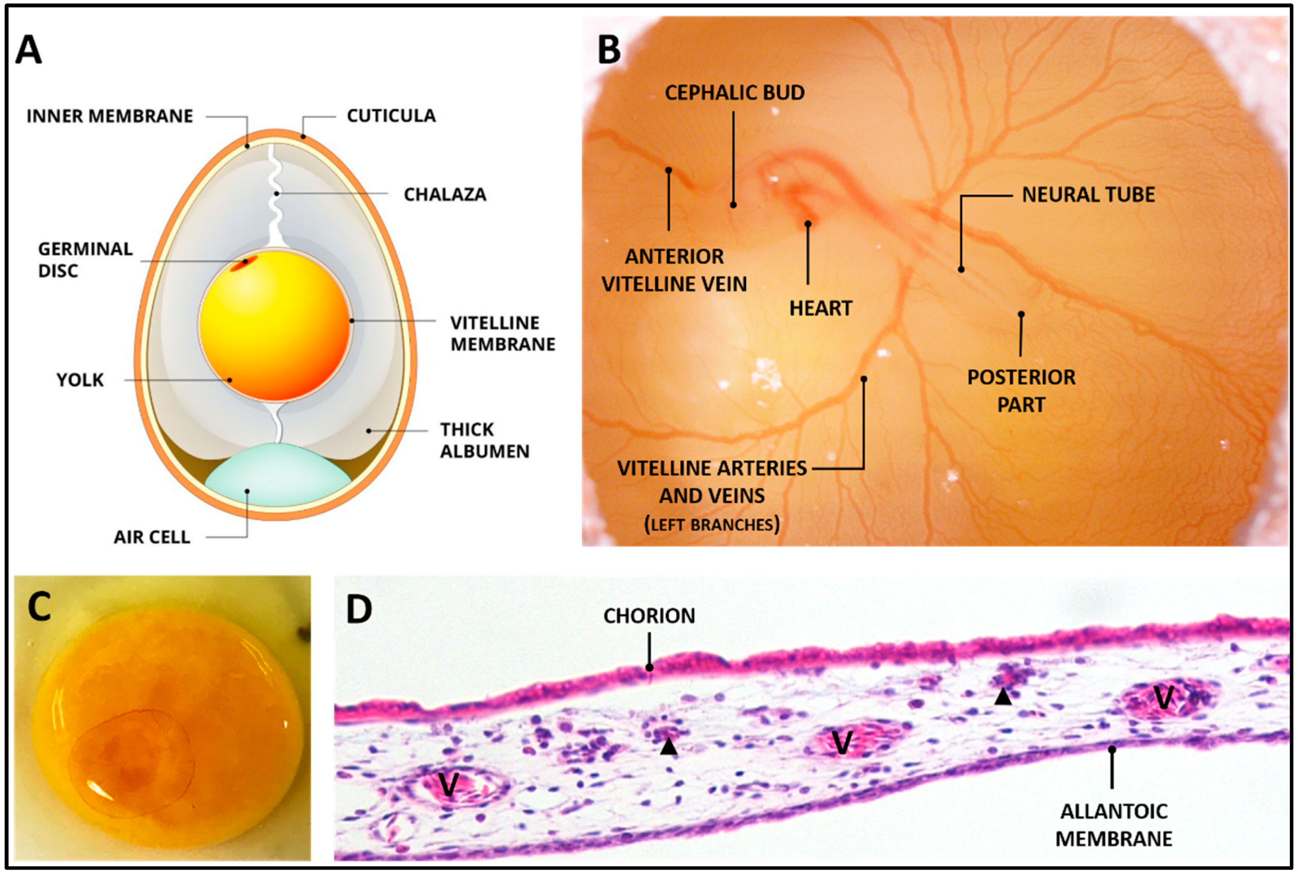

Disclaimer/Publisher’s Note: The statements, opinions and data contained in all publications are solely those of the individual author(s) and contributor(s) and not of MDPI and/or the editor(s). MDPI and/or the editor(s) disclaim responsibility for any injury to people or property resulting from any ideas, methods, instructions or products referred to in the content. |
© 2023 by the authors. Licensee MDPI, Basel, Switzerland. This article is an open access article distributed under the terms and conditions of the Creative Commons Attribution (CC BY) license (http://creativecommons.org/licenses/by/4.0/).
Copyright: This open access article is published under a Creative Commons CC BY 4.0 license, which permit the free download, distribution, and reuse, provided that the author and preprint are cited in any reuse.


