
Submitted:
31 July 2023
Posted:
02 August 2023
You are already at the latest version
Abstract
Chiari malformation type 1 (CM1) includes a series of congenital anomalies that share ectopia of the cerebellar tonsils below the foramen magnum, in some cases associated with syringomyelia or hydrocephalus. CM1 can cause dysfunction of the brainstem, spinal cord, and cranial nerves. This functional alteration of the nervous system can be detected by various modalities of neurophysiological tests, such as brainstem auditory evoked potentials, somatosensory evoked potentials, motor evoked potentials, electromyography and nerve conduction studies of the cranial nerves and spinal roots, as well as brainstem reflexes. The main goal of this study is to review the findings of multimodal neurophysiological examinations in published studies of patients with CM1 and their indication in the diagnosis, treatment, and follow-up of these patients, as well as their utility in intraoperative monitoring.
Keywords:
blink reflex
; brainstem auditory evoked potentials
; brainstem reflexes
; Chiari type 1 malformation
; electromyography
; evoked potentials
; intraoperative monitoring
; motor evoked potentials
; somatosensory evoked potentials
; syringomyelia.
Introduction
Chiari malformations (CMs) comprise a series of neurodevelopmental disorders characterized by a descent of the cerebellar tonsils through the foramen magnum (FM) [1,2,3,4,5]. CM type 1 (CM1) was first described by the Austrian pathologist Hans Chiari in two papers published in 1891 and 1895 [2,3,4]. Nevertheless, the term “Arnold-Chiari malformation” must still be introduced to search in PubMed or other databases for literature on this abnormality. However, due to Arnold’s minor role in the original description, “Chiari malformation” is the most widely accepted eponym [6]. In Chiari’s original description, four types of malformations were differentiated (CM1, CM2, CM3, and CM4). CM1 is traditionally defined as a tonsillar descent of 3-5 mm below the FM, quantified in a mid-sagittal section of magnetic resonance imaging (MRI). The choice of cut-off point for tonsillar descent (3 or 5 mm) is somewhat arbitrary and varies depending on the criteria of the different authors [7,8,9,10,11,12,13]. CM type 2 (CM2) is characterized by a descent of the structures of the brainstem or vermis below the FM and is always associated with spinal dysraphisms (spina bifida) in addition to tonsillar ectopia. Patients with CM2 also present a series of brain anomalies associated with spina bifida that are not observed in any other type of CM (gray matter heterotopia, polygyria, and descended tentorium, among others). CM type 3 (craniocervical encephalocele) and CM type 4 (cerebellar hypoplasia) [14] are severe malformations with a low incidence that most authors consider unrelated to CM1 and CM2. The recent International Consensus Conference recommended considering CM3 and CM4 as separate entities [15].
Other variants of CM1 have been described as minor or major forms of CM1. In 1998, Iskandar et al. coined the term “Chiari 0” (CM0) to describe five pediatric patients with syringomyelia, no tonsillar herniation, and a “tight” posterior fossa (PF), who improved after PF decompression. Subsequent studies by this and other groups have confirmed this entity and observed a significant volumetric reduction in the PF and alterations in cerebrospinal fluid (CSF) dynamics around the FM as the common etiopathogenic factor in CM0. Some authors suggested the term “tight cisterna magna” to define the same entity [16,17,18,19]. In 2004, Tubbs et al. coined the term “Chiari 1.5” to define those patients without spina bifida that presented, in addition to the tonsillar ectopia, a variable descent of the brainstem and displacement of the obex below the FM [13]. We found that once this variant was defined, a considerable percentage of patients in our series initially classified as CM1 actually corresponded to CM1.5 [19,20,21,22].
An additional type, often not included in canonical classifications, is comprised of patients in which any type of CM is associated with different osseous malformations of the craniovertebral junction (CVJ). In a previous paper, we proposed the term ‘complex CVJ abnormalities’ when patients present tonsillar herniation and at least two of the following abnormalities: a significant retroflexed odontoid, a basilar impression (BI), platybasia, severe bone abnormalities in the C0–C2 complex, uni- or bilateral occipital condyle hypoplasia, atlantooccipital assimilation, or other abnormalities that condition an anterior compression of the cervical-medullary junction [19,23] (Figure 1). These patients need different clinical management and often require multiple surgical procedures (such as anterior approaches and occipito-cervical fusions) [24].
A common feature of CMs is that the cerebellar tonsillar descent causes compression of the neural structures and hinders CSF passage at the cervico–medullary junction, consequently altering brainstem and upper spinal cord function. The compressive effect on neural structures at this level may produce alterations in brainstem pathways, cranial nerve nuclei, or their exit from the brainstem, sleep-regulating regions, and cardiorespiratory centers [25]. The frequent association of syringomyelia with any form of CM can induce additional damage to the spinal cord and the spinal roots that emerge from the gray matter. These functional alterations of the nervous system can be detected by multimodal neurophysiological tests: brainstem auditory evoked potentials (BAEPs), somatosensory evoked potentials (SEPs), motor evoked potentials (MEPs), electromyography (EMG) of the cranial nerves/ spinal roots, and brainstem reflexes.
Our paper aims to review the literature and summarize the findings reported in multimodal neurophysiological examinations in patients with CM0, CM1, and CM1.5 and their role in managing these patients, establishing indications for surgical treatment and follow-up. In addition, we will update their role in intraoperative neurophysiological monitoring (IONM). The role of neurophysiology in the diagnosis of sleep-related disorders, a frequent finding in CM1 [22,25,26], has deliberately been excluded from this review. We also excluded articles referring to cognitive evoked potentials.
Methods
This study is an updated narrative review of neurophysiological studies (except for polysomnography and sleep disorders) published up until 30/08/2022. We searched the following databases: PubMed, EMBASE, and WoS. The search strategy used a combination of the keywords: “Arnold Chiari malformation,” “Chiari type 1 malformation*,” “Chiari type 1.5 malformation*,” “Type I Arnold Chiari malformation*,” “Chiari malformation type I with Syringomyelia,” “CM1 with Syringomyelia,” “Syringomyelia” and “evoked potentials,” “blink reflex*,” “brainstem auditory evoked potential*,” “brainstem reflex*,” “electromyography,” “motor evoked potential*,” “intraoperative neuromonitor*.” The flowchart used is shown in Figure 2.
Exploring Brainstem and Spinal Cord Functionality
Brainstem Auditory Evoked Potentials (BAEPs)
BAEPs were first described in humans in 1970 [19]. They allow the recording of a series of five to seven positive electrical signals generated in response to sounds, each of which has a well-defined electrical generator [20,21] (Figure 3); because waves VI and VII are not constant in the healthy population, they are not routinely evaluated. BAEPs explore the functional integrity of a limited portion of the brainstem, both in the rostrocaudal direction—from the VIII cranial nerve entry at the pontomedullary junction to the upper part of the pons-midbrain— as well as in the transversal plane. The ventral part of the brainstem is not explored with BAEPs. It is widely accepted that alterations in waves I to III reflect the involvement of neural structures ipsilateral to the auditive stimulation. In contrast, abnormalities in the amplitude or latency of waves IV and V indicate contralateral structural impairment [22]. BAEPs can be abnormal in several processes: 1) peripheral auditory pathology (conductive or cochlear hearing loss); 2) primary anatomical modifications of the brainstem due to congenital pathologies (CM, Joubert syndrome, Dandy-Walker syndrome, etc.,) or secondary (vascular, tumor, demyelinating, or degenerative); 3) anoxia and ischemia; and 4) intracranial hypertension with transtentorial herniation [23]. Due to the close relationship between the cranial nerves and the brainstem, BAEPs are routinely used in IONM and are important in surgical procedures in which the brainstem and cranial nerves are at risk [27].
Somatosensory Evoked Potentials (SEPs)
SEPs explore the conduction of the electrical impulse through the lemniscal or posterior cordonal system (Figure 4), collecting the integrated responses obtained at different anatomical levels after applying a repetitive electrical stimulus on a peripheral sensory nerve or the sensory portion of a mixed nerve—usually the median nerve (MN) for the upper limbs and the posterior tibial nerve (PTN) for the lower limbs. In addition, other nerves, such as the cubital, trigeminal, saphenous, or pudendal, can be explored [28,29,30,31]. SEPs explore the somesthetic pathway from the peripheral nerve, preganglionic or postganglionic plexus, roots, spinal cord, brainstem, thalamus, and suprathalamic structures [23]. In anoxic or traumatic coma, they are useful for evaluating cortical and subcortical function. SEPs are now routinely used in the IONM of surgical procedures with a risk of damaging structures that generate and transmit the electrical signal [32]. In addition, ‘dorsal column mapping’ has also been introduced to guide the surgical team in procedures with a risk of spinal cord damage [33,34].
Cutaneous heat stimulations with a laser beam can selectively activate thermoalgesic A-delta and C nociceptors, leading to the generation of laser-evoked potentials (LEPs) in the cortex [35,36]. LEPs have been recognized as reliable neurophysiological tools for investigating neuropathic pain [37]. Pure spinothalamic lesions, such as syringomyelia, brainstem syndromes, or small fiber neuropathy, are characterized by normal SEPs but abnormal LEPs [36,38]. However, spinal injuries involving the cervical or lumbar dorsal horn can also alter or abolish components of classical SEPs, such as N13 or N22 [31,39]. Despite their potential utility, LEPs have not been widely adopted due to technical difficulties, the need for skilled personnel, and associated risks like skin burns and hyperpigmentation. Contact heat-evoked potentials (CHEPs) have emerged as a safer alternative to LEPs and are easier to use but require patient cooperation and are unsuitable for IONM [40]. In recent research, Leandri et al. introduced a new noninvasive electrode that selectively stimulates nociceptive nerve-free endings, enabling the evaluation of disorders affecting the nociceptive pathway by enlarging the stimulated surface area [41].
Motor Evoked Potentials
The term “motor evoked potentials” refers to potentials recorded from muscle or nerve after stimulation of the primary motor areas in the CNS. MEPs are the responses elicited distally along the spinal cord, peripheral nerve, or muscle with stimulation of the central motor pathways (spinal cord or cerebral hemispheres) using either transcranial magnetic stimulation (TMS) or electrical stimulation. Electrical stimulation is utilized routinely for IONM, whereas magnetic stimulation is generally used for diagnostic studies in awake patients because of its better tolerance.
TMS is performed with coils of different sizes and shapes. A magnetic field applied through the skull induces a stimulus in the underlying brain tissue with minimal current affecting the skin and subcutaneous tissue. For lower extremity MEPs, the coil is centered in the Cz region; for the upper limb, it is just lateral to this reference. With peripheral recordings, three MEP characteristics are used: latency, amplitude, and threshold. MEP amplitude is highly variable but is generally considered abnormal if it is less than 20% of the compound muscle action potential (CMAP) amplitude obtained on peripheral neurography [42]. Central motor conduction time (CMCT) is the most commonly used parameter to identify CNS disorders [43] and is calculated by subtracting the latency of spinal MEPs from the latency of cortical MEPs [44]. TMS MEPs are used to study central demyelinating disease, motoneuron disease, epilepsy, movement disorders, ataxia, myelopathies, neuropathies, and radiculopathies [45].
In order to elicit an MEP by transcranial electric stimulation (TcMEP) for IONM, subcutaneous electrodes are placed on the scalp in C1-C2 or C3-C4 (10-20 EEG International System). In general, a 50% reduction in amplitude from the average baseline value during surgery is considered significant and a warning sign of damage to the corticospinal tract. Similarly, corticobulbar motor evoked potentials (CoMEPs) elicit cranial nerve responses [46]. In addition to the two aforementioned techniques, which assess the integrity of the corticospinal tract, several mapping methods have been described for identifying the corticospinal tract by directly stimulating the exposed neural structures with a bipolar or monopolar electrode [47,48].
Electromyography and Nerve Conduction Studies
Virtually all primary neuromuscular diseases cause changes in the electrical activity recorded in muscle fibers. These changes can best be explored using needle electrodes inserted into the muscle to record spontaneous and voluntary electromyography (EMG). EMG is one of the major diagnostic tools for identifying and characterizing motor unit disorders, including anterior horn cells, peripheral nerves, neuromuscular junctions, and muscles. EMG complements nerve conduction studies (NCS) in the localization of neuromuscular disorders [44,45]. Normal muscle fibers at rest show no spontaneous electrical activity outside the end plate region. All voluntary muscle activity is mediated by the lower motor neurons and their corresponding innervated muscle fibers that form the ‘motor unit potentials ‘(MUPs). Voluntary MUPs are the sum of the potentials of individual muscle fibers innervated by a single anterior horn cell that occurs in the region of the needle electrode. MUPs are characterized by their firing pattern, rates of fire (recruitment), and configuration and appearance; MUPs fire in a semi-rhythmic pattern. Neuromuscular diseases may show abnormal spontaneous discharges, abnormal voluntary MUPs, or both. Abnormal spontaneous activity includes fibrillation potentials, fasciculation potentials, myotonic discharges, complex repetitive discharges, myochemical discharges, cramps, and neuromyotonic discharges. MUPs may have abnormal duration and amplitude, an abnormal number of phases, or morphology. The extent and distribution of these abnormalities allow us to define the type and severity of the disease, its stage, and the anatomical location of the lesion [44].
EMG is also used in IONM via continuous free-run and stimulated EMG. Free-run EMG consists of recording spontaneous muscle activity in real time. In intraoperative neurophysiology, we use this technique as a monitoring tool to detect surgically induced mechanical irritation of the peripheral nervous system and cranial nerves before irreversible damage to these structures may occur. Stimulated EMG consists of applying an electrical stimulus directly to the peripheral motor nerves or roots to be registered in the corresponding muscles and can be used as a mapping tool to detect the location of peripheral or cranial nerves that are difficult to distinguish from tumors, fibers, or fatty tissues. Stimulated EMG is also used to check the function of injured (or at risk of injury) nerves, roots, or trunks by evaluating electrical transmission through these structures and comparing it to the baseline [49].
Brainstem Reflexes
Blink reflex. The blink reflex (BR) is the neural response obtained in the orbicularis oculi muscle after stimulating the trigeminal nerve with electrical, mechanical, or other stimuli. The BR is the most widely used brainstem reflex in clinical practice. The trigeminal BR is mediated by the first division of the trigeminal nerve (afferent branch) and facial nerve (efferent branch). In normal individuals, two responses (R1 and R2) are obtained when recording from the ipsilateral muscles to the stimulated nerve, and a single (R2) response is obtained from the contralateral muscles [50]. R1 is constituted by the oligosynaptic reflex arc, which includes trigeminal afferents, brainstem connections between the sensory part of the trigeminal nucleus, the motor nucleus of the facial nerve, the facial nerve proper, and the orbicularis oculi muscle (Figure 5). The R2 component has polysynaptic connections within the brainstem but has the same afferent/efferent pathways as R1. BR is useful in the study of trigeminal and facial nerve lesions, peripheral neuropathy, posterior fossa lesions, multiple sclerosis, and extrapyramidal diseases [51]. Under general anesthesia in the operating room, only the R1 response can be recorded [52,53,54,55].
Trigeminal-hypoglossal reflex. Brainstem trigeminal-hypoglossal reflexes (THRs), also known as jaw-tongue reflexes, coordinate the tongue’s position in the mouth during chewing, swallowing, vocalization, and breathing. Recently, a novel methodology for obtaining jaw-opening THR of the brainstem under general anesthesia has been described. This technique could be helpful for the intraoperative monitoring of surgeries involving the trigeminal, hypoglossal nerves, and lower brainstem lesions, however, further studies are still required to validate it [56,57,58,59,60].
H reflex in the masseter muscle. The H reflex in the masseter muscle is a monosynaptic trigeminal-trigeminal reflex transmitted through the mesencephalic nucleus of the trigeminal nerve. It reflects conduction through the midbrain and the median pons [61,62,63]. It can be helpful in studying patients with brainstem or trigeminal lesions, and can be obtained under general anesthesia, thus representing a new method for intraoperative monitoring, especially for lesions involving the midbrain and pons. Together with the BR, the H reflex is a straightforward method for intraoperative monitoring of brainstem reflex circuits and the functional integrity of the trigeminal and facial nerves. Nevertheless, yet more studies are necessary to demonstrate their usefulness [64].
Laryngeal adductor reflex. Recording of the laryngeal adductor reflex (LAR), a brainstem reflex entirely governed by the vagus nerve, has been introduced to monitor the vagus and laryngeal nerves in thyroid surgery [65,66]. Preservation of the reflex prevents potentially harmful substances from entering the tracheobronchial tree by activating the adductor muscles of the larynx to close both vocal folds [67,68]. Recently, a study was published using this reflex in surgeries around the brainstem to monitor cranial nerve X and related intrinsic brainstem pathways (Figure 5), showing that IONM with continuous LAR monitoring, together with vocal-CoMEPs, may be useful to reduce swallowing and laryngeal complications, such as aspiration and pneumonia, enhancing safety in resecting complex PF lesions [69,70].
Neurophysiological Limitations in the Developing Central Nervous System
Neurophysiological test references change during the maturational process of the CNS and, therefore, exhibit some peculiarities depending on the patient’s age. In term newborns, the most reproducible waves in BAEPs are I, III, and V [71]. However, unlike adults, it is normal for wave I to have a greater amplitude than complex IV/V. The myelin maturation process shortens the latency of the successive waves and the central conduction time (CCT or I-V interval). It has also been demonstrated that the auditory pathway responsible for generating the first and last components of BAEP responses matures at a different rate [72]. In a normal subject, wave I latency reaches the adult value around six months of life, while wave V does so by one year of life [71].
A similar process occurs in SEPs, the maturation of peripheral segments of the sensory pathway progresses more rapidly than that of central segments. N9 latency—brachial plexus potential—shortens during the first year of life and subsequently increases as the patient’s size increases. The N9-N11 interval—conduction between the brachial plexus and the dorsal column—decreases with age. In contrast, the N13-N20 (CCT) presents a rapid shortening during the first year of life, reaching adult characteristics at four years of age [73].
Independent of the changes described, the motor stimulation threshold is higher in children than in adults under 18 years of age [74]. CMCT is a dependent value of CNS maturation but has different times. For example, with relaxation (resting motor threshold), children reach adult values approximately at the age of ten, however, with facilitation (active motor threshold), they reach normal values at two years of age [74,75]. Also, MEPs could be obtained at a resting motor threshold in the upper limb after the first year of life and in the lower limb after four years. Maturation also affects the duration, morphology, and amplitude of MEPs.
When evaluating peripheral nerves in children, the myelination process also affects parameters commonly used for assessing NCS [76]. For instance, conduction velocity increases in proportion to nerve fiber diameters and the distance between the nodes of Ranvier, achieving faster motor and sensitive velocities with growth; distal latencies and amplitudes also change. For example, the CMAP increases threefold in the upper limbs and twofold in the lower limbs [76].
The LAR in children is vital for airway protection. In humans, the larynx descends from the neck at 4-6 months. This descent necessitates a change in the airway protection mechanism from a pattern in which an upright epiglottis in the newborn is grasped by an intact palatopharyngeal sphincter to one in which the epiglottis folds down over the aditus to the larynx and the closing vocal folds. This also coincides with the transition from obligate nasal breathing in early infancy to the adult pattern of facultative nasal or oral breathing. It has been suggested that difficulties in adjusting to this transition may be a factor in sudden infant death syndrome, which peaks in incidence at approximately the same age that laryngeal descent is complete (4–6 months) [77,78]. In theory, as of six months of life, we should record the LAR (either in the clinic or the operating theatre), although it may be necessary to do so with hook wire electrodes if there is no age-appropriate endotracheal tube [79,80].
A discussion of the changes from children to adults is beyond the scope of this paper. However, it is essential to bear in mind that neurophysiological tests have some peculiarities according to age, basically due to myelin maturation, which must be considered when evaluating children.
Follow-Up Neurophysiological Studies in Patients with CM1
There are many publications on the follow-up of CM1 patients, however, practically half of them refer to case reports or series with fewer than ten patients (Table 1). Here, we summarize the most relevant information.
Brainstem Auditory Evoked Potentials
To our knowledge, the first paper was published in 1983 by Stone et al. [81], describing BAEP findings in a 16-year-old boy with CM2 and symptoms suggestive of brainstem involvement and neuroimaging revealing brainstem compression. The authors diagnosed CM2, however, as the reported patient did not have any visible spinal lesion, we believe that CM1.5 was the most likely diagnosis [13]. BAEPs were abnormally bilaterally, with I-V interpeak latency (IPL) prolongations and no visible wave III identified upon stimulation of the right ear. The patient underwent bilateral posterior fossa decompression (PFD) and cervical laminectomy. Six months later, he presented neurological improvement and bilateral normalization of his BAEPs. A few authors have also described case reports with retrocochlear findings in the BAEPs of CM1 patients, in both adults [82,83] and children [84,85] (Table 1).

Table 1.
Published neurophysiological follow-up studies in patients with Chiari 1 malformation.
| Author | Year | N | Age/Sex | SYR | SCOL | BAEPs | SEPs | MEPs | EMG/NCS | Brainstem reflexes |
|---|---|---|---|---|---|---|---|---|---|---|
| Cocito et al. [104] |
2022 | 100 Symptomatic CM1=34 CM1+SYR=53 SYR=13 |
23–75-y-o 25♂ / 75♀ |
66 | TMS of phrenic nerve: In 48%--> alt. C5-MEP. In 20%--> absence or delay of Cz-MEP Alterations of the Cz-MEP and C5-MEP were prevalent in patients with cervical SYR/syringobulbia, most associated with CM1 |
|||||
| Di Stefano et al. [90] | 2019 | Comparison between groups: IH = 18 CM1 = 18 SNHL= 20 Normal controls = 52 |
CM1 49 ± 11-y-o 12♂ / 6♀ |
Wave V, I—III, and III-V IPL were higher in CM1 than in controls | ||||||
| Guvenc et al. [98] | 2019 | T=27 | 15-62-y-o 7♂ / 20♀ |
Abnormal SEP in 22.2%. Much more frequent in PTN than in MN. There was a significant correlation between CSF flow disturbance, the degree of TE (P=0.038), and the presence of SEP abnormalities (P=0.016). CSF flow disturbance and SEP abnormalities are more frequently seen in patients with platybasia | ||||||
| Jayamanne et al. [111] | 2018 | T=1 Presenting as left foot drop |
6-y-o ♀ |
YES (holocord SYR) |
NCS: CPN, PTN, and sural nerve were normal. EMG at left TA revealed fibrillations and scant MUPs. EMG of left medial gastrocnemius and right TA were normal | |||||
| Stancanelli et al. [102] | 2018 | T=1 Presenting as excessive sweating on all the left side of the body |
22-y-o ♂ |
BAEPs were normal. | SEPs were normal. | MEP showed an asymmetric response with increased CMCT on the right upper and lower limbs |
Sudoscan test: Asymmetric sweating with a higher electrochemical skin conductance on the left hand and foot |
|||
| Moncho et al. [19] | 2016 | 200 | 15-70-y-o 58♂ / 142♀ CM0=14 CM1=137 CM1.5=49 |
YES (96) | Of all the variables introduced into the logistic regression, only age, the degree of TE, and lower cranial nerve dysfunction had a statistically significant influence on predicting abnormal BAEPs | Of all the variables introduced into the logistic regression model, only age and the degree of TE were statistically significant at predicting abnormal SEPs. BAEPs and SEPs play an essential role in clinically asymptomatic/oligosymptomatic patients |
||||
| Akakin et al. [96] | 2015 | T=1 | 34-y-o ♂ |
Large SYR from just below FM to T5 vertebral body. The spinal cord was thinned at these levels | Preoperatively, SEPs were abnormal: Increased N13-20 IPL of the MN SEP and reduced cortical AMP of the PTN SEP. After syringo-subarachnoid-peritoneal shunt insertion using a conventional lumbo-peritoneal shunt and a T-tub, the SEP test returned to normal | |||||
| Awai and Curt [101] | 2015 | T=7 SYR (1 CM1) |
32-53-y-o 6♂ / 1♀ MC1: 32-y-o ♂ |
YES | Differently affected dSEPs and dCHEPs. dCHEPs in at least one dermatome were abnormal in all patients | All patients had normal NCS and MEPs (of the upper limbs) | ||||
| Moncho et al. [20] | 2015 | 50 | 16-67-y-o 11 ♂ / 39♀ |
YES (20 patients) |
BAEPs were altered in 52%. The most common finding was increased I–V IPL and LAT of wave V (48%). A greater TE was observed in patients with pathological BAEPs compared with patients with normal BAEPs (but not statistically significant, possible type II error) | SEPs were altered by 50%. The most frequent alteration of MN SEP was increased N13-N20 IPL; the most frequent alteration of PTN SEP was an increased N22-P37 IPL, sometimes associated with alteration of the cervical potential. A greater TE was observed in patients with altered PTN SEPs and MN SEPs compared with patients with normal SEPs (but not statistically significant, possible type II error) |
||||
| Isik et al. [87] | 2013 | T= 44 SYR CM1= 32 |
14-71-y-o 24♂ / 20♀ |
YES | BAEP was only pathological in ten (31.2%) patients with CM1. Except for one patient, improvement was seen and correlated with neurological and radiological improvement (90%). This series suggested that BAEPs were more correlated with clinical and radiological findings than SEPs were | MN/PTN SEPs were abnormal in 54.2% of patients (Difficult to know the exact number of patients since the figures do not match). In this series, SEPs did not always correlate with the clinical findings |
||||
| Panda and Kaur [114] | 2013 | T=1 Rapidly progressive right foot drop |
16-y-o ♂ |
YES (holocord SYR) |
NCS: CPN normal BIL with absent bilateral F waves. PTN, sural, superficial peroneal, and saphenous nerve were normal. EMG: fibrillation potentials and positive sharp waves in the right TA, peroneus longus, medial gastrocnemius, gluteus medius, and lumbar paraspinal muscles confirming the lesion to be proximal (lumbar roots or anterior horn cell) |
|||||
| Vidmer et al. [89] | 2011 | T=66 (MC1 and 2) 1 MC1 |
3-months-60-y-o MC1: 9-y-o ♀ |
|
Peripheral or cochlear alteration in a single pediatric patient with CM1 | SEP of MN abnormal with increased LAT N20 and CCT UNIL Normal PTN SEP BIL |
||||
| Mc Millan et al. [113] | 2011 | T=2 Abrupt onset UNIL foot drop |
5 and 4.5-y-o 2♀ |
YES | Case 1) Not included or provided. Case 2) MN SEPs revealed prolonged cervical and cortical responses. PTN SEPs responses were poorly formed with normal latencies |
NCS: Case 1) CPN showed low motor AMP. EMG: fibrillation potentials and positive sharp waves confirming a proximal lesion. Case 2) EMG of the right TA revealed active denervation |
||||
| Saifudheen et al. [112] | 2011 | T=1 Rapidly progressive BIL foot drop |
14-y-o ♀ |
YES (holocord SYR) |
NCS: Low AMP CMAP and normal LAT and velocity for both peroneal and left ulnar nerves. The F wave was normal. Sensory median, ulnar, and sural nerves were normal. EMG: On both sides, chronic neurogenic changes in the first dorsal interossei, TA, and medial gastrocnemius muscles |
|||||
| Berciano et al. [100] | 2007 | T=1 Lancinating left cervico-brachial pain provoked by coughing fits |
54-y-o ♀ |
Cervico-dorsal SYR extending into the left posterolateral quadrant in axial sections passing through C7 and D1 vertebral levels | dSEPs left side: N20 of the C8 dermatome severely attenuated, both pre- and postoperatively. All other left-side dermatomes and right-hand dSEPs were normal Persistence of segmental hypoalgesia and altered SEPs despite the disappearance of SYR on MRI after PFD |
Bilateral motor and sensory conduction parameters of MN and UN, including F responses, were normal | ||||
| Henriques Filho and Pratesi [88] | 2006 | T=75 MC1=27 MC2=48 |
27 patients = 19-70-y-o 48 patients = 2-16-y-o |
First in frequency: alteration of wave I or cochlear level (“segment 1”) Second in frequency: alteration I-III or “between the acoustic nerve near the cochlea and the pontomedullary junction” (“segment 2”) Two patients with an abnormality in the AMP V/I ratio |
||||||
| Brookler [82] | 2005 | T=1 Dizziness |
63-y-o ♀ |
Delay of wave V and III-V BIL, with normal audiology | ||||||
| Utzig et al. [95] | 2003 | T=1 Headache, paresthesia, and SCOL |
15-y-o ♀ |
YES | YES | Left UN/ PTN SEPs: Absence of cortical responses. After SUR: Cortical responses of reduced amplitude for left extremities | ||||
| Hausmann et al. [144] | 2003 | T=100 MC1 =1 |
15.3 ± 2.2-y-o 20♂ / 80♀ |
3 | 100 | Normality in PTN SEPs in the patient with MC1 | ||||
| Muhn et al. [110] | 2002 | T=1 Rapidly progressive UNIL leg weakness |
5-y-o ♂ |
YES | NCS showed a low-amplitude CMAP for the left peroneal and normal sural nerve. EMG of the left TA, tibialis posterior, and medial gastrocnemius muscles showed fibrillation potentials at rest and reduced voluntary recruitment action potentials. A follow-up eight weeks after PFD showed improvement in leg strength with a co-temporal resolution of the previously observed fibrillation potentials | |||||
| Paulig and Prosiegel [107] | 2002 | T=1 Progressive dysphagia for a year |
78-y-o ♀ |
SEPs were normal | NRL examination: BIL paresis and atrophy of the tongue showed diffuse fibrillations. Further neurological examination was normal. NCS and EMG of various muscles of the upper and lower limbs were normal |
|||||
| Bagnato et al. [108] | 2001 | T=1 Presenting as spinal myoclonus |
48-y-o ♂ |
YES | Left MN SEPs revealed normal P14 and N20, while the N13, obtained after glottis reference, was nearly abolished | EMG: 1) Chronic partial denervation in C8/D1 muscles; 2) Rhythmic contractions in FDI and ABP muscles (spinal myoclonus), and 3) Peripheral silent period after supramaximal electrical stimulation of UN at FDI muscle | ||||
| Jacome [122] | 2001 | T = 4 CM1 presented with blepharoclonus |
17-52-y-o 1♂ / 3♀ |
1 | Facial EMG: complex repetitive discharges of the right mentalis muscle in one patient | Blink reflexes were abnormal in 3 patients | ||||
| Scelsa [109] | 2000 | T=1 Presenting as ulnar neuropathy at the elbow |
24-y-o ♀ |
YES | YES | NCS: Marked AMP reduction of the left ulnar CMAP without focal slowing or conduction block across the elbow. EMG: Fibrillations and high AMP MUP in left arm muscles innervated by C7-D1 spinal segments and the median and ulnar nerves |
||||
| Cheng et al. [99] | 1999 | T=164 MC1=12 |
10-20-y-o (m₁=14.2) (m₂=13.6) 22♂ / 142♀ |
6 | 164 | MN and PTN SEPs. Association between TE and SEP dysfunction (P<0.001; c.c 0.672 Spearman). No differences in the degree of TE in patients with normal SEPs and those with abnormal SEPs (P=0.864; Mann-Whitney) |
||||
| Hort-Legrand and Emery [86] | 1999 | T=79 SYR -Foraminal (64) -Meningitis (5) -Trauma (15) CM1=48 (CVJM=11 BI=7) |
SYR foraminal 16-71-y-o 27♂ / 22♀ |
YES | 11 | BAEPs were performed in all patients except 20, in whom the upper level of the SYR was in the lower cervical or the dorsal vertebrae. BAEPs were abnormal in 13 of the 59 patients studied (total cohort), 6 with CVJM. The more frequent finding was I-V IPL PROL UNIL, less frequently BIL. (They did not specify how many patients with CM1 underwent BAEP or what the results were in this subgroup) |
MN and PTN SEPs in all patients. Abnormality (PTN or MN) was noted in 77 of the 79 patients. The most frequent findings for MN SEPs were altered cervical N13 response, an anomaly of the P14-N20 interval, or altered cortical response. The most frequent findings of PTN SEP were an absence of cortical waves or a decrease in their amplitude. SEPs of the trigeminal nerve (V3) were recorded in 60 patients. Prolonged LAT UNIL (less frequently BIL). Much more sensitive than BAEPs: always altered when bulbar symptoms present, while MRI in no case showed syringobulbia. (They did not specify test results in the CM1 subgroup) |
MEPs of the upper limbs in 60 patients. More frequent findings were: PROL CCT, reduced AMP, or very polyphasic responses. (They did not specify test results in the CM1 subgroup) |
||
| Ahmmed et al. [85] | 1996 | T=1 Tinnitus and mild hearing loss in the left ear |
13-y-o ♀ |
Asymmetry in III, V LAT, and I-V IPL, more prolonged in the left ear that returned to normal values after PFD | ||||||
| Amoiridis et al. [121] | 1996 | T=1 Dysesthesia and weakness with urinary retention |
25-y-o ♂ Mild hydrocephalus |
YES | YES | PTN SEPs: No lumbar potential (N24; Ll/iliac crest) could be obtained, whereas a high cervical potential (N33; C2/Fz) and a cortical (P40; Cz' /Fz) potential with a normal latency was registered. The MN SEP was normal | AMP of the H reflex in the soleus muscle was low bilaterally, and the H reflex LAT was prolonged on the right. Motor and sensory NCS were normal. F waves in AH, EDB, or hypothenar muscles, all on the left, were not elicited |
BR: Rl was absent bilaterally, and R2 latency with left side stimulation was prolonged BIL | ||
| Kaneko et al. [120] | 1996 | T=5 | Mean age 18-y-o ♂ |
YES (cervical) | Absent or reduced cervical N13 SEP with preserved cortical responses of the upper limb were observed in 3 of the 5 patients | MEP latency and amplitude were normal in all patients | CMAP latency and amplitude were normal in all patients. Symptomatically, all patients showed diminished cutaneous silent periods (CSPs) up to a stimulus intensity of 15 times the sensory threshold. Diminished CSP was the only abnormal finding in 2 subjects. Also, CSP decrease was more sensitive to syringomyelic changes than abnormal cervical N13 SEP |
|||
| Cristante et al. [105] | 1994 | T=26 CM1 (8 with MEPs) |
YES (5) | Preoperative TMS MEPs: functionally impaired muscles with an abnormal recording. The postoperative functional motor recovery was not reflected by improvement in MEP parameters | ||||||
| Johnson et al. [84] | 1994 | T=1 Steadily progressive bilateral asymmetrical SNHL |
10-y-o ♂ |
NO | CCT or I-V BIL increased: Increased I-III in one ear and III-V in the other | |||||
| Strowitzki et al. [97] | 1993 | T=18 CM1=9 |
YES | MN SEPs: Abnormal in 4 patients. PTN SEPs: Abnormal in 7 patients. No cortical responses were found in 6 patients and delayed responses in 2 (does not specify MN or PTN) | ||||||
| Morioka et al. [94] | 1992 | T= 11 (cervical SYR) CM1= 10 |
24-56-y-o 3♂ / 7♀ |
YES | The most common MN SEP abnormality was UNIL attenuation or absence of N13 often with normal N20 potentials. Spinal EPs provided information regarding abnormality responsible for the dorsal column dysfunction: the syrinx, the TE, or both | |||||
| Nogués et al. [103] | 1992 | T=13 MC1=7 BI=1 | 19-53-y-o (m=37.4) 7♂ / 6♀ |
T=13 | SEPs: Alteration of cortical PTN and reduction or absence of cervical potential of MN | The most frequent finding was an increase in CMTC | ||||
| Gerard et al. [106] | 1992 | T=1 BI Presenting as velar insufficiency |
5-y-o ♀ |
Velar insufficiency: EMG BIL in levator palatini and anterior faucial pillars showed ample biphasic or polyphasic action potentials at rest; when the child cried, the frequency of these potentials increased poorly, and recruitment was impaired. Suspicion of denervation of the IX, X, and XI cranial nerves | ||||||
| Hendrix et al. [83] | 1992 | T=3 | 59,34, 64-y-o | BAEPs were abnormal in one patient: prolonged I-V IPL on the right side. The other two patients had normal BAEPs BIL |
||||||
| Restuccia and Maguière [39] | 1991 | T=24 MC1=16 (9 *) |
20-74-y-o (m=56) 10♂ / 14♀ |
T=24 | MN SEPs: abnormal or absent N13 in 83% of patients with cervical SYR. Good correlation between loss of thermoalgesic sensation and absence of tendon reflexes. With associated CM: increased P14-N20 IPL |
|||||
| Jabbari et al. [93] | 1990 | T=22 MC1=4 |
15-69-y-o (m=28) 15♂ / 7♀ |
T=22 | No significant relationship between SEPs in SYR when CM coexists: In 3 of 4 patients with SYR and CM, SEPs were normal | |||||
| Forcadas et al. [92] | 1988 | T=18 MC1=17 |
12 | MN SEPs: more frequent abnormal N11-N13 with or without abnormal CCT. Good correlation with clinical symptoms. Two of the three patients with normal N11-N13 and abnormal CCT had MC1 without SYR | ||||||
| Anderson et al. [91] | 1986 | T=9 MC1= 8 |
16-65-y-o (m=41) 1♂ / 8♀ |
T=9 | MN SEPs: AMP reduction or absence of the cervical potential, consistent with the clinically more affected side. Seven of the 8 cases with CM1 had a prolonged or asymmetric CCT. One case with MC-1 presented normal MN SEPs |
|||||
| Stone et al. [81] | 1983 | T=1 Classified by the authors as MC2 (but without overt spinal defects), probably CM1.5 |
16-y-o ♂ With associated hydrocephalus |
BAEPs: PROL I-III and I-V IPL in the left ear. Absence of wave III; I-V IPL more PROL in the right ear. Postoperative BAEPs showed BIL normalization |
AMP: amplitude; APB: abductor pollicis brevis; BAEPs: brainstem auditory evoked potentials; BI: basilar impression; BIL: bilateral; BR: blink reflex; CCT: Central conduction time; CM1: Chiari 1 malformation; CM2: Chiari 2 malformation; CMCT: central motor conduction time; CPN: common peroneal nerve; CSF: cerebrospinal fluid; CVJM: craniovertebral junction malformation; dCHEPs: dermatomal contact heat evoked potentials; dSEPs: dermatomal somatosensory evoked potentials; EMG: electromyography; FA: Fractional anisotropy; FDI: first dorsal interosseus; IH: intracranial hypotension; IPL: interpeak latency; LAT: latency; m: mean; MEPs: motor evoked potentials; MN: median nerve; MUP: motor unit potentials; NCS: nerve conduction studies; PFD: posterior fossa decompression; PROL: prolonged; PTN: posterior tibial nerve; SCOL: scoliosis; SEPs: somatosensory evoked potentials; SNHL: sensory neural hearing loss; SUR: Surgery; SYR: Syringomyelia; T: Total; TA: tibialis anterior; TE: tonsillar ectopia; TMS: transcranial magnetic stimulation; UN: ulnar nerve; UNIL: unilateral; *: Previous surgery.
In 1999, Hort-Legrand and Emery [86] reported 79 patients with syringomyelia, of which 48 cases were associated with CM1. BAEPs were abnormal in 13 of the 59 patients studied, and the most frequent finding was prolongation of the I-V IPL, more often unilaterally. Another study on syringomyelia and CM1 [87] found that BAEP abnormalities had a better correlation with clinical and radiological findings than SEPs.
In 2006, Henriques Filho and Pratesi used BAEPs to study 75 CM1 and CM2 patients [88]. Abnormalities were detected in most cases (71%); however, more were observed in CM2 patients. According to these authors, these findings reinforce the concept that pontine alterations are a consequence of brainstem dysgenesis in CM2. They also observed that patients with CM1 preferentially had peripheral alterations—cochlea or cochlear nerve close to the cochlea—with less in the area located between the cochlear nerve and the pontomedullary junction. This study concludes that the assessment of the V/I amplitude ratio, together with other abnormalities, allows the identification of a more significant number of BAEP alterations in patients with CM1 and is a valuable tool for the diagnosis. Vidmer et al. [89] in 2011, described BAEP findings in a single pediatric patient with CM1 (a nine-year-old girl), in whom the abnormalities were located at the peripheral or cochlear level, supporting the results of Henriques Filho and Pratesi [88].
In studies carried out by our group―the latest including 200 patients―we found that BAEPs were altered in 38.5% of patients with CM1 [19,20]. The most common finding was increased I–V interpeak latency and increased wave V latency (31%). The logistic regression model used to predict the probability of an abnormal BAEP value showed that age, the degree of tonsillar ectopia, and the clinical detection of lower cranial nerve dysfunction had a statistically significant influence on predicting abnormal BAEPs. This finding could be explained by the fact that a more severe distortion of brainstem structures induces more BAEP abnormalities. Therefore, patients with a greater degree of tonsillar ectopia or lower cranial nerve dysfunction had a higher frequency of abnormal BAEPs.
Di Stefano et al. studied BAEPs and MRI in three groups of patients: with intracranial hypotension (n = 18), CM1 without intracranial hypotension (n = 18), and sensorineural hearing loss (n = 20) vs. controls (n = 52). In the CM1 group, they found abnormal BAEPs in 33% of patients, the main finding being a prolonged latency of wave V and I-III and III-V intervals [90], comparable results to those obtained by our group [19,20].
Somatosensory Evoked Potentials
In 1986, Anderson et al. [91] described the SEP alterations in nine patients with syringomyelia, eight of whom had an associated CM1. The most frequent findings were unilateral or bilateral reduction or absence of the cervical potential. In all but one patient with CM1 and syringomyelia, a prolonged or asymmetric CCT was found; in 1988, Forcadas et al. reported similar results [92]. In 1990, Jabbari et al. published a study with SEPs from the MN and PTN in 22 patients with syringomyelia, four with CM. Three of the four patients with CM1 and syringomyelia had normal SEPs [93]. Restuccia and Mauguière [39] studied the MN SEPs of 24 patients with syringomyelia, 16 associated with CM1. They observed several anomalies, the most frequent being an alteration or absence of the N13 wave in 83% of patients with cervical syringomyelia. When syringomyelia was associated with CM, these authors, in contrast to Jabbari et al. [93], observed an increase in the P14-N20 interval; Morioka et al. reported similar results [94]. Several case reports and small series with SEP investigations in CM1 patients show abnormal SEPs at a central level, with an absence of cortical responses and an increased latency of cortical potentials or CCT [89,95,96,97].
Moncho et al. found that SEPs were altered in 43.5% of CM1 patients. The most frequent alteration in MN SEPs was an increase in N13-N20 IPL, whereas the most frequent alteration in PTN SEPs was an increase in N22-P37 IPL, sometimes associated with an alteration of the cervical potential. In a logistic regression model, only age and degree of tonsillar herniation were statistically significant in predicting abnormal SEPs. These findings indicated a greater probability of finding abnormal SEPs in older patients with greater tonsillar descent [19,20]; recently, Guvenc et al. described similar findings [98].
Some studies specify the relation between idiopathic scoliosis and CM1, in which abnormal SEPs can be due to tonsillar ectopia or syringomyelia rather than spinal deformity [95,99]. Cheng et al. found a relationship between tonsillar ectopia and SEP alterations when studying 164 patients with idiopathic scoliosis, 12 of whom had associated CM1 [99]. Utzig et al. reported the case of a 15-year-old girl complaining of intense headache, recurrent left-sided paresthesia, and progressing scoliosis, who had CM1 with cervical syringomyelia. Ulnar and PTN SEPs showed an absence of cortical responses that improved after surgical treatment with PFD, C1 laminectomy, and partial removal of the cerebellar tonsils with the reappearance of cortical responses of acceptable latency, however with reduced amplitude for the left extremities [95].
The somatosensory tract in CM1 can also be evaluated by trigeminal SEPs from V3, which can present prolonged latency. Hort-Legrand and Emery found that trigeminal SEPs were much more sensitive than BAEPs, being altered when patients had bulbar symptoms [86].
Berciano et al. reported a CM1 patient who presented with sudden lancinating episodes of left cervicobrachial pain provoked by coughing fits, correlated with cervico-dorsal syringomyelia extending into the left posterolateral quadrant in axial sections passing through vertebral levels C7 and D1. Pre- and postoperative upper limb dermatomal SEPs (dSEPs) elicited by stimulation at levels C6, C7, and C8 showed N20 attenuation only after stimulation of the left fifth finger (left C8 level). Given the preservation of peripheral Sensory nerve action potentials (SNAPs) in both upper limbs, such a selective dSEP attenuation is probably caused by extension of the paracentral posterior horn cavity into the C8 posterior entry zone. Under such circumstances, dSEP is a useful method of evaluating this radicular semiology [100]. In 2015, Awai and Curt reported seven patients with syringomyelia showing differences in dSEP and CHEP affectations. Dermatomal CHEPs were abnormal in all patients in at least one dermatome [101].
Motor Evoked Potentials
Few authors have described TMS MEP alterations in patients with CM1, most of them with syringomyelia. The most frequent findings were increased CMTCs [86,102,103,104].
Referring to pre- and post-surgical MEP evaluations, Cristante et al. [105] described the MEP findings in eight CM1 patients. Preoperative TMS MEPs showed that functionally impaired muscles exhibited neurophysiologic abnormalities, and even one patient without any motor deficit had abnormal MEPs. Interestingly, the postoperative functional motor recovery of five of the eight patients―mostly partial―was not reflected by a similar improvement in MEPs.
NCS, EMG, and other Peripheral Nerve Studies
In 1992, Gerard et al. described the case of a five-year-old girl―with CM1 and BI―admitted due to insufficiency of the soft palate [106]. EMG activity was recorded bilaterally in the levator palatini and anterior faucial pillars, showing ample biphasic or polyphasic action potentials at rest (probably spontaneous activity). When the child cried, the frequency of these potentials increased poorly, and recruitment was impaired. Clinical examination and EMG findings led to a suspicion of denervation of the IX, X, and XI cranial nerves.
CM1 should be a differential diagnosis in patients with adult dysphagia onset requiring MRI examination, even when presenting with “typical” lower motor neuron signs in bulbar muscles. SEPs, NCS, and EMG can help diagnose these cases [107].
In patients with CM1 and associated syringomyelia, NCS and EMG are useful in detecting anterior horn affectation caused by the syringomyelia. This syndrome characteristically presents with spontaneous activity while performing needle EMG if acute or denervating activity occurs, with large, polyphasic (neurogenic) MUPs, reduced voluntary recruitment, and normal sensory nerve conductions [108,109,110,111,112,113]. Sometimes, this can be misdiagnosed as focal mononeuropathy if only motor NCSs are performed since a common finding is a small amplitude CMAP [108,109,110,112,113], although just one group reported unaffected CMAPs [111]. Another study found a confounding ulnar neuropathy with a tricky focal slowing at the elbow in a patient with cervical syringomyelia at C7-D1. A few studies reported a clinical foot drop syndrome in children, with neurogenic findings in most muscles that were dependent on the common peroneal nerve but also the PTN, and with normal sensory evaluation. The MRI confirmed the presence of holocord syrinx [110,111,112,113,114]. Therefore, it is essential to keep in mind that foot drop is a common problem that can present, and even though the most frequent cause is peroneal neuropathy, other causes include anterior horn cell disease, lumbar plexopathies, L5 radiculopathy, partial sciatic neuropathy, and, rarely, parasagittal lesion.
Painful cutaneous nerve stimulation can suppress EMG activity in voluntarily contracting muscle [115,116,117,118,119]. This phenomenon is designated the “cutaneous silent period” (CSP). In 1996, Kaneko et al. [120] studied five male patients (mean age 18 years) with cervical syringomyelia using CSPs, CMAPs, F waves, MEPs, and SEPs of upper limbs. MEP latency and amplitude were within normal limits in all patients. Three of the five patients presented absent or reduced cervical N13 potentials from SEPs of the upper limbs with preserved cortical responses. In the symptomatic hand, all patients exhibited reduced CSPs, constituting the only abnormal finding in two subjects. The laterality of the abnormal CSPs and the reduced N13 potentials of the SEPs were consistent with the site of the syrinx on MRI. Previous reports demonstrated that absent or reduced cervical N13 potentials with preserved scalp responses of upper limb SEPs are characteristic of early cervical syringomyelia [39,91,92]. Kanebo et al. further demonstrated that the absence of CSPs with normal conduction in large, myelinated fibers that form motor and sensory pathways is characteristic of patients with cervical syringomyelia. In addition, the CSP decrease was more sensitive to syringomyelic changes than abnormal cervical N13 potentials were.
Brainstem Reflexes
We only detected two reports regarding CM1 follow-up with brainstem reflexes, specifically, the blink reflex. Amoiridis et al. [121] reported a case of a 25-year-old man with CM1 and holocord syrinx who experienced dysesthesia and weakness with urinary retention during shooting practice. The PTN SEPs showed no lumbar potential (N24; Ll/iliac crest), whereas a high cervical potential (N33; C2/Fz) and a cortical potential (P40; Cz’/Fz) with normal latencies were registered. The MN SEP was normal bilaterally. The amplitude of the H reflex in the soleus muscle was low bilaterally, and H reflex latency was prolonged on the right. Motor and sensory NCS were normal. F waves in legs and arms were abolished. They performed the BR that showed Rl absent bilaterally and R2 latency with left-side stimulation prolonged on both sides. However, neurological examination of the cranial nerves was unremarkable. Jacome, in 2001, described four CM1 patients presenting with blepharoclonus, one with altered R1, two with altered R2, and one with normal BR. Also, facial EMG showed complex repetitive discharges of the right mentalis muscle in one patient [122].
Intraoperative Neurophysiological Monitoring in CM1
Most articles on this topic refer to neuromonitoring during PF surgeries. A few refer to neuromonitoring during the insertion of shunts for the treatment of syringomyelia. A third category comprises CM1 and scoliosis surgery, as well as studies reporting new techniques (Table 2).
Posterior fossa surgery
The role of intraoperative neuromonitoring (IONM) during PF surgery for CM1 is controversial, and there is still no consensus on the usefulness of the technique for the prevention of new neurological deficits nor on the most effective modality to use. Anderson et al. studied changes in intraoperative BAEPs and SEPs in 11 pediatric patients undergoing suboccipital decompression for CM1. BAEP conduction times were compared at four different surgical stages: before positioning, after positioning, after craniotomy, and after durotomy. Their data indicated that significant improvement in conduction times occurs after bony decompression and division of the atlantooccipital membrane rather than after dura opening. They also highlighted the risk of altering SEPs and BAEPs during positioning [123]. Zamel et al. reviewed the records of 80 pediatric patients, with or without syringomyelia, who underwent suboccipital decompression to treat CM1. They divided patients into two groups according to whether or not the dura mater was opened. Their data suggest that PFD with bone removal alone significantly improves BAEP conduction times in most pediatric patients with CM1 and that using duraplasty led to a further, albeit slight, improvement in conduction times in only 20% of patients [124]. Kim et al. [125] performed an alternate strategy for treating 11 symptomatic children presenting with basilar invagination associated with CM1. Their technique involved suboccipital decompression, C1 laminectomy, and duraplasty to treat CM1 and subsequent intraoperative manual reduction using accentuated cervical distraction and extension. Their results showed that immediately after reduction and fusion, SEPs improved significantly in 10 patients, while remaining unchanged from baseline in one.
In 2012, Chen et al. analyzed BAEP and SEP parameters in 13 consecutive pediatric patients who underwent suboccipital decompression to treat symptomatic CM1. They recorded the MN SEP, PTN SEP, and BAEP latencies at four stages: preoperatively, following craniotomy, following durotomy, and following closure [126]. In contrast to that reported by Anderson et al. [123] and Zamel et al. [124], they demonstrated clinically notable improvements in overall latency times in both MN SEP N20 and BAEP wave V, not only after bony decompression but also after duraplasty. Since their results showed more significant improvements in MN SEPs, they consider them the most sensitive for evaluating sufficient decompression. Incorporating an additional replacement of the bone flap retained the improvements in MN SEP and BAEPs, suggesting that this technique does not compromise the decompressive effect of the surgery.
The largest pediatric population series―with 156 patients―was reviewed in 2015 by Kennedy et al., in children with PFD but not dural opening [127]. IONM with SEPs and BAEPs was performed before and after prone positioning and during surgery. They found that 78% of patients exhibited significant BAEP conduction time improvement after bony decompression. SEPs were used to assess the lack of problems with patient positioning during surgery.
The first study evaluating the efficacy of multimodality IONM using both TcMEPs and SEPs during PFD in 21 pediatric patients was presented in 2016 by Barzilai et al. [128]. They found significant signal changes in three patients, all concomitant with patient positioning on the surgical table: in two cases, the signals recovered at closure, whereas in one case, SEPs remained attenuated even after repositioning. No new neurological deficits were observed post-operatively. They concluded that IONM could be useful during patient positioning in PFD surgery.
In 2019, Rasul et al. also analyzed IONM data with SEPs and BAEPs at two time points—baseline before skin incision and final at the time of skin closure—in 37 symptomatic patients who underwent PFD for CM1. They found significant SEP latency reduction in all patients. As for BAEPs, 13 patients had a reduction in their conduction time [129]. However, they could not establish a definite link between clinical outcomes and IONM changes, unlike other authors [130].
In 2020, a case report published by Krishnakumar et al. [131] evaluated the use of IONM to identify neurological safety during critical surgical steps. They reported a 13-year-old patient diagnosed with atlantooccipital dislocation with BI, hypoplastic C1 arch with CM1, and thoracic and lumbar scoliosis. Loss of TcMEPs was recorded during positioning and screw fixation, recognizing the need for a new surgical strategy. Changes in TcMEPs were reverted, and even an increase in amplitude was recorded at the end of surgery compared with baseline. During the three-month follow-up, the patient showed no new neurological deficit.
Contrary to most studies presented in the pediatric population― in which a beneficial role of IONM is suggested―the literature regarding the adult population is more skeptical as to the benefits of IONM during PFD for CM1 treatment. The results of these studies are more heterogeneous than those in pediatric patients; however, most are single case reports and, therefore, represent anecdotal evidence.
Grossauer et al. [132] published the case of a 32-year-old woman who underwent surgery for CM1 associated with extensive cervicothoracic syringomyelia and whose SEPs did not notably improve following craniotomy or durotomy. Instead, they only improved after opening the fourth ventricle and restoring CSF flow at the level of the craniocervical junction.
Roser et al. [133] presented IONM data from a small series of 39 consecutive adult patients (84.6% presenting with syringomyelia) who underwent suboccipital decompression and duraplasty for the treatment of symptomatic CM1. SEPs and TcMEPs were monitored in all patients, reporting significant changes in both SEPs and TcMEPs in only two cases related to patient positioning. They found no significant change in SEP and TcMEP amplitudes or latencies between baseline and final measurements.
Heffez et al. examined the relationship between the extent of tonsillar ectopia and intraoperative changes in BAEP conduction times in an extensive series of adult CM1 cases [134]. They divided the patients into four groups depending on the position of the cerebellar tonsil. They noted a reduction of > 0.1 ms in conduction times in 35 - 49% of patients with no statistical difference between groups or clinical outcomes. This suggests that, even with minimal tonsillar herniation, a disruption in the function of at least one pathway within the brainstem may occur and that this disturbance is improved by brainstem decompression.
In 2022, Schaefer et al. [135] suggested that CM1 suboccipital decompression surgery may be performed safely without IONM after evaluating SEPs, TcMEPs, and/or BAEPs in 93 adult patients, as they found only one instance of a transient decrease in TcMEPs, which resolved without intervention.
Some articles describe MEPs or SEPs during IONM in CM1 patients treated with other surgical techniques, like atlantoaxial stabilization surgery or endoscopic suboccipital decompression, with inconclusive results [136,137] (Table 2).
Surgery for the direct treatment of syringomyelia in MC1
The first articles about IONM for syringomyelia in CM1 patients were published by Milhorat et al., with 32 syringomyelia patients (21 with CM1). IONM with MN SEPs demonstrated a significant decrease in N20 latency and a nonsignificant increase in N20 amplitude 30 minutes after syrinx decompression. They concluded that the improvement in N20 latency was indirect evidence of preexisting long tract compression. However, these conclusions are questionable in patients with CM1, in whom SEP improvements may have been caused, in part, by previous PF surgery [138,139].
Pencovich et al., in 2013, were the first to address the benefits of multimodality IONM during syringomyelia surgery. They monitored SEPs and MEPs in 13 patients with syringomyelia, six of whom also had CM1. One of the CM1 patients presented severe attenuation of left leg MEP data noted upon the midline approach to the syrinx. The catheter was subsequently removed and PFD was completed without syrinx drainage. IONM might be a valuable adjunct to spinal cord surgeries for the treatment of syringomyelia, supporting the routine use of multimodal IONM in this kind of surgery [140,141].
CM1 in scoliosis surgery
The association between Chiari malformation syrinx and scoliosis has been widely documented during the past few decades [99,142,143,144]. The concomitant rate of scoliosis ranges from 15 to 50% in patients with Chiari malformation [145,146,147]. There is a potential risk from scoliosis surgery in patients with syringomyelia-associated scoliosis, which might be higher with longer and wider syrinxes. We found two articles with different conclusions concerning IONM during scoliosis surgery in patients with CM1. Tan et al. [148] compared radiographic features, clinical outcomes, and surgical complications after posterior spinal fusion between 21 patients with CM1 and syringomyelia-associated scoliosis and 21 with idiopathic syringomyelia-associated scoliosis. As for the IONM data, TcMEPs were implemented in all patients. They noted no differences in the successful recording of baseline TcMEPs or evident TcMEP deterioration between the two groups. Patients with CM-associated scoliosis had longer syrinxes than the syringomyelia-associated scoliosis group; however, their preoperative neurological status and IONM signals during the operation were similar. In addition, no progression of the original neurological deficits or neurological complications were detected in either group. Shi et al. [149] studied 60 neurologically asymptomatic CM-associated scoliosis (CMS) patients (48 presented with syringomyelia) and 210 idiopathic scoliosis (IS) patients. CMS patients showed similar absolute values of IONM, including SEPs latency and amplitude and MEPs amplitude compared to IS patients, however, the syringomyelia in CMS patients indicated a more severe curvature and lower SEP amplitude, even though these patients had previously undergone PFD.
Exploratory Research on the Subject
Two articles stand out in this section. In the first, Brînzeu and Sindou carried out a research study on the functional anatomy of the accessory nerve (XI CN) via IONM (mapping) [150]. Forty-nine patients undergoing surgery with intradural exposure at the craniocervical junction―22 CM1 patients―were enrolled in the study. The authors concluded that the function of each of the XI CN rootlets appears to be specific. Thus, the cranial root contributes, independently of the spinal root, to the innervation of the vocal folds, which makes it a specific entity. The spinal root innervates the sternocleidomastoid and trapezius muscles with a craniocaudal motor organization of its cervical rootlets. Giampicolo et al. conducted an exploratory and preliminary study on cerebello-cortical stimulation in 10 patients undergoing PF surgery, one of them with CM1. A third of children undergoing cerebellar resections can present cerebellar mutism. According to recent evidence, it is suggested that this may arise from damage to cerebellar efferents, either uni- or bilaterally, affecting the cortex along the cerebello-dento-thalamo-cortical pathway. There is currently no neurophysiological procedure available to intraoperatively monitor this pathway. This study has promising implications for pediatric PF surgery to preserve cerebello-cortical paths and thus prevent cerebellar mutism [151].
Conclusions
Many published articles deal with the techniques performed in the neurophysiology laboratory in CM1 patients, however, most are of limited usefulness as they focus on heterogeneous, non-systematized series or case reports, where only the most affected patients (most with syringomyelia) are investigated. Most studies are about SEPs, probably due to the frequent association of CM1 with syringomyelia. The most common SEP alteration (both MN and PTN) was increased CCT, occasionally associated with altering the cervical potential. We did not find any publication regarding LEPs and only one about CHEPs and CM1 with syringomyelia, although, as we mentioned previously, the sensibility of these tests is superior to classic SEPs in detecting pure spinothalamic lesions. BAEPs are the second most frequently studied modality in the literature, with the most common finding being an increased I–V interval, suggesting central or retrocochlear involvement. MEPs are the third most frequently studied parameter, and the most commonly observed finding is an increased CMCT. Regarding EMG, most results indicate major or minor denervation, confirming the presence of an anterior horn cell lesion when patients have syringomyelia.
Our view is that the utility of neurophysiological studies in patients with CM1 depends on the clinical context. Thus, when there is apparent symptomatology, our findings suggest that neurophysiological explorations do not add clinically relevant information, nor are they useful to establish which patients should undergo surgical treatment. Hence, it is not necessary to include them in the routine study. Nevertheless, an important finding in our series was that half of the patients in whom MC1 was discovered incidentally had abnormal evoked potentials, establishing objective evidence of subclinical dysfunction. Therefore, this is useful in the follow-up of patients whose symptoms have apparently stabilized and who require follow-up to define further the subclinical progression.
Concerning PF surgery IONM, the published studies we found were also heterogeneous series, not only as regards patient characteristics (such as different age ranges, with or without syringomyelia, and CVJ malformations) but also the type of neurophysiologic modality used for monitoring. Most authors used SEPs, with BAEPs or MEPs having been used less frequently. Surprisingly, we did not find any publication on the control of cranial nerves by reflexes (such as BR, LAR), Co-MEP, or by evaluating free-EMG activity. Since the lower cranial nerves can be affected by this pathology and may be at risk in certain patients in whom tonsillar coagulation or resection are required or in patients at high risk from surgery, such as patients with CM1.5, with the currently available equipment, when intraoperative monitoring is decided, the goal is multimodal IONM including monitoring of the lower cranial nerves. Some authors have proposed IONM during surgery in patients with CM1 in the following three scenarios: 1) during patient placement before surgery, 2) to determine when adequate decompression has been achieved and, therefore, to design surgery on an individualized basis, and 3) to detect intraoperative evoked potential worsening when intradural manipulation is planned. For surgical positioning in patients with CM1 (scenario 1), the rationale for IONM is that it can reduce the risk of neurological injury while it can still be reversed, particularly in patients with CM1.5 in whom retrocurved odontoids and partial anterior compression of the cervico-medullary junction are present. In scenario 2, the most controversial aspect is, in our opinion, the use of IONM to limit the degree of PFD and guide the decision of whether or not to open the dura mater. Analyzing some series that compare MRI studies of CM1 pediatric patients treated with an extradural procedure and others undergoing duraplasty, the normalization rate of tonsillar descent is seen to be clearly higher in the group of patients treated with duraplasty [124,152,153]. Given these results, we cannot consider that optimal treatment can be decided based on IONM findings. Regarding the use of IONM for monitoring neurological worsening during surgery (scenario 3), none of the reports reviewed concerning CM1―without associated complex CVJ malformations―showed any significant worsening during surgery that would have prompted the surgical team to modify their surgical strategy. These findings confirm that the incidence of neurological complications in the PFD surgery of CM1 patients without complex CVJ malformations is very low once the patient has been positioned.
In conclusion, the evidence to date shows that the only potential benefit of IONM in CM1 is during patient positioning and only in certain patients (CM1.5), making the routine use of IONM difficult to justify, at least with the IONM modalities studied so far.
Author contributions
Dr. Moncho had full access to all the data in the study and takes responsibility for the integrity of the data and the accuracy of the data analysis. Author contributions to the study and manuscript preparation include the following: Conception and design: Moncho, Poca, Sahuquillo. Acquisition of data: Moncho, Rahnama, Sánchez-Roldán, Santa-Cruz. Analysis and interpretation of data: Moncho, Poca, Sahuquillo. Drafting the article: Moncho, Rahanama, Sánchez-Roldán, Poca, Sahuquillo. Critically revising the article: all authors. Reviewed the submitted version of manuscript: all authors. Approved the final version of the manuscript on behalf of all authors: Moncho, Poca, Sahuquillo. Study supervision: Poca, Sahuquillo. Role of the Funder/Sponsor: The funders had no role in the design and conduct of the study, neither in the collection, analysis, and interpretation of the data nor in the review or approval of the manuscript.
Author Disclosure Statement
The authors report no conflict of interest concerning the materials or methods used in this study or the findings specified in this paper.
Acknowledgments
This study was partially supported by grants FIS PI22/01082, which was co-financed by the European Regional Development Fund (ERDF), awarded to M.A. Poca and Agència de Gestió d’Ajuts Universitaris i de Recerca (AGAUR), Spain, grant 2021SGR/00810. The non governmental organizations: 1) Asociación Nacional de Amigos de Arnold-Chiari (ANAC, http://www.arnoldchiari.es), 2) Asociación Chiari Y Siringomielia del Principado de Asturias (CHySPA, https://chyspa.org), 3) Federación Española de Malformación de Chiari y Patologías Asociadas (FEMACPA), and 4) Mariana Dañobeitia (http://en.vhir.org/portal1/news-detail.asp?t=donacio-de-mes-de-21000%80-per-impulsar-la-recerca-de-la-malformacio-de-chiari&contentid=211288&s=actualitat4), generously donated funding to support this research.
References
- Cleland, null Contribution to the Study of Spina Bifida, Encephalocele, and Anencephalus. J Anat Physiol 1883, 17, 257–292.
- Chiari H Über Veränderungen Des Kleinhirns Infolge von Hydrocephalie Des Grosshirns. Dtsch Med Wochenschr 1891, 17, 1172–1175. [CrossRef]
- Chiari H Über veränderungen des Kleinhirns, des Pons un der Medulla Oblongata in folge von congenitaler Hydrocephalie des Grosshirns. Denkschr Akad Wiss Wien 1896, 63, 71–116.
- Chiari, H. Concerning Alterations in the Cerebellum Resulting from Cerebral Hydrocephalus. 1891. Pediatr Neurosci 1987, 13, 3–8. [Google Scholar] [CrossRef] [PubMed]
- Cruveilhier J L’anatomie Pathologique Du Corps Humain; Descriptions Avec Figures Lithographiées et Coloriées; Diverses Altérations Morbides Dont Le Corps Humain et Susceptible; Bailliere: Paris, 1829.
- Mortazavi, M.M.; Tubbs, R.S.; Brockerhoff, M.A.; Loukas, M.; Oakes, W.J. The First Description of Chiari I Malformation with Intuitive Correlation between Tonsillar Ectopia and Syringomyelia. J Neurosurg Pediatr 2011, 7, 257–260. [Google Scholar] [CrossRef] [PubMed]
- Barkovich, A.J.; Wippold, F.J.; Sherman, J.L.; Citrin, C.M. Significance of Cerebellar Tonsillar Position on MR. AJNR Am J Neuroradiol 1986, 7, 795–799. [Google Scholar]
- Bogdanov, E.I.; Heiss, J.D.; Mendelevich, E.G.; Mikhaylov, I.M.; Haass, A. Clinical and Neuroimaging Features of “Idiopathic” Syringomyelia. Neurology 2004, 62, 791–794. [Google Scholar] [CrossRef]
- Botelho, R.V.; Bittencourt, L.R.A.; Rotta, J.M.; Tufik, S. A Prospective Controlled Study of Sleep Respiratory Events in Patients with Craniovertebral Junction Malformation. J. Neurosurg. 2003, 99, 1004–1009. [Google Scholar] [CrossRef]
- Meadows, J.; Kraut, M.; Guarnieri, M.; Haroun, R.I.; Carson, B.S. Asymptomatic Chiari Type I Malformations Identified on Magnetic Resonance Imaging. J. Neurosurg. 2000, 92, 920–926. [Google Scholar] [CrossRef]
- Milhorat, T.H.; Nishikawa, M.; Kula, R.W.; Dlugacz, Y.D. Mechanisms of Cerebellar Tonsil Herniation in Patients with Chiari Malformations as Guide to Clinical Management. Acta Neurochir (Wien) 2010, 152, 1117–1127. [Google Scholar] [CrossRef]
- Sekula, R.F.; Jannetta, P.J.; Casey, K.F.; Marchan, E.M.; Sekula, L.K.; McCrady, C.S. Dimensions of the Posterior Fossa in Patients Symptomatic for Chiari I Malformation but without Cerebellar Tonsillar Descent. Cerebrospinal Fluid Res 2005, 2, 11. [Google Scholar] [CrossRef] [PubMed]
- Tubbs, R.S.; Iskandar, B.J.; Bartolucci, A.A.; Oakes, W.J. A Critical Analysis of the Chiari 1.5 Malformation. J. Neurosurg. 2004, 101, 179–183. [Google Scholar] [CrossRef] [PubMed]
- Tubbs, R.S.; Demerdash, A.; Vahedi, P.; Griessenauer, C.J.; Oakes, W.J. Chiari IV Malformation: Correcting an over One Century Long Historical Error. Childs Nerv Syst 2015. [Google Scholar] [CrossRef]
- Ciaramitaro, P.; Massimi, L.; Bertuccio, A.; Solari, A.; Farinotti, M.; Peretta, P.; Saletti, V.; Chiapparini, L.; Barbanera, A.; Garbossa, D.; et al. Diagnosis and Treatment of Chiari Malformation and Syringomyelia in Adults: International Consensus Document. Neurol Sci 2022, 43, 1327–1342. [Google Scholar] [CrossRef]
- Isik, N.; Elmaci, I.; Kaksi, M.; Gokben, B.; Isik, N.; Celik, M. A New Entity: Chiari Zero Malformation and Its Surgical Method. Turk Neurosurg 2011, 21, 264–268. [Google Scholar] [CrossRef]
- Kyoshima, K.; Kuroyanagi, T.; Oya, F.; Kamijo, Y.; El-Noamany, H.; Kobayashi, S. Syringomyelia without Hindbrain Herniation: Tight Cisterna Magna. Report of Four Cases and a Review of the Literature. J. Neurosurg. 2002, 96, 239–249. [Google Scholar] [CrossRef]
- Tubbs, R.S.; Elton, S.; Grabb, P.; Dockery, S.E.; Bartolucci, A.A.; Oakes, W.J. Analysis of the Posterior Fossa in Children with the Chiari 0 Malformation. Neurosurgery 2001, 48, 1050–1054, discussion 1054–1055. [Google Scholar] [PubMed]
- Moncho, D.; Poca, M.A.; Minoves, T.; Ferré, A.; Cañas, V.; Sahuquillo, J. Are Evoked Potentials Clinically Useful in the Study of Patients with Chiari Malformation Type 1? J. Neurosurg. 2016, 1–14. [Google Scholar] [CrossRef]
- Moncho, D.; Poca, M.A.; Minoves, T.; Ferré, A.; Rahnama, K.; Sahuquillo, J. Brainstem Auditory and Somatosensory Evoked Potentials in Relation to Clinical and Neuroimaging Findings in Chiari Type 1 Malformation. J Clin Neurophysiol 2015, 32, 130–138. [Google Scholar] [CrossRef]
- Ferré, Á.; Poca, M.A.; de la Calzada, M.D.; Moncho, D.; Romero, O.; Sampol, G.; Sahuquillo, J. Sleep-Related Breathing Disorders in Chiari Malformation Type 1: A Prospective Study of 90 Patients. Sleep 2017, 40. [Google Scholar] [CrossRef]
- Ferré, Á.; Poca, M.A.; de la Calzada, M.D.; Moncho, D.; Urbizu, A.; Romero, O.; Sampol, G.; Sahuquillo, J. A Conditional Inference Tree Model for Predicting Sleep-Related Breathing Disorders in Patients With Chiari Malformation Type 1: Description and External Validation. J Clin Sleep Med 2019, 15, 89–99. [Google Scholar] [CrossRef]
- Moncho, D.; Poca, M.A.; Minoves, T.; Ferré, A.; Rahnama, K.; Sahuquillo, J. [Brainstem auditory evoked potentials and somatosensory evoked potentials in Chiari malformation]. Rev Neurol 2013, 56, 623–634. [Google Scholar] [PubMed]
- Sahuquillo, J.; Moncho, D.; Ferré, A.; López-Bermeo, D.; Sahuquillo-Muxi, A.; Poca, M.A. A Critical Update of the Classification of Chiari and Chiari-like Malformations. Journal of Clinical Medicine 2023, 12, 4626. [Google Scholar] [CrossRef]
- Ferré Masó, A.; Poca, M.A.; de la Calzada, M.D.; Solana, E.; Romero Tomás, O.; Sahuquillo, J. Sleep Disturbance: A Forgotten Syndrome in Patients with Chiari I Malformation. Neurologia 2011. [Google Scholar] [CrossRef]
- Ferré, Á.; Poca, M.A.; de la Calzada, M.D.; Moncho, D.; Romero, O.; Sampol, G.; Sahuquillo, J. Sleep-Related Breathing Disorders in Chiari Malformation Type 1. A Prospective Study of 90 Patients. Sleep 2017. [Google Scholar] [CrossRef]
- Møller, A.R. Monitoring Auditory Evoked Potentials. In Intraoperative Neurophysiological Monitoring; Humana Press: Totowa, New Jersey, 2006; pp. 85–124. ISBN 978-1-59745-018-8. [Google Scholar]
- Guérit, Jean-Michel Les potentiels évoqués; 2e éd.; Masson (Paris), 1993; Vol. 1 vol; ISBN 2-225-84107-1.
- Desmedt, J.E. [Physiology and Physiopathology of Somatic Sensations Studied in Man by the Method of Evoked Potentials]. J. Physiol. (Paris) 1987, 82, 64–136. [Google Scholar]
- Cracco, R.Q.; Cracco, J.B. Somatosensory Evoked Potential in Man: Far Field Potentials. Electroencephalogr Clin Neurophysiol 1976, 41, 460–466. [Google Scholar] [CrossRef] [PubMed]
- Cruccu, G.; Aminoff, M.J.; Curio, G.; Guerit, J.M.; Kakigi, R.; Mauguiere, F.; Rossini, P.M.; Treede, R.-D.; Garcia-Larrea, L. Recommendations for the Clinical Use of Somatosensory-Evoked Potentials. Clin Neurophysiol 2008, 119, 1705–1719. [Google Scholar] [CrossRef]
- Cruccu, G.; Aminoff, M.J.; Curio, G.; Guerit, J.M.; Kakigi, R.; Mauguiere, F.; Rossini, P.M.; Treede, R.-D.; Garcia-Larrea, L. Recommendations for the Clinical Use of Somatosensory-Evoked Potentials. Clin Neurophysiol 2008, 119, 1705–1719. [Google Scholar] [CrossRef] [PubMed]
- Nair, D.; Kumaraswamy, V.M.; Braver, D.; Kilbride, R.D.; Borges, L.F.; Simon, M.V. Dorsal Column Mapping via Phase Reversal Method: The Refined Technique and Clinical Applications. Neurosurgery 2014, 74, 437–446. [Google Scholar] [CrossRef]
- Simon, M.V.; Chiappa, K.H.; Borges, L.F. Phase Reversal of Somatosensory Evoked Potentials Triggered by Gracilis Tract Stimulation: Case Report of a New Technique for Neurophysiologic Dorsal Column Mapping. Neurosurgery 2012, 70, E783–788. [Google Scholar] [CrossRef]
- Cruccu, G.; García-Larrea, L. Clinical Utility of Pain--Laser Evoked Potentials. Suppl Clin Neurophysiol 2004, 57, 101–110. [Google Scholar] [CrossRef] [PubMed]
- Garcia-Larrea L Pain Evoked Potentials. In Handbook of neurology; Elsevier: Amsterdam, 2006; Vol. 81, pp. 439–462.
- Cruccu, G.; Anand, P.; Attal, N.; Garcia-Larrea, L.; Haanpää, M.; Jørum, E.; Serra, J.; Jensen, T.S. EFNS Guidelines on Neuropathic Pain Assessment. Eur J Neurol 2004, 11, 153–162. [Google Scholar] [CrossRef] [PubMed]
- Kakigi, R.; Inui, K.; Tamura, Y. Electrophysiological Studies on Human Pain Perception. Clin Neurophysiol 2005, 116, 743–763. [Google Scholar] [CrossRef] [PubMed]
- Restuccia, D.; Mauguière, F. The Contribution of Median Nerve SEPs in the Functional Assessment of the Cervical Spinal Cord in Syringomyelia. A Study of 24 Patients. Brain 1991, 114 Pt 1B, 361–379. [Google Scholar] [CrossRef]
- Dotson, R.M.; Sandroni, P. CONTACT HEAT EVOKED POTENTIALS. In Clinical Neurophsyiology; Contemporary Neurology Series; OUP USA, 2009; pp. 688–692 ISBN 978-0-19-538511-3.
- Leandri, M.; Marinelli, L.; Siri, A.; Pellegrino, L. Micropatterned Surface Electrode for Massive Selective Stimulation of Intraepidermal Nociceptive Fibres. J Neurosci Methods 2018, 293, 17–26. [Google Scholar] [CrossRef]
- Magnetic Stimulation in Clinical Neurophysiology; Hallett, M. , Chokroverty, S., Eds.; 2nd ed.; Elsevier Butterworth-Heinemann: Philadelphia, Pa, 2005; ISBN 978-0-7506-7373-0. [Google Scholar]
- Claus, D. Central Motor Conduction: Method and Normal Results. Muscle Nerve 1990, 13, 1125–1132. [Google Scholar] [CrossRef]
- Strommen, J.A. Motor Evoked Potentials. In Clinical Neurophsyiology; Contemporary Neurology Series; OUP USA, 2009; pp. 385–397 ISBN 978-0-19-538511-3.
- Kimura, J. Motor Evoked Potentials. In Electrodiagnosis in Diseases of Nerve and Muscle: Principles and Practice; Oxford University Press, 2013; pp. 526–551 ISBN 978-0-19-935316-3.
- Deletis, V.; Fernández-Conejero, I. Intraoperative Monitoring and Mapping of the Functional Integrity of the Brainstem. J Clin Neurol 2016, 12, 262–273. [Google Scholar] [CrossRef]
- Deletis, V. Intraoperative Neurophysiology of the Corticospinal Tract of the Spinal Cord. Suppl Clin Neurophysiol 2006, 59, 107–112. [Google Scholar] [CrossRef]
- Deletis, V.; Seidel, K.; Sala, F.; Raabe, A.; Chudy, D.; Beck, J.; Kothbauer, K.F. Intraoperative Identification of the Corticospinal Tract and Dorsal Column of the Spinal Cord by Electrical Stimulation. J. Neurol. Neurosurg. Psychiatry 2018. [Google Scholar] [CrossRef]
- Simon, M.V. Intraoperative Clinical Neurophysiology: A Comprehensive Guide to Monitoring and Mapping; Demos Medical: New York, 2010; ISBN 978-1-933864-46-4. [Google Scholar]
- Kugelberg, E. [Facial reflexes]. Brain 1952, 75, 385–396. [Google Scholar] [CrossRef] [PubMed]
- Smith, B.E. Cranial Reflexes and Related Techniques. In Clinical Neurophsyiology; OUP USA, 2009; pp. 529–542 ISBN 978-0-19-538511-3.
- Deletis, V.; Urriza, J.; Ulkatan, S.; Fernandez-Conejero, I.; Lesser, J.; Misita, D. The Feasibility of Recording Blink Reflexes under General Anesthesia. Muscle Nerve 2009, 39, 642–646. [Google Scholar] [CrossRef]
- Aydinlar, E.I.; Kocak, M.; Soykam, H.O.; Mat, B.; Dikmen, P.Y.; Sezerman, O.U.; Elmaci, İ.; Pamir, M.N. Intraoperative Neuromonitoring of Blink Reflex During Posterior Fossa Surgeries and Its Correlation With Clinical Outcome. J Clin Neurophysiol 2022, 39, 299–306. [Google Scholar] [CrossRef]
- Fernández-Conejero, I.; Ulkatan, S.; Deletis, V. Chapter 10 - Monitoring Cerebellopontine Angle and Skull Base Surgeries. In Handbook of Clinical Neurology; Nuwer, M.R., MacDonald, D.B., Eds.; Intraoperative Neuromonitoring; Elsevier, 2022; Vol. 186, pp. 163–176.
- Karakis, I.; Simon, M.V. Neurophysiologic Mapping and Monitoring of Cranial Nerves and Brainstem. In Intraoperative Neurophysiology; Simon, M.V., Ed.; Springer Publishing Company: New York, 2018; pp. 345–388. ISBN 978-1-62070-117-1. [Google Scholar]
- Mirallave Pescador, A.; Téllez, M.J.; Sánchez Roldán, M. de L.Á.; Samusyte, G.; Lawson, E.C.; Coelho, P.; Lejarde, A.; Rathore, A.; Le, D.; Ulkatan, S. Methodology for Eliciting the Brainstem Trigeminal-Hypoglossal Reflex in Humans under General Anesthesia. Clin Neurophysiol 2022, 137, 1–10. [Google Scholar] [CrossRef] [PubMed]
- Ishiwata, Y.; Ono, T.; Kuroda, T.; Nakamura, Y. Jaw-Tongue Reflex: Afferents, Central Pathways, and Synaptic Potentials in Hypoglossal Motoneurons in the Cat. J Dent Res 2000, 79, 1626–1634. [Google Scholar] [CrossRef] [PubMed]
- Lowe, A.A. The Neural Regulation of Tongue Movements. Prog Neurobiol 1980, 15, 295–344. [Google Scholar] [CrossRef]
- Morimoto, T.; Kawamura, Y. Properties of Tongue and Jaw Movements Elicited by Stimulation of the Orbital Gyrus in the Cat. Arch Oral Biol 1973, 18, 361–372. [Google Scholar] [CrossRef]
- Luo, P.; Zhang, J.; Yang, R.; Pendlebury, W. Neuronal Circuitry and Synaptic Organization of Trigeminal Proprioceptive Afferents Mediating Tongue Movement and Jaw-Tongue Coordination via Hypoglossal Premotor Neurons. Eur J Neurosci 2006, 23, 3269–3283. [Google Scholar] [CrossRef]
- Godaux, E.; Desmedt, J.E. Human Masseter Muscle: H- and Tendon Reflexes. Their Paradoxical Potentiation by Muscle Vibration. Arch Neurol 1975, 32, 229–234. [Google Scholar] [CrossRef]
- Kimura, J. H, T, and Masseter Reflexes and the Silent Period. In Electrodiagnosis in Diseases of Nerve and Muscle: Principles and Practice; Oxford University Press, 2013; pp. 216–218 ISBN 978-0-19-935316-3.
- Szentagothai, J. Anatomical Considerations on Monosynaptic Reflex Arcs. J Neurophysiol 1948, 11, 445–454. [Google Scholar] [CrossRef] [PubMed]
- Ulkatan, S.; Jaramillo, A.M.; Téllez, M.J.; Goodman, R.R.; Deletis, V. Feasibility of Eliciting the H Reflex in the Masseter Muscle in Patients under General Anesthesia. Clin Neurophysiol 2017, 128, 123–127. [Google Scholar] [CrossRef]
- Sinclair, C.F.; Téllez, M.J.; Tapia, O.R.; Ulkatan, S.; Deletis, V. A Novel Methodology for Assessing Laryngeal and Vagus Nerve Integrity in Patients under General Anesthesia. Clin Neurophysiol 2017, 128, 1399–1405. [Google Scholar] [CrossRef] [PubMed]
- Sinclair, C.F.; Téllez, M.J.; Ulkatan, S. Noninvasive, Tube-Based, Continuous Vagal Nerve Monitoring Using the Laryngeal Adductor Reflex: Feasibility Study of 134 Nerves at Risk. Head Neck 2018, 40, 2498–2506. [Google Scholar] [CrossRef] [PubMed]
- Ludlow, C.L.; Van Pelt, F.; Koda, J. Characteristics of Late Responses to Superior Laryngeal Nerve Stimulation in Humans. Ann Otol Rhinol Laryngol 1992, 101, 127–134. [Google Scholar] [CrossRef]
- Sasaki, C.T.; Suzuki, M. Laryngeal Reflexes in Cat, Dog, and Man. Arch Otolaryngol 1976, 102, 400–402. [Google Scholar] [CrossRef]
- Téllez, M.J.; Mirallave-Pescador, A.; Seidel, K.; Urriza, J.; Shoakazemi, A.; Raabe, A.; Ghatan, S.; Deletis, V.; Ulkatan, S. Neurophysiological Monitoring of the Laryngeal Adductor Reflex during Cerebellar-Pontine Angle and Brainstem Surgery. Clin Neurophysiol 2021, 132, 622–631. [Google Scholar] [CrossRef]
- Kaneoka, A.; Pisegna, J.M.; Inokuchi, H.; Ueha, R.; Goto, T.; Nito, T.; Stepp, C.E.; LaValley, M.P.; Haga, N.; Langmore, S.E. Relationship Between Laryngeal Sensory Deficits, Aspiration, and Pneumonia in Patients with Dysphagia. Dysphagia 2018, 33, 192–199. [Google Scholar] [CrossRef]
- Tarantino, V.; Stura, M.; Vallarino, R. [Development of Auditory Evoked Potentials of the Brainstem in Relation to Age]. Pediatr Med Chir 1988, 10, 73–76. [Google Scholar] [PubMed]
- Reroń, E.; Sekuła, J. Maturation of the Acoustic Path-Way in Brain Stem Responses (ABR) in Neonates. Otolaryngol Pol 1994, 48, 363–369. [Google Scholar]
- Boor, R.; Goebel, B. Maturation of Near-Field and Far-Field Somatosensory Evoked Potentials after Median Nerve Stimulation in Children under 4 Years of Age. Clin Neurophysiol 2000, 111, 1070–1081. [Google Scholar] [CrossRef]
- Hameed, M.Q.; Dhamne, S.C.; Gersner, R.; Kaye, H.L.; Oberman, L.M.; Pascual-Leone, A.; Rotenberg, A. Transcranial Magnetic and Direct Current Stimulation in Children. Curr Neurol Neurosci Rep 2017, 17, 11. [Google Scholar] [CrossRef]
- Garvey, M.A. Transcranial Magnetic Stimulation in Children. In Magnetic stimulation in clinical neurophysiology; Hallett, M., Chokroverty, S., Eds.; Elsevier Butterworth-Heinemann: Philadelphia, Pa, 2005; pp. 429–433. ISBN 978-0-7506-7373-0. [Google Scholar]
- Kuntz, N.L. Chapter 6 - Clinical Neurophysiology of the Motor Unit in Infants and Children. In Clinical Neurophysiology of Infancy, Childhood, and Adolescence; Holmes, G.L., Jones, H.R., Moshé, S.L., Eds.; Butterworth-Heinemann: Philadelphia, 2006; pp. 130–145. ISBN 978-0-7506-7251-1. [Google Scholar]
- Sasaki, C.T.; Levine, P.A.; Laitman, J.T.; Crelin, E.S. Postnatal Descent of the Epiglottis in Man. A Preliminary Report. Arch Otolaryngol 1977, 103, 169–171. [Google Scholar] [CrossRef] [PubMed]
- Wang, X.; Guo, R.; Zhao, W.; Pilowsky, P.M. Medullary Mediation of the Laryngeal Adductor Reflex: A Possible Role in Sudden Infant Death Syndrome. Respir Physiol Neurobiol 2016, 226, 121–127. [Google Scholar] [CrossRef] [PubMed]
- Costa, P.; Gaglini, P.P.; Tavormina, P.; Ricci, F.; Peretta, P. A Method for Intraoperative Recording of the Laryngeal Adductor Reflex during Lower Brainstem Surgery in Children. Clin Neurophysiol 2018, 129, 2497–2498. [Google Scholar] [CrossRef] [PubMed]
- Ulkatan, S.; Téllez, M.J.; Sinclair, C. Laryngeal Adductor Reflex and Future Projections for Brainstem Monitoring. Reply to “A Method for Intraoperative Recording of the Laryngeal Adductor Reflex during Lower Brainstem Surgery in Children.” Clin Neurophysiol 2018, 129, 2499–2500. [Google Scholar] [CrossRef]
- Stone, J.L.; Bouffard, A.; Morris, R.; Hovsepian, W.; Meyers, H.L. Clinical and Electrophysiologic Recovery in Arnold-Chiari Malformation. Surg Neurol 1983, 20, 313–317. [Google Scholar] [CrossRef]
- Brookler, K.H. Vestibular Findings in a 62-Year-Old Woman with Dizziness and a Type I Chiari Malformation. Ear Nose Throat J 2005, 84, 630–631. [Google Scholar] [CrossRef]
- Hendrix, R.A.; Bacon, C.K.; Sclafani, A.P. Chiari-I Malformation Associated with Asymmetric Sensorineural Hearing Loss. J Otolaryngol 1992, 21, 102–107. [Google Scholar]
- Johnson, G.D.; Harbaugh, R.E.; Lenz, S.B. Surgical Decompression of Chiari I Malformation for Isolated Progressive Sensorineural Hearing Loss. Am J Otol 1994, 15, 634–638. [Google Scholar]
- Ahmmed, A.U.; Mackenzie, I.; Das, V.K.; Chatterjee, S.; Lye, R.H. Audio-Vestibular Manifestations of Chiari Malformation and Outcome of Surgical Decompression: A Case Report. J. LARYNGOL. OTOL. 1996, 110, 1060–1064. [Google Scholar] [CrossRef]
- Hort-Legrand, C.; Emery, E. Motor and sensory evoked potentials in syringomyelia. Neurochirurgie 1999, 45, 95–104. [Google Scholar] [PubMed]
- Isik, N.; Elmaci, I.; Isik, N.; Cerci, S.; Basaran, R.; Gura, M.; Kalelioglu, M. Long-Term Results and Complications of the Syringopleural Shunting for Treatment of Syringomyelia: A Clinical Study. BRITISH JOURNAL OF NEUROSURGERY 2013, 27, 91–99. [Google Scholar] [CrossRef] [PubMed]
- Henriques Filho, P.S.A.; Pratesi, R. Abnormalities in Auditory Evoked Potentials of 75 Patients with Arnold-Chiari Malformations Types I and II. Arq Neuropsiquiatr 2006, 64, 619–623. [Google Scholar] [CrossRef] [PubMed]
- Vidmer, S.; Sergio, C.; Veronica, S.; Flavia, T.; Silvia, E.; Sara, B.; Valentini, L.G.; Daria, R.; Solero, C.L. The Neurophysiological Balance in Chiari Type 1 Malformation (CM1), Tethered Cord and Related Syndromes. Neurol. Sci. 2011, 32 Suppl 3, S311–316. [Google Scholar] [CrossRef]
- Di Stefano, V.; Ferrante, C.; Telese, R.; Caulo, M.; Bonanni, L.; Onofrj, M.; Franciotti, R. Brainstem Evoked Potentials and Magnetic Resonance Imaging Abnormalities in Differential Diagnosis of Intracranial Hypotension. Neurophysiol Clin 2019, 49, 217–226. [Google Scholar] [CrossRef]
- Anderson, N.E.; Frith, R.W.; Synek, V.M. Somatosensory Evoked Potentials in Syringomyelia. J. Neurol. Neurosurg. Psychiatr. 1986, 49, 1407–1410. [Google Scholar] [CrossRef]
- Forcadas, I.; Hurtado, P.; Madoz, P.; Zarranz, J.J. [Somatosensory Evoked Potentials in Syringomyelia and the Arnold-Chiari Anomaly. Clinical and Imaging Correlations]. Neurologia 1988, 3, 172–175. [Google Scholar]
- Jabbari, B.; Geyer, C.; Gunderson, C.; Chu, A.; Brophy, J.; McBurney, J.W.; Jonas, B. Somatosensory Evoked Potentials and Magnetic Resonance Imaging in Syringomyelia. Electroencephalogr Clin Neurophysiol 1990, 77, 277–285. [Google Scholar] [CrossRef]
- Morioka, T.; Kurita-Tashima, S.; Fujii, K.; Nakagaki, H.; Kato, M.; Fukui, M. Somatosensory and Spinal Evoked Potentials in Patients with Cervical Syringomyelia. Neurosurgery 1992, 30, 218–222. [Google Scholar] [CrossRef]
- Utzig, N.; Burtzlaff, C.; Wiersbitzky, H.; Lauffer, H. [Evoked Potentials in Chiari-Malformation Type I with Syringomyelia--a Case History]. Klin Padiatr 2003, 215, 241–243. [Google Scholar] [CrossRef]
- Akakın, A.; Yılmaz, B.; Ekşi, M.Ş.; Kılıç, T. Treatment of Syringomyelia Due to Chiari Type I Malformation with Syringo-Subarachnoid-Peritoneal Shunt. J. Korean Neurosurg. Soc. 2015, 57, 311–313. [Google Scholar] [CrossRef] [PubMed]
- Strowitzki, M.; Schwerdtfeger, K.; Donauer, E. The Value of Somato-Sensory Evoked Potentials in the Diagnosis of Syringomyelia. Acta Neurochir (Wien) 1993, 123, 184–187. [Google Scholar] [PubMed]
- Guvenc, G.; Kızmazoglu, C.; Aydın, H.E.; Coskun, E. Somatosensory Evoked Potentials and Cerebrospinal Fluid Flow in Chiari Malformation. Annals of Clinical and Analytical Medicine 2019, 10, 348–353. [Google Scholar] [CrossRef]
- Cheng, J.C.; Guo, X.; Sher, A.H.; Chan, Y.L.; Metreweli, C. Correlation between Curve Severity, Somatosensory Evoked Potentials, and Magnetic Resonance Imaging in Adolescent Idiopathic Scoliosis. Spine 1999, 24, 1679–1684. [Google Scholar] [CrossRef]
- Berciano, J.; Poca, M.-A.; García, A.; Sahuquillo, J. Paroxysmal Cervicobrachial Cough-Induced Pain in a Patient with Syringomyelia Extending into Spinal Cord Posterior Gray Horns. J. Neurol. 2007, 254, 678–681. [Google Scholar] [CrossRef] [PubMed]
- Awai, L.; Curt, A. Preserved Sensory-Motor Function despite Large-Scale Morphological Alterations in a Series of Patients with Holocord Syringomyelia. J. Neurotrauma 2015, 32, 403–410. [Google Scholar] [CrossRef] [PubMed]
- Stancanelli, C.; Mazzeo, A.; Gentile, L.; Vita, G. Unilateral Hyperhidrosis as Persistently Isolated Feature of Syringomyelia and Arnold Chiari Type 1. Neurol. Sci. 2018, 39, 1607–1608. [Google Scholar] [CrossRef]
- Nogués, M.A.; Pardal, A.M.; Merello, M.; Miguel, M.A. SEPs and CNS Magnetic Stimulation in Syringomyelia. Muscle Nerve 1992, 15, 993–1001. [Google Scholar] [CrossRef] [PubMed]
- Cocito, D.; Peci, E.; Garbossa, D.; Ciaramitaro, P. Neurophysiological Correlates in Patients with Syringomyelia and Chiari Malformation: The Cortico-Diaphragmatic Involvement. J Clin Med 2022, 11, 5080. [Google Scholar] [CrossRef]
- Cristante, L.; Westphal, M.; Herrmann, H.D. Cranio-Cervical Decompression for Chiari I Malformation. A Retrospective Evaluation of Functional Outcome with Particular Attention to the Motor Deficits. Acta Neurochir (Wien) 1994, 130, 94–100. [Google Scholar] [CrossRef]
- Gerard, C.L.; Dugas, M.; Narcy, P.; Hertz-Pannier, J. Chiari Malformation Type I in a Child with Velopharyngeal Insufficiency. Dev Med Child Neurol 1992, 34, 174–176. [Google Scholar] [CrossRef]
- Paulig, M.; Prosiegel, M. Misdiagnosis of Amyotrophic Lateral Sclerosis in a Patient with Dysphagia Due to Chiari I Malformation. JOURNAL OF NEUROLOGY NEUROSURGERY AND PSYCHIATRY 2002, 72, 270–270. [Google Scholar] [CrossRef] [PubMed]
- Bagnato, S.; Rizzo, V.; Quartarone, A.; Majorana, G.; Vita, G.; Girlanda, P. Segmental Myoclonus in a Patient Affected by Syringomyelia. Neurol Sci 2001, 22, 27–29. [Google Scholar] [CrossRef] [PubMed]
- Scelsa, S. Syringomyelia Presenting as Ulnar Neuropathy at the Elbow. CLINICAL NEUROPHYSIOLOGY 2000, 111, 1527–1530. [Google Scholar] [CrossRef]
- Muhn, N.; Baker, S.K.; Hollenberg, R.D.; Meaney, B.F.; Tarnopolsky, M.A. Syringomyelia Presenting as Rapidly Progressive Foot Drop. J Clin Neuromuscul Dis 2002, 3, 133–134. [Google Scholar] [CrossRef]
- Jayamanne, C.; Fernando, L.; Mettananda, S. Chiari Malformation Type 1 Presenting as Unilateral Progressive Foot Drop: A Case Report and Review of Literature. BMC Pediatr 2018, 18, 34. [Google Scholar] [CrossRef]
- Saifudheen, K.; Jose, J.; Gafoor, V.A. Holocord Syringomyelia Presenting as Rapidly Progressive Foot Drop. J Neurosci Rural Pract 2011, 2, 195–196. [Google Scholar] [CrossRef] [PubMed]
- McMillan, H.J.; Sell, E.; Nzau, M.; Ventureyra, E.C.G. Chiari 1 Malformation and Holocord Syringomyelia Presenting as Abrupt Onset Foot Drop. Child’s Nerv. Syst. 2011, 27, 183–186. [Google Scholar] [CrossRef] [PubMed]
- Panda, A.K.; Kaur, M. Rapidly Progressive Foot Drop: An Uncommon and Underappreciated Cause of Chiari I Malformation and Holocord Syrinx. BMJ Case Rep 2013, 2013. [Google Scholar] [CrossRef]
- Leis, A.A.; Kofler, M.; Ross, M.A. The Silent Period in Pure Sensory Neuronopathy. Muscle Nerve 1992, 15, 1345–1348. [Google Scholar] [CrossRef]
- Leis, A.A.; Stĕtkárová, I.; Berić, A.; Stokić, D.S. Spinal Motor Neuron Excitability during the Cutaneous Silent Period. Muscle Nerve 1995, 18, 1464–1470. [Google Scholar] [CrossRef]
- McLellan, D.L. The Electromyographic Silent Period Produced by Supramaximal Electrical Stimulation in Normal Man. J Neurol Neurosurg Psychiatry 1973, 36, 334–341. [Google Scholar] [CrossRef] [PubMed]
- Shefner, J.M.; Logigian, E.L. Relationship between Stimulus Strength and the Cutaneous Silent Period. Muscle Nerve 1993, 16, 278–282. [Google Scholar] [CrossRef]
- Uncini, A.; Kujirai, T.; Gluck, B.; Pullman, S. Silent Period Induced by Cutaneous Stimulation. Electroencephalogr Clin Neurophysiol 1991, 81, 344–352. [Google Scholar] [CrossRef]
- Kaneko, K.; Kawai, S.; Fuchigami, Y.; Morita, H.; Ofuji, A. Cutaneous Silent Period in Syringomyelia. MUSCLE NERVE 1997, 20, 884–886. [Google Scholar] [CrossRef]
- Amoiridis, G.; Meves, S.; Schols, L.; Przuntek, H. Reversible Urinary Retention as the Main Symptom in the First Manifestation of a Syringomyelia. JOURNAL OF NEUROLOGY NEUROSURGERY AND PSYCHIATRY 1996, 61, 407–408. [Google Scholar] [CrossRef] [PubMed]
- Jacome, D.E. Blepharoclonus and Arnold-Chiari Malformation. Acta Neurol Scand 2001, 104, 113–117. [Google Scholar] [CrossRef]
- Anderson, R.C.; Dowling, K.C.; Feldstein, N.A.; Emerson, R.G. Chiari I Malformation: Potential Role for Intraoperative Electrophysiologic Monitoring. J Clin Neurophysiol 2003, 20, 65–72. [Google Scholar] [CrossRef]
- Zamel, K.; Galloway, G.; Kosnik, E.J.; Raslan, M.; Adeli, A. Intraoperative Neurophysiologic Monitoring in 80 Patients with Chiari I Malformation: Role of Duraplasty. J Clin Neurophysiol 2009, 26, 70–75. [Google Scholar] [CrossRef]
- Kim, I.-K.; Wang, K.-C.; Kim, I.-O.; Cho, B.-K. Chiari 1.5 Malformation: An Advanced Form of Chiari I Malformation. J Korean Neurosurg Soc 2010, 48, 375–379. [Google Scholar] [CrossRef]
- Chen, J.A.; Coutin-Churchman, P.E.; Nuwer, M.R.; Lazareff, J.A. Suboccipital Craniotomy for Chiari I Results in Evoked Potential Conduction Changes. Surg Neurol Int 2012, 3, 165. [Google Scholar] [CrossRef]
- Kennedy, B.; Kelly, K.; Phan, M.; Bruce, S.; McDowell, M.; Anderson, R.; Feldstein, N. Outcomes after Suboccipital Decompression without Dural Opening in Children with Chiari Malformation Type I. JOURNAL OF NEUROSURGERY-PEDIATRICS 2015, 16, 150–158. [Google Scholar] [CrossRef]
- Barzilai, O.; Roth, J.; Korn, A.; Constantini, S. The Value of Multimodality Intraoperative Neurophysiological Monitoring in Treating Pediatric Chiari Malformation Type I. Acta Neurochir (Wien) 2015. [Google Scholar] [CrossRef] [PubMed]
- Rasul, F.T.; Matloob, S.A.; Haliasos, N.; Jankovic, I.; Boyd, S.; Thompson, D.N.P. Intraoperative Neurophysiological Monitoring in Paediatric Chiari Surgery-Help or Hindrance? Childs Nerv Syst 2019, 35, 1769–1776. [Google Scholar] [CrossRef]
- Kawasaki, Y.; Uchida, S.; Onishi, K.; Toyokuni, M.; Okanari, K.; Fujiki, M. Intraoperative Neurophysiologic Monitoring for Prediction of Postoperative Neurological Improvement in a Child With Chiari Type I Malformation. J Craniofac Surg 2017, 28, 1837–1841. [Google Scholar] [CrossRef] [PubMed]
- Krishnakumar, M.; Ramesh, V.; Goyal, A.; Srinivas, D. Utility of Motor Evoked Potential in Identification and Treatment of Suboptimal Positioning in Pediatric Craniovertebral Junction Abnormalities: A Case Report. A&A Practice 2020, 14, e01323. [Google Scholar] [CrossRef]
- Grossauer, S.; Koeck, K.; Vince, G.H. Intraoperative Somatosensory Evoked Potential Recovery Following Opening of the Fourth Ventricle during Posterior Fossa Decompression in Chiari Malformation: Case Report. JNS 2015, 122, 692–696. [Google Scholar] [CrossRef]
- Roser, F.; Ebner, F.H.; Liebsch, M.; Tatagiba, M.S.; Naros, G. The Role of Intraoperative Neuromonitoring in Adults with Chiari I Malformation. Clinical Neurology and Neurosurgery 2016, 150, 27–32. [Google Scholar] [CrossRef]
- Heffez, D.S.; Golchini, R.; Ghorai, J.; Cohen, B. Operative Findings and Surgical Outcomes in Patients Undergoing Chiari 1 Malformation Decompression: Relationship to the Extent of Tonsillar Ectopia. Acta Neurochir (Wien) 2020, 162, 1539–1547. [Google Scholar] [CrossRef]
- Schaefer, J.; Atallah, E.; Tecce, E.; Thalheimer, S.; Harrop, J.; Heller, J.E. Utility of Intraoperative Neuromonitoring for Decompression of Chiari Type I Malformation in 93 Adult Patients. Journal of Neurosurgery 2022, 1–6. [Google Scholar] [CrossRef]
- Di, X. Endoscopic Suboccipital Decompression on Pediatric Chiari Type I. Minim Invasive Neurosurg 2009, 52, 119–125. [Google Scholar] [CrossRef] [PubMed]
- Shah, A.; Patil, A.; Vutha, R.; Thakar, K.; Goel, A. Recovery of Transcranial Motor Evoked Potentials After Atlantoaxial Stabilization for Chiari Formation: Report of 20 Cases. World Neurosurg 2019, 127, e644–e648. [Google Scholar] [CrossRef] [PubMed]
- Milhorat, T.H.; Kotzen, R.M.; Capocelli, A.L.; Bolognese, P.; Bendo, A.A.; Cottrell, J.E. Intraoperative Improvement of Somatosensory Evoked Potentials and Local Spinal Cord Blood Flow in Patients with Syringomyelia. J Neurosurg Anesthesiol 1996, 8, 208–215. [Google Scholar] [CrossRef]
- Milhorat, T.H.; Capocelli, A.L.; Kotzen, R.M.; Bolognese, P.; Heger, I.M.; Cottrell, J.E. Intramedullary Pressure in Syringomyelia: Clinical and Pathophysiological Correlates of Syrinx Distension. Neurosurgery 1997, 41, 1102–1110. [Google Scholar] [CrossRef] [PubMed]
- Pencovich, N.; Korn, A.; Constantini, S. Intraoperative Neurophysiologic Monitoring during Syringomyelia Surgery: Lessons from a Series of 13 Patients. Acta Neurochir (Wien) 2013, 155, 785–791, discussion 791. [Google Scholar] [CrossRef]
- Sánchez-Roldán, M.A.; Moncho, D.; Rahnama, K.; Santa-Cruz, D.; Lainez, E.; Baiget, D.; Chocron, I.; Gandara, D.; Bescos, A.; Sahuquillo, J.; et al. Intraoperative Neurophysiological Monitoring in Syringomyelia Surgery: A Multimodal Approach 2023. [CrossRef]
- Zhu, Z.; Qiu, Y.; Wang, B.; Yu, Y.; Qian, B.; Zhu, F. Abnormal Spreading and Subunit Expression of Junctional Acetylcholine Receptors of Paraspinal Muscles in Scoliosis Associated with Syringomyelia. Spine (Phila Pa 1976) 2007, 32, 2449–2454. [Google Scholar] [CrossRef]
- Eule, J.M.; Erickson, M.A.; O’Brien, M.F.; Handler, M. Chiari I Malformation Associated with Syringomyelia and Scoliosis: A Twenty-Year Review of Surgical and Nonsurgical Treatment in a Pediatric Population. Spine (Phila Pa 1976) 2002, 27, 1451–1455. [Google Scholar] [CrossRef]
- Hausmann, O.N.; Böni, T.; Pfirrmann, C.W.A.; Curt, A.; Min, K. Preoperative Radiological and Electrophysiological Evaluation in 100 Adolescent Idiopathic Scoliosis Patients. Eur Spine J 2003, 12, 501–506. [Google Scholar] [CrossRef]
- Zhu, Z.; Yan, H.; Han, X.; Jin, M.; Xie, D.; Sha, S.; Liu, Z.; Qian, B.; Zhu, F.; Qiu, Y. Radiological Features of Scoliosis in Chiari I Malformation Without Syringomyelia. Spine (Phila Pa 1976) 2016, 41, E276–281. [Google Scholar] [CrossRef]
- Tubbs, R.S.; Beckman, J.; Naftel, R.P.; Chern, J.J.; Wellons, J.C.; Rozzelle, C.J.; Blount, J.P.; Oakes, W.J. Institutional Experience with 500 Cases of Surgically Treated Pediatric Chiari Malformation Type I. J Neurosurg Pediatr 2011, 7, 248–256. [Google Scholar] [CrossRef]
- Sha, S.; Zhu, Z.; Lam, T.P.; Sun, X.; Qian, B.; Jiang, J.; Cheng, J.C.Y.; Qiu, Y. Brace Treatment versus Observation Alone for Scoliosis Associated with Chiari I Malformation Following Posterior Fossa Decompression: A Cohort Study of 54 Patients. Eur Spine J 2014, 23, 1224–1231. [Google Scholar] [CrossRef] [PubMed]
- Tan, H.; Lin, Y.; Rong, T.; Shen, J.; Zhang, J.; Feng, E.; Jiao, Y.; Liang, J.; Li, Z. Surgical Scoliosis Correction in Chiari-I Malformation with Syringomyelia Versus Idiopathic Syringomyelia. J Bone Joint Surg Am 2020, 102, 1405–1415. [Google Scholar] [CrossRef] [PubMed]
- Shi, B.; Qiu, J.; Xu, L.; Li, Y.; Jiang, D.; Xia, S.; Liu, Z.; Sun, X.; Shi, B.; Zhu, Z.; et al. Somatosensory and Motor Evoked Potentials during Correction Surgery of Scoliosis in Neurologically Asymptomatic Chiari Malformation-Associated Scoliosis: A Comparison with Idiopathic Scoliosis. Clin Neurol Neurosurg 2020, 191, 105689. [Google Scholar] [CrossRef] [PubMed]
- Brinzeu, A.; Sindou, M. Functional Anatomy of the Accessory Nerve Studied through Intraoperative Electrophysiological Mapping. JOURNAL OF NEUROSURGERY 2017, 126, 913–921. [Google Scholar] [CrossRef]
- Giampiccolo, D.; Basaldella, F.; Badari, A.; Squintani, G.M.; Cattaneo, L.; Sala, F. Feasibility of Cerebello-Cortical Stimulation for Intraoperative Neurophysiological Monitoring of Cerebellar Mutism. Child’s Nerv. Syst. 2021, 37, 1505–1514. [Google Scholar] [CrossRef]
- Caldarelli, M.; Novegno, F.; Vassimi, L.; Romani, R.; Tamburrini, G.; Di Rocco, C. The Role of Limited Posterior Fossa Craniectomy in the Surgical Treatment of Chiari Malformation Type I: Experience with a Pediatric Series. J. Neurosurg. 2007, 106, 187–195. [Google Scholar] [CrossRef]
- Barzilai, O.; Roth, J.; Korn, A.; Constantini, S. Letter to the Editor: Evoked Potentials and Chiari Malformation Type 1. J. Neurosurg. 2016, 1–4. [Google Scholar] [CrossRef]
Figure 1.
Patient with a complex craniovertebral junction (CVJ) malformation. This 17-year-old male with a VACTERL association (https://rarediseases.info.nih.gov/diseases/5443/vacterl-association)—a non-random association of congenital disabilities that affects multiple parts of the body—was referred to our institution due to a Chiari malformation type 1 (CM1) detected as the patient presented recurrent episodes of severe occipital headaches. The neurological examination was normal. The patient had CM1 with an asymmetrical tonsillar descent 11 mm below the McRae line. (A): T1*weighted sagittal midline image; (B): T2*weighted sagittal, and (C): T2*weighted coronal slices; (D) and (E): Three-dimensional CT reconstruction of the CVJ. White asterisks in (B) and (C) indicate the right cerebellar tonsil. The patient presented complete assimilation of the anterior and posterior C1 arches with an associated C2-C3 fusion and partial agenesis of the right C2 lamina. C0: posterior part of the foramen magnum. C1 labels the right lateral mass of the atlas. The red arrow in (D) shows the assimilated posterior C1 arch. Arrow in (F) indicates the valve (Polaris® adjustable valve, Sophysa, Orsay, France) of a ventriculoperitoneal shunt implanted one month before because of hydrocephalus. This patient was treated with halo-vest stabilization for three weeks, followed by a one-step surgical procedure in which a posterior fossa reconstruction was conducted and instrumented occipito-C4 posterior fusion with bone-bank allograft (F).
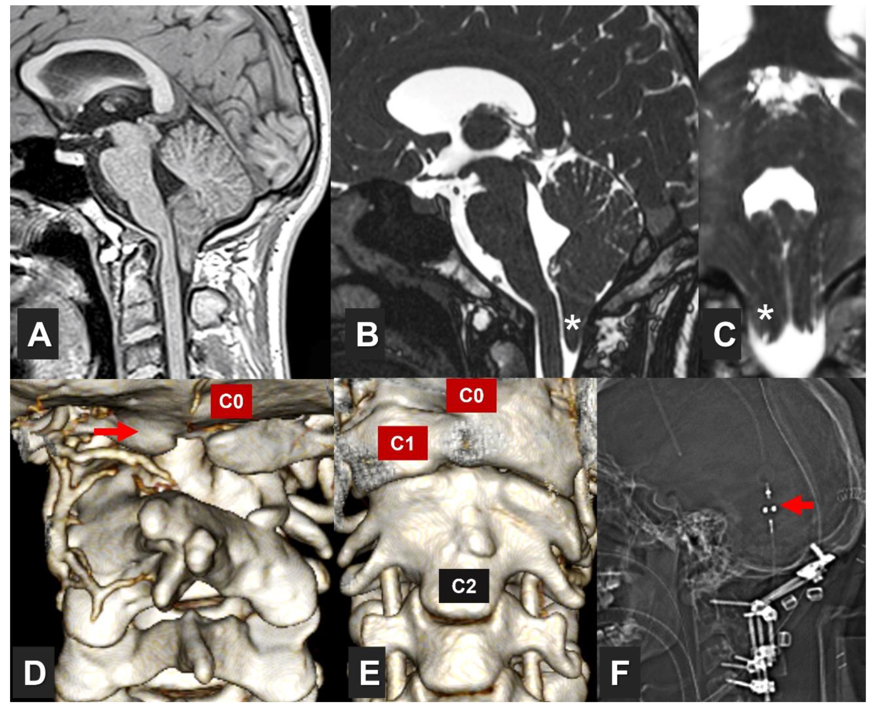
Figure 2.
Flowchart. Diagram of the process used to select the articles for this review. CM1: Chiari malformation type 1. IONM: Intraoperative neurophysiological monitoring.
Figure 2.
Flowchart. Diagram of the process used to select the articles for this review. CM1: Chiari malformation type 1. IONM: Intraoperative neurophysiological monitoring.
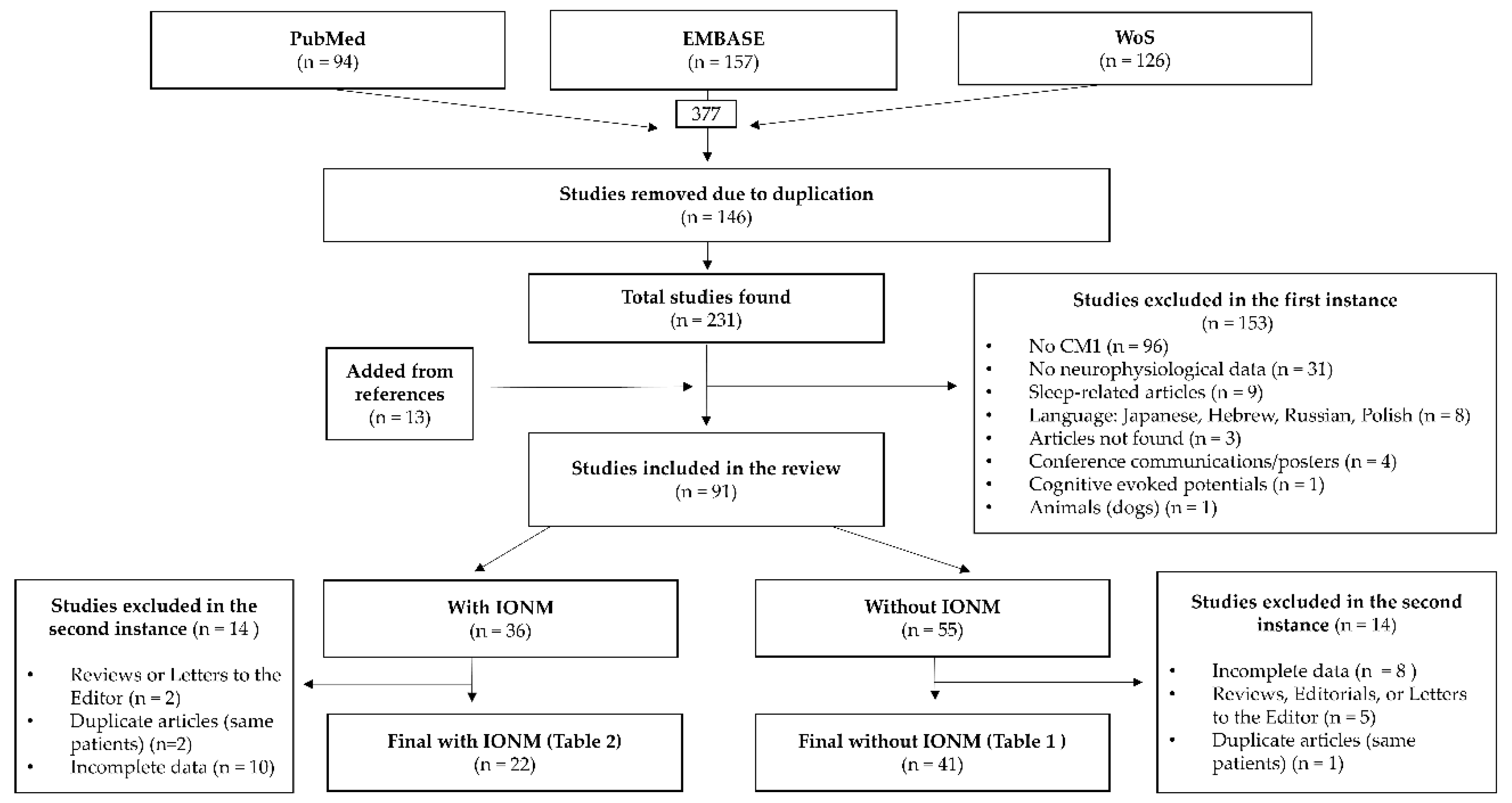
Figure 3.
Example of normal brainstem auditory evoked potentials. The image summarizes the waves recorded after applying the acoustic stimulus with their respective neural generators. Wave I: organ of Corti of the cochlea (the distal portion of the auditory nerve). Wave II: cochlear nucleus (and the proximal intracranial portion of the auditory nerve). Wave III: superior olivary complex, trapezoid body. Wave IV: ventral nucleus of the lateral lemniscus. Wave V: inferior colliculus. Wave VI: medial geniculate nucleus of the thalamus. Wave VII: thalamocortical radiations and primary auditory cortex (Heschl’s) or Brodmann’s Area 41. Ai: ipsilateral earlobe. (Modified from Moncho et al., with permission [23]).
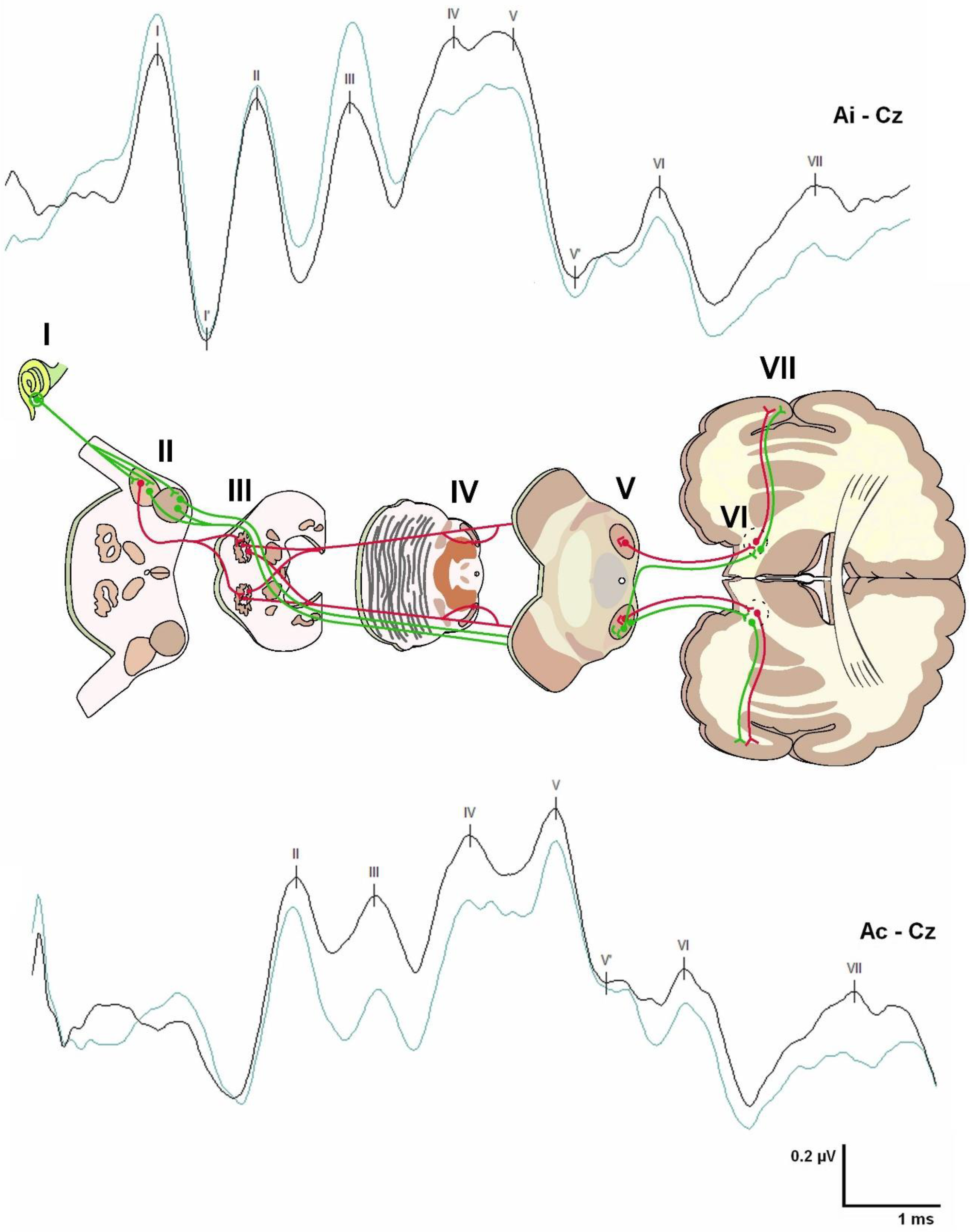
Figure 4.
Example of normal somatosensory evoked potentials. The image shows the responses recorded at different levels after stimulation of the median nerve in the wrist and its main generators. N9: brachial plexus. N11: entry of roots in dorsal horns and posterior columns. N13: high cervical and medullary structures (nuclei of the posterior columns). P14: most caudal part of the medial lemniscus. N18: medial lemniscus at the mesencephalic level. N20: primary somatosensory cortex (Brodmann areas 3, 1, 2). Cc’: contralateral parietal cortex. Hc: Contralateral shoulder. Cv7: the spinous process of the seventh cervical vertebra. (Modified from Moncho et al., with permission [23]).
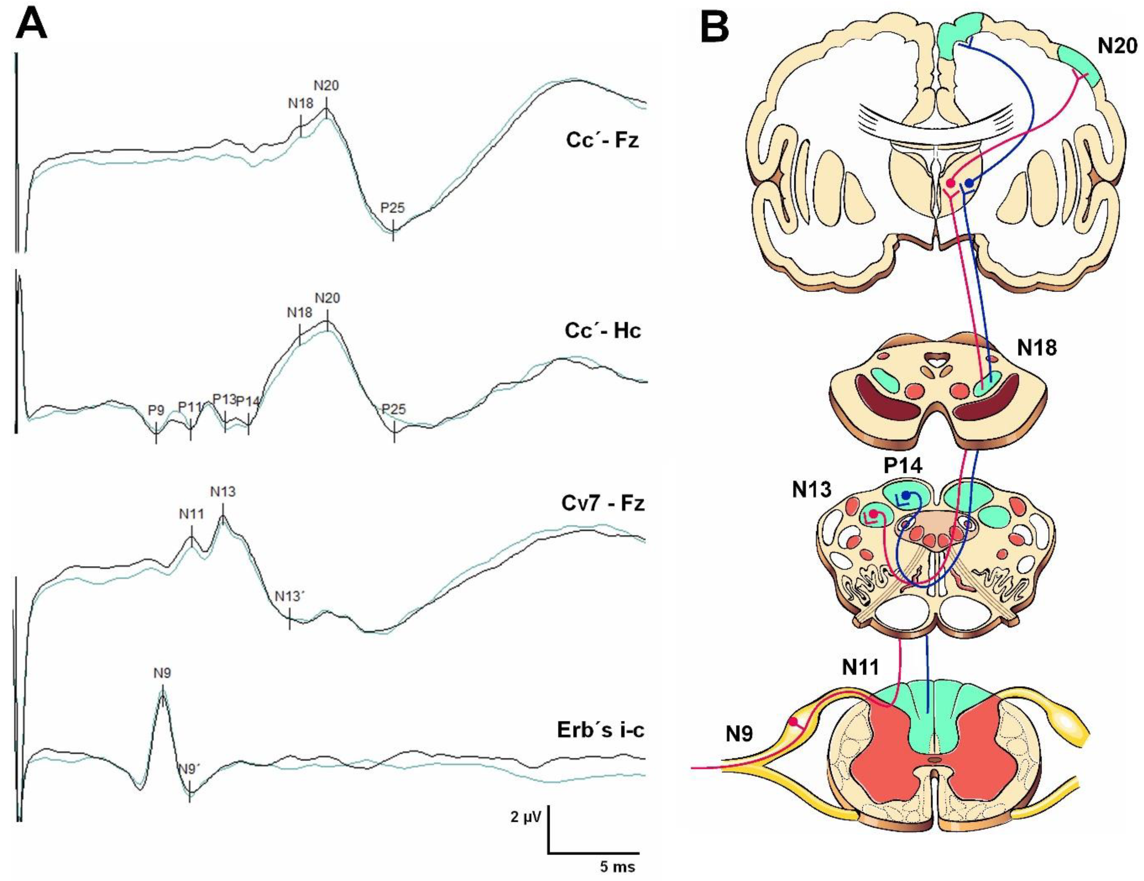
Figure 5.
Blink reflex and laryngeal adductor reflex. Panel (A) shows exemplary traces of the R1 response of the blink reflex (BR) (A1) and the R1 and R2 components of the laryngeal adductor reflex (LAR) (A2) intraoperatively recorded under general anesthesia. Panel (B) illustration represents the setup used to stimulate and record the BR (top scheme, green lines) and the LAR (bottom scheme, purple lines), including a schematic representation of the arc reflex pathways (please, note that discontinued lines are used for afferent fibers and continued lines for efferent pathways), and the brainstem structures involved. V1 is the first branch of the cranial nerve V; CN: cranial nerve; NA: ambiguous nucleus; NST: nucleus of the solitary tract; RLN: recurrent laryngeal nerve; SLN: superior laryngeal nerve; VF: vocal fold.
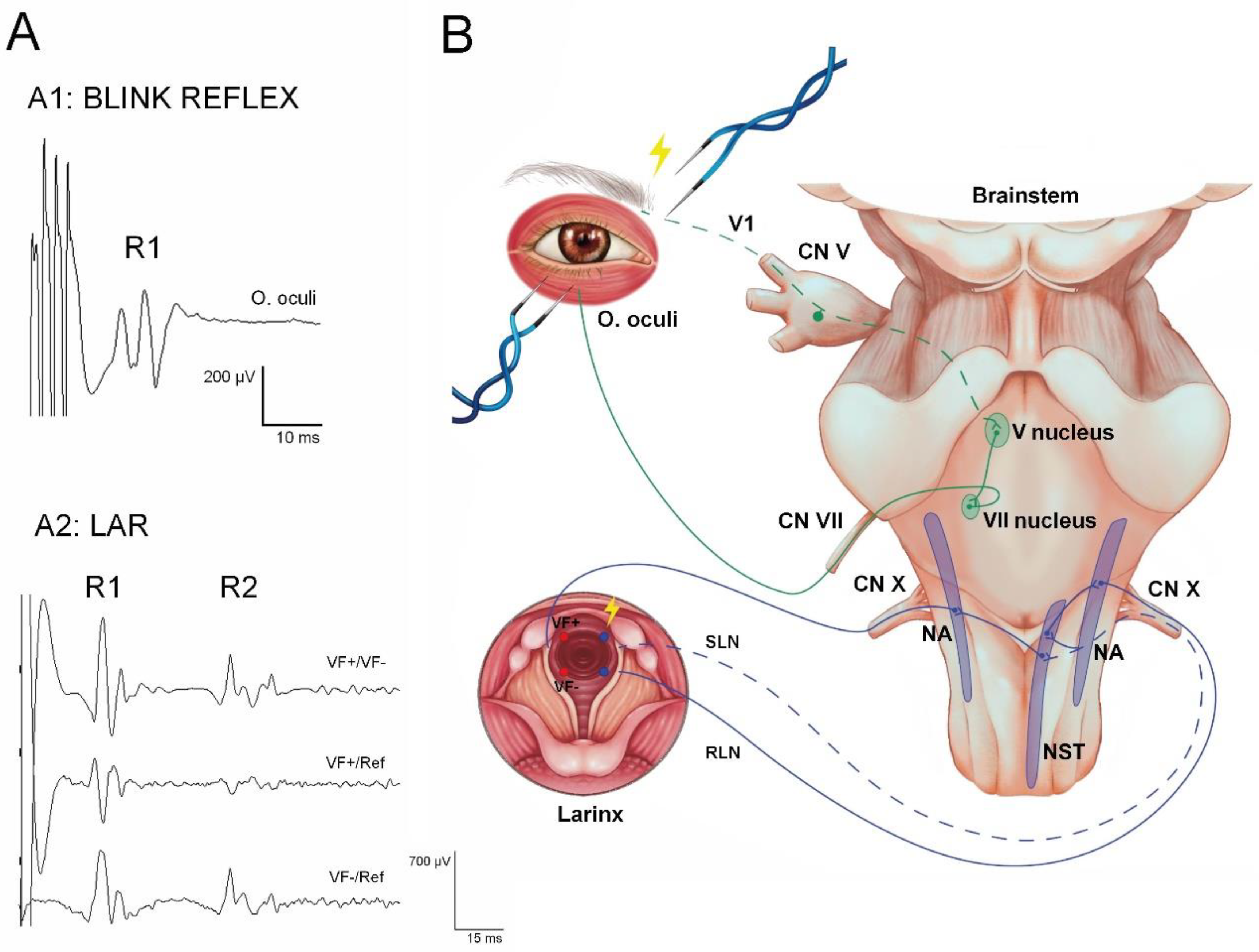
Table 2.
Published neurophysiological studies in neuromonitoring of patients with CM1/Syringomyelia.
Table 2.
Published neurophysiological studies in neuromonitoring of patients with CM1/Syringomyelia.
| Author | Year | No. of cases |
Study type | Patient characteristics | SEP | MEP | BAEP | BR or CN mapping | Alarm criteria | Alerts | Findings | Conclusion |
|---|---|---|---|---|---|---|---|---|---|---|---|---|
| Schaefer et al. [135] | 2022 | 93 | Retrospective | 17-76-y-o SYR= 53 |
93 | 92 | 83 | ― | Decreased SEP AMP by 50%, increased SEP LAT by 10%, or decreased MEP AMP by 50%. A significant change in BAEPs is a decrease in wave V AMP of 50% or an increase in wave V LAT by 1 msec, or total loss of waves (I, III, and V) | 1 (1.1%), which resolved spontaneously after 10 minutes in a patient who had concomitant stenosis as a result of pannus formation at C1–2 (suboccipital craniectomy and C1–2 laminectomy with DP) | TE was significantly associated with unmonitorable MEPs but not with unmonitorable SEPs or BAEPs. SYR characteristics were not significantly associated with unmonitorable MEPs, SEPs, or BAEPs. Cerebellar symptoms were associated with unmonitorable MEPs and BAEPs but not SEPs | PFD in CM1 may be performed safely without IONM. In patients with additional occipitocervical pathology, IONM should be left as an option to be assessed by the surgeon on a case-by-case basis |
| Giampiccolo et al. [151] | 2021 | T = 10 CM1 = 1 |
An exploratory and preliminary study of cerebello-cortical stimulation |
6–73-y-o 2 children |
― | 1 | ― | ― |
N
o MEP was evoked with direct cerebellar stimulation in any of the patients. Paired cortico-transcortical stimulation: M1 stimulation occurred after cerebellar stimulation at fixed intervals between 8 and 24 ms. They observed significant modulation of MEPs in 8/10 patients. 5 patients showed MEP inhibition, 1 patient MEP facilitation, and 2 patients showed both conditions at different interstimulus intervals |
Electrical conditioning stimuli delivered to the exposed cerebellar cortex (cStim) alone did not produce any MEP. cStim preceding Tc Stim produced a significant inhibition at 8 ms (P <0.0001). This inhibition is likely the product of activity along the cerebello-dento-thalamo-cortical pathway | The authors show that monitoring efferent cerebellar pathways to the motor cortex is feasible in intraoperative settings. This study has promising implications for pediatric posterior fossa surgery to preserve the cerebello-cortical pathways and thus prevent cerebellar mutism | |
| Heffez et al. [134] | 2020 | 488 | Observational, prospective | > 18-y-o They divided patients into four groups depending on the position of the cerebellar tonsils: GR 1: 0-3 mm GR 2: 3-5 mm GR 3: 5-10 mm GR 4: > 10 mm They included some patients with tethered cord in GR 4 (CM2?) |
― | ― | 488 | ― | Any improvement or worsening was defined as a change of at least 0.1 msec from their initial BL | A reduction in III-V IPL of at least 0.1 msec was observed in 35-49% of patients | Reduction in III–V IPL was observed towards completing intradural dissection or during DP, with no statistical difference between groups | The observed reduction in IPLs indicates that even with minimal TE, there may be impairment of the function of at least one pathway within the brainstem similar to that seen with much more extreme TE and that this impairment improves after brainstem decompression |
| Krishnakumar et al. [131] | 2020 | 1 | Case report | 13-y-o ♂ with atlantooccipital dislocation, BI, hypoplastic C1 arch, and CM1 |
1 | 1 | ― | ― | N/A | After the prone position, there was a loss of MEP in all four limbs, so neck flexion was reduced by 15°, which reverted the changes in MEP. Following screw tightening, there was a loss of MEP in both upper limbs, and the decision to perform a C1 arch excision was made; MEP reappeared in both upper limbs following this step | IONM can contribute to safe surgical positioning and performance. It is essential for the team involved to promptly identify and rectify any changes in neuronal, structural, and vascular integrity to help minimize neurologic sequelae | |
| Shi et al. [149] | 2020 | 270 | Retrospective & case-matched subgroup analysis | Scoliosis surgery 60 asymptomatic CMS patients (48 presented with SYR) vs 210 IS patients. Case-matched patients: 60 CMS vs. 60 IS. -PFD was performed 8–12 months before correction surgery in whole patients with SYR |
270 | 258 | ― | ― | Absent SEPs, UNIL or BIL prolonged LAT. LAT normalized with body height and > 2.5 SD (P37 LAT = 0.277*height −7.144, SD = 1.071). And asymmetric LAT when interside difference of LAT or AMP > 2.5 SD of normal control |
In terms of SEP LAT and AMP as well as MEP amplitude, no significant difference was found between CMS and total IS patients. There was no significant difference in SEP LAT and AMP and MEP AMP between CMS and matched IS patients. The CMS patients with SYR were correlated with lower SEPs amplitudes |
Neurologically asymptomatic CMS patients showed similar absolute values of IONM, including SEP LAT and AMP, and MEP AMP as compared with IS patients. The SYR in CMS patients indicated more severe curvature and a lower SEP AMP even after PFD | |
| Tan et al. [148] | 2020 | 42 | Retrospective & case-matched | Scoliosis surgery 21 patients with SCOL secondary to CM1 and SYR matched with 21 patients with SCOL secondary to idiopathic SYR |
― | 42 | ― | ― | -Obvious MEP degeneration was defined as 40% to 80% MEP AMP loss. -Significant MEP loss with monitoring alerts was defined as >80% of MEP loss associated with high-risk surgical maneuvers |
-Obvious MEP degeneration in 5 patients -Significant MEP loss in none |
There were no differences in the successful recording of MEP BL | Although patients with CM1 had longer SYR, their IONM signals during the operation were similar to those of the SCOL secondary to idiopathic SYR group. The potential risk of SCOL surgery in patients with SYR-associated SCOL should not be ignored |
| Shah et al. [137] | 2019 | 20 | Observational, prospective | 7-52-y-o, with CM1 that were surgically treated by atlantoaxial stabilization surgery. No bone, dural, or neural decompression |
20 | 20 | ― | ― | N/A | All patients had immediate intraoperative improvement in their BL MEPs after fixation was completed, ranging from 20% to 35%. No change in SEPs during the entire surgery in any of the patients |
The improvement in electrophysiologic parameters after atlantoaxial fixation fortifies their belief that atlantoaxial instability is the cause of Chiari formation and atlantoaxial fixation is the treatment | |
| Rasul et al. [129] | 2019 | 37 | Retrospective | < 17-y-o undergoing PFD for CM1 SYR = 24 SCOL = 13 |
33 | ― | 19 | ― | N/A | SEP → 2/33 ↑AMP 18/33 ↓ AMP 31/33 ↓ LAT BAEP→ 13/19 ↓ LAT 4/19 ↑ LAT |
SEP LAT ↓ in 93.9% of patients. More than 50% of patients had a ↓ in their SEP AMP BAEPs ↓ in 68.4% of patients |
PFD for CM1 is associated with changes in SEPs and BAEPs. However, the authors were unable to identify a definite link between clinical outcomes and IONM |
| Brînzeu and Sindou [150] |
2017 | T=49 22 CM1 |
Research study about the functional anatomy of the accessory nerve (XI CN) studied through IONM (mapping) |
20-73-y-o Far-lateral decompression of the tonsils at the FM in addition to posterior decompression, followed by a Y-shaped dural incision without opening the arachnoid |
Rootlet and cranial root stimulation in the majority of CM1 patients | The CN XI pair is organized around two components: a cranial root and a spinal root with several cervical rootlets. The cranial component contributes at least to the motor innervation of the larynx. The spinal component largely contributes to the innervation of the sternocleidomastoid and trapezius with a precise craniocaudal myotopic organization; the rootlets destined to innervate the sternocleidomastoid are more cranial |
||||||
| Kawasaki et al. [130] | 2017 | 1 | Case report | Symptomatic 7-y-o boy who underwent surgery of PFD with tonsillectomy and DP for CM1 with cervicomedullary compression + pre-SYR state at the C3-4 dorsal spinal cord | 1 | 1 | ― | ― | Changes of 50% in AMP and 10% in LAT from the BL value were considered significant for intraoperative use both in MEPs and SEPs | MEPs improved, showing ↑ AMP and ↓ LAT after craniotomy and durotomy, whereas SEPs improved only after durotomy | The improvement in MEPs and SEPs observed during decompression may be a good indicator for the prediction of the clinical improvement seen postoperatively | |
| Roser et al. [133] | 2016 | 39 | Retrospective | 13–65-y-o patients with CM1, undergoing suboccipital decompression and DP SYR = 33 |
38 | 33 | ― | ― | N/A |
SEP→ Deterioration during positioning 2/39, ↑ >10% LAT in 4 recordings, ↓ or ↑ >50% AMP in 9 and 10 recordings; MEP→ Deterioration during positioning 2/39, ↓ and ↑ > 10% LAT in 11 and 10 recordings, ↓ and ↑ > 50% AMP in 14 and 17 recordings |
There were no significant differences in absolute BL and final LAT and AMP of MN and PTN SEPs. There were no significant differences in absolute BL and final LAT and AMP of APB and TA MEPs |
IONM during primary treatment of CM1 shows only subtle nonsignificant changes in SEPs and MEPs without clinical correlation during suboccipital decompression |
| Barzilai et al. [153] | 2015 | 22 | Retrospective | 21 pediatric patients aged 2–17-y-o with CM1 SYR =18 PFD + C1 (C2/C3) laminectomy (22 surgeries) |
22 | 22 | ― | ― | N/A | IONM position-related alarms in 3 patients: 1 attenuation only of SEPs, 1 attenuation only of MEPs, and one attenuation in SEPs and MEPs together | None of the 3 patients displayed new immediate postoperative deficits |
Multimodality IONM can be helpful in PFD surgery, particularly during patient positioning. MEP attenuations may occur independently of SEPs. The clinical implications of these monitoring alerts have yet to be defined |
| Grossauer et al. [132] | 2015 | 1 | Case report | A 32-y-o woman who underwent surgery for CM1 associated with extensive cervicothoracic SYR | 1 | ― | ― | ― | A change of 50% in N20 amplitude from the BL value and a change of 10% in N20 LAT from the BL value | From positioning to completion of dura opening, there were no significant changes in MN SEP. A few minutes after opening the IV ventricle, they observed a 230% ↑ in the N20 AMP and an 8% ↓ in the N20 LAT compared with the BL value. This clear SEP improvement persisted until the end of the operation |
Conduction improvement in SEPs during CM1 decompression may not always occur after bone decompression or DP. It may also happen after opening the IV ventricle and establishing CSF flow at the level of the CVJ. Additional studies are needed to adapt the degree of decompression to each CM1 patient based on IONM data | |
| Kennedy et al. [127] | 2015 | 156 | Retrospective | 7-20.6-y-o Suboccipital decompression without dural opening SYR = 68 SCOL = 18 m Cobb angle= 25° |
156? | ― | 156? | ― | N/A | N/A | BAEPs and SEPs were performed before (supine) and after (prone) positioning and throughout the surgery. 121 (78%) of patients exhibited at least UNIL improvement in I–V IPL after bony decompression, with a mean improvement of 0.26 msec. The remaining 34 patients had stable BAEPs throughout the surgery. Once a preoperative neck position was established in the prone position with SEPs unchanged from BL, there were no adverse changes in the SEPs during any patient’s surgery |
|
| Pencovich et al. [140] | 2013 | T= 13 CM1= 6 |
SYR Surgery 4-61-y-o →1 CM1= Syrinx drainage and PFD. Level T4-T5 →5 CM1= Syringo-subarachnoid shunt SYR approach: →4 midline →2 DREZ |
6 | 6 | ― | ― | SEP alert criteria included non-linear AMP ↓ beyond 50 % or LAT ↑ of over 10 %. Alert criteria for MEP were significant changes, including sudden attenuation beyond 85 % AMP in at least two reproducible traces after ruling out technical and anesthetic considerations |
In one, absent right leg MEP BL signal. While draining the SYR before PFD, severe attenuation of left leg MEP data was noted upon midline approach to the SYR. The catheter was subsequently removed, and the PFD was completed without SYR drainage. New neurologic deficit: Transient worsening of right hemiparesis with gradual improvement over a few days | This study demonstrates the reversibility of intraoperative neurological damage identified by IONM. An immediate response resulted in rapid recovery of cord functionality | Data collected in this study support routine usage of IONM in SYR surgeries. IONM can inform the surgeon of potential intraoperative threats to the functional integrity of the spinal cord, providing a helpful adjunct to spinal cord surgeries for the treatment of SYR. More extensive prospective studies are required to conclusively show that using IONM in these operations is indeed advantageous. The first study to specifically address the benefits of multimodality IONM during SYR surgery |
|
| Chen et al. [126] | 2012 | 13 | 2-17-y-o Suboccipital craniotomy for symptomatic CM1. In 3, the bone flap was not replaced (craniectomy), and in 9 cases, it was (craniotomy) SYR = 3 |
12 | ― | 9 | ― | N/A | MN or PTN SEP LAT improved in all patients. BAEPs improved in 8 patients. No significant SEP or BAEP deterioration was seen |
Improvements in neurophysiological parameters were sustained for the duration of the procedure. The interaction terms of the ANOVA were insignificant at the α =0.05 level | IONM may be used to perform suboccipital decompression in a step-by-step fashion, enlarging the craniectomy or adding additional procedures (laminectomy, DP) until positive changes are observed | |
| Di [136] | 2009 | 26 | Endoscopic suboccipital decompression 18 months-16-y-o 0° and 30° endoscopes were adapted to perform the procedure of suboccipital craniectomy and upper cervical laminectomies SYR = 5 SCOL = 1 |
11 | ― | ― | ― | SEPs were monitored throughout the entire procedure for the first 11 patients, and it was then discontinued due to lack of significant benefit for the patients |
SEP is still necessary, especially for the beginners of this procedure, to preclude the development of intraoperative spinal cord injury | |||
| Zamel et al. [124] | 2009 | 80 | Retrospective | 2-36-y-o Group A: PFD Group B: PFD+ DP SYR = 33 |
80 | ― | 80 | ― | BAEP waves I, III, and V were determined, and IPLs of waves I to III, III to V, and I to V were compared at BL, after positioning, immediately after bony decompression, and at closure | Neurophysiologic improvement in CCT was defined as a reduction in I-V IPL at closure compared with BL | A significant main interaction was found between the presence of SYR and reduction of I-V IPL from BL to decompression. Patients with SYR showed a significantly decreased I-V interval between BL and decompression compared with those without an SYR. No SEP findings were detailed |
PFD with bone removal alone significantly improves conduction time in most pediatric patients with CM1. DP allowed for a further but slight improvement in conduction time in only 20% of patients, beyond that achieved by decompression alone. None of the patients had any significant worsening of their BAEPs that would have alerted the neurosurgeon to modify the course of the surgery |
| Kim et al. [125] | 2004 |
11 | Retrospective | 1.5–17-y-o Significant BI and CM1 They underwent a novel treatment method involving decompression, manual reduction, and posterior instrumentation-augmented fusion. SYR= 3 |
11 | ― | ― | ― | N/A | ― | SEPs remained stable in 10 cases and improved intraoperatively after decompression and manual reduction in 1 case. Immediately after reduction and fusion, SEPs in 10 patients improved significantly, and in one, remained unchanged from BL | |
| Anderson et al. [123] | 2003 | 11 | Observational, prospective | 3-19-y-oSuboccipital decompressive procedure with DPSYR = 6 | 11 | ― | 11 | ― | N/A | In one case, the left MN SEP dramatically deteriorated after turning the patient to the prone position with neck flexion. The patient was immediately repositioned in a neutral position, improving the left MN SEP.BL BAEPs were abnormal, withdelayed I–III IPLs bilaterally. On flexion, BAEPs remained at BL | BAEPs: Decreased I-V IPLs after bone decompression but not after dural opening, statistically significant (compared with BL in supine).SEP for both sides in 10 patients remained stable throughout the procedure | Suggests that, in pediatric CM1 patients, the maximum improvement in conduction through the brainstem occurs after bony decompression and division of the atlantooccipital membrane rather than after dural opening. BAEPs and SEPs indicate that these patients are at risk for neurologic injury during operative positioning with neck flexion |
| Milhorat et al. [139] | 1997 | T=3221 CM1 | 5-72-y-o In SYR with CM1: Patients with CM1 underwent suboccipital craniectomies without opening the dura + SYR shunting procedures | 21 | ― | ― | ― | N/A | Eight patients had normal SEPs, and 6 had SEP abnormalities that were unchanged 30 minutes after SYR decompression. In 18 of 24 patients with pre-drainage abnormalities, there was a slight but consistent ↓ N20 LAT and less consistent ↑ of N20 AMP | Significant correlation between SEP findings and SYR morphology: Of 8 patients with normal SEPs, 6 had central core cavities, and 2 had central cavities that extended paracentrally. These types of cavities were more likely than eccentric cavities to be associated with SEP improvements after shunting (Pearson Χ2 = 6.47, P = 0.039) | The improvement in N20 LAT is indirect evidence of preexisting long tract compression, whereas improvements in N20 amplitudes were presumably caused by the decompression of perisyrinx neurons. These conclusions were less certain for patients with CM1 in whom SEP improvements may have been caused, in part, by decompression of the cerebellar hernia | |
| Milhorat et al. [138] | 1996 | T=1311 CM1 | 12-72-y-o In SYR with CM1: PFD and SYR shunt to the PF cisterns (syringo-cisternostomy) | 11 | ― | ― | ― | Continuous BL values recorded2-3 h before SYR shunting were compared with values obtained 30 min after shunting.SEP data were analyzed by the paired t-test | Bilateral MN SEPs: 30 min after SYR decompressiondemonstrated a significant ↓ N20 LAT and nonsignificant ↑ N20 AMP | Findings suggest that distended spinal cord cavities can produce regional ischemia possibly reflected by SEP abnormalities and reversed by SYR decompression. Preliminary, the IONM of SEPs can provide useful information during surgical procedures for SYR | ||
AMP: amplitude; APB: abductor pollicis brevis; BAEPs: brainstem auditory evoked potentials; BI: basilar impression; BL: baseline; BIL: bilateral; BR: blink reflex; CCT: central conduction time; CM1: Chiari 1 malformation; CM2: Chiari 2 malformation; CMCT: central motor conduction time; CMS: Chiari malformation-associated scoliosis; CN: cranial nerve; CPN: common peroneal nerve; CSF: cerebrospinal fluid; CVJ: craniovertebral junction; dCHEPs: dermatomal contact heat evoked potentials; DP: duraplasty; dSEPs: dermatomal somatosensory evoked potentials; EMG: electromyography; FA: Fractional anisotropy; FDI: first dorsal interosseus; IONM: intraoperative monitoring; IPL: interpeak latency; IS: idiopathic scoliosis; LAT: latency; m: mean; MEPs: motor evoked potentials; MN: median nerve; MUP: motor unit potentials; NCS: nerve conduction studies; PF: posterior fossa; PFD: posterior fossa decompression; PROL: prolonged; PTN: posterior tibial nerve; SCOL: scoliosis; SEPs: somatosensory evoked potentials; SNHL: sensory neural hearing loss; SYR: Syringomyelia; T: Total; TA: tibialis anterior; TcMEP: transcranial-electric motor evoked potential; TE: tonsillar ectopia; TMS: transcranial magnetic stimulation; UNIL: unilateral; *: Previous surgery.
Disclaimer/Publisher’s Note: The statements, opinions and data contained in all publications are solely those of the individual author(s) and contributor(s) and not of MDPI and/or the editor(s). MDPI and/or the editor(s) disclaim responsibility for any injury to people or property resulting from any ideas, methods, instructions or products referred to in the content. |
© 2023 by the authors. Licensee MDPI, Basel, Switzerland. This article is an open access article distributed under the terms and conditions of the Creative Commons Attribution (CC BY) license (http://creativecommons.org/licenses/by/4.0/).
Copyright: This open access article is published under a Creative Commons CC BY 4.0 license, which permit the free download, distribution, and reuse, provided that the author and preprint are cited in any reuse.


