
Submitted:
31 July 2023
Posted:
01 August 2023
You are already at the latest version
Abstract
There is an increasing interest in biomarkers of nitric oxide dysregulation and oxidative stress to guide management and identify new therapeutic targets in patients with chronic obstructive pulmonary disease (COPD). We conducted a systematic review and meta-analysis of the association between circulating metabolites within the arginine (arginine, citrulline, ornithine, asymmetric, ADMA, and symmetric, SDMA dimethylarginine), transsulfuration (methionine, homocysteine, and cysteine) and folic acid (folic acid, vitamin B6, and vitamin B12) metabolic pathways and COPD. We searched electronic databases from inception to 30 June 2023 and assessed the risk of bias and the certainty of evidence. In 21 eligible studies, compared to healthy controls, patients with stable COPD had significantly lower methionine (standardized mean difference, SMD=-0.50, 95% CI -0.95 to -0.05, p=0.029) and folic acid (SMD=-0.37, 95% CI -0.65 to -0.09, p=0.009), and higher homocysteine (SMD=0.78, 95% CI 0.48 to 1.07, p˂0.001) and cysteine concentrations (SMD=0.34, 95% CI 0.02 to 0.66, p=0.038). Additionally, COPD was associated with significantly higher ADMA (SMD=1.27, 95% CI 0.08 to 2.46, p=0.037), SDMA (SMD=3.94, 95% CI 0.79 to 7.08, p=0.014), and ornithine concentrations (SMD=0.67, 95% CI 0.13 to 1.22, p=0.015). In subgroup analysis, the SMD of homocysteine was significantly associated with the biological matrix assessed and the forced expiratory volume in the first second to forced vital capacity ratio, but not with age, study location, or analytical method used. Our study suggests the presence of significant alterations in metabolites within the arginine, transsulfuration, and folic acid pathways which can be useful for assessing nitric oxide dysregulation and oxidative stress and identifying novel treatment targets in COPD. (PROSPERO registration number: CRD42023448036)
Keywords:
folic acid
; transsulfuration
; oxidative stress
; nitric oxide
; biomarkers
; chronic obstructive pulmonary disease
; homocysteine
; asymmetric dimethylarginine
; symmetric dimethylarginine
; ornithine
1. Introduction
The global public health and financial burden of chronic obstructive pulmonary disease (COPD) remains unacceptably high despite the availability of different pharmacological and non-pharmacological treatments in this ever-increasing patient group [1,2,3,4,5,6,7]. Such challenges have stimulated a significant body of research to better understand the molecular, biochemical, and cellular mechanisms underpinning the pathophysiology of COPD and identify novel druggable targets and therapies [8,9,10,11]. Whilst the role of local (airway) and systemic inflammation in COPD is well established, using conventional biomarkers (e.g., C-reactive protein) and specific blood cell types [12,13,14,15], studies have also focused on the dysregulation of the endogenous messenger nitric oxide (NO) and the redox state [16,17,18,19,20,21,22,23,24]. The investigation of possible alterations in the NO pathway and redox balance are also important in this context given their involvement in other disease states, some of them, e.g., atherosclerosis and cardiovascular disease, frequently associated with COPD [25,26,27,28,29,30,31]. For example, in epidemiological studies the prevalence of atherosclerotic cardiovascular disease in patients with COPD has been shown to range between 20% and 60% [32,33,34]. Furthermore, the coexistence of COPD and cardiovascular disease is associated with poorer quality of life and functional capacity and a higher risk of COPD exacerbations, hospitalizations, and mortality [35,36,37,38].
A significant limitation in the development of analytical platforms for the assessment of NO and biomarkers of oxidative stress in biological samples is represented by the highly reactive nature of these compounds, the relatively short half-life of NO, and the influence of other factors in the assessment of circulating NO metabolites such as nitrite and nitrate [39,40,41,42,43,44,45]. Therefore, an alternative approach consists in measuring stable metabolites within metabolic pathways that are closely associated with NO synthesis and oxidative stress. In this context, several metabolites within the arginine, transsulfuration, and folic acid metabolic pathways have been shown to reflect alterations in NO synthesis and/or redox state. Furthermore, these metabolites can be measured in serum or plasma using a wide range of analytical methods (Figure 1) [46,47,48,49,50,51,52]. The arginine pathway includes a) arginine, a critical amino acid and substrate for several enzymes, e.g., protein arginine methyltransferases (PRMTs), arginase 1 and 2, and NO synthases (NOS) [46,53]; b) citrulline, the end product of enzymatic reactions catalyzed by NOS and isoform 1 of dimethylarginine dimethylaminohydrolase (DDAH1) [46,54]; c) the methylated arginine analogues, asymmetric (ADMA) and symmetric (SDMA) methylarginine, which directly (ADMA) or indirectly (SDMA) downregulate NO synthesis [53,54,55,56,57]; and d) ornithine, the end product of arginase 1 and 2 (Figure 1) [46,58]. The transsulfuration pathway regulates sulfur metabolism and redox balance and primarily involves the transfer of sulfur from homocysteine, a highly reactive amino acid derived from the dietary compound, methionine, to cysteine through the intermediate cystathionine, in enzymatic reactions that require vitamin B6 (Figure 1) [48,59]. Finally, the folic acid pathway plays a critical role in regulating several intracellular homeostatic mechanisms that also include the lowering of homocysteine concentrations through the regeneration of methionine in enzymatic reactions that involve vitamin B12 (Figure 1) [60,61].
Importantly, the known associations between the arginine, transsulfuration, and folic acid pathways, vascular homeostasis, and cardiovascular outcomes might also allow investigating the complex interplay between COPD, NO, oxidative stress, and atherosclerotic cardiovascular disease [53,54,56,62,63,64,65,66,67,68,69,70,71,72]. This knowledge would be potentially useful for identifying new therapeutic targets and management approaches in patients with COPD.
We investigated this issue by a) appraising the available evidence, through a systematic review and meta-analysis, of the association between the circulating concentrations of key metabolites within the arginine, transsulfuration, and folic acid metabolic pathways and COPD, and b) assessing, where possible, the relationship between the effect size of the observed differences vs. healthy controls and clinical and demographic characteristics.
2. Materials and Methods
2.1. Study selection
A systematic search of publications was conducted in the electronic databases PubMed, Web of Science, and Scopus from inception to 30 June 2023. The search utilized the following terms and their combinations: “COPD” OR “chronic obstructive pulmonary disease” AND “methionine” OR “homocysteine” OR “cysteine” OR “cystathionine” OR” S-adenosylmethyonine” OR “ S-adenosylhomocysteine” OR “S-adenosyl-methyonine” OR “ S-adenosyl-homocysteine” OR “betaine” OR “dimethylglycine” OR “folates” OR “folic acid” OR “B12” OR “cobalamin” OR “B6” OR “pyridoxine” OR “arginine” OR “asymmetric dimethylarginine” OR “ADMA” OR “symmetric dimethylarginine” OR “citrulline” OR “ornithine”.
Two investigators independently screened the abstracts, full-text articles, and relevant references according to the following inclusion criteria: (a) the assessment of homocysteine, cysteine, methionine, vitamin B6, vitamin B12, folic acid, arginine, ADMA, SDMA, citrulline, or ornithine in plasma or serum, (b) the study of patients with stable COPD and healthy controls using a case-control design, (c) the inclusion of participants ≥18 years, and (d) the availability of full text in English language. The main exclusion criterion was the assessment of patients with acute exacerbations of COPD. The two investigators independently extracted the following variables into an electronic spreadsheet for further analysis: year of publication, first author, study country, participant number, age, male to female ratio, forced expiratory volume in the first second (FEV1), FEV1/forced vital capacity (FVC), biological matrix (plasma or serum), and analytical method used. A third investigator was involved in case of disagreement.
The Joanna Briggs Institute Critical Appraisal Checklist was used to assess the risk of bias [73], whereas the Grades of Recommendation, Assessment, Development, and Evaluation (GRADE) Working Group system was used to assess the certainty of evidence [74]. The Preferred Reporting Items for Systematic Reviews and Meta-Analyses 2020 statement was followed to present the results [75], and the International Prospective Register of Systematic Reviews was used to register our review (PROSPERO registration number: CRD42023448036).
2.2. Statistical analysis
We created forest plots of standardized mean differences (SMDs) and 95% confidence intervals (CIs) (p-value <0.05 for statistical significance), and estimated means and standard deviations from medians and interquartile ranges or ranges [76,77], or using the Graph Data Extractor software (San Diego, CA, USA). The heterogeneity of SMD was evaluated using the Q statistic (significance level set at p<0.10) [78,79]. Sensitivity analysis was used to assess the stability of the results [80]. The Egger’s and Begg’s tests and the "trim-and-fill" method were used to assess publication bias [81,82,83]. Univariate meta-regression and subgroup analyses investigated associations between the effect size and the following parameters: year of publication, study continent, sample size, age, male to female ratio, FEV1, FEV1/FVC, biological matrix, and analytical method used. Statistical analyses were performed using Stata 14 (Stata Corp., College Station, TX, USA).
3. Results
3.1. Literature search
From a total of 1,788 articles, we excluded 1,759 as they were either duplicates or irrelevant. A full-text revision of the remaining 29 articles led to the exclusion of further eight because they had missing data (n=2), unsuitable (not case-control) design (n=4) or included patients with acute exacerbation of COPD (n=2). The 21 studies included in the final analysis were published between 1998 and 2020 (Figure 2 and Table 1) [84,85,86,87,88,89,90,91,92,93,94,95,96,97,98,99,100,101,102,103,104]. There was no disagreement between the two independent investigators, therefore input from a third investigator was not required. The cross-sectional design of all studies was primarily responsible for the initial low level of certainty given (rating 2, ⊕⊕⊝⊝). The risk of bias was low in all studies (Supplementary Table S3) [84,85,86,87,88,89,90,91,92,93,94,95,96,97,98,99,100,101,102,103,104].
3.2. Homocysteine
Homocysteine was measured in 11 studies investigating a total of 610 COPD patients (mean age 57 years, 72% males) and 468 healthy controls (mean age 44years, 66% males) [85,87,88,89,90,91,97,98,102,103,104], six conducted in Asia [87,91,97,98,102,103], four in Europe [85,88,90,104], and one in Africa [89]. Liquid chromatography was used in four studies [85,87,89,90], an enzyme-linked immunosorbent assay in two [91,102], capillary electrophoresis laser induced with fluorescence detection in one [104], and a fluorescence polarization immunoassay in the remaining one [88]. No information regarding the analytical method was reported in three studies [97,98,103]. In liquid chromatography studies, two used a fluorimetric detector [87,90], and the remaining two an ultraviolet detector [85,89]. Homocysteine was measured in plasma in eight studies [85,87,88,90,91,102,103,104], and in serum in the remaining three [89,97,98]. The FEV1 was reported in eight studies (range between 39% and 70%) [87,88,90,97,98,102,103,104], and the FEV1/FVC in five (range between 53% and 68%) [88,90,97,103,104].
Homocysteine concentrations were significantly higher in COPD patients compared to controls (SMD=0.78, 95% CI 0.48 to 1.07, p˂0.001; I2=79.4%, p<0.001; Figure 3). The results were stable in sensitivity analysis (SMD range between 0.69 and 0.85; Figure 4). There was no publication bias (Begg’s test, p=0.64); Egger’s test, p=0.51). No additional study was identified using the "trim-and-fill" method (Figure 5).
There were no significant associations in meta-regression between the effect size and male to female ratio (t=-0.36, p=0.73), number of participants (t=1.39, p=0.20), or publication year (t=0.10, p=0.92). In subgroup analysis, no significant differences (p=0.47) in the pooled SMD were observed between studies in patients ≤70 years (SMD=0.81, 95% CI 0.39 to 1.24, p˂0.001; I2=84.0%, p<0.001), or >70 years (SMD=0.58, 95% CI 0.22 to 0.93, p=0.001; I2=56.8%, p=0.074; Figure 6), with a lower between-study variance in the >70 years subgroup. Similarly, no significant differences (p=0.86) in effect size were observed between studies conducted in Europe (SMD=0.69, 95% CI 0. 45 to 0.93, p˂0.001; I2=0.0%, p=0.998) and Asia (SMD=0.74, 95% CI 0.26 to 1.23, p=0.003; I2=88.6%, p˂0.001; Figure 7), with a virtually absent heterogeneity in the European subgroup. Additionally, no significant differences (p=0.95) in the pooled SMD were observed between studies using high performance liquid chromatography (SMD=0.90, 95% CI 0.58 to 1.22, p˂0.001; I2=17.9%, p=0.30) and other methods (SMD=0.89, 95% CI 0.39 to 1.39, p˂0.001; I2=81.5%, p=0.001; Figure 8), with a lower between-study variance in the liquid chromatography subgroup. Among the liquid chromatography studies, no significant differences (p=0.64) in the pooled SMD were observed between studies using ultraviolet detection (SMD=1.04, 95% CI 0.29 to 1.79, p=0.007; I2=64.7%, p=0.092), and fluorimetric detection (SMD=0.81 95% CI 0.43 to 1.19, p=0.001; I2=0.0%, p=0.55; Figure 9), with a virtually absent heterogeneity in the fluorimetric detection subgroup. The pooled SMD was statistically significant in studies assessing plasma (SMD=0.90, 95% CI 0.64 to 1.16, p˂0.001; I2=63.9%, p=0.007), but not serum (SMD=0.51, 95% CI -0.19 to 1.21, p=0.16; I2=84.1%, p=0.002; Figure 10). Furthermore, the pooled SMD was statistically significant in studies of patients with FEV1 ≤55% (SMD=0.72, 95% CI 0.32 to 1.13, p˂0.001; I2=75.9%, p=0.002), but not FEV1 ˃55% (SMD=0.79, 95% CI -0.03 to 1.61, p=0.06; I2=92.6%, p˂0.001; Figure 11). Finally, the pooled SMD was statistically significantly in studies of patients with FEV1/FVC ≤60% (SMD=0.92, 95% CI 0.63 to 1.22, p˂0.001; I2=33.9%, p=0.22), but not FEV1/FVC ˃60% (SMD=0.39, 95% CI -0.16 to 0.94, p=0.17; I2=73.7%, p=0.051; Figure 12), with a lower heterogeneity in the FEV1/FVC ≤60% subgroup.
The level of certainty remained low (rating 2, ⊕⊕⊝⊝) after considering the low risk of bias in all studies, the high but partially explainable heterogeneity, the lack of indirectness, the relatively low imprecision, the moderate effect size, and the lack of publication bias.
3.2. Cysteine
Cysteine was measured plasma in two European studies including a total of 73 COPD patients (mean age 72 years, 66% males) and 83 healthy controls (mean age 70 years, 65% males) [85,104]. Liquid chromatography with ultraviolet detection was used in one study [85], and capillary electrophoresis with laser-induced fluorescence in the other [104]
Cysteine concentrations were significantly higher in COPD patients compared to controls (SMD=0.34, 95% CI 0.02 to 0.66, p=0.038; I2=0.0 %, p=0.83; Figure 13). The limited number of studies prevented sensitivity analysis, the assessment of publication bias, and the conduct of meta-regression and subgroup analyses.
The level of certainty was downgraded to very low (rating 1, ⊕⊝⊝⊝) after considering the low risk of bias in all studies, the virtually absent heterogeneity, the lack of indirectness, the relatively low imprecision, the relatively small effect size, and the lack of assessment of publication bias (downgrade one level).
3.3. Methionine
Two studies investigated plasma methionine in a total of 42 COPD patients and 38 healthy controls [84,86]. One study was conducted in Europe [84], and the other in Asia one [86]. Liquid chromatography with fluorimetric detection was used in both studies [84,86].
Methionine concentrations were significantly lower in COPD patients compared to controls (SMD=-0.50, 95% CI -0.95 to -0.05, p=0.029; I2=0.0 %, p=0.80; Figure 14). The limited number of studies prevented sensitivity analysis, the assessment of publication bias, and the conduct of meta-regression and subgroup analyses.
The level of certainty was downgraded to very low (rating 1, ⊕⊝⊝⊝) after considering the low risk of bias in all studies, the virtually absent heterogeneity, the lack of indirectness, the relatively low imprecision, the relatively moderate effect size, and the lack of assessment of publication bias (downgrade one level).
3.4. Vitamin B12
Vitamin B12 was measured in three studies including a total of 125 patients (mean age 71 years, 84% males) and 85 healthy controls (mean age 71 years, 83% males) [89,90,97]. One study was conducted in Africa [89], one in Europe [90], and one in Asia [97]. One study used liquid chromatography with ultraviolet detection [89], the second a chemiluminometric immunoassay [90], and the third did not provide relevant details regarding the analytical method used [97]. Two studies assessed serum [89,90], and the third plasma [97].
There were non-significant differences in Vitamin B12 concentrations between COPD patients and controls (SMD=-0.20, 95% CI -0.48 to 0.08, p=0.16; I2=54.7 %, p=0.11; Figure 15). The limited number of studies prevented sensitivity analysis, the assessment of publication bias, and the conduct of meta-regression and subgroup analyses.
The level of certainty was downgraded to very low (rating 1, ⊕⊝⊝⊝) after considering the low risk of bias in all studies, the moderate heterogeneity, the lack of indirectness, and the lack of assessment of publication bias (downgrade one level).
3.5. Folic acid
Three studies measured folic acid in a total of 125 COPD patients (mean age 71 years, 84% males) and 85 healthy controls (mean age 71 years, 83% males) [89,90,97]. One study was conducted in Africa [89], one in Europe [90], and one in Asia [97]. One study used liquid chromatography with ultraviolet detection [89], the second a chemiluminometric immunoassay [90], and the third did not provide relevant details regarding the analytical method used [97]. Two studies assessed serum [89,90], and the third plasma [97].
Folic acid concentrations were significantly lower in COPD patients compared to controls (SMD=-0.37, 95% CI -0.65 to -0.09, p=0.009; I2=0.0 %, p=0.88; Figure 16). The limited number of studies prevented sensitivity analysis, the assessment of publication bias, and the conduct of meta-regression and subgroup analyses.
The level of certainty was downgraded to very low (rating 1, ⊕⊝⊝⊝) after considering the low risk of bias in all studies, the virtually absent heterogeneity, the lack of indirectness, the relatively low imprecision, the relatively moderate effect size, and the lack of assessment of publication bias (downgrade one level).
3.5. Arginine
Arginine was measured in four studies including a total of 117 COPD patients (mean age 67 years) and 111 healthy controls (mean age 64 years) [84,86,92,93]. Three were conducted in Europe [84,92,93], and the remaining one in Asia [86]. Three studies used liquid chromatography with fluorimetric detection [84,86,92], and the remaining one capillary electrophoresis with ultraviolet detection [23]. Three studied assessed plasma [86,92,93], whilst the remaining one serum [84].
There were non-significant between-group differences in arginine concentrations (SMD=1.53, 95% CI -0.69 to 3.75, p=0.18; I2=97.7 %, p˂0.001; Figure 17). The limited number of studies prevented sensitivity analysis, the assessment of publication bias, and the conduct of meta-regression and subgroup analyses.
The level of certainty was downgraded to extremely low (rating 0, ⊝⊝⊝⊝) after considering the low risk of bias in all studies, the high and unexplained heterogeneity (downgrade one level), the lack of indirectness), and the lack of assessment of publication bias (downgrade one level).
3.6. Asymmetric dimethylarginine
ADMA was measured in six studies including a total of 314 COPD patients (mean age 67 years, males 69%) and 218 healthy controls (mean age 66 years, males 63%) were evaluated [92,93,94,95,96,99]. Four studies were conducted in Europe [92,93,95,96], and two in Asia [94,99]. Four studies used liquid chromatography [92,95,96,99], one capillary electrophoresis with ultraviolet detection [93], and the remaining one an enzyme-linked immunosorbent assay [94]. Among the liquid chromatography studies, three utilized a fluorimetric detection [92,96,99], whereas the remaining one did not provide relevant information [95]. Plasma was assessed in four studies [92,94,95,99], and serum in the remaining two [93,96].
ADMA concentrations were significantly higher in COPD patients compared to controls (SMD=1.27, 95% CI 0.08 to 2.46, p=0.037; I2=97.2 %, p˂0.001; Figure 18). The limited number of studies prevented sensitivity analysis, the assessment of publication bias, and the conduct of meta-regression and subgroup analyses.
The level of certainty was downgraded to very low (rating 1, ⊕⊝⊝⊝) after considering the low risk of bias in all studies, the high and unexplained heterogeneity (downgrade one level), the lack of indirectness, the relatively low imprecision, the relatively large effect size (upgrade one level), and the lack of assessment of publication bias (downgrade one level).
3.7. Symmetric dimethylarginine
Three European studies measured SDMA in a total of 104 COPD patients (mean age 67 years, males 59%) and 88 healthy controls (mean age 62 years, males 63%) [92,93,100]. Two studies used liquid chromatography with fluorimetric detection [92,100], and the remaining one capillary electrophoresis with ultraviolet detection [93]. Two studies assessed serum [92,100], and the remaining one plasma [93].
SDMA concentrations were significantly higher in COPD patients compared to controls (SMD=3.94, 95% CI 0.79 to 7.08, p=0.014; I2=98.1 %, p˂0.001; Figure 19). The limited number of studies prevented sensitivity analysis, the assessment of publication bias, and the conduct of meta-regression and subgroup analyses.
The level of certainty was downgraded to very low (rating 1, ⊕⊝⊝⊝) after considering the low risk of bias in all studies, the high and unexplained heterogeneity (downgrade one level), the lack of indirectness, the relatively low imprecision, the relatively large effect size (upgrade one level), and the lack of assessment of publication bias (downgrade one level).
3.8. Ornithine
Plasma ornithine was measured in three studies including a total of 117 COPD patients (mean age 69 years) and 82 healthy controls (mean age 58 years) [84,86,101]. Two studies were conducted in Asia [86,101], and one in Europe [84]. Liquid chromatography with fluorimetric detection was used in two studies [84,86], and liquid chromatography with ultraviolet detection in the remaining one [101].
Ornithine concentrations were significantly higher in COPD patients than controls (SMD=0.67, 95% CI 0.13 to 1.22, p=0.015; I2=62.9 %, p=0.067; Figure 20). The limited number of studies prevented sensitivity analysis, the assessment of publication bias, and the conduct of meta-regression and subgroup analyses.
The level of certainty was downgraded to very low (rating 1, ⊕⊝⊝⊝) after considering the low risk of bias in all studies, the moderate heterogeneity, the lack of indirectness, the relatively low imprecision, the moderate effect size, and the lack of assessment of publication bias (downgrade one level).
3.9. Vitamin B6 and citrulline
In a study comparing 42 COPD patients (71±8 years) and 29 age-matched healthy controls (71±6 years), COPD patients had significantly lower vitamin B6 concentrations compared to controls (5.6±5.1 vs. 9.1±6.4 pg/mL, p=0.036) using a radioimmunoassay method [90].
In a study comparing 12 COPD patients (66±2 years) and eight age-matched healthy controls (64±3 years), there were non-significant differences in plasma citrulline concentrations between the two groups (48±6 vs. 54±7 μmol/L) using a liquid chromatography assay with fluorimetric detection [84].
4. Discussion
We observed significant alterations in the circulating concentrations of key metabolites within the arginine, transsulfuration, and folic acid metabolic pathways in COPD. Compared to healthy controls, patients with stable COPD had significantly lower concentrations of methionine and folic acid, and higher concentrations of homocysteine and cysteine. In the context of arginine pathways, COPD was also associated with significant elevations of ADMA, SDMA, and ornithine. Subgroup analysis, only possible for studies investigating homocysteine, showed that the SMD of this metabolite was significantly associated to the biological matrix assessed (plasma vs. serum) and the FEV1 to FVC ratio, but not with age, study location, or analytical method used.
Homocysteine, a highly reactive sulphur-containing amino acid and a metabolite of methionine (Figure 1), has been extensively investigated in view of its capacity to disrupt vascular homeostasis through the inhibition of NO synthesis, endothelial dysfunction, and stimulation of pro-inflammatory and pro-oxidative pathways in the vascular wall and systemically [66,105,106,107,108,109,110,111,112,113]. Not surprisingly, higher circulating homocysteine concentrations have been associated with an increased risk of cardiovascular morbidity and mortality in several observational studies [66,114,115]. Notably, homocysteine can also inhibit DDAH1 with a consequent accumulation of ADMA [107], whereas folic acid and vitamin B12 stimulate the conversion of homocysteine into methionine [66], with consequent homocysteine lowering. These effects further highlight the complex interplay between the arginine, transsulfuration, and folic acid metabolic pathways (Table 1).
The results of our systematic review and meta-analysis, particularly the increased circulating concentrations of homocysteine and ADMA, and the reduced concentrations of folic acid and methionine suggests a significant dysregulation of these pathways in COPD. Such dysregulation would manifest biologically as an impaired synthesis of NO via ADMA accumulation, a pro-oxidative state via homocysteine accumulation, and an overall pro-atherosclerotic state. Furthermore, epidemiological studies have reported that higher ADMA concentrations are independently associated with a significant reduction of FEV1 and FVC [116]. Similar negative associations with FEV1 and FVC have been reported specifically in healthy smokers [117]. In further support of these observations, a study has also reported that patients with COPD have a significantly lower dietary intake of folic acid compared to healthy controls (231±90 vs. 261±110 μg/day, p<0.001) [118]. Notably, in this study COPD patients in the upper quartile of folic acid intake had significantly lower breathlessness and higher FEV1 and FVC values compared to patients in the bottom quartile. In a more recent nationwide survey of COPD patients, folic acid concentrations were positively associated with FEV1 and FVC values, particularly in males and in current smokers [119]. Given the well-known homocysteine-lowering effects of folic acid supplementation [66,69,110], and the emerging evidence of additional lowering effects on circulating ADMA [120,121,122,123,124], further studies are warranted to determine whether folic acid supplementation, with or without vitamin B12, can improve symptoms, lung function, and clinical outcomes in patients with COPD.
The observed increases in circulating SDMA and cysteine in COPD are intriguing. Like ADMA, SDMA is derived from the methylation of arginine residues in proteins by PRMT 2 [125,126] (Figure 1). However, differently from ADMA, SDMA does not directly inhibit NOS nor is metabolized by DDAH1 and is eliminated in the urine unchanged [54,56]. In experimental studies, SDMA has been shown to indirectly reduce NO availability by favoring the uncoupling of NOS and by competing with the transport of the essential NOS substrate arginine [127,128,129]. The relatively high prevalence of chronic kidney disease in patients with COPD might potentially account for the reduced renal elimination and consequent accumulation of SDMA in this group [130,131,132]. However, recent studies have also reported an association between COPD and PRMTs. For example, an increased expression of PRMT 7, which has also been demonstrated to synthesise SDMA [133,134,135], has been observed in lung tissue macrophages of patients with COPD. Furthermore, a reduced expression of PRMT 7 in mice models of COPD was associated with a reduction in markers of lung injury [135]. The increase in cysteine concentrations in COPD is counterintuitive given that this thiol is essential for protein synthesis, exerts antioxidant effects, and is a precursor of the major antioxidant glutathione and another metabolite with antioxidant effects, taurine [48,136,137,138]. Additional research is required to confirm these findings and elucidate the mechanisms involved in cysteine elevations, including a selective dysregulation of enzymes responsible for its synthesis and degradation [48].
Another interesting observation in our systematic review and meta-analysis was the higher concentration of circulating ornithine in patients with stable COPD compared to healthy controls. As previously described (Figure 1), ornithine is the end product of the arginase 1 and 2 enzymes [58]. Therefore, an increase in ornithine concentrations is suggestive of an increased expression and/or activity of arginase which, in turn, reduces the availability of arginine as a NOS substrate for the synthesis of NO. Accordingly, arginase upregulation has been reported in experimental models of COPD and clinical studies. For example, mice exposed to cigarette smoking for 13 weeks showed a significant increase in the expression of arginase [139]. Similar smoking-mediated increases in arginase expression have been observed in rabbits, with a concomitant reduction in NOS expression and activity [140]. Furthermore, treatment with arginase inhibitors significantly suppressed bronchial reactivity in patients with COPD [141]. An increased arginase activity has also been reported in platelets and erythrocytes in this group [142]. Pending confirmatory studies, this observation suggests that pharmacological strategies downregulating arginase might provide beneficial effects in COPD, also by increasing the availability of arginine for the synthesis of NO [143,144,145,146].
Our study has several strengths, including the comprehensive assessment of arginine, transsulfuration, and folic acid metabolomics in stable COPD and the robust evaluation of the risk of bias and the certainty of evidence for each studied metabolite. Limitations include the small group of selected studies for most metabolites, with the exception of homocysteine, which prevented sensitivity analysis, the assessment of publication bias, and the conduct of meta-regressions and subgroup analyses to investigate associations between the effect size and several clinical and demographic variables and identify possible sources of heterogeneity. Further studies are also necessary to investigate the potential pathophysiological role of citrulline and vitamin B6 given that our systematic search identified only one relevant study for each metabolite.
5. Conclusions
Our study has shown significant alterations in the circulating concentrations of methionine, homocysteine, and cysteine (transsulfuration pathway), folic acid (folic acid pathway), and ADMA, SDMA, and ornithine (arginine pathway) in COPD. These alterations are suggestive of impaired NO synthesis and redox balance and may also explain the frequent occurrence of specific comorbidities, particularly atherosclerotic cardiovascular disease, in this patient group. Further research is warranted to confirm these findings and to investigate the effects of ADMA/homocysteine lowering therapies and arginase inhibitors on lung function, symptom burden, disease progression, and mortality in COPD.
Supplementary Materials
The following supporting information can be downloaded at the website of this paper posted on Preprints.org. Table S1: PRISMA 2020 for abstracts checklist; Table S2: PRISMA 2020 checklist; Table S3: The Joanna Briggs Institute critical appraisal checklist.
Author Contributions
A.Z. and A.A.M. conceived the study and conducted the literature search. A.Z. analysed the data. A.A.M. wrote the first draft. A.Z. and A.A.M. reviewed further drafts and the final version. All authors have read and agreed to the published version of the manuscript.
Funding
This research received no external funding.
Data Availability Statement
The relevant data are available from A.Z. upon reasonable request.
Conflicts of Interest
The authors declare no conflict of interest.
References
- Wang, H.; Ye, X.; Zhang, Y.; Ling, S. Global, regional, and national burden of chronic obstructive pulmonary disease from 1990 to 2019. Front Physiol 2022, 13, 925132. [CrossRef]
- May, S.M.; Li, J.T. Burden of chronic obstructive pulmonary disease: Healthcare costs and beyond. Allergy Asthma Proc 2015, 36, 4-10. [CrossRef]
- Adeloye, D.; Song, P.; Zhu, Y.; Campbell, H.; Sheikh, A.; Rudan, I.; Unit, N.R.G.R.H. Global, regional, and national prevalence of, and risk factors for, chronic obstructive pulmonary disease (COPD) in 2019: A systematic review and modelling analysis. Lancet Respir Med 2022, 10, 447-458. [CrossRef]
- Safiri, S.; Carson-Chahhoud, K.; Noori, M.; Nejadghaderi, S.A.; Sullman, M.J.M.; Ahmadian Heris, J.; Ansarin, K.; Mansournia, M.A.; Collins, G.S.; Kolahi, A.A.; et al. Burden of chronic obstructive pulmonary disease and its attributable risk factors in 204 countries and territories, 1990-2019: Results from the Global Burden of Disease Study 2019. BMJ 2022, 378, e069679. [CrossRef]
- Matera, M.G.; Cazzola, M.; Page, C. Prospects for COPD treatment. Curr Opin Pharmacol 2021, 56, 74-84. [CrossRef]
- Riley, C.M.; Sciurba, F.C. Diagnosis and Outpatient Management of Chronic Obstructive Pulmonary Disease: A Review. JAMA 2019, 321, 786-797. [CrossRef]
- Wouters, E.F.; Posthuma, R.; Koopman, M.; Liu, W.Y.; Sillen, M.J.; Hajian, B.; Sastry, M.; Spruit, M.A.; Franssen, F.M. An update on pulmonary rehabilitation techniques for patients with chronic obstructive pulmonary disease. Expert Rev Respir Med 2020, 14, 149-161. [CrossRef]
- Christenson, S.A.; Smith, B.M.; Bafadhel, M.; Putcha, N. Chronic obstructive pulmonary disease. Lancet 2022, 399, 2227-2242. [CrossRef]
- Brandsma, C.A.; Van den Berge, M.; Hackett, T.L.; Brusselle, G.; Timens, W. Recent advances in chronic obstructive pulmonary disease pathogenesis: From disease mechanisms to precision medicine. J Pathol 2020, 250, 624-635. [CrossRef]
- Ferrera, M.C.; Labaki, W.W.; Han, M.K. Advances in Chronic Obstructive Pulmonary Disease. Annu Rev Med 2021, 72, 119-134. [CrossRef]
- Ko, F.W.S.; Sin, D.D. Twenty-five years of Respirology: Advances in COPD. Respirology 2020, 25, 17-19. [CrossRef]
- Leuzzi, G.; Galeone, C.; Taverna, F.; Suatoni, P.; Morelli, D.; Pastorino, U. C-reactive protein level predicts mortality in COPD: A systematic review and meta-analysis. Eur Respir Rev 2017, 26. [CrossRef]
- Barnes, P.J. Inflammatory endotypes in COPD. Allergy 2019, 74, 1249-1256. [CrossRef]
- Brightling, C.; Greening, N. Airway inflammation in COPD: Progress to precision medicine. Eur Respir J 2019, 54. [CrossRef]
- David, B.; Bafadhel, M.; Koenderman, L.; De Soyza, A. Eosinophilic inflammation in COPD: From an inflammatory marker to a treatable trait. Thorax 2021, 76, 188-195. [CrossRef]
- Duca, L.; Ottolenghi, S.; Coppola, S.; Rinaldo, R.; Dei Cas, M.; Rubino, F.M.; Paroni, R.; Samaja, M.; Chiumello, D.A.; Motta, I. Differential Redox State and Iron Regulation in Chronic Obstructive Pulmonary Disease, Acute Respiratory Distress Syndrome and Coronavirus Disease 2019. Antioxidants (Basel) 2021, 10. [CrossRef]
- Kiyokawa, H.; Hoshino, Y.; Sakaguchi, K.; Muro, S.; Yodoi, J. Redox Regulation in Aging Lungs and Therapeutic Implications of Antioxidants in COPD. Antioxidants (Basel) 2021, 10. [CrossRef]
- Mumby, S.; Adcock, I.M. Recent evidence from omic analysis for redox signalling and mitochondrial oxidative stress in COPD. J Inflamm (Lond) 2022, 19, 10. [CrossRef]
- Sotgia, S.; Paliogiannis, P.; Sotgiu, E.; Mellino, S.; Zinellu, E.; Fois, A.G.; Pirina, P.; Carru, C.; Mangoni, A.A.; Zinellu, A. Systematic Review and Meta-Analysis of the Blood Glutathione Redox State in Chronic Obstructive Pulmonary Disease. Antioxidants (Basel) 2020, 9. [CrossRef]
- Singh, S.; Verma, S.K.; Kumar, S.; Ahmad, M.K.; Nischal, A.; Singh, S.K.; Dixit, R.K. Evaluation of Oxidative Stress and Antioxidant Status in Chronic Obstructive Pulmonary Disease. Scand J Immunol 2017, 85, 130-137. [CrossRef]
- Liu, X.; Zhang, H.; Wang, Y.; Lu, Y.; Gao, Y.; Lu, Y.; Zheng, C.; Yin, D.; Wang, S.; Huang, K. Fractional Exhaled Nitric Oxide is Associated with the Severity of Stable COPD. COPD 2020, 17, 121-127. [CrossRef]
- Zhang, C.; Zhang, M.; Wang, Y.; Su, X.; Lei, T.; Yu, H.; Liu, J. Diagnostic value of fractional exhaled nitric oxide in differentiating the asthma-COPD overlap from COPD: A systematic review and meta-analysis. Expert Rev Respir Med 2022, 16, 679-687. [CrossRef]
- Bayarri, M.A.; Milara, J.; Estornut, C.; Cortijo, J. Nitric Oxide System and Bronchial Epithelium: More Than a Barrier. Front Physiol 2021, 12, 687381. [CrossRef]
- Antus, B.; Barta, I. Blood Eosinophils and Exhaled Nitric Oxide: Surrogate Biomarkers of Airway Eosinophilia in Stable COPD and Exacerbation. Biomedicines 2022, 10. [CrossRef]
- Lundberg, J.O.; Weitzberg, E. Nitric oxide signaling in health and disease. Cell 2022, 185, 2853-2878. [CrossRef]
- Gantner, B.N.; LaFond, K.M.; Bonini, M.G. Nitric oxide in cellular adaptation and disease. Redox Biol 2020, 34, 101550. [CrossRef]
- Gliozzi, M.; Scicchitano, M.; Bosco, F.; Musolino, V.; Carresi, C.; Scarano, F.; Maiuolo, J.; Nucera, S.; Maretta, A.; Paone, S.; et al. Modulation of Nitric Oxide Synthases by Oxidized LDLs: Role in Vascular Inflammation and Atherosclerosis Development. Int J Mol Sci 2019, 20. [CrossRef]
- Wang, L.; Cheng, C.K.; Yi, M.; Lui, K.O.; Huang, Y. Targeting endothelial dysfunction and inflammation. J Mol Cell Cardiol 2022, 168, 58-67. [CrossRef]
- Batty, M.; Bennett, M.R.; Yu, E. The Role of Oxidative Stress in Atherosclerosis. Cells 2022, 11. [CrossRef]
- Forman, H.J.; Zhang, H. Targeting oxidative stress in disease: Promise and limitations of antioxidant therapy. Nat Rev Drug Discov 2021, 20, 689-709. [CrossRef]
- Steven, S.; Frenis, K.; Oelze, M.; Kalinovic, S.; Kuntic, M.; Bayo Jimenez, M.T.; Vujacic-Mirski, K.; Helmstadter, J.; Kroller-Schon, S.; Munzel, T.; et al. Vascular Inflammation and Oxidative Stress: Major Triggers for Cardiovascular Disease. Oxid Med Cell Longev 2019, 2019, 7092151. [CrossRef]
- Roversi, S.; Fabbri, L.M.; Sin, D.D.; Hawkins, N.M.; Agusti, A. Chronic Obstructive Pulmonary Disease and Cardiac Diseases. An Urgent Need for Integrated Care. Am J Respir Crit Care Med 2016, 194, 1319-1336. [CrossRef]
- Chen, W.; Thomas, J.; Sadatsafavi, M.; FitzGerald, J.M. Risk of cardiovascular comorbidity in patients with chronic obstructive pulmonary disease: A systematic review and meta-analysis. Lancet Respir Med 2015, 3, 631-639. [CrossRef]
- Mullerova, H.; Agusti, A.; Erqou, S.; Mapel, D.W. Cardiovascular comorbidity in COPD: Systematic literature review. Chest 2013, 144, 1163-1178. [CrossRef]
- Miller, J.; Edwards, L.D.; Agusti, A.; Bakke, P.; Calverley, P.M.; Celli, B.; Coxson, H.O.; Crim, C.; Lomas, D.A.; Miller, B.E.; et al. Comorbidity, systemic inflammation and outcomes in the ECLIPSE cohort. Respir Med 2013, 107, 1376-1384. [CrossRef]
- Westerik, J.A.; Metting, E.I.; van Boven, J.F.; Tiersma, W.; Kocks, J.W.; Schermer, T.R. Associations between chronic comorbidity and exacerbation risk in primary care patients with COPD. Respir Res 2017, 18, 31. [CrossRef]
- Mannino, D.M.; Thorn, D.; Swensen, A.; Holguin, F. Prevalence and outcomes of diabetes, hypertension and cardiovascular disease in COPD. Eur Respir J 2008, 32, 962-969. [CrossRef]
- Divo, M.; Cote, C.; de Torres, J.P.; Casanova, C.; Marin, J.M.; Pinto-Plata, V.; Zulueta, J.; Cabrera, C.; Zagaceta, J.; Hunninghake, G.; et al. Comorbidities and risk of mortality in patients with chronic obstructive pulmonary disease. Am J Respir Crit Care Med 2012, 186, 155-161. [CrossRef]
- Marrocco, I.; Altieri, F.; Peluso, I. Measurement and Clinical Significance of Biomarkers of Oxidative Stress in Humans. Oxid Med Cell Longev 2017, 2017, 6501046. [CrossRef]
- Frijhoff, J.; Winyard, P.G.; Zarkovic, N.; Davies, S.S.; Stocker, R.; Cheng, D.; Knight, A.R.; Taylor, E.L.; Oettrich, J.; Ruskovska, T.; et al. Clinical Relevance of Biomarkers of Oxidative Stress. Antioxid Redox Signal 2015, 23, 1144-1170. [CrossRef]
- Dalle-Donne, I.; Rossi, R.; Colombo, R.; Giustarini, D.; Milzani, A. Biomarkers of oxidative damage in human disease. Clin Chem 2006, 52, 601-623. [CrossRef]
- Tsikas, D. Methods of quantitative analysis of the nitric oxide metabolites nitrite and nitrate in human biological fluids. Free Radic Res 2005, 39, 797-815. [CrossRef]
- Goshi, E.; Zhou, G.; He, Q. Nitric oxide detection methods in vitro and in vivo. Med Gas Res 2019, 9, 192-207. [CrossRef]
- Moller, M.N.; Rios, N.; Trujillo, M.; Radi, R.; Denicola, A.; Alvarez, B. Detection and quantification of nitric oxide-derived oxidants in biological systems. J Biol Chem 2019, 294, 14776-14802. [CrossRef]
- Viinikka, L. Nitric oxide as a challenge for the clinical chemistry laboratory. Scand J Clin Lab Invest 1996, 56, 577-581. [CrossRef]
- Morris, S.M., Jr. Arginine Metabolism Revisited. J Nutr 2016, 146, 2579S-2586S. [CrossRef]
- Stanhewicz, A.E.; Kenney, W.L. Role of folic acid in nitric oxide bioavailability and vascular endothelial function. Nutr Rev 2017, 75, 61-70. [CrossRef]
- Sbodio, J.I.; Snyder, S.H.; Paul, B.D. Regulators of the transsulfuration pathway. Br J Pharmacol 2019, 176, 583-593. [CrossRef]
- van Dyk, M.; Mangoni, A.A.; McEvoy, M.; Attia, J.R.; Sorich, M.J.; Rowland, A. Targeted arginine metabolomics: A rapid, simple UPLC-QToF-MS(E) based approach for assessing the involvement of arginine metabolism in human disease. Clin Chim Acta 2015, 447, 59-65. [CrossRef]
- Sotgia, S.; Zinellu, A.; Paliogiannis, P.; Pinna, G.A.; Mangoni, A.A.; Milanesi, L.; Carru, C. A diethylpyrocarbonate-based derivatization method for the LC-MS/MS measurement of plasma arginine and its chemically related metabolites and analogs. Clin Chim Acta 2019, 492, 29-36. [CrossRef]
- Zinellu, A.; Sotgia, S.; Scanu, B.; Pisanu, E.; Sanna, M.; Sati, S.; Deiana, L.; Sengupta, S.; Carru, C. Determination of homocysteine thiolactone, reduced homocysteine, homocystine, homocysteine-cysteine mixed disulfide, cysteine and cystine in a reaction mixture by overimposed pressure/voltage capillary electrophoresis. Talanta 2010, 82, 1281-1285. [CrossRef]
- Zinellu, A.; Pinna, A.; Zinellu, E.; Sotgia, S.; Deiana, L.; Carru, C. High-throughput capillary electrophoresis method for plasma cysteinylglycine measurement: Evidences for a clinical application. Amino Acids 2008, 34, 69-74. [CrossRef]
- Mangoni, A.A.; Rodionov, R.N.; McEvoy, M.; Zinellu, A.; Carru, C.; Sotgia, S. New horizons in arginine metabolism, ageing and chronic disease states. Age Ageing 2019, 48, 776-782. [CrossRef]
- Jarzebska, N.; Mangoni, A.A.; Martens-Lobenhoffer, J.; Bode-Boger, S.M.; Rodionov, R.N. The Second Life of Methylarginines as Cardiovascular Targets. Int J Mol Sci 2019, 20. [CrossRef]
- Wadham, C.; Mangoni, A.A. Dimethylarginine dimethylaminohydrolase regulation: A novel therapeutic target in cardiovascular disease. Expert Opin Drug Metab Toxicol 2009, 5, 303-319. [CrossRef]
- Mangoni, A.A. The emerging role of symmetric dimethylarginine in vascular disease. Adv Clin Chem 2009, 48, 73-94. [CrossRef]
- Ragavan, V.N.; Nair, P.C.; Jarzebska, N.; Angom, R.S.; Ruta, L.; Bianconi, E.; Grottelli, S.; Tararova, N.D.; Ryazanskiy, D.; Lentz, S.R.; et al. A multicentric consortium study demonstrates that dimethylarginine dimethylaminohydrolase 2 is not a dimethylarginine dimethylaminohydrolase. Nat Commun 2023, 14, 3392. [CrossRef]
- Caldwell, R.W.; Rodriguez, P.C.; Toque, H.A.; Narayanan, S.P.; Caldwell, R.B. Arginase: A Multifaceted Enzyme Important in Health and Disease. Physiol Rev 2018, 98, 641-665. [CrossRef]
- Brosnan, J.T.; Brosnan, M.E. The sulfur-containing amino acids: An overview. J Nutr 2006, 136, 1636S-1640S. [CrossRef]
- Bailey, L.B.; Gregory, J.F., 3rd. Folate metabolism and requirements. J Nutr 1999, 129, 779-782. [CrossRef]
- Zheng, Y.; Cantley, L.C. Toward a better understanding of folate metabolism in health and disease. J Exp Med 2019, 216, 253-266. [CrossRef]
- Shah, R.J.; Tommasi, S.; Faull, R.; Gleadle, J.M.; Mangoni, A.A.; Selvanayagam, J.B. Arginine Metabolites as Biomarkers of Myocardial Ischaemia, Assessed with Cardiac Magnetic Resonance Imaging in Chronic Kidney Disease. Biomolecules 2021, 11. [CrossRef]
- Mangoni, A.A.; Tommasi, S.; Sotgia, S.; Zinellu, A.; Paliogiannis, P.; Piga, M.; Cauli, A.; Pintus, G.; Carru, C.; Erre, G.L. Asymmetric Dimethylarginine: A Key Player in the Pathophysiology of Endothelial Dysfunction, Vascular Inflammation and Atherosclerosis in Rheumatoid Arthritis? Curr Pharm Des 2021, 27, 2131-2140. [CrossRef]
- Mangoni, A.A.; Zinellu, A.; Carru, C.; Attia, J.R.; McEvoy, M. Transsulfuration pathway thiols and methylated arginines: The Hunter Community Study. PLoS ONE 2013, 8, e54870. [CrossRef]
- Mangoni, A.A.; Zinellu, A.; Carru, C.; Attia, J.R.; McEvoy, M. Serum thiols and cardiovascular risk scores: A combined assessment of transsulfuration pathway components and substrate/product ratios. J Transl Med 2013, 11, 99. [CrossRef]
- Mangoni, A.A.; Jackson, S.H. Homocysteine and cardiovascular disease: Current evidence and future prospects. Am J Med 2002, 112, 556-565. [CrossRef]
- Mangoni, A.A.; Sherwood, R.A.; Asonganyi, B.; Swift, C.G.; Thomas, S.; Jackson, S.H. Short-term oral folic acid supplementation enhances endothelial function in patients with type 2 diabetes. Am J Hypertens 2005, 18, 220-226. [CrossRef]
- Mangoni, A.A.; Arya, R.; Ford, E.; Asonganyi, B.; Sherwood, R.A.; Ouldred, E.; Swift, C.G.; Jackson, S.H. Effects of folic acid supplementation on inflammatory and thrombogenic markers in chronic smokers. A randomised controlled trial. Thromb Res 2003, 110, 13-17. [CrossRef]
- Mangoni, A.A.; Sherwood, R.A.; Swift, C.G.; Jackson, S.H. Folic acid enhances endothelial function and reduces blood pressure in smokers: A randomized controlled trial. J Intern Med 2002, 252, 497-503. [CrossRef]
- Liu, Y.; Geng, T.; Wan, Z.; Lu, Q.; Zhang, X.; Qiu, Z.; Li, L.; Zhu, K.; Liu, L.; Pan, A.; et al. Associations of Serum Folate and Vitamin B12 Levels With Cardiovascular Disease Mortality Among Patients With Type 2 Diabetes. JAMA Netw Open 2022, 5, e2146124. [CrossRef]
- Huo, Y.; Li, J.; Qin, X.; Huang, Y.; Wang, X.; Gottesman, R.F.; Tang, G.; Wang, B.; Chen, D.; He, M.; et al. Efficacy of folic acid therapy in primary prevention of stroke among adults with hypertension in China: The CSPPT randomized clinical trial. JAMA 2015, 313, 1325-1335. [CrossRef]
- Li, Y.; Huang, T.; Zheng, Y.; Muka, T.; Troup, J.; Hu, F.B. Folic Acid Supplementation and the Risk of Cardiovascular Diseases: A Meta-Analysis of Randomized Controlled Trials. J Am Heart Assoc 2016, 5. [CrossRef]
- Moola, S.; Munn, Z.; Tufanaru, C.; Aromataris, E.; Sears, K.; Sfetcu, R.; Currie, M.; Qureshi, R.; Mattis, P.; Lisy, K.; et al. Systematic reviews of etiology and risk. In Joanna Briggs Institute Reviewer’s Manual, Aromataris, E., Munn, Z., Eds.; Johanna Briggs Institute: Adelaide, Australia, 2017.
- Balshem, H.; Helfand, M.; Schunemann, H.J.; Oxman, A.D.; Kunz, R.; Brozek, J.; Vist, G.E.; Falck-Ytter, Y.; Meerpohl, J.; Norris, S.; et al. GRADE guidelines: 3. Rating the quality of evidence. J Clin Epidemiol 2011, 64, 401-406. [CrossRef]
- Page, M.J.; McKenzie, J.E.; Bossuyt, P.M.; Boutron, I.; Hoffmann, T.C.; Mulrow, C.D.; Shamseer, L.; Tetzlaff, J.M.; Akl, E.A.; Brennan, S.E.; et al. The PRISMA 2020 statement: An updated guideline for reporting systematic reviews. BMJ 2021, 372, n71. [CrossRef]
- Wan, X.; Wang, W.; Liu, J.; Tong, T. Estimating the sample mean and standard deviation from the sample size, median, range and/or interquartile range. BMC Med Res Methodol 2014, 14, 135. [CrossRef]
- Hozo, S.P.; Djulbegovic, B.; Hozo, I. Estimating the mean and variance from the median, range, and the size of a sample. BMC Med Res Methodol 2005, 5, 13. [CrossRef]
- Higgins, J.P.; Thompson, S.G. Quantifying heterogeneity in a meta-analysis. Stat Med 2002, 21, 1539-1558. [CrossRef]
- Higgins, J.P.; Thompson, S.G.; Deeks, J.J.; Altman, D.G. Measuring inconsistency in meta-analyses. BMJ 2003, 327, 557-560. [CrossRef]
- Tobias, A. Assessing the influence of a single study in the meta-analysis estimate. Stata Technical Bulletin 1999, 47, 15-17.
- Begg, C.B.; Mazumdar, M. Operating characteristics of a rank correlation test for publication bias. Biometrics 1994, 50, 1088-1101. [CrossRef]
- Sterne, J.A.; Egger, M. Funnel plots for detecting bias in meta-analysis: Guidelines on choice of axis. J Clin Epidemiol 2001, 54, 1046-1055. [CrossRef]
- Duval, S.; Tweedie, R. Trim and fill: A simple funnel-plot-based method of testing and adjusting for publication bias in meta-analysis. Biometrics 2000, 56, 455-463. [CrossRef]
- Pouw, E.M.; Schols, A.M.; Deutz, N.E.; Wouters, E.F. Plasma and muscle amino acid levels in relation to resting energy expenditure and inflammation in stable chronic obstructive pulmonary disease. Am J Respir Crit Care Med 1998, 158, 797-801. [CrossRef]
- Andersson, A.; Ankerst, J.; Lindgren, A.; Larsson, K.; Hultberg, B. Hyperhomocysteinemia and changed plasma thiol redox status in chronic obstructive pulmonary disease. Clin Chem Lab Med 2001, 39, 229-233. [CrossRef]
- Yoneda, T.; Yoshikawa, M.; Fu, A.; Tsukaguchi, K.; Okamoto, Y.; Takenaka, H. Plasma levels of amino acids and hypermetabolism in patients with chronic obstructive pulmonary disease. Nutrition 2001, 17, 95-99. [CrossRef]
- Kai, S.; Nomura, A.; Morishima, Y.; Ishii, Y.; Sakamoto, T.; Hegab, A.E.; Sekizawa, K. The effect of smoking-related hyperhomocysteinemia on spirometric declines in chronic obstructive pulmonary disease in elderly Japanese. Arch Gerontol Geriatr 2006, 42, 117-124. [CrossRef]
- Seemungal, T.A.; Lun, J.C.; Davis, G.; Neblett, C.; Chinyepi, N.; Dookhan, C.; Drakes, S.; Mandeville, E.; Nana, F.; Setlhake, S.; et al. Plasma homocysteine is elevated in COPD patients and is related to COPD severity. Int J Chron Obstruct Pulmon Dis 2007, 2, 313-321. [CrossRef]
- Abdallah, G.M.; Abdullah, A.; Omran, G.A.; Mariee, A.D. Serum Homocystein Level In COPD Patients: Possible Beneficial Effect Of Antioxidants. Res J Med Med Sci 2009, 4, 306-310.
- Fimognari, F.L.; Loffredo, L.; Di Simone, S.; Sampietro, F.; Pastorelli, R.; Monaldo, M.; Violi, F.; D’Angelo, A. Hyperhomocysteinaemia and poor vitamin B status in chronic obstructive pulmonary disease. Nutr Metab Cardiovasc Dis 2009, 19, 654-659. [CrossRef]
- Khan, N.A.; Saini, H.; Mawari, G.; Kumar, S.; Hira, H.S.; Daga, M.K. The Effect of Folic Acid Supplementation on Hyperhomocysteinemia and Pulmonary Function Parameters in Chronic Obstructive Pulmonary Disease: A Pilot Study. J Clin Diagn Res 2016, 10, OC17-OC21. [CrossRef]
- Ruzsics, I.; Nagy, L.; Keki, S.; Sarosi, V.; Illes, B.; Illes, Z.; Horvath, I.; Bogar, L.; Molnar, T. L-Arginine Pathway in COPD Patients with Acute Exacerbation: A New Potential Biomarker. COPD 2016, 13, 139-145. [CrossRef]
- Zinellu, A.; Fois, A.G.; Sotgia, S.; Sotgiu, E.; Zinellu, E.; Bifulco, F.; Mangoni, A.A.; Pirina, P.; Carru, C. Arginines Plasma Concentration and Oxidative Stress in Mild to Moderate COPD. PLoS ONE 2016, 11, e0160237. [CrossRef]
- Aydin, M.; Altintas, N.; Cem Mutlu, L.; Bilir, B.; Oran, M.; Tulubas, F.; Topcu, B.; Tayfur, I.; Kucukyalcin, V.; Kaplan, G.; et al. Asymmetric dimethylarginine contributes to airway nitric oxide deficiency in patients with COPD. Clin Respir J 2017, 11, 318-327. [CrossRef]
- Costanzo, L.; Pedone, C.; Battistoni, F.; Chiurco, D.; Santangelo, S.; Antonelli-Incalzi, R. Relationship between FEV(1) and arterial stiffness in elderly people with chronic obstructive pulmonary disease. Aging Clin Exp Res 2017, 29, 157-164. [CrossRef]
- Urban, M.H.; Eickhoff, P.; Funk, G.C.; Burghuber, O.C.; Wolzt, M.; Valipour, A. Increased brachial intima-media thickness is associated with circulating levels of asymmetric dimethylarginine in patients with COPD. Int J Chron Obstruct Pulmon Dis 2017, 12, 169-176. [CrossRef]
- Lin, C.H.; Chen, K.H.; Chen, C.M.; Chang, C.H.; Huang, T.J.; Lin, C.H. Risk factors for osteoporosis in male patients with chronic obstructive pulmonary disease in Taiwan. PeerJ 2018, 6, e4232. [CrossRef]
- Moayyedkazemi, A.; Rahimirad, M. Evaluating the Serum Homocysteine Level in the Patients with Chronic Obstructive Pulmonary Disease and its Correlation with Severity of the Disease. J Res Med Dent Sci 2018, 6.
- Telo, S.; Kirkil, G.; Kuluozturk, M.; Balin, M.; Deveci, F. Can ADMA play a role in determining pulmonary hypertension related to chronic obstructive pulmonary disease? Clin Respir J 2018, 12, 1433-1438. [CrossRef]
- Csoma, B.; Bikov, A.; Nagy, L.; Toth, B.; Tabi, T.; Szucs, G.; Komlosi, Z.I.; Muller, V.; Losonczy, G.; Lazar, Z. Dysregulation of the endothelial nitric oxide pathway is associated with airway inflammation in COPD. Respir Res 2019, 20, 156. [CrossRef]
- Kuo, W.K.; Liu, Y.C.; Chu, C.M.; Hua, C.C.; Huang, C.Y.; Liu, M.H.; Wang, C.H. Amino Acid-Based Metabolic Indexes Identify Patients With Chronic Obstructive Pulmonary Disease And Further Discriminates Patients In Advanced BODE Stages. Int J Chron Obstruct Pulmon Dis 2019, 14, 2257-2266. [CrossRef]
- Wei, B.; Tian, T.; Liu, Y.; Li, C. The diagnostic value of homocysteine for the occurrence and acute progression of chronic obstructive pulmonary disease. BMC Pulm Med 2020, 20, 237. [CrossRef]
- Yu, T.; Niu, W.; Niu, H.; Duan, R.; Dong, F.; Yang, T. Chitinase 3-like 1 polymorphisms and risk of chronic obstructive pulmonary disease and asthma in a Chinese population. J Gene Med 2020, 22, e3208. [CrossRef]
- Zinellu, A.; Zinellu, E.; Sotgiu, E.; Fois, A.G.; Paliogiannis, P.; Scano, V.; Piras, B.; Sotgia, S.; Mangoni, A.A.; Carru, C.; et al. Systemic transsulfuration pathway thiol concentrations in chronic obstructive pulmonary disease patients. Eur J Clin Invest 2020, e13267. [CrossRef]
- Lai, W.K.; Kan, M.Y. Homocysteine-Induced Endothelial Dysfunction. Ann Nutr Metab 2015, 67, 1-12. [CrossRef]
- Hou, H.T.; Wang, J.; Zhang, X.; Wang, Z.Q.; Chen, T.N.; Zhang, J.L.; Yang, Q.; He, G.W. Endothelial nitric oxide synthase enhancer AVE3085 reverses endothelial dysfunction induced by homocysteine in human internal mammary arteries. Nitric Oxide 2018, 81, 21-27. [CrossRef]
- Stuhlinger, M.C.; Tsao, P.S.; Her, J.H.; Kimoto, M.; Balint, R.F.; Cooke, J.P. Homocysteine impairs the nitric oxide synthase pathway: Role of asymmetric dimethylarginine. Circulation 2001, 104, 2569-2575. [CrossRef]
- Zhang, X.; Li, H.; Jin, H.; Ebin, Z.; Brodsky, S.; Goligorsky, M.S. Effects of homocysteine on endothelial nitric oxide production. Am J Physiol Renal Physiol 2000, 279, F671-678. [CrossRef]
- McCully, K.S. Chemical pathology of homocysteine. IV. Excitotoxicity, oxidative stress, endothelial dysfunction, and inflammation. Ann Clin Lab Sci 2009, 39, 219-232.
- McCully, K.S. Homocysteine and the pathogenesis of atherosclerosis. Expert Rev Clin Pharmacol 2015, 8, 211-219. [CrossRef]
- Weiss, N.; Heydrick, S.J.; Postea, O.; Keller, C.; Keaney, J.F., Jr.; Loscalzo, J. Influence of hyperhomocysteinemia on the cellular redox state--impact on homocysteine-induced endothelial dysfunction. Clin Chem Lab Med 2003, 41, 1455-1461. [CrossRef]
- Wu, X.; Zhang, L.; Miao, Y.; Yang, J.; Wang, X.; Wang, C.C.; Feng, J.; Wang, L. Homocysteine causes vascular endothelial dysfunction by disrupting endoplasmic reticulum redox homeostasis. Redox Biol 2019, 20, 46-59. [CrossRef]
- Bourgonje, A.R.; Abdulle, A.E.; Al-Rawas, A.M.; Al-Maqbali, M.; Al-Saleh, M.; Enriquez, M.B.; Al-Siyabi, S.; Al-Hashmi, K.; Al-Lawati, I.; Bulthuis, M.L.C.; et al. Systemic Oxidative Stress Is Increased in Postmenopausal Women and Independently Associates with Homocysteine Levels. Int J Mol Sci 2020, 21. [CrossRef]
- Veeranna, V.; Zalawadiya, S.K.; Niraj, A.; Pradhan, J.; Ference, B.; Burack, R.C.; Jacob, S.; Afonso, L. Homocysteine and reclassification of cardiovascular disease risk. J Am Coll Cardiol 2011, 58, 1025-1033. [CrossRef]
- Yuan, S.; Mason, A.M.; Carter, P.; Burgess, S.; Larsson, S.C. Homocysteine, B vitamins, and cardiovascular disease: A Mendelian randomization study. BMC Med 2021, 19, 97. [CrossRef]
- McEvoy, M.A.; Schofield, P.W.; Smith, W.T.; Agho, K.; Mangoni, A.A.; Soiza, R.L.; Peel, R.; Hancock, S.J.; Carru, C.; Zinellu, A.; et al. Serum methylarginines and spirometry-measured lung function in older adults. PLoS ONE 2013, 8, e58390. [CrossRef]
- Nunomiya, K.; Shibata, Y.; Abe, S.; Inoue, S.; Igarashi, A.; Yamauchi, K.; Aida, Y.; Kishi, H.; Sato, M.; Watanabe, T.; et al. Hyperhomocysteinaemia predicts the decline in pulmonary function in healthy male smokers. Eur Respir J 2013, 42, 18-27. [CrossRef]
- Hirayama, F.; Lee, A.H.; Terasawa, K.; Kagawa, Y. Folate intake associated with lung function, breathlessness and the prevalence of chronic obstructive pulmonary disease. Asia Pac J Clin Nutr 2010, 19, 103-109.
- Kim, T.; Oak, C.H.; Jung, M.H.; Jang, T.W.; Kim, J. High Serum Folate Concentration Is Associated with Better Lung Function in Male Chronic Obstructive Pulmonary Disease Patients Who Are Current Smokers: Analysis of Nationwide Population-Based Survey. Nutrients 2020, 12. [CrossRef]
- Koyama, K.; Ito, A.; Yamamoto, J.; Nishio, T.; Kajikuri, J.; Dohi, Y.; Ohte, N.; Sano, A.; Nakamura, H.; Kumagai, H.; et al. Randomized controlled trial of the effect of short-term coadministration of methylcobalamin and folate on serum ADMA concentration in patients receiving long-term hemodialysis. Am J Kidney Dis 2010, 55, 1069-1078. [CrossRef]
- Xia, X.S.; Li, X.; Wang, L.; Wang, J.Z.; Ma, J.P.; Wu, C.J. Supplementation of folic acid and vitamin B(1)(2) reduces plasma levels of asymmetric dimethylarginine in patients with acute ischemic stroke. J Clin Neurosci 2014, 21, 1586-1590. [CrossRef]
- Paul, B.; Whiting, M.J.; De Pasquale, C.G.; Mangoni, A.A. Acute effects of 5-methyltetrahydrofolate on endothelial function and asymmetric dimethylarginine in patients with chronic heart failure. Nutr Metab Cardiovasc Dis 2010, 20, 341-349. [CrossRef]
- Vukotic, R.; Di Donato, R.; Roncarati, G.; Simoni, P.; Renzulli, M.; Gitto, S.; Schepis, F.; Villa, E.; Berzigotti, A.; Bosch, J.; et al. 5-MTHF enhances the portal pressure reduction achieved with propranolol in patients with cirrhosis: A randomized placebo-controlled trial. J Hepatol 2023. [CrossRef]
- Wu, C.J.; Wang, L.; Li, X.; Wang, C.X.; Ma, J.P.; Xia, X.S. [Impact of adding folic acid, vitamin B(12) and probucol to standard antihypertensive medication on plasma homocysteine and asymmetric dimethylarginine levels of essential hypertension patients]. Zhonghua Xin Xue Guan Bing Za Zhi 2012, 40, 1003-1008.
- Ghosh, S.K.; Paik, W.K.; Kim, S. Purification and molecular identification of two protein methylases I from calf brain. Myelin basic protein- and histone-specific enzyme. J Biol Chem 1988, 263, 19024-19033.
- Rawal, N.; Rajpurohit, R.; Paik, W.K.; Kim, S. Purification and characterization of S-adenosylmethionine-protein-arginine N-methyltransferase from rat liver. Biochem J 1994, 300 ( Pt 2), 483-489. [CrossRef]
- Closs, E.I.; Basha, F.Z.; Habermeier, A.; Forstermann, U. Interference of L-arginine analogues with L-arginine transport mediated by the y+ carrier hCAT-2B. Nitric Oxide 1997, 1, 65-73. [CrossRef]
- Feliers, D.; Lee, D.Y.; Gorin, Y.; Kasinath, B.S. Symmetric dimethylarginine alters endothelial nitric oxide activity in glomerular endothelial cells. Cell Signal 2015, 27, 1-5. [CrossRef]
- Strobel, J.; Muller, F.; Zolk, O.; Endress, B.; Konig, J.; Fromm, M.F.; Maas, R. Transport of asymmetric dimethylarginine (ADMA) by cationic amino acid transporter 2 (CAT2), organic cation transporter 2 (OCT2) and multidrug and toxin extrusion protein 1 (MATE1). Amino Acids 2013, 45, 989-1002. [CrossRef]
- Gaddam, S.; Gunukula, S.K.; Lohr, J.W.; Arora, P. Prevalence of chronic kidney disease in patients with chronic obstructive pulmonary disease: A systematic review and meta-analysis. BMC Pulm Med 2016, 16, 158. [CrossRef]
- Chen, C.Y.; Liao, K.M. Chronic Obstructive Pulmonary Disease is associated with risk of Chronic Kidney Disease: A Nationwide Case-Cohort Study. Sci Rep 2016, 6, 25855. [CrossRef]
- Corsonello, A.; Aucella, F.; Pedone, C.; Antonelli-Incalzi, R. Chronic kidney disease: A likely underestimated component of multimorbidity in older patients with chronic obstructive pulmonary disease. Geriatr Gerontol Int 2017, 17, 1770-1788. [CrossRef]
- Zakrzewicz, D.; Zakrzewicz, A.; Preissner, K.T.; Markart, P.; Wygrecka, M. Protein Arginine Methyltransferases (PRMTs): Promising targets for the treatment of pulmonary disorders. Int J Mol Sci 2012, 13, 12383-12400. [CrossRef]
- Lee, J.H.; Cook, J.R.; Yang, Z.H.; Mirochnitchenko, O.; Gunderson, S.I.; Felix, A.M.; Herth, N.; Hoffmann, R.; Pestka, S. PRMT7, a new protein arginine methyltransferase that synthesizes symmetric dimethylarginine. J Biol Chem 2005, 280, 3656-3664. [CrossRef]
- Jain, K.; Clarke, S.G. PRMT7 as a unique member of the protein arginine methyltransferase family: A review. Arch Biochem Biophys 2019, 665, 36-45. [CrossRef]
- Mosharov, E.; Cranford, M.R.; Banerjee, R. The quantitatively important relationship between homocysteine metabolism and glutathione synthesis by the transsulfuration pathway and its regulation by redox changes. Biochemistry 2000, 39, 13005-13011. [CrossRef]
- Stipanuk, M.H.; Ueki, I. Dealing with methionine/homocysteine sulfur: Cysteine metabolism to taurine and inorganic sulfur. J Inherit Metab Dis 2011, 34, 17-32. [CrossRef]
- Vitvitsky, V.; Garg, S.K.; Banerjee, R. Taurine biosynthesis by neurons and astrocytes. J Biol Chem 2011, 286, 32002-32010. [CrossRef]
- Gebel, S.; Gerstmayer, B.; Kuhl, P.; Borlak, J.; Meurrens, K.; Muller, T. The kinetics of transcriptomic changes induced by cigarette smoke in rat lungs reveals a specific program of defense, inflammation, and circadian clock gene expression. Toxicol Sci 2006, 93, 422-431. [CrossRef]
- Imamura, M.; Waseda, Y.; Marinova, G.V.; Ishibashi, T.; Obayashi, S.; Sasaki, A.; Nagai, A.; Azuma, H. Alterations of NOS, arginase, and DDAH protein expression in rabbit cavernous tissue after administration of cigarette smoke extract. Am J Physiol Regul Integr Comp Physiol 2007, 293, R2081-2089. [CrossRef]
- Tadie, J.M.; Henno, P.; Leroy, I.; Danel, C.; Naline, E.; Faisy, C.; Riquet, M.; Levy, M.; Israel-Biet, D.; Delclaux, C. Role of nitric oxide synthase/arginase balance in bronchial reactivity in patients with chronic obstructive pulmonary disease. Am J Physiol Lung Cell Mol Physiol 2008, 294, L489-497. [CrossRef]
- Guzman-Grenfell, A.; Nieto-Velazquez, N.; Torres-Ramos, Y.; Montoya-Estrada, A.; Ramirez-Venegas, A.; Ochoa-Cautino, L.; Flores-Trujillo, F.; Hicks, J.J. Increased platelet and erythrocyte arginase activity in chronic obstructive pulmonary disease associated with tobacco or wood smoke exposure. J Investig Med 2011, 59, 587-592. [CrossRef]
- Petta, I.; Peene, I.; Elewaut, D.; Vereecke, L.; De Bosscher, K. Risks and benefits of corticosteroids in arthritic diseases in the clinic. Biochem Pharmacol 2019, 165, 112-125. [CrossRef]
- van den Berg, M.P.; Meurs, H.; Gosens, R. Targeting arginase and nitric oxide metabolism in chronic airway diseases and their co-morbidities. Curr Opin Pharmacol 2018, 40, 126-133. [CrossRef]
- Scott, J.A.; Maarsingh, H.; Holguin, F.; Grasemann, H. Arginine Therapy for Lung Diseases. Front Pharmacol 2021, 12, 627503. [CrossRef]
- Doman, A.J.; Tommasi, S.; Perkins, M.V.; McKinnon, R.A.; Mangoni, A.A.; Nair, P.C. Chemical similarities and differences among inhibitors of nitric oxide synthase, arginase and dimethylarginine dimethylaminohydrolase-1: Implications for the design of novel enzyme inhibitors modulating the nitric oxide pathway. Bioorg Med Chem 2022, 72, 116970. [CrossRef]
Figure 1.
Schematic representation of the arginine, transsulfuration, and folic acid metabolic pathways. 5,10-MeTHF, 5,10-methylenetetrahydrofolate; CBS, cystathionine β-synthase; CGL, cystathionine γ-lyase; ADMA, asymmetric dimethylarginine; SDMA, symmetric dimethylarginine; DDAH1, dimethylarginine dimethylaminohydrolase 1; MAT, methionine adenosyltransferase; MHTFR, 5,10-methylenetetrahydrofolate reductase; MS, methionine synthase; NOS, nitric oxide synthase; PRMTs, protein arginine methyltransferases; SAH, S-adenosyl-homocysteine; SAM, S-adenosyl-methionine; SAHH, S-Adenosylhomocysteine hydrolase; SHMT, serine hydroxymethyltransferase. CBS and CGL are vitamin B6 dependent; methionine synthase is vitamin B12 dependent.
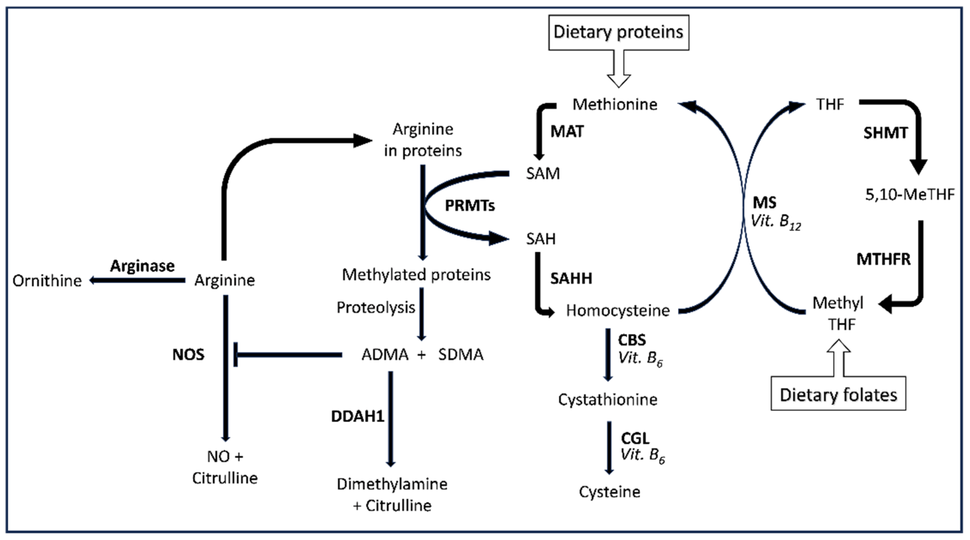
Figure 2.
PRISMA 2020 flow diagram.
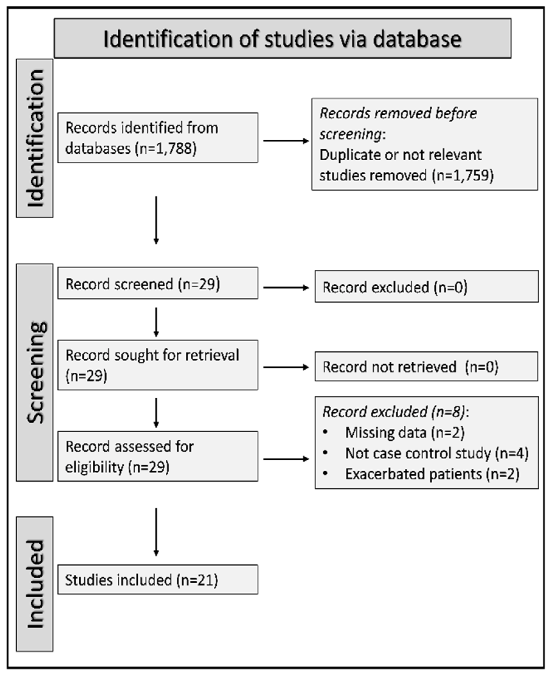
Figure 3.
Forest plot of homocysteine concentrations in COPD patients and controls [85,87,88,89,90,91,97,98,102,103,104].
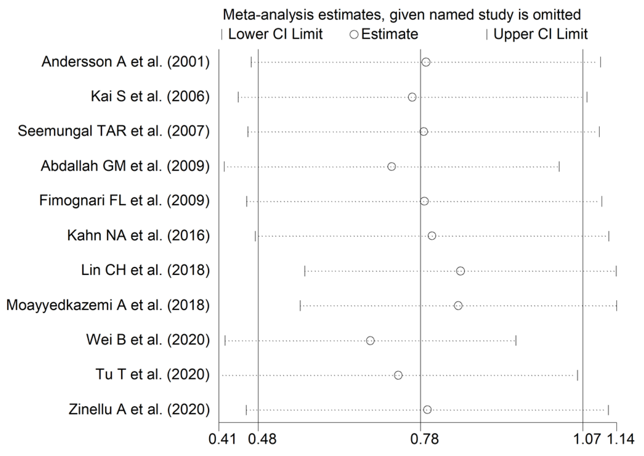
Figure 4.
Sensitivity analysis of the association between homocysteine and COPD [85,87,88,89,90,91,97,98,102,103,104].
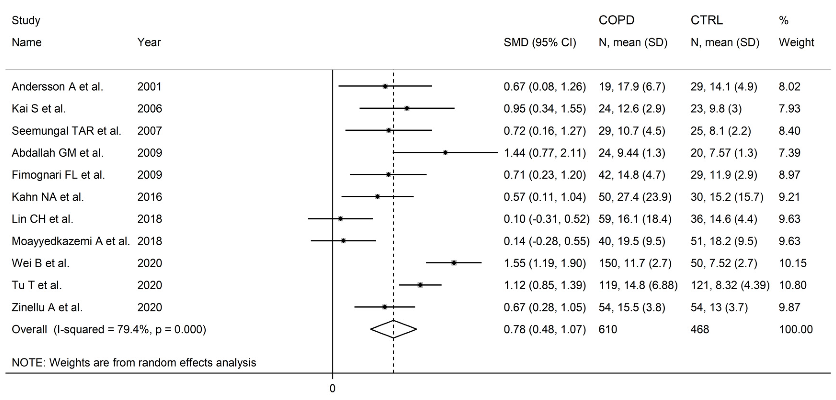
Figure 5.
Funnel plot of studies investigating homocysteine in COPD after “trimming-and-filling”.
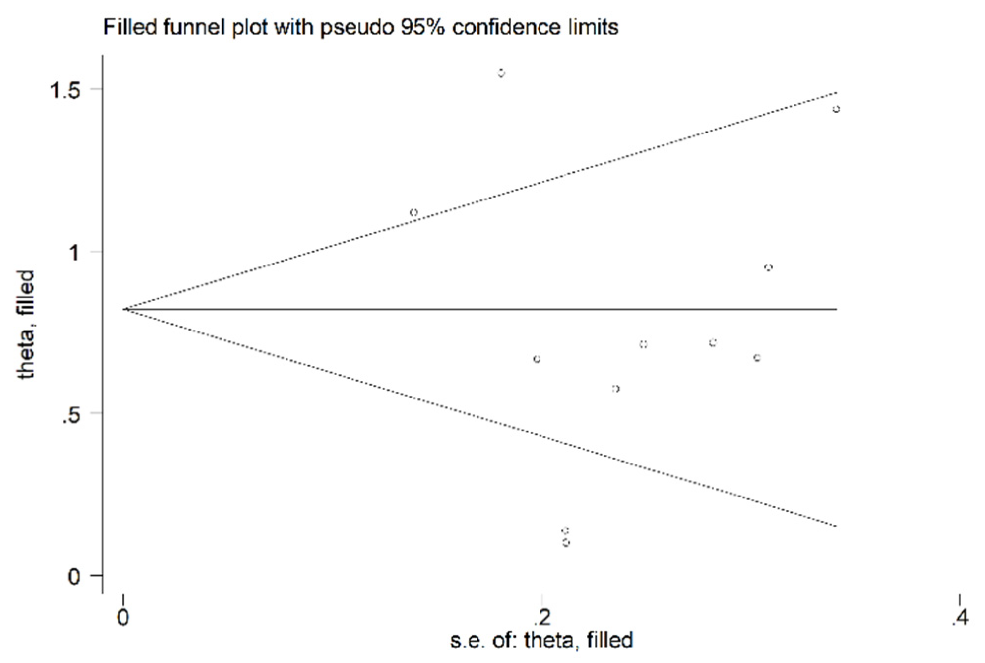
Figure 6.
Forest plot of studies investigating homocysteine concentrations in COPD patients and controls according to patient age (≤70 years or ˃70 years) [85,87,88,90,91,97,98,102,103,104].
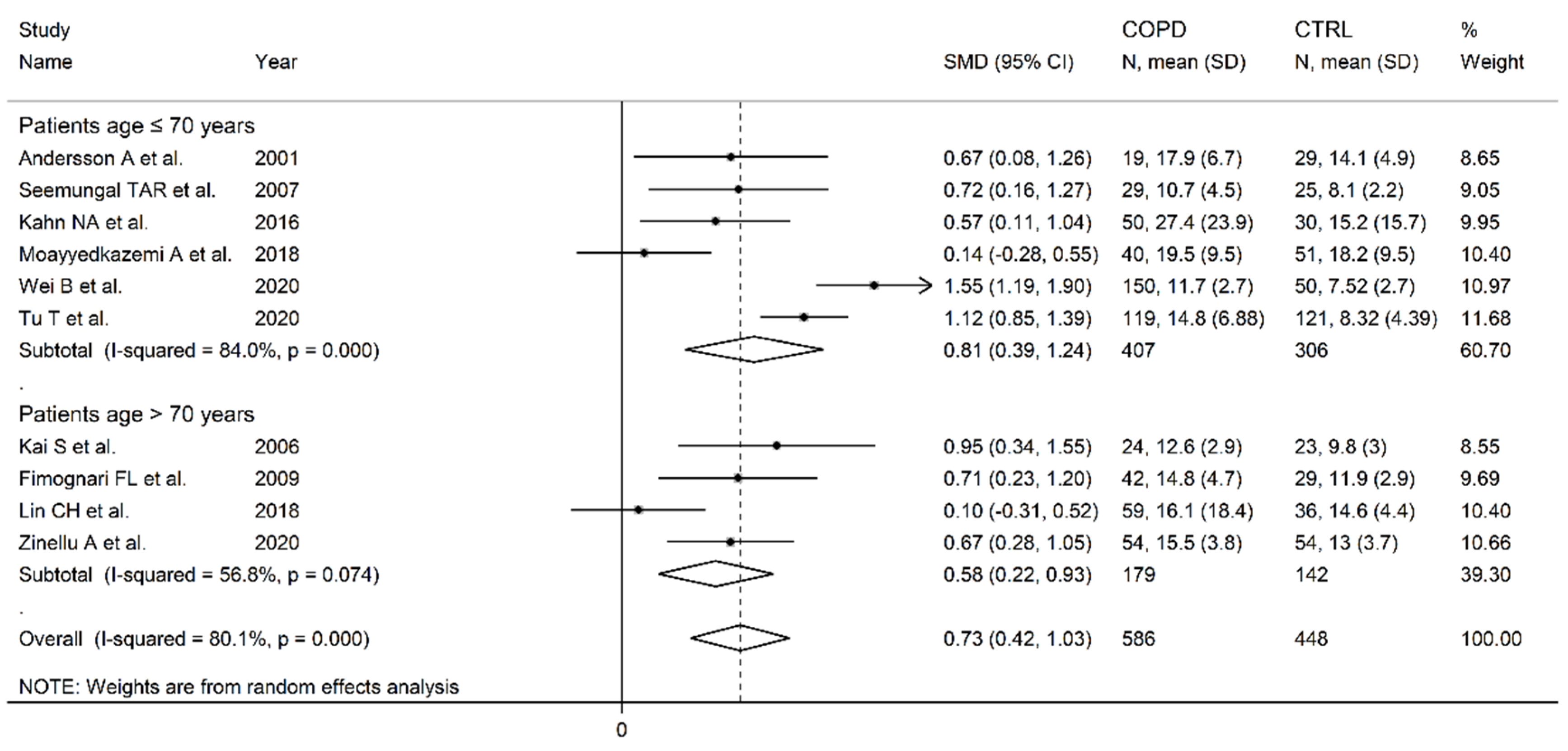
Figure 7.
Forest plot of studies investigating homocysteine concentrations in COPD patients and controls according to study continent [85,87,88,89,90,91,97,98,102,103,104].
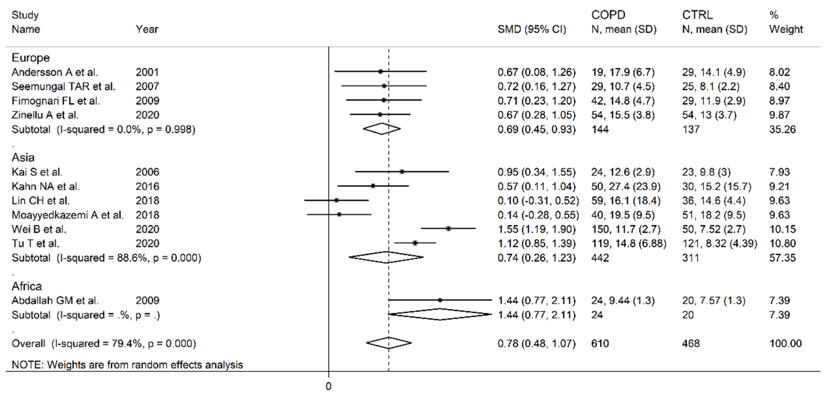
Figure 8.
Forest plot of studies investigating homocysteine concentrations in COPD patients and controls according to analytical method [85,87,88,89,90,91,102,104].
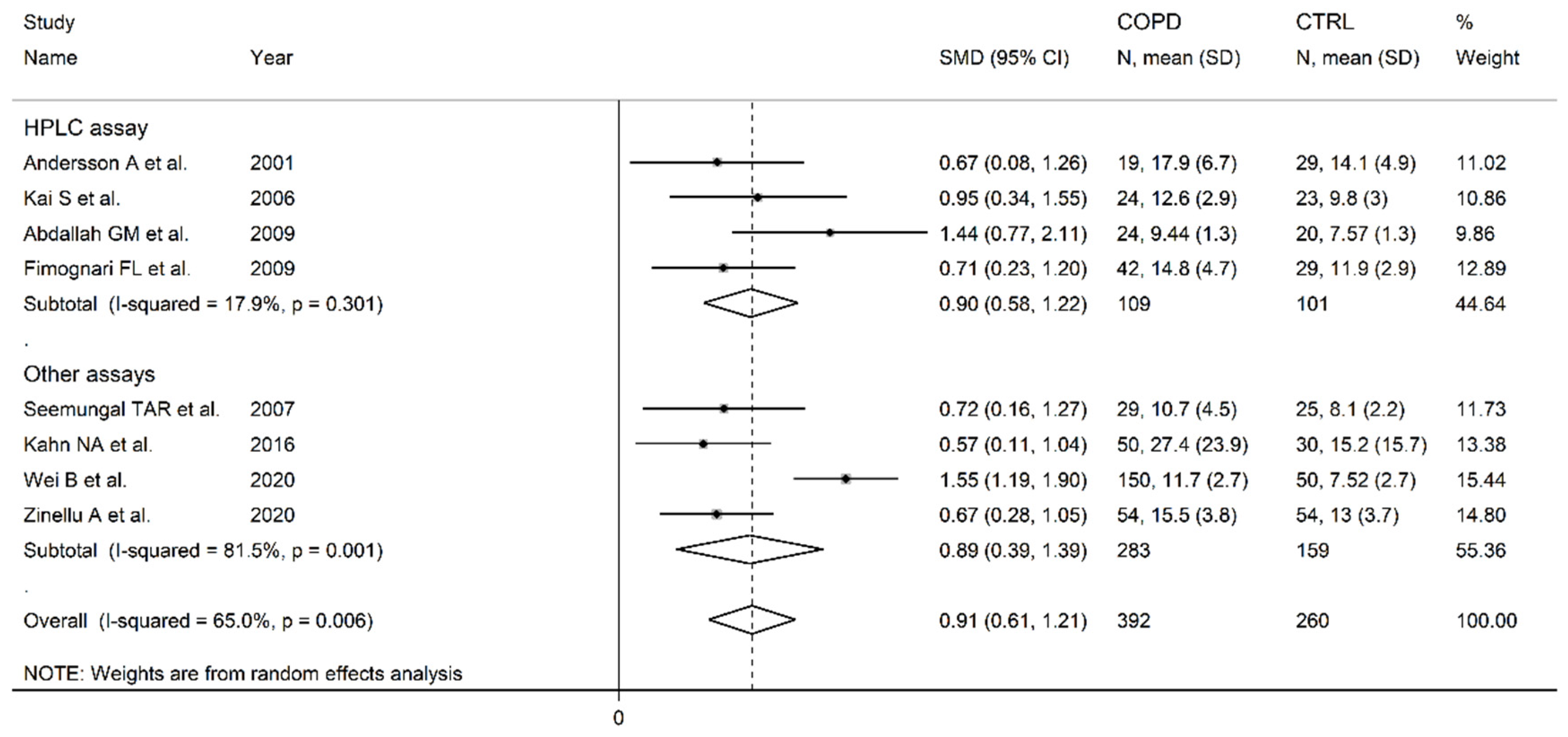
Figure 9.
Forest plot of studies investigating homocysteine concentrations in COPD patients and controls according to the detection method used with liquid chromatography [85,87,89,90].
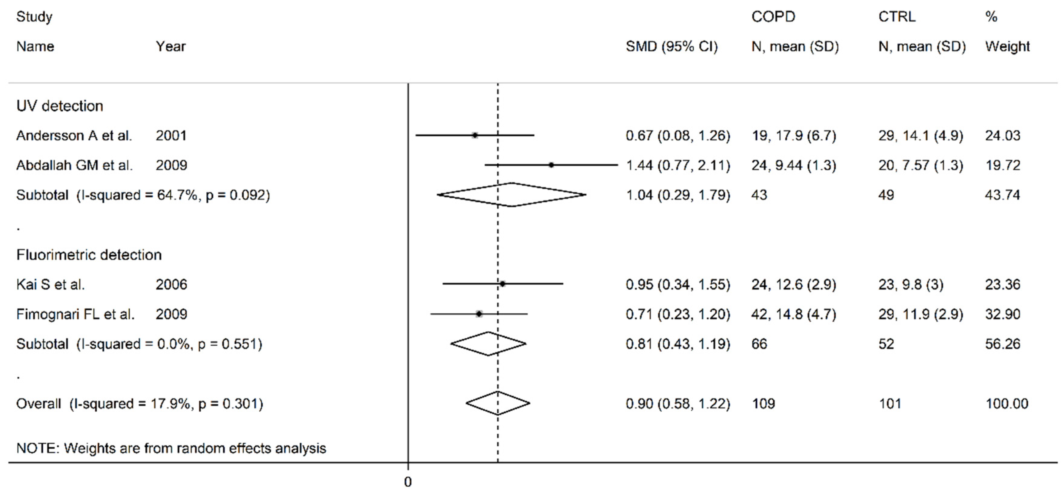
Figure 10.
Forest plot of studies investigating homocysteine concentrations in COPD patients and controls according to measurement in serum or plasma [85,87,88,89,90,91,97,98,102,103,104].
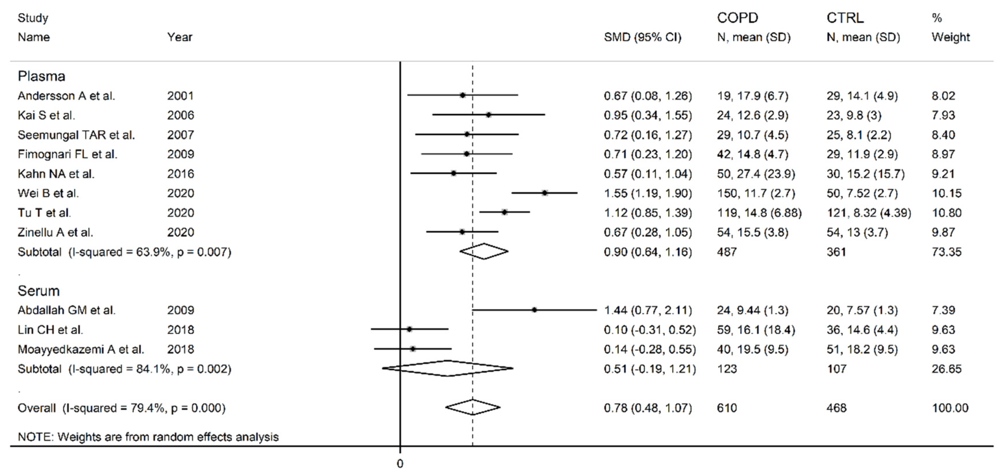
Figure 11.
Forest plot of studies investigating homocysteine concentrations in COPD patients and controls according to FEV1 (≤55% or ˃55% years) [87,88,90,97,98,102,103,104].
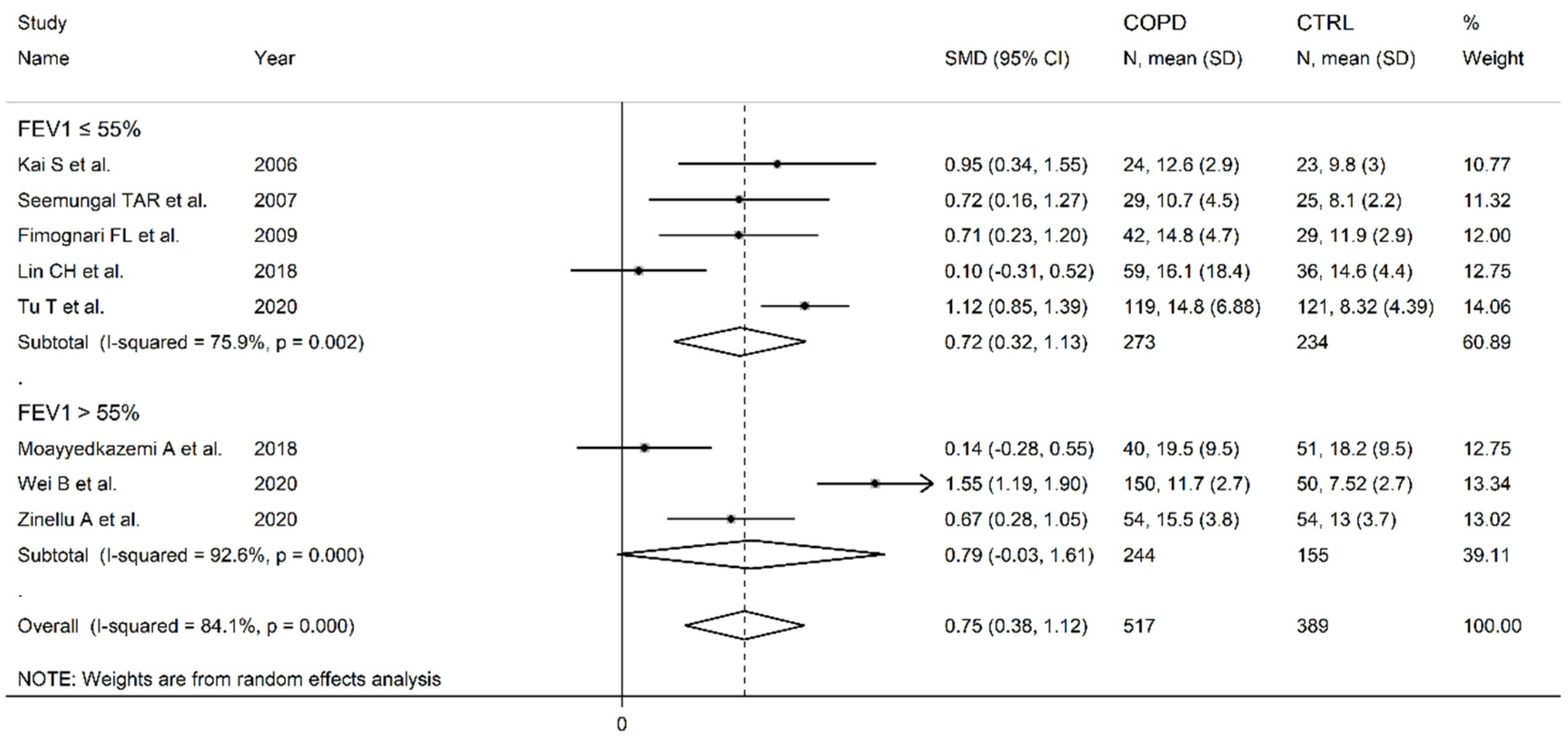
Figure 12.
Forest plot of studies examining homocysteine concentration in COPD patients and controls according to FEV1/FVC (≤60% vs. ˃60%) [88,90,97,103,104].
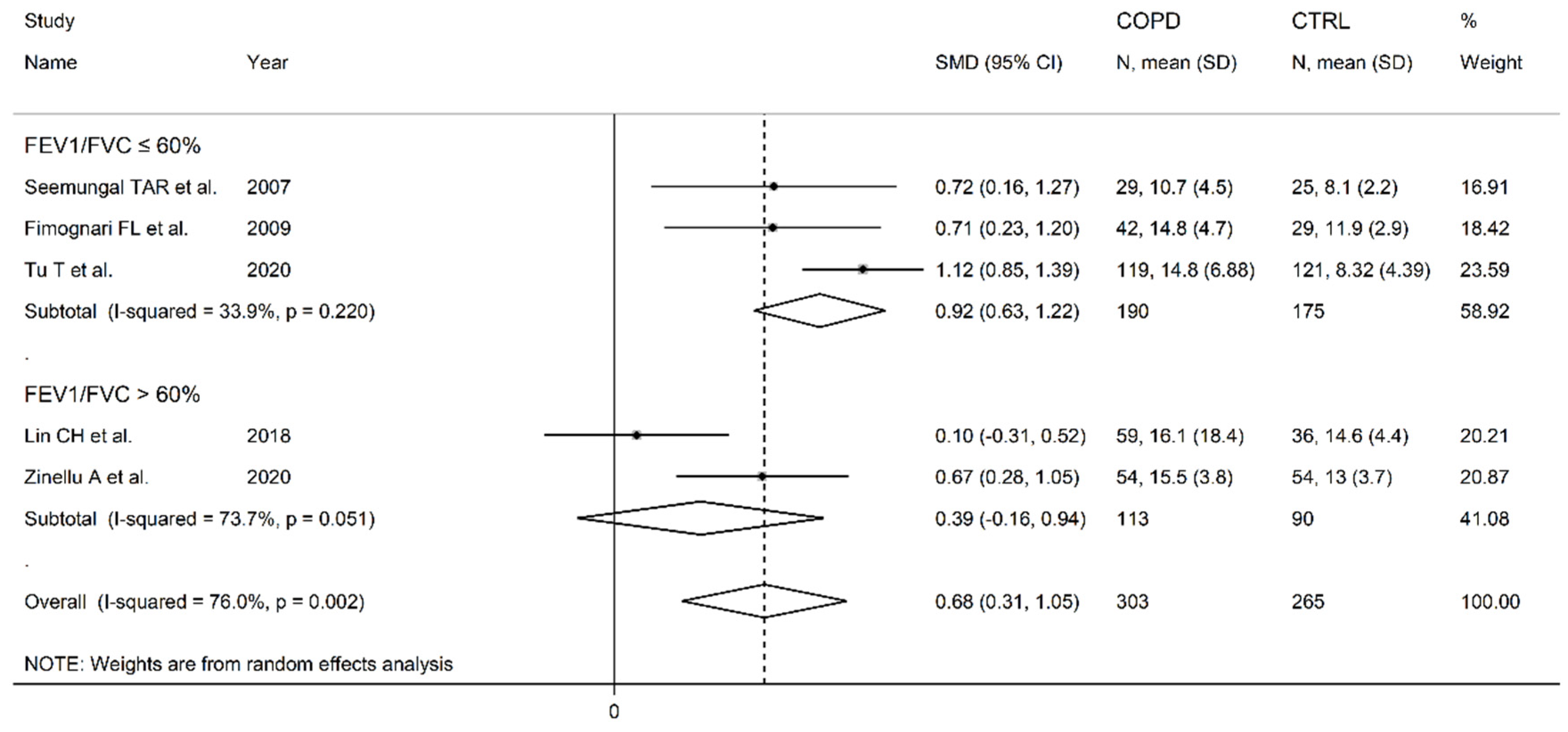
Figure 13.
Forest plot of studies investigating cysteine concentrations in COPD patients and controls [85,104].
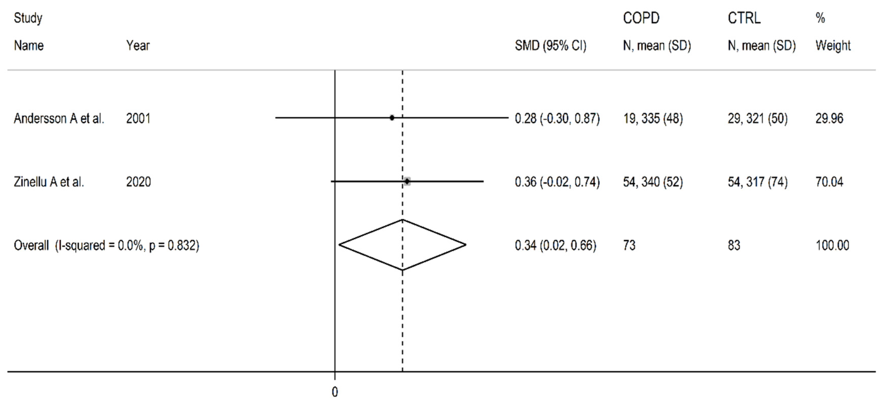
Figure 14.
Forest plot of studies investigating methionine concentrations in COPD patients and controls [84,86].
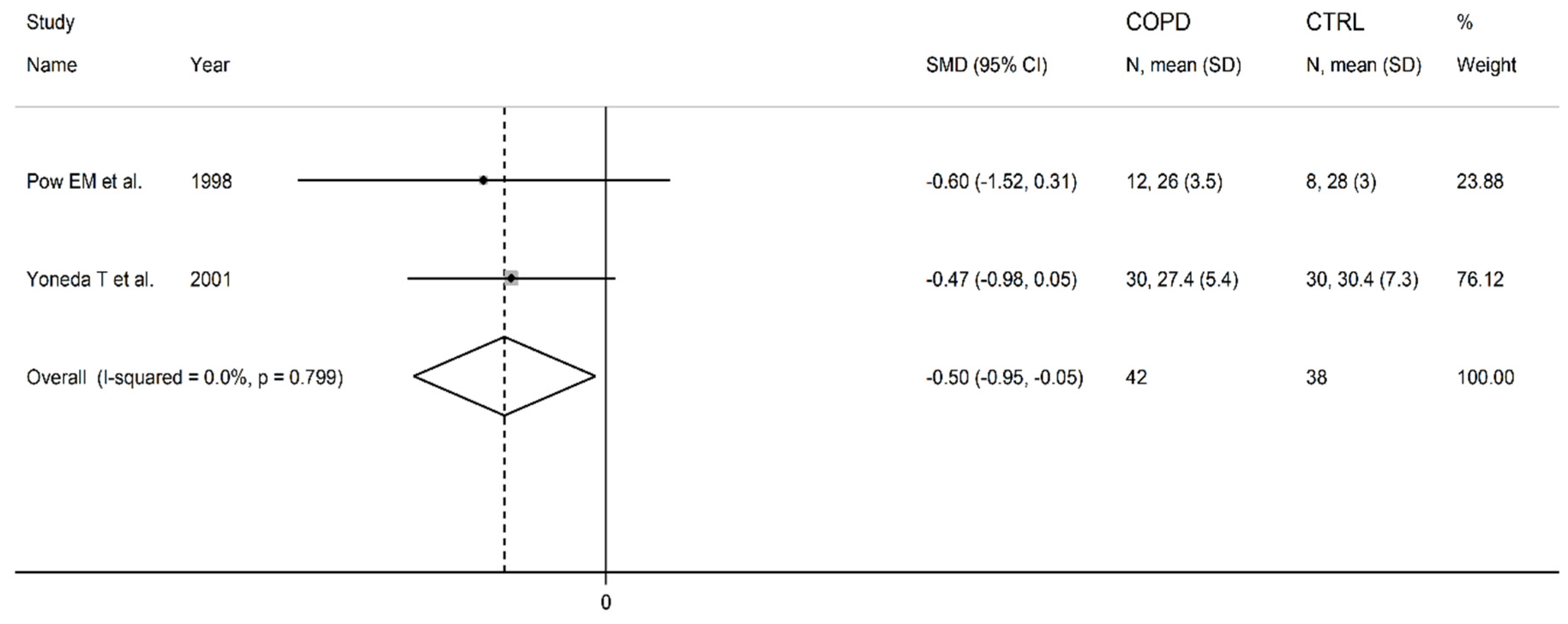
Figure 15.
Forest plot of studies investigating Vitamin B12 concentrations in COPD patients and controls [89,90,97].
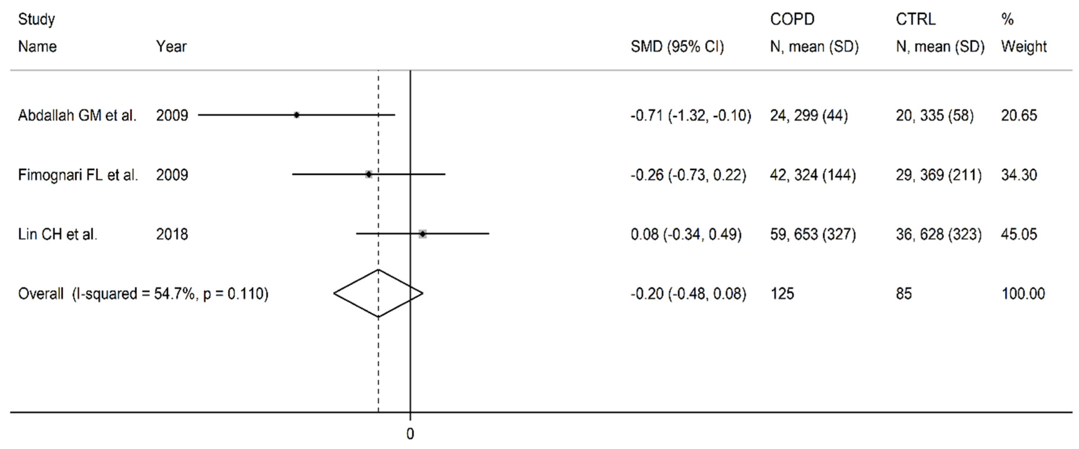
Figure 16.
Forest plot of studies investigating folic acid concentrations in COPD patients and controls [89,90,97].
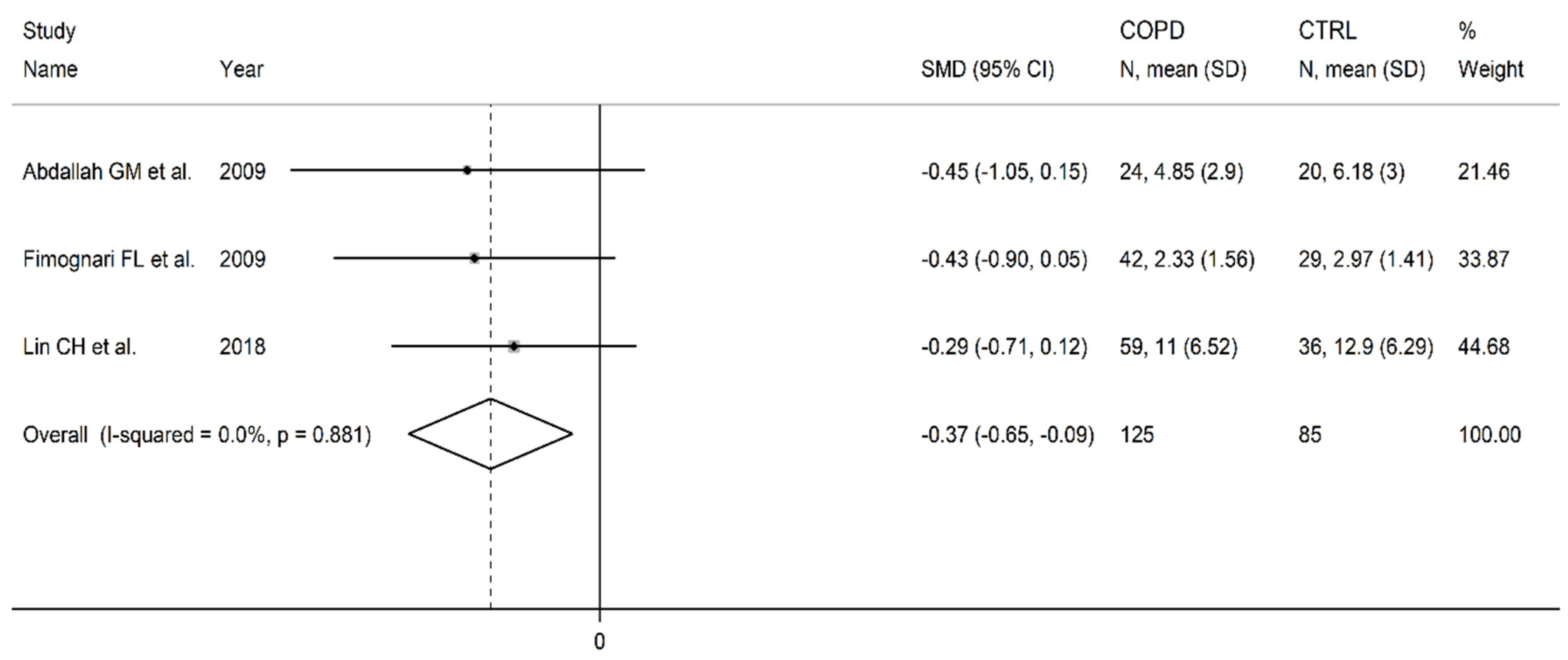
Figure 17.
Forest plot of studies investigating arginine concentrations in COPD patients and controls [84,86,92,93].
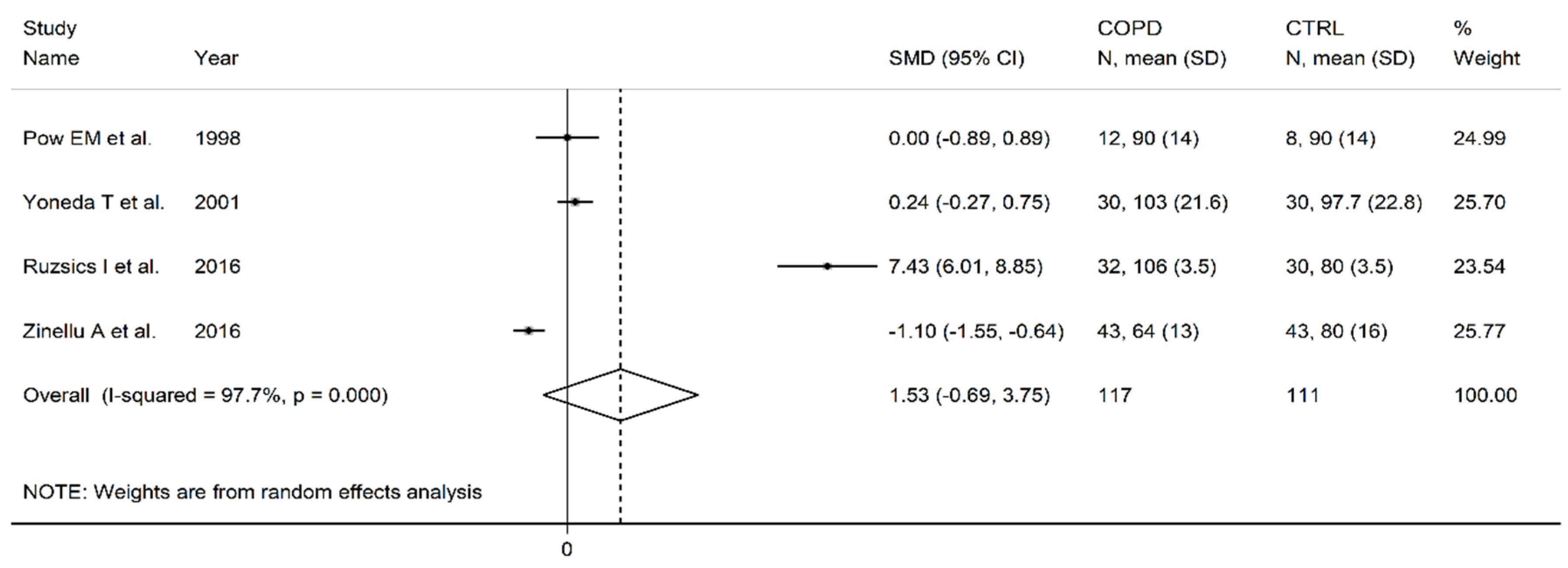
Figure 18.
Forest plot of studies investigating ADMA concentrations in COPD patients and controls [92,93,94,95,96,99].
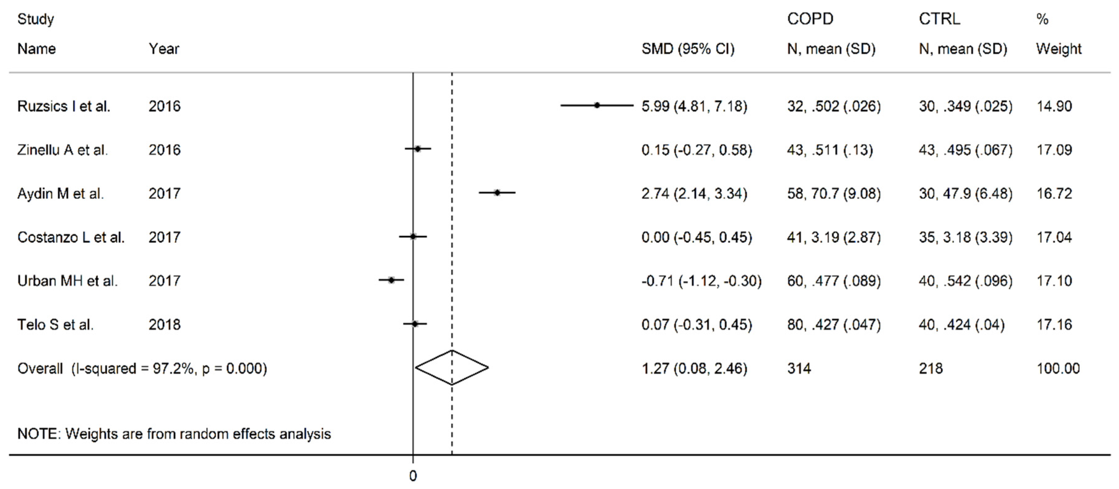
Figure 19.
Forest plot of studies investigating SDMA concentrations in COPD patients and controls [92,93,100].
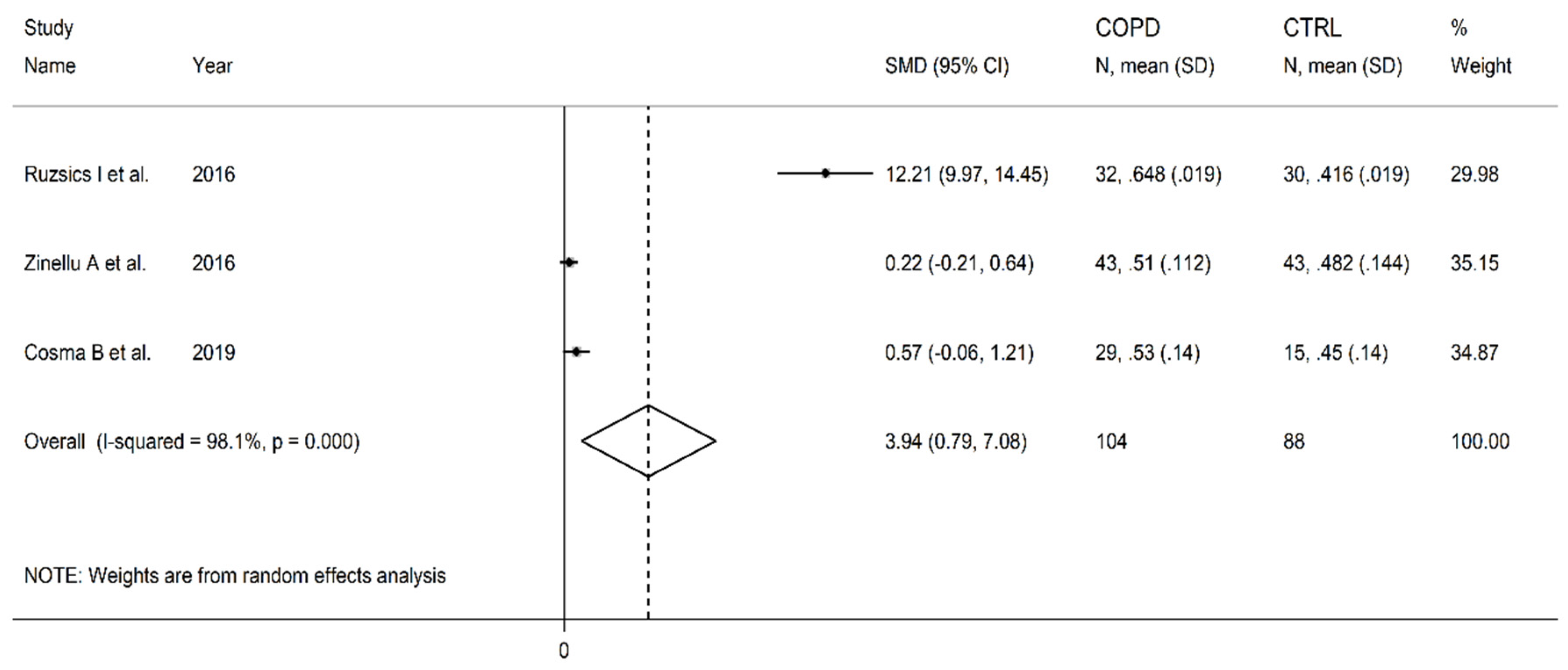
Figure 20.
Forest plot of studies investigating ornithine concentrations in COPD patients and controls [84,86,101].
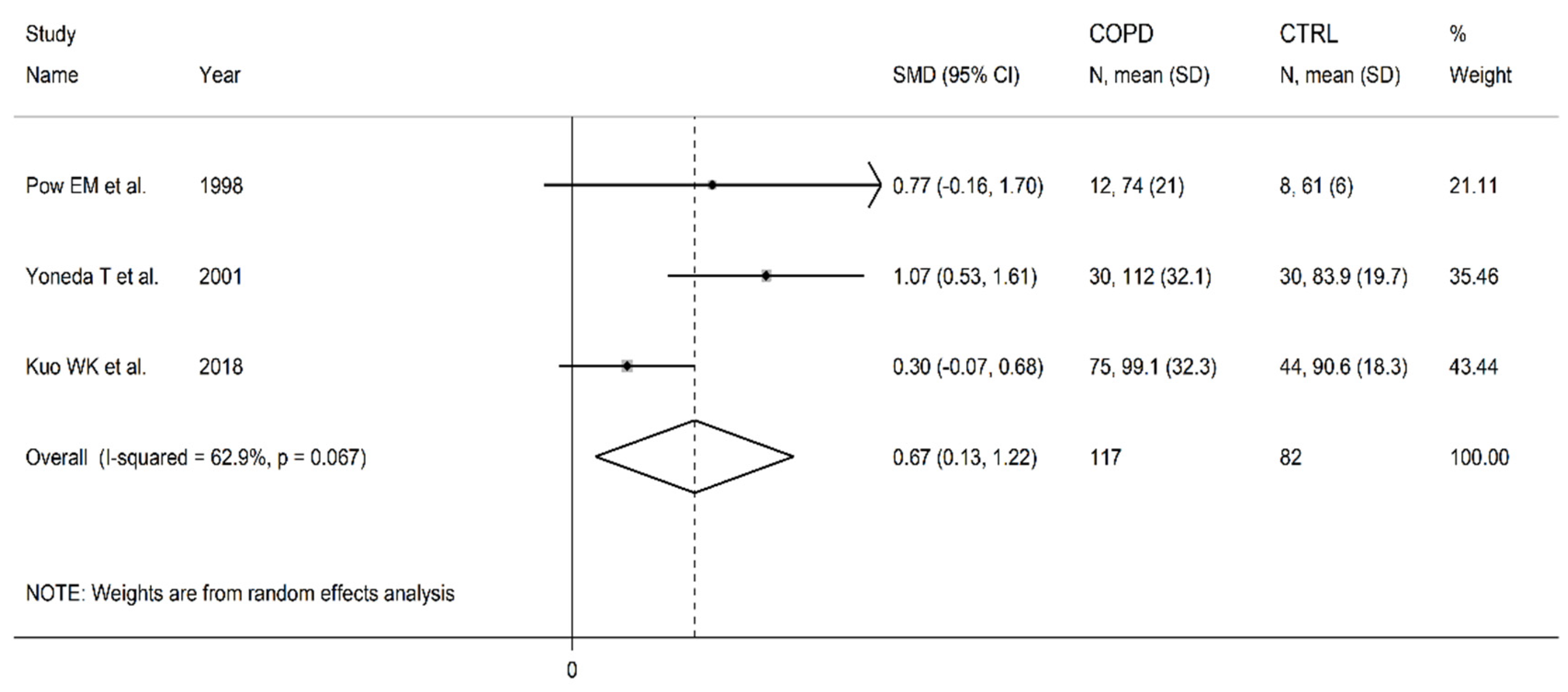

Table 1.
Study characteristics.
| Healthy controls | Patients with COPD | |||||||||
|---|---|---|---|---|---|---|---|---|---|---|
| Study |
n |
Age (Years) |
M/F | Homocysteine Cysteine Methionine Vitamin B6 Vitamin B12 Folic acid (Mean±SD) |
Arginine ADMA SDMA Ornithine Citrulline (Mean±SD) |
n | Age (Years) |
M/F | Homocysteine Cysteine Methionine Vitamin B6 Vitamin B12 Folic acid (Mean±SD) |
Arginine ADMA SDMA Ornithine Citrulline (Mean±SD) |
| Pow EM et al. 1998, The Netherlands [84] | 8 | 64 | NR | NR NR 28±3 NR NR NR |
90±14 NR NR 61±6 54±7 |
12 | 66 | NR | NR NR 26±3.5 NR NR NR |
90±14 NR NR 74±21 48±6 |
| Andersson A et al. 2001, Sweden [85] | 29 | 64 | 14/15 | 14.1±4.9 321±50 NR NR NR NR |
NR NR NR NR NR |
19 | 68 | 8/11 | 17.9±6.7 340±52 NR NR NR NR |
NR NR NR NR NR |
| Yoneda T et al. 2001, Japan [86] | 30 | NR | NR | NR NR 30.4±7.3 NR NR |
97.7±22.8 NR NR 83.9±19.7 NR |
30 | 64 | NR | NR NR 27.4±5.4 NR NR |
103±21.6 NR NR 112.4±32.1 NR |
| Kai S et al. 2006, Japan [87] |
23 | 63 | 23/0 | 9.8±3.0 NR NR NR NR NR |
NR NR NR NR NR |
24 | 71 | 24/0 | 12.6±2.9 NR NR NR NR NR |
NR NR NR NR NR |
| Seemungal TAR et al. 2007, England [88] | 25 | 65 | 16/9 | 8.1±2.2 NR NR NR NR NR |
NR NR NR NR NR |
29 | 69 | 23/6 | 10.7±4.5 NR NR NR NR NR |
NR NR NR NR NR |
| Abdallah GM et al. 2009, Egypt [89] | 20 | NR | 12/8 | 7.6±1.3 NR NR NR 335±58 6.2±3.0 |
NR NR NR NR NR |
24 | NR | 18/6 | 9.4±1.3 NR NR NR 299±44 4.8±2.9 |
NR NR NR NR NR |
| Fimognari FL et al. 2009, Italy [90] | 29 | 71 | 21/8 | 11.9±2.9 NR NR 9.1±6.4 369±211 3.0±1.4 |
NR NR NR NR NR |
42 | 71 | 36/6 | 14.8±4.7 NR NR 5.6±5.1 324±144 2.3±1.6 |
NR NR NR NR NR |
| Kahn NA et al. 2016, India [91] | 30 | 52 | 13/17 | 15.2±15.7 NR NR NR NR NR |
NR NR NR NR NR |
50 | 58 | 43/7 | 27.4±27.9 NR NR NR NR NR |
NR NR NR NR NR |
| Ruzsics I et al. 2016, Hungary [92] | 30 | 51 | 15/15 | NR NR NR NR NR NR |
80±3.5 0.35±0.02 0.42±0.02 NR NR |
32 | 59 | 14/18 | NR NR NR NR NR NR |
106±3.5 0.50±0.03 0.65±0.02 NR NR |
| Zinellu A et al. 2016, Italy [93] | 43 | 73 | 34/9 | NR NR NR NR NR NR |
80±16 0.50±0.07 0.48±0.18 NR NR |
43 | 75 | 34/9 | NR NR NR NR NR NR |
64±13 0.51±0.13 0.51±0.11 NR NR |
| Aydin M et al. 2017, Turkey [94] | 30 | 64 | 21/9 | NR NR NR NR NR NR |
NR 47.9±6.5 NR NR NR |
58 | 62 | 48/10 | NR NR NR NR NR NR |
NR 70.7±9.1 NR NR NR |
| Costanzo L et al. 2017, Italy [95] | 35 | 74 | 16/9 | NR NR NR NR NR NR |
NR 3.18±3.39 NR NR NR |
41 | 74 | 23/18 | NR NR NR NR NR NR |
NR 3.19±2.87 NR NR NR |
| Urban MH et al. 2017, Austria [96] | 40 | 62 | 14/26 | NR NR NR NR NR NR |
NR 0.54±0.10 NR NR NR |
60 | 64 | 32/28 | NR NR NR NR NR NR |
NR 0.48±0.09 NR NR NR |
| Lin CH et al. 2018, Taiwan [97] | 36 | 71 | 36/0 | 14.6±4.4 NR NR NR 628±323 12.9±6.3 |
NR NR NR NR NR |
59 | 71 | 59/0 | 16.1±18.4 NR NR NR 653±327 11.0±6.5 |
NR NR NR NR NR |
| Moayyedkazemi A et al. 2018, Iran [98] | 51 | 66 | 29/22 | 18.2±9.5 NR NR NR NR NR |
NR NR NR NR NR |
40 | 67 | 22/18 | 19.5±9.5 NR NR NR NR NR |
NR NR NR NR NR |
| Telo S et al. 2018, Turkey [99] | 40 | 69 | 31/9 | NR NR NR NR NR NR |
NR 0.42±0.04 NR NR NR |
80 | 69 | 65/15 | NR NR NR NR NR NR |
NR 0.43±0.05 NR NR NR |
| Csoma B et al. 2019, Hungary [100] | 15 | 51 | 6/9 | NR NR NR NR NR NR |
NR NR 0.45±0.14 NR NR |
29 | 63 | 13/16 | NR NR NR NR NR NR |
NR NR 0.53±0.14 NR NR |
| Kuo WK et al. 2018, Taiwan [101] | 44 | 53 | 36/8 | NR NR NR NR NR NR |
NR NR NR 90.6±18.3 NR |
75 | 72 | 67/8 | NR NR NR NR NR NR |
NR NR NR 99.1±32.3 NR |
| Wei B et al. 2020, China [102] | 50 | 58 | 28/22 | 7.5±2.7 NR NR NR NR NR |
NR NR NR NR NR |
150 | 62 | 90/60 | 11.7±2.7 NR NR NR NR NR |
NR NR NR NR NR |
| Yu T et al. 2020, China [103] | 121 | 59 | 77/44 | 8.3±4.4 NR NR NR NR NR |
NR NR NR NR NR |
119 | 59 | 86/33 | 14.8±6.9 NR NR NR NR NR |
NR NR NR NR NR |
| Zinellu A et al. 2020, Italy [104] | 54 | 73 | 40/14 | 13.0±3.7 317±74 NR NR NR NR |
NR NR NR NR NR |
54 | 73 | 40/14 | 15.5±3.8 340±52 NR NR NR NR |
NR NR NR NR NR |
Legend: NR, not reported; M, male; F, female; COPD, chronic obstructive pulmonary disease; ADMA, asymmetric dimethylarginine; SDMA, symmetric dimethylarginine. The concentration of homocysteine, cysteine, methionine, arginine, citrulline, SDMA, and ornithine is expressed in µmol/L. The concentration of ADMA is expressed in µmol/L or ng/mL. The concentration of vitamin B6 and folic acid is expressed in ng/mL. The concentration of vitamin B12 is expressed in pg/mL.
Disclaimer/Publisher’s Note: The statements, opinions and data contained in all publications are solely those of the individual author(s) and contributor(s) and not of MDPI and/or the editor(s). MDPI and/or the editor(s) disclaim responsibility for any injury to people or property resulting from any ideas, methods, instructions or products referred to in the content. |
© 2023 by the authors. Licensee MDPI, Basel, Switzerland. This article is an open access article distributed under the terms and conditions of the Creative Commons Attribution (CC BY) license (http://creativecommons.org/licenses/by/4.0/).
Copyright: This open access article is published under a Creative Commons CC BY 4.0 license, which permit the free download, distribution, and reuse, provided that the author and preprint are cited in any reuse.


