
Submitted:
26 July 2023
Posted:
27 July 2023
You are already at the latest version
Abstract
In recent years, antimicrobial resistance in many human pathogens has become a serious health concern. Since infections with resistant pathogens cannot be treated with traditional antimicro-bial drugs, new strategies are necessary to fight bacterial infections. Hybrid nano-systems may provide a solution to this problem, by combining multiple mechanisms for killing bacteria to synergistically increase the effectiveness of the antimicrobial treatment. In this review, we high-light recent advances in the development of hybrid nano-systems for the treatment of bacterial infections. We discuss the use of hybrid nano-systems for combinational therapy, focusing on various triggering mechanisms for drug release and the development of biomimetic nanomateri-als. We also examine inherently antimicrobial nano-systems and their uses in preventing infec-tions due to wounds and medical implants. This review summarizes recent advances and pro-vides insight into the future development of antimicrobial treatments using hybrid nanomateri-als.
Keywords:
Stimuli-triggered nano-systems
; hybrid nano-systems
; biomimetics
; bacterial infections
1. Introduction
Bacterial antimicrobial resistance is a major global health concern. Antimicrobial resistance (AMR) occurs naturally in bacteria due to genetic variability, but bacterial AMR has become more prevalent due to the strong selective pressure caused by widespread overuse of antimicrobial treatments in clinical, industrial, and agricultural settings. 1 The problem of AMR can be exacerbated in developing countries, due to the lack of regulation in the sales of antimicrobials, the lack of diagnostic and medical facilities for the identification and treatment of drug-resistant pathogens, and a lack of sanitation, which can contribute to the spread of AMR pathogens. 2 In a recent study from The Lancet, it was estimated that in 2019 there were 4.95 million deaths worldwide associated with AMR. 3 For these reasons, the World Health Organization lists AMR as one of the top ten global public health threats. 4
Traditional antimicrobial treatments rely on a variety of mechanisms to kill or inhibit the growth of pathogenic bacteria. Many of these mechanisms target essential processes in the microbe, such as synthesis of proteins and nucleic acids, while other mechanisms target key structural components of the bacterial cell, such as the cell wall or cell membrane. 5 Many mutations can confer AMR by changing how the bacterial cell interacts with the antimicrobial agent. The main mechanisms by which a mutation imparts resistance are by the inactivation of the antibiotic, the modification of the antibiotic target site, or the alteration of bacterial membrane permeability to the antibiotic. 5,6 Because AMR bacteria can no longer be treated with traditional antibiotic strategies, new treatments must be developed that evade AMR.
Nanomaterials have been suggested as potential therapeutic agents for combatting AMR. 7-9 Undoubtedly part of the attraction of antimicrobial nanomaterials relates to the difficulty of bacteria developing resistance to their mechanism of action; however, some researchers have indicated that microbes can develop “nanoresistance.” 10 Although further research is needed on the development of resistance to antimicrobial nanomaterials, nanomaterials are still appealing options for combatting microbial infections due to their ability to be tuned for specific applications and targeted to specific cellular sites Furthermore, nanomaterials can be combined to create hybrid nano-systems, which can synergistically improve treatment outcomes.
In this review, we will discuss recent advances in the use of hybrid nano-systems as antimicrobial agents, highlighting how these hybrid nano-systems can be used to treat AMR bacteria. We will first discuss the use of nanomaterials as drug carrier systems, which combine the antimicrobial activity of various drugs with various endogenous or exogenous triggers, allowing for controlled drug release. We will also discuss biomimetic nano-systems, which increase biocompatibility and allow for targeted treatment of infections. Then, we will discuss inherently antimicrobial nano-systems, which combine the antimicrobial properties of metals with various external stimuli to achieve antimicrobial activity.
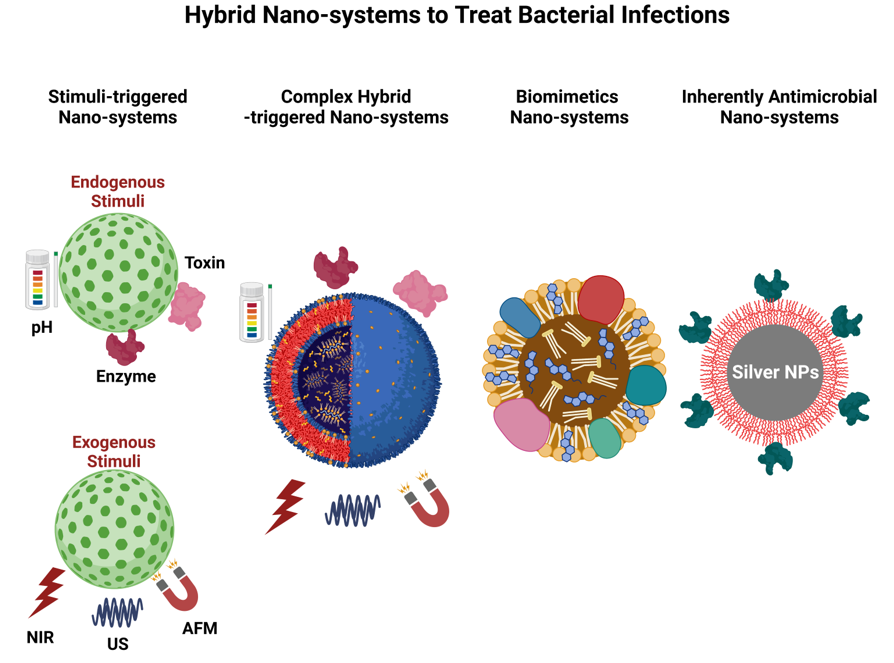
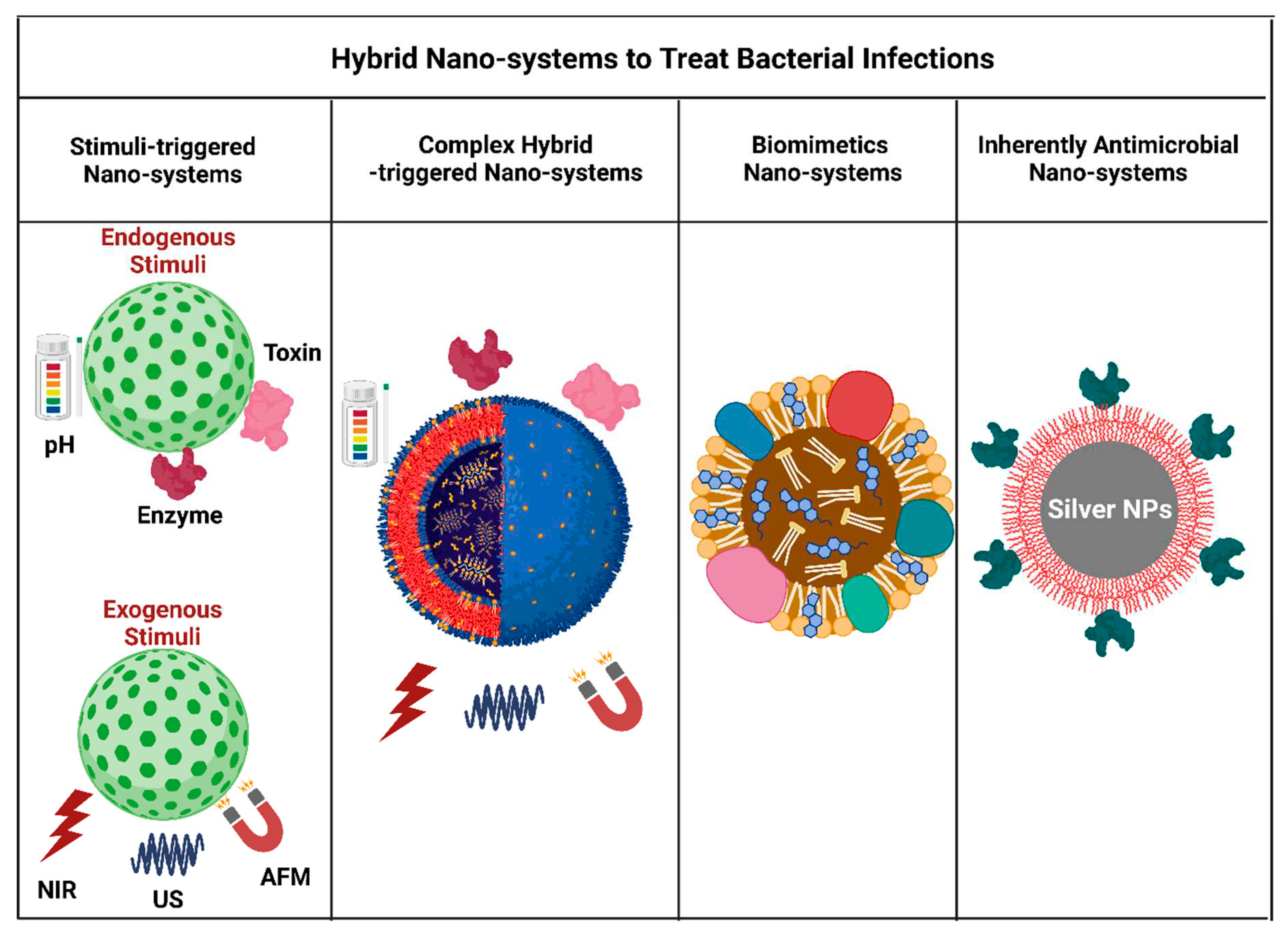
Scheme 1.
Schematics of hybrid nano-systems used to combat bacterial infections.
2. Hybrid Nano-systems for Combinational Therapy
Stimuli-responsive hybrid nano-systems, a combination of metallic inorganic nanoparticles with “soft matter” drug-delivery systems such as liposomes, polymers, and hydrogels, have been developed in recent years. This approach shows great potential for providing a targeted release of antibiotics in response to specific environmental cues. Inorganic nanoparticles, including metal, mesoporous silica, and quantum dots, are increasingly being used as vehicles for delivering antibiotics.11 By incorporating inorganic nanoparticles into soft-matter systems, a range of unique benefits can be achieved. This approach combines the desirable drug-delivery features of traditional vehicles, including excellent colloidal stability, ease of fabrication, biocompatibility, and high drug-loading capacities, with innovative stimuli-responsive mechanisms for releasing antibiotics that are imparted by the inorganic nanoparticles.12 When combined, inorganic nanoparticles and traditional drug-delivery systems offer a straightforward and adaptable method to create unique antibiotic release mechanisms that respond to stimuli. This can lead to a boost in the effectiveness of antimicrobial treatments. In this review, we aim to fill the gap in existing literature by discussing the creation of innovative antibiotic delivery systems that respond to stimuli. This is achieved by co-formulating inorganic nanoparticles with soft-matter drug delivery systems (Table 1).
2.1. Stimuli-triggered drug delivery
In order to achieve smart therapeutic responses, drug-delivery systems can be influenced by external or internal stimuli to enhance their antibiotic activity. This can result in controlled release of the payload in the specific biological compartment, as well as prompt treatment of the pathological event. The following sections provide a detailed explanation of each individual stimulus. h covers the hybrid nanomaterials for combinational therapy against bacterial infections which includes delivery systems triggered by endogenous, exogenous stimuli and hybrid nano-systems reported in the literature for the treatment of bacterial infections.
2.1.1. Drug Delivery Triggered by Endogenous Stimuli
Nanomaterials that respond to endogenous stimuli can effectively utilize the unique changes that occur in the microenvironment of certain diseases, such as inflammatory conditions, bacterial infections, and tumors. This principle has been the inspiration for the development of many chemical-stimuli responsive nanomaterials. Microorganisms have a particular microenvironment surrounding them, and many microbial lesions exhibit low pH levels, the secretion of distinctive bacterial enzymes, and bacterial toxins. One can create highly effective and multi-purpose stimuli triggered nano-systems by utilizing environmental cues.
A highly effective drug delivery nano-system exists that responds to pH levels and can combat harmful microbes including Helicobacter pylori, Listeria, Salmonella, Campylobacter rectus, E. coli, and Shigella flexneri.13 These microbes are known to thrive in acidic environments within the body, including the stomach (pH 1.0-2.0), vagina (pH 4.0-5.0), skin (pH 4.0-5.5), intestines (pH 5.0-8.0), and bladder (pH 4.5-8.0).14 The lining of the stomach, also known as the stomach mucosa, is highly susceptible to the harmful effects of the spiral bacterium H. Pylori. This bacterium is linked to the development of gastritis, gastric ulcers, and associated forms of cancer. When treating H. Pylori infection, antibiotics are often paired with proton pump inhibitors (PPIs) to lessen the production of gastric acid. This is necessary as gastric acid can decrease the effectiveness of antibiotics. PPIs are effective due to their binding to proton pumps, which helps in reducing acid secretion. However, prolonged use can lead to several adverse effects like headaches and diarrhea, and in severe cases, anxiety or depression. Therefore, it would be advantageous to develop an alternative treatment with equivalent or better therapeutic efficacy compared to current antibiotic treatments, without the need for PPIs. A new micromotor made of Magnesium (Mg) and covered with a pH-sensitive polymer coating has been reported by Li, et al.15 A micromotor, made of Mg and gold coated with Au, can temporarily neutralize stomach fluid acidity in vitro and in vivo.15 Its payload is encapsulated within a pH-sensitive polymer layer, and when acidic, Mg reacts with acids to produce hydrogen bubbles, propelling the motor and reducing protons.15 These artificial motors can neutralize gastric pH in under 20 minutes, modifying the surrounding area without interfering with proton pumps' function.15 This method has minimal effect on stomach function, ensuring no undesirable outcomes compared to conventional PPIs. Recently, Song, et al. have reported the first design of an active drug combination therapy for treating H. Pylori which is based on self-propelled micromotors with a magnesium/gold Janus structure as shown in Figure 1.16 These micromotors can penetrate the mucus layer, deliver amoxicillin and clarithromycin simultaneously, and release them in a pH-responsive manner.16 The self-propelled micromotors improve antimicrobial efficiency and offer a promising future for active treatment of bacterial infections due to their self-propulsion, biocompatibility, and biodegradability.16
Bacteria surfaces contain numerous physiological and molecular cues that can be harnessed for effective targeting purposes. H. Pylori effectively utilizes the urea transport protein located on its membrane, allowing for the transportation of urea into the cytoplasm, ultimately enabling the urease enzyme to generate ammonia. This is crucial for the bacterium, as it serves as a protective mechanism against the highly acidic environment of the stomach The Zhang group utilized urea as a targeted head group to conjugate with chitosan through two different linkages, resulting in urea-modified chitosan derivative UCCs-2. This derivative served as a targeting moiety to the urea transport channel protein UreI of H. Pylori and was used as a targeting ligand on pH-sensitive bilayer PLGA/UCCs-2 nanoparticles. In achieving optimal results for the eradication of H. Pylori, a variety of chitosan (CS) microparticles and NPs loaded with antibiotics were utilized by Zhang group. 17-20 Another method of targeting the bacterial surface is through electrostatic targeting, which makes use of the negative charge of the bacterial surface. A considerable number of bacteria exhibit a negative surface charge at physiological pH levels, making them susceptible to targeting through the use of commonly available ligands like cationic peptides or lipopeptides.21-23 These compounds operate by interacting with and damaging the cell membranes of bacteria.24, 25
Pathogenic bacteria have various virulence factors that enable them to colonize, invade, and replicate in immune-competent hosts. Along with negatively charged surfaces and pH gradients, these factors include bacterial toxins, fibronectin proteins, and collagen adhesins, as well as a range of enzymes such as phosphatase, phospholipase, and lipase.26 A straightforward approach to pinpointing the infection location involves adding a complementary substrate or antibody to the surface of the antibiotic delivery vehicle. As the enzyme breaks down the substrate, it punctures the carrier and releases the antibiotic close to the site of infection.
Since the past ten years, there have been several nano-systems that are sensitive to hyaluronidase,27, phosphatase28 and lipase29, 30 which have been utilized to deliver antibiotics in synchronization to infection sites. Rathnayake, et al. have developed a novel method for delivering antibiotics within cells as shown in Figure 2.31 They utilized mesoporous silica nanoparticles that were loaded with colistin and coated with a bacteria-targeting peptide called LL-37, which was conjugated on the liposomal shell. The liposomal shell prevents premature drug release before the nanoassembly approaches the targeted bacteria. Upon encountering lipase in the local environment of Pseudomonas aeruginosa (P.aeruginosa), the liposome bilayer degrades and releases the encapsulated Col. In comparison to free Col, Col@MSN@LL-(LL-37) encapsulated Col showed a significant 6.7-fold rise in antimicrobial effectiveness. The researchers found that this approach effectively inhibited the growth of P. aeruginosa, both in vitro for planktonic and intracellular infections.31

Table 1.
Hybrid nanomaterials for combinational therapy against bacterial infections.
| Nanomaterials | Trigger | Target Pathogen | Drug | Active Targeting | Ref. |
|---|---|---|---|---|---|
| Endogenous Triggered DDS | |||||
| PLGA-PLH-PEG nanoparticles | pH |
S. aureus E. coli |
Vancomycin | Electrostatic | 21 |
| Squalenoylated Penicillin Bioconjugates | pH | S. aureus | β-lactam antibiotics | NA | 32 |
| Chitosan modified gold nanoparticles, Liposome | pH | H. Pylori | Doxycycline | NA | 17 |
| Mesoporous silica nanoparticle, pH sensitive Nanovalves | pH | F. tularemia | Moxifloxacin | NA | 33 |
| Ureido-conjugated chitosan/TPP multifunctional nanoparticle | pH | H. Pylori | Amoxicillin | Ureido targeting groups | 18 |
| Triblock polymers PEG-b-PCL-b-PAE | pH | S. aureus | Vancomycin | Electrostatic | 22 |
| Mg-based micromotor | pH | H. Pylori | pH sensitive polymer coating | NA | 15 |
| Mg-based micromotor | pH | H. Pylori | Ciprofloxacin | NA | 34 |
| Solid lipid Np | pH | MSSA, MRSA | Vancomycin | NA | 35 |
| AMX-PLGA/UCCs-2 nanoparticles | pH | H. Pylori | Amoxicillin | UCCs-2 as targeting moiety | 20 |
| Cysteine conjugated chitosan/PMLA | pH | H. Pylori | Amoxicillin | NA | 19 |
| AMP (LL37) and lipid (OA) selfassembly | pH | E. coli | LL 37 | Electrostatic | 24 |
| Antimicrobial Peptide-Reduced Gold Nanoclusters | pH | E. coli, P. aeruginosa, S. aureus, S. epidermidis | Antimicrobial peptide | Electrostatic | 25 |
| Hyaluronic acid (HA)-based nanocapsules | Enzyme (hyaluronidase) |
S. aureus,E. coli | polyhexanide | NA | 27 |
| Chitosan-modified gold nanoparticles (AuChi-liposome) | Enzyme (phospholipase A2) | H. Pylori | Doxycycline | NA | 28 |
| PGA and Bla-responsive polymeric vesicles | Enzyme (penicillin Gamidase (PGA) and b-lactamase (Bla) | MRSA, B. longum, L. acidophilus,and E. faecalis | Vancomycin, Gentamycine, quinupristin/dalfopristin (Synercid) | NA | 36 |
| MSNP/LIPID Bilayer | Enzyme (Lipase) | S. aureus | Gentamycin | bacteria-targeting peptide ubiquicidin (UBI29−41) | 29 |
| Monoolein liquid crystal nanoparticles (MO-LCNPs) | Enzyme (Lipase) | P. aeruginosa S. aureus | Rifampicin Ciprofloxacin |
NA | 30 |
| Gold nanoparticle-stabilized phospholipid liposomes. | Alpha-toxin | MRSA | Vancomycin | NA | 37 |
| Liposome-based nanoreactors | Alpha-toxin | MRSA | Rifampicin | NA | 38 |
| Exogenous Triggered DDS | |||||
| Hollow microspheres (HMs) shell PLGA Core- Van, polypyrrole nanoparticles |
PPT (808 nm, 0.5 W/cm2, 15 min |
S. aureus (subcutaneous bacterial abscesses) | Vancomycin | NA | 39 |
| Reduced graphene oxide (rGO)-embedded polymeric nanofiber mats | PPT (980 nm, 1 W/cm2), 10 min) |
E. coli K12 S. aureus S. epidermidis |
ampicillin cefepime | NA | 40 |
| (PDA-PEG-Van | PPT (808 nm, 0.78 W/cm210 min | MRSA | Vancomycin | 41 | |
| Vancomycin (Van)-modified gold nanostars | PPT | MRSA | Vancomycin | Vancomycin | 42 |
| Bubble liposomes | US (0.15 or 0.44 W/ cm2) | C. trachomatis | Doxycycline Ceftizoxime | NA | 43 |
| microbubble-mediated low-intensity ultrasound | US (100mW/cm2; 46.5 KHz; 33% duty cycle; 12 h. | E. coli | Gentamycin | NA | 44 |
| dextran sulfate-shelled perfluoropentane (PFP)-cored NBs | US (f = 2.5 MHz; P = 5 W; t = 10 min) | MRSA | Vancomycin | NA | 45 |
| microbubble suspension | US (1.1 MHz, 2.5 Mpa, 5500 cycles at 20 ms pulse duration) for 20 s) | UTI (E. faecalis) | Gentamycin | NA | 46 |
| (Pd@Pt-T790) | US | MRSA | T790 as sonosensitizer | NA | 47 |
| iron oxide nanoparticles (NPs) encapsulated into polymeric microspheres | Magnetic | S. aureus | ciprofloxacin | NA | 48 |
| MnFe2O4 superparamagnetic nanoparticles, pegalyated chitosan as shell | Magnetic |
S. aureus S. Epidermitis, B. subtilis , E. coli, P.aeruginosa, and MRSA |
Vancomycin | NA | 49 |
| iron oxide nanoparticles | Magnetic | S. aureus, B. subtilis , E. coli, andP.aeruginosa | Gentamicin. | NA | 50 |
| Fe3O4 nanoparticles, chitosan microbeads cross-linked with varying lengths of polyethylene glycol dimethacrylate. | Magnetic | S. aureus | vancomycin | NA | 51 |
| MNPs@Ag@HA | Magnetic |
S. aureus, E. coli S. aureus biofilm |
gentamicin | NA | 52 |
| Hybrid Nano-systems | |||||
| MNP Eudragit®S100 |
pH Magnetic |
H. Pylori | Amoxicillin | NA | 53 |
| SiO2-Cy-Van | Bacteria-activated polyelectrolyte dissociation | MRSA | Vancomycin conjugate poly(acrylic acid) | Vancomycin | 54 |
| Amphiphilic block copolymer consisting of biotinylated poly(ethylene glycol)-b-poly(β-amino ester)-b-poly(ethylene glycol) grafted with PEGylated lipid (Biotin-PEG-b-PAE(-g-PEGb-DSPE)-b-PEG-Biotin) | pH, Enzyme |
P. aeruginosa, (Sepsis) |
Ciprofloxacin, and an anti-inflammatory agent (2-[(aminocarbonyl)amino]-5-(4-fluorophenyl)-3-thiophenecarboxamide, TPCA-1) | Intercellular adhesion molecule-1 antibodies | 55 |
| amphiphilic poly (ethylene glycol)−poly(ε-caprolactone) (PECL) copolymers, | pH, Enzyme (Lipase) | P. aeruginosa | Ciprofloxacin | Vancomycin as targeting ligand | 56 |
| Amp-MSN@FA@CaP@FA | pH | E. coli, S. aureus | AMP | Folic acid | 57 |
| Multimetallic microparticles (MMPs), PLGA, AgNP, ZnO NP | NA | M. tuberculosis | Rifampicin | NA | 58 |
| MSN, PGEDA, CB[7],TPE-(COOH)4 | NA | S. aureus, E. coli | Amoxicillin | Electrostatic | 59 |
| Mesoporous silica nanoparticles decorated with polycationic dendrimers | NA | E. coli | Levofloxacin | Electrostatic | 60 |
| DAP-GCS-PDA@GNRs) | pH, PPT808 nm Laser (0.5 W cm−2) for 0–8 min | MRSA | Daptomycin (DAP | Electrostatic | 61 |
| Porous silicon np, Peptide carg peptide identified by phage library |
NA |
S. aureus, P. aeruginosa |
VAN | CARG Peptide | 62 |
| Fusogenic pSi nanoparticle system (F-pSi) | NA | S. aureus | siRNA | Macrophage-targeting peptide (F-siIRF5-CRV) | 63 |
| Au@AgNP@SiO2@Nc-Van | PTT, (780 nm, 30 mW/cm2 for 30 min) | Van-sensitive B. subtilis, Van-resistant E. faecium, E. faecalis, E. coli |
Van | NA | 64 |
| TRIDENT, Natural fatty acid, Lecitin, DSPEPEG2000,IR780 | PTT (808 nm, 0.5W/cm2) | MDR S. aureus E. coli (Sepsis) |
Imipenem | NA | 65 |
| Black phosphorus quantum dots (BPQDs) and thermal-sensitive liposome. | PTT (1 W, 808nm, 15 min) | MRSA | Vancomycin | NA | 66 |
| AA@Ru@HA-MoS | Enzyme, PTT (808 nm, 0.5 W/cm2 for 7 min) | S. aureus and MDR P. aeruginosa | Pro drug Ascorbic acid | Ciprofloxacin as a catalyst with targeting effect | 67 |
| Lipid−dendrimer hybrid nanoparticles (LDH-NPs) | pH | MRSA | Vancomycin | Electrostatic | 68 |
| Maltohexaose-decorated cholesterol and bacteria-responsive lipid compositions, a smart nanoliposomes platform (MLP18) | Enzyme, US | MRSA | Purpurin 18 as sonosensitizer | Bacteria-targeting maltohexaose | 69 |
| Metal organic frameworks(MOFs)/antibiotics | pH | S. aureus | Tetracycline | Hyaluronic acid (HA) targeting | 70 |
| P(HEMA-co-DMA) as templet, Van-OA@PPy | PTT (808 nm, 1.0 W/cm2 for 5 min) |
MRSA | Vancomycin | Vancomycin conjugated oleic acid | 71 |
| AIE flurophore TTD, Micelle | White light irradiation (250 mW/ cm2 | M. tuberculosis | Rifampicin | TTD targeting | 72 |
| D-TiO2/Au@UCN nanocomposites. | PTT (980 nm laser 0.68 W/cm2) |
E. coli and MRSA |
Ampicillin | NA | 73 |
| Ison@Man-Se NPs | M. tuberculosis | Isoniazid | Mannose targeting | 74 | |
Pathogenic bacteria secrete virulence factors known as bacterial toxins, which are highly sophisticated proteins that can disrupt cellular membranes or act intracellularly with a high level of specificity for their target cells. One such toxin is α-toxin, a pore-forming toxin secreted by Staphylococcus aureus (S. aureus).75 α-Toxin disrupts the cellular membrane by forming pores, which alters the permeability of the membrane.76 The small pores, measuring approximately 2.5 nm in size, enable water, ions, and small molecules to pass through without control. This can lead to the rapid release of vital molecules like ATP, loss of membrane potential and ionic gradients, and irreversible swelling due to osmosis, ultimately resulting in cell lysis.75, 77 These pore-forming toxins can be exploited in the targeted treatment of bacterial infections to avoid ligand off-target problems and any damage to normal cells. Wu, et al. have developed a new method using phase changing materials (PCMs) for treating bacterial infections using toxin pore-formation activity as an endogenous stimulus to deliver drugs and control their release. This system utilizes lipids as a gate material to coat a eutectic mixture of two fatty acids. Encapsulating calcium peroxide (CaO2) and rifampicin into the eutectic mixture forms liposome-based nanoreactors that can release drugs in response to toxins . When these nanoreactors encounter pathogenic bacteria in vivo, they are penetrated by bacterial toxins, creating pores. Water molecules then enter the nanoreactors through the pores and react with CaO2 to produce hydrogen peroxide (H2O2). At the same time, some of the H2O2 breaks down into oxygen (O2), which powers the release of antibiotics. Additionally, the nanoreactors stimulate the body's immune response by capturing bacterial toxins and reducing their toxicity. This process significantly improves the therapy effect of bacterial-infected mice.38 In another interesting approach, bacterial toxins were utilized to trigger antibiotic release from liposomes stabilized with gold nanoparticles, which subsequently inhibited the growth of toxin-secreting S. aureus bacteria.37 Chitosan-modified gold nanoparticles attached to liposome surfaces improve their stability and prevent drug leakage. However, bacterial toxins can still penetrate the liposome membrane and form pores, through which the drug is released to combat the toxin-secreting MRSA and other bacterial infections. This method shows promise in treating various bacterial infections caused by pore-forming toxins.
2.1.2. Drug Delivery Triggered by Exogenous Stimuli
Nanomaterials with the ability to react to external physical stimuli such as light, ultrasound, and magnetic fields have demonstrated significant potential in precisely delivering therapeutic agents. This is achieved by utilizing the unique optical, magnetic, and physiochemical properties of metallic inorganic NPs. These smart materials, which respond to exogenous stimuli, can control drug release. . Numerous strategies have been explored to formulate these nano-systems with multiple functionalities, a lower degree of variability, and high precision in order to address the pressing need for on-demand and targeted drug delivery for bacterial infections.
Light is often used as a trigger for the development of responsive drug delivery systems that utilize frequencies in the ultraviolet, visible, or near infrared (NIR).78 NIR-triggered systems have received attention for applications in treating in bacterial infections as well as in cancer research.78-85 NIR-responsive materials are usually called photothermal agents. Photothermal heating occurs due to the absorption of light by ensemble of electrons on the surface of certain conductive materials and the subsequent dissipation of that energy as heat. Plasmonically active metal NPs such as Au NPs, Ag NPs, and Cu NPs, black phosphorus, carbon tubes, and polydopamine nanoparticles are particularly powerful photothermal agents because of their large absorption cross sections, tunable optical properties, and highly efficient light-to-heat conversion.86 In such metallic NPs, most of the energy gained by the electrons during photo-excitation at this resonance is dissipated as heat, through electron-photon collisions, leading to the heating of the NPs and the surrounding environment. Photothermal heating can either be used to deliver thermal energy to a localized area, or, alternatively to the entire drug carrier, causing destruction of the capsule material and triggering release of the drug from the charged therapeutics.87, 88 Some early demonstrations of the use of photothermal effects to release antibacterial agents from drug carriers have been reported in a number of research studies.39-41 Wang, et al. developed a new strategy for effective control and killing of gram-positive bacteria based on vancomycin (Van)-modified gold nanostars (AuNSs). The Van-modified AuNSs (AuNSs@Van) can not only selectively detect methicillin-resistant Staphylococcus aureus (MRSA), but also MRSA under near-infrared laser irradiation (808 nm for 10 minutes, 2.5 W/cm2) in vitro and in vivo kill .42 In addition, AuNSs@Van shows satisfactory biocompatibility and antibacterial activity in treating bacterial infections in vivo. The attractive feature of AuNSs@Van is that it can actively attack the gram-positive bacteria by binding to the D-AlaD-Ala moiety of the cell wall of the gram- positive bacteria and anchoring to the surface of the bacteria.89, 90 Subsequently, irradiation with an NIR laser produces hyperthermia in situ, which leads to the death of bacteria.
Recently, Patel, et al. have reported the gold nanorod-based hybrid liposomal nano-system to combat intracellular bacterial infections as shown in Figure 3.91 Gold nanorods act as a photothermic agent, which is then coated with mesoporous silica that provides a surface for drug loading. This entire nanoassembly was then wrapped in a thermosensitive liposome that was functionalized with a mycobacteria-targeting peptide. The bacteria-targeting nano-system specifically targets the mycobacteria and then, upon exposure to NIR light, converts the NIR light to heat, causing the heat-sensitive liposome to melt and releasing the drug in a controlled manner.91
In addition to photothermal therapy, the ultrasound (US) based drug delivery system has gained importance in bacterial infections. Sonodynamic Therapy (SDT) is a novel US-based modality that responds to ultrasonic stimulation and activates sonosensitizers to produce reactive oxygen species (ROS), cavitation, gas bubbles, hyperthermia, etc.92 The exact mechanism by which SDT works is still remained unresolved due to complexity of SDT process but possible theories include generation of ROS,93 ultrasonic cavitation effect94 and thermal destruction.95, 96 The most acceptable mechanism at present is ROS production induced by cavitation or sonosensitizers, which causes apoptosis. US at a frequency of 0.8–3 MHz is an external stimulus which is used for diagnostic and therapeutic purposes.97 US at low frequency can be used to diagnose a disease or at high frequency to treat a disease by responsive drug delivery. When designing a US mediated delivery system, three main criteria must be considered. These are: 1) high drug loading capacity, 2) effective drug release after exposure to a specific US frequency, and 3) the ability to monitor drug release for imaging and therapeutic applications.98 US can be used for drug delivery and drug permeability through biological barriers by increasing temperature and the cavitation bubble.99 Ultrasound-activated microbubbles/nanobubbles are one attractive intracellular delivery modality solution for various bacterial infections such as Chlamydia trachomatis,43 E. coli,44 MRSA wound infections,45 and Enterococcus faecalis intracellular urinary track infections (UTI).46Gas bubbles stabilized by a polymer or surfactant coating have been in clinical use as ultrasound imaging contrast agents for over two decades.100 Once introduced into the body, the passage of the microbubbles/nanobubbles can be easily monitored by diagnostic imaging, while drug delivery is achieved through the application of a higher intensity ultrasound pulse to the target site, thereby limiting side effects elsewhere. The movement of the microbubbles in response to ultrasound not only releases the drug, but also helps promote its convection into surrounding tissue and the permeabilization of cell membranes, through a process known as sonoporation. The combination of these phenomena enhances both the distribution of the drug throughout the target site and its intracellular uptake.46, 101 Xin Pang, et al. used purpurin 18 as a potent sonosensitizer and loaded it into a bacteria-responsive nanoliposome platform for precise diagnosis and SDT of MDR bacteria as shown in Figure 4.69 The nanoliposomes (MLP18) consist of purpurin 18, maltohexaose-modified cholesterol and dioctadecanoyl-sn-glycero-3-phospho-(1-racglycerol) (DSPG). Maltohexaose-modified cholesterol shows bacterial specificity through the maltodextrin transporter pathway; in addition, DSPG is susceptible to bacterial overexpression of phospholipase A2 (PLA2). As a result, the as-synthesized MLP18 enables bacterial targeting and the release of PLA2-activated sonosensitizers. Importantly, this also allows bacterial infections to be distinguished from aseptic inflammation caused by lipopolysaccharide (LPS). Subsequently, due to considerable bacteria-specific drug accumulation, MLP18 showed an excellent sonodynamic effect on MDR bacteria in vitro and completely inhibited bacterial infections in vivo by US irradiation.69 Another interesting study on nanoparticle-mediated SDT for acne was published by Xiang, et al. They reported the development of a sodium hyaluronate microneedle patch that facilitates the transdermal delivery of ultrasound-responsive nanoparticles for the effective treatment of acne.102 The patch contains nanoparticles made of a zinc porphyrin-based metal-organic framework and zinc oxide (ZnTCPP@ZnO). They demonstrated activated oxygen-mediated killing of Propionibacterium acnes with an antibacterial efficiency of 99.73% after 15 minutes of ultrasound irradiation, resulting in a decrease in levels of acne-related factors such as tumor necrosis factor, interleukins, and matrix metalloproteinases.102 Furthermore, zinc ions increased the expression of DNA replication-related genes, promoting fibroblast proliferation and, as a result, skin repair. Through the interface engineering of ultrasound response, this research leads to a highly effective acne treatment strategy.
Finally, magnetic stimuli-responsive drug delivery systems were first developed by Freeman in 1960.103 The use of magnetic force is safer than light irradiation, ultrasound, or electrical fields because it does not physically interact with the body. Magnetically guided systems have been widely explored for cancer diagnosis/therapy,104 as a drug delivery vehicle for bacterial infections/biofilms, and as diagnosis tools.105, 106 Magnetic nanoparticles (MNPs) have been encapsulated in shells made of a functionalized polymeric microspheres,48 PEGylated chitosan,49 or chitosan microbeads.50, 51 These MNP-based nano-systems have been extensively used to load various antibiotics such as ciprofloxacin , vancomycin and gentamycin have been widely investigated for their potential to penetrate into bacteria cells, which may inactivate bacteria and antibiotic-resistant bacteria.48-51, 107 MNPs show great potential as magnetic drug delivery systems, which can control the movement and location of antibiotics, resulting in a rapid, and efficient treatment specifically for biofilms.108 In one study by Alwarze, et al., magnetic hyperthermia together with controlled antibiotic delivery was used to create a unique magnetic-responsive nanocarrier for a combination therapy against biofilms.109 The antibiotic-loaded mesoporous silica nanoparticles (MSNs) were functionalized with a thermo-responsive polymer layer and decorated with super-paramagnetic iron oxide nanoparticles (SPIONs) on the outermost surface. When an alternating magnetic field (AMF) is applied, the SPIONs generate heat, reaching the temperature required to induce a change in polymer conformation from linear to globular, resulting in pore uncapping and antibiotic cargo release. According to the findings of their study, combining hyperthermia and antibiotic treatment is a promising approach for the effective management of biofilm-associated infections.109
2.2. Complex Hybrid Triggered Nano-systems
In order to combine the unique properties of organic and inorganic NPs in one nanoplatform, a number of hybrid nanocomposites have been developed in recent years. The advantage of designing complex hybrid nano-systems is that multiple different stimuli can be accommodated in a single package.110 This requires the use of different stimuli-responsive linkers with two or more nano-systems. These hybrid nano-systems offer numerous opportunities to selectively target and kill intracellular MDR bacterial infections. Intracellular MDR bacterial infections are very difficult to treat because they evade the host's immune system.111 Researchers have developed strategies to treat intracellular MDR bacterial infections using hybrid nano-systems, specifically focusing on 1) targeting bacteria directly, 2) targeting the infectious microenvironment (IME), and 3) targeting host cells (macrophages).
Targeted delivery of antibiotics to bacteria would be a novel strategy to reduce antimicrobial resistance since the drug dosage is low. However, most antibiotics lack the ability to act in a targeted manner. Recent advances in nanotechnology allow investigators to load drugs inside nanoparticles, and simultaneously, nanoparticles can be conjugated with cationic polymer/ligand (electrostatic)60, 61 or with a targeting peptide62, 63 to recognize specific bacterial infections. Hussain, et al. proposed in vivo screening of S. aureus in a lung infection model to identify a targeting ligand that specifically recognizes S. aureus and could thus improve antibiotic delivery at sites of infection.62 In their studies, they found that the 9-amino acid long cyclic peptide CARGGLKSC (CARG) was able to increase vancomycin release after the peptide was conjugated to vancomycin-loaded porous silicon nanoparticles, thereby significantly enhancing mouse survival during bacterial lung infection and decreasing the systemic toxicity of vancomycin. It is interesting to note that the CARG peptides bound specifically to S. aureus and not to P. aeruginosa in vitro. This result is consistent with in vivo studies in lungs and skin of S. aureus-infected mice compared to results in healthy mice and P. aeruginosa-infected mouse models. These studies demonstrate the potential of targeting pathogens directly at the site of the infection using nanoparticles to improve antibiotic bioavailability at infectious sites, potentially preventing antimicrobial resistance caused by antibiotic overdose administration.
Hybrid strategies for targeting the IME include, dual endogenous stimuli-responsive nano-systems (pH/enzyme)55, 56 and endo/exogeneous stimuli-responsive nano-systems, which utilize pH/magnetic53, pH/PTT61, enzyme/PTT67 and enzyme/US69 triggers. The codelivery of an antibiotic and an anti-inflammatory agent to the site of infection was proposed as a novel strategy for sepsis therapy, to diminish the bacteria and the inflammation response simultaneously.55 Sepsis is a life-threatening organ dysfunction caused by a dysregulated host response to infection, particularly bacterial infection. Two major factors are involved in the pathogenesis of sepsis: 1) invading pathogens and 2) activation of the host's immune system. When bacteria enter the body, the host's immune system is activated to eradicate invading bacteria by recruiting macrophages that recognize lipopolysaccharide on a Gram-negative bacterium via toll-like receptors. Macrophages also release pro-inflammatory cytokines (such as TNF-, IL-1, etc. ) that activate endothelial cells lining the blood vessels to increase the expression of intracellular adhesion molecules (ICAM-1) for the recruitment of leukocytes to eliminate bacteria.112-114 Based on this observation, Zhang, et al. designed a novel polymer nanoparticle coated with ICAM-1 antibody to target the affected vasculature.115 The nanoparticles were designed to be broken down by the IME in response to the low pH and bacterial enzymes, triggering drug release. An antibiotic and an anti-inflammatory agent were loaded into the polymeric micelle core through hydrophobic interactions. Finally, the anti-ICAM-1 antibody was applied to the surface of the polymeric micelles. It is expected that the multifunctional polymeric micelles can simultaneously eliminate the bacteria and attenuate the inflammatory responses caused by the bacterial invasion. In an infectious lesion, the activated endothelium strongly expresses ICAM-1 molecules and vascular permeability is increased. After intravenous injection, antibody-coated nanoparticles can recognize and bind to the infected vasculature to facilitate deposition of NPs at the site of infection. The nanoparticles then release the charged drugs in response to the acidity and bacterial enzymes present in the IME. Zhao, et al. have reported a nano-system responsive to pathogen infection that targets the endoplasmic reticulum of macrophages.116 The nanoplatform consists of large-pore mesoporous silica nanoparticles (MSNs) grafted with an endoplasmic reticulum-targeting peptide and a pathogen infection-responsive cap containing the reactive oxygen species(ROS)-cleavable borobenzylic acid linker and bovine serum albumin.116 The capped MSNs demonstrated the ability to efficiently load the antimicrobial peptide melittin and rapidly release the load when triggered by H2O2 or the pathogen-macrophage interaction system, but had no apparent toxicity to macrophages. In a murine model of systemic infection, the nanoplatform efficiently prevented renal dysfunction, alleviated inflammatory symptoms, and protected the mice from death. This study developed a nanoplatform that targeted macrophage organelles to treat life-threatening systemic infections.116
Antimicrobial resistance is associated with bacterial survival inside host cells111 because the cells have low permeability to antibiotic entry that leads to chronic and recurrent infections117, 118 including pulmonary infections,119 endocarditis120 and osteomyelitis.121 Thus, targeting intracellular bacteria may be a key to clinical success. In addition, targeting phagocytes is a smart route for the targeted delivery of antibiotics to infectious lesions. Macrophages are important scavenger cells that remove bacteria. However, sometimes the elimination of the bacteria is not complete, which can lead to chronic infections. One of the main features of tuberculosis (TB) pathogenesis is the escape of Mycobacterium tuberculosis (Mtb) from phagolysosomal destruction. Pi, et al. studied a macrophage-targeted mannosyl-selenium nanoparticle hybrid nano-system (Man-Se NPs) to deliver isoniazid to the intracellular Mtb.74 The authors synthesized Man-Se NPs for macrophage targeting and encapsulated isoniazid in the nanoparticles. The hybrid system Ison@Man-Se NPs could be used for the synergistic treatment of TB. Mtb can survive in host cells because the intracellular drug concentration is limited, and Mtb escapes destruction by phagolysosomes. Ison@Man-Se NPs promote fusion of Mtb in lysosomes to initiate innate immunity against lysosomal destruction of Mtb, and isoniazid release would be accelerated to allow more effective direct Mtb killing in lysosomes. Ison@Man-Se NPs also promote autophagosome formation to initiate innate immunity for Mtb sequestration by autophagosome lysosomes. Finally, Ison@Man-Se NPs regulate macrophage polarization and cytokine production of host cells to trigger innate immunity for Mtb antibacterial M1 inhibition. This macrophage-targeted nanomaterial-assisted synergistic bactericidal strategy with broad innate immunity functions and remarkably low cytotoxicity could potentially serve as more effective therapeutics against tuberculosis and multidrug-resistant tuberculosis.74
Hybrid nano-systems consisting combination of photothermal therapy,65, 66, 71 photodynamic therapy64 and antibacterial properties are widely used to combat MDR bacterial infections.64-66, 71 Xu, et al. developed an effective hybrid nano-platform to treat ampicillin-resistant MRSA.73 The NIR-active (980 nm laser (0.68 W cm2)) photocatalytic platform was composed of a shell structure of upconversion nanocrystals (UCN) decorated on an Au/dark TiO2 core as shown in Figure 5. The heart of this system is the strong photocatalytic activity in the visible region enabled by gold surface plasmon resonance on dark TiO2 (DTiO2). Simulation and experiment show a greatly increased light absorption in the visible range for an optimized Au/D-TiO2 combination. Using ampicillin sodium (AMP) as a model drug, the authors illustrated the effective use of this principle by demonstrating NIR light-triggered photocatalytic payload release. Importantly, the photocatalytically generated ROS can effectively inactivate AMP-resistant bacterial strains, thus maintaining an antibacterial effect even after full drug release. A major advantage of this strategy is the long-lasting secondary antibacterial effect arising directly from photogenerated ROS, which was sufficient to significantly destroy drug-resistant bacteria. Importantly, this NIR light-triggered system is effective in the biologically transparent window for mammalian tissue and is therefore able to inactivate bacteria even through 2mm of skin tissue. Overall, the authors believed that this NIR light-active photocatalytic cascade can greatly expand TiO2-based photocatalysis and its applications in destroying microorganisms resistant to antibiotics and heat.73
Considering exogenous stimuli-reactive drug delivery, sonodynamic therapy using ultrasound waves is now proving to be a promising approach to eradicate bacterial infections.122Antimicrobial sonodynamic therapy (aSDT) relies on the interaction of low-frequency ultrasound (US) and a nontoxic sonosensitizer to generate ROS that are highly cytotoxic in virtually all bacteria without concern for resistance.123, 124 Compared to photo/hyperthermia-induced antibacterial therapies that are limited to skin lesion,125, 126 aSDT takes advantage of superior tissue penetrability and non-invasiveness of ultrasound, showing great potential in deeply seated diseases.127, 128 Sun, et al. developed a Pd@Pt-T790 nanoplatform to combat MRSA-induced myositis by using Pd@Pt nanoplatelets to act as sonosensitizer carriers, effectively delivering not only a sonosensitizer (T790) to the lesions, but also providing the sonosensitizer with rich oxygen through the nanozyme activity of the nanoplatelets.47 Noble metal nanoparticles (NPs) such as Au, Pd, and Pt act as nanozymes to produce ROS, resulting in remarkable antibacterial activity against both Gram-negative and Gram-positive bacteria.129-133
Even more remarkable, the modification of Pd@Pt with a sonosensitizer significantly blocked the catalase-like activity of Pd@Pt, whereas upon US irradiation, nanozyme activity was effectively restored to enhance oxygen generation to catalyze. Such blocking and activating enzyme activity has been particularly important for reducing the potential toxicity and side effects of nanozymes on normal tissues and has the potential to realize active, controllable, and disease site-specific catalytic behavior of nanozymes. The Pd@Pt-T790-based SDT hybrid nano-system took advantage of this US-switchable enzyme activity, excellent accumulation at infection sites, as well as excellent biocompatibility and could be successfully applied to eradicate MRSA-induced myositis. Furthermore SDT could be used simultaneously for non-invasive monitoring by photoacoustic imaging and magnetic resonance imaging. The developed US-switchable nanoenzyme system offers a promising strategy to actively, controllably, and precisely enhance sonodynamic eradication of deep-seated bacterial infections.47
2.3. Biomimetic Nano-systems
Due to the biocompatibility, toxicity, and stability issues of existing nanoparticles, new nanotherapeutics approaches are still needed. Effective drug delivery systems must allow for the shielding of cargo from rapid degradation, long-term in vivo retention, immune escape, controlled and targeted cargo release, and the ability to cross specific barriers in vivo. 134 Hence, various bioengineering and biomimetic strategies have been applied, whereby cell membranes are isolated and used to coat NP cores for drug delivery and vaccine development.135 In an effort to replicate mammalian physiology, there have been many recent efforts to produce biomimetic systems better suited to in vivo drug delivery. One of the most prominent approaches to NP functionalization relies upon the use of cell membrane coating. 136 Cell membrane coating technology is a simple top-down approach that utilizes the cell membrane as a carrier, facilitating the undetected targeted delivery of core NPs without specific regard to the properties of the inner core nanomaterial. As the membrane coatings are structurally and functionally similar to those of host cells, they can express specific markers useful for appropriate NP delivery.134
Such cell biomimetic approaches include efforts to replicate the surface composition, shape, and movement of normal cellular physiology.137 NPs coated with cell membrane could offer dual solutions, as translocated cell membranes could easily perform the function of the natural cell membrane, due to successful transfer of membrane proteins along with the surface chemistry of lipid bilayer, effectively camouflaging the NPs. For example, CD47, an integral membrane protein expressed on red blood cells (RBCs) and platelets, functions as a “do not eat me” signal that prevents the macrophage-mediated clearance from circulation. 134 When NPs are encompassed in a natural cell membrane, additional external modifications are no longer required. To date, cell membrane coating approaches have sought to mimic the surfaces of bacteria, cancer cells, platelets, RBCs, stem cells, and leukocytes. This coating strategy has been explored in fields including drug delivery, vascular injury repair, tumor imaging, optical therapy, detoxification, and immunotherapy.134
These biomimetic nanoparticles possess multiple advantages, such as the diversity, tunability, and reproducibility of synthetic nanomaterials, as well as the functionality, complexity, and biocompatibility of biological materials. Due to their intrinsic activity, nature-inspired nanoparticles themselves can function as effective nanotherapies or nanovaccines against infectious diseases. Alternatively, they may serve as advanced nanocarriers for site-specific delivery of therapeutics or vaccines.135Additionally, a localized drug delivery within target cells may be obtained by biomimetic surface functionalization of these nanocarriers with specific biological ligands or moieties similar to the combination and functionality of external cell membrane. There are two main types of biomimetic drug carriers: (1) existing biological entities, like inactivated viral vectors, bacteria vectors, and (2) synthetic drug carriers, which are engineered biomimetic nanoparticles with properties similar to biological materials. It should be noted that the second types are safer for in vivo therapeutic applications.138
In this review, we summarize recent advances of membrane-cloaked nanoplatforms to mimic the natural entities in biological fluids ranging from inherent host cells (e.g., erythrocytes, leukocytes, platelets, and exosomes) to invasive pathogens (e.g., bacteria and viruses) based on cell membrane-mimicking and pathogen-mimicking strategies for the management of infectious diseases through of drug delivery, vaccination, detoxification, and immune modulation strategies (Table 2). These approaches provide excellent means for an in-depth exploration of how nanomedicine interacts with the surrounding environment and how to optimize the structures of nanomaterials for improved theranostics in vivo.136
Marygorret Obonyo and Liangfang Zhang, et al. have reported the coating of plasma membranes of gastric epithelial cells onto clarithromycin-loaded polymeric cores, which showed superior therapeutic efficacy towards H. pylori in an infected mouse model, when compared with the free drug counterpart as well as a non-targeted nanoparticle control group.139 Lihua Yang, et al. demonstrated antibiotic-loaded polymeric nanoparticles coated with the membrane of an extracellular vesicle secreted by S. aureus as an active-targeting antibiotic carrier to alleviate metastatic infection in an S. aureus bacteremia-bearing mouse model. They also confirmed that these biomimetic nanoparticles were internalized at higher efficiency by S. aureus-infected macrophages than by the naive counterpart and confer their cargoes with strikingly improved efficacy in kidney and lung, which bear the highest metastatic bacterial burden and represent the most common sites for S. aureus infection.140 Another biomimetic approach described by Liangfang Zhang, et al. was the use of polymeric nanoparticles coated with the membrane of intact red blood cells. These biomimetic nano-systems act as nanosponges that absorb and neutralize toxins, such as bacterial virulence factors associated with numerous bacterial infections. These biomimetic nanosponges were used to treat severe MRSA infections, such as MRSA bacteremia and MRSA-induced sepsis. This study shows that, more significantly, inhibition of toxins prevents infection through nonbactericidal pathways and therefore is expected to pose less selective pressure for resistance development. Using a mouse model, in vivo studies further demonstrate that, by neutralizing the hemolytic activity, RBC-NS confer significant survival benefits against whole secreted protein-induced lethality.141 Jordan J. Green, et al. have reported the use of biodegradable polymeric nanoparticles coated with red blood cells to circulate through the blood for extended periods of time while also acting as a detoxification device, specifically to detoxify the bacterial pore-forming toxin, alpha toxin, from the blood in a mouse model of sepsis. They have shown that the anisotropic shape and membrane coating synergize to resist cellular uptake and reduce clearance from the blood while enhancing drug delivery efficacy.142
Table 2.
List of Biomimetic nano-systems for tretament of bacterial infections.
| Drug delivery NP | Biomimetic membrane | Pathogen | Drug | Ref. |
|---|---|---|---|---|
| Polymeric cores | plasma membranes of gastric epithelial cells | H. Pylori | clarithromycin | 139 |
| Polymeric nanoparticles, poly(lactic-co-glycolic acid)(PLGA) nanoparticle | membrane of extracellular vesicle secreted by S. aureus | S. aureus | Vancomycin (Van) and rifampicin (Rif) | 140 |
| Polymeric nanoparticles | red blood cells | Methicillin-resistant Staphylococcus aureus (MRSA) |
N/A | 141 |
| Polymeric nanoparticles | red blood cells | bacterial pore-forming toxin | N/A | 142 |
| neutrophil membrane-coated nanoparticles | MRSA | Sparfloxacin (SPX) | 143 | |
| Gold nanoparticle | bacteria outer membrane | E. coli | N/A | 144 |
| BSA nanoparticles | hollow outer membrane vesicles of bacteria | carbapenem-resistant K. pneumoniae | N/A | 145 |
Yuqiang Ma, et al. have reported the use of neutrophil membrane-coated nanoparticles loaded with sparfloxacin (SPX) in the treatment of inflammation, because neutrophils can participate in various inflammatory responses and accumulate at inflammatory sites to eliminate pathogens. These neutrophil membrane-coated nanoparticles (NM-NP-SPX) possessed precise targeting ability and, just like naturally occurring neutrophils, could accumulate at inflammatory sites when inflammation burst. Through in vivo experiments, they have found that the concentration of three representative inflammatory cytokines in blood, bacteria, and inflammatory cells in the lungs of the mice with pneumonia was reduced significantly in the initial 24 h after the injection of NM-NP-SPX, which meant that NM-NP-SPX could greatly reduce the risk of death for patients with inflammation.143 Another interesting approach are biomimetic nanoparticle-based vaccine systems that have been developed to better manipulate immune responses and to potentially enhance antimicrobial immunity. Liangfang Zhang, et al. has published another article about biomimetic NPs in which a bacterial outer membrane-coated gold nanoparticle system is used as a new and exciting antibacterial vaccine. They have reported that this nano-system is capable of generating antibody responses that are durable and of higher avidity than those elicited by outer membrane vesicles only.144 Bin He, et al. has developed a nano-system with hollow outer membrane vesicles of carbapenem-resistant Klebsiella pneumoniae, reinforced internally by size-controlled BSA nanoparticles to obtain uniform and stable vaccines through hydrophobic interaction to treat carbapenem-resistant K. pneumoniae. They have found subcutaneous BN-OMVs vaccination mediated significantly higher CRKP specific antibody titers and was able to increase survival rate of the mice infected with a lethal dose of CRKP after BN-OMV immunization.145
3. Applications of Inherently Antimicrobial Nano-systems
Inherently antimicrobial properties have been established for a number of materials, including silver, copper, and certain polymers, such as chitosan. Nanomaterials made of these inherently antimicrobial materials have attracted attention for combatting AMR, because bacterial resistance to these materials is rare. However, many inherently antimicrobial nanomaterials can be cytotoxic, so they are often combined with other materials in hybrid nano-systems to increase their biocompatibility. These inherently antimicrobial nano-systems have many practical applications. In this review, we will discuss the use of inherently antimicrobial nano-systems for wound healing applications, focusing on the hybridization of inherently antimicrobial nanomaterials with photothermal therapies. We will also discuss the modification of implant surfaces with inherently antimicrobial nanomaterials to combat implant-associated infections and biofilms.
3.1. Wound Healing
Wounds can be a serious health problem, especially when they become infected. If not properly treated, wounds with chronic infections can progress to sepsis, multi-organ dysfunction, and in severe cases, death. Difficulties in treating infected wounds are exacerbated by multidrug resistant (MDR) bacteria, which can make traditional treatment options ineffective. Thus, it has been suggested that inherently antimicrobial nano-systems could be useful for treating MDR infections due to the rarity of antimicrobial resistance to these materials. While many of these inherently antimicrobial nanomaterials are cytotoxic at high concentrations, they are ideal for topical treatments, such as antimicrobial creams and adhesive patches. Due to these characteristics, inherently antimicrobial nanomaterials are a promising treatment for combatting wound infections.
Table 3 summarizes recent advances in the use of inherently antimicrobial nano-systems for wound healing applications. Wound healing via inherently antimicrobial nanomaterials has long focused on silver nanoparticles due to their well-established antimicrobial properties. Recent works have improved the known antibacterial properties of silver nanoparticles (AgNPs) by incorporation into a hydrogel. 146, 147 Another approach to improve the properties of AgNPs is by hybridization with other nanomaterials to synergistically increase the antimicrobial efficacy. For example, both black phosphorous nanosheets doped with AgNP and AgNP-carrying mesoporous silica-coated single-walled carbon nanotubes have shown increased antibacterial properties when compared to AgNPs alone and have promoted wound healing in mice and animal skin models. 148, 149 Other studies have combined the inherent antimicrobial properties of AgNPs with phototherapy to develop hybrid nanotherapies, using of a wide variety of photothermal and photosensitizing agents. 147, 150-152 Furthermore, both photothermal therapy (PTT), which uses near-infrared (NIR) light to induce localized hyperthermia, and photodynamic therapy (PDT), which uses photochemical reactions to generate reactive oxygen species (ROS), have been combined with many different inherently antimicrobial nanomaterials to increase their efficacy.
The photothermal and photodynamic properties of copper sulfide nanomaterials have made them attractive options for inherently antimicrobial hybrid nano-systems since phototherapy can be combined with the release of Cu2+ ions. 125, 153 Qiao, et al. developed dual-functional copper sulfide nanodots (CuS NDs) to address chronic nonhealing wounds associated with diabetes mellitus. 153 The CuS NDs were shown to kill MDR bacteria through synergistic mechanisms: hyperthermia due to the photothermal properties of CuS NDs, the release of antibacterial Cu2+ ions, and ROS generation due to the photodynamic properties of CuS NDs. 153 Furthermore, the CuS NDs were found to accelerate wound healing in diabetic mice, due to their hypoxia-mimicking capabilities. 153 Cu2+ ions have been shown to increase the expression of hypoxia-induced factors, which promotes angiogenesis. Angiogenesis, or the formation of new blood vessels, is a crucial part of wound healing and may be impaired in chronic diabetic wounds. 154 Thus, due to their combined antimicrobial, photothermal, photodynamic, and angiogenesis-promoting properties, CuS NDs were an effective hybrid nano-system for the treatment of chronic wound infections and the promotion of wound healing.
Table 3.
Antimicrobial wound healing nano-systems.
| Material | Infection | Ref. |
|---|---|---|
| Silver-based Nanomaterials | ||
| AgNP in hyaluronic acid hydrogel | E. coli, S. aureus, P. aeruginosa | 146 |
| GA-AgNP hydrogel + NIR laser | E. coli, S. aureus | 147 |
| SWCNTs@mSiO2-TSD@Ag | MDR E. coli, MDR S. aureus | 148 |
| BPN-AgNP | E. coli | 149 |
| Ag2S QD/mSiO2 NP hydrogel + NIR laser | E. coli, MRSA | 150 |
| CG/PDA@Ag + NIR laser | E. coli, S. aureus | 151 |
| Au/AgNPs | E. coli, MRSA | 152 |
| IM-POP-AgNPs | E. coli, S. aureus | 155 |
| S-nitroso-MSA/AgNP in alginate hydrogel | E. coli, S. aureus, S. mutans | 156 |
| Biogenic AgNPs/PLA/PEG nanofilm | S. aureus, P. aeruginosa | 157 |
| OCOS-AgNPs-pADMs | E. coli, S. aureus | 158 |
| Electrospun CA/SSD nanofibers | E. coli, B. subtilis | 159 |
| Copper-based Nanomaterials | ||
| PATA-C4@CuS nanoclusters | Levoflaxin-resistant S. aureus, E. coli, P. aeruginosa, B. amyloloquefaciens | 125 |
| CuS NDs + NIR laser | MRSA, ESBL-producing E. coli | 153 |
| BSA-CuS + NIR laser | S. aureus, A. baumannii, S. haemolyticus | 160 |
| Polyphenol-crosslinked CMCS-CuNPs | E. coli, S. aureus | 161 |
| Molybdenum-based Nanomaterials | ||
| CF-MoS2 + NIR laser | E. coli, S. aureus | 162 |
| PEG-MoS2 NFs + NIR laser | B. subtilis, AmpR E. coli | 163 |
| MoS2-BNN6 + NIR laser | AmpR E. coli, E. faecalis, & S. aureus | 164 |
| Gold-based Nanomaterials | ||
| UsAuNPs/MOFs | E. coli, S. aureus | 165 |
| CSAu@ MMT/gelatin | E. coli, S. aureus, MRSA | 166 |
| PDA@Au-HAp NPs + NIR laser | E. coli, S. aureus | 167 |
| Polymer-based Nanomaterials | ||
| Guanidine nanogel | E. coli, S. aureus | 168 |
| PDMAPS-co-PMA-Ade/Chitosan hydrogel | E. coli, S. aureus | 169 |
| PHCI hydrogel | E. coli, S. aureus | 170 |
| rGB/QCS/PDA-PAM | MRSA | 171 |
| Other Nanomaterials | ||
| Y2O3 in lauric acid-peptide conjugate gel | E. coli, S. aureus | 172 |
The photothermal properties of molybdenum sulfate nanoparticles have also generated interest in their use as hybrid nano-systems with inherently antimicrobial properties. 162-164 In one prominent study, MoS2 nanoflowers functionalized with polyethylene glycol (PEG-MoS2 NFs) were used to catalyze the decomposition of H2O2 to the hydroxyl radical to kill bacteria. 163 This peroxidase (POD)-like activity was combined with PTT to increase the antibacterial efficiency of the nano-system against ampicillin-resistant E. coli and B. subtilis. 163 Additionally, the hyperthermia caused by PTT increased the rate of glutathione oxidation, which contributed to the antibacterial activity, due to the role that glutathione has in preventing cellular damage due to oxidative stress. 163 The combined effects of hyperthermia and the catalytic activity of PEG-MoS2 NFs made them a potent hybrid nano-system for wound healing applications. The ability to behave as a POD-like catalyst has been observed in other nanomaterials, making them appealing candidates for wound healing applications. The POD-like activity of ultra-small gold nanoparticles (UsAuNPs) was investigated by Hu and colleagues for the treatment of infected wounds (Figure 6). 165 The POD-like activity of these nanoparticles not only gave them inherently antimicrobial properties but also enabled the UsAuNPs to act as nanozymes, that is nanoparticles that mimic the reaction mechanisms of naturally occurring enzymes. Due to their ultra-small size, UsAuNPs have high enzymatic activity; however, UsAuNPs are unstable and tend to for aggregates due to their large surface energy. Hybridization with a molecular organic framework (MOF) stabilized the UsAuNPs and improved their catalytic performance. 165 Compared with pure MOFs, the UsAuNPs/MOFs showed increased catalytic efficiency in the presence of H2O2, which was maintained under a wide range of pH, salinity, and temperature, indicating increased stability of the hybrid nanozyme. 165 The POD-like activity of this hybrid nano-system was confirmed via the oxidation of 3,3′,5,5′-tetramethylbenzidine (TMB), with the TMB oxidation peak used as an indicator of the conversion of H2O2 to the hydroxyl radical. 165 In vitro assays showed that the UsAuNP/MOF hybrids were effective against S. aureus and E. coli. 165 Furthermore, the POD-like activity of the UsAuNPs/MOFs was shown to shorten wound healing time in BALB/c mice, with a smaller amount of H2O2 required than the 100 × 10−3 to 1 M normally used for wound disinfection. 165 The POD-like activity of the UsAuNP/MOF nanozyme, which was effective in killing bacteria, and its biocompatibility made it excellent for wound healing applications.
While the treatment of infected wounds has been an area of major importance for inherently antimicrobial nano-systems, another application of these systems has been in monitoring wound healing through imaging. Different imaging techniques have emerged as a method to monitor infections and their treatment with nano-systems in real time. Previously photoacoustic imaging has been used with Au/Ag hybrid nanoparticles to monitor silver ion release and improve wound healing in methicillin-resistant S. aureus (MRSA)-infected rat models. 152 More recently, He and colleagues have used gold-silver nanoshells (AuAgNSs) for PTT-assisted antibacterial treatment of wounds and real-time imaging of infected wounds. 173 The conjugation of 3,3′-diethylthiatricarbocyanine iodide (DTTC), a Raman reporter, to the AuAgNSs allows real-time monitoring of residual bacteria via surface-enhanced Raman scattering (SERS) imaging. SERS imaging using the AuAgNSs-DTTC showed suitable sensitivity, with the ability to detect 600 CFU/mL extended-spectrum β-lactamase (ESBL) E. coli and 300 CFU/mL MRSA. 173 Furthermore, the SERS imaging of AuAgNSs-DTTC allowed for in vivo tracking of bacteria up to 8 days. 173 In addition to their effectiveness in SERS imaging of bacteria both in vitro and in vivo, the AuAgNSs-DTTC also showed effective antibacterial properties in vitro and wound healing applications in vivo. The antibacterial effect of the AuAgNSs-DTTC is due to the synergistic effect of PTT and silver ions release. 173 However, recent research has focused on treating chronic wounds, photothermal ablation and imaging, and the use of novel materials to generate reactive oxygen species through enzyme-like nanoparticles.
3.2. Surface Modification of Implants
Implant-associated infections can contribute to medical complications, extend hospitalizations, and increase treatment costs. Current antibiotic treatments for implant-associated infections are often ineffective because many of the bacteria that infect the cells around the implant can form biofilms. Biofilms, which contain bacteria adhered to a surface and surrounded by an extracellular matrix, are difficult to combat with traditional antimicrobial drugs, because they impede the penetration of the drugs and block host defenses. This action of biofilms can lead to antibiotic resistance and chronic infections. While some work has been done on the antimicrobial targeting of biofilms, other research has found success in creating inherently antimicrobial coatings for implants to prevent bacterial adhesion to the surface (Table 4).
The modification of implant surfaces with metallic nanoparticles, such as gold, silver, or copper, is a useful strategy for combatting biofilms due to the inherently antimicrobial properties of these nanomaterials. These nanomaterials have broad-spectrum antibacterial activity, long term stability, and low risk of causing antibiotic resistance, as well as other properties to support the function of the implant in vivo. For example, electrophoretic deposition of ZnO coatings on stainless steel implants has been shown to be effective in the preventing the corrosion of the implant material and the preventing growth of S. aureus and S. enteric. 174 Titanium joint implants coated with AgNPs have been shown to support similar bone formation in uninfected rat tibias over the course of 12 weeks compared to uncoated implants. 175 In addition, the silver ions released by these AgNPs formed less toxic Ag2S, which was accumulated mostly in the osseous tissue directly surrounding the implant surface. 175 Tissue engineering has also been accomplished with nonmetal materials, such as silk fibroin scaffolds containing AgNPs, which have been shown to support the differentiation of human mesenchymal stem cells and to be effective against Gram negative bacteria and antibiotic resistant bacteria. 176 Recently, inherently antimicrobial nano-systems have been used to modify bone implants, stents, and catheters in order to prevent implant associated infections.
Table 4.
Inherently Antimicrobial Nano-systems Used for Implants.
| Material | Infection | Ref. |
|---|---|---|
| Nanomaterial-Modified Implants | ||
| Ag-coated Ti joint implants | N/A | 175 |
| nZnO-coated implants | S. enteric | 174 |
| TNTs-AgNPs-(CHI/ADA)10 | E. coli, S. aureus | 177 |
| TNT/AgNP composite coated Ti6Al4V surface | E. coli, S. aureus | 178 |
| PDA-AgNP coated titanium surface | S. aureus | 179 |
| Nanomaterial-Modified Stents | ||
| TiO2 NT@AgNP stents | S. aureus | 180 |
| AgNP biliary stents | E. coli, S. aureus Quail chicken enterococcus D, E. cloacae, K. pneumoniae, E. faecalis | 181 |
| PU/PU-PTX-PCL/PU-AgNP tri-layer membrane stents | E. coli & S. aureus | 182 |
| JQ alloy stents | N/A | 183 |
| hCOLIII-based ECM-mimetic coated stents | N/A | 184 |
| EVA/BS@SN ureteral J-shaped stents | E. coli | 185 |
| SF/CS/Cu coating for cardiovascular stents | N/A | 186 |
| PVP-AgNPs coated on silicone hydrogel | E. coli | 187 |
| Nanomaterial-Modified Catheters | ||
| Ag/Cu-coated catheters | MRSA | 188 |
| ACPs@AgNP-coated catheter | Drug resistant S. aureus | 189 |
| AgPEI NP-coated catheter | Candida species | 190 |
| PDA-CMC-AgNP-coated urinary catheter | E. coli, S. aureus | 191 |
| ZnO coated central venous catheter | P. aeruginosa, E. coli, S. aureus | 192 |
| ZnO NP-grafted silicone catheter | P. aeruginosa | 193 |
| AgNP-coated mini catheters | P. aeruginosa | 194 |
| GO/CU coating | C. parapsilosis | 195 |
| Ag/TiOx-PDMS nanoflim | P. aeruginosa, E. coli, S. aureus | 196 |
| Nanomaterial Modified Tissue Scaffolds | ||
| AgNP-silk fibroin scaffold | E. coli, S. aureus | 176 |
| PCL/AgNP-coated tissue scaffold | E. coli | 197 |
| Chitosan-CMC-FZO@Hap scaffold | E. coli, S. paratyphi, S. aureus, & L. monocytogenes | 198 |
| Hap/AgNP loaded cellulose scaffold | E. coli, S. aureus | 199 |
| CuFe2O4-MXene/PLLA tracheal scaffold | S. aureus, P. aeruginosa | 200 |
| Ag/MBG scaffold | E. coli, S. aureus | 201 |
| LgNP/PCL nanofiber scaffold | S. aureus | 202 |
Antimicrobial properties and biocompatibility are crucial factors in the long-term success of titanium bone implants. Yuan, et al. 177 developed a nano-system for bone implants with inherently antimicrobial activity and high biocompatibility by using titanium dioxide nanotubes (TNTs or TiO2-NTs) loaded with AgNPs and coated with chitosan (CHI) and dialdehyde alginate (ADA) using a layer by layer (LBL) technique. They designed this hybrid nanomaterial with the (CHI/ADA)10 multilayer film that would degrade in physiological conditions and slowly release the silver ions from the loaded AgNPs. The results showed that the TNTs-AgNPs-(CHI/ADA)10 slowed silver ion release when compared to TNTs-AgNPs without the LBL addition. 177 Furthermore, osteoblast adhesion in vitro was shown to be increased in the TNTs-AgNPs-(CHI/ADA)10 when compared to TNTs or TNTs-AgNPs, and lactate dehydrogenase, a marker of cytotoxicity, was lower in the TNTs-AgNPs-(CHI/ADA)10 than in TNT-AgNPs. 177 These results indicated that the TNTs-AgNPs-(CHI/ADA)10 had greater biocompatibility and less cytotoxicity than similar nano-systems without the (CHI/ADA)10 multilayer film. TNTs-AgNPs-(CHI/ADA)10 were also shown to prevent the bacterial adhesion of S. aureus and E. coli, and the TNTs-AgNPs-(CHI/ADA)10 nano-system was shown to have an antibacterial efficiency only slightly less than TNTs-AgNPs without the LBL addition of (CHI/ADA)10. 177 The biocompatibility and antimicrobial activity of TNTs-AgNPs-(CHI/ADA)10 due to their gradual release of ions were demonstrated by Yuan and colleagues, but similar approaches to inherently antimicrobial nano-systems have been applied to stents. The most common treatment for patients with cardiovascular atherosclerosis is stent implantation, which can lead to numerous complications such as blood clotting or delayed endothelialization. Dai, et al. 180 developed an inherently antimicrobial nano-system for use in vascular stents using TiO2-NTs loaded with AgNPs and pre-treated with UV radiation . While TiO2-NTs are excellent drug carriers, they have been reported to induce thrombosis, which reduces their biocompatibility. However, it has previously been shown that UV irradiation can improve the anticoagulant activity of TiO2 films by inhibiting fibrinogen adsorption and platelet adhesion through the generation of ROS. 203, 204 Furthermore, TiO2-NTs can also carry inorganic materials, such as AgNPs, which are more stable during UV irradiation than organic drugs. For these reasons, Dai, et al. tested UV-treated TiO2-NTs loaded with AgNPs (UV-NTs@AgNPs) as a potential material for stents. 180 UV-NTs@AgNPs exhibited anticoagulant properties and selective inhibition of smooth muscle cells and macrophages in vitro.180 When implanted in the abdominal aorta of Sprague-Dawley rats, UV-NTs@AgNPs had decreased thickness of tissue formed on the surface of the nano-system when compared to the untreated TiO2-NTs@AgNPs, showing greater histocompatibility. Additionally, tissue around the implants showed more inflammation in the non-irradiated TiO2-NTs@AgNPs, and the UV-NTs@AgNPs had lower intimal hyperplasia thickness than the non-irradiated nano-systems. 180 Furthermore, the UV-NTs@AgNPs showed greater biocompatibility than the non-irradiated TiO2-NTs@AgNPs. These results indicated that the UV-NTs@AgNPs were a good candidate for vascular stents. The antibacterial properties of AgNPs made them not only useful for stents but also for other implants, such as catheters.
The use of intravenous catheters (IVCs) in patients can often lead to the development of infections. IVC-related infections are most commonly caused by Staphylococcus bacteria, including methicillin-resistant S. aureus (MRSA). While using aseptic techniques for inserting the catheter can reduce IVC-related infections, it does not completely prevent IVC-associated infections and is insufficient to prevent infections caused by antimicrobial resistant organisms, such as MRSA. Due to their effectiveness against a broad spectrum of microorganisms, silver and copper nanoparticles are a natural choice for the coating of polyurethane catheters. Ballo and colleagues 188 used direct-current magnetron sputtering to coat polyurethane catheters with a 67% AgNPs 33% copper NPs mixture and tested their effectiveness against MRSA both in vitro and in vivo. While the Ag/Cu coating was effective in vitro, preventing 80% of catheter colonization by MRSA, the catheter coating was less effective in vivo. 188 When inserted into the jugular vein of female Wistar rats, the Ag/Cu-coated catheters only prevented 22% of infections. 188 However, the Ag/Cu-coated catheters did show a decrease in bacteremia compared to uncoated catheters. The adhesion of plasma proteins and the formation of a fibrin sheath on the surface of the catheter likely inhibited the antimicrobial activity of the Ag/Cu-coated catheters in vivo, which probably contributed to their poorer performance. 188 While the Ag/Cu-coated catheters did not perform as well in vivo as they did in vitro, further studies could improve on the nano-system by preventing the formation of the fibrin sheath or the deposition of plasma proteins.
4. Conclusions
The continued incidence of AMR bacteria is a major health concern that has highlighted the need for new and innovative treatments for bacterial infections. Hybrid nano-systems offer promising treatments of AMR infections due to their abilities to specifically target and kill bacterial cells through a variety of different mechanisms. In this review, we have summarized recent advances in the use of hybrid nano-systems for the treatment of bacterial infections, focusing on stimuli-responsive nano-systems and inherently antimicrobial nano-systems. These hybrid nano-systems can overcome some of the limitations of traditional antimicrobial treatments due to their unique properties, such as their photothermal properties or their antimicrobial properties. Furthermore, these nano-systems can be modified with various ligands to allow for the specific binding to biomolecules. While there are still some challenges to the clinical implementation of hybrid nano-systems for the treatment of bacterial infections, based on the literature review presented here, these nanomaterials-based treatments show great potential for the treatment of bacterial infections and may be essential in fighting antimicrobial resistant pathogens.
Author Contributions
U.P.; resources, U.P.; writing—original draft preparation, U.P.; writing—review and editing, E.C.H.; Writing—review and editing E.C.H.; visualization, UP.; supervision. All authors have read and agreed to the published version of the manuscript.
Funding
This research received no external funding.
Data Availability Statement
This study did not report any data.
Conflicts of Interest
The authors declare no conflict of interest.
References
- Michael, C.A.; Dominey-Howes, D.; Labbate, M. The Antimicrobial Resistance Crisis: Causes, Consequences, and Management. Front. Public Health 2014, 2, 145. [Google Scholar] [CrossRef] [PubMed]
- Ayukekbong, J.A.; Ntemgwa, M.; Atabe, A.N. The threat of antimicrobial resistance in developing countries: causes and control strategies. Antimicrob. Resist. Infect. Control. 2017, 6, 1–8. [Google Scholar] [CrossRef] [PubMed]
- Antimicrobial Resistance, C. Global burden of bacterial antimicrobial resistance in 2019: a systematic analysis. Lancet 2022, 399, 629–655. [Google Scholar]
- World Health Organization Antimicrobial resistance. https://www.who.int/news-room/fact-sheets/detail/antimicrobial-resistance (accessed June 8). 8 June.
- Abushaheen, M. A.; Muzaheed; Fatani, A. J.; Alosaimi, M.; Mansy, W.; George, M.; Acharya, S.; Rathod, S.; Divakar, D. D.; Jhugroo, C.; Vellappally, S.; Khan, A. A.; Shaik, J.; Jhugroo, P. Antimicrobial resistance, mechanisms and its clinical significance. Disease-a-Month 2020, 66, 100971.
- Reygaert, W.C. An overview of the antimicrobial resistance mechanisms of bacteria. AIMS Microbiol. 2018, 4, 482–501. [Google Scholar] [CrossRef] [PubMed]
- Huh, A.J.; Kwon, Y.J. “Nanoantibiotics”: A new paradigm for treating infectious diseases using nanomaterials in the antibiotics resistant era. J. Control. Release 2011, 156, 128–145. [Google Scholar] [CrossRef]
- Pelgrift, R.Y.; Friedman, A.J. Nanotechnology as a therapeutic tool to combat microbial resistance. Adv. Drug Deliv. Rev. 2013, 65, 1803–1815. [Google Scholar] [CrossRef]
- Gupta, A.; Mumtaz, S.; Li, C.-H.; Hussain, I.; Rotello, V.M. Combatting antibiotic-resistant bacteria using nanomaterials. Chem. Soc. Rev. 2019, 48, 415–427. [Google Scholar] [CrossRef]
- Zhang, C.; Sun, R.; Xia, T. Adaption/resistance to antimicrobial nanoparticles: Will it be a problem? Nano Today 2020, 34, 100909. [Google Scholar] [CrossRef]
- Jayawardena, H.S.N.; Liyanage, S.H.; Rathnayake, K.; Patel, U.; Yan, M. Analytical Methods for Characterization of Nanomaterial Surfaces. Anal. Chem. 2021, 93, 1889–1911. [Google Scholar] [CrossRef]
- Patel, U. S. Nanomaterials in Theranostics: Therapeutics and Diagnosis against Infectious Diseases. Ph.D., The University of Alabama in Huntsville, Ann Arbor, 2022.
- Mu, Y.; Gong, L.; Peng, T.; Yao, J.; Lin, Z. Advances in pH-responsive drug delivery systems. OpenNano 2021, 5, 100031. [Google Scholar] [CrossRef]
- Morey, M.; Pandit, A. Responsive triggering systems for delivery in chronic wound healing. Adv. Drug Deliv. Rev. 2018, 129, 169–193. [Google Scholar] [CrossRef] [PubMed]
- Li, J.; Angsantikul, P.; Liu, W.; Esteban-Fernández de Ávila, B.; Thamphiwatana, S.; Xu, M.; Sandraz, E.; Wang, X.; Delezuk, J.; Gao, W.; Zhang, L.; Wang, J. Micromotors Spontaneously Neutralize Gastric Acid for pH-Responsive Payload Release. Angewandte Chemie International Edition 2017, 56, 2156–2161. [Google Scholar] [CrossRef] [PubMed]
- Song, Q.; Ding, X.; Liu, Y.; Liu, W.; Li, J.; Wang, B.; Gu, Z. Gastric acid powered micromotors for combined-drug delivery to eradiate helicobacter pylori. Appl. Mater. Today 2023, 31. [Google Scholar] [CrossRef]
- Thamphiwatana, S.; Fu, V.; Zhu, J.; Lu, D.; Gao, W.; Zhang, L. Nanoparticle-Stabilized Liposomes for pH-Responsive Gastric Drug Delivery. Langmuir 2013, 29, 12228–12233. [Google Scholar] [CrossRef]
- Jing, Z.-W.; Jia, Y.-Y.; Wan, N.; Luo, M.; Huan, M.-L.; Kang, T.-B.; Zhou, S.-Y.; Zhang, B.-L. Design and evaluation of novel pH-sensitive ureido-conjugated chitosan/TPP nanoparticles targeted to Helicobacter pylori. Biomaterials 2016, 84, 276–285. [Google Scholar] [CrossRef]
- Arif, M.; Dong, Q. J.; Raja, M. A.; Zeenat, S.; Chi, Z.; Liu, C. G. Development of novel pH-sensitive thiolated chitosan/PMLA nanoparticles for amoxicillin delivery to treat Helicobacter pylori. Mater Sci Eng C Mater Biol Appl 2018, 83, 17–24. [Google Scholar] [CrossRef]
- Luo, M.; Jia, Y.-Y.; Jing, Z.-W.; Li, C.; Zhou, S.-Y.; Mei, Q.-B.; Zhang, B.-L. Construction and optimization of pH-sensitive nanoparticle delivery system containing PLGA and UCCs-2 for targeted treatment of Helicobacter pylori. Colloids and Surfaces B: Biointerfaces 2018, 164, 11–19. [Google Scholar] [CrossRef]
- Radovic-Moreno, A.F.; Lu, T.K.; Puscasu, V.A.; Yoon, C.J.; Langer, R.; Farokhzad, O.C. Surface Charge-Switching Polymeric Nanoparticles for Bacterial Cell Wall-Targeted Delivery of Antibiotics. ACS Nano 2012, 6, 4279–4287. [Google Scholar] [CrossRef]
- Chu, L.; Gao, H.; Cheng, T.; Zhang, Y.; Liu, J.; Huang, F.; Yang, C.; Shi, L.; Liu, J. A charge-adaptive nanosystem for prolonged and enhanced in vivo antibiotic delivery. Chem. Commun. 2016, 52, 6265–6268. [Google Scholar] [CrossRef]
- Spicer, C.D.; Jumeaux, C.; Gupta, B.; Stevens, M.M. Peptide and protein nanoparticle conjugates: versatile platforms for biomedical applications. Chem. Soc. Rev. 2018, 47, 3574–3620. [Google Scholar] [CrossRef] [PubMed]
- Gontsarik, M.; Yaghmur, A.; Ren, Q.; Maniura-Weber, K.; Salentinig, S. From Structure to Function: pH-Switchable Antimicrobial Nano-Self-Assemblies. ACS Applied Materials & Interfaces 2019, 11, 2821–2829. [Google Scholar]
- Pranantyo, D.; Liu, P.; Zhong, W.; Kang, E.-T.; Chan-Park, M.B. Antimicrobial Peptide-Reduced Gold Nanoclusters with Charge-Reversal Moieties for Bacterial Targeting and Imaging. Biomacromolecules 2019, 20, 2922–2933. [Google Scholar] [CrossRef] [PubMed]
- Rahme, L.G.; Stevens, E.J.; Wolfort, S.F.; Shao, J.; Tompkins, R.G.; Ausubel, F.M. Common Virulence Factors for Bacterial Pathogenicity in Plants and Animals. Science 1995, 268, 1899–1902. [Google Scholar] [CrossRef] [PubMed]
- Baier, G.; Cavallaro, A.; Vasilev, K.; Mailänder, V.; Musyanovych, A.; Landfester, K. Enzyme Responsive Hyaluronic Acid Nanocapsules Containing Polyhexanide and Their Exposure to Bacteria To Prevent Infection. Biomacromolecules 2013, 14, 1103–1112. [Google Scholar] [CrossRef]
- Thamphiwatana, S.; Gao, W.; Pornpattananangkul, D.; Zhang, Q.; Fu, V.; Li, J.; Li, J.; Obonyo, M.; Zhang, L. Phospholipase A2-responsive antibiotic delivery via nanoparticle-stabilized liposomes for the treatment of bacterial infection. J. Mater. Chem. B 2014, 2, 8201–8207. [Google Scholar] [CrossRef]
- Yang, S.; Han, X.; Yang, Y.; Qiao, H.; Yu, Z.; Liu, Y.; Wang, J.; Tang, T. Bacteria-Targeting Nanoparticles with Microenvironment-Responsive Antibiotic Release To Eliminate Intracellular Staphylococcus aureus and Associated Infection. ACS Appl. Mater. Interfaces 2018, 10, 14299–14311. [Google Scholar] [CrossRef]
- Thorn, C.R.; Clulow, A.J.; Boyd, B.J.; Prestidge, C.A.; Thomas, N. Bacterial lipase triggers the release of antibiotics from digestible liquid crystal nanoparticles. J. Control. Release 2019, 319, 168–182. [Google Scholar] [CrossRef]
- Rathnayake, K.; Patel, U.; Pham, C.; McAlpin, A.; Budisalich, T.; Jayawardena, S.N. Targeted Delivery of Antibiotic Therapy to Inhibit Pseudomonas aeruginosa Using Lipid-Coated Mesoporous Silica Core–Shell Nanoassembly. ACS Appl. Bio Mater. 2020, 3, 6708–6721. [Google Scholar] [CrossRef]
- Sémiramoth, N.; Di Meo, C.; Zouhiri, F.; Saïd-Hassane, F.; Valetti, S.; Gorges, R.; Nicolas, V.; Poupaert, J.H.; Chollet-Martin, S.; Desmaële, D.; et al. Self-Assembled Squalenoylated Penicillin Bioconjugates: An Original Approach for the Treatment of Intracellular Infections. ACS Nano 2012, 6, 3820–3831. [Google Scholar] [CrossRef]
- Li, Z.; Clemens, D. L.; Lee, B.-Y.; Dillon, B. J.; Horwitz, M. A.; Zink, J. I. Mesoporous Silica Nanoparticles with pH-Sensitive Nanovalves for Delivery of Moxifloxacin Provide Improved Treatment of Lethal Pneumonic Tularemia. ACS Nano 2015, 9, 10778–10789. [Google Scholar] [CrossRef]
- de Ávila, B. E.-F.; Angsantikul, P.; Li, J.; Angel Lopez-Ramirez, M.; Ramírez-Herrera, D. E.; Thamphiwatana, S.; Chen, C.; Delezuk, J.; Samakapiruk, R.; Ramez, V.; Obonyo, M.; Zhang, L.; Wang, J. Micromotor-enabled active drug delivery for in vivo treatment of stomach infection. Nature Communications 2017, 8, 272. [Google Scholar] [CrossRef] [PubMed]
- Kalhapure, R.S.; Sikwal, D.R.; Rambharose, S.; Mocktar, C.; Singh, S.; Bester, L.; Oh, J.K.; Renukuntla, J.; Govender, T. Enhancing targeted antibiotic therapy via pH responsive solid lipid nanoparticles from an acid cleavable lipid. Nanomedicine: Nanotechnology, Biol. Med. 2017, 13, 2067–2077. [Google Scholar] [CrossRef] [PubMed]
- Li, Y.; Liu, G.; Wang, X.; Hu, J.; Liu, S. Enzyme-Responsive Polymeric Vesicles for Bacterial-Strain-Selective Delivery of Antimicrobial Agents. Angew. Chem. Int. Ed. 2015, 55, 1760–1764. [Google Scholar] [CrossRef] [PubMed]
- Pornpattananangkul, D.; Zhang, L.; Olson, S.; Aryal, S.; Obonyo, M.; Vecchio, K.; Huang, C.-M.; Zhang, L. Bacterial Toxin-Triggered Drug Release from Gold Nanoparticle-Stabilized Liposomes for the Treatment of Bacterial Infection. J. Am. Chem. Soc. 2011, 133, 4132–4139. [Google Scholar] [CrossRef]
- Wu, Y.; Song, Z.; Wang, H.; Han, H. Endogenous stimulus-powered antibiotic release from nanoreactors for a combination therapy of bacterial infections. Nat. Commun. 2019, 10, 1–10. [Google Scholar] [CrossRef]
- Chiang, W.-L.; Lin, T.-T.; Sureshbabu, R.; Chia, W.-T.; Hsiao, H.-C.; Liu, H.-Y.; Yang, C.-M.; Sung, H.-W. A rapid drug release system with a NIR light-activated molecular switch for dual-modality photothermal/antibiotic treatments of subcutaneous abscesses. J. Control. Release 2015, 199, 53–62. [Google Scholar] [CrossRef]
- Altinbasak, I.; Jijie, R.; Barras, A.; Golba, B.; Sanyal, R.; Bouckaert, J.; Drider, D.; Bilyy, R.O.; Dumych, T.; Paryzhak, S.; et al. Reduced Graphene-Oxide-Embedded Polymeric Nanofiber Mats: An “On-Demand” Photothermally Triggered Antibiotic Release Platform. ACS Appl. Mater. Interfaces 2018, 10, 41098–41106. [Google Scholar] [CrossRef]
- Hu, D.; Zou, L.; Li, B.; Hu, M.; Ye, W.; Ji, J. Photothermal Killing of Methicillin-Resistant Staphylococcus aureus by Bacteria-Targeted Polydopamine Nanoparticles with Nano-Localized Hyperpyrexia. ACS Biomater. Sci. Eng. 2019, 5, 5169–5179. [Google Scholar] [CrossRef]
- Wang, H.; Song, Z.; Li, S.; Wu, Y.; Han, H. One Stone with Two Birds: Functional Gold Nanostar for Targeted Combination Therapy of Drug-Resistant Staphylococcus aureus Infection. ACS Appl. Mater. Interfaces 2019, 11, 32659–32669. [Google Scholar] [CrossRef]
- Ikeda-Dantsuji, Y.; Feril, L.B.; Tachibana, K.; Ogawa, K.; Endo, H.; Harada, Y.; Suzuki, R.; Maruyama, K. Synergistic effect of ultrasound and antibiotics against Chlamydia trachomatis-infected human epithelial cells in vitro. Ultrason. Sonochemistry 2011, 18, 425–430. [Google Scholar] [CrossRef] [PubMed]
- Zhu, H.-X.; Cai, X.-Z.; Shi, Z.-L.; Hu, B.; Yan, S.-G. Microbubble-Mediated Ultrasound Enhances the Lethal Effect of Gentamicin on Planktonic Escherichia coli. BioMed Research International 2014, 2014, 142168. [Google Scholar] [CrossRef] [PubMed]
- Argenziano, M.; Banche, G.; Luganini, A.; Finesso, N.; Allizond, V.; Gulino, G.R.; Khadjavi, A.; Spagnolo, R.; Tullio, V.; Giribaldi, G.; et al. Vancomycin-loaded nanobubbles: A new platform for controlled antibiotic delivery against methicillin-resistant Staphylococcus aureus infections. Int. J. Pharm. 2017, 523, 176–188. [Google Scholar] [CrossRef]
- Horsley, H.; Owen, J.; Browning, R.; Carugo, D.; Malone-Lee, J.; Stride, E.; Rohn, J. Ultrasound-activated microbubbles as a novel intracellular drug delivery system for urinary tract infection. J. Control. Release 2019, 301, 166–175. [Google Scholar] [CrossRef] [PubMed]
- Sun, D.; Pang, X.; Cheng, Y.; Ming, J.; Xiang, S.; Zhang, C.; Lv, P.; Chu, C.; Chen, X.; Liu, G.; et al. Ultrasound-Switchable Nanozyme Augments Sonodynamic Therapy against Multidrug-Resistant Bacterial Infection. ACS Nano 2020, 14, 2063–2076. [Google Scholar] [CrossRef] [PubMed]
- Sirivisoot, S.; Harrison, B.S. Magnetically stimulated ciprofloxacin release from polymeric microspheres entrapping iron oxide nanoparticles. Int. J. Nanomed. 2015, ume 10, 4447–4458. [Google Scholar] [CrossRef]
- Esmaeili, A.; Ghobadianpour, S. Vancomycin loaded superparamagnetic MnFe 2 O 4 nanoparticles coated with PEGylated chitosan to enhance antibacterial activity. Int. J. Pharm. 2016, 501, 326–330. [Google Scholar] [CrossRef]
- Bhattacharya, P.; Neogi, S. Gentamicin coated iron oxide nanoparticles as novel antibacterial agents. Mater. Res. Express 2017, 4, 095005. [Google Scholar] [CrossRef]
- Harris, M.; Ahmed, H.; Barr, B.; LeVine, D.; Pace, L.; Mohapatra, A.; Morshed, B.; Bumgardner, J.D.; Jennings, J.A. Magnetic stimuli-responsive chitosan-based drug delivery biocomposite for multiple triggered release. Int. J. Biol. Macromol. 2017, 104, 1407–1414. [Google Scholar] [CrossRef]
- Wang, X.; Wu, J.; Li, P.; Wang, L.; Zhou, J.; Zhang, G.; Li, X.; Hu, B.; Xing, X. Microenvironment-Responsive Magnetic Nanocomposites Based on Silver Nanoparticles/Gentamicin for Enhanced Biofilm Disruption by Magnetic Field. ACS Appl. Mater. Interfaces 2018, 10, 34905–34915. [Google Scholar] [CrossRef]
- Silva-Freitas, E.L.; Pontes, T.R.F.; Araújo-Neto, R.P.; Damasceno, .H.M.; Silva, K.L.; Carvalho, J.F.; Medeiros, A.C.; Silva, R.B.; Silva, A.K.A.; Morales, M.A.; et al. Design of Magnetic Polymeric Particles as a Stimulus-Responsive System for Gastric Antimicrobial Therapy. AAPS PharmSciTech 2016, 18, 2026–2036. [CrossRef]
- Zhao, Z.; Yan, R.; Yi, X.; Li, J.; Rao, J.; Guo, Z.; Yang, Y.; Li, W.; Li, Y.-Q.; Chen, C. Bacteria-Activated Theranostic Nanoprobes against Methicillin-Resistant Staphylococcus aureus Infection. ACS Nano 2017, 11, 4428–4438. [Google Scholar] [CrossRef] [PubMed]
- Zhang, C.Y.; Gao, J.; Wang, Z. Bioresponsive Nanoparticles Targeted to Infectious Microenvironments for Sepsis Management. Adv. Mater. 2018, 30, e1803618. [Google Scholar] [CrossRef] [PubMed]
- Chen, M.; Xie, S.; Wei, J.; Song, X.; Ding, Z.; Li, X. Antibacterial Micelles with Vancomycin-Mediated Targeting and pH/Lipase-Triggered Release of Antibiotics. ACS Applied Materials & Interfaces 2018, 10, 36814–36823. [Google Scholar]
- Chen, X.; Liu, Y.; Lin, A.; Huang, N.; Long, L.; Gang, Y.; Liu, J. Folic acid-modified mesoporous silica nanoparticles with pH-responsiveness loaded with Amp for an enhanced effect against anti-drug-resistant bacteria by overcoming efflux pump systems. Biomaterials Science 2018, 6, 1923–1935. [Google Scholar] [CrossRef] [PubMed]
- Ellis, T.; Chiappi, M.; García-Trenco, A.; Al-Ejji, M.; Sarkar, S.; Georgiou, T.K.; Shaffer, M.S.P.; Tetley, T.D.; Schwander, S.; Ryan, M.P.; et al. Multimetallic Microparticles Increase the Potency of Rifampicin against Intracellular Mycobacterium tuberculosis. ACS Nano 2018, 12, 5228–5240. [Google Scholar] [CrossRef]
- Li, Q.; Wu, Y.; Lu, H.; Wu, X.; Chen, S.; Song, N.; Yang, Y.-W.; Gao, H. Construction of Supramolecular Nanoassembly for Responsive Bacterial Elimination and Effective Bacterial Detection. ACS Appl. Mater. Interfaces 2017, 9, 10180–10189. [Google Scholar] [CrossRef]
- González, B.; Colilla, M.; Díez, J.; Pedraza, D.; Guembe, M.; Izquierdo-Barba, I.; Vallet-Regí, M. Mesoporous silica nanoparticles decorated with polycationic dendrimers for infection treatment. Acta Biomater. 2018, 68, 261–271. [Google Scholar] [CrossRef]
- He, D.; Yang, T.; Qian, W.; Qi, C.; Mao, L.; Yu, X.; Zhu, H.; Luo, G.; Deng, J. Combined photothermal and antibiotic therapy for bacterial infection via acidity-sensitive nanocarriers with enhanced antimicrobial performance. Appl. Mater. Today 2018, 12, 415–429. [Google Scholar] [CrossRef]
- Hussain, S.; Joo, J.; Kang, J.; Kim, B.; Braun, G.B.; She, Z.-G.; Kim, D.; Mann, A.P.; Mölder, T.; Teesalu, T.; et al. Antibiotic-loaded nanoparticles targeted to the site of infection enhance antibacterial efficacy. Nat. Biomed. Eng. 2018, 2, 95–103. [Google Scholar] [CrossRef]
- Kim, B.; Pang, H.-B.; Kang, J.; Park, J.-H.; Ruoslahti, E.; Sailor, M.J. Immunogene therapy with fusogenic nanoparticles modulates macrophage response to Staphylococcus aureus. Nat. Commun. 2018, 9, 1–13. [Google Scholar] [CrossRef] [PubMed]
- Zhou, Z.; Peng, S.; Sui, M.; Chen, S.; Huang, L.; Xu, H.; Jiang, T. Multifunctional nanocomplex for surface-enhanced Raman scattering imaging and near-infrared photodynamic antimicrobial therapy of vancomycin-resistant bacteria. Colloids Surfaces B: Biointerfaces 2018, 161, 394–402. [Google Scholar] [CrossRef] [PubMed]
- Qing, G.; Zhao, X.; Gong, N.; Chen, J.; Li, X.; Gan, Y.; Wang, Y.; Zhang, Z.; Zhang, Y.; Guo, W.; et al. Thermo-responsive triple-function nanotransporter for efficient chemo-photothermal therapy of multidrug-resistant bacterial infection. Nat. Commun. 2019, 10, 1–12. [Google Scholar] [CrossRef] [PubMed]
- Zhang, L.; Wang, Y.; Wang, J.; Wang, Y.; Chen, A.; Wang, C.; Mo, W.; Li, Y.; Yuan, Q.; Zhang, Y. Photon-Responsive Antibacterial Nanoplatform for Synergistic Photothermal-/Pharmaco-Therapy of Skin Infection. ACS Applied Materials & Interfaces 2019, 11, 300–310. [Google Scholar]
- Liu, Y.; Lin, A.; Liu, J.; Chen, X.; Zhu, X.; Gong, Y.; Yuan, G.; Chen, L.; Liu, J. Enzyme-Responsive Mesoporous Ruthenium for Combined Chemo-Photothermal Therapy of Drug-Resistant Bacteria. ACS Appl. Mater. Interfaces 2019, 11, 26590–26606. [Google Scholar] [CrossRef]
- Maji, R.; Omolo, C. A.; Agrawal, N.; Maduray, K.; Hassan, D.; Mokhtar, C.; Mackhraj, I.; Govender, T. , pH-Responsive Lipid–Dendrimer Hybrid Nanoparticles: An Approach To Target and Eliminate Intracellular Pathogens. Molecular Pharmaceutics 2019, 16, 4594–4609. [Google Scholar] [CrossRef]
- Pang, X.; Xiao, Q.; Cheng, Y.; Ren, E.; Lian, L.; Zhang, Y.; Gao, H.; Wang, X.; Leung, W.; Chen, X.; et al. Bacteria-Responsive Nanoliposomes as Smart Sonotheranostics for Multidrug Resistant Bacterial Infections. ACS Nano 2019, 13, 2427–2438. [Google Scholar] [CrossRef]
- Zhang, X.; Liu, L.; Huang, L.; Zhang, W.; Wang, R.; Yue, T.; Sun, J.; Li, G.; Wang, J. The highly efficient elimination of intracellular bacteria via a metal organic framework (MOF)-based three-in-one delivery system. Nanoscale 2019, 11, 9468–9477. [Google Scholar] [CrossRef]
- Guo, X.; Cao, B.; Wang, C.; Lu, S.; Hu, X. In vivo photothermal inhibition of methicillin-resistant Staphylococcus aureus infection by in situ templated formulation of pathogen-targeting phototheranostics. Nanoscale 2020, 12, 7651–7659. [Google Scholar] [CrossRef]
- Liao, Y.; Li, B.; Zhao, Z.; Fu, Y.; Tan, Q.; Li, X.; Wang, W.; Yin, J.; Shan, H.; Tang, B.Z.; et al. Targeted Theranostics for Tuberculosis: A Rifampicin-Loaded Aggregation-Induced Emission Carrier for Granulomas Tracking and Anti-Infection. ACS Nano 2020, 14, 8046–8058. [Google Scholar] [CrossRef]
- Xu, J.; Liu, N.; Wu, D.; Gao, Z.; Song, Y.-Y.; Schmuki, P. Upconversion Nanoparticle-Assisted Payload Delivery from TiO2 under Near-Infrared Light Irradiation for Bacterial Inactivation. ACS Nano 2019, 14, 337–346. [Google Scholar] [CrossRef] [PubMed]
- Pi, J.; Shen, L.; Yang, E.; Shen, H.; Huang, D.; Wang, R.; Hu, C.; Jin, H.; Cai, H.; Cai, J.; et al. Macrophage-Targeted Isoniazid–Selenium Nanoparticles Promote Antimicrobial Immunity and Synergize Bactericidal Destruction of Tuberculosis Bacilli. Angew. Chem. Int. Ed. 2019, 59, 3226–3234. [Google Scholar] [CrossRef] [PubMed]
- Bhakdi, S.; Tranum-Jensen, J. Alpha-toxin of Staphylococcus aureus. Microbiol. Rev. 1991, 55, 733–51. [Google Scholar] [CrossRef] [PubMed]
- Füssle, R.; Bhakdi, S.; Sziegoleit, A.; Tranum-Jensen, J.; Kranz, T.; Wellensiek, H.J. On the mechanism of membrane damage by Staphylococcus aureus alpha-toxin. J. Cell Biol. 1981, 91, 83–94. [Google Scholar] [CrossRef]
- Bantel, H.; Sinha, B.; Domschke, W.; Peters, G.; Schulze-Osthoff, K.; Jänicke, R.U. α-Toxin is a mediator of Staphylococcus aureus–induced cell death and activates caspases via the intrinsic death pathway independently of death receptor signaling. J. Cell Biol. 2001, 155, 637–648. [Google Scholar] [CrossRef]
- Xu, J.-W.; Yao, K.; Xu, Z.-K. Nanomaterials with a photothermal effect for antibacterial activities: an overview. Nanoscale 2019, 11, 8680–8691. [Google Scholar] [CrossRef]
- Tang, H.; Shen, S.; Guo, J.; Chang, B.; Jiang, X.; Yang, W. Gold nanorods@mSiO2 with a smart polymer shell responsive to heat/near-infrared light for chemo-photothermal therapy. Journal of Materials Chemistry 2012, 22, 16095–16103. [Google Scholar] [CrossRef]
- Jiang, Z.; Dong, B.; Chen, B.; Wang, J.; Xu, L.; Zhang, S.; Song, H. Multifunctional Au@mSiO2/rhodamine B isothiocyanate nanocomposites: cell imaging, photocontrolled drug release, and photothermal therapy for cancer cells. Small 2013, 9, 604–12. [Google Scholar] [CrossRef]
- Piao, J.-G.; Wang, L.; Gao, F.; You, Y.-Z.; Xiong, Y.; Yang, L. Erythrocyte Membrane Is an Alternative Coating to Polyethylene Glycol for Prolonging the Circulation Lifetime of Gold Nanocages for Photothermal Therapy. ACS Nano 2014, 8, 10414–10425. [Google Scholar] [CrossRef]
- Tian, Y.; Shen, S.; Feng, J.; Jiang, X.; Yang, W. Mussel-Inspired Gold Hollow Superparticles for Photothermal Therapy. Adv. Heal. Mater. 2015, 4, 1009–1014. [Google Scholar] [CrossRef]
- Luo, G.-F.; Chen, W.-H.; Lei, Q.; Qiu, W.-X.; Liu, Y.-X.; Cheng, Y.-J.; Zhang, X.-Z. A Triple-Collaborative Strategy for High-Performance Tumor Therapy by Multifunctional Mesoporous Silica-Coated Gold Nanorods. Adv. Funct. Mater. 2016, 26, 4339–4350. [Google Scholar] [CrossRef]
- Guo, B.; Sheng, Z.; Hu, D.; Li, A.; Xu, S.; Manghnani, P.N.; Liu, C.; Guo, L.; Zheng, H.; Liu, B. Molecular Engineering of Conjugated Polymers for Biocompatible Organic Nanoparticles with Highly Efficient Photoacoustic and Photothermal Performance in Cancer Theranostics. ACS Nano 2017, 11, 10124–10134. [Google Scholar] [CrossRef] [PubMed]
- Nam, J.; Son, S.; Ochyl, L.J.; Kuai, R.; Schwendeman, A.; Moon, J.J. Chemo-photothermal therapy combination elicits anti-tumor immunity against advanced metastatic cancer. Nat. Commun. 2018, 9, 1–13. [Google Scholar] [CrossRef]
- Jaque, D.; Martínez Maestro, L.; del Rosal, B.; Haro-Gonzalez, P.; Benayas, A.; Plaza, J. L.; Martín Rodríguez, E.; García Solé, J. Nanoparticles for photothermal therapies. Nanoscale 2014, 6, 9494–9530. [Google Scholar] [CrossRef] [PubMed]
- Cui, X.; Ruan, Q.; Zhuo, X.; Xia, X.; Hu, J.; Fu, R.; Li, Y.; Wang, J.; Xu, H. Photothermal Nanomaterials: A Powerful Light-to-Heat Converter. Chem. Rev. 2023, 123, 6891–6952. [Google Scholar] [CrossRef]
- Patel, U.; Rathnayake, K.; Singh, N.; Hunt, E.C. Dual Targeted Delivery of Liposomal Hybrid Gold Nano-Assembly for Enhanced Photothermal Therapy against Lung Carcinomas. ACS Appl. Bio Mater. 2023, 6, 1915–1933. [Google Scholar] [CrossRef] [PubMed]
- Walsh, C. Molecular mechanisms that confer antibacterial drug resistance. Nature 2000, 406, 775–781. [Google Scholar] [CrossRef]
- Yang, C.; Ren, C.; Zhou, J.; Liu, J.; Zhang, Y.; Huang, F.; Ding, D.; Xu, B.; Liu, J. Dual Fluorescent- and Isotopic-Labelled Self-Assembling Vancomycin for in vivo Imaging of Bacterial Infections. Angew. Chem. Int. Ed. 2017, 56, 2356–2360. [Google Scholar] [CrossRef]
- Patel, U.; Rathnayake, K.; Jani, H.; Jayawardana, K.W.; Dhakal, R.; Duan, L.; Jayawardena, S.N. Near-infrared responsive targeted drug delivery system that offer chemo-photothermal therapy against bacterial infection. Nano Sel. 2021, 2, 1750–1769. [Google Scholar] [CrossRef]
- Yumita, N.; Iwase, Y.; Nishi, K.; Komatsu, H.; Takeda, K.; Onodera, K.; Fukai, T.; Ikeda, T.; Umemura, S.-I.; Okudaira, K.; et al. Involvement of Reactive Oxygen Species in Sonodynamically Induced Apoptosis Using a Novel Porphyrin Derivative. Theranostics 2012, 2, 880–888. [Google Scholar] [CrossRef]
- Rosenthal, I.; Sostaric, J.Z.; Riesz, P. Sonodynamic therapy??a review of the synergistic effects of drugs and ultrasound. Ultrason. Sonochemistry 2004. [Google Scholar] [CrossRef] [PubMed]
- McHale, A.P.; Callan, J.F.; Nomikou, N.; Fowley, C.; Callan, J.F. Sonodynamic Therapy: Concept, Mechanism and Application to Cancer Treatment. In Therapeutic Ultrasound; Springer Science and Business Media LLC: Cham, Switzerland, 2016; Volume 880, pp. 429–450. [Google Scholar]
- Chen, H.; Zhou, X.; Gao, Y.; Zheng, B.; Tang, F.; Huang, J. Recent progress in development of new sonosensitizers for sonodynamic cancer therapy. Drug Discov. Today 2014, 19, 502–509. [Google Scholar] [CrossRef]
- Rengeng, L.; Qianyu, Z.; Yuehong, L.; Zhongzhong, P.; Libo, L. Sonodynamic therapy, a treatment developing from photodynamic therapy. Photodiagnosis Photodyn. Ther. 2017, 19, 159–166. [Google Scholar] [CrossRef] [PubMed]
- Hall, D. O.; Selfridge, A. R. Multi-frequency ultrasound therapy systems and methods. Google Patents: 1995.
- Karimi, M.; Eslami, M.; Sahandi-Zangabad, P.; Mirab, F.; Farajisafiloo, N.; Shafaei, Z.; Ghosh, D.; Bozorgomid, M.; Dashkhaneh, F.; Hamblin, M.R. pH -Sensitive stimulus-responsive nanocarriers for targeted delivery of therapeutic agents. WIREs Nanomed. Nanobiotechnology 2016, 8, 696–716. [Google Scholar] [CrossRef] [PubMed]
- Unger, E.C.; McCREERY, T.P.; Sweitzer, R.H. Ultrasound Enhances Gene Expression of Liposomal Transfection. Investig. Radiol. 1997, 32, 723–727. [Google Scholar] [CrossRef] [PubMed]
- Cosgrove, D. Ultrasound contrast agents: An overview. Eur. J. Radiol. 2006, 60, 324–330. [Google Scholar] [CrossRef] [PubMed]
- Bazan-Peregrino, M.; Arvanitis, C.D.; Rifai, B.; Seymour, L.W.; Coussios, -C.C. Ultrasound-induced cavitation enhances the delivery and therapeutic efficacy of an oncolytic virus in an in vitro model. J. Control. Release 2012, 157, 235–242. [CrossRef]
- Xiang, Y.; Lu, J.; Mao, C.; Zhu, Y.; Wang, C.; Wu, J.; Liu, X.; Wu, S.; Kwan, K.Y.H.; Cheung, K.M.C.; et al. Ultrasound-triggered interfacial engineering-based microneedle for bacterial infection acne treatment. Sci. Adv. 2023, 9, eadf0854. [Google Scholar] [CrossRef]
- Freeman, M. W.; Arrott, A.; Watson, J. H. L. Magnetism in Medicine. Journal of Applied Physics 1960, 31, S404–S405. [Google Scholar] [CrossRef]
- Kang, T.; Li, F.; Baik, S.; Shao, W.; Ling, D.; Hyeon, T. Surface design of magnetic nanoparticles for stimuli-responsive cancer imaging and therapy. Biomaterials 2017, 136, 98–114. [Google Scholar] [CrossRef]
- Yuan, P.; Ding, X.; Yang, Y.Y.; Xu, Q.-H. Metal Nanoparticles for Diagnosis and Therapy of Bacterial Infection. Adv. Heal. Mater. 2018, 7, e1701392. [Google Scholar] [CrossRef] [PubMed]
- Jayawardena, H. S.; Rathnayake, K. M.; Patel, U.; Sentell, A.; Johnson, J.; Mustain, M.; Devarasetty, V. V. N. M. Rapid diagnostics of mycobacteria with lectin conjugated particles. U.S. Patent No. 20210311049A1, 7 October 2021.
- Mabrouk, M.; El-Wahab, R.M.A.; Abo-Elfadl, M.T.; Beherei, H.H.; Selim, M.M.; Ibrahim, A.M.; Das, D.B. Magnetic nanosystems substituted with zinc for enhanced antibacterial, drug delivery and cell viability behaviours. Colloids Surfaces A: Physicochem. Eng. Asp. 2022, 650. [Google Scholar] [CrossRef]
- Xu, C.; Akakuru, O.U.; Zheng, J.; Wu, A. Applications of Iron Oxide-Based Magnetic Nanoparticles in the Diagnosis and Treatment of Bacterial Infections. Front. Bioeng. Biotechnol. 2019, 7, 141. [Google Scholar] [CrossRef] [PubMed]
- Álvarez, E.; Estévez, M.; Gallo-Cordova, A.; González, B.; Castillo, R.R.; Morales, M.d.P.; Colilla, M.; Izquierdo-Barba, I.; Vallet-Regí, M. Superparamagnetic Iron Oxide Nanoparticles Decorated Mesoporous Silica Nanosystem for Combined Antibiofilm Therapy. Pharmaceutics 2022, 14, 163. [Google Scholar] [CrossRef]
- Patel, U.; Rathnayake, K.; Hunt, E.C.; Singh, N. Role of Nanomaterials in COVID-19 Prevention, Diagnostics, Therapeutics, and Vaccine Development. J. Nanotheranostics 2022, 3, 151–176. [Google Scholar] [CrossRef]
- Thwaites, G. E.; Gant, V. Are bloodstream leukocytes Trojan Horses for the metastasis of Staphylococcus aureus? Nature Reviews Microbiology 2011, 9, 215–222. [Google Scholar] [CrossRef]
- Phillipson, M.; Kubes, P. The neutrophil in vascular inflammation. Nat. Med. 2011, 17, 1381–1390. [Google Scholar] [CrossRef]
- Medzhitov, R. Origin and physiological roles of inflammation. Nature 2008, 454, 428–435. [Google Scholar] [CrossRef]
- van der Poll, T.; van de Veerdonk, F. L.; Scicluna, B. P.; Netea, M. G. The immunopathology of sepsis and potential therapeutic targets. Nature Reviews Immunology 2017, 17, 407. [Google Scholar] [CrossRef]
- Zhang, C.Y.; Gao, J.; Wang, Z. Bioresponsive Nanoparticles Targeted to Infectious Microenvironments for Sepsis Management. Adv. Mater. 2018, 30, e1803618. [Google Scholar] [CrossRef]
- Zhao, Y.; Liu, S.; Shi, Z.; Zhu, H.; Li, M.; Yu, Q. Pathogen infection-responsive nanoplatform targeting macrophage endoplasmic reticulum for treating life-threatening systemic infection. Nano Res. 2022, 15, 6243–6255. [Google Scholar] [CrossRef] [PubMed]
- Proctor, R.A.; Von Eiff, C.; Kahl, B.C.; Becker, K.; McNamara, P.; Herrmann, M.; Peters, G. Small colony variants: a pathogenic form of bacteria that facilitates persistent and recurrent infections. Nat. Rev. Microbiol. 2006, 4, 295–305. [Google Scholar] [CrossRef] [PubMed]
- Foster, T. J. Immune evasion by staphylococci. Nature reviews microbiology 2005, 3, 948–958. [Google Scholar] [CrossRef] [PubMed]
- Thompson, B. T.; Chambers, R. C.; Liu, K. D. Acute respiratory distress syndrome. New England Journal of Medicine 2017, 377, 562–572. [Google Scholar] [CrossRef] [PubMed]
- McDonald, J. R. Acute infective endocarditis. Infectious disease clinics of North America 2009, 23, 643–664. [Google Scholar] [CrossRef]
- Wright, J. A.; Nair, S. P. Interaction of staphylococci with bone. International journal of medical microbiology 2010, 300, 193–204. [Google Scholar] [CrossRef]
- Serpe, L.; Giuntini, F. Sonodynamic antimicrobial chemotherapy: First steps towards a sound approach for microbe inactivation. J. Photochem. Photobiol. B: Biol. 2015, 150, 44–49. [Google Scholar] [CrossRef]
- Liu, Y.; Qin, R.; Zaat, S.A.J.; Breukink, E.; Heger, M. Antibacterial photodynamic therapy: overview of a promising approach to fight antibiotic-resistant bacterial infections. J. Clin. Transl. Res. 2015, 1, 140–167. [Google Scholar]
- Kashef, N.; Huang, Y.-Y.; Hamblin, M.R. Advances in antimicrobial photodynamic inactivation at the nanoscale. Nanophotonics 2017, 6, 853–879. [Google Scholar] [CrossRef]
- Dai, X.; Zhao, Y.; Yu, Y.; Chen, X.; Wei, X.; Zhang, X.; Li, C. Single Continuous Near-Infrared Laser-Triggered Photodynamic and Photothermal Ablation of Antibiotic-Resistant Bacteria Using Effective Targeted Copper Sulfide Nanoclusters. ACS Appl. Mater. Interfaces 2017, 9, 30470–30479. [Google Scholar] [CrossRef]
- Mao, C.; Xiang, Y.; Liu, X.; Cui, Z.; Yang, X.; Li, Z.; Zhu, S.; Zheng, Y.; Yeung, K.W.K.; Wu, S. Repeatable Photodynamic Therapy with Triggered Signaling Pathways of Fibroblast Cell Proliferation and Differentiation To Promote Bacteria-Accompanied Wound Healing. ACS Nano 2018, 12, 1747–1759. [Google Scholar] [CrossRef] [PubMed]
- Pang, X.; Xu, C.; Jiang, Y.; Xiao, Q.; Leung, A.W. Natural products in the discovery of novel sonosensitizers. Pharmacol. Ther. 2016, 162, 144–151. [Google Scholar] [CrossRef] [PubMed]
- Wang, Z.; Liu, C.; Zhao, Y.; Hu, M.; Ma, D.; Zhang, P.; Xue, Y.; Li, X. Photomagnetic nanoparticles in dual-modality imaging and photo-sonodynamic activity against bacteria. Chem. Eng. J. 2018, 356, 811–818. [Google Scholar] [CrossRef]
- Cai, S.; Jia, X.; Han, Q.; Yan, X.; Yang, R.; Wang, C. Porous Pt/Ag nanoparticles with excellent multifunctional enzyme mimic activities and antibacterial effects. Nano Res. 2017, 10, 2056–2069. [Google Scholar] [CrossRef]
- Xu, B.; Wang, H.; Wang, W.; Gao, L.; Li, S.; Pan, X.; Wang, H.; Yang, H.; Meng, X.; Wu, Q.; et al. A Single-Atom Nanozyme for Wound Disinfection Applications. Angew. Chem. Int. Ed. 2019, 58, 4911–4916. [Google Scholar] [CrossRef]
- Wang, Z.; Dong, K.; Liu, Z.; Zhang, Y.; Chen, Z.; Sun, H.; Ren, J.; Qu, X. Activation of biologically relevant levels of reactive oxygen species by Au/g-C3N4 hybrid nanozyme for bacteria killing and wound disinfection. Biomaterials 2017, 113, 145–157. [Google Scholar] [CrossRef]
- Tao, Y.; Ju, E.; Ren, J.; Qu, X. Bifunctionalized Mesoporous Silica-Supported Gold Nanoparticles: Intrinsic Oxidase and Peroxidase Catalytic Activities for Antibacterial Applications. Adv. Mater. 2014, 27, 1097–1104. [Google Scholar] [CrossRef]
- Fang, G.; Li, W.; Shen, X.; Perez-Aguilar, J.M.; Chong, Y.; Gao, X.; Chai, Z.; Chen, C.; Ge, C.; Zhou, R. Differential Pd-nanocrystal facets demonstrate distinct antibacterial activity against Gram-positive and Gram-negative bacteria. Nat. Commun. 2018, 9, 1–9. [Google Scholar] [CrossRef]
- Liu, Y.; Luo, J.; Chen, X.; Liu, W.; Chen, T. Cell Membrane Coating Technology: A Promising Strategy for Biomedical Applications. Nano-Micro Lett. 2019, 11, 1–46. [Google Scholar] [CrossRef]
- Yang, G.; Chen, S.; Zhang, J. Bioinspired and Biomimetic Nanotherapies for the Treatment of Infectious Diseases. Front. Pharmacol. 2019, 10, 751. [Google Scholar] [CrossRef]
- Ai, X.; Hu, M.; Wang, Z.; Zhang, W.; Li, J.; Yang, H.; Lin, J.; Xing, B. Recent Advances of Membrane-Cloaked Nanoplatforms for Biomedical Applications. Bioconjugate Chem. 2018, 29, 838–851. [Google Scholar] [CrossRef] [PubMed]
- Narain, A.; Asawa, S.; Chhabria, V.; Patil-Sen, Y.; Cottam, E.; Pierini, R.; Roberts, R.; Wileman, T.; Kim, S.-W.; Kim, H.; et al. Cell membrane coated nanoparticles: next-generation therapeutics. Nanomedicine 2017, 12, 2677–2692. [Google Scholar] [CrossRef] [PubMed]
- Sheikhpour, M.; Barani, L.; Kasaeian, A. Biomimetics in drug delivery systems: A critical review. J. Control. Release 2017, 253, 97–109. [Google Scholar] [CrossRef] [PubMed]
- Angsantikul, P.; Thamphiwatana, S.; Zhang, Q.; Spiekermann, K.; Zhuang, J.; Fang, R.H.; Gao, W.; Obonyo, M.; Zhang, L. Coating Nanoparticles with Gastric Epithelial Cell Membrane for Targeted Antibiotic Delivery against Helicobacter pylori Infection. Adv. Ther. 2018, 1. [Google Scholar] [CrossRef]
- Gao, F.; Xu, L.; Yang, B.; Fan, F.; Yang, L. Kill the Real with the Fake: Eliminate Intracellular Staphylococcus aureus Using Nanoparticle Coated with Its Extracellular Vesicle Membrane as Active-Targeting Drug Carrier. ACS Infect. Dis. 2018, 5, 218–227. [Google Scholar] [CrossRef]
- Chen, Y.; Zhang, Y.; Chen, M.; Zhuang, J.; Fang, R.H.; Gao, W.; Zhang, L. Biomimetic Nanosponges Suppress In Vivo Lethality Induced by the Whole Secreted Proteins of Pathogenic Bacteria. Small 2019, 15, e1804994. [Google Scholar] [CrossRef]
- Ben-Akiva, E.; Meyer, R.A.; Yu, H.; Smith, J.T.; Pardoll, D.M.; Green, J.J. Biomimetic anisotropic polymeric nanoparticles coated with red blood cell membranes for enhanced circulation and toxin removal. Sci. Adv. 2020, 6, eaay9035. [Google Scholar] [CrossRef]
- Wang, K.; Lei, Y.; Xia, D.; Xu, P.; Zhu, T.; Jiang, Z.; Ma, Y. Neutrophil membranes coated, antibiotic agent loaded nanoparticles targeting to the lung inflammation. Colloids Surfaces B: Biointerfaces 2020, 188, 110755. [Google Scholar] [CrossRef]
- Gao, W.; Fang, R.H.; Thamphiwatana, S.; Luk, B.T.; Li, J.; Angsantikul, P.; Zhang, Q.; Hu, C.-M.J.; Zhang, L. Modulating Antibacterial Immunity via Bacterial Membrane-Coated Nanoparticles. Nano Lett. 2015, 15, 1403–1409. [Google Scholar] [CrossRef]
- Wu, G.; Ji, H.; Guo, X.; Li, Y.; Ren, T.; Dong, H.; Liu, J.; Liu, Y.; Shi, X.; He, B. Nanoparticle reinforced bacterial outer-membrane vesicles effectively prevent fatal infection of carbapenem-resistant Klebsiella pneumoniae. Nanomedicine: Nanotechnology, Biol. Med. 2020, 24, 102148. [Google Scholar] [CrossRef]
- Massironi, A.; Franco, A. R.; Babo, P. S.; Puppi, D.; Chiellini, F.; Reis, R. L.; Gomes, M. E. Development and Characterization of Highly Stable Silver NanoParticles as Novel Potential Antimicrobial Agents for Wound Healing Hydrogels International Journal of Molecular Sciences [Online], 2022.
- Liu, Y.; Li, F.; Guo, Z.; Xiao, Y.; Zhang, Y.; Sun, X.; Zhe, T.; Cao, Y.; Wang, L.; Lu, Q.; et al. Silver nanoparticle-embedded hydrogel as a photothermal platform for combating bacterial infections. Chem. Eng. J. 2019, 382, 122990. [Google Scholar] [CrossRef]
- Zhu, Y.; Xu, J.; Wang, Y.; Chen, C.; Gu, H.; Chai, Y.; Wang, Y. Silver nanoparticles-decorated and mesoporous silica coated single-walled carbon nanotubes with an enhanced antibacterial activity for killing drug-resistant bacteria. Nano Res. 2020, 13, 389–400. [Google Scholar] [CrossRef]
- Liang, M.; Zhang, M.; Yu, S.; Wu, Q.; Ma, K.; Chen, Y.; Liu, X.; Li, C.; Wang, F. Silver-Laden Black Phosphorus Nanosheets for an Efficient In Vivo Antimicrobial Application. Small 2020, 16, e1905938. [Google Scholar] [CrossRef] [PubMed]
- Du, T.; Xiao, Z.; Cao, J.; Wei, L.; Li, C.; Jiao, J.; Song, Z.; Liu, J.; Du, X.; Wang, S. NIR-activated multi-hit therapeutic Ag2S quantum dot-based hydrogel for healing of bacteria-infected wounds. Acta Biomater. 2022, 145, 88–105. [Google Scholar] [CrossRef]
- Qi, X.; Huang, Y.; You, S.; Xiang, Y.; Cai, E.; Mao, R.; Pan, W.; Tong, X.; Dong, W.; Ye, F.; et al. Engineering Robust Ag-Decorated Polydopamine Nano-Photothermal Platforms to Combat Bacterial Infection and Prompt Wound Healing. Adv. Sci. 2022, 9, e2106015. [Google Scholar] [CrossRef]
- Kim, T.; Zhang, Q.; Li, J.; Zhang, L.; Jokerst, J.V. A Gold/Silver Hybrid Nanoparticle for Treatment and Photoacoustic Imaging of Bacterial Infection. ACS Nano 2018, 12, 5615–5625. [Google Scholar] [CrossRef]
- Qiao, Y.; Ping, Y.; Zhang, H.; Zhou, B.; Liu, F.; Yu, Y.; Xie, T.; Li, W.; Zhong, D.; Zhang, Y.; et al. Laser-Activatable CuS Nanodots to Treat Multidrug-Resistant Bacteria and Release Copper Ion to Accelerate Healing of Infected Chronic Nonhealing Wounds. ACS Appl. Mater. Interfaces 2019, 11, 3809–3822. [Google Scholar] [CrossRef]
- Okonkwo, U.A.; DiPietro, L.A. Diabetes and Wound Angiogenesis. Int. J. Mol. Sci. 2017, 18, 1419. [Google Scholar] [CrossRef]
- Luo, H.; Huang, T.; Li, X.; Wang, J.; Lv, T.; Tan, W.; Gao, F.; Zhang, J.; Zhou, B. Synergistic antibacterial and wound-healing applications of an imidazole-based porous organic polymer encapsulated silver nanoparticles composite. Microporous Mesoporous Mater. 2022, 337. [Google Scholar] [CrossRef]
- Urzedo, A.L.; Gonçalves, M.C.; Nascimento, M.H.M.; Lombello, C.B.; Nakazato, G.; Seabra, A.B. Cytotoxicity and Antibacterial Activity of Alginate Hydrogel Containing Nitric Oxide Donor and Silver Nanoparticles for Topical Applications. ACS Biomater. Sci. Eng. 2020, 6, 2117–2134. [Google Scholar] [CrossRef]
- Bardania, H.; Mahmoudi, R.; Bagheri, H.; Salehpour, Z.; Fouani, M.H.; Darabian, B.; Khoramrooz, S.S.; Mousavizadeh, A.; Kowsari, M.; Moosavifard, S.E.; et al. Facile preparation of a novel biogenic silver-loaded Nanofilm with intrinsic anti-bacterial and oxidant scavenging activities for wound healing. Sci. Rep. 2020, 10, 1–14. [Google Scholar] [CrossRef]
- Chen, Y.; Dan, N.; Dan, W.; Liu, X.; Cong, L. A novel antibacterial acellular porcine dermal matrix cross-linked with oxidized chitosan oligosaccharide and modified by in situ synthesis of silver nanoparticles for wound healing applications. Mater. Sci. Eng. C 2018, 94, 1020–1036. [Google Scholar] [CrossRef] [PubMed]
- Khan, M. Q.; Kharaghani, D.; Sanaullah; Shahzad, A.; Saito, Y.; Yamamoto, T.; Ogasawara, H.; Kim, I. S. Fabrication of antibacterial electrospun cellulose acetate/ silver-sulfadiazine nanofibers composites for wound dressings applications. Polym. Test. 2019, 74, 39-44.
- Zhao, Y.; Cai, Q.; Qi, W.; Jia, Y.; Xiong, T.; Fan, Z.; Liu, S.; Yang, J.; Li, N.; Chang, B. BSA-CuS Nanoparticles for Photothermal Therapy of Diabetic Wound Infection In Vivo. ChemistrySelect 2018, 3, 9510–9516. [Google Scholar] [CrossRef]
- Wei, Q.; Zhao, Y.; Wei, Y.; Wang, Y.; Jin, Z.; Ma, G.; Jiang, Y.; Zhang, W.; Hu, Z. Facile preparation of polyphenol-crosslinked chitosan-based hydrogels for cutaneous wound repair. Int. J. Biol. Macromol. 2023, 228, 99–110. [Google Scholar] [CrossRef]
- Tang, Y.; Qin, Z.; Zhong, Y.; Yan, X.; Kong, L.; Yang, X.; Yin, S.; Li, M.; Liu, Z.; Sun, H. Bioinspired MoS2 Nanosheet-Modified Carbon Fibers for Synergetic Bacterial Elimination and Wound Disinfection. Adv. Heal. Mater. 2022, 12, e2202270. [Google Scholar] [CrossRef]
- Yin, W.; Yu, J.; Lv, F.; Yan, L.; Zheng, L.R.; Gu, Z.; Zhao, Y. Functionalized Nano-MoS2 with Peroxidase Catalytic and Near-Infrared Photothermal Activities for Safe and Synergetic Wound Antibacterial Applications. ACS Nano 2016, 10, 11000–11011. [Google Scholar] [CrossRef]
- Gao, Q.; Zhang, X.; Yin, W.; Ma, D.; Xie, C.; Zheng, L.; Dong, X.; Mei, L.; Yu, J.; Wang, C.; et al. Functionalized MoS2Nanovehicle with Near-Infrared Laser-Mediated Nitric Oxide Release and Photothermal Activities for Advanced Bacteria-Infected Wound Therapy. Small 2018, 14, 1802290. [Google Scholar] [CrossRef]
- Hu, W.-C.; Younis, M. R.; Zhou, Y.; Wang, C.; Xia, X.-H. In Situ Fabrication of Ultrasmall Gold Nanoparticles/2D MOFs Hybrid as Nanozyme for Antibacterial Therapy. Small 2020, n/a (n/a), 2000553.
- Lu, B.; Ye, H.; Shang, S.; Xiong, Q.; Yu, K.; Li, Q.; Xiao, Y.; Dai, F.; Lan, G. Novel wound dressing with chitosan gold nanoparticles capped with a small molecule for effective treatment of multiantibiotic-resistant bacterial infections. Nanotechnology 2018, 29, 425603. [Google Scholar] [CrossRef]
- Xu, X.; Liu, X.; Tan, L.; Cui, Z.; Yang, X.; Zhu, S.; Li, Z.; Yuan, X.; Zheng, Y.; Yeung, K. W. K.; Chu, P. K.; Wu, S. Controlled-temperature photothermal and oxidative bacteria killing and acceleration of wound healing by polydopamine-assisted Au-hydroxyapatite nanorods. Acta Biomaterialia 2018, 77, 352–364. [Google Scholar] [CrossRef]
- Han, H.; Zhu, J.; Wu, D.-Q.; Li, F.-X.; Wang, X.-L.; Yu, J.-Y.; Qin, X.-H. Inherent Guanidine Nanogels with Durable Antibacterial and Bacterially Antiadhesive Properties. Adv. Funct. Mater. 2019, 29, n/a. [Google Scholar] [CrossRef]
- Yan, J.; Ji, Y.; Huang, M.; Li, T.; Liu, Y.; Lü, S.; Liu, M. Nucleobase-Inspired Self-Adhesive and Inherently Antibacterial Hydrogel for Wound Dressing. ACS Mater. Lett. 2020, 2, 1375–1380. [Google Scholar] [CrossRef]
- Cheng, S.; Wang, H.; Pan, X.; Zhang, C.; Zhang, K.; Chen, Z.; Dong, W.; Xie, A.; Qi, X. Dendritic Hydrogels with Robust Inherent Antibacterial Properties for Promoting Bacteria-Infected Wound Healing. ACS Appl. Mater. Interfaces 2022, 14, 11144–11155. [Google Scholar] [CrossRef]
- Zhao, P.; Zhang, Y.; Chen, X.; Xu, C.; Guo, J.; Deng, M.; Qu, X.; Huang, P.; Feng, Z.; Zhang, J. Versatile Hydrogel Dressing with Skin Adaptiveness and Mild Photothermal Antibacterial Activity for Methicillin-Resistant Staphylococcus Aureus-Infected Dynamic Wound Healing. Adv. Sci. 2023, 10. [Google Scholar] [CrossRef]
- Chawla, V.; Sharma, S.; Singh, Y. Yttrium Oxide Nanoparticle-Loaded, Self-Assembled Peptide Gel with Antibacterial, Anti-Inflammatory, and Proangiogenic Properties for Wound Healing. ACS Biomater. Sci. Eng. 2023, 9, 2647–2662. [Google Scholar] [CrossRef] [PubMed]
- He, J.; Qiao, Y.; Zhang, H.; Zhao, J.; Li, W.; Xie, T.; Zhong, D.; Wei, Q.; Hua, S.; Yu, Y.; et al. Gold–silver nanoshells promote wound healing from drug-resistant bacteria infection and enable monitoring via surface-enhanced Raman scattering imaging. Biomaterials 2020, 234, 119763. [Google Scholar] [CrossRef]
- Karbowniczek, J.; Cordero-Arias, L.; Virtanen, S.; Misra, S.K.; Valsami-Jones, E.; Tuchscherr, L.; Rutkowski, B.; Górecki, K.; Bała, P.; Czyrska-Filemonowicz, A.; et al. Electrophoretic deposition of organic/inorganic composite coatings containing ZnO nanoparticles exhibiting antibacterial properties. Mater. Sci. Eng. C 2017, 77, 780–789. [Google Scholar] [CrossRef]
- Geng, H.; Poologasundarampillai, G.; Todd, N.; Devlin-Mullin, A.; Moore, K.L.; Golrokhi, Z.; Gilchrist, J.B.; Jones, E.; Potter, R.J.; Sutcliffe, C.; et al. Biotransformation of Silver Released from Nanoparticle Coated Titanium Implants Revealed in Regenerating Bone. ACS Appl. Mater. Interfaces 2017, 9, 21169–21180. [Google Scholar] [CrossRef]
- Patil, S.; Singh, N. Antibacterial silk fibroin scaffolds with green synthesized silver nanoparticles for osteoblast proliferation and human mesenchymal stem cell differentiation. Colloids Surfaces B: Biointerfaces 2018, 176, 150–155. [Google Scholar] [CrossRef]
- Yuan, Z.; Liu, P.; Hao, Y.; Ding, Y.; Cai, K. Construction of Ag-incorporated coating on Ti substrates for inhibited bacterial growth and enhanced osteoblast response. Colloids Surfaces B: Biointerfaces 2018, 171, 597–605. [Google Scholar] [CrossRef]
- Chen, D.; Li, Y.; Zhao, X.; He, H.; Sun, G.; Li, W.; Wang, X. Spray-deposited Ag nanoparticles on micro/nano structured Ti6Al4V surface for enhanced bactericidal property and cytocompatibility. Surf. Coatings Technol. 2021, 431, 128010. [Google Scholar] [CrossRef]
- Wang, S.; Wu, Z.; Wang, Y.; Hong, H.; Zhang, L.; Chen, Z.; Zhang, P.; Chen, Z.; Zhang, W.; Zheng, S.; et al. A homogeneous dopamine–silver nanocomposite coating: striking a balance between the antibacterial ability and cytocompatibility of dental implants. Regen. Biomater. 2022, 10, rbac082. [Google Scholar] [CrossRef] [PubMed]
- Dai, S.; Jiang, L.; Liu, L.; Chen, J.; Liao, Y.; He, S.; Cui, J.; Liu, X.; Zhao, A.-S.; Yang, P.; et al. Photofunctionalized and Drug-Loaded TiO2 Nanotubes with Improved Vascular Biocompatibility as a Potential Material for Polymer-Free Drug-Eluting Stents. ACS Biomater. Sci. Eng. 2020, 6, 2038–2049. [Google Scholar] [CrossRef] [PubMed]
- Yang, F.; Ren, Z.; Chai, Q.; Cui, G.; Jiang, L.; Chen, H.; Feng, Z.; Chen, X.; Ji, J.; Zhou, L.; et al. A novel biliary stent coated with silver nanoparticles prolongs the unobstructed period and survival via anti-bacterial activity. Sci. Rep. 2016, 6, 21714. [Google Scholar] [CrossRef] [PubMed]
- Rezk, A.I.; Park, J.; Moon, J.Y.; Lee, S.; Park, C.H.; Kim, C.S. A Novel Design of Tri-Layer Membrane with Controlled Delivery of Paclitaxel and Anti-Biofilm Effect for Biliary Stent Applications. Nanomaterials 2021, 11, 486. [Google Scholar] [CrossRef] [PubMed]
- Tie, D.; Hort, N.; Chen, M.; Guan, R.; Ulasevich, S.; Skorb, E.V.; Zhao, D.; Liu, Y.; Holt-Torres, P.; Liu, H. In vivo urinary compatibility of Mg-Sr-Ag alloy in swine model. Bioact. Mater. 2022, 7, 254–262. [Google Scholar] [CrossRef]
- Yang, L.; Wu, H.; Lu, L.; He, Q.; Xi, B.; Yu, H.; Luo, R.; Wang, Y.; Zhang, X. A tailored extracellular matrix (ECM) - Mimetic coating for cardiovascular stents by stepwise assembly of hyaluronic acid and recombinant human type III collagen. Biomaterials 2021, 276, 121055. [Google Scholar] [CrossRef]
- Mohammadi, A.; Ebrahimi, N.G.; Rouhi, H. Investigation of radiopacity and antibacterial properties of ethylene vinyl acetate hollow fiber utilizable in ureteral J-shaped stents. Mater. Today Commun. 2023, 34. [Google Scholar] [CrossRef]
- Hua, J.; Yang, H.; Wang, B.; Dai, Y.; Li, X.; Yan, K.; You, R.; Ma, L. Silk fibroin/chitosan coating with tunable catalytic nitric oxide generation for surface functionalization of cardiovascular stents. Int. J. Biol. Macromol. 2023, 228, 261–272. [Google Scholar] [CrossRef]
- Wuppaladhodi, V.; Yang, S.; Pouri, H.; Zhang, J. Laser-assisted process for the deposition of nanostructured anti-microbial coatings on hydrogels. Opt. Laser Technol. 2023, 164. [Google Scholar] [CrossRef]
- Ballo, M.K.S.; Rtimi, S.; Pulgarin, C.; Hopf, N.; Berthet, A.; Kiwi, J.; Moreillon, P.; Entenza, J.M.; Bizzini, A. In Vitro and In Vivo Effectiveness of an Innovative Silver-Copper Nanoparticle Coating of Catheters To Prevent Methicillin-Resistant Staphylococcus aureus Infection. Antimicrob. Agents Chemother. 2016, 60, 5349–5356. [Google Scholar] [CrossRef] [PubMed]
- Liu, C.; Feng, S.; Ma, L.; Sun, M.; Wei, Z.; Wang, J.; Chen, Z.; Guo, Y.; Shi, J.; Wu, Q. An Amphiphilic Carbonaceous/Nanosilver Composite-Incorporated Urinary Catheter for Long-Term Combating Bacteria and Biofilms. ACS Appl. Mater. Interfaces 2021, 13, 38029–38039. [Google Scholar] [CrossRef] [PubMed]
- Ortega, M.P.; López-Marín, L.M.; Millán-Chiu, B.; Manzano-Gayosso, P.; Acosta-Torres, L.S.; García-Contreras, R.; Manisekaran, R. Polymer mediated synthesis of cationic silver nanoparticles as an effective anti-fungal and anti-biofilm agent against Candida species. Colloid Interface Sci. Commun. 2021, 43, 100449. [Google Scholar] [CrossRef]
- Cai, Y.; Gu, R.; Dong, Y.; Zhao, Q.; Zhang, K.; Cheng, C.; Yang, H.; Li, J.; Yuan, X. Fabrication of antibacterial polydopamine-carboxymethyl cellulose-Ag nanoparticle hydrogel coating for urinary catheters. J. Biomater. Appl. 2023, 38, 73–84. [Google Scholar] [CrossRef]
- Malhotra, A.; Chauhan, S.R.; Rahaman, M.; Tripathi, R.; Khanuja, M.; Chauhan, A. Phyto-assisted synthesis of zinc oxide nanoparticles for developing antibiofilm surface coatings on central venous catheters. Front. Chem. 2023, 11. [Google Scholar] [CrossRef]
- Elzahaby, D. A.; Farrag, H. A.; Haikal, R. R.; Alkordi, M. H.; Abdeltawab, N. F.; Ramadan, M. A. Inhibition of Adherence and Biofilm Formation of Pseudomonas aeruginosa by Immobilized ZnO Nanoparticles on Silicone Urinary Catheter Grafted by Gamma Irradiation Microorganisms [Online], 2023.
- Gallingani, T.; Resca, E.; Dominici, M.; Gavioli, G.; Laurita, R.; Liguori, A.; Mari, G.; Ortolani, L.; Pericolini, E.; Sala, A.; et al. A new strategy to prevent biofilm and clot formation in medical devices: The use of atmospheric non-thermal plasma assisted deposition of silver-based nanostructured coatings. PLOS ONE 2023, 18, e0282059. [Google Scholar] [CrossRef]
- Cacaci, M.; Squitieri, D.; Palmieri, V.; Torelli, R.; Perini, G.; Campolo, M.; Di Vito, M.; Papi, M.; Posteraro, B.; Sanguinetti, M.; Bugli, F. Curcumin-Functionalized Graphene Oxide Strongly Prevents Candida parapsilosis Adhesion and Biofilm Formation Pharmaceuticals [Online], 2023.
- Bletsa, E.; Merkl, P.; Thersleff, T.; Normark, S.; Henriques-Normark, B.; Sotiriou, G.A. Highly durable photocatalytic titanium suboxide–polymer nanocomposite films with visible light-triggered antibiofilm activity. Chem. Eng. J. 2023, 454. [Google Scholar] [CrossRef]
- Radhakrishnan, S.; Nagarajan, S.; Belaid, H.; Farha, C.; Iatsunskyi, I.; Coy, E.; Soussan, L.; Huon, V.; Bares, J.; Belkacemi, K.; Teyssier, C.; Balme, S.; Miele, P.; Cornu, D.; Kalkura, N.; Cavaillès, V.; Bechelany, M. Fabrication of 3D printed antimicrobial polycaprolactone scaffolds for tissue engineering applications. Materials Science and Engineering: C 2021, 118, 111525. [Google Scholar] [CrossRef]
- Saxena, V.; Hasan, A.; Pandey, L.M. Antibacterial nano-biocomposite scaffolds of Chitosan, Carboxymethyl Cellulose and Zn & Fe integrated Hydroxyapatite (Chitosan-CMC-FZO@HAp) for bone tissue engineering. Cellulose 2021, 28, 9207–9226. [Google Scholar] [CrossRef]
- Sofi, H.S.; Akram, T.; Shabir, N.; Vasita, R.; Jadhav, A.H.; Sheikh, F.A. Regenerated cellulose nanofibers from cellulose acetate: Incorporating hydroxyapatite (HAp) and silver (Ag) nanoparticles (NPs), as a scaffold for tissue engineering applications. Mater. Sci. Eng. C 2020, 118, 111547. [Google Scholar] [CrossRef]
- Qian, G.; Zhang, L.; Shuai, Y.; Wu, X.; Zeng, Z.; Peng, S.; Shuai, C. 3D-printed CuFe2O4-MXene/PLLA antibacterial tracheal scaffold against implantation-associated infection. Appl. Surf. Sci. 2023, 614. [Google Scholar] [CrossRef]
- Sánchez-Salcedo, S.; García, A.; González-Jiménez, A.; Vallet-Regí, M. Antibacterial effect of 3D printed mesoporous bioactive glass scaffolds doped with metallic silver nanoparticles. Acta Biomater. 2023, 155, 654–666. [Google Scholar] [CrossRef] [PubMed]
- Haider, K.; Kharaghani, D.; Sun, L.; Ullah, S.; Sarwar, M.N.; Ullah, A.; Khatri, M.; Yoshiko, Y.; Gopiraman, M.; Kim, I.S. Synthesized bioactive lignin nanoparticles/polycaprolactone nanofibers: A novel nanobiocomposite for bone tissue engineering. Mater. Sci. Eng. C 2023, 144, 213203. [Google Scholar] [CrossRef]
- Chen, J.; Yang, P.; Liao, Y.; Wang, J.; Chen, H.; Sun, H.; Huang, N. Effect of the Duration of UV Irradiation on the Anticoagulant Properties of Titanium Dioxide Films. ACS Appl. Mater. Interfaces 2015, 7, 4423–4432. [Google Scholar] [CrossRef] [PubMed]
- Chen, J.; Zhao, A.; Chen, H.; Liao, Y.; Yang, P.; Sun, H.; Huang, N. The effect of full/partial UV-irradiation of TiO 2 films on altering the behavior of fibrinogen and platelets. Colloids Surfaces B: Biointerfaces 2014, 122, 709–718. [Google Scholar] [CrossRef] [PubMed]
Figure 1.
(a) Schematic illustration of in vivo propulsion and drug delivery of Mg/Au/Eur micromotors in a mouse stomach. (b) Drug-loaded Mg/Au/Eur micromotor. Adopted with permission from Ref. [16]. Copyright 2020, Applied Materials Today.
Figure 1.
(a) Schematic illustration of in vivo propulsion and drug delivery of Mg/Au/Eur micromotors in a mouse stomach. (b) Drug-loaded Mg/Au/Eur micromotor. Adopted with permission from Ref. [16]. Copyright 2020, Applied Materials Today.
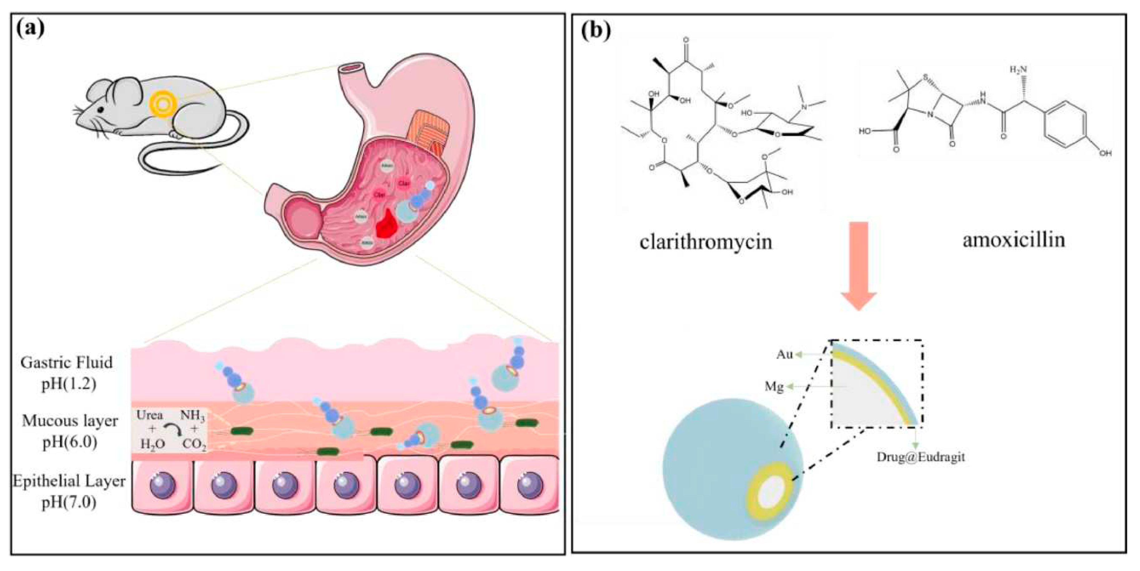
Figure 2.
(a) Synthetic route of Col@MSN@LL-(LL-37), final nanoassembly. (b) Hypothetical mechanism of the action of Col@MSN@LL-(LL-37) upon interaction with PA14. Adopted with permission from Ref. [31]. Copyright 2020, American Chemical Society.
Figure 2.
(a) Synthetic route of Col@MSN@LL-(LL-37), final nanoassembly. (b) Hypothetical mechanism of the action of Col@MSN@LL-(LL-37) upon interaction with PA14. Adopted with permission from Ref. [31]. Copyright 2020, American Chemical Society.
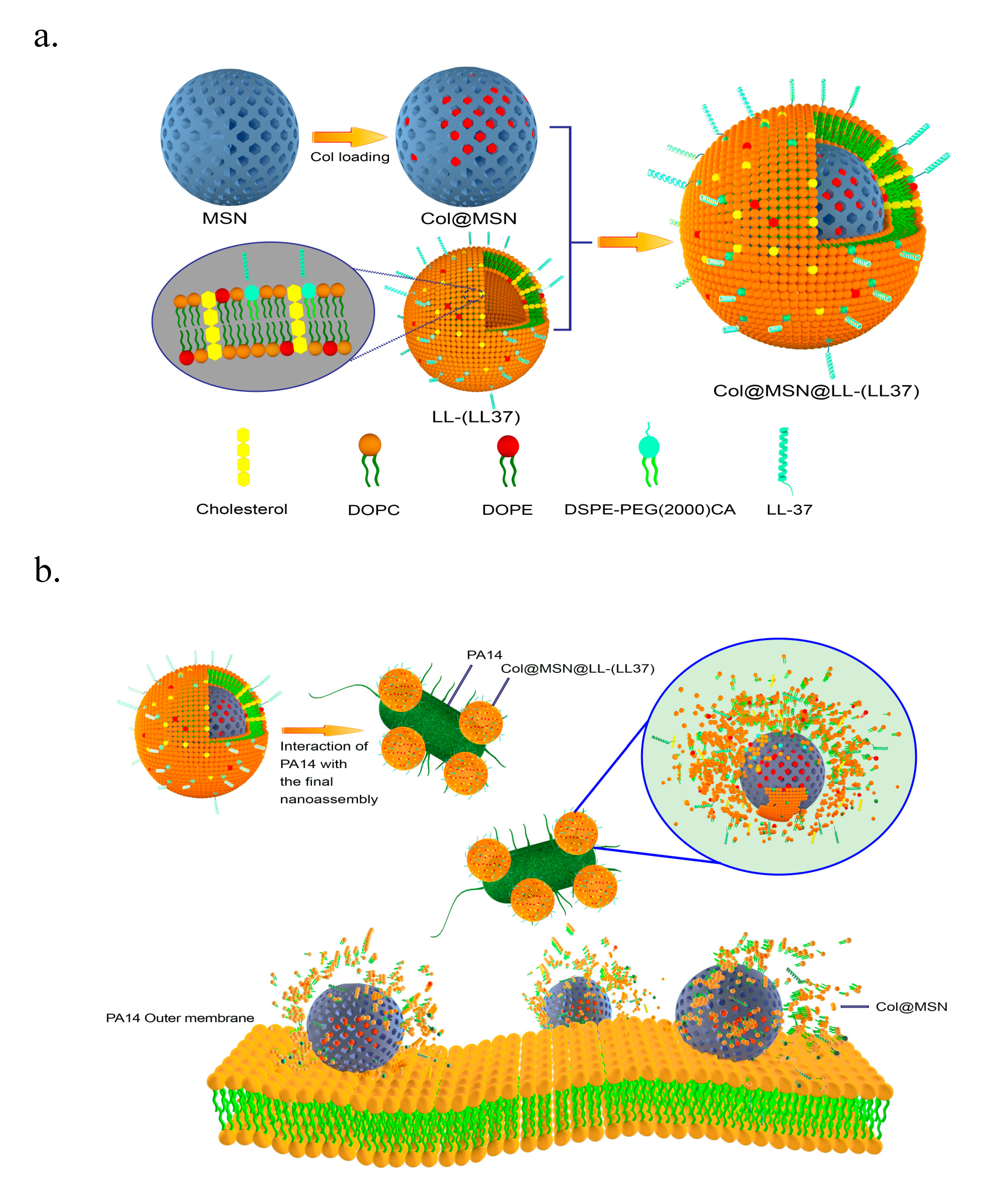
Figure 3.
(A) Schematic illustration of stepwise synthesis of the final nano-assembly: (B) GNR@MSNP@BDQ@TSL@NZX. B, Application of final nano-assembly: GNR@MSNP@BDQ@TSL@NZX for targeting and killing Msmeg using 808 nm NIR laser light. Adopted with permission from Ref. [91]. Copyright 2020 Nano select Wiley.
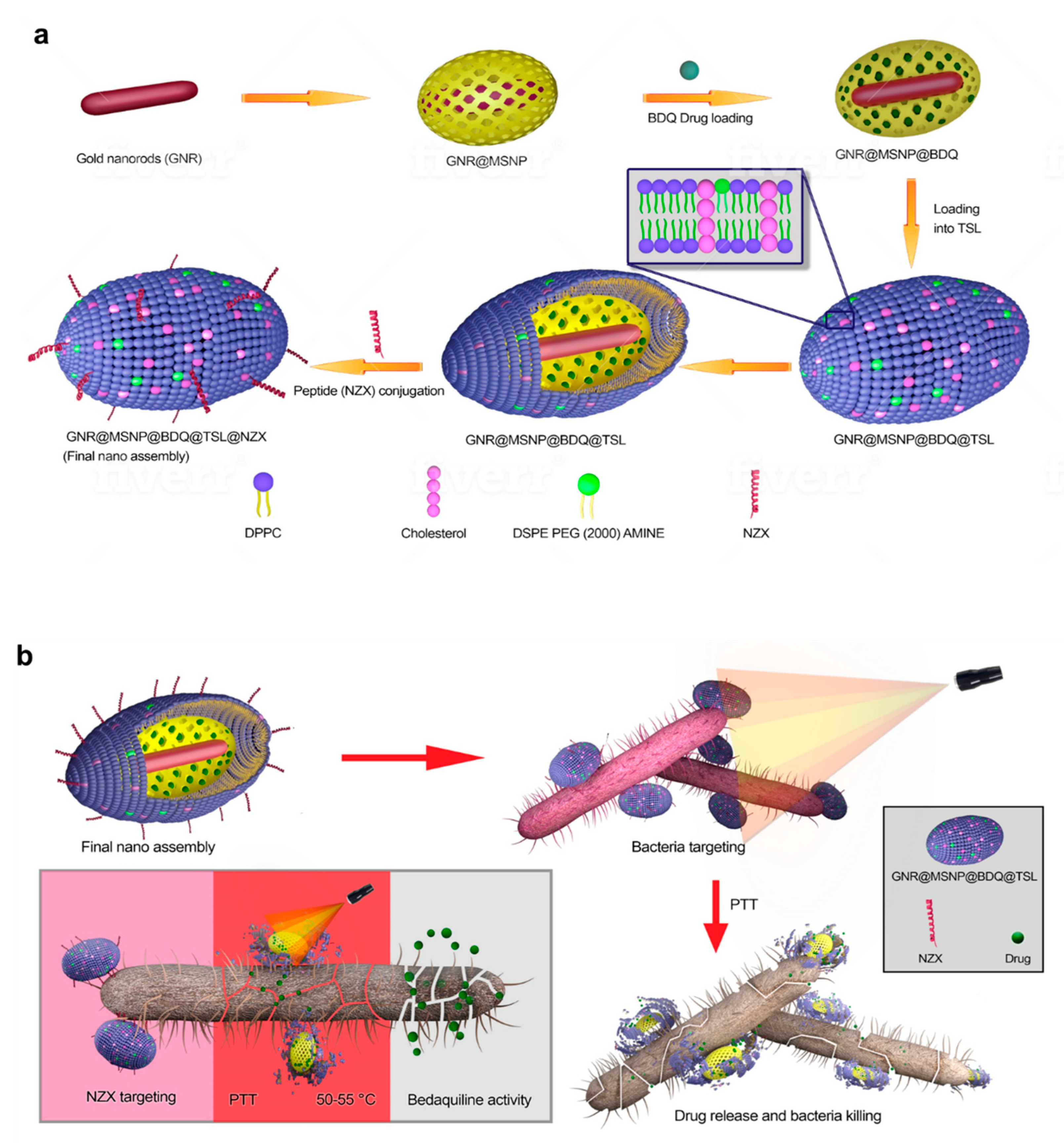
Figure 4.
Scheme illustration of MLP18 nanoliposomes for diagnosis and eradiation of MDR bacterial infection. Taking advantage of the bacteria-specific maltodextrin transporter pathway, MLP18 after intravenous injection selectively navigates to the bacterial infection site (a). Afterward, the oversecreted PLA2 enzyme in bacterial infection microenvironment effectively degrades the lipid skeleton of MLP18, activating an efficient release and internalization of sonoensitizer into bacteria. Due to the excellent optical properties of P18, the bacteria show an obvious NIR FL/PA signal, changing from yellow to red (b). Upon ultrasound irradiation, cytotoxic ROS are generated and then mediate bacterial death (c). Adopted with permission from Ref. [69]. Copyright 2020, American Chemical Society.

Figure 5.
Preparation of NIR light-controlled drug-release D-TiO2/Au@UCN nanocomposites and its mechanism of action. Adopted with permission from Ref. [73]. Copyright 2020, American Chemical Society.
Figure 5.
Preparation of NIR light-controlled drug-release D-TiO2/Au@UCN nanocomposites and its mechanism of action. Adopted with permission from Ref. [73]. Copyright 2020, American Chemical Society.
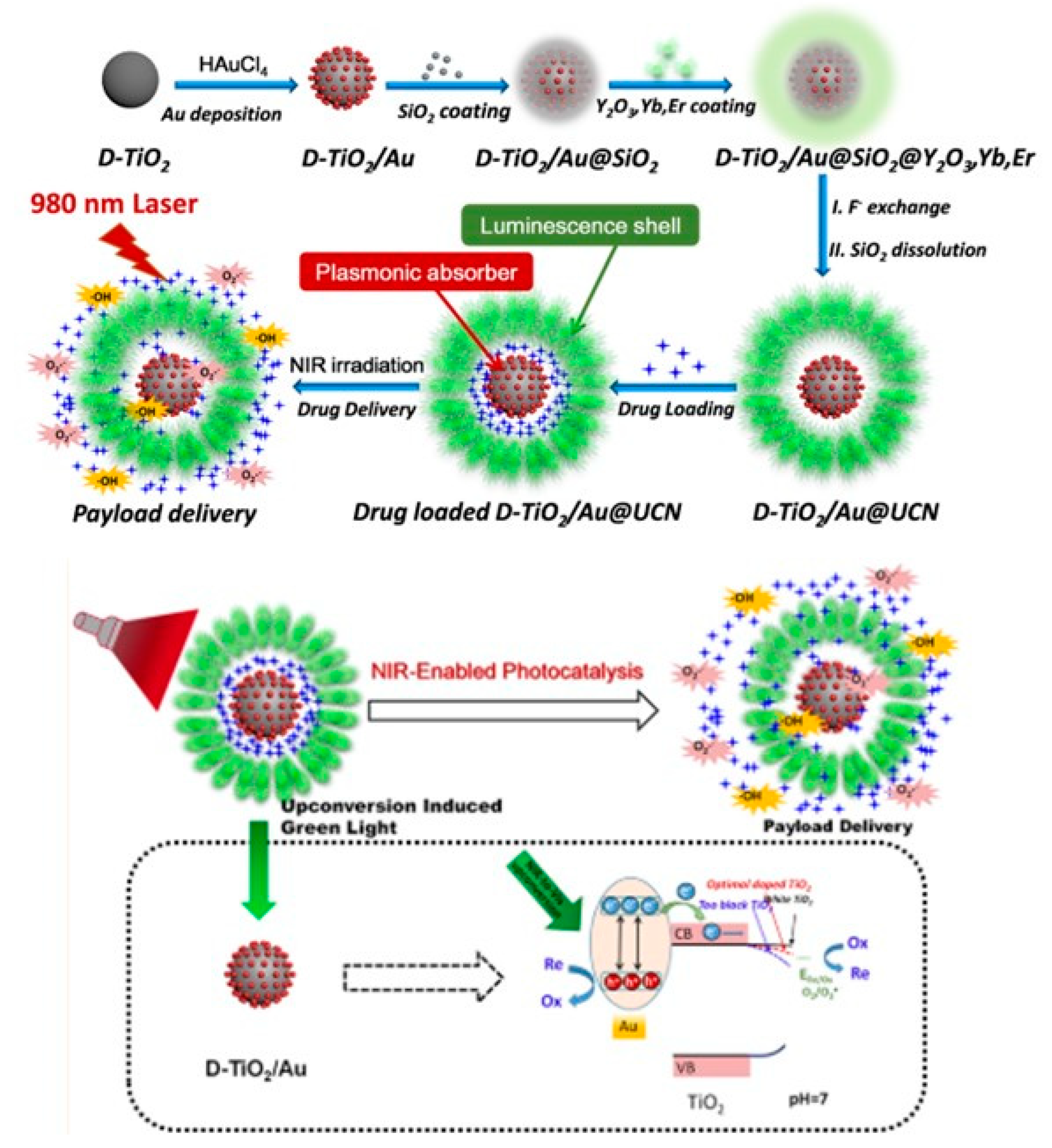
Figure 6.
Schematic illustration of the preparation and the POD-like activity of UsAuNPs/MOFs hybrid for antibacterial therapy. Adopted with permission from Ref. [165]. Copyright 2020 Small, Wiley.
Figure 6.
Schematic illustration of the preparation and the POD-like activity of UsAuNPs/MOFs hybrid for antibacterial therapy. Adopted with permission from Ref. [165]. Copyright 2020 Small, Wiley.
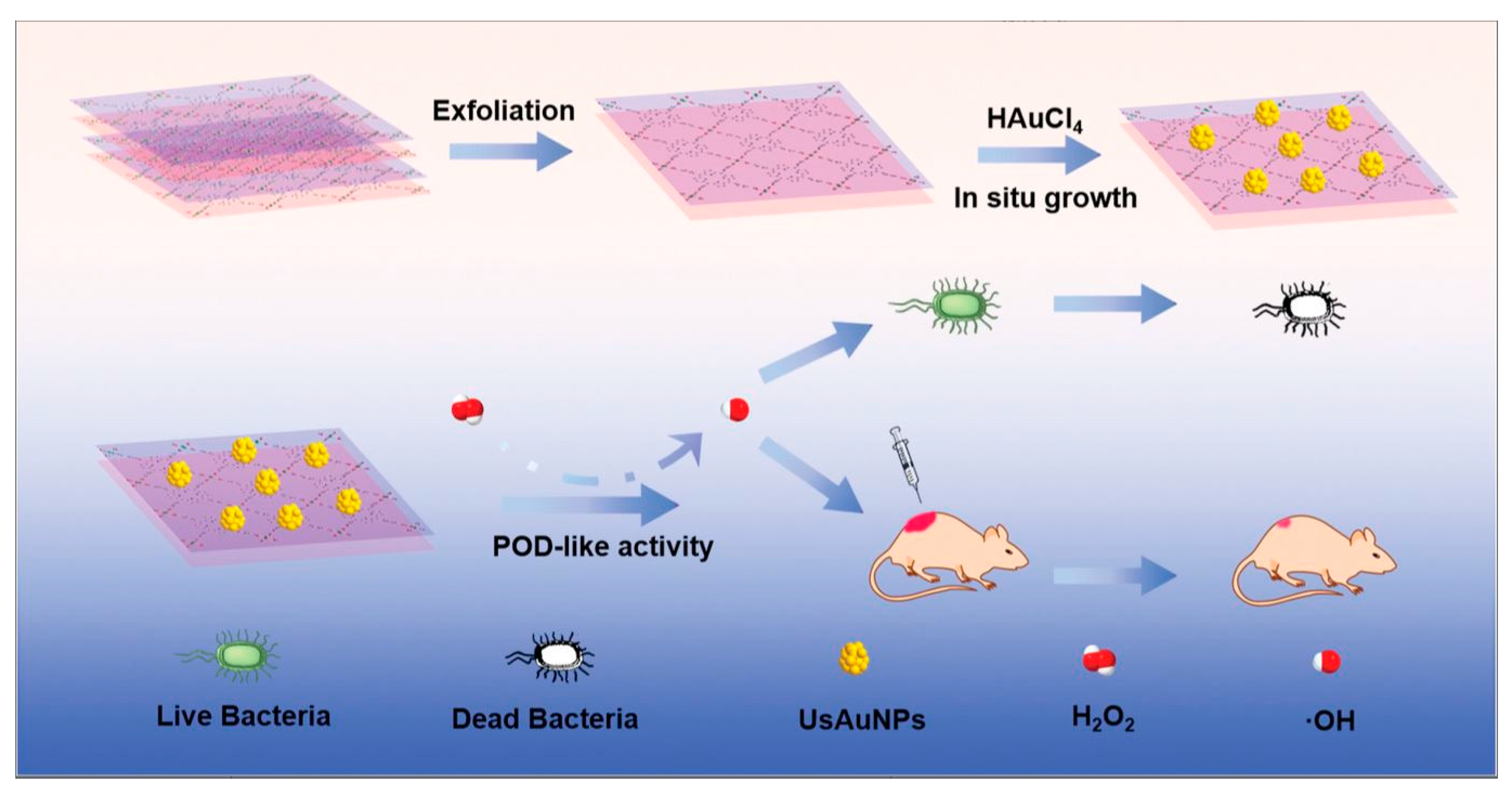
Disclaimer/Publisher’s Note: The statements, opinions and data contained in all publications are solely those of the individual author(s) and contributor(s) and not of MDPI and/or the editor(s). MDPI and/or the editor(s) disclaim responsibility for any injury to people or property resulting from any ideas, methods, instructions or products referred to in the content. |
© 2023 by the authors. Licensee MDPI, Basel, Switzerland. This article is an open access article distributed under the terms and conditions of the Creative Commons Attribution (CC BY) license (http://creativecommons.org/licenses/by/4.0/).
Copyright: This open access article is published under a Creative Commons CC BY 4.0 license, which permit the free download, distribution, and reuse, provided that the author and preprint are cited in any reuse.


