Submitted:
20 June 2023
Posted:
20 June 2023
You are already at the latest version
Abstract
In this study, a case of acute cattle death was investigated. The pathogen was identified, and its molecular characteristics and vector were analysed. This study provides a reference for the prevention and control of babesiosis and the healthy breeding of cattle. In this study, unengorged and engorged Rhipicephalus microplus ticks were collected from the Chongqing area. The unengorged ticks were cultured on cattle under laboratory conditions, and the engorged ticks were cultured to lay eggs. In the process, the cattle suddenly died at 12 days from the bite of an unengorged R. microplus tick. In addition, the larvae hatched from R. microplus eggs, which were cultured on the other cattle, and the experimental cattle died in approximately 14 days. Blood was collected from a second dying and stored at 4 ℃ for one week. Two millilitres of anticoagulated blood was injected subcutaneously into the third cow without piriformis infection. On the fourth day, the body temperature rose to 41 ℃ with slight lymphadenopathy. On the fifth day, the cow suddenly fell and died approximately 4 hours later. DNA was extracted from the blood of all dead cattle and amplified by PCR with piriformis universal primers. The results showed that the cattle were infected with Babesia bovis. The phylogenetic tree based on 18S rRNA showed that the Chongqing strain of B. bovis, which caused the death of cattle, was closely related to the Yunnan strain in China and had the same taxonomic status as the Spanish strain. This case report will draw people's attention to B. bovis once again, and positive control measures should be taken to reduce the losses of farmers to achieve the goal of healthy breeding.
Keywords:
Ticks
; Rhipicephalus microplus
; Babesia bovis
; Acute death
1. Introduction
Babesia bovis is a tick-borne intracellular protozoan parasite that causes the most pathogenic form of bovine babesiosis [1]. It can lead to high fever, mental depression, loss of appetite, and even death, causing serious harm to the world cattle industry and human public health [2]. Many researchers have carried out a series of prevention and control studies based on B. bovis. The use of chemicals and acaricides has caused serious environmental pollution and resistance to pathogens, and they can no longer adapt to current social development and health needs [3]. Therefore, vaccines have become the focus of research. At present, live attenuated vaccines have achieved good efficiency in disease prevention and animal immune protection and have been widely used in Australia, Argentina, Brazil, Uruguay and Israel [4]. In addition, the technology of in vitro culture of B. bovis is convenient for biochemical, immunological and antigen source research [5]. In several studies conducted in Mexico, an attenuated vaccine of B. bovis in vitro produced good protection for host animals, but the disadvantage of this method is that it requires a large-scale culture of B. bovis. In recent years, the screening of immune molecules has also become an important means to control B. bovis. Examples include natural killer cells from the spleen, antigens identified by immune CD4+ T lymphocytes, MHC class II restricting elements, etc. [6]. Ideally, the B. bovis vaccine must induce a humoral immune response, which is characterized by neutralizing antibodies and cellular Th1 immune responses against conservative epitopes. The AMA-1, MSA-2c and RAP-1 proteins have been studied in B. bovis, and antibodies to these proteins have shown good neutralizing effects, suggesting the role of B cells and T-cell epitopes in the immune response. However, whether there are conserved peptides in the epitopes of B cells and T cells in all strains and their role in producing lasting immunity remain to be determined [7]. The spread and epidemic characteristics of B. bovis are important components in the prevention and control of bovine worm disease caused by this pathogen. Studies have shown that R. microplus ticks are an important biological medium for B. bovis [8,9]. Therefore, based on the prevalence of ticks, the prevalence of B. bovis is seasonal and time-sensitive.
In this study, ticks (R. microplus) were collected from Chongqing city, China, and cultured in the laboratory on cattle. However, the cattle died after being bitten by the ticks. Here, the blood of infected cattle was collected, the pathogens were detected with piriformis universal primers for PCR, and positive PCR products were used for sequencing analysis. Based on the sequencing results, it is speculated that the pathogen may have caused the cows’ deaths. To further clarify the prevalence of this pathogen in China, ticks in the main provinces experiencing an epidemic of R. microplus were investigated, and the status of their infection with the pathogen was analysed to provide a reference for the formulation of prevention and control measures and early warning of the pathogen.
2. Materials and Methods
2.1. Ethics Statement
All animal experiments were performed according to the protocols approved by the Animal Care and Use Committee of the Lanzhou Veterinary Research Institute (permit number 2023–03).
2.2. Tick Collection and DNA Extraction
Engorged adult R. microplus ticks were collected from cattle in Chongqing city and identified by morphology in the Department of Veterinary Parasitology, Lanzhou Veterinary Research Institute, Chinese Academy of Agricultural Sciences. These ticks were maintained at a temperature of 30 ± 2℃ and relative humidity of 80 ± 5%. Some of the ticks were immediately placed into phosphate buffered saline (PBS) and washed twice in a solution containing 0.133 M NaCl, 1.11% sodium dodecyl sulfate (SDS) and 0.0088 M ethylenediaminetetraacetic acid (EDTA). These ticks were mixed and stored in liquid nitrogen until ground using a mortar and pestle with liquid nitrogen, and genomic DNA (gDNA) was extracted with a QIAamp DNA Mini Kit (QIAGEN, China) following the manufacturer's instructions. Other unengorged ticks were used to bite experimental cattle.
2.3. Cattle Experiment
Experimental cattle had their back hair removed and cloth bags with openings at both ends that had been sewn in advance were glued to the sheared areas. The above mentioned hatched R. microplus were released into the cloth bag to bite the cattle to further verify the nonaccidental case of cattle death.
2.4. Blood Genome Extraction and PCR Amplification
Genomic DNA (gDNA) was extracted from cattle blood with a Blood &Tissue Kit (Lot 163043916) from QIAGEN, China, following the manufacturer's instructions. The gDNA of cow blood and ticks was analysed using common primers of piroplasma, 989/990 (Theileria) and Prio-As/S (Babesia) primers [10,11]. The reaction system was 50 μL volume: 36.5 μL of sterilized deionized water, 4 μL of dNTP mixture (2.5 mmol/L), 5 μL of 10×PCR buffer, 1 μL of each primer (25 pmol/μL), 2 μL of gDNA template, and 0.5 μL of rTaq DNA polymerase (5 U/μL). The reaction conditions were as follows: 94°C denaturation for 4 min; 40 cycles of 94°C for 1 min, 57°C for 1 min, and 72°C for 1 min; and extension at 72°C for 7 min. The products were electrophoresed in agarose gel at 10 g/L. The PCR products were purified using a TaKaRa Agarose Gel DNA Purification Kit Ver. 2.0 (TaKaRa, Dalian, China), and the amplified products were ligated into the vector pMD®19-T (TaKaRa, Dalian, China). The positive clones were sequenced with vector-specific primers (T7 and SP6) by Sangon (Shanghai, China).
2.5. Sequence Analysis and Phylogenetic Tree
For homology analyses, the 18S rRNA sequences were searched against the BlastN/X (NCBI) (http://www.ncbi.nlm.nih.gov/BLAST/). These sequences from ticks and cattle blood were aligned with previously identified 18S rRNA sequences. Nucleotide sequences from other species retrieved from NCBI GenBank were aligned using Clustal version 1.81. A neighbour-joining (NJ) phylogenetic tree was constructed using Molecular Evolutionary Genetics Analysis (MEGA) version 11.0 [12], and the likelihood of branching was tested using bootstrap resampling (1000 pseudoreplicates).
2.6. Sample Testing
A total of 1678 wild ticks from Guangxi, Yunnan, Qinghai, Fujian and Gansu provinces were analysed using specific primers for B. bovis by PCR [13].
3. Result
3.1. Pathological Replication
In the experiment, unengorged R. microplus ticks from Chongqing were released to healthy cattle not infected by piroplasma. Other engorged ticks were cultured under laboratory conditions to lay eggs, and the hatched larvae were released to healthy cattle not infected by piroplasma. As a result, all the cattle died suddenly in approximately two weeks. In the process, the ticks and cattle received attention; unfortunately, the experimental cattle died suddenly after approximately 13 days, and they showed some clinical symptoms, such as high fever, enlarged anterior shoulder lymph nodes, mental fatigue and loss of appetite. Blood was collected from ill cattle, and 7% dimethyl sulfoxide (DMSO) was added to the blood for storage in liquid nitrogen for later use. Similarly, the blood was inoculated into other experimental cattle, which also exhibited the same clinical symptoms as the previous cattle and ultimately died.
3.2. Pathological Examination
An autopsy of sick and dead cows showed thin blood and poor coagulation; tissue oedema, yellow stains on the organs; flushing of the intestines and wrinkling of gastric mucosa with point bleeding. There were bleeding points in the inner and external membranes of the heart, and the myocardial texture was soft; the liver and spleen were yellow‒brown and obviously swollen; the kidneys were light red and yellow and enlarged, and there was dot bleeding on their surface (Figure 1).
3.3. PCR Detection and Sequencing
Specific primers for B. bovis were used to amplify 18S rRNA by PCR from the blood of dead cattle and R. microplus ticks. The results of agarose gel electrophoresis and sequencing showed that the pathogen that caused the death of the cattle at this time was B. bovis transmitted by R. microplus, and the blood smear also showed the typical morphology of B. bovis (Figure 2).
3.4. Sequence Analysis and Evolutionary Tree
The 18S rRNA gene sequence of B. bovis was obtained and compared with the 18S rRNA nucleic acid sequence of different local strains of this species in the GenBank database. The results show that these sequences are highly conserved, and their identity is more than 92%. The similarity comparison results are shown in Table 1.
MEGA 11.0 software was used to evaluate evolutionary relationships of the B. bovis 18S rRNA gene and showed that the Chongqing strain of B. bovis in this case was similar to the Yunnan strain, China (KY805830), and they have the same taxonomic status as the Spanish strain (FJ426364) (Figure 3). On the other hand, the 18S rRNA of B. bovis can be divided into two main genotypes, both of which have been found in China.
3.5. Sample Detection
In this study, a total of 1768 R. microplus were collected in Guangxi, Fujian, Qinghai, Gansu, Chongqing, Shandong, Henan and Yunnan provinces, and these samples were tested for B. bovis infection. The results show that the population density of R. microplus in Gansu and Yunnan is relatively high, at 358/per square kilometre in Gansu and 356/per square kilometre in Yunnan; the population density of R. microplus in Henan is relatively low, with an average of 83/per square kilometre. In these data, the infection rate was high in Chongqing at 21.35%, 18.44% in Gansu Province, and 17.70% in Yunnan Province. (Table 2)
4. Discussion
B. bovis is the most economically important tick-borne disease and is mainly transmitted by R. microplus ticks [14]. The disease causes substantial economic losses through high animal mortality, low growth rates, reduced milk production in diseased or rehabilitated animals, and the direct costs of tick control and disease treatment. In severe cases, it can lead to the death of an infected animal [15]. In this study, a case of acute death of multiple cattle by bites from field-captured R. microplus was discussed. Blood smears and PCR were used to detect pathogens. To observe whether the death of cattle is caused by blood protozoa, we inoculated the blood of previously dying cattle into another cow. However, for approximately 6 days, the cow experienced elevated body temperature, loss of appetite and mental depression. On the 8th day, the cow fell to the ground and could not stand. It died in approximately 4 hours, leaving dark red blood from the nasal cavity. The anatomy showed that there was no obvious change in the lungs, but the spleen was swollen, and the liver also had a small number of bleeding points. According to the clinical features of cattle disease, as well as blood smears, PCR sequencing studies showed that the death was caused by B. bovis infection. Pathological anatomy also showed the same pathological features as B. bovis disease, such as the gastric surface mucosa being flushed with punctate haemorrhage, poor blood coagulation, serious spleen, and haemorrhage. These pathological changes are the main clinical indicators of Babesia infection [16]. These lesions may also be the main cause of death.
The occurrence and development of B. bovis are closely related to the distribution and prevalence of ticks. Available studies have shown that R. microplus is mainly distributed in Asia [17], India [18], West Africa [19], and South America [20]. This also implies that the transmission of the R. microplus carrying the B. bovis pathogen is linked with animal migration and the long-distance transmission of the livestock and poultry trade. This results in the presence of the same strain or the same genotype in different regions. It also provides a basis for analysing the migration relationship and evolutionary characteristics of different local strains and species. Here, the 18S rRNA of B. bovis was amplified in R. microplus from Chongqing city, and the 18S rRNA sequences were used to construct the phylogenetic tree. The results showed that the Chongqing strain had high identity with the Yunnan strain (KY805830) in China, but they had the same taxonomic status as the Spanish strain (FJ426364), and the Spanish strain had an earlier evolutionary history. This also suggests that the pathogenic strains will be prevalent in different regions along with animal migration or livestock trade. On the other hand, B. bovis 18S rRNA can be divided into two major genotypes (I and II), a result that is consistent with the findings of Ramos CM (2010) [21]. Type I is mainly distributed in China, Spain, West Africa, South America and Australia, and type II is relatively limited, mainly distributed in Turkey, China and Germany. Although the genotype of the B. bovis species is simple, its distribution is extremely widespread. The frequent exchanges of livestock and poultry trade around the world have also led to the widespread prevalence of babesiosis, causing certain losses to the local farming industry and threats to human public health security.
The distribution of ticks is closely related to tick-borne diseases. The diseases caused by tick-borne pathogens will occur and develop regularly with tick activity, which shows typical seasonal and spatiotemporal specificity. To understand the distribution of R. microplus in different areas in China, R. microplus ticks were collected from Guangxi, Fujian, Shaanxi, Gansu, Chongqing, Shandong, Henan, and Yunnan, and B. bovis was detected in 1768 ticks using Prio-As/S primers. The results showed that the population density of R. microplus varied greatly in different regions, with a relatively high density in Gansu and Yunnan and a relatively low density in Henan. Moreover, the infection rate of B. bovis transmitted by R. microplus also showed a positive correlation with changes in tick population density. Of course, this inference also requires a comprehensive and detailed investigation of the distribution of local R. microplus and the epidemic characteristics of B. bovis carried by ticks. Therefore, the data on the population density and infection status of B. bovis in different regions of China may not be accurate. However, these data have certain reference value for the prevention and control of R. microplus and bovine babesiosis and are important for early warning of bovine babesiosis.
5. Conclusions
In conclusion, B. bovis has already occurred in vast areas of our country, seriously endangering the healthy development of the local cattle industry. Therefore, the prevention and control of ticks, the reduction of tick breeding population density, and the avoidance of pathogen transmission during animal migration and livestock trade are important measures to prevent the occurrence of B. bovis. Healthy animal breeding and scientific management are also important strategies for the prevention and control of bovine babesiosis.
Author Contributions
LGY and LJ designed the experiments. LJ, CLY and GMY performed the experiments. LJX and YH analysed the data. LJ and HR wrote the manuscript. CZ, GKF, LDJ and GGQ collected ticks. All authors read and approved the final version of the manuscript.
Funding
This study was financially supported by grants from the National Parasitic Resources Center (NPRC-2019-194-30), ASTIP (CAAS-ASTIP-2016-LVRI), NBCITS (CARS-37),The Science Fund for Creative Research Groups of Gansu Province (22JR5RA024).
Acknowledgments
All listed authors made substantial, direct and intellectual contributions to this work and approved its publication.
Conflicts of interest: The authors declare that they have no conflicts of interest.
References
- Hakimi, H.; Asada, M.; Ishizaki, T.; Kawazu, S. Isolation of viable Babesia bovis merozoites to study parasite invasion. Sci Rep 2021, 11, 16959. [Google Scholar] [CrossRef] [PubMed]
- Giglioti, R.; Okino, C.H.; Azevedo, B.T.; Wedy, B.C.R.; Gutmanis, G.; Veríssimo, C.J.; Katiki, L.M.; Filho, A.E.V.; de Oliveira, H.N.; Oliveira, M.C.S. Semi-quantitative evaluation of Babesia bovis and B. bigemina infection levels estimated by HRM analysis. Ticks Tick Borne Dis 2021, 12, 101753. [Google Scholar] [CrossRef]
- Cuy-Chaparro, L.; Reyes, C.; Díaz-Guiot, E.V.; Moreno-Pérez, D.A.; Patarroyo, M.A. Developing Anti-Babesia bovis Blood Stage Vaccines: A New Perspective Regarding Synthetic Vaccines. Int J Mol Sci 2023, 24, 5219. [Google Scholar] [CrossRef]
- Alvarez, J.A.; Rojas, C.; Figueroa, J.V. An Overview of Current Knowledge on in vitro Babesia Cultivation for Production of Live Attenuated Vaccines for Bovine Babesiosis in Mexico. Front Vet Sci 2020, 7, 364. [Google Scholar] [CrossRef] [PubMed]
- Álvarez Martínez, J.A.; Figueroa Millán, J.V.; Ueti, M.W.; Rojas-Martínez, C. Establishment of Babesia bovis In Vitro Culture Using Medium Free of Animal Products. Pathogens 2021, 10, 770. [Google Scholar] [CrossRef] [PubMed]
- Brown, W.C.; Norimine, J.; Knowles, D.P.; Goff, W.L. Immune control of Babesia bovis infection. Vet Parasitol 2006, 138, 75–87. [Google Scholar] [CrossRef]
- Hidalgo-Ruiz, M.; Mejia-López, S.; Pérez-Serrano, R.M.; Zaldívar-Lelo de Larrea, G.; Ganzinelli, S.; Florin-Christensen, M.; Suarez, C.E.; Hernández-Ortiz, R.; Mercado-Uriostegui, M.A.; Rodríguez-Torres, A.; Carvajal-Gamez, B.I.; Camacho-Nuez, M.; Wilkowsky, S.E.; Mosqueda, J. Babesia bovis AMA-1, MSA-2c and RAP-1 contain conserved B and T-cell epitopes, which generate neutralizing antibodies and a long-lasting Th1 immune response in vaccinated cattle. Vaccine 2022, 40, 1108–1115. [Google Scholar] [CrossRef]
- Githaka, N.W.; Bishop, R.P.; Šlapeta, J.; Emery, D.; Nguu, E.K.; Kanduma, E.G. Molecular survey of Babesia parasites in Kenya: first detailed report on occurrence of Babesia bovis in cattle. Parasit Vectors 2022, 15, 161. [Google Scholar] [CrossRef]
- Guglielmone, A.A. Epidemiology of babesiosis and anaplasmosis in South and Central America. Vet Parasitol 1995, 57, 109–19. [Google Scholar] [CrossRef]
- Li, Y.; Guan, G.; Ma, M.; Liu, J.; Ren, Q.; Luo, J.; Yin, H. Theileria ovis discovered in China. Exp Parasitol 2011, 127, 304–7. [Google Scholar] [CrossRef]
- Gou, H.; Guan, G.; Liu, A.; Ma, M.; Xu, Z.; Liu, Z.; Ren, Q.; Li, Y.; Yang, J.; Chen, Z.; Yin, H.; Luo, J. A DNA barcode for Piroplasmea. Acta Trop 2012, 124, 92–7. [Google Scholar] [CrossRef] [PubMed]
- Kumar, S.; Stecher, G.; Li, M.; Knyaz, C.; Tamura, K. MEGA X: Molecular Evolutionary Genetics Analysis across Computing Platforms. Mol Biol Evol 2018, 35, 1547–1549. [Google Scholar] [CrossRef]
- Mahdy, O.A.; Nassar, A.M.; Elsawy, B.S.M.; Alzan, H.F.; Kandil, O.M.; Mahmoud, M.S.; Suarez, C.E. Cross-sectional analysis of Piroplasma species-infecting camel (Camelus dromedaries) in Egypt using a multipronged molecular diagnostic approach. Front Vet Sci 2023, 10, 1178511. [Google Scholar] [CrossRef] [PubMed]
- Oliveira-Sequeira, T.C.; Oliveira, M.C.; Araujo, J.P. , Jr Amarante, A.F. PCR-based detection of Babesia bovis and Babesia bigemina in their natural host Boophilus microplus and cattle. Int J Parasitol 2005, 35, 105–11. [Google Scholar] [CrossRef] [PubMed]
- El-Sayed, S.; Rizk, M.A.; Terkawi, M.; Igarashi, I. Cocktail Babesia bovis antigens for global detection of Babesia bovis infection in cattle. Exp Parasitol 2019, 206, 107758. [Google Scholar] [CrossRef] [PubMed]
- Eamens, G.J.; Bailey, G.; Jenkins, C.; Gonsalves, J.R. Significance of Theileria orientalis types in individual affected beef herds in New South Wales based on clinical, smear and PCR findings. Vet Parasitol 2013, 196, 96–105. [Google Scholar] [CrossRef] [PubMed]
- Zhang, X.L.; Deng, Y.P.; Yang, T.; Li, L.Y.; Cheng, T.Y.; Liu, G.H.; Duan, D.Y. Metagenomics of the midgut microbiome of Rhipicephalus microplus from China. Parasit Vectors 2022, 15, 48. [Google Scholar] [CrossRef] [PubMed]
- Kumar, R.; Sharma, A.K.; GhoshS. Menace of acaricide resistance in cattle tick, Rhipicephalus microplus in India: Status and possible mitigation strategies. Vet Parasitol 2020, 278, 108993. [Google Scholar] [CrossRef]
- Makenov, M.T.; Toure, A.H.; Korneev, M.G.; Sacko, N.; Porshakov, A.M.; Yakovlev, S.A.; Radyuk, E.V.; Zakharov, K.S.; Shipovalov, A.V.; Boumbaly, S.; Zhurenkova, O.B.; Grigoreva, Y.E.; Morozkin, E.S.; Fyodorova, M.V.; Boiro, M.Y.; Karan, L.S. Rhipicephalus microplus and its vector-borne haemoparasites in Guinea: further species expansion in West Africa. Parasitol Res 2021, 120, 1563–1570. [Google Scholar] [CrossRef]
- Gómez, G.F.; Isaza, J.P.; Segura, J.A.; Alzate, J.F.; Gutiérrez, L.A. Metatranscriptomic virome assessment of Rhipicephalus microplus from Colombia. Ticks Tick Borne Dis 2020, 11, 101426. [Google Scholar] [CrossRef]
- Ramos, C.M.; Cooper, S.M.; Holman, P.J. Molecular and serologic evidence for Babesia bovis-like parasites in white-tailed deer (Odocoileus virginianus) in south Texas. Vet Parasitol 2010, 172, 214–20. [Google Scholar] [CrossRef] [PubMed]
Figure 1.
Pathological features of the anatomical tissues of cattle with Babesia bovine. A, There is a small amount of nasal fluid in the nasal cavity, mixed with mucus and blood; B, The mucosa of the intestine and wrinkled stomach is flushed with spot-like bleeding; C, The blood is thin and poorly clotting; D, The spleen is markedly enlarged with petechial bleeding on the surface.
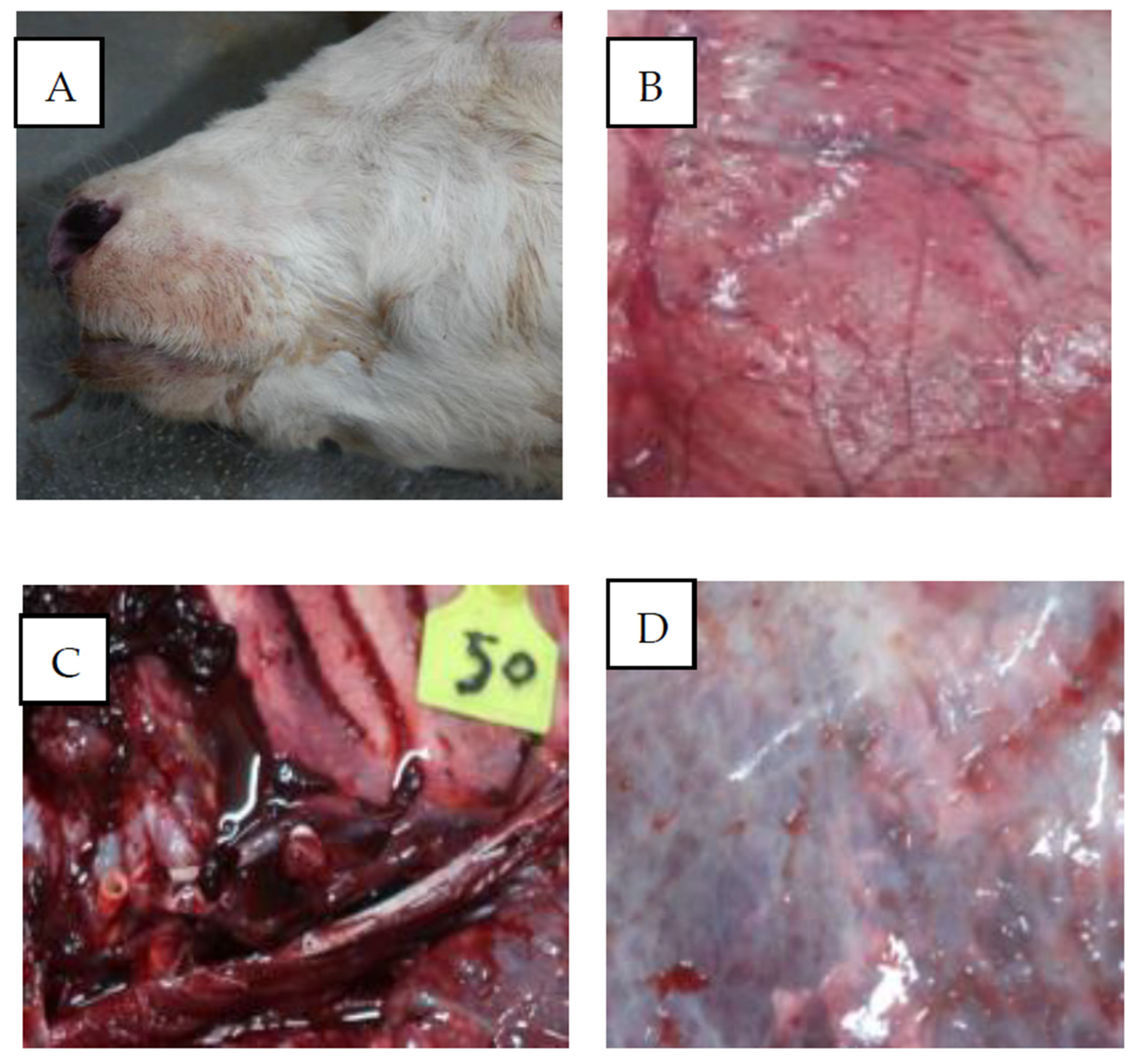
Figure 2.
Morphological characteristics of Babesia bovine.
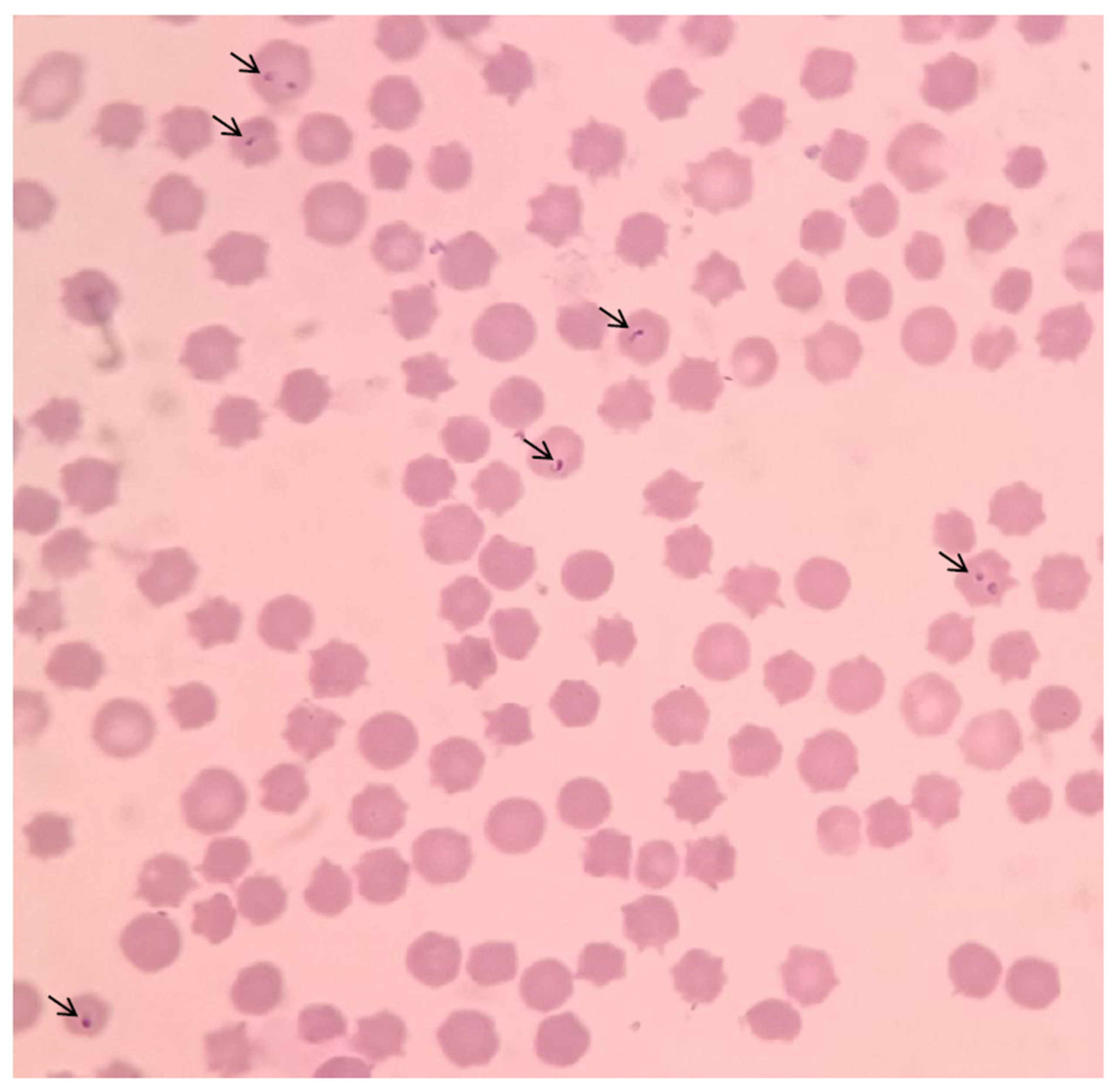
Figure 3.
Neighbour-joining tree showing the phylogenetic relationships of the 18S rRNA based on nucleotide sequences of Babesia bovine. The numbers represent the percentage of 1000 replicates (bootstrap support) for which the same branching patterns were obtained.
Figure 3.
Neighbour-joining tree showing the phylogenetic relationships of the 18S rRNA based on nucleotide sequences of Babesia bovine. The numbers represent the percentage of 1000 replicates (bootstrap support) for which the same branching patterns were obtained.
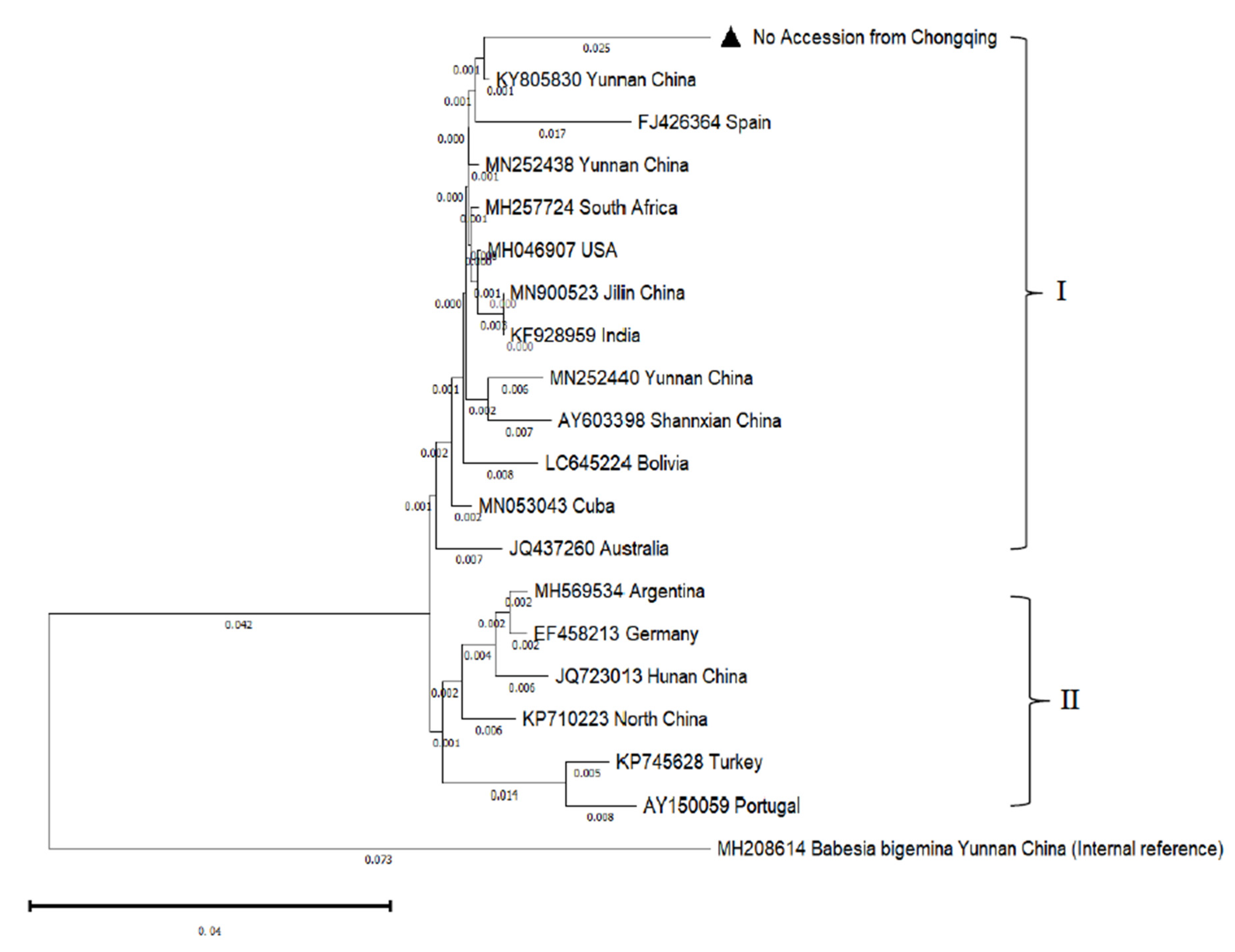

Table 1.
Information on the 18S rRNA gene sequences of different Babesia bovis strains from NCBI.
| Species | Isolate | Accession No. | Identity | Species | Isolate | Accession No. | Identity |
|---|---|---|---|---|---|---|---|
| B. bovis | Chongqing | No |  |
B. bovis | Yunnan | KY805830 | 96.39 |
| B. bovis | Yunnan | MN252438 | 96.31 | B. bovis | Hunan | JQ723013 | 95.16 |
| B. bovis | Yunnan | MN252440 | 95.50 | B. bovis | North | KP710223 | 94.48 |
| B. bovis | Shannxian | AY603398 | 95.41 | B. bovis | Turkey | KP745628 | 94.11 |
| B. bovis | Jilin | MN900523 | 95.82 | B. bovis | Argentina | MH569534 | 94.49 |
| B. bovis | Spain | FJ426364 | 94.84 | B. bovis | Germany | EF458213 | 94.49 |
| B. bovis | South Africa | MH257724 | 96.23 | B. bovis | Australia | JQ437260 | 95.08 |
| B. bovis | USA | MH046907 | 96.15 | B. bovis | Cuba | MN053043 | 95.90 |
| B. bovis | Portugal | KP745628 | 94.11 | B. bovis | Bolivia | LC645224 | 95.54 |
| B. bovis | India | KF928959 | 95.82 | B. bigemina | Yunnan | MH208614 | |
Table 2.
Babesia bovis was detected from Rhipicephalus microplus ticks in China by PCR.
| Province | Rhipicephalus microplus ticks (Number) | Babesia Bovis Positive (%) |
|---|---|---|
| Guangxi | 186 | 12.40(n=23) |
| Yunnan | 356 | 17.70(n=63) |
| Fujian | 136 | 0 |
| Qinghai | 225 | 10.67(n=24) |
| Gansu | 358 | 18.44(n=66) |
| Chongqing | 178 | 21.35(n=38) |
| Shandong | 246 | 9.75(n=24) |
| Henan | 83 | 0 |
| Total | 1768 | 13.46(n=238) |
Disclaimer/Publisher’s Note: The statements, opinions and data contained in all publications are solely those of the individual author(s) and contributor(s) and not of MDPI and/or the editor(s). MDPI and/or the editor(s) disclaim responsibility for any injury to people or property resulting from any ideas, methods, instructions or products referred to in the content. |
© 2023 by the authors. Licensee MDPI, Basel, Switzerland. This article is an open access article distributed under the terms and conditions of the Creative Commons Attribution (CC BY) license (http://creativecommons.org/licenses/by/4.0/).
Copyright: This open access article is published under a Creative Commons CC BY 4.0 license, which permit the free download, distribution, and reuse, provided that the author and preprint are cited in any reuse.


