Submitted:
13 June 2023
Posted:
13 June 2023
You are already at the latest version
Abstract
Panax ginseng C. A. Meyer (P. ginseng), has been widely used in traditional Chinese medicine (TCM). It contains a number of chemical components and possesses a variety of pharmacological activities. Alzheimer's disease (AD) is neurodegenerative disease that creates a huge burden on the lives and health of individuals. In recent years, studies have indicated that the chemical components of P. ginseng, especially ginsenosides, play a pronounced positive role in the prevention and treatment of neurological diseases. This review aimed to summarize currently studies on chemical components and the mechanisms action in AD intervention treatment of P. ginseng, especially ginsenosides. In this review, the components of P. ginseng and their respective active effects were first introduced. Then the key molecular mechanisms and signaling pathways of P. ginseng were introduced, and its different active ingredients in the prevention and treatment of Alzheimer's disease-related pathogenesis were also summarized from the pathogenesis of AD Aβ generation and aggregation, hyperphosphorylation of tau protein,oxidant stress,neuroinflammation,mitochondrial damage,disorder of neurotransmitter and gut microbiota. Signaling pathway networks related to the action of ginseng active ingredients was constructed, which could serve as a therapeutic target for Alzheimer's disease. In addition to a detailed report on P. ginseng in improving Alzheimer's disease-related pathogenesis, the application of the current technology, spatial metabonomics in AD therapeutics and diagnostics were later discussed. Spatial metabonomics could be applied to investigate the multi-target intervention of P. ginseng on Alzheimer's disease. Research perspectives for the study of P. ginseng in the treatment of Alzheimer's disease were provided for further studies.
Keywords:
P. ginseng
; chemical component
; ginsenoside
; Alzheimer's disease
; signaling pathways
; spatial metabolomics
1. Introduction
Panax ginseng C. A. Meyer is perennial herb belonging to the family Araliaceae and genus Panax.P.ginseng comprises many biologically active components including Ginsenosides, Gintonin, Polysaccharides, Peptides, Glycoconjugated compounds, and other compounds. The most commonly studied bioactive component for health benefit claims was Ginsenosides [1]. Ginsenosides has been extensively applied for many years to treat various diseases, especially neurodegenerative diseases, including Alzheimer's disease. Ginsenosides, as the active components isolated from P.ginseng, have been reported to have therapeutic effects on AD, and pharmacological mechanisms were further studied as well. In addition, other active ingredients including Gintonin and Ginseng polysaccharides of ginseng have also been found to have certain therapeutic effects on nervous system disease.
Alzheimer's disease (AD) is a neurodegenerative disease characterized by the decline of cognitive and memory function, which seriously affects the health and life of patients. Several factors such as age, sex, environmental factors play roles in the disease. It is mostly seen in people over 65 years old. In recent years, the research on Alzheimer's disease has attracted extensive attention as a multifactorial neurodegenerative disease [2],there is still no perfect treatment plan to improve Alzheimer's disease. Moreover, Alzheimer's disease is considered to start decades earlier before clinical symptoms occur [3]. Thus, it is important to clarify the pathogenesis of Alzheimer's disease for the early diagnosis, prevention and treatment.
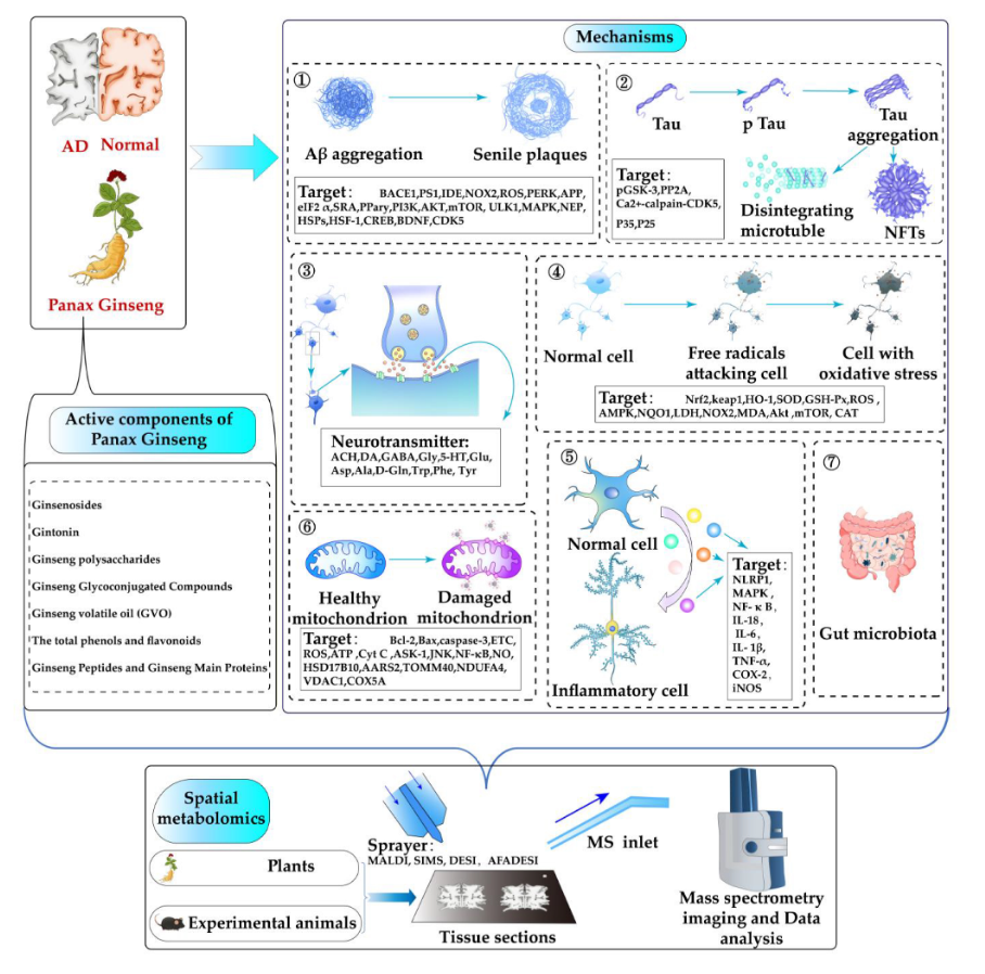
Research on the mechanisms of AD mainly focuses on the amyloid cascade and tau protein [4]. Senile plaques formed by aggregation of amyloid beta Protein (Aβ) and neurofibrillary tangles (NFT) formed by hyperphosphorylation of tau protein are two hallmark pathologies of Alzheimer's disease. It is generally believed that AD is caused by multiple pathological processes resulting from Aβ abnormality, tau phosphorylation, neurotransmitter dysregulation, neuroinflammation, oxidative stress, mitochondrial damage, and gut microbiota disorders in recently reported. Therefore, Multi-target and multi-channel therapies as an emerging strategy for Alzheimer's disease treatment need be highlighted. Moreover, evidence for the medicinal and health benefits of ingredient in P.ginseng, mainly ginsenosides, in the prevention of Alzheimer's disease is increasing. The active ingredients in P. ginseng can also improve Alzheimer's disease through the interaction of different mechanisms of action and signal pathways. In addition, some classical Chinese traditional prescription and herb pairs related to P. ginseng, was also widely applied in the treatment of Alzheimer's disease.
Mass spectrometry imaging (MSI) is a tool capable of simultaneously providing in situ untargeted chemical information as well as the spatial distribution of vast molecular species with high efficiency [5]. MSI combines the mass spectrometry with microscopic imaging technology, enabling the precise location of several analytes in the tissue. Common mass spectrometry imaging technologies mainly include MALDI, SIMS, DESI and AFADESI. With the rise of metabolomics, more and more studies are using MSI to detect the tissue distribution of key metabolites [6]. Spatial metabolomics has been widely used in phytochemistry and medicine. It was suggested that spatial metabolomics would be a promising direction to study the complexity of AD pathophysiology. MS imaging allows the direct visualization of metabolite distributions in tissues, which is beneficial for the treatment of diseases.
This review introduces the active ingredients in P. ginseng and their active effects. The role targets of the active ingredients in P. ginseng were summarized, especially ginsenosides,in mechanisms of Alzheimer's disease. Related therapeutic signal networks for P. ginseng active ingredients acting on different mechanisms of Alzheimer's disease were constructed. In addition, this review summarizes the application of spatial metabolomics in the treatment of Alzheimer's disease with P. ginseng, and discusses its technical support role.
2. Active Ingredients of P. ginseng
P. ginseng contains comprehensive bioactive and potentially effective therapeutic compounds including Ginsenosides, Gintonin, Polysaccharides, Peptides, Glycoconjugated compounds, and other compounds. These active ingredients in P. ginseng have been found to have various pharmacological effects and can exert good therapeutic effects on different diseases (Figure 1). The various components of P. ginseng and their active roles were reviewed in order to provide theoretical basis for better development and utilization of P. ginseng.
2.1. Ginsenosides
Ginsenosides are the main active ingredients existing the roots, fruits, stems, and leaves of P. ginseng. Ginsenosides are steroidal glycosides. The reported ginsenosides are based on the type of saponin, the number, type, and site of glycosyl units. According to their aglycone skeletons, they can be divided into dammarane and oleanane types, which respectively form a four ring skeleton with sugar connecting parts and a five ring skeleton with oleanolic acid (OA) and C17 side chain variant (C17SCV) subtypes. According to the connection position of the sugar part, the dammarane skeleton is further classified as protopanaxadiol (PPD) and protopanaxatriol (PPT) ginsenosides, whose sugar components are respectively connected at the third and sixth positions of the triterpenoid structure [7]. In addition, malonyl saponins containing malonyl groups are also detected in different parts of P. ginseng. Molecular formulas, molecular weights, and structural categories of 170 ginsenosides isolated from different parts of P. ginseng. As a result, 59 ginsenosides were OA type, 42 ginsenosides were PPD type, 65 ginsenosides were PPT type, and 17 ginsenosides were C17CSV type. Four PPD type ginsenosides (Rb2, Rb1, Rc, Rd), three PPT type ginsenosides (Re, Rf, Rg1), and one OA type ginsenoside Ro have the highest content in P. ginseng, accounting for more than 70% of the total saponins. Protopanaxadiol, protopanaxatriol and C17 side chain variation (C17SCV) are the main types of ginsenosides, and the composition of ginsenosides in different parts is significantly different. 16 types of ginsenosides are commonly present in all tissues, including 9 PPD types (Rc, Rd, Rb2, Rb1, Rb3, m-ginsenoside Rb1, m-ginsenoside Rc, m-ginsenoside Rb2, m-ginsenoside Rd), 6 PPT types (Re, Rg1, Rf, 20 (R) - ginsenoside Rg2, Panax notoginseng saponin R1, m-ginsenoside Re), 1 OA type (Ro), and no C17SCV type. Protopanaxadiol type ginsenosides dominate in roots, rhizomes, leaves, stems, and fruits, while malonyl and C17SCV type ginsenosides account for a greater proportion in flowers and buds compared to other parts [1].Each type of ginsenoside has different pharmacological effects. As the main active ingredients in P.ginseng, Ginsenosides exhibit significant pharmacological activities,such as anti-cancer [8], anti-tumor [9], anti-inflammatory [10], anti-virus [11], anti-aging [12], anti fatigue, anti diabetes [13], treatment of cardiovascular diseases [14,15] and neurodegenerative diseases [16,17] including Alzheimer's disease [18].Therefore, increasing the production of ginsenosides and developing more types of ginsenosides play an important role in the treatment of various diseases. Conventional tissue culture, cell suspension culture, protoplast culture, polyploidy, in vitro mutagenesis, hair root culture and other biotechnology have been proposed to improve the shortcomings of traditional extraction methods that are time-consuming and low in yield, greatly increasing the yield of ginsenosides [19]. In addition, the method of producing ginsenosides using microbial hosts has also been proposed for more efficient and selective production of ginsenosides [20]. Rare ginsenosides have various biological activities, but their content is extremely low in P. ginseng. Different extraction methods can have an impact on the structure of ginsenosides [21]. An effective combination biotechnology method, including tissue culture, fixation, and hydrolysis methods, has been developed for the production of rare ginsenosides, including ginsenoside Rh2 [22]. In the biosynthesis process of ginsenosides, glycosyltransferases play an important role in the formation of ginsenosides with diverse structures and biological activities by transferring various sugar parts into ginsenosides. The discovery of UDP glycosyltransferases (UGTs) can help produce more rare ginsenosides [23].
2.2. Gintonin
Gintonin, a glycoprotein complex isolated from P. ginseng, contains three lipid-derived G protein-coupled receptor ligands: lysophosphatidic acids (LPAs), lysophosphatidylinositols (LPIs), and linoleic acid (LA). Gintonin plays an important role in the treatment of neurodegenerative diseases, including Alzheimer's disease. It can not only help drugs penetrate the blood-brain barrier, but also exert neuroprotective effects through anti-inflammatory, antioxidant, and induced autophagy effects. Gintonin-enriched fraction (GEF) exhibits anti-brain senescence and effects against disorders such as Alzheimer's disease (AD), Huntington's disease (HD), and Parkinson's disease (PD). Gintonin can regulate the levels of neurotransmitters such as acetylcholine, dopamine, norepinephrine, and serotonin exert antioxidant and anti-inflammatory effects. And it plays a role in treating neurodegenerative diseases by inducing autophagy to reduce Aβ production and other mechanisms [24]. Gintonin induces a rapid and transient opening of the BBB and that gintonin enhances the delivery of low and high molecular weight molecules to the brain via a paracellular pathway. A gintonin-mediated enhancement of the delivery of molecules to the brain may be applied to enhance the therapeutic effect of drugs for brain diseases [25].
2.3. Ginseng Polysaccharides
Ginseng polysaccharides are a class of bioactive compounds present in the P.ginseng. Studies have shown that ginseng polysaccharide has the activity of anti-tumor [26], anti-fatigue [27], anti-cancer [28], anti-inflammatory [29], anti-oxidant [30], immune regulation [31], intestinal microbiota regulation [32] and treatment of nervous system disease [33]. Ginseng root polysaccharides were the main research subjects in the past. The polysaccharides in P. ginseng flowers, fruits, stems, and leaves also have important biological activities. So far, about 80 types of polysaccharides have been isolated from P. ginseng. The structure and activity of ginseng polysaccharides vary. The structure, molecular weight, side chain, main chain configuration, monosaccharide composition, and functional groups of ginseng polysaccharides can all affect their biological activity. Ginseng polysaccharides contain neutral polysaccharides and pectins. Neutral polysaccharides are the main components of ginseng polysaccharides, mainly including amyloid mixtures, dextrans, and arabinogalactans. Acidic polysaccharides account for a small portion of ginseng polysaccharides. The acidic polysaccharides are considered to be pectin-containing rhamnose and homogalacturonic acid [34]. The crude polysaccharides from ginseng stems and leaves have antioxidant activity, and research has found that the antioxidant activity of polysaccharides from aboveground parts is higher than that from underground parts; The antioxidant activity of polysaccharides above ground is as follows: neutral polysaccharides>acidic polysaccharides. The antioxidant activity of acidic polysaccharides in the underground part is not significant [30]. Ginseng pectin (GP) may play a neuroprotective role in preventing apoptosis through activation of ERK1/2 and Akt phosphorylation [35].
2.4. Ginseng Peptides and Ginseng Main Proteins
The proteins isolated from P. ginseng have anti fatigue, antifungal, and antiviral activities. Ginseng Main Proteins (GMP) are the main proteins present in P. ginseng. Peptidomics can be defined as the study of low molecular weight components of proteins, including endogenous peptides, protein fragments, and protein degradation products [36].Compared with proteins, peptides have a simple structure that is easy to analyze and can reflect the synthesis, processing, and degradation processes of proteins. Peptide fragments can provide a deeper understanding of the properties and functions of precursor proteins. Peptides are expected to be candidate drugs for quality control of medicinal plants [37].
2.5. Ginseng Glycoconjugated Compounds
Glycoconjugates are compounds that covalently connect one or more monosaccharide or oligosaccharide units with non carbohydrate (proteins, lipids, etc.) parts. As bioinformatics molecules, glycoconjugates can participate in regulating various life activities in organisms. In recent years, it has been found that Glycoconjugated Compounds have various biological activities such as anti-inflammatory and analgesic effects. Luo et al. Used P. ginseng as the raw material, extracted with 85% ethanol reflux, and then purified with a G-15 column of silk fatty amide to prepare ginseng glycopeptides (Gg). They found that ginseng glycopeptides have anti-inflammatory and analgesic effects [38].
2.6. Other Compounds
P. ginseng has various active effects, and the material basis for its active effects is mainly ginsenosides, ginseng polysaccharides, etc. In recent years, some new secondary metabolites of ginseng have been discovered to have certain pharmacological activities. The content of ginsenosides and amino acids in wild ginseng is much higher than that in cultivated ginseng, but wild ginseng is relatively scarce. The preparation of wild ginseng adventitious root culture induced by methyl jasmonic acid can effectively solve the problem of insufficient supply of wild ginseng, and new secondary metabolites have been found in wild ginseng adventitious root culture. Chromatographic fractionation of wild ginseng adventitious root cultures led to the isolation of eleven compounds. The chemical structures of isolated compounds were identified as four known flavanone derivatives (1–4), one new curcubinoyl derivative, jasmogin A (5) and six new curcubinoyl-flavanone conjugates, jasmoflagins A-F (6–11) by extensive spectroscopic analysis.And it was found that these compounds have inhibitory effects on lipopolysaccharide (LPS) induced NO production recently reported by Liu [39].The total phenols and flavonoids in ginseng shoot extracts (GSEs) have been found to have anti-inflammatory and antioxidant activities. And research has found that when GSEs are cooked, the lower polarity of ginsenosides, total phenols, total flavonoids, their antioxidant capacity, and anti-inflammatory effects all increase with increasing cooking time [40].Ginseng volatile oil (GVO) is one of the main components of P. ginseng. Research has found that ginseng volatile oil can delay the aging of the elegans elegans by activating autophagy and antioxidant effects [41].
3. Mechanism of P.ginseng in the Treatment of Alzheimer's Disease
Alzheimer's disease is caused by many factors. The pathogenesis of AD is complex. Nevertheless, the pathogenesis is closely related to Aβ Aggregation and deposition, tau hyperphosphorylation, oxidative stress, neuroinflammation, the disorder of neurotransmitter levels and the related signal pathway of those pathogenesis. Therefore, by intervening in these molecules and signal transductions, we could advance the treatment of neurological diseases. In recent years, more and more studies have found that there are more potential mechanisms of AD pathogenesis, including mitochondrial damage and disorder of gut microbiota. The occurrence and development of AD resulted by the interaction of various pathogenesis, and multiple targets jointly. Many studies have shown that various active substances in P. ginseng, especially ginsenosides, can alleviate Alzheimer's disease by regulating different signaling pathways related to the mechanisms of AD. Those pathological mechanisms are main contain that Aβ aggregation, tau hyperphosphorylation, the disorder of neurotransmitter, neuroinflammation, oxidative stress, mitochondrial damage and the disorder of gut microbiota .
3.1. Interference Aβ Generation and Aggregation
Aβ Amyloid plaque (senile plaques, SP) produced by aggregation is an important marker of AD [42]. Aβ Aggregation is neurotoxic, leading to membrane rupture, abnormal cell signal and organelle dysfunction [43]. Aβ accumulation can cause a series of detrimental neurodegeneration cascades such as induction of mitochondrial apoptosis, neuroinflammation, Tau hyperphosphorylation, and synaptic dysfunction,further inducing the generation of AD. It is found that the active substances in P. ginseng, especially ginsenosides can prevent Aβ Aggregation to improve cognitive and memory impairment caused by Alzheimer's disease. The active substances of P. ginseng can be used to regulate Aβ related signaling pathways and exert anti AD effects (Table 1) ( Figure 2). Imbalance in production and clearance of amyloid beta (Aβ) is the primary reason for its deposition in Alzheimer disease. Aβ is caused by abnormal processing of amyloid precursor protein (APP), which can pass through β- Secretory enzymes and γ- Secretase cleaved into Aβ, You can also pass α- Secretase cleaves into secretory app α (sAPP α), Block Aβ Generation of. Macroautophagy/autophagy is one of the important mechanisms for clearance of both intracellular and extracellular Aβ [44]. PS1 is the key catalytic component of γ-secretase.BACE1 is the key component of β-secretase.IDE is a important Aβ-degrading enzyme in the brain that can reduce the production of Aβ monomers and prevent the aggregation of Aβ .Ginsenoside compound K(CK)reduced the expression level of APP, inhibited BACE1 and PS1 expressions, increased IDE activity, and reduced Aβ expression recently reported by Yang et al [45]. It was found that ginsenoside Rg1 can improve cognitive impairment and neuronal damage in APP / PS1 mice, and reduce the expression of amyloid precursor protein (APP) and Aβ. Ginsenoside Rg1 can significantly reduce the expression of cyclin-dependent kinase 5 (CDK5), inhibit the phosphorylation of peroxisome proliferator-activated receptor y (PPary), increase the expression of insulin-degrading enzyme (IDE), and down-regulate the expression of amyloidolytic enzyme 1 (BACE1) and APP. ginsenoside Rg1 inhibits PPARγ phosphorylation possibly through the downregulation of CDK5 expression, thereby affecting the expression of PPARγ target genes (IDE and BACE1) and decreased Aβ level recently reported by Quan et al [46]. The BACE1-ginsenosides complex was subjected to a molecular dynamics simulation t, showing ginsenosides CK, F1, Rh1 and Rh2 are potential BACE1 inhibitors from Panax ginseng recently reported by Karpagam et al [47]. Neprilysin (NEP) inhibits the progression of AD by degrading Aβ plaques. Ginsenoside F1, the metabolite of Rg1, is one of the most important constituents of P. ginseng. Ginsenoside F1 reduces Aβ level or Aβ plaques through the regulation of IDE and NEP in vitro and in vivo,playing an anti AD role [48]. The Western blot analysis showed that Rg3 reduced the levels of Aβ through enhancing NEP gene expression [49]. Aβ deposition may increase NADPH oxidase 2 (NOX2) expression and ROS generation, which further upregulate the expression of APP and BACE, resulting in a vicious circle in APP/PS1 mice. Rg1 treatment significantly decreases NOX2 expression and ROS production and thus breaks the vicious circle in APP/PS1 mice [50]. Mitogen-activated protein kinase (MAPK) is a group of highly conserved serine/threonine proteases in eukaryotes that regulate cell proliferation, differentiation, growth and apoptosis. The role for MAPK in the pathogenesis of AD has been identified, inducing Aβ aggregation, oxidative stress, neuroinflammation, mitochondrial dysfunction and memory impairment. Ginsenoside Rd was able to inhibit the mRNA levels of Aβ by suppressing the MAPK signaling pathway and reducing the ROS content [51]. Ginsenoside Re inhibits BACE1 through activation of PPARγ, which ultimately reduces the generation of Aβ1-40 and Aβ1-42. Therefore, ginsenoside Re may be a promising agent for the modulation of Aβ-related pathology in AD recently reported by Cao et al [52]. Rb1 and Rb2 have potential inhibitory activity on BACE1recently reported by Choi et al [53]. Many studies have shown that it can play an anti AD role by preventing PI3K/AKT/mTOR activation and inducing autophagy, reducing Aβ. Activation of mTOR, a conserved Ser/Thr protein kinase, disrupts autophagy via phosphorylation of ULK1, which is an initiator of autophagy process. AKT is a positive regulator of mTOR, and increases mTOR activity through direct or indirect phosphorylation of mTOR, whereas AKT is regulated by PI3K. Blockade of the metabotropic glutamate receptor 5(mGluR5 )efficiently alleviates AD-like pathologies by inhibiting the PI3K/AKT/mTOR pathway and activates autophagy in 5XFAD mice recently reported by Chen et al [54]. Phosphatase and Tensin Homolog deleted on Chromosome 10 (PTEN) is a tumor suppressor gene, which can induce autophagy of Aβ through down-regulated the phosphorylation level of PI3K/Akt-mTOR pathway-related proteins reported byWaniet al [44]. 20(S)protopanaxadiol (PPD) ginsenoside is generally believed to be one of the most potently bioactive components. PPD improves Aβ-induced memory impairment. The inferior aqueous solubility and low oral bioavailability of PPD largely hinder its therapeutic applications and developments in anti-ADresearch.The compound 1-(3,4-dimethoxyphenethyl)-3-(3dehydroxyl-20(s)-protopanaxadiol-3b-yl)-urea (DDPU) is one of the derivatives of PPD and has been reported to promote autophagy. DDPU suppressed the phosphorylation of PI3K, AKT, mTOR, ULK1, and Thus, DDPU stimulated the PI3K/AKT/mTOR pathway-mediated autophagy to reduce Aβ. In addition,the activation of protein kinase RNA-like endoplasmic reticulum kinase (PERK) will increases the phosphorylation of eukaryotic translation initiation factor-2α (eIF2 α) to promote the preferential synthesis of BACE1 and A β generation .DDPU could be used to decreased BACE1 protein level by inhibiting PERK/eIF2 α pathway [55]. Ginsenoside Rg3 could increase the expression of scavenger receptor type A(SRA) and upregulate the uptake of Aβ42 in an in vitro model recently reported by Ahn et al [56]. It is expected to be used as an intervention treatment for AD. Selected the ginsenosides combination suml were applied to AD worms. Combined ginsenoside SumI are composed of 5.67% Rg1, 26.67% Rc, 13.41% Rd, 10.36% Re, 21.59% Rb1, 16.03% Rb2, 3.15% Rb3, 3.1% Rf. It was found that its anti AD mechanism is mainly to inhibit a through HSF-1 pathway β Deposition .Heat shock factor 1 (HSF-1) serves as a nuclear transcription factor to up-regulate the expression of heat shock proteins (HSPs). HSPs act as important molecular chaperones to be required for efficiently promoting the protein folding process. HSPs can directly inhibit misfolded or aggregated Aβ to be produced and accelerate their degradation. recently reported by Zhi et al [57]. Brain-derived neurotrophic factor (BDNF) and cyclic adenosine monophosphate (cAMP) response element binding protein (CREB) have been suggested as key downstream mediators of Aβ toxicity. It has suggested that Aβ via some pathways such as decreasing CREB phosphorylation decreases BDNF expression and consequently exerts its toxic effects on synaptic loss and learning and memory deficits in AD [20]. Designing pharmacologic or genetic therapeutic approaches based on the targeting of CREB-BDNF signaling could be a promising treatment potential for AD [21]. Ginsenoside F1 plays a therapeutic role in Alzheimer's disease by rescuing the expression level of a phosphorylated form of CREB in the hippocampus and increasing the expression level of BDNF in the cortex of APP/PS1 mice [58].In addition, ginsenoside Re can improve Aβ aggregation induced mitochondrial damage [59]. Ginsenoside Rg1 can increase the content of acetylcholine in AD model rats induced by Aβ aggregation [60]. Ginsenoside compound K (CK) can preventing oxidative stress caused by Aβ aggregation plays a neuroprotective role [45]. Ginsenosides not only prevent Aβ The effect of aggregation can also improve Aβ Aggregation of other damage to improve the memory and cognitive impairment of AD is expected to become Aβ Potential therapeutic drugs for aggregation related Alzheimer's disease.In addition, P. ginseng related formulas can also play a role in treating AD by reducing Aβ. Qisheng Wan formula (QWF), a classic Chinese formulation, comprises seven herbal drugs: the sclerotium of Poria cocos (Schw.) Wolf, bark of Cinnamomum cassia Presl, root of Polygala tenuifolia Willd., root and rhizome of Panax ginseng C. A. Mey., root of Asparagus cochinchinensis (Lour.) Merr., root and rhizome of Acorus tatarinowii Schott, and root bark of Lycium chinense Mill. QWF significantly ameliorated the cognition processes and histopathological damages due to AD in rats by decreasing the deposition of Aβ1-42 [61]. Based on the above research, it can be found that P. ginseng and its corresponding formulas can effectively treat AD by reducing Aβ aggregation.
3.2. Inhibition of the Hyperphosphorylation of Tau
The hyperphosphorylation of microtubule associated Tau protein is one of the recognized pathogenesis of Alzheimer's disease. The microtubule system plays an important role in the structure, function, and plasticity of neurons. Many neurodegenerative diseases are characterized by changes in the structure and tissue of microtubules and microtubule regulatory proteins (such as microtubule associated protein tau) [62]. Deposition of tau aggregates is a pathological hallmark of Alzheimer's disease that is closely linked both spatially and temporally to emergence of neurodegeneration and manifestation of clinical symptoms [63]. NFTs are composed of aberrantly hyperphosphorylated cytoskeletal components like tau and neurofilament proteins. Normally, tau functions to regulate microtubule (MT) assembly and transport. When tau becomes hyperphosphorylated in AD, it dissociates from MT assembly, resulting in destabilizing MTs and impairment of axonal transport. The phospho-tau aggregates form filamentous structures called paired helical filaments (PHFs), which further combine to form the aggregates of insoluble NFTs. Increasing evidence have shown that β-amyloid (Aβ) induced hyperphosphorylation of tau, which eventually resulted in the disruption of microtubule (MT) integrity [64].The active substances in P. ginseng, especially ginsenosides were found to inhibit the hyperphosphorylation of tau protein. The active substances of P. ginseng regulate tau related signaling pathways and exert anti AD effects (Table 2) (Figure 3). GSK-3 and Protein phosphatase 2A (PP2A) as major enzymes are responsible for regulations of tau phosphorylation in vivo. GSK-3 exists as two isoforms, alfa and beta,which both phosphorylate tau in vitro. Ginsenoside Rb1 can exert neuroprotective effects by reducing pGSK-3 levels and increasing PP2A levels, preventing tau hyperphosphorylation. It is considered that ginsenoside Rb1 can be used as a potential therapeutic drug for AD [65]. It was proved that ginsenoside Rd had the effect of resisting OA neurotoxicity, and played a neuroprotective role by increasing the level of PP2A to prevent tau hyperphosphorylation. It can be used as a candidate drug for the treatment of AD [66]. Ginsenoside Rb1 can significantly prevent tau hyperphosphorylation through Ca2+-calpain-CDK5 pathway .CDK5 is one of the main protein kinases of tau, and the conversion of p35 to p25 can lead to the dislocation of CDK5. Ginsenoside Rb1 can effectively maintain intracellular calcium homeostasis and block the activation of calpain, prevent calpain mediated transformation of P35 to P25, and reduce the activity of CDK5 by down-regulation of p25 [67]. In addition, ginsenoside Rg1, ginsenoside Rk3 and ginsenosides also have the effect of inhibiting tau protein hyperphosphorylation, but the relevant therapeutic mechanisms have not been elucidated [50,68,69].
3.3. Regulation of Neurotransmitter Levels
Alzheimer's disease (AD) is often accompanied by changes in the level of neurotransmitters, including cholinergic, glutamatergic and GABA( γ-Aminobutyric acid), serotonin, noradrenergic and histaminergic changes. Neurotransmitters are endogenous chemical messengers that enable neurotransmission. These neurotransmitters transmit the signals across the synapse (between the neurons) and neuromuscular junctions[70]. It is found that the change of neurotransmitter level is an important mechanism of AD. The change of neurotransmitter level will have different effects on cognitive and memory function through different ways of action, and then lead to nervous system diseases. The change of neurotransmitter level is an important factor in the pathogenesis of Alzheimer's disease. Therefore, regulating neurotransmitter level and ensuring the stability of neurotransmitter level will be an effective treatment direction. The role of various active substances in P. ginseng, especially ginsenosides, in regulating neurotransmitter levels has been reported. In addition, P. ginseng related formulas and herb pairs can also play corresponding roles. It is considered that they can be used as potential drugs for the treatment of AD (Table 3). Acetylcholine (Ach) is an important neurotransmitter in the cholinergic system. The decrease of acetylcholine level is an important factor in the production of AD. Acetylcholine can be degraded by two kinds of cholinesterase (CHS), namely acetylcholinesterase (AChE) and butyrylcholinesterase (BChE). Therefore, the regulation of ACHE and BChE plays a certain role in the treatment of AD reported by Kamecki et al [71]. Excitatory amino acids, especially glutamic acid (Glu) and aspartic acid (Asp), are the main excitatory neurotransmitters in the hippocampus and cortex of the brain. Excitotoxicity caused by excitatory amino acid neurotransmitters can lead to various neurodegenerative diseases. γ-aminobutyric acid (GABA) is an important inhibitory neurotransmitter in the central nervous system. Reduced GABA levels may be a potential cause of behavioral and psychological symptoms in AD. Glutamatergic and GABAergic neurotransmitters for Aβ Induced damage have the opposite effect. Extracellular vesicles (EVS) are the main carrier of intercellular information transmission. It is found that EVs of neurons are regulated by the balance of neurotransmitter level, and sev released by GABA treated neurons reduces Aβ Induced injury, while the SEV released by neurons treated with glutamate aggravated Aβ Toxicity reported by Dou et al [72]. The changes of glutamatergic, GABAergic and cholinergic will affect the dysfunction of neural activity, the distribution of amyloid and tau, and then induce AD, among which GABAergic has the greatest impact on cognitive ability recently reported by khan et al [73]. DA is synthesized in midbrain neurons and spreads to the hippocampus, cortex, and basal ganglia. Dopaminergic dysfunction plays a pathogenic role in the cognitive decline of AD symptoms. Ginsenoside Rh2 is a rare ginsenoside in P. ginseng. It is found that ginsenoside Rh2 can produce neuroprotective effect by regulating cholinergic transmission and inhibiting oxidative stress, and then have therapeutic effect on scopolamine induced AD model mice [74]. It can be used as a candidate drug for AD. It was found that ginsenoside Rg1 can improve Aβ The effect of induced Alzheimer's disease model on cognitive ability of rats, and can increase the amount of ACh acetylcholine in hip cells of AD mice. It is considered that the therapeutic mechanism of Ginsenoside Rg1 on AD may be realized by regulating acetylcholinergic [60]. Ginseng stem and leaf saponins (GSL) can improve the cognitive impairment of patients with Alzheimer's disease. Through the activity screening and analysis of GSL, 31 ginsenosides were found, of which 27 compounds have acetylcholine enzyme binding activity, and 11 of them were identified by enzyme method. It was found that most of them have acetylcholine enzyme inhibitory activity. It is suggested that the mechanism of GSL in the treatment of AD may be to increase the level of acetylcholine [75]. 20 (s) - protopanaxatriol (PPT) is a ginsenoside in P. ginseng. The experiment found that PPT can improve the cognitive and memory impairment caused by scopolamine and play a certain role in the treatment of AD. The mechanism may be that PPT can inhibit the activity of acetylcholinesterase, increase the level of acetylcholine [76].Re has significant AChE inhibitory activity, and Rg3 can inhibit BChE recently reported by Choi et al [53]. In addition, ginsenoside Rb1 can promote the metabolism of acetylcholine in the central nervous system [77], ginsenoside Re can increase the levels of extracellular dopamine (DA) and acetylcholine [78], and Ginsenoside Rg3 can inhibit the activity of acetylcholinesterase [79]. It has been reported that these effects can improve cognitive deficits and play an anti Alzheimer's disease role [80]. Although most of the studies on the regulation of Ginsenoside on the level of neurotransmitters are devoted to the study of the changes of choline level, in recent years, it has been found that ginsenoside has also been reported on the regulation of other neurotransmitters. It was found that ginsenosides can reduce the spatial memory damage induced by D-galactose and aluminum and contribute to the treatment of AD. It was found that ginsenosides can increase the levels of GABA, acetylcholine and dopamine, reduce the levels of glutamate and aspartate in hippocampus and cortex, and increase the levels of glycine and serotonin (5-HT) in blood. It is considered that the main mechanism of its treatment of AD is to regulate the level of neurotransmitters and restore neurotransmitter related dysfunction [68]. Mitochondrial dysfunction can lead to abnormal amino acid metabolism. Abnormal amino acid metabolisms, including alanine (Ala), aspartate (Asp) , glutamate (Glu), D-glutamine (D-Gln) , D-glutamate (D-Glu), and tryptophan (Trp) metabolisms, have been repeatedly reported in the pathogenesis of AD. Rg3 could regulate disordered amino acid metabolism of AD [81].In addition, P. ginseng related herb pairs (Ginseng Schisandra chinensis) can prevent abnormal metabolism of phenylalanine (Phe), tyrosine (Tyr), and tryptophan (Trp) by regulating intestinal microbiota disorder [82]. Ginsenosides can improve the cognitive impairment of Alzheimer's disease, which has been reported, and it is considered to be a potential drug for the treatment of AD. Many studies have also found that ginsenoside can regulate the levels of various neurotransmitters, especially acetylcholine, which may be an important mechanism of Ginsenoside in the treatment of AD.
3.4. Antioxidant Stress and Anti-Inflammation
It has been reported that the occurrence of neurodegenerative diseases is often accompanied by oxidative stress [83] and neuroinflammation [84]. Classic Chinese formulation (QWF), Ginseng oligopeptides (GOPs) and Ginsenosides such as ginsenoside Rg1,ginsenoside Rg3,ginsenoside Rk3 ,ginsenoside Rh2, 20(s)- protopanaxatriol (PPT) , ginsenoside compound K (CK) and ginseng fibrous root enzymatic hydrolysate (GFREH) have been reported to have the characteristics of antioxidant stress and anti-inflammatory. The effect of Rk3 on oxidative stress was detected by measuring the content of ROS. It was found that Rk3 can attenuate intracellular ROS production. AMPK is a major regulator of mitochondrial energy homeostasis, plays an important role in oxidative stress, and participates in the pathogenesis of AD. Studies have shown that ginsenoside Rk3 can mediate the activation of AMPK, significantly increasing the phosphorylation of AMPK. Nrf2 has been identified as one of the main transcription factors associated with oxidative stress responses. Nrf2 and its downstream proteins such as heme oxygenase 1 (HO-1) and NAD (P) H: quinone oxidoreductase 1 (NQO1) can be enhanced by activation of upstream AMPK. Rk3 increased Nrf2, HO-1, and NQO1 levels. Nrf2 activity is precisely regulated by Kelch like ECH related protein 1 (Keap1). Research has found that Rk3 can increase the expression of keap1. Therefore, it is concluded that the antioxidant effect of Rk3 is through the AMPK/Nrf2 pathway [69].The decrease of superoxide dismutase(SOD)and glutathione peroxidase (GSH-Px) levels and the increase of malondialdehyde (MDA) levels caused by Aβ aggregation will cause oxidative stress and oxidative damage. The body responds to oxidative stress conditions by activating the Nrf2/Keap1 signaling pathway and initiating anti-oxidative mechanisms to reduce oxidative damage. Ginsenoside compound K (CK) can reverse scopolamine induced Aβ Aggregation can increase the levels of superoxide dismutase and glutathione peroxidase and reduce the level of malondialdehyde, and can activate Nrf2/Keap1 signaling pathway, which enhanced expression of the downstream molecule HO-1, to deal with oxidative stress and reduce oxidative damage recently reported by Yang et al [45]. HO-1 is the main antioxidant protein in the body, which can promote the degradation of heme and the production of biliverdin, and eliminate free radicals accumulated by oxidation-reduction imbalance, thereby exerting antioxidant effects. Ginsenoside Rg1 can resist cell death, protect nerve cells and play its role in Alzheimer's disease by exerting antioxidant stress. Ginsenoside Rg1 can alleviate H2O2 induced oxidative stress injury in N2a cells by activating Nrf2/HO-1 signal pathway, increasing the level of SOD and decreasing the level of lactate dehydrogenase (LDH), MDA recently reported by ZHAO et al [85]. NOX2 is the main source of reactive oxygen species (ROS) in the brain. Some Aβ will combine with metal ions, cause damage to the steady state of redox active Fe (II / III) and Cu (I / II), produce reactive oxygen species (ROS), and then cause damage to neurons reported by Han et al [43]. Ginsenoside Rg1 can reduce the production of ROS mediated by inhibiting the expression ofNADPH oxidase 2 ( NOX2) [50]. ROS induced oxidative damage can activate the Akt / mTOR cascade to induce NSCs aging and lead to neurodegenerative diseases. Ginsenoside Rg1 can significantly improve memory and cognitive impairment. increasing the activities of endogenous antioxidant enzymes such as superoxide dismutase (SOD), glutathione peroxidase (GSH-Px) and reduced the levels of malondialdehyde (MDA) ,and down regulate Akt / mTOR signal pathway by reducing the production of ROS reported by Chen et al [86].After administration of Ginsenoside Rg3 (GRg3), the levels of SOD, CAT, and GSH-Px increased and that of MDA decreased. And GRg3 could resist the oxidative stress of AD rats by scavenging ROS [81]. Ginsenoside Rh2 can significantly reverse the decrease of SOD activity, the increase of malondialdehyde (MDA) level and the decrease of GSH content induced by scopolamine, so as to play the role of antioxidant stress [74]. PPT can exert antioxidant activity by increasing superoxide dismutase activity and reducing malondialdehyde level in hippocampus [76]. Ginseng fibrous root enzymatic hydrolysate (GFREH) was first prepared by digesting ginseng fibrous roots with alkaline protease. GFREH can reduce ROS levels , increase SOD and CAT levels and decrease MDA and LDH levels in the worms, and play an antioxidant role [87]. Ginsenoside Rg1 can also play an anti-inflammatory role by regulating the expression of proinflammatory cytokines(IL-1β and IL-18) and inhibiting the activation of NLRP1 inflammatory body. Ginsenoside Rg1 can inhibit NLRP1 inflamma some to reduce neuroinflammation and prevent neuronal damage recently reported by Zhang et al [88]. Rg3 suppressed LPS-induced expression of pro-inflammatory mediators such as TNF-α, IL- 1β, and COX-2 in the hippocampus. Thus, Rg3 could be used to improve cognitive and memory functions due to its anti-inflammatory activity in the brain [89]. GinsenosideRg3 and its stereoisomers, 20(S)-Rg3, 20(R)-Rg3, had their functions in human microglia with complementary activities. Rg3 can ameliorate chronic inflammatory state by reducing proinflammatory repertoires such as iNOS, IL-6, and TNF-α [56]. Ginseng oligopeptides (GOPs) are considered a kind of nutrient with high bioavailability and absorption features, which was extracted from P. ginseng protein.GOPs can reduce the proinflammatory cytokines tumor necrosis factor-α (TNF-α), interleukin IL-β. Which may be due to the inhibition of genes that cause inflammation (MAPK and NF- κ B) Expression to inhibit inflammation [90]. The expression levels of NF-κB, IL-6, and TNF-α remarkably decreased after respectively treatment of QWF, a classic Chinese formulation [61]. There are many different active substances in P. ginseng, which exert anti-inflammatory and antioxidant effects through different ways and signal pathways in synergy, and have proved to improve cognitive and memory impairment (Table 4) (Figure 4). They can be used as potential drugs for AD.
3.5. Prevention of Mitochondrial Damage
Studies have shown that mitochondrial dysfunction is related to AD. Mitochondrial dysfunction is often found in the early stage of AD. Mitochondrial damage is often the cause of synaptic failure, cell apoptosis and neuronal degeneration [91]. There is also interaction between mitochondrial damage and other pathogenesis of Alzheimer's disease, which plays a role in the occurrence of Alzheimer's disease. The increase of Aβ and phosphorylated tau (p-tau) will cause the increase of reactive oxygen species (ROS) reported by Pradeepkiran et al [92]. Oxidative stress caused by the increase of reactive oxygen species or the decrease of antioxidants may cause mitochondrial DNA damage, loss of integrity and mitochondrial dysfunction. Mitochondrial dysfunction is often accompanied by abnormal energy metabolism, which can not produce enough energy for the brain to maintain learning and memory function. In addition, abnormal lipid metabolism will also cause abnormal energy metabolism, and lipid metabolism may produce more ROS and aggravate mitochondrial damage. Mitochondrial dysfunction may also cause amino acid metabolism disorder, cause amino acid level changes, and affect the occurrence of AD. The active substances of P. ginseng can be used to regulate mitochondrial related signaling pathways and exert anti AD effects (Table 5) (Figure 5). Bcl-2 is an important anti-apoptotic factor located on the mitochondrial membrane,Bax, which is structurally homologous to Bcl-2, is an apoptotic promoter that can lead to the release of cytochrome C (Cyt C), expression of caspase-3, and consequently apoptosis recently reported by Wang et al [93].Studies have shown that cell death in AD is related to the changes in the expression of anti-apoptotic proteins (Bcl-2, Bcl-xL), which play an anti-apoptotic role by stabilizing the permeability of mitochondrial membrane and preventing the release of mitochondrial Cyt C. The change in mitochondrial membrane permeability is controlled by the mitochondrial permeability transition pore (MPTP). Bax and Bcl-2 take a pivotal regulatory role in turning on and off the MPTP pores [81]. After the treatment of CK, Bcl-2 expression increased while Bax and caspase-3 expressions decreased [45]. Mitochondrial dysfunction in AD is also accompanied by electron transport chain (ETC) perturbations. ETC is the main determiner of the balance between the intracellular ROS and the cell redox state including NAD+ and NADH. As reported, the ETC activity could decline in AD based on ROS overproduction. Turbulence of the mitochondrial ETC can lead to the decrease of ATP synthesis. ATP can inhibit Cyt C activity by directly binding to it. Low level of ATP causes a high activation of Cyt C. Administration of GRg3 improved the mitochondrial dysfunction of AD rats by directly or indirectly regulating the energy metabolism abnormality and recovering mitochondrial ETC perturbations. Ginsenoside Rg3 can improve mitochondrial dysfunction and prevent cognitive and memory impairment in AD rats by regulating abnormal energy metabolism, electron transport chain, amino acid metabolism, purine metabolism and anti apoptosis. In addition, GRg3 can regulate levels of apoptotic-related factors ( Bax and Bcl-2),Preventing mitochondrial damage reported by Zhang et al [81]. Ginsenoside Re could significantly elevate the Bcl-2/Bax ratio in Aβ-challenged cells,suppressed Cyt C release. Ginsenoside Re can down-regulate the expression of BAX by reducing the production of reactive oxygen species (ROS), decreasing the phosphorylation of apoptosis signal kinase 1 (ASK-1) and c-Jun N-terminal kinase (JNK). and inhibit Aβ Triggered mitochondrial apoptosis pathway has potential therapeutic effect on the treatment of AD [59]. Up-regulation of NF-κB/NO signaling pathway can also cause mitochondrial damage. Rg1 decreased NF-κB activation by “mopping up” redundant cellular ROS in primary neurons. And decrease in NO production NF-κB-mediated. Pretreatment of the cells with Rg1 elevated the proportion of Bcl-2/Bax, lessened the release of Cyt C from mitochondria into cytoplasm and then blocked mitochondrial apoptotic cascades by lowering NO generation. Indicating that Rg1 blocked mitochondrion-mediated apoptosis through downregulation of the NF-κB/NO signaling pathway suppressing of NO generation reported by Wu et al [94]. In addition, Ginsenoside Rg1 can change a variety of mitochondrial proteins, which can reduce AD related mitochondrial damage and produce a certain therapeutic effect on AD. Mitochondrial proteins containing hydroxysteroid 17-beta dehydrogenase 10 (HSD17B10), alanyl-tRNA synthetase 2(AARS2), translocase of outer mitochondrial membrane 40 (TOMM40), voltage dependent anion channel 1 (VDAC1), Cyt C Oxidase Subunit 5A (COX5A) and Renamed COXFA4 (NDUFA4) are associated with AD related mitochondrial dysfunction. It is reported that Rg1 might have a protective role in the regulation of mitochondrial functions in AD by up-regulating mitochondrial proteins HSD17B10, AARS2 and TOMM40 and down-regulating NDUFA4, VDAC1 and COX5A [95].
3.6. Regulation of Gut Microbiota
In recent years, studies have found that Gut microbiota is related to the pathogenesis of a variety of neurodegenerative diseases, including Alzheimer's disease. Although a causal relationship between gut dysbiosis and neural dysfunction remains elusive, emerging evidence indicates that gut dysbiosis may promote amyloid-beta aggregation, neuroinflammation, oxidative stress, and insulin resistance in the pathogenesis of Alzheimer's disease (AD) [96]. Active substances such as ginsenosides and polysaccharides in P. ginseng have been found to have the effect of regulating intestinal microbiota disorders. The study found that Gut microbiota may stimulate MAPK signaling pathway in the brain and promote amyloid deposition [97]. The secretion of lipopolysaccharide and amyloid protein can interfere with the permeability of intestinal tract and blood-brain barrier [98]. The increase of intestinal and blood brain permeability affects the production and absorption of neurotransmitters such as serotonin and GABA. Gut microbiota can produce a variety of neurotransmitters and neuromodulators, such as serotonin, kynurenine, catecholamine and so on [99]. After pretreatment with Rb1, the relative abundance of specific probiotics (Bif. L, Bif. D, Lac. B, Lac. H, and Lac. R) can be significantly enhanced, and Lac. H is upregulated far more that the other studied probiotics. Enhanced Lac. H levels can then upregulate the expression of GABAA (α2, β2, and γ2) and GABAB (1b and 2) receptor subunits in the rat hippocampus and striatum. Upregulation of GABAA receptors may play a crucial role in mediating neuroprotective effects of Rb1 and Lac.H [100]. Ginseng Schisandra chinensis can prevent abnormal metabolism of phenylalanine, tyrosine, and tryptophan by regulating intestinal microbiota disorder [82]. Intestinal microorganisms produce neuroactive substances, which affect neuronal function, host metabolism and immunity, and then affect the neural pathways of intestinal tract and nervous system, which has an impact on the occurrence of AD [101]. The increased permeability of gut and blood-brain barrier may also cause pathogens to reach the nervous system through gut and blood-brain barrier, and may also induce AD [102]. Gut microbiota and neuroactive substances realize two-way communication through the gut brain axis. Some components of the intestinal microbiota can synthesize and release cytokines to activate inflammatory signals [103]. Intestinal microbiota may also regulate kynurenine pathway (KP), and then regulate neuroinflammation [104]. The disorder of gut microbiota in Alzheimer's disease patients affects tryptophan metabolism, and the ratio of canine urine to tryptophan (Kyn/Trp) increases [105]. Ginseng polysaccharides (GPs) can increase the microbial metabolite valeric acid and reduce the proportion of L-canine uric acid and Kyn/Trp. However, further research is needed to determine whether it can exert therapeutic effects on AD through this pathway [106]. The metabolites produced by some intestinal microorganisms can affect the activity of microglia, further mediate neuroinflammation, cause neuronal necrosis, and play a potential role in the occurrence of AD [107]. Intestinal microbial imbalance may also promote the secretion of lipopolysaccharide and amyloid protein and accelerate the induction of Aβ The formation of fibrous plaques and Alzheimer's disease [108]. Under certain pathological conditions, the intestinal microenvironment may be more conducive to the overgrowth of some bacteria. QWF reduces the richness and diversity of gut microbiota, exerting anti AD effects [61]. In addition GRg1 may alter the composition and abundance of gut microbiota to improve AD [109]. Dushen Tang (DST, also called “Ginseng decoction”), which is composed of a single herbal material (Panax ginseng C.A. Meyer, regarded as “the king of herbs” in China), could correct the disturbance of the gut microbiota, including Lactobacillus, Bacteroidales, and Bacteroides, to improve the memory impairment induced by D-gal [110]. Gut microbiota imbalance can induce the onset of Alzheimer's disease by prometing Aβ aggregation and the formation of NFT, affecting the production and absorption of neurotransmitters, and neuroinflammation. The increased richness and diversity of gut microbiota can lead to neurodegenerative diseases, including AD. However, there are some healthy microorganisms in the gut that can reduce neuroinflammation, vascular pathology and Aβ Aggregation and maintenance of brain balance play a beneficial role in the treatment of AD [111]. Gut microbiota play a two-way role in the occurrence of AD. Therefore, maintaining the homeostasis of intestinal microbiota and preventing intestinal microbial disorder are important for the prevention and treatment of Alzheimer's disease. Various active substances in P. ginseng and the related Classic Chinese formulation and Herb pairs of P. ginseng can improve AD related pathological status by regulating intestinal microbiota disorder, playing an important role in the treatment of Alzheimer's disease (Table 6).
4. Application of Spatial Metabolomics in Alzheimer's Disease Research
Spatial metabolomics technology is widely used in various fields, and has also received extensive attention in the field of medicine. It can detect the distribution differences of active ingredients in different tissues of herbs. It has played an important role in the treatment of nervous system disease, including Alzheimer's disease and can detect spatial and quantitative changes in neurotransmitters and Aβ levels .The study of protein deposition process and early changes of lipid metabolism can also understand the distribution of therapeutic drugs, so as to understand the therapeutic mechanism of drugs for diseases. The discovery of spatial metabolomics is of great significance for the study of Alzheimer's disease, which has greatly promoted the progress of the study of Alzheimer's disease.
Mass spectrometry and imaging technology can analyze the chemical components of different parts of herbs, playing an important role in the study of the distribution of active substances in different parts of P. ginseng.Ultra high performance liquid chromatography quadrupole/time of flight mass spectrometry (UPLC-QTOF MS) and desorption spray ionization mass spectrometry (DESI-MSI) were used to analyze P. ginseng in different years. PPT, PPD and some other compounds in P. ginseng can be used as biomarker to identify P. ginseng in different years. In addition, the distribution of some malonyl ginsenosides, neutral ginsenoside Rg1, and other various components in different tissues of P. ginseng was also obtained [119].
Spatial metabonomics is widely used in the field of medicine. It can be used for the detection of pathological markers [112], drug metabolism [113] and the study of disease pathogenesis and treatment mechanism. Spatial metabonomics technology plays an important role in the study of the pathogenesis and treatment mechanism of a variety of diseases, including cancer [114], cardiovascular [115], tumor [116], diabetes nephropathy [117], nervous system diseases and other diseases. Spatial metabonomics plays an important role in the study of depression [117], Alzheimer's disease, Parkinson's disease [118], diabetes encephalopathy [119] and other neurological diseases. Spatial metabolomics can be used to detect the content change and spatial distribution of Aβ, neurotransmitter and lipid in the onset and treatment of Alzheimer's disease. It can also visualize the mechanism and site of action of Alzheimer's disease treatment drugs. Through the environmental airflow assisted desorption electrospray spray ionization mass spectrometry (AFADESI-MSI) and metabonomics analysis, hundreds of different polar functional metabolites involved in different metabolic pathways, including neurotransmitters, organic acids, purines, carbohydrates, etc., can be mapped and distributed in micro regions, which can be applied to the discovery of dysfunctional metabolites in brain micro regions in the pathological study of scopolamine treatment model of Alzheimer's disease, Provide spatial information of metabolic events of diseases [120]. Amyloid plaques are the early inducement of Alzheimer's disease. The use of mass spectrometry imaging technology can visualize the formation of plaques and peptide deposition with different structures in different brain regions, and understand the characteristics of early plaque formation, which can promote the research on the early prevention of Alzheimer's disease [121]. The dysfunction of lipid metabolism is closely related to the pathogenesis of Alzheimer's disease. Using MALDI-MSI can intuitively reveal the spatial distribution and metabolism of mouse brain region. It can help to understand the lipid changes in the early stage of Alzheimer's disease [122]. Space metabolomics plays an important role in the study of drug metabolism in vivo. LC-MS/MS combined with nano spray desorption electrospray ionization (DESI) mass spectrometry was used to analyze the drug distribution of ginsenoside Rg1 (Rg1) in rats given intravenously at different times, and its distribution and content changes in the kidney, liver, lung, spleen, heart, and brain at different times were obtained, providing a theoretical basis for drug development [123]. Mass spectrometry imaging can visualize the distribution and metabolism of neurochemicals in the brain caused by drugs. Desorption electrospray ionization mass spectrometry (DESI-MSI) was used to study the effects of P. ginseng and American ginseng on the distribution of brain neurochemicals in rats, screening out neurochemical substances related to the cold and warm characteristics of P. ginseng and American ginseng. It is divided into the warm markers that promotes energy metabolism in the body, improves the function of the endocrine system, enhances central nervous system excitability, and the cool property markers that reduces central nervous system excitability, weakens metabolism and stress response ability. It can provide help for the treatment and research of nervous system disease [124].
In summary, spatial metabolomics is expected to serve as a technical support for studying the mechanism of action and pharmacological substance basis of various active ingredients in P. ginseng in the treatment of Alzheimer's disease.
5. Conclusions
P. ginseng contains various active ingredients including ginsenosides, polysaccharides,gintonin,ginseng glycoconjugated compounds,peptides and proteins,volatile oil and the total phenols and flavonoids et al. And in the last few decades, the pharmacological activities of these bioacompounds including analgesic,anti-inflammatory,anti-fungal,anti-viral,anti-aging,antioxidant,anti-cancer,anti-tumor,anti-fatigue,anti-diabetes,immune regulation,intestinal microbiota regulation,treatment of cardiovascular diseases and neurodegenerative dieases have been studied in both basic and clinical research. Increasing studies suggest that ginsenosides show effects of treatment in Alzheimer's disease.
The pathogenesis of Alzheimer's disease includes Aβ Accumulation, tau hyperphosphorylation, neurotransmitter level, oxidative stress , neuroinflammation, mitochondrial apoptosis and gut microflora disorder. The effects of every pathogenesis are related to different signaling pathways in AD. The components in P. ginseng play a role in the treatment of Alzheimer's disease by acting on different targets and pathways. This article constructs a signal pathway network related to the action of P. ginseng active ingredients and Alzheimer's disease, which is related to various pathogenesis, to explain the relevant process of P. ginseng treatment for Alzheimer's disease, and provide a theoretical basis for developing P. ginseng as a therapeutic herb for Alzheimer's disease. This review summarizes the effect of P. ginseng on the pathogenesis of Alzheimer's disease, and provides theoretical support for the development of P. ginseng for the treatment of Alzheimer's disease.
Spatial metabolomics can be used to visualize the changes of pathological substances in the early stage of disease, study the distribution of compounds in herbs, and promote the study of the treatment mechanism in herbs. Many studies have shown that spatial metabolomics can be applied to the study of early pathological components of Alzheimer's disease, including neurotransmitters and Aβ. In addition, it can also detect the distribution of effective ingredients in P. ginseng after administration and its regulatory effect on neuroactive substances. Therefore, it is believed that spatial metabolomics provides technical support for the more effective development of P. ginseng as a medicinal herb for treating Alzheimer's disease.
Author Contributions
Conceptualization, M. Z. finished original draft preparation. W. W. finished review and editing. M. Z., M. G., and Q. L. prepared and arranged references. M. Z., H. L., and L. J. finished the figures works. All authors have read and agreed to the published version of the manuscript.
Funding
This research was funded by National College Students Innovation and Entrepreneurship Training Program (202110199015).And National Natural Science Foundation of China (Grant No: 82003969).
References
- Piao, X.; Zhang, H.; Kang, J.P.; Yang, D.U.; Li, Y.; Pang, S.; Jin, Y.; Yang, D.C.; Wang, Y. Advances in Saponin Diversity of Panax ginseng. Molecules 2020, 25, 3452. [Google Scholar] [CrossRef]
- Villain, N.; Dubois, B. Alzheimer’s disease including focal presentations. Semin. Neurol. 2019, 39, 213. [Google Scholar] [CrossRef] [PubMed]
- Zhang, X.-X.; Tian, Y.; Wang, Z.-T.; Ma, Y.-H.; Tan, L.; Yu, J.-T. The Epidemiology of Alzheimer’s Disease Modifiable Risk Factors and Prevention. J. Prev. Alzheimers Dis. 2021, 8, 313–321. [Google Scholar] [CrossRef] [PubMed]
- Weller, J.; Budson, A. Current understanding of Alzheimer’s disease diagnosis and treatment. F1000Research 2018, 7, F1000. [Google Scholar] [CrossRef] [PubMed]
- Zou, Y.; Tang, W.; Li, B. Mass spectrometry imaging and its potential in food microbiology. Int. J. Food Microbiol. 2022, 371, 109675. [Google Scholar] [CrossRef] [PubMed]
- He, M.J.; Pu, W.; Wang, X.; Zhang, W.; Tang, D.; Dai, Y. Comparing DESI-MSI and MALDI-MSI Mediated Spatial Metabolomics and Their Applications in Cancer Studies. Front. Oncol. 2022, 12, 891018. [Google Scholar] [CrossRef] [PubMed]
- Chopra, P.; Chhillar, H.; Kim, Y.-J.; Jo, I.H.; Kim, S.T.; Gupta, R. Phytochemistry of ginsenosides: Recent advancements and emerging roles. Crit. Rev. Food Sci. Nutr. 2023, 63, 613–640. [Google Scholar] [CrossRef]
- Yao, W.; Guan, Y. Ginsenosides in cancer: A focus on the regulation of cell metabolism. Biomed. Pharmacother. Biomedecine Pharmacother. 2022, 156, 113756. [Google Scholar] [CrossRef]
- Liu, Z.; Liu, T.; Li, W.; Li, J.; Wang, C.; Zhang, K. Insights into the antitumor mechanism of ginsenosides Rg3. Mol. Biol. Rep. 2021, 48, 2639–2652. [Google Scholar] [CrossRef]
- Im, D.-S. Pro-Resolving Effect of Ginsenosides as an Anti-Inflammatory Mechanism of Panax ginseng. Biomolecules 2020, 10, 444. [Google Scholar] [CrossRef]
- Yang, X.; Sun, H.; Zhang, Z.; Ou, W.; Xu, F.; Luo, L.; Liu, Y.; Chen, W.; Chen, J. Antiviral Effect of Ginsenosides rk1 against Influenza a Virus Infection by Targeting the Hemagglutinin 1-Mediated Virus Attachment. Int. J. Mol. Sci. 2023, 24, 4967. [Google Scholar] [CrossRef]
- de Oliveira Zanuso, B.; de Oliveira Dos Santos, A.R.; Miola, V.F.B.; Guissoni Campos, L.M.; Spilla, C.S.G.; Barbalho, S.M. Panax ginseng and aging related disorders: A systematic review. Exp. Gerontol. 2022, 161, 111731. [Google Scholar] [CrossRef]
- Zhou, P.; Xie, W.; He, S.; Sun, Y.; Meng, X.; Sun, G.; Sun, X. Ginsenoside Rb1 as an Anti-Diabetic Agent and Its Underlying Mechanism Analysis. Cells 2019, 8, 204. [Google Scholar] [CrossRef]
- Fan, W.; Huang, Y.; Zheng, H.; Li, S.; Li, Z.; Yuan, L.; Cheng, X.; He, C.; Sun, J. Ginsenosides for the treatment of metabolic syndrome and cardiovascular diseases: Pharmacology and mechanisms. Biomed. Pharmacother. Biomedecine Pharmacother. 2020, 132, 110915. [Google Scholar] [CrossRef]
- Zhu, G.-X.; Zuo, J.-L.; Xu, L.; Li, S.-Q. Ginsenosides in vascular remodeling: Cellular and molecular mechanisms of their therapeutic action. Pharmacol. Res. 2021, 169, 105647. [Google Scholar] [CrossRef] [PubMed]
- Lu, J.; Wang, X.; Wu, A.; Cao, Y.; Dai, X.; Liang, Y.; Li, X. Ginsenosides in central nervous system diseases: Pharmacological actions, mechanisms, and therapeutics. Phytother. Res. PTR 2022, 36, 1523–1544. [Google Scholar] [CrossRef] [PubMed]
- Zheng, M.; Xin, Y.; Li, Y.; Xu, F.; Xi, X.; Guo, H.; Cui, X.; Cao, H.; Zhang, X.; Han, C. Ginsenosides: A Potential Neuroprotective Agent. BioMed Res. Int. 2018, 2018, 8174345. [Google Scholar] [CrossRef]
- Zhi, D.; Yang, W.; Yue, J.; Xu, S.; Ma, W.; Zhao, C.; Wang, X.; Wang, D. HSF-1 mediated combined ginsenosides ameliorating Alzheimer’s disease like symptoms in Caernorhabditis elegans. Nutr. Neurosci. 2022, 25, 2136–2148. [Google Scholar] [CrossRef] [PubMed]
- Gantait, S.; Mitra, M.; Chen, J.-T. Biotechnological Interventions for Ginsenosides Production. Biomolecules 2020, 10, 538. [Google Scholar] [CrossRef] [PubMed]
- Chu, L.L.; Huy, N.Q.; Tung, N.H. Microorganisms for Ginsenosides Biosynthesis: Recent Progress, Challenges, and Perspectives. Mol. Basel Switz. 2023, 28, 1437. [Google Scholar] [CrossRef] [PubMed]
- H, L.; H, J.; L, X.; Y, D.; J, X.; Y, Z. Effects of Different Extraction Methods in Pharmacopoeia on the Content and Structure Transformation of Ginsenosides. Mol. Basel Switz. 2022, 27. [Google Scholar] [CrossRef]
- Cao, L.; Wu, H.; Zhang, H.; Zhao, Q.; Yin, X.; Zheng, D.; Li, C.; Kim, M.-J.; Kim, P.; Xue, Z.; et al. Highly efficient production of diverse rare ginsenosides using combinatorial biotechnology. Biotechnol. Bioeng. 2020, 117, 1615–1627. [Google Scholar] [CrossRef] [PubMed]
- Zhao, J.-N.; Wang, R.-F.; Zhao, S.-J.; Wang, Z.-T. Advance in glycosyltransferases, the important bioparts for production of diversified ginsenosides. Chin. J. Nat. Med. 2020, 18, 643–658. [Google Scholar] [CrossRef]
- Choi, S.-H.; Lee, R.; Nam, S.M.; Kim, D.-G.; Cho, I.-H.; Kim, H.-C.; Cho, Y.; Rhim, H.; Nah, S.-Y. Ginseng gintonin, aging societies, and geriatric brain diseases. Integr. Med. Res. 2021, 10, 100450. [Google Scholar] [CrossRef]
- Kim, D.-G.; Jang, M.; Choi, S.-H.; Kim, H.-J.; Jhun, H.; Kim, H.-C.; Rhim, H.; Cho, I.-H.; Nah, S.-Y. Gintonin, a ginseng-derived exogenous lysophosphatidic acid receptor ligand, enhances blood-brain barrier permeability and brain delivery. Int. J. Biol. Macromol. 2018, 114, 1325–1337. [Google Scholar] [CrossRef]
- Tao, R.; Lu, K.; Zong, G.; Xia, Y.; Han, H.; Zhao, Y.; Wei, Z.; Lu, Y. Ginseng polysaccharides: Potential antitumor agents. J. Ginseng Res. 2023, 47, 9–22. [Google Scholar] [CrossRef]
- Jiao, L.; Li, J.; Liu, F.; Wang, J.; Jiang, P.; Li, B.; Li, H.; Chen, C.; Wu, W. Characterisation, Chain Conformation and Antifatigue Effect of Steamed Ginseng Polysaccharides With Different Molecular Weight. Front. Pharmacol. 2021, 12, 712836. [Google Scholar] [CrossRef] [PubMed]
- Zhai, F.-G.; Liang, Q.-C.; Wu, Y.-Y.; Liu, J.-Q.; Liu, J.-W. Red ginseng polysaccharide exhibits anticancer activity through GPX4 downregulation-induced ferroptosis. Pharm. Biol. 2022, 60, 909–914. [Google Scholar] [CrossRef]
- Li, S.; Huo, X.; Qi, Y.; Ren, D.; Li, Z.; Qu, D.; Sun, Y. The Protective Effects of Ginseng Polysaccharides and Their Effective Subfraction against Dextran Sodium Sulfate-Induced Colitis. Foods Basel Switz. 2022, 11, 890. [Google Scholar] [CrossRef]
- Chen, F.; Huang, G. Antioxidant activity of polysaccharides from different sources of ginseng. Int. J. Biol. Macromol. 2019, 125, 906–908. [Google Scholar] [CrossRef]
- Lee, D.-Y.; Park, C.W.; Lee, S.J.; Park, H.-R.; Kim, S.H.; Son, S.-U.; Park, J.; Shin, K.-S. Anti-Cancer Effects of Panax ginseng Berry Polysaccharides via Activation of Immune-Related Cells. Front. Pharmacol. 2019, 10, 1411. [Google Scholar] [CrossRef]
- Wang, D.; Shao, S.; Zhang, Y.; Zhao, D.; Wang, M. Insight Into Polysaccharides From Panax ginseng C. A. Meyer in Improving Intestinal Inflammation: Modulating Intestinal Microbiota and Autophagy. Front. Immunol. 2021, 12, 683911. [Google Scholar] [CrossRef]
- Wang, N.; Wang, X.; He, M.; Zheng, W.; Qi, D.; Zhang, Y.; Han, C.-C. Ginseng polysaccharides: A potential neuroprotective agent. J. Ginseng Res. 2021, 45, 211–217. [Google Scholar] [CrossRef]
- Guo, M.; Shao, S.; Wang, D.; Zhao, D.; Wang, M. Recent progress in polysaccharides from Panax ginseng C. A. Meyer. Food Funct. 2021, 12, 494–518. [Google Scholar] [CrossRef]
- Fan, Y.; Sun, C.; Gao, X.; Wang, F.; Li, X.; Kassim, R.M.; Tai, G.; Zhou, Y. Neuroprotective effects of ginseng pectin through the activation of ERK/MAPK and Akt survival signaling pathways. Mol. Med. Rep. 2012, 5, 1185–1190. [Google Scholar] [CrossRef] [PubMed]
- Zhao, Q.; Bai, Y.; Liu, D.; Zhao, N.; Gao, H.; Zhang, X. Quinetides: diverse posttranslational modified peptides of ribonuclease-like storage protein from Panax quinquefolius as markers for differentiating ginseng species. J. Ginseng Res. 2020, 44, 680–689. [Google Scholar] [CrossRef] [PubMed]
- Zhao, N.; Cheng, M.; Lv, W.; Wu, Y.; Liu, D.; Zhang, X. Peptides as Potential Biomarkers for Authentication of Mountain-Cultivated Ginseng and Cultivated Ginseng of Different Ages Using UPLC-HRMS. J. Agric. Food Chem. 2020, 68, 2263–2275. [Google Scholar] [CrossRef]
- Luo, H.; Zhu, D.; Wang, Y.; Chen, Y.; Jiang, R.; Yu, P.; Qiu, Z. Study on the Structure of Ginseng Glycopeptides with Anti-Inflammatory and Analgesic Activity. Mol. Basel Switz. 2018, 23, 1325. [Google Scholar] [CrossRef]
- Liu, Q.; Kim, S.B.; Jo, Y.H.; Ahn, J.H.; Turk, A.; Kim, D.E.; Chang, B.Y.; Kim, S.Y.; Jeong, C.-S.; Hwang, B.Y.; et al. Curcubinoyl flavonoids from wild ginseng adventitious root cultures. Sci. Rep. 2021, 11, 12212. [Google Scholar] [CrossRef]
- Yao, F.; Xue, Q.; Li, K.; Cao, X.; Sun, L.; Liu, Y. Phenolic Compounds and Ginsenosides in Ginseng Shoots and Their Antioxidant and Anti-Inflammatory Capacities in LPS-Induced RAW264.7 Mouse Macrophages. Int. J. Mol. Sci. 2019, 20, 2951. [Google Scholar] [CrossRef] [PubMed]
- Wang, L.; Qiao, P.; Ouyang, Z.; Li, D.; Zheng, J.; Wang, G.; Wang, F. Ginseng volatile oil prolongs the lifespan and healthspan of Caenorhabditis elegans. Biogerontology 2022, 23, 485–497. [Google Scholar] [CrossRef] [PubMed]
- Reiss, A.B.; Arain, H.A.; Stecker, M.M.; Siegart, N.M.; Kasselman, L.J. Amyloid toxicity in Alzheimer’s disease. Rev. Neurosci. 2018, 29, 613–627. [Google Scholar] [CrossRef]
- Han, J.; Du, Z.; Lim, M.H. Mechanistic Insight into the Design of Chemical Tools to Control Multiple Pathogenic Features in Alzheimer’s Disease. Acc. Chem. Res. 2021, 54, 3930–3940. [Google Scholar] [CrossRef] [PubMed]
- Wani, A.; Gupta, M.; Ahmad, M.; Shah, A.M.; Ahsan, A.U.; Qazi, P.H.; Malik, F.; Singh, G.; Sharma, P.R.; Kaddoumi, A.; et al. Alborixin clears amyloid-β by inducing autophagy through PTEN-mediated inhibition of the AKT pathway. Autophagy 2019, 15, 1810–1828. [Google Scholar] [CrossRef] [PubMed]
- Yang, Q.; Lin, J.; Zhang, H.; Liu, Y.; Kan, M.; Xiu, Z.; Chen, X.; Lan, X.; Li, X.; Shi, X.; et al. Ginsenoside Compound K Regulates Amyloid β via the Nrf2/Keap1 Signaling Pathway in Mice with Scopolamine Hydrobromide-Induced Memory Impairments. J. Mol. Neurosci. MN 2019, 67, 62–71. [Google Scholar] [CrossRef]
- Quan, Q.; Li, X.; Feng, J.; Hou, J.; Li, M.; Zhang, B. Ginsenoside Rg1 reduces β-amyloid levels by inhibiting CDK5-induced PPARγ phosphorylation in a neuron model of Alzheimer’s disease. Mol. Med. Rep. 2020, 22, 3277–3288. [Google Scholar] [CrossRef]
- Karpagam, V.; Sathishkumar, N.; Sathiyamoorthy, S.; Rasappan, P.; Shila, S.; Kim, Y.-J.; Yang, D.-C. Identification of BACE1 inhibitors from Panax ginseng saponins-An Insilco approach. Comput. Biol. Med. 2013, 43, 1037–1044. [Google Scholar] [CrossRef]
- Yun, Y.-J.; Park, B.-H.; Hou, J.; Oh, J.-P.; Han, J.-H.; Kim, S.-C. Ginsenoside F1 Protects the Brain against Amyloid Beta-Induced Toxicity by Regulating IDE and NEP. Life 2022, 12, 58. [Google Scholar] [CrossRef]
- Yang, L.; Hao, J.; Zhang, J.; Xia, W.; Dong, X.; Hu, X.; Kong, F.; Cui, X. Ginsenoside Rg3 promotes beta-amyloid peptide degradation by enhancing gene expression of neprilysin. J. Pharm. Pharmacol. 2009, 61, 375–380. [Google Scholar] [CrossRef]
- Zhang, H.; Su, Y.; Sun, Z.; Chen, M.; Han, Y.; Li, Y.; Dong, X.; Ding, S.; Fang, Z.; Li, W.; et al. Ginsenoside Rg1 alleviates Aβ deposition by inhibiting NADPH oxidase 2 activation in APP/PS1 mice. J. Ginseng Res. 2021, 45, 665–675. [Google Scholar] [CrossRef]
- Mi, L.; Fan, M.; Liu, T.; Wu, D.; Wang, Y.; Li, F.; Cai, Y.; Qiu, Z.; Liu, D.; Cao, L. Ginsenoside Rd protects transgenic Caenorhabditis elegans from β-amyloid toxicity by activating oxidative resistant. Front. Pharmacol. 2022, 13, 1074397. [Google Scholar] [CrossRef] [PubMed]
- Cao, G.; Su, P.; Zhang, S.; Guo, L.; Zhang, H.; Liang, Y.; Qin, C.; Zhang, W. Ginsenoside Re reduces Aβ production by activating PPARγ to inhibit BACE1 in N2a/APP695 cells. Eur. J. Pharmacol. 2016, 793, 101–108. [Google Scholar] [CrossRef] [PubMed]
- Choi, R.J.; Roy, A.; Jung, H.J.; Ali, M.Y.; Min, B.-S.; Park, C.H.; Yokozawa, T.; Fan, T.-P.; Choi, J.S.; Jung, H.A. BACE1 molecular docking and anti-Alzheimer’s disease activities of ginsenosides. J. Ethnopharmacol. 2016, 190, 219–230. [Google Scholar] [CrossRef] [PubMed]
- Chen, Y.; Zhang, Y.; Chen, Q.; Liu, Y.; Wei, X.; Wu, M.; Zhang, K.; Liu, Y.; Wei, W. Inhibition of mGluR5/PI3K-AKT Pathway Alleviates Alzheimer’s Disease-Like Pathology Through the Activation of Autophagy in 5XFAD Mice. J. Alzheimers Dis. 2023, 91, 1197–1214. [Google Scholar] [CrossRef] [PubMed]
- Guo, X.; Lv, J.; Lu, J.; Fan, L.; Huang, X.; Hu, L.; Wang, J.; Shen, X. Protopanaxadiol derivative DDPU improves behavior and cognitive deficit in AD mice involving regulation of both ER stress and autophagy. Neuropharmacology 2018, 130, 77–91. [Google Scholar] [CrossRef] [PubMed]
- Ahn, J.W.; Jang, S.K.; Jo, B.R.; Kim, H.S.; Park, J.Y.; Park, H.Y.; Yoo, Y.-M.; Joo, S.S. A therapeutic intervention for Alzheimer’s disease using ginsenoside Rg3: its role in M2 microglial activation and non-amyloidogenesis. J. Physiol. Pharmacol. Off. J. Pol. Physiol. Soc. 2021, 72. [Google Scholar] [CrossRef]
- Zhi, D.; Yang, W.; Yue, J.; Xu, S.; Ma, W.; Zhao, C.; Wang, X.; Wang, D. HSF-1 mediated combined ginsenosides ameliorating Alzheimer’s disease like symptoms in Caernorhabditis elegans. Nutr. Neurosci. 2022, 25, 2136–2148. [Google Scholar] [CrossRef]
- Han, J.; Oh, J.-P.; Yoo, M.; Cui, C.-H.; Jeon, B.-M.; Kim, S.-C.; Han, J.-H. Minor ginsenoside F1 improves memory in APP/PS1 mice. Mol. Brain 2019, 12, 77. [Google Scholar] [CrossRef]
- Liu, M.; Bai, X.; Yu, S.; Zhao, W.; Qiao, J.; Liu, Y.; Zhao, D.; Wang, J.; Wang, S. Ginsenoside Re Inhibits ROS/ASK-1 Dependent Mitochondrial Apoptosis Pathway and Activation of Nrf2-Antioxidant Response in Beta-Amyloid-Challenged SH-SY5Y Cells. Molecules 2019, 24, 2687. [Google Scholar] [CrossRef]
- Liu, Y.; Gao, Y.; Li, K.-X.; Xue, W. Pharmacokinetics and acetylcholine releasing effects of ginsenoside Rg1 in hippocampus of beta-amyloid model rats. J. Asian Nat. Prod. Res. 2019, 21, 772–781. [Google Scholar] [CrossRef]
- Xiong, W.; Zhao, X.; Xu, Q.; Wei, G.; Zhang, L.; Fan, Y.; Wen, L.; Liu, Y.; Zhang, T.; Zhang, L.; et al. Qisheng Wan formula ameliorates cognitive impairment of Alzheimer’s disease rat via inflammation inhibition and intestinal microbiota regulation. J. Ethnopharmacol. 2022, 282, 114598. [Google Scholar] [CrossRef] [PubMed]
- Soliman, A.; Bakota, L.; Brandt, R. Microtubule-modulating Agents in the Fight Against Neurodegeneration: Will it ever Work? Curr. Neuropharmacol. 2022, 20, 782–798. [Google Scholar] [CrossRef] [PubMed]
- Ossenkoppele, R.; van der Kant, R.; Hansson, O. Tau biomarkers in Alzheimer’s disease: towards implementation in clinical practice and trials. Lancet Neurol. 2022, 21, 726–734. [Google Scholar] [CrossRef] [PubMed]
- Shukla, V.; Skuntz, S.; Pant, H.C. Deregulated Cdk5 activity is involved in inducing Alzheimer’s disease. Arch. Med. Res. 2012, 43, 655–662. [Google Scholar] [CrossRef] [PubMed]
- Zhao, H.; Di, J.; Liu, W.; Liu, H.; Lai, H.; Lü, Y. Involvement of GSK3 and PP2A in ginsenoside Rb1’s attenuation of aluminum-induced tau hyperphosphorylation. Behav. Brain Res. 2013, 241, 228–234. [Google Scholar] [CrossRef]
- Li, L.; Liu, J.; Yan, X.; Qin, K.; Shi, M.; Lin, T.; Zhu, Y.; Kang, T.; Zhao, G. Protective effects of ginsenoside Rd against okadaic acid-induced neurotoxicity in vivo and in vitro. J. Ethnopharmacol. 2011, 138, 135–141. [Google Scholar] [CrossRef]
- Chen, X.; Huang, T.; Zhang, J.; Song, J.; Chen, L.; Zhu, Y. Involvement of calpain and p25 of CDK5 pathway in ginsenoside Rb1’s attenuation of beta-amyloid peptide25-35-induced tau hyperphosphorylation in cortical neurons. Brain Res. 2008, 1200, 99–106. [Google Scholar] [CrossRef] [PubMed]
- Zhang, Y.; Pi, Z.; Song, F.; Liu, Z. Ginsenosides attenuate d-galactose- and AlCl3-inducedspatial memory impairment by restoring the dysfunction of the neurotransmitter systems in the rat model of Alzheimer’s disease. J. Ethnopharmacol. 2016, 194, 188–195. [Google Scholar] [CrossRef]
- She, L.; Xiong, L.; Li, L.; Zhang, J.; Sun, J.; Wu, H.; Ren, J.; Wang, W.; Zhao, X.; Liang, G. Ginsenoside Rk3 ameliorates Aβ-induced neurotoxicity in APP/PS1 model mice via AMPK signaling pathway. Biomed. Pharmacother. Biomedecine Pharmacother. 2023, 158, 114192. [Google Scholar] [CrossRef]
- Kandimalla, R.; Reddy, P.H. Therapeutics of Neurotransmitters in Alzheimer’s Disease. J. Alzheimers Dis. JAD 2017, 57, 1049–1069. [Google Scholar] [CrossRef]
- Kamecki, F.; Knez, D.; Carvalho, D.; Marcucci, C.; Rademacher, M.; Higgs, J.; Žakelj, S.; Marcos, A.; de Tezanos Pinto, F.; Abin-Carriquiry, J.A.; et al. Multitarget 2’-hydroxychalcones as potential drugs for the treatment of neurodegenerative disorders and their comorbidities. Neuropharmacology 2021, 201, 108837. [Google Scholar] [CrossRef] [PubMed]
- Dou, Y.; Xie, J.; Tan, Y.; Zhang, M.; Zhao, Y.; Liu, X. Neurotransmitter-stimulated neuron-derived sEVs have opposite effects on amyloid β-induced neuronal damage. J. Nanobiotechnology 2021, 19, 324. [Google Scholar] [CrossRef] [PubMed]
- Khan, A.F.; Adewale, Q.; Baumeister, T.R.; Carbonell, F.; Zilles, K.; Palomero-Gallagher, N.; Iturria-Medina, Y. Personalized brain models identify neurotransmitter receptor changes in Alzheimer’s disease. Brain 2021, 145, 1785–1804. [Google Scholar] [CrossRef]
- Lv, J.; Lu, C.; Jiang, N.; Wang, H.; Huang, H.; Chen, Y.; Li, Y.; Liu, X. Protective effect of ginsenoside Rh2 on scopolamine-induced memory deficits through regulation of cholinergic transmission, oxidative stress and the ERK-CREB-BDNF signaling pathway. Phytother. Res. 2021, 35, 337–345. [Google Scholar] [CrossRef]
- Yang, Y.; Liang, X.; Jin, P.; Li, N.; Zhang, Q.; Yan, W.; Zhang, H.; Sun, J. Screening and determination for potential acetylcholinesterase inhibitory constituents from ginseng stem–leaf saponins using ultrafiltration (UF)-LC-ESI-MS2. Phytochem. Anal. 2019, 30, 26–33. [Google Scholar] [CrossRef]
- Lu, C.; Lv, J.; Dong, L.; Jiang, N.; Wang, Y.; Wang, Q.; Li, Y.; Chen, S.; Fan, B.; Wang, F.; et al. Neuroprotective effects of 20(S)-protopanaxatriol (PPT) on scopolamine-induced cognitive deficits in mice. Phytother. Res. 2018, 32, 1056–1063. [Google Scholar] [CrossRef]
- Benishin, C.G.; Lee, R.; Wang, L.C.; Liu, H.J. Effects of ginsenoside Rb1 on central cholinergic metabolism. Pharmacology 1991, 42, 223–229. [Google Scholar] [CrossRef]
- Shi, J.; Xue, W.; Zhao, W.; Li, K. Pharmacokinetics and dopamine/acetylcholine releasing effects of ginsenoside Re in hippocampus and mPFC of freely moving rats. Acta Pharmacol. Sin. 2013, 34, 214–220. [Google Scholar] [CrossRef]
- Kim, J.; Shim, J.; Lee, S.; Cho, W.-H.; Hong, E.; Lee, J.H.; Han, J.-S.; Lee, H.J.; Lee, K.W. Rg3-enriched ginseng extract ameliorates scopolamine-induced learning deficits in mice. BMC Complement. Altern. Med. 2016, 16, 66. [Google Scholar] [CrossRef]
- Jiang, Y.; Gao, H.; Turdu, G. Traditional Chinese medicinal herbs as potential AChE inhibitors for anti-Alzheimer’s disease: A review. Bioorganic Chem. 2017, 75, 50–61. [Google Scholar] [CrossRef]
- Zhang, Y.; Yang, X.; Wang, S.; Song, S. Ginsenoside Rg3 Prevents Cognitive Impairment by Improving Mitochondrial Dysfunction in the Rat Model of Alzheimer’s Disease. J. Agric. Food Chem. 2019, 67, 10048–10058. [Google Scholar] [CrossRef] [PubMed]
- Wang, A.; Pi, Z.; Liu, S.; Zheng, Z.; Liu, Z.; Song, F. Mass spectrometry-based urinary metabolomics for exploring the treatment effects of Radix ginseng-Schisandra chinensis herb pair on Alzheimer’s disease in rats. J. Sep. Sci. 2021, 44, 3158–3166. [Google Scholar] [CrossRef] [PubMed]
- Ionescu-Tucker, A.; Cotman, C.W. Emerging roles of oxidative stress in brain aging and Alzheimer’s disease. Neurobiol. Aging 2021, 107, 86–95. [Google Scholar] [CrossRef]
- Ozben, T.; Ozben, S. Neuro-inflammation and anti-inflammatory treatment options for Alzheimer’s disease. Clin. Biochem. 2019, 72, 87–89. [Google Scholar] [CrossRef]
- ZHAO, Y.; CHEN, J.; XIAO, H.; TIAN, Y.; LI, H.; TIAN, J.; YANG, J. Study on the Inhibition of Oxidative Stress Injury in H2O2-induced N2a Cells by Ginsenoside Rg1 via Activation of Nrf2/HO-1 Signaling Pathway. Res. Pract. Chin. Med. 2022, 36, 26–31. [Google Scholar] [CrossRef]
- Chen, L.; Yao, H.; Chen, X.; Wang, Z.; Xiang, Y.; Xia, J.; Liu, Y.; Wang, Y. Ginsenoside Rg1 Decreases Oxidative Stress and Down-Regulates Akt/mTOR Signalling to Attenuate Cognitive Impairment in Mice and Senescence of Neural Stem Cells Induced by d-Galactose. Neurochem. Res. 2018, 43, 430–440. [Google Scholar] [CrossRef] [PubMed]
- Zhu, Y.; Wang, Z.; Yu, S.; Zhao, C.; Xu, B.; Liu, R.; Xu, L.; Guo, Y. Neuroprotective Effect of Ginseng Fibrous Root Enzymatic Hydrolysate against Oxidative Stress. Molecules 2022, 27, 7824. [Google Scholar] [CrossRef]
- Zhang, Y.; Hu, W.; Zhang, B.; Yin, Y.; Zhang, J.; Huang, D.; Huang, R.; Li, W.; Li, W. Ginsenoside Rg1 protects against neuronal degeneration induced by chronic dexamethasone treatment by inhibiting NLRP-1 inflammasomes in mice. Int. J. Mol. Med. 2017, 40, 1134–1142. [Google Scholar] [CrossRef]
- Lee, B.; Sur, B.; Park, J.; Kim, S.-H.; Kwon, S.; Yeom, M.; Shim, I.; Lee, H.; Hahm, D.-H. Ginsenoside rg3 alleviates lipopolysaccharide-induced learning and memory impairments by anti-inflammatory activity in rats. Biomol. Ther. 2013, 21, 381–390. [Google Scholar] [CrossRef]
- Xu, M.; Chen, Q.; Fan, R.; Wang, J.; Li, Y. Anti-Inflammation Effect of Small Molecule Oligopeptides Prepared from Panax ginseng C. A. Meyer in Rats. Mol. Basel Switz. 2019, 24, 858. [Google Scholar] [CrossRef]
- Piaceri, I.; Rinnoci, V.; Bagnoli, S.; Failli, Y.; Sorbi, S. Mitochondria and Alzheimer’s disease. J. Neurol. Sci. 2012, 322, 31–34. [Google Scholar] [CrossRef]
- Pradeepkiran, J.A.; Reddy, P.H. Defective Mitophagy in Alzheimer’s Disease. Ageing Res. Rev. 2020, 64, 101191. [Google Scholar] [CrossRef] [PubMed]
- Wang, X.; Qi, L.; Cheng, Y.; Ji, X.; Chi, T.; Liu, P.; Zou, L. PINK1 overexpression prevents forskolin-induced tau hyperphosphorylation and oxidative stress in a rat model of Alzheimer’s disease. Acta Pharmacol. Sin. 2022, 43, 1916–1927. [Google Scholar] [CrossRef]
- Wu, J.; Yang, H.; Zhao, Q.; Zhang, X.; Lou, Y. Ginsenoside Rg1 exerts a protective effect against Aβ25-35-induced toxicity in primary cultured rat cortical neurons through the NF-κB/NO pathway. Int. J. Mol. Med. 2016, 37, 781–788. [Google Scholar] [CrossRef] [PubMed]
- Shim, J.S.; Song, M.-Y.; Yim, S.-V.; Lee, S.-E.; Park, K.-S. Global analysis of ginsenoside Rg1 protective effects in β-amyloid-treated neuronal cells. J. Ginseng Res. 2017, 41, 566. [Google Scholar] [CrossRef]
- Liu, S.; Gao, J.; Zhu, M.; Liu, K.; Zhang, H.-L. Gut Microbiota and Dysbiosis in Alzheimer’s Disease: Implications for Pathogenesis and Treatment. Mol. Neurobiol. 2020, 57, 5026–5043. [Google Scholar] [CrossRef]
- Li, Z.; Zhu, H.; Guo, Y.; Du, X.; Qin, C. Gut microbiota regulate cognitive deficits and amyloid deposition in a model of Alzheimer’s disease. J. Neurochem. 2020, 155, 448–461. [Google Scholar] [CrossRef]
- Shabbir, U.; Arshad, M.S.; Sameen, A.; Oh, D.-H. Crosstalk between Gut and Brain in Alzheimer’s Disease: The Role of Gut Microbiota Modulation Strategies. Nutrients 2021, 13, 690. [Google Scholar] [CrossRef]
- Sochocka, M.; Donskow-Łysoniewska, K.; Diniz, B.S.; Kurpas, D.; Brzozowska, E.; Leszek, J. The Gut Microbiome Alterations and Inflammation-Driven Pathogenesis of Alzheimer’s Disease—a Critical Review. Mol. Neurobiol. 2019, 56, 1841–1851. [Google Scholar] [CrossRef]
- Chen, H.; Shen, J.; Li, H.; Zheng, X.; Kang, D.; Xu, Y.; Chen, C.; Guo, H.; Xie, L.; Wang, G.; et al. Ginsenoside Rb1 exerts neuroprotective effects through regulation of Lactobacillus helveticus abundance and GABAA receptor expression. J. Ginseng Res. 2020, 44, 86–95. [Google Scholar] [CrossRef]
- Benakis, C.; Martin-Gallausiaux, C.; Trezzi, J.-P.; Melton, P.; Liesz, A.; Wilmes, P. The microbiome-gut-brain axis in acute and chronic brain diseases. Curr. Opin. Neurobiol. 2020, 61, 1–9. [Google Scholar] [CrossRef]
- Panza, F.; Lozupone, M.; Solfrizzi, V.; Watling, M.; Imbimbo, B.P. Time to test antibacterial therapy in Alzheimer’s disease. Brain 2019, 142, 2905–2929. [Google Scholar] [CrossRef]
- de J.R. De-Paula, V.; Forlenza, A.S.; Forlenza, O.V. Relevance of gutmicrobiota in cognition, behaviour and Alzheimer’s disease. Pharmacol. Res. 2018, 136, 29–34. [Google Scholar] [CrossRef] [PubMed]
- Garcez, M.L.; Jacobs, K.R.; Guillemin, G.J. Microbiota Alterations in Alzheimer’s Disease: Involvement of the Kynurenine Pathway and Inflammation. Neurotox. Res. 2019, 36, 424–436. [Google Scholar] [CrossRef]
- Leblhuber, F.; Steiner, K.; Schuetz, B.; Fuchs, D.; Gostner, J.M. Probiotic Supplementation in Patients with Alzheimer’s Dementia - An Explorative Intervention Study. Curr. Alzheimer Res. 2018, 15, 1106–1113. [Google Scholar] [CrossRef]
- Huang, J.; Liu, D.; Wang, Y.; Liu, L.; Li, J.; Yuan, J.; Jiang, Z.; Jiang, Z.; Hsiao, W.W.; Liu, H.; et al. Ginseng polysaccharides alter the gut microbiota and kynurenine/tryptophan ratio, potentiating the antitumour effect of antiprogrammed cell death 1/programmed cell death ligand 1 (anti-PD-1/PD-L1) immunotherapy. Gut 2022, 71, 734–745. [Google Scholar] [CrossRef]
- Kesika, P.; Suganthy, N.; Sivamaruthi, B.S.; Chaiyasut, C. Role of gut-brain axis, gut microbial composition, and probiotic intervention in Alzheimer’s disease. Life Sci. 2021, 264, 118627. [Google Scholar] [CrossRef]
- Narengaowa; Kong, W.; Lan, F.; Awan, U.F.; Qing, H.; Ni, J. The Oral-Gut-Brain AXIS: The Influence of Microbes in Alzheimer’s Disease. Front. Cell. Neurosci. 2021, 15, 633735. [Google Scholar] [CrossRef]
- Wang, L.; Lu, J.; Zeng, Y.; Guo, Y.; Wu, C.; Zhao, H.; Zheng, H.; Jiao, J. Improving Alzheimer’s disease by altering gut microbiota in tree shrews with ginsenoside Rg1. FEMS Microbiol. Lett. 2020, 367, fnaa011. [Google Scholar] [CrossRef] [PubMed]
- Wang, J.; He, M.; Guo, W.; Zhang, Y.; Sui, X.; Lin, J.; Liu, X.; Li, H.; Li, J.; Yang, Q.; et al. Microbiome-Metabolomics Reveals Endogenous Alterations of Energy Metabolism by the Dushen Tang to Attenuate D-Galactose-Induced Memory Impairment in Rats. BioMed Res. Int. 2021, 2021, 6649085. [Google Scholar] [CrossRef]
- Janeiro, M.H.; Ramírez, M.J.; Solas, M. Dysbiosis and Alzheimer’s Disease: Cause or Treatment Opportunity? Cell. Mol. Neurobiol. 2022, 42, 377–387. [Google Scholar] [CrossRef]
- Schwamborn, K.; Kriegsmann, M.; Weichert, W. MALDI imaging mass spectrometry — From bench to bedside. Biochim. Biophys. Acta BBA - Proteins Proteomics 2017, 1865, 776–783. [Google Scholar] [CrossRef]
- Nishidate, M.; Hayashi, M.; Aikawa, H.; Tanaka, K.; Nakada, N.; Miura, S.; Ryu, S.; Higashi, T.; Ikarashi, Y.; Fujiwara, Y.; et al. Applications of MALDI mass spectrometry imaging for pharmacokinetic studies during drug development. Drug Metab. Pharmacokinet. 2019, 34, 209–216. [Google Scholar] [CrossRef]
- Connolly, L.; Jamzad, A.; Kaufmann, M.; Farquharson, C.E.; Ren, K.; Rudan, J.F.; Fichtinger, G.; Mousavi, P. Combined Mass Spectrometry and Histopathology Imaging for Perioperative Tissue Assessment in Cancer Surgery. J. Imaging 2021, 7, 203. [Google Scholar] [CrossRef]
- Drake, R.R.; West, C.A.; Mehta, A.S.; Angel, P.M. MALDI Mass Spectrometry Imaging of N-Linked Glycans in Tissues. In Glycobiophysics; Advances in Experimental Medicine and Biology; Yamaguchi, Y., Kato, K., Eds.; Springer: Singapore, 2018; pp. 59–76. ISBN 9789811321580. [Google Scholar]
- Sun, C.; Li, T.; Song, X.; Huang, L.; Zang, Q.; Xu, J.; Bi, N.; Jiao, G.; Hao, Y.; Chen, Y.; et al. Spatially resolved metabolomics to discover tumor-associated metabolic alterations. Proc. Natl. Acad. Sci. U. S. A. 2019, 116, 52–57. [Google Scholar] [CrossRef]
- Wang, Z.; Fu, W.; Huo, M.; He, B.; Liu, Y.; Tian, L.; Li, W.; Zhou, Z.; Wang, B.; Xia, J.; et al. Spatial-resolved metabolomics reveals tissue-specific metabolic reprogramming in diabetic nephropathy by using mass spectrometry imaging. Acta Pharm. Sin. B 2021, 11, 3665–3677. [Google Scholar] [CrossRef] [PubMed]
- Shariatgorji, M.; Nilsson, A.; Fridjonsdottir, E.; Vallianatou, T.; Källback, P.; Katan, L.; Sävmarker, J.; Mantas, I.; Zhang, X.; Bezard, E.; et al. Comprehensive mapping of neurotransmitter networks by MALDI–MS imaging. Nat. Methods 2019, 16, 1021–1028. [Google Scholar] [CrossRef] [PubMed]
- Huo, M.; Wang, Z.; Fu, W.; Tian, L.; Li, W.; Zhou, Z.; Chen, Y.; Wei, J.; Abliz, Z. Spatially Resolved Metabolomics Based on Air-Flow-Assisted Desorption Electrospray Ionization–Mass Spectrometry Imaging Reveals Region-Specific Metabolic Alterations in Diabetic Encephalopathy. J. Proteome Res. 2021, 20, 3567–3579. [Google Scholar] [CrossRef] [PubMed]
- Pang, X.; Gao, S.; Ga, M.; Zhang, J.; Luo, Z.; Chen, Y.; Zhang, R.; He, J.; Abliz, Z. Mapping Metabolic Networks in the Brain by Ambient Mass Spectrometry Imaging and Metabolomics. Anal. Chem. 2021, 93, 6746–6754. [Google Scholar] [CrossRef] [PubMed]
- Michno, W.; Stringer, K.M.; Enzlein, T.; Passarelli, M.K.; Escrig, S.; Vitanova, K.; Wood, J.; Blennow, K.; Zetterberg, H.; Meibom, A.; et al. Following spatial Aβ aggregation dynamics in evolving Alzheimer’s disease pathology by imaging stable isotope labeling kinetics. Sci. Adv. 2021, 7, eabg4855. [Google Scholar] [CrossRef] [PubMed]
- Zhang, X.; Wu, C.; Tan, W. Brain Lipid Dynamics in Amyloid Precursor Protein/Presenilin 1 Mouse Model of Early Alzheimer’s Disease by Desorption Electrospray Ionization and Matrix Assisted Laser Desorption Ionization–Mass Spectrometry Imaging Techniques. J. Proteome Res. 2021, 20, 2643–2650. [Google Scholar] [CrossRef] [PubMed]
- Wei, W.; Li, Z.; Li, H.; An, Y.; Qu, H.; Yao, C.; Zhang, J.; Li, J.; Zhang, G.; Shi, Y.; et al. Exploration of tissue distribution of ginsenoside Rg1 by LC-MS/MS and nanospray desorption electrospray ionization mass spectrometry. J. Pharm. Biomed. Anal. 2021, 198, 113999. [Google Scholar] [CrossRef] [PubMed]
- Huang, X.; Wang, R.; Wang, Y.; Chen, C.; Liu, S. Investigation on property differences of ginseng and American ginseng by spatial metabolomics of neurochemicals with desorption electrospray ionization mass spectrometry imaging. J. Ethnopharmacol. 2023, 303, 116006. [Google Scholar] [CrossRef]
Figure 1.
Active ingredients and pharmacological effects of P. ginseng.
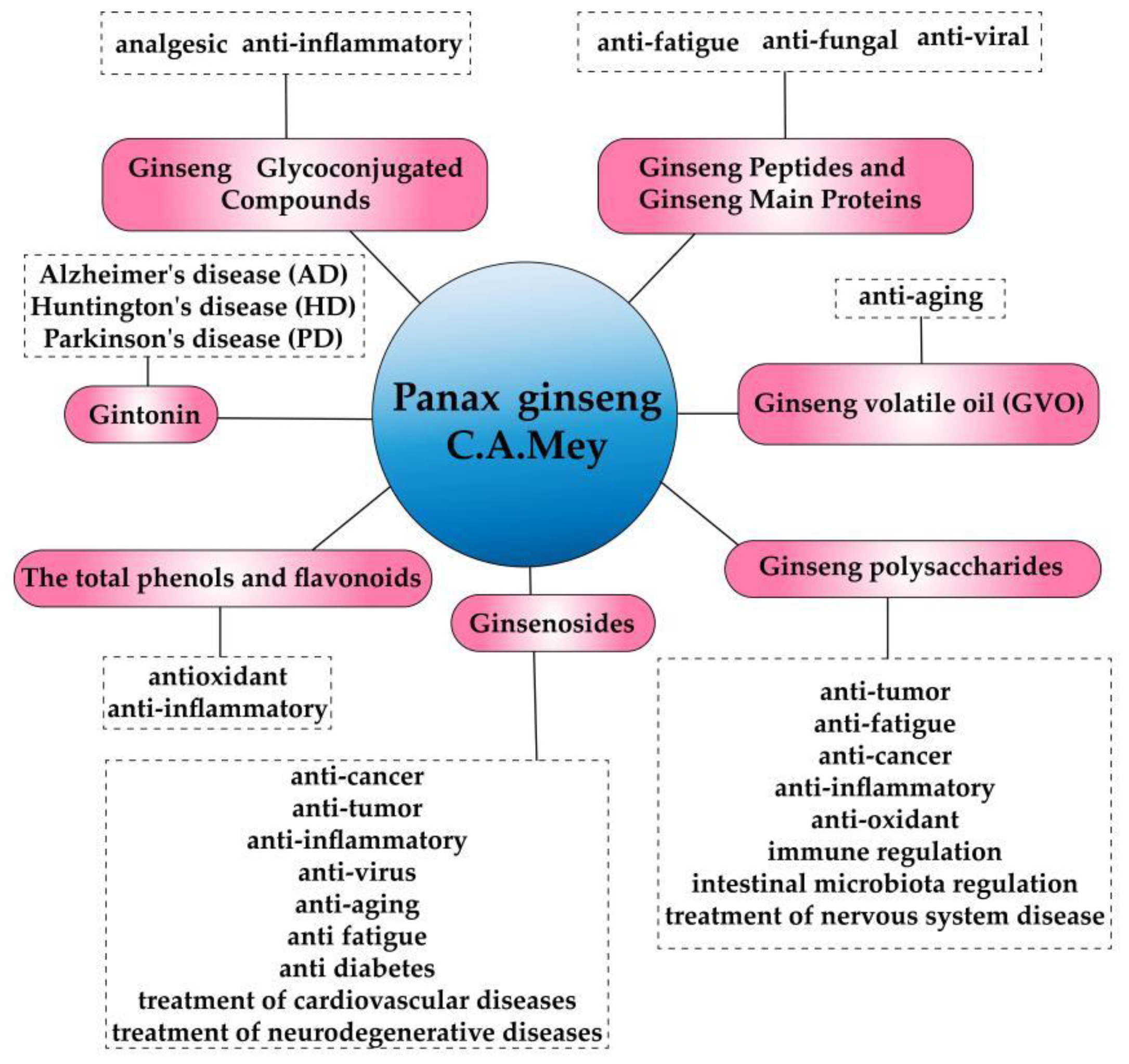
Figure 2.
Signal path related to Aβ accumulation.
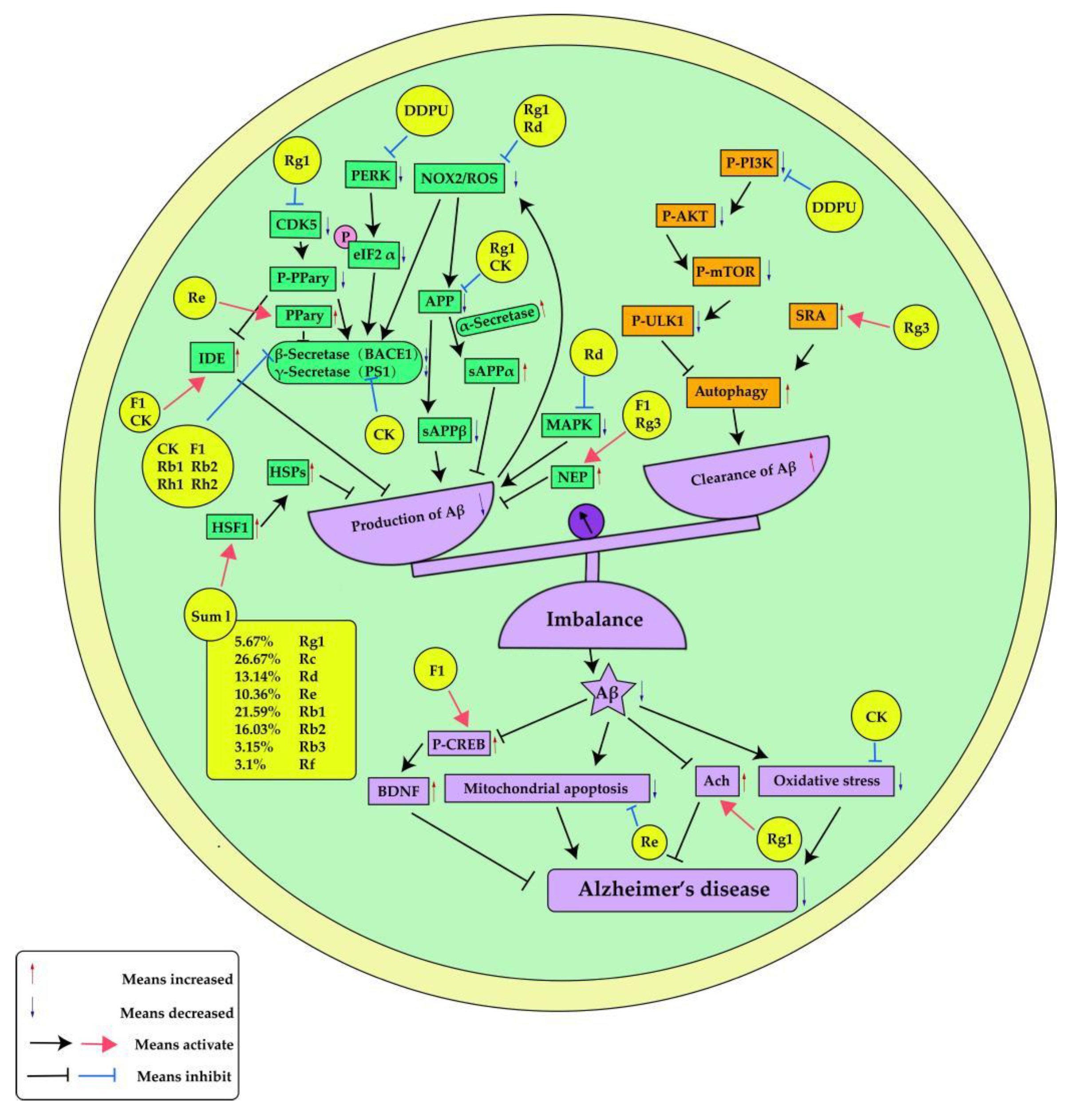
Figure 3.
Signal path related to Tau Hyperphosphorylation.
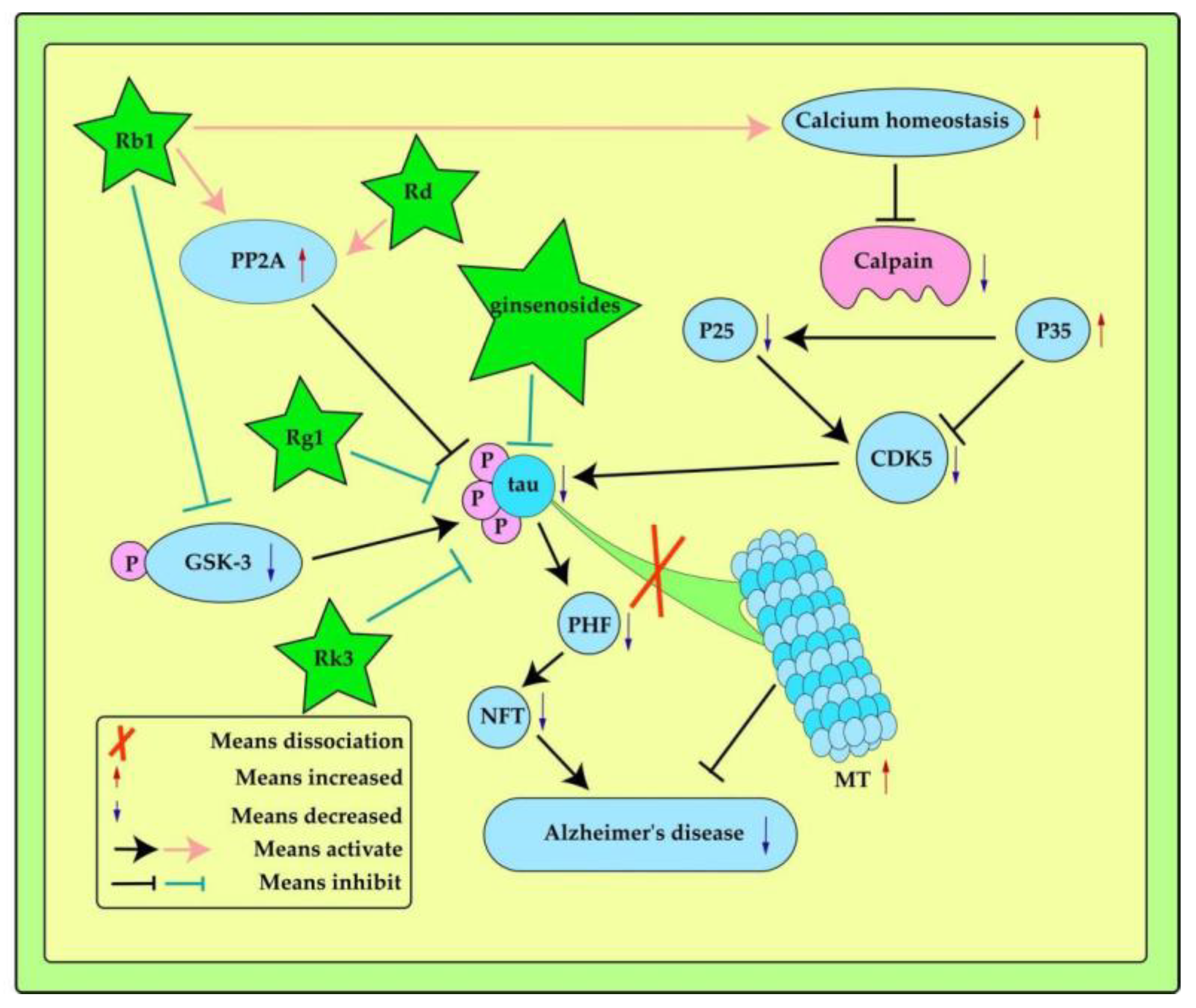
Figure 4.
Signal path related to oxidative stress and neuroinflammation.
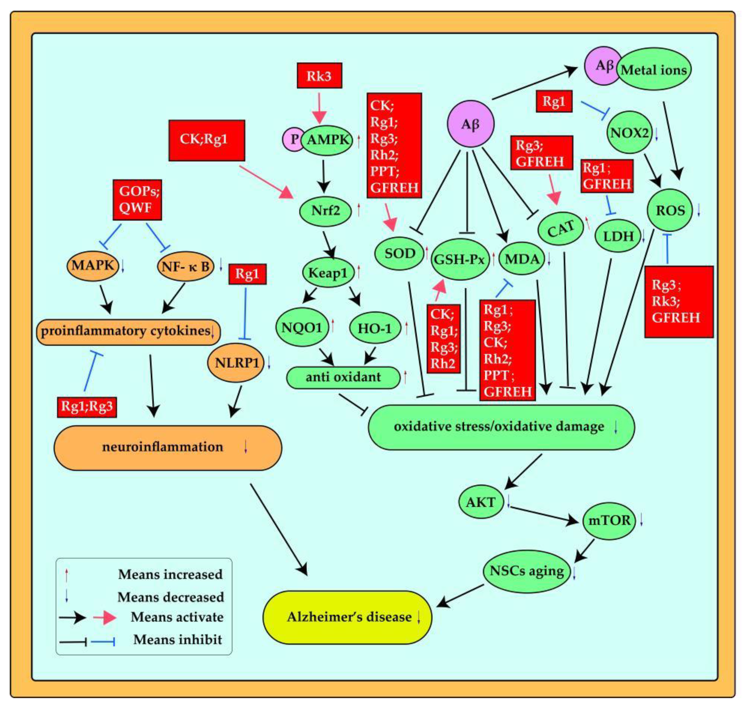
Figure 5.
Signal path related to mitochondrial apoptosis.
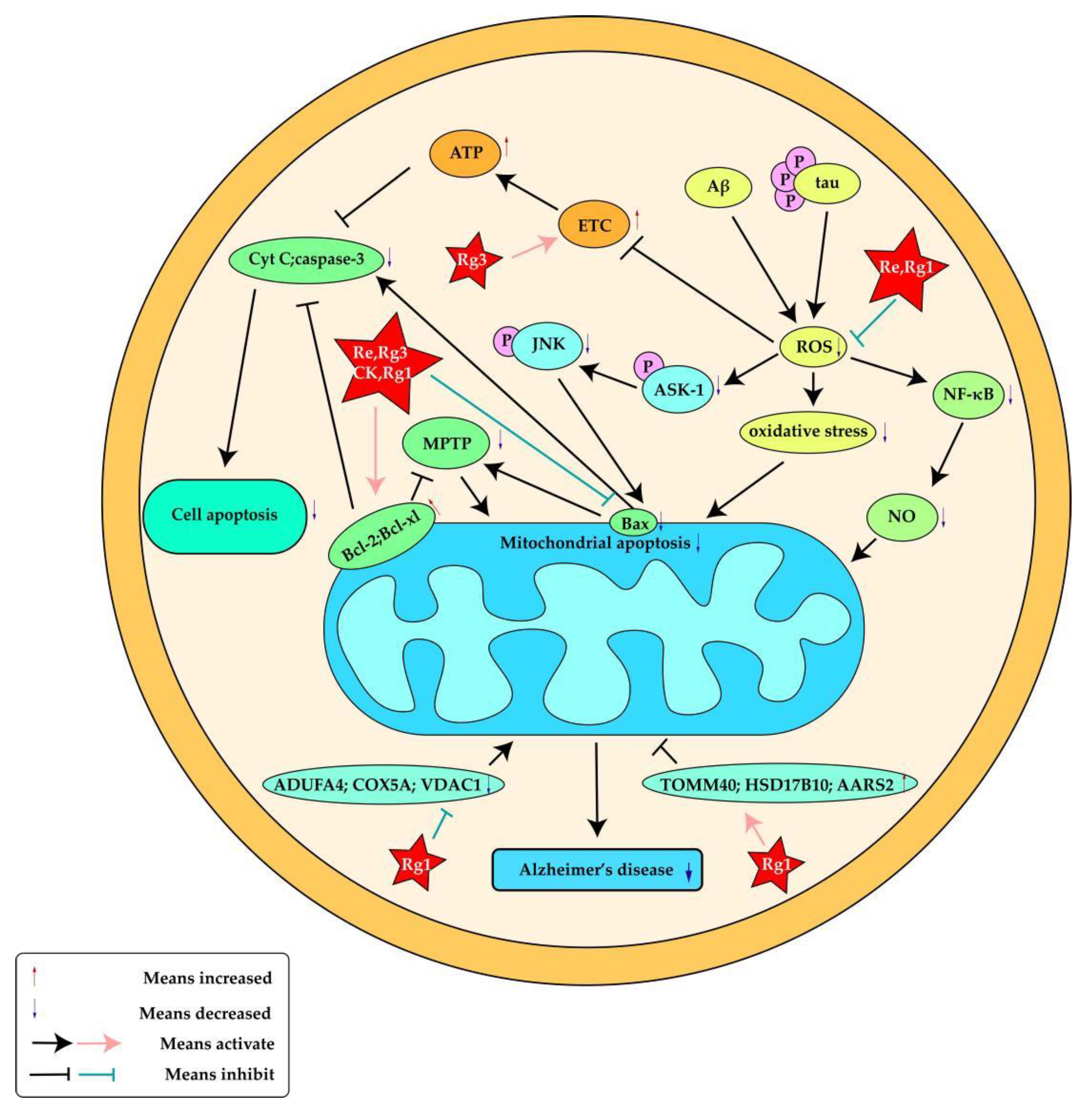

Table 1.
The effects of P. ginseng on the pathogenesis of Alzheimer's disease related to Aβ.
| Ginseng ingredients | Experimental model | Effects | Ref. |
|---|---|---|---|
| Ginsenoside compound K(CK) | ICR mice | BACE1,PS1,IDE | [45] |
| Ginsenoside Rg1 | Sprague Dawley rats | APP,CDK5,PPary,IDE,BACE1 | [46] |
| Ginsenoside CK, F1, Rh1 and Rh2 | Molecular docking | BACE1 | [47] |
| Ginsenoside F1 | Mouse neuroblastoma neuro-2a (N2a) cells and Human neuroblastoma SH-SY5Y cells | IDE,NEP | [48] |
| Ginsenoside Rg3 | SK-N-SH cells | NEP | [49] |
| Ginsenoside Rg1 | Wild type (WT) and APP/PS1 AD mice | NOX2,ROS,APP,BACE | [50] |
| Ginsenoside Rd | C. elegans | ROS,MAPK | [51] |
| Ginsenoside Re | N2a/APP695 cells | BACE1,PPary | [52] |
| Ginsenoside Rb1,Rb2 | Molecular docking | BACE1 | [53] |
| DDPU | APP/PS1 transgenic mice,SH-SY5Y cells, HEK293-APP swe cells, CHO-APP cells, primary neurons or primary astrocytes | BACE1,PERK,eIF2 α,PI3K, AKT, mTOR, ULK1, | [55] |
| Ginsenoside Rg3 | Neuro-2a (N2a) murine neuroblastoma and HMO6 humanmicroglial cells | SRA | [56] |
| combined ginsenosides(SumI) | AD worms | HSPs,HSF-1 | [57] |
| Ginsenoside F1 | APPswe/PSEN1dE9 double-transgenic AD mice with a B6 × C3 background and B6 × C3 wild type mice | CREB,BDNF | [58] |
Table 2.
The effects of P. ginseng on the pathogenesis of Alzheimer's disease related to Tau protein.
Table 2.
The effects of P. ginseng on the pathogenesis of Alzheimer's disease related to Tau protein.
| Ginseng ingredients | Experimental model | Effects | Ref. |
|---|---|---|---|
| Ginsenoside Rb1 | ICR mice | pGSK-3,PP2A | [65] |
| Ginsenoside Rd | SD rats | PP2A | [66] |
| Ginsenoside Rb1 | SD rats | Ca2+-calpain-CDK5,P35,P25 | [67] |
Table 3.
The effects of P. ginseng on the pathogenesis of Alzheimer's disease related to neurotransmitters.
Table 3.
The effects of P. ginseng on the pathogenesis of Alzheimer's disease related to neurotransmitters.
| Ginseng ingredients or Herb pairs | Experimental model | Effects | Ref. |
|---|---|---|---|
| Ginsenoside Rh2 | Male ICR mice | Ach | [74] |
| Ginsenoside Rg1 | Adult male Sprague–Dawley rats | Ach | [60] |
| GSL | Cell | Ach | [75] |
| PPT | Male Institute of Cancer Research mice | Ach | [76] |
| Ginsenoside Re,Rg3 | In vitro enzyme assays | ACHE,BCHE | [53] |
| Ginsenoside Rb1 | Male Sprague-Dawley rats | Ach | [77] |
| Ginsenoside Re | Adults male Sprague Dawley rats | DA,Ach | [78] |
| Ginsenoside Rg3 | Male C57BL/6 mice | Ach | [79] |
| Ginsenosides | Male Wistar rats | GABA,Ach,DA,Gly,5-HT,Glu,Asp | [68] |
| Ginsenoside Rg3 | Male Wistar rats | Ala,Asp,Glu,D-Gln,D-Glu,Trp | [81] |
| Ginseng Schisandra chinensis | Sprague-Dawley male rats | Phe, Tyr,Trp | [82] |
Table 4.
The effects of P. ginseng on the pathogenesis of Alzheimer's disease related tooxidative stress and neuroinflammation.
Table 4.
The effects of P. ginseng on the pathogenesis of Alzheimer's disease related tooxidative stress and neuroinflammation.
| Ginseng ingredients and Classic Chinese formulation | Experimental model | Effects | Ref. |
|---|---|---|---|
| Ginsenoside compound K(CK) | ICR mice | Nrf2, keap1, HO-1,SOD, GSH-Px, MDA | [45] |
| Ginsenoside Rk3 | APP/PS1 mice,PC12 Cells | ROS, AMPK, Nrf2,HO -1, NQO1, Keap1 | [69] |
| Ginsenoside Rg1 | mouse neuroblastoma N2a cells | Nrf2, HO-1, SOD, MDA, LDH | [85] |
| Ginsenoside Rg1 | Male APP/PS1 mice | ROS, NOX2 | [50] |
| Ginsenoside Rg1 | C57BL/6 mice,mice NSCs | SOD, GSH-Px, MDA, ROS, Akt, mTOR | [86] |
| Ginsenoside Rg3 | Male Wistar rats | SOD, CAT, GSH-Px, MDA, ROS | [81] |
| Ginsenoside Rh2 | Male ICR mice | SOD, GSH, MDA | [74] |
| PPT | Male Institute of Cancer Research mice | SOD, MDA | [76] |
| GFREH | C. elegans | ROS, SOD, CAT, MDA, LDH | [87] |
| Ginsenoside Rg1 | Male ICR mice | NLRP1,proinflammatory cytokines( IL-1β ,IL-18) | [88] |
| Ginsenoside Rg3 | Male Sprague-Dawley (SD) rats | proinflammatory cytokines(TNF-α, IL- 1β, and COX-2) | [89] |
| Ginsenoside Rg3 | Neuro-2a (N2a) murine neuroblastoma and HMO6 humanmicroglial cells | proinflammatory cytokines(iNOS, IL-6, and TNF-α) | [56] |
| Ginseng oligopeptides (GOPs) | Male Sprague-Dawley (SD) rats | MAPK , NF- κ Bproinflammatory cytokines(TNF-α IL-β) | [90] |
| Qisheng Wan formula (QWF) | Adult Sprague Dawley rats | NF-κB, IL-6, TNF-α | [61] |
Table 5.
The effects of P. ginseng on the pathogenesis of Alzheimer's disease related to mitochondrial apoptosis.
Table 5.
The effects of P. ginseng on the pathogenesis of Alzheimer's disease related to mitochondrial apoptosis.
| Ginseng ingredients | Experimental model | Effects | Ref. |
|---|---|---|---|
| Ginsenoside compound K (CK) | ICR mice | Bcl-2, Bax, caspase-3 | [45] |
| Ginsenoside Rg3 | Male Wistar rats | ETC, ROS, ATP ,Cyt C, Bax, Bcl-2 | [81] |
| Ginsenoside Re | SH-SY5Y human neuroblastoma cells | Bax, ROS, ASK-1, JNK | [59] |
| Ginsenoside Rg1 | Primary cultured cortical neurons were preparedfrom embryonic day (D17-18) Sprague Dawley (SD) rat fetuses | NF-κB, NO, ROS, Bcl-2, Bax | [94] |
| Ginsenoside Rg1 | SH-SY5Y cells | HSD17B10, AARS2, TOMM40,NDUFA4, VDAC1, COX5A | [95] |
Table 6.
The effects of P. ginsengon the pathogenesis of Alzheimer's disease related toGut microbiota.
Table 6.
The effects of P. ginsengon the pathogenesis of Alzheimer's disease related toGut microbiota.
| Ginseng ingredients and Classic Chinese formulation and Herb pairs | Experimental model | Effects | Ref. |
|---|---|---|---|
| Ginsenoside Rb1 | Sprague–Dawley rats | GABAA (α2, β2, and γ2) , GABAB (1b and 2) ,Bif.L, Bif.D, Lac.B, Lac.H, Lac.R | [100] |
| Ginseng Schisandra chinensis | Sprague-Dawley male rats | regulating intestinal microbiota disorderPhe, Tyr,Trp | [82] |
| Qisheng Wan formula (QWF) | Adult Sprague Dawley rats | Reduces the richness and diversity of gut microbiota | [61] |
| Ginsenoside Rg1 | Male conventional tree shrews | Alter the composition and abundance of gut microbiota | [109] |
| DushenTang (DST) | Adult male Sprague-Dawley rats | Correct the disturbance of the gut microbiota | [110] |
Disclaimer/Publisher’s Note: The statements, opinions and data contained in all publications are solely those of the individual author(s) and contributor(s) and not of MDPI and/or the editor(s). MDPI and/or the editor(s) disclaim responsibility for any injury to people or property resulting from any ideas, methods, instructions or products referred to in the content. |
© 2023 by the authors. Licensee MDPI, Basel, Switzerland. This article is an open access article distributed under the terms and conditions of the Creative Commons Attribution (CC BY) license (http://creativecommons.org/licenses/by/4.0/).
Copyright: This open access article is published under a Creative Commons CC BY 4.0 license, which permit the free download, distribution, and reuse, provided that the author and preprint are cited in any reuse.


