
Submitted:
29 March 2023
Posted:
03 April 2023
You are already at the latest version
Preprints on COVID-19 and SARS-CoV-2
Abstract
(1) Background: In the year of 2020 Covid-19 was declared epidemic by WHO. From that time millions of people were affected and died by this disease. The main detection process for this epidemic is RT-PCR test or reverse polymerase transcription chain reaction test. One of the reason of spreading of this disease so much is lack of efficiency in RT-PCR test. Sampling error and low viral load were two main reasons for what the testing process faced such problems. Lung infection is a very common symptom for covid-19 patients, so, CT scan and chest X-ray imaging technique can be applied to detect patient at a very early stage of infection. Which will be very effective and also can be a better option to RT-PCR test; (2) Methods: We searched data in Scopus for articles published between 2020 and 2023. The initial set of articles was 189, from which 21 articles will eventually be selected by exclusion criteria; (3) Results: A total of thirteen (61.90%) articles were found working on detecting Covid-19 by extracting the X-ray and CT scan data individually. Three (14.28%) of those articles focused on a hybrid model of CT scan and X-ray Image Data. Another four articles made a comparison between Covid-19, pneumonia and a normal person to identify a Covid-19 patient. Where others have worked on unsupervised learning methods or SVM to detect Covid-19.; (4) Conclusions: We have conducted a systematic review of the studies that have been published up to this time, with the purpose to present a summary of the evidence about the detection of COVID-19. In this article, we have summarized and critically reviewed literatures which worked on development or application or both of different AI or ML technique from chest CT and X-ray images to find a solution to detect covid-19.
Keywords:
Covid-19
; Deep Learning
; Computerized Tomography
; Artificial Intelligence
; Machine Learning
; X-ray
1. Introduction
Within the period of 2020-2022 Covid-19 was the main reason of death all over the World [1]. It affects a wide geographical area, especially in countries with large populations like India [2,3]. WHO announced Covid-19 as pandemic because coronavirus has been found in every nation on earth, and it was also advised that slowdown the growth rate of the disease is only possible by early detection, isolation and immediate treatment of the affected person.[4,5]. For this reason several institutions had started research work on Covid-19 [6].
Artificial intelligence (AI) has come at the fore front in the scientific field as surprisingly new discoveries are made [7]. AI as a part of computer science, can be applied to the creation of intelligence systems and is often implemented as software [8]. The recent use of AI in disease diagnosis has expanded its potential. In mid-20th century, medicine and healthcare system was most auspicious field for application among other fields [9]. Various decision support systems had been developed by researchers to diagnose disease effectively and accurately [10]. AI system, which was applied for detection of disease, decoding EKG images and other tasks, choosing the best course of action and generation of hypothesis, were adequate in late 1970s [11,12]. Machine learning algorithms are applied by Modern AI to find patterns and associations in data, as opposed to previously used knowledge-based AI systems that rely on medical expertise based on the evidence-based AI system [13]. The modern revolution in AI can largely be characterized as successful and trained artificial neural network, worked on a huge labelled data set by use the of deep learning method.[14,15,16]. According to researchers, this systems can essentially help doctors make decisions faster and more effectively, and even occasionally eliminate the need for human decisions [17]. Success of AI, at present, in healthcare can be attributed to the growing amount of data in the industry because of more use of digital technologies and huge development of big data analytics [18]. Collection of data becomes so easy through mobile applications, because of frequent use of mobile at present scenario. While Artificial Intelligence in healthcare is still in its infancy. Cancer, neurology, and cardiovascular disease are three target field on which AI is mainly working [19]. Powerful AI can use evidence to uncover insights from medical data. Those are usually used mainly for forecasting with support in decision making system also. [16,20].
Researchers believe AI could be effective tool to fight against Covid-19 as it has already shown promise in the healthcare industry [21]. Present researches on Covid-19 infections indicate, AI could be helpful to identify the infection, assume the next outbreak, and chalk out the pattern of attack and discovery a way to cure. Image processing, data mining, and ML have been shown to be useful to diagnose, classify, and develop vaccines for Covid-19 in recent studies. [22,23]. Our study examines some specific work limited to the usage of AI to detect Covid-19 from X-ray and CT scan image data. We also suggest some ways for future researchers how AI and machine learning could be used to fight other pandemics besides Covid-19. Together, our study examines the research conducted using AI to combat the Covid-19 pandemic.
The following sections are arranged as follows. Section 2 of this review study discusses the methods and materials used to carry it out. Section 3 covers the analysis and results of the review data. Conclusions and the different scopes in future for researcher in Covid-19 are presented in Section 4, and the final comments, limitations, research proposals are presented in Section 5.
2. Materials and Methods
2.1. Search Strategy
We searched Scopus for related articles with X-ray [24,25] and CT scan [26,27] imaging process for Covid-19 detection published between 2019 to march 2023 with preferred reporting items for systematic review to identify Covid-19 through X-ray and CT scan image. The search term included ( covid* AND detect* AND ( xray OR scan ) AND ( ( artificial AND intelligence ) OR AI OR ( machine AND learning ))) and further it was limited to its language, subject field, publication stage, source type. The following Figure 1 will show the documents by subject area after applying the search query and Figure 2 will show the number of document published in the year 2020 to 2023.
2.2. Inclusion and Exclusion Criteria
We included the articles for analysis according to the criteria of: (a) The article which belongs to computer science, engineering, medical, genetics and biotechnology field (b) the articles use or develop Artificial Intelligence or Machine Learning technique, Algorithms for detection of Covid-19 (c) The articles which associated with X-ray and CT Scan Data. (d) Only published journal Article.
With these inclusion criteria we have also used the following exclusion criteria: (a) studies which does not meet the aim of research (b) Studies, which are not written in English language (c) Articles which do not contain Computational data. (d) Articles which consider only CT scan or X-ray image data not the both.
2.3. Study Selection
After Searching Scopus database 498 articles were found in the specific research topic. Figure 3 which is a PRISMA Flowchart, exhibit the article selection procedure through various steps, what we get by the inclusion- exclusion criteria. From 498 articles, at first round the study selection process was done by limiting the selected article by written language, publication stage and field. After the first round of selection process total 309 articles were excluded. In Second round 189 Articles written in English language and from relevant field were screened based on their title and abstract, to understand the data from those are suitable with inclusion criteria or not. From these articles, after applying all of the exclusion inclusion principle, for the final review 21 articles were selected. The final selected set of articles include original research the applied or implemented different AI or ML model to get the result from CT and X-ray image data.
2.4. Data Extraction
Data extraction was carried out to scrutiny various Artificial Intelligence or Machine Learning techniques, algorithms and systems. Their applications for detection Covid-19 from patient’s X-ray and CT scan data are also explored. Selected articles were read minutely and carefully to gathered research data to write the review article. Researchers from AI, ML, and health informatics backgrounds oversaw and kept an eye on the complete research process, to secure the accuracy and excellence of this review article. The data extraction process for each chosen article focused mainly on the type and context of the study, research goals, results, methods followed by them, used technique and algorithm and dataset. After extraction the collected data were combined, analysed to provide a summary of the previous study and to pinpoint potential areas for further investigation.
3. Data Analysis
3.1. Publication Type
All articles are collected from the Scopus database. These articles are published between 2020 and March 2023. All of the articles are published in reputed Journal. We only reviewed journal articles because they have already been reviewed, so we assume the articles will be more reliable and the data quality will be higher. In the 21 articles, 16 (76.19%) of them proposed a new method to detect Covid-19 using X-ray and CT scans.
3.2. Research Purposes and Objectives
We have collected all the research done so far to analyse how it can help detect Covid-19 early, easily and effectively. All of this summarized data is presented in Table 1 to highlight our findings and the precise purpose of the research linking these articles to detection of Covid-19 by AI. They used various algorithms, like convolutional neural network (CNN) model [28], support vector machine (SVM) [29], Gaussian Naive Bayes (GNB) [30], Decision Tree (DT), Logistic Regression (LR) [31], K-Nearest Neighbour (KNN) [32].
Though we have searched in database with the key word “Detect” so according to the search string, objectives of maximum of the selected papers are detection of Covid-19.
Total thirteen (61.90%) articles were focused only on the early, accurate and easy detection of Covid-19 through the individual extraction of the X-ray and CT scan data. In these articles CT scan and X-ray data were collected and examined without mixing them. Within these some articles develops/propose new methods. For example Elpeltagy M. et al. [33] develops a model, Modified ResNet50, where ResNet model was applied with modification of three layers, named, ′Conv′, ′Batch_Normaliz′ and ′Activation_Relu′ for layers which is an accurate, fast, and low-cost auxiliary diagnostic tools to detect Covid-19, where Gupta R.K. et al. proposed InceptionResNetV2 model [34]. Some researchers worked through other convenient CNN models like Baz M. et.al. [35] experimented on CNN-based model to detect Covid-19 and achieved 99% accuracy by existing VGG-16, VGG-19, ResNet50 and other methods.
Three (14.28%) of those articles were worked on a hybrid model of Computer Tomography scan and X-ray Image Data. These models are more efficient like, Irfan M. et.al. proposed Hybrid Deep Neural Networks (HDNN) which gave an accuracy of 99% on the test data set [36].
In the article by Hayat A et.al. compared Covid-19 pneumonia and normal healthy sample by the proposed SCoVNet architecture [37]. Article by Ahsan, M.M. et.al. [38] and Ko H. et.al. [39] also provided few opportunities to differentiate among Covid-19 patients, pneumonia patients and healthy person. Ullah F. et.al. [40] contributed in detection and classification Covid-19 in CT and X-ray images. Ravi V. et.al. [41] discovered the way for identification and analysis of Covid-19 through unsupervised learning. Some studies have perspective to develop efficient AI model to detect Covid-19 where some other articles focused on, within X-ray or CT scan data, which one will give better result through a particular CNN model and some worked on various models to differentiate Covid-19 from normal pneumonia.

Table 1.
Main Objectives of Reviewed Studies.
| Brief Description | Reference | Frequency | |
|---|---|---|---|
| Detection | Early detection of this disease | [42] | 4 |
| Detect Covid-19 patient | [43] | ||
| Detection of Covid-19 | [44] | ||
| Covid-19 detection via features extraction | [45] | ||
| Detection by CT and X-ray image together (with developed architecture/model) |
Detect pneumonia and Covid-19. | [46] | 3 |
| Detection via Convolutional Neural Network (CNN) -tailored Deep Neural Network (DNN) | [47] | ||
| Detect through hybrid deep neural networks (HDNNs), | [36] | ||
| Detection by CT and X-ray image separately (with developed architecture/model) |
Predicts Covid-19 detection by multimodal covid network (MMCovid-NET) | [48] | 9 |
| Accurate diagnose of Covid-19 by light CNN model with watershed-based region-growing segmentation | [49] | ||
| Automatic detection by Multilayer Spatial Covid Convolutional Neural Network (MSCovCNN) | [50] | ||
| Early detection of Covid patient by proposed InceptionResNetV2 model | [34] | ||
| Differentiate Covid pneumonia normal sample by proposed SCoVNet architecture | [37] | ||
| Modified MobileNetV2 model to understand the what features of CT/X-ray images used to know the reason behind Covid-19 | [51] | ||
| Develop Modified ResNet50, accurate, fast, and cheap auxiliary diagnostic tool for detection |
[33] | ||
| DenseNet-121 model to image segregation |
[52] | ||
| Intelligent decision support system for Covid-19 empowered with deep learning (ID2S-Covid19-DL) To detect covid-19 | [53] | ||
| detection and analysis of Covid-19 through unsupervised learning (with developed architecture) |
Unsupervised deep learning-based Covid-19 detection by Autoencoder3-ResNet50 (GMM) model | [41] | 1 |
| Detection through differentiation between covid, pneumonia and normal patient (with developed architecture/model) |
Accurate and efficient method to detect Covid-19 by CovidCon | [54] | 4 |
| Distinguish between Covid-19 patients with others by NasNetMobile model | [38] | ||
| Differentiate Covid-19 pneumonia, non-Covid-19 pneumonia and nonpneumonia diseases | [39] | ||
| Differentiation between Covid-19, non-Covid-19 and pneumonia by CNN-based Covid-19 detection model | [35] |
3.3. Exploration of used data
All of the studies use only image data as our searching key word was “X-ray” and “CT Scan”. But there is only article by Nasir N. et al. [43] which include clinical notes along with the X-ray and CT scan images. They found only 485 clinical notes available for 535 images. Seven of them used only Kaggle dataset to collect their data for training or testing purpose of developed model [35,38,42,44,46,49,51]. Three of them used only GitHub repository which was developed by Dr. Joseph Cohen [33,43,48], other four of them used both of the Kaggle and GitHub database [34,47,50,54]. One used all of the GitHub, Covid-19 radiography database, Kaggle Covid-19 image data collection, and Actual Med Covid-19 Chest X-ray Dataset [36]. One article collected data From D. S. Kermany, et al., "Identifying medical diagnoses and treatable diseases by image-based deep learning”, J. P. Cohen, P. Morrison, and L. Dao, "Covid-19 image data collection" [45]. Another one from Git hub, Italian Society of Medical and Interventional Radiology, Radiological Society of North America (RSNA), Radiopaedia, and SIRM) and Kaggle repository [52].
Article [36,46] worked on only Hybrid model, Where [47] worked on both of the individual and hybrid model of chest X-ray and Computer Tomography scan. In Hybrid model, CT scan and X-ray image is merged in a single Image and processed. All of them have worked with the global data which are free available for researcher. Seventeen of them developed new algorithm and tested those model on the collected dataset. They divided the collected data for testing and training purpose while most of them used 80% data for training and 20% for testing procedure. The following Table 2 will describes details of the data used is the selected review articles.
3.4. Context of Study
Usually contextual studies mainly focus epidemic forecasting and continuous development [13]. Articles on disease detection used public datasets and worldwide views which were not specified by the context. These results shows disease detection via various machine learning method methods were generally not reliant on context.
3.5. Exploring the AI Techniques
One Study focusing on real time surveillance on Covid-19 detection by Ullah F. et al. proposed the basics of Internet of Things platform is the connection of a web service to a cloud infrastructure service [52]. To identify Covid19, a combined model is formed that consists of Gaussian Naive Bayes [30], Support Vector Machine [29], Decision Tree [55,56], Logistic Regression [31], K-Nearest Neighbour [32], and Random Forest [57]. For extraction of 350 prominent feature CNN model was used. Hyper-parameter tuning is used to select the suitable filter length and frequency. As a consequence, ReLU [58], a non-linear activation function, is applied to each component. In the suggested CNN model, two convolution layers employ 64 and 128 filters, four-size kernels, and the padding numbers which is determined by the number of the input which was taken based on the precise shape. In this model the performance measure have been done through the Gradient-weighted Class Activation Mapping (Grad-CAM) [59,60]. Besides this they have use another performance measuring method called t-distributed Stochastic Neighbour Embedding (t-SNE) [61] which actually used for visualizing the multidimensional data by initializing any data point. Here authors have done this analysis on SARS-CoV-2 CT scan data from the Kaggle database.
A total of 6 papers worked on different CNN procedures like VGG19 [62], VGG16 [63], InceptionResNetV2 [64], MobileNetV2 [65], Densenet121 [66], InceptionV3 [67], Xception [68], and Resnet50 [68]. For example deep learning based Convolutional Neural Network, Inception v3, VGG-19, ResNet-50 and Xception models are used by Mukhi S.E et al. [42].
After pre-processing the accuracy of models are analysed and estimated by obtained accuracy. They found that fine-tuned VGG-19 model has a highest accuracy at detection of covid-19. VGG-19 model shows an accuracy of 93% from CT scan and 94.17% from the chest X-ray data. Kathamuthu N.D. et al. [46], applied Densenet121, VGG16 and 19, InceptionV3, Xception, and Resnet50 models to detect Covid and they found VGG16 model performed much better than other models with the accuracy of 98.00%. In another article Baz M. et al [17] achieved 99% accuracy by applying a Covid-19 detection tool which works by CNN model.
Some articles proposed new method to detect Covid-19. In article [48] Padmapriya T. et al. proposed a new bespoke convolutional neural network (CNN) architecture named multimodal Covid network (MMCovid-NET). This model functions by varying the layer number from two to seven. 24 models were build up to for the testing purpose of the algorithm. MMCovid-NET-I, MMCovid-NET-II, MMCovid-NET-III, and MMCovid-NET-IV were the four models among them. MMCovid-NET-III gave best result with an accuracy of 99.75%. A novel light CNN model architecture was developed by Khan H.A. et al., which is a lighter model gives mean of 98.8% accuracy for X-ray and 98.6% for CT scans. SCoVNet architecture [37] and InceptionResNetV2 [54] are two different architecture, are also developed to detect Covid, these were pre-trained by VGGNet-19, ResNet50, InceptionResNetV2 and MobileNet etc. SCoVNet architecture shows 99% accuracy on CT scan and 98% on X-ray data set. InceptionResNetV2 results 99.2%, 98%, 97% training, validation and test accuracy respectively. Convolutional Neural Network (CNN)-tailored Deep Neural Network (DNN) [47] was developed for training and testing both of the X-ray and CT scan data together. Which gives 96.28% accuracy and the Area Under Curve (AUC) is 0.9808 and 0.0208 is the false negative rate. Another automatic detection process is provided by the Multilayer Spatial Covid Convolutional Neural Network (MSCovCNN) [50], with detection accuracy of 93.63%, for X-ray images 97.88% AUC and detection accuracy of 91.44% and AUC of chest CT scan 95.92%. 5-tiered 2D-CNN frameworks, softmax classifier [69] and ANN [70] are used in this model. Detection of Covid-19 pneaumonia can be done by Fast-track Covid-19 classification network (FCONet) [39], based on a single CT image with sensitivity 99.58%, specificity 100.00%, and accuracy 99.87%. VGG16 has fewer parameters and needs less training time, which leads to better results. It therefore works better than other CNN models [44].
Ravi V. et al. shows an 84% accuracy for CT scan and chest X-ray data by their model Autoencoder3-ResNet50 (GMM). Separate four sets of experiments are performed on both CT and CXR scan data. The convolutional autoencoders, pre-trained CNNs, hybrid, and PCA-based models are applied methods for feature extraction. for clustering both of the Autoencoder1-VGG16 (KMeans and GMM) models, K-means and GMM techniques are applied in the experiment and these models reached at accuracy of 70% [41]. Reis H.C develop COVID-DSNet model toclassify Covid and typical pneumonia (bacterial, viral). COVID-DSNet model gives an accuracy of 97.60 % and sensitivity of 97.60 %. Sensitivity values of 100 % for typical pneumonia, 96.30 % for common pneumonia, and 96.58 % for COVID-19 are collected. [46]. The Table 3 will show the list of various algorithm and there respected obtained accuracy, used or developed in the selected research articles. And following Figure 4 will describe the accuracy performance chart of various selected articles for individual imaging methods like X-ray, CT, and X-ray or CT both.
4. Future Research Opportunities
We've briefly discussed the difficulties and potential directions for future study in this section with regards to using AI and machine learning to combat the Covid-19 pandemic as well as potential future pandemics.
- Use of a Large Set of Data in Research – There are opportunities to gather substantial amounts of data and make them accessible to researchers so they can run various tests. Such initiatives will be incredibly helpful in the battle against the pandemic. Global data were used in all disease detection study projects. However, we suggest that more work in this area could benefit from using a variety of worldwide data sources. Future studies could look into whether larger databases could produce more well-structured, verified, and generalised results. Additional research could be done to create an algorithm that is more efficient and effective. With enough information in the future, the claims made in the research can be investigated further.
- Development of New Algorithm – New algorithms can be developed in future for more efficient and accurate detection of Covid-19. CNN with multiple layer can be developed for more refined result. The structural architecture of machine learning algorithm can be modified in near future by observing the potential performance level through this literature review.
- Hybrid Data – Among 21 articles only three of them used Hybrid data to detect Covid-19. Use of hybrid data should be more. Because hybrid data gives more accurate and effective result in diagnosis of Covid-19. Because hybrid data consider CT scan image and X-ray image together as a single image, the retrieved data from that image is more accurate as input to any algorithm.
- Other Data Should Include – In these articles we noticed that when researcher use CT scan and X-ray data usually they do not consider any other kind of data like health history, pathological, clinical etc. with exception of one Article [43]. If researcher include other related data for the research, the study will be strong and detection process will be more accurate.
- Managing the ICU Surge during the Covid-19 Crisis - According to reports, some hospitals chose to only treat young patients, abandoning elderly patients who had a lower chance of surviving because the hospitals were running low on supplies. Further study can be done to determine which individuals, based on their X-ray and CT scan report, are more likely to be critical cases. This would assist hospitals in identifying patients who can be treated at home versus those who require intensive care unit assistance. Studies that concentrate on ICU admission could help some patients be discharged early, freeing up room for those who really need it. Studies can also help to delay the early discharge of ICU patients.
5. Conclusion
Within this research study, significance of artificial intelligence, machine learning, deep learning is intended to search for an early, easy, and accurate detection of global pandemic, Covid-19. Based on the method used to detect diseases, we divided the study into five different categories. In our research, we also found that, with acceptable accuracy, application of AI and ML could be useful for the differentiation among seasonal flu, normal pneumonia and Covid-19 pneumonia [38,39]. Besides that, our study found that Artificial Intelligence has performed very efficiently with existing or new models for detection of Covid. Thoes studies which are critically reviewed, they used deep learning algorithms for analysing image data, particularly only on X-rays and CT scans with exception [43], which used clinical notes along with image data. We have suggested five possible way to research in future based on these findings.
Conducted systematic review has given an extensive overview to combat with Covid-19 when taken as a whole. These articles were examined and compared in a number of ways, taking into account the input features, the techniques they used for data processing, as well as their different goals to achieve. We have offered a variety of insightful details throughout the study, such as the application type, the use of machine learning, related assessment carried out in every article.
Our review study may have a number of flaws, but at the same 6+time it also opens up some possibilities for further research work in the indicated areas. First, we conducted a thorough keyword searching to find relevant. Despite the fact that our search terms produced useful results for achieving our study's objective, there is a chance that we might have missed some crucial materials. Second, we believe that the main components we have outlined, examined, and summarised in this paper are current materials pertaining to covid-19 virus and machine learning techniques. Third, we are only concerned with using AI and ML on the retrieved data from CT scan and X-ray images to combat Covid-19.
Despite the fact that there are many studies that either use CT scan or X-ray data, we did not include those kind of articles in the review the main cause for this, they do not accurately align with the aim of our research study. We view above factor as a limitation of our work. Although, when we broaden our search criteria and our study goal in the future, this can be addressed and assessed.
Future work is therefore required to gather and examine more pertinent data. Supportive data, like datasets which includes high data points features or more variable or observations and collaborate to healthcare field, could do some betterment in how AI and ML handle this health danger and help governments and communities control the virus's or any similar types of pandemic effects early on.
Future study could be done on examination of data security in the pertinent fields in addition to information extraction. Furthermore, some pioneering technologies like internet of things (IoT) should be the main tool for the enhanced and enriched AI and ML techniques with more effectiveness in order to remotely assist Covi-19 patients.
Supplementary Materials
None.
Funding
This research received no external funding
Author Contributions
Conceptualization, Vinayak Majhi. and Sudip Paul.; methodology, Vinayak Majhi.; software, Vinayak Majhi.; validation, Sudip Paul.; formal analysis, Vinayak Majhi.; investigation, Vinayak Majhi, Sudip Paul.; resources, Vinayak Majhi; data curation, Vinayak Majhi, Sudip Paul.; writing—original draft preparation, Vinayak Majhi.; writing—review and editing, Sudip Paul.; visualization, Vinayak Majhi.; supervision, Sudip Paul.; All authors have read and agreed to the published version of the manuscript.
Institutional Review Board Statement
Not applicable.
Informed Consent Statement
Not Applicable.
Data Availability Statement
The journal searched data is generated from the Scopus Document Search https://www.scopus.com/.
Acknowledgments
None.
Conflicts of Interest
The authors declare no conflict of interest.
References
- Wong, M.K. , et al., COVID-19 mortality and Progress toward vaccinating older adults—World Health Organization, worldwide, 2020–2022. MMWR. Morbidity and Mortality Weekly Report, 2023. 72(5): p. 113-118.
- Bhadra, A., A. Mukherjee, and K. Sarkar, Impact of population density on Covid-19 infected and mortality rate in India. Modeling Earth Systems and Environment, 2021. 7(1): p. 623-629. [CrossRef]
- Sadowski, A. , et al., Big data insight on global mobility during the Covid-19 pandemic lockdown. Journal of Big Data, 2021. 8(1): p. 78. [CrossRef]
- Chakraborty, I. and P. Maity, COVID-19 outbreak: Migration, effects on society, global environment and prevention. Science of The Total Environment, 2020. 728: p. 138882. [CrossRef]
- Banakar, M. , et al., COVID-19 transmission risk and protective protocols in dentistry: a systematic review. BMC Oral Health, 2020. 20(1): p. 275.
- Ciotti, M. , et al., The COVID-19 pandemic. Critical Reviews in Clinical Laboratory Sciences, 2020. 57(6): p. 365-388.
- Islam, M.N. , et al., A Survey on the Use of AI and ML for Fighting the COVID-19 Pandemic. ArXiv, 2020. abs/2008.07449.
- Zakharov, V. About the Evolution of the Concept of “Artificial Intelligence”. in 2021 International Conference Engineering Technologies and Computer Science (EnT). 2021.
- Bashshur, R.L. , et al., Sustaining and Realizing the Promise of Telemedicine. Telemedicine and e-Health, 2013. 19(5): p. 339-345. [CrossRef]
- S.K, L., et al., Online clinical decision support system using optimal deep neural networks. Applied Soft Computing, 2019. 81: p. 105487. [CrossRef]
- Belciug, S. and F. Gorunescu, Era of Intelligent Systems in Healthcare, in Intelligent Decision Support Systems—A Journey to Smarter Healthcare, S. Belciug and F. Gorunescu, Editors. 2020, Springer International Publishing: Cham. p. 1-55.
- Rahmani, A.M. , et al. Machine Learning (ML) in Medicine: Review, Applications, and Challenges. Mathematics, 2021. 9. [CrossRef]
- Islam, M.N. , et al., A Systematic Review on the Use of AI and ML for Fighting the COVID-19 Pandemic. IEEE Transactions on Artificial Intelligence, 2020. 1(3): p. 258-270. [CrossRef]
- Kaul, V., S. Enslin, and S.A. Gross, History of artificial intelligence in medicine. Gastrointestinal Endoscopy, 2020. 92(4): p. 807-812. [CrossRef]
- Haenlein, M. and A. Kaplan, A Brief History of Artificial Intelligence: On the Past, Present, and Future of Artificial Intelligence. California Management Review, 2019. 61(4): p. 5-14. [CrossRef]
- Benko, A. and C. Sik Lányi, History of Artificial Intelligence, in Encyclopedia of Information Science and Technology, Second Edition, D.B.A.M. Khosrow-Pour, Editor. 2009, IGI Global: Hershey, PA, USA. p. 1759-1762.
- Garbuio, M. and N. Lin, Artificial Intelligence as a Growth Engine for Health Care Startups: Emerging Business Models. California Management Review, 2019. 61(2): p. 59-83. [CrossRef]
- Dash, S. , et al., Big data in healthcare: management, analysis and future prospects. Journal of Big Data, 2019. 6(1): p. 54. [CrossRef]
- Kaur, S. , et al., Medical Diagnostic Systems Using Artificial Intelligence (AI) Algorithms: Principles and Perspectives. IEEE Access, 2020. 8: p. 228049-228069. [CrossRef]
- Yu, K.-H., A. L. Beam, and I.S. Kohane, Artificial intelligence in healthcare. Nature Biomedical Engineering, 2018. 2(10): p. 719-731.
- Chowdhury, M.E.H. , et al., Can AI Help in Screening Viral and COVID-19 Pneumonia? IEEE Access, 2020. 8: p. 132665-132676.
- Abdulkareem, M. and S.E. Petersen, The Promise of AI in Detection, Diagnosis, and Epidemiology for Combating COVID-19: Beyond the Hype. Frontiers in Artificial Intelligence, 2021. 4. [CrossRef]
- Dong, D. , et al., The Role of Imaging in the Detection and Management of COVID-19: A Review. IEEE Reviews in Biomedical Engineering, 2021. 14: p. 16-29. [CrossRef]
- Howell, J.D. , EARLY CLINICAL USE OF THE X-RAY. Trans Am Clin Climatol Assoc, 2016. 127: p. 341-349.
- Behling, R. , Medical X-ray sources now and for the future. Nuclear Instruments and Methods in Physics Research Section A: Accelerators, Spectrometers, Detectors and Associated Equipment, 2017. 873: p. 43-50.
- Schmidt Charles, W. , CT Scans: Balancing Health Risks and Medical Benefits. Environmental Health Perspectives, 2012. 120(3): p. a118-a121.
- Al-Sharify, Z.T., T. A. Al-Sharify, and N.T. Al-Sharify. A critical review on medical imaging techniques (CT and PET scans) in the medical field. in IOP Conference Series: Materials Science and Engineering. 2020. IOP Publishing. [CrossRef]
- Albawi, S., T. A. Mohammed, and S. Al-Zawi. Understanding of a convolutional neural network. in 2017 International Conference on Engineering and Technology (ICET). 2017.
- Noble, W.S. , What is a support vector machine? Nature Biotechnology, 2006. 24(12): p. 1565-1567.
- Jahromi, A.H. and M. Taheri. A non-parametric mixture of Gaussian naive Bayes classifiers based on local independent features. in 2017 Artificial Intelligence and Signal Processing Conference (AISP). 2017.
- LaValley, M.P. , Logistic Regression. Circulation, 2008. 117(18): p. 2395-2399.
- Mucherino, A., P. J. Papajorgji, and P.M. Pardalos, k-Nearest Neighbor Classification, in Data Mining in Agriculture, A. Mucherino, P.J. Papajorgji, and P.M. Pardalos, Editors. 2009, Springer New York: New York, NY. p. 83-106.
- Elpeltagy, M. and H. Sallam, Automatic prediction of COVID- 19 from chest images using modified ResNet50. Multimed Tools Appl, 2021. 80(17): p. 26451-26463. [CrossRef]
- Gupta, R.K. , et al., An AI-enabled pre-trained model-based Covid detection model using chest X-ray images. Multimed Tools Appl, 2022. 81(26): p. 37351-37377. [CrossRef]
- Baz, M. , et al., Utilization of Artificial Intelligence in Medical Image Analysis for COVID-19 Patients Detection. Intelligent Automation & Soft Computing, 2021. 30(1). [CrossRef]
- Irfan, M. , et al. Role of Hybrid Deep Neural Networks (HDNNs), Computed Tomography, and Chest X-rays for the Detection of COVID-19. International Journal of Environmental Research and Public Health, 2021. 18. [CrossRef]
- Hayat, A. , et al., Novel Comparative Study for the Detection of COVID-19 Using CT Scan and Chest X-ray Images. Int J Environ Res Public Health, 2023. 20(2). [CrossRef]
- Ahsan, M.M. , et al. COVID-19 Symptoms Detection Based on NasNetMobile with Explainable AI Using Various Imaging Modalities. Machine Learning and Knowledge Extraction, 2020. 2, 490-504. [CrossRef]
- Ko, H. , et al., COVID-19 Pneumonia Diagnosis Using a Simple 2D Deep Learning Framework With a Single Chest CT Image: Model Development and Validation. J Med Internet Res, 2020. 22(6): p. e19569. [CrossRef]
- Ullah, F. , et al., Explainable artificial intelligence approach in combating real-time surveillance of COVID19 pandemic from CT scan and X-ray images using ensemble model. J Supercomput, 2022. 78(17): p. 19246-19271. [CrossRef]
- Ravi, V. and T.D. Pham, Unsupervised Deep learning-based Feature Fusion Approach for Detection and Analysis of COVID-19 using X-ray and CT Images. The Open Bioinformatics Journal, 2022. 15(1). [CrossRef]
- Mukhi, S.E., R. T. Varshini, and S.E.F. Sherley, Diagnosis of COVID-19 from Multimodal Imaging Data Using Optimized Deep Learning Techniques. SN Computer Science, 2023. 4(3): p. 212.
- Nasir, N. , et al., Multi-modal image classification of COVID-19 cases using computed tomography and X-rays scans. Intelligent Systems with Applications, 2023. 17: p. 200160. [CrossRef]
- Kathamuthu, N.D. , et al., A deep transfer learning-based convolution neural network model for COVID-19 detection using computed tomography scan images for medical applications. Advances in Engineering Software, 2023. 175: p. 103317. [CrossRef]
- Kamil, M.Y. , A deep learning framework to detect Covid-19 disease via chest X-ray and CT scan images. International Journal of Electrical & Computer Engineering (2088-8708), 2021. 11(1). [CrossRef]
- Reis, H.C. and V. Turk, COVID-DSNet: A novel deep convolutional neural network for detection of coronavirus (SARS-CoV-2) cases from CT and Chest X-Ray images. Artificial Intelligence in Medicine, 2022. 134: p. 102427.
- Mukherjee, H. , et al., Deep neural network to detect COVID-19: one architecture for both CT Scans and Chest X-rays. Applied Intelligence, 2021. 51(5): p. 2777-2789. [CrossRef]
- Padmapriya, T., T. Kalaiselvi, and V. Priyadharshini, Multimodal covid network: Multimodal bespoke convolutional neural network architectures for COVID-19 detection from chest X-ray's and computerized tomography scans. International Journal of Imaging Systems and Technology, 2022. 32(3): p. 704-716.
- Khan, H.A. , et al. Novel Light Convolutional Neural Network for COVID Detection with Watershed Based Region Growing Segmentation. Journal of Imaging, 2023. 9. [CrossRef]
- Khattak, M.I. , et al., Automated detection of COVID-19 using chest x-ray images and CT scans through multilayer-spatial convolutional neural networks. International Journal of Interactive Multimedia and Artificial Intelligence, 2021. 6: p. 15-24. [CrossRef]
- Ahsan, M.M. , et al. Detection of COVID-19 Patients from CT Scan and Chest X-ray Data Using Modified MobileNetV2 and LIME. Healthcare, 2021. 9. [CrossRef]
- Ullah, F. , et al., Explainable artificial intelligence approach in combating real-time surveillance of COVID19 pandemic from CT scan and X-ray images using ensemble model. The Journal of Supercomputing, 2022. 78(17): p. 19246-19271. [CrossRef]
- Siddiqui, S.Y. , et al., Intelligent decision support system for COVID-19 empowered with deep learning. Comput. Mater. Contin, 2021. 66: p. 1719-1732. [CrossRef]
- Sahoo, P. , et al., Potential diagnosis of COVID-19 from chest X-ray and CT findings using semi-supervised learning. Physical and Engineering Sciences in Medicine, 2022. 45(1): p. 31-42. [CrossRef]
- Myles, A.J. , et al., An introduction to decision tree modeling. Journal of Chemometrics, 2004. 18(6): p. 275-285. [CrossRef]
- Song, Y.Y. and Y. Lu, Decision tree methods: applications for classification and prediction. Shanghai Arch Psychiatry, 2015. 27(2): p. 130-5. [CrossRef]
- More, A.S. and D. P. Rana. Review of random forest classification techniques to resolve data imbalance. in 2017 1st International Conference on Intelligent Systems and Information Management (ICISIM). 2017.
- Rasamoelina, A.D., F. Adjailia, and P. Sinčák. A Review of Activation Function for Artificial Neural Network. in 2020 IEEE 18th World Symposium on Applied Machine Intelligence and Informatics (SAMI). 2020.
- Vinogradova, K., A. Dibrov, and G. Myers, Towards Interpretable Semantic Segmentation via Gradient-Weighted Class Activation Mapping (Student Abstract). Proceedings of the AAAI Conference on Artificial Intelligence, 2020. 34(10): p. 13943-13944. [CrossRef]
- Das, P. and A. Ortega. Gradient-Weighted Class Activation Mapping for Spatio Temporal Graph Convolutional Network. in ICASSP 2022 - 2022 IEEE International Conference on Acoustics, Speech and Signal Processing (ICASSP). 2022.
- Belkina, A.C. , et al., Automated optimized parameters for T-distributed stochastic neighbor embedding improve visualization and analysis of large datasets. Nature Communications, 2019. 10(1): p. 5415. [CrossRef]
- Rajinikanth, V. , et al. A Customized VGG19 Network with Concatenation of Deep and Handcrafted Features for Brain Tumor Detection. Applied Sciences, 2020. 10. [CrossRef]
- Qassim, H., A. Verma, and D. Feinzimer. Compressed residual-VGG16 CNN model for big data places image recognition. in 2018 IEEE 8th Annual Computing and Communication Workshop and Conference (CCWC). 2018.
- Baldassarre, F., D. G. Morín, and L. Rodés-Guirao, Deep koalarization: Image colorization using cnns and inception-resnet-v2. arxiv preprint . arXiv:1712.03400, 2017.
- Liu, J. and X. Wang, Early recognition of tomato gray leaf spot disease based on MobileNetv2-YOLOv3 model. Plant Methods, 2020. 16(1): p. 83. [CrossRef]
- Ezzat, D. and H.A. Ella, GSA-DenseNet121-COVID-19: a hybrid deep learning architecture for the diagnosis of COVID-19 disease based on gravitational search optimization algorithm. arXiv preprint. arXiv:2004.05084, 2020.
- Shadin, N.S., S. Sanjana, and N.J. Lisa. COVID-19 Diagnosis from Chest X-ray Images Using Convolutional Neural Network(CNN) and InceptionV3. in 2021 International Conference on Information Technology (ICIT). 2021.
- Rahimzadeh, M. and A. Attar, A modified deep convolutional neural network for detecting COVID-19 and pneumonia from chest X-ray images based on the concatenation of Xception and ResNet50V2. Informatics in Medicine Unlocked, 2020. 19: p. 100360. [CrossRef]
- Wu, Q. , Image retrieval method based on deep learning semantic feature extraction and regularization softmax. Multimedia Tools and Applications, 2020. 79(13): p. 9419-9433. [CrossRef]
- Krogh, A. , What are artificial neural networks? Nature Biotechnology, 2008. 26(2): p. 195-197.
Figure 1.
Available documents in various subjects domain.
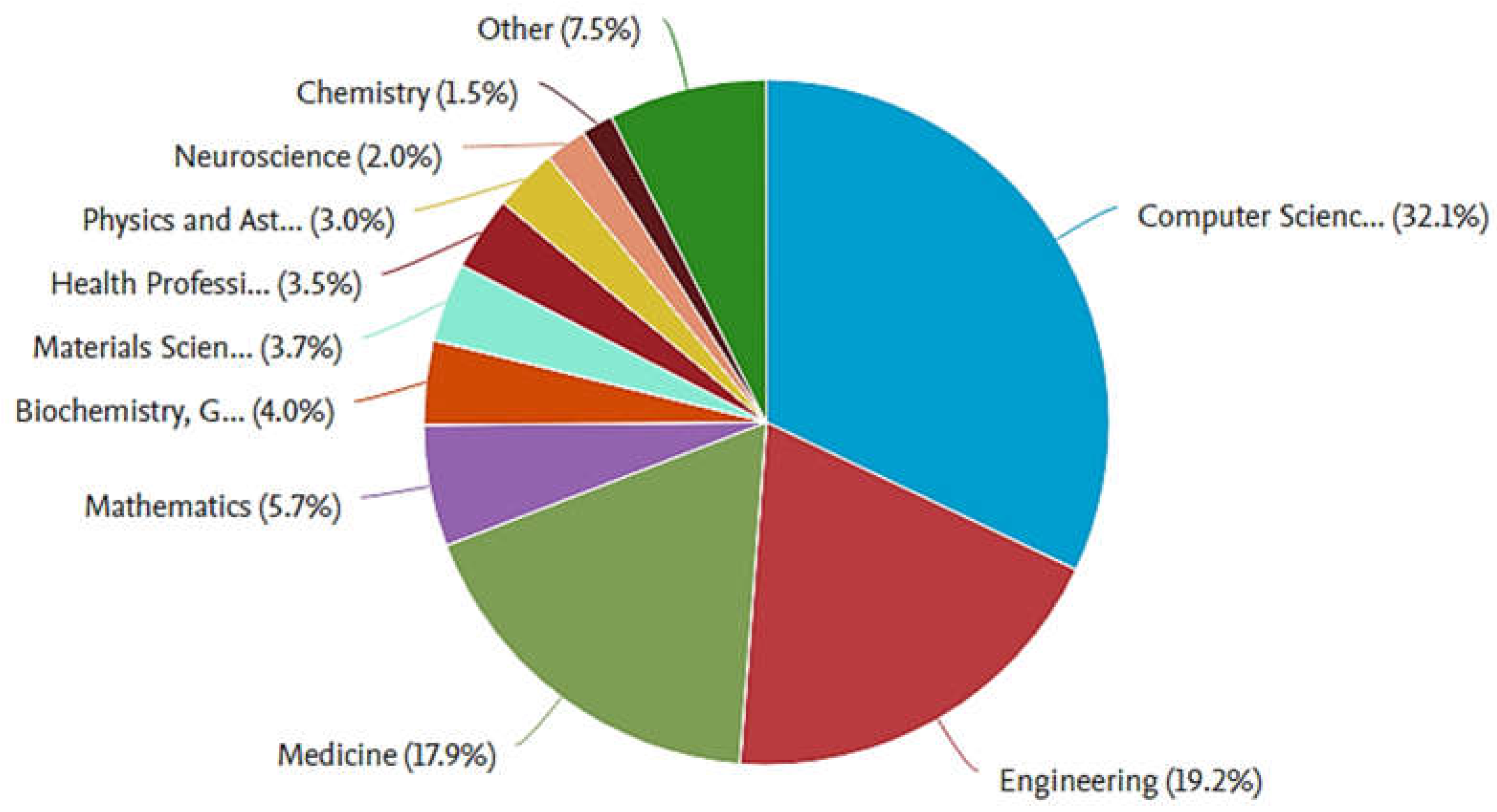
Figure 2.
Research paper published in various years from 2020.
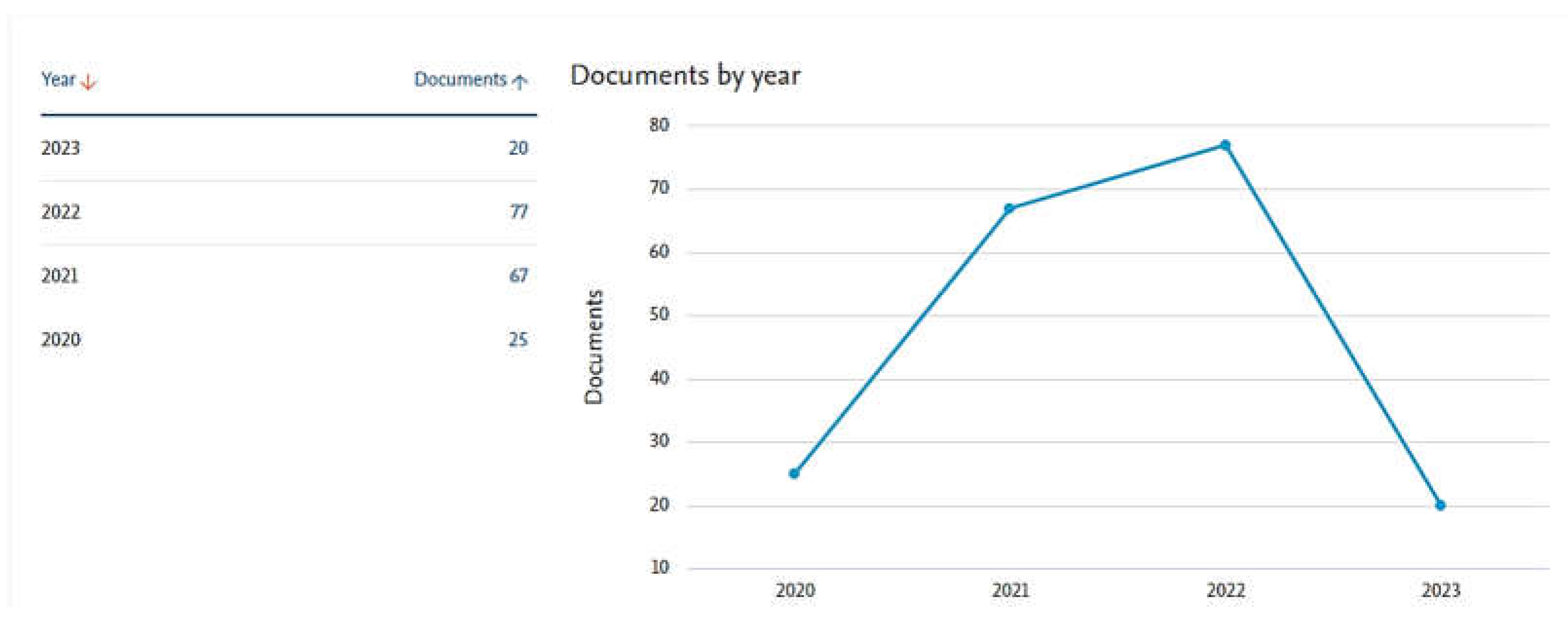
Figure 3.
Inclusion and Exclusion Criteria for selection of article for analysis.
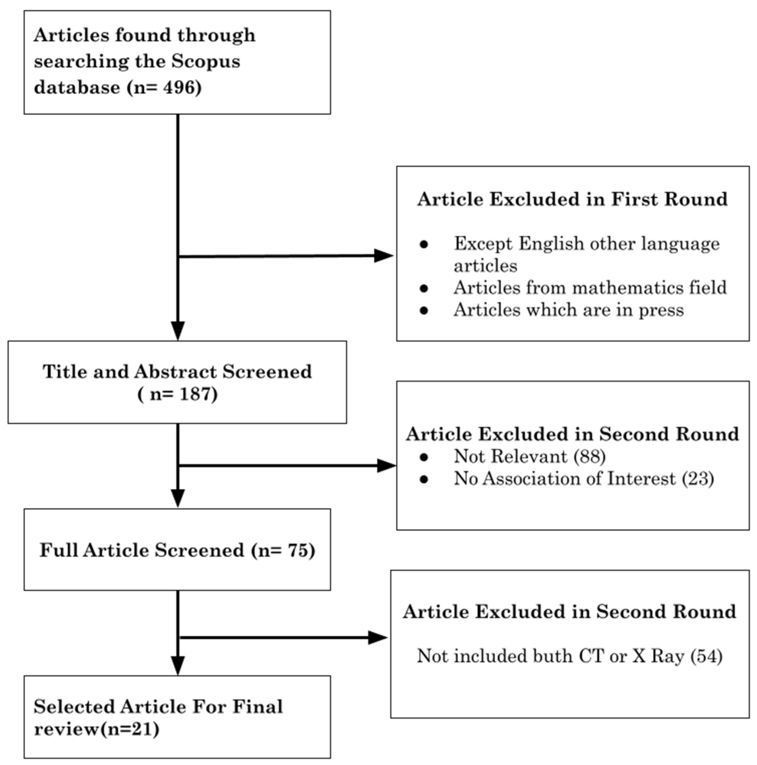
Figure 4.
The Accuracy obtained by various selected research articles with different imaging techniques.
Figure 4.
The Accuracy obtained by various selected research articles with different imaging techniques.
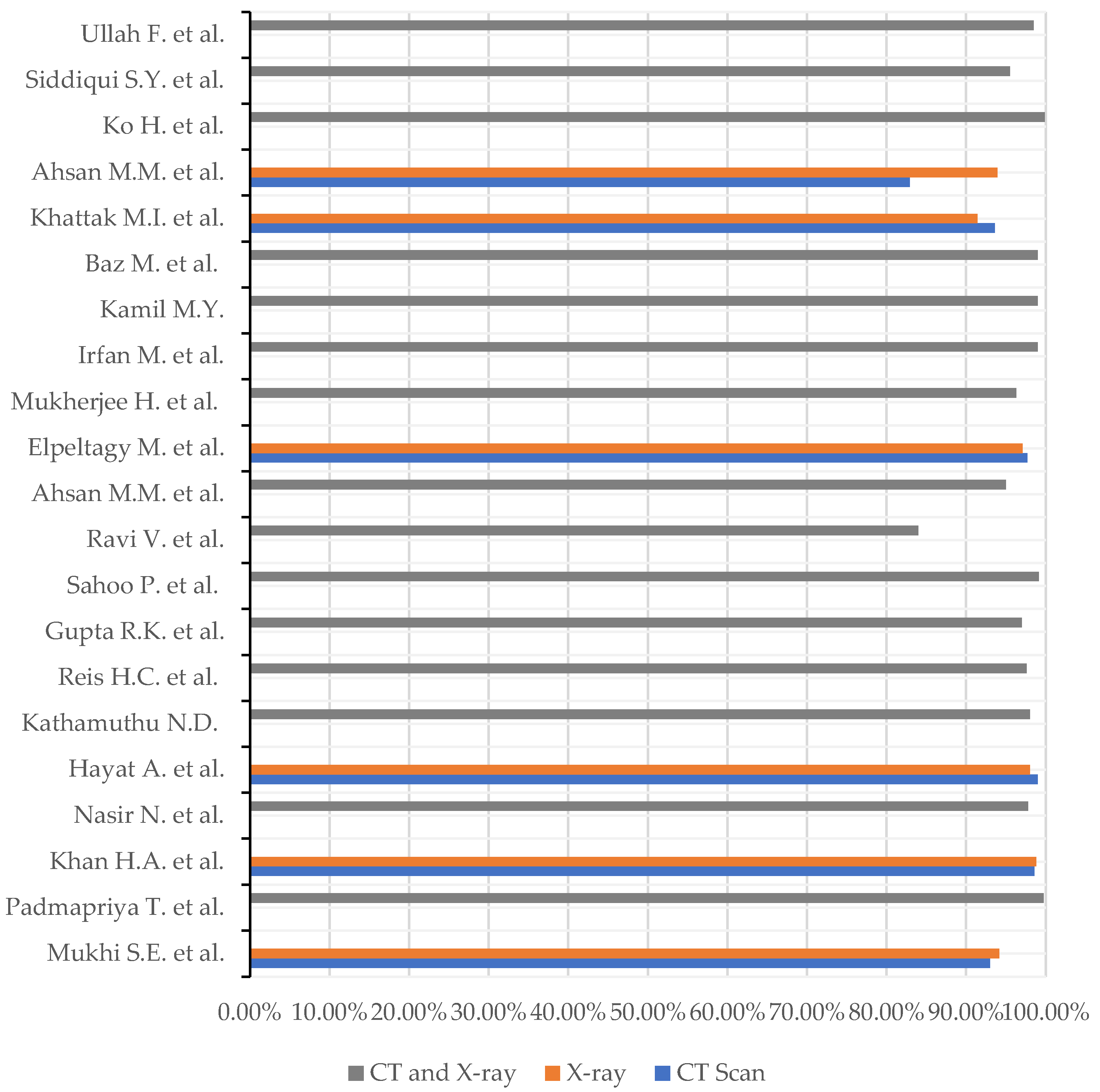
Table 2.
Details of Data used in Review.
| Literature | Data Source | Data Volume |
|---|---|---|
| [42] | Kaggle | 2482 CT scans image and 31 Covid positive along with 10,192 normal X-ray image |
| [48] | GitHub | 108,948 images |
| [49] | Kaggle | 3829 X-rays and 3829 X-rays |
| [43] | GitHub | 535 CT and X-ray images and 485 clinical notes related with them |
| [37] | Mendeley Data | 17,599 images |
| [44] | Kaggle | 2481 images |
| [46] | Kaggle | 2357 CT scan data, 2515 chest X-ray data and 2400 CT and chest X-ray hybrid data |
| [34] | Kaggle, GitHub | 5856 chest X-ray & CT dataset |
| [54] | Kaggle, GitHub | 2905 unique images for X-ray 617,775 images from 4154 patients |
| [41] | Mendeley Data | 8,055 CT scan and 9,544 X-ray images |
| [51] | Kaggle | 2591 mixed data |
| [33] | GitHub | 17100 X−ray and CT images |
| [47] | Kaggle, GitHub | 168 Covid and 168 normal cases for the both X-ray and CT scan images |
| [36] | GitHub, Covid-19 radiography database, Kaggle, Covid-19 image data collection, and Actual Med Covid-19 Chest X-ray Dataset | 3500 infected and 1500 healthy controls |
| [45] | D. S. Kermany, et al., "Identifying medical diagnoses and treatable diseases by image-based deep learning," J. P. Cohen, P. Morrison, and L. Dao, "Covid-19 image data collection," |
images of normal 805 and Covid-19 195 |
| [35] | Kaggle | Covid-19 400 Pneumonia 402 non-Covid 406 |
| [50] | Kaggle and Git Hub | 723 X-ray and 3228 CT scans images |
| [38] | Kaggle | 400 chest X-ray images, and 400 CT scan images |
| [39] | Wonkwang University Hospital(WKUH) and Chonnam National University Hospital (CNUH), Italian Society of Medical and Interventional Radiology(SIRM) public database. | 3993 images. |
| [53] | - | 527 images |
| [52] | From Git hub, Italian Society of Medical and Interventional Radiology, Radiological Society of North America (RSNA), Radiopaedia, and SIRM) and Kaggle repository. | 1229 SARS-CoV-2,1161 negative |
Table 3.
A Summary of algorithms used in the articles.
| Article | Algorithm and Model | Result |
|---|---|---|
| [42] | CNN with Deep Neural
|
|
| [48] | Bespoke CNN
|
99.75% accuracy |
| [49] |
Light CNN (Watershed based region growing segmentation) |
|
| [43] | Multi-modal system
|
Accuracy with 97.8% |
| [37] | CNN
|
|
| [44] | CNN
|
|
| [46] |
Deep CNN (Covid-DSNet developed) |
∙ 97.60 % accuracy |
| [34] | CNN
|
|
| [54] | CovidCon |
|
| [41] | Convolutional auto encoders with pre-trained CNN
|
|
| [51] | Deep CNN
|
|
| [33] |
CNN ResNet50 |
|
| [47] | Tailored Deep CNN | 96.28% Accuracy |
| [36] | Hybrid Deep Neural Networks (HDNNs) |
|
| [45] | VGG-19 |
|
| [35] | CNN |
|
| [50] | MSCovCNN |
|
| [38] | CNN
|
|
| [39] | FCONet |
|
| [53] | ID2S-Covid19-DL |
|
| [52] | CLAHE
|
|
Disclaimer/Publisher’s Note: The statements, opinions and data contained in all publications are solely those of the individual author(s) and contributor(s) and not of MDPI and/or the editor(s). MDPI and/or the editor(s) disclaim responsibility for any injury to people or property resulting from any ideas, methods, instructions or products referred to in the content. |
© 2023 by the authors. Licensee MDPI, Basel, Switzerland. This article is an open access article distributed under the terms and conditions of the Creative Commons Attribution (CC BY) license (http://creativecommons.org/licenses/by/4.0/).
Copyright: This open access article is published under a Creative Commons CC BY 4.0 license, which permit the free download, distribution, and reuse, provided that the author and preprint are cited in any reuse.


