Submitted:
07 July 2021
Posted:
08 July 2021
You are already at the latest version
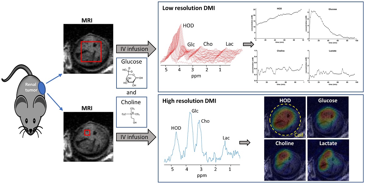
Abstract
: Increased glucose and choline uptake are hallmarks of cancer. We investigated if the uptake and conversion of [2H9]choline alone and together with that of [6,6’ 2H2]glucose can be assessed in subcutaneous tumors by deuterium metabolic imaging (DMI) after bolus administration of these compounds. Therefore tumors with human renal carcinoma cells were grown subcutaneously in mice up to ~0.5 cm3. Mice were anesthetized with isoflurane and IV infused in the MR magnet for ~20 sec with ~0.2 ml solutions containing either [2H9]choline (0.05g/kg) alone or together with [6,6’ 2H2]glucose (1.3g/kg). 2H MR was performed on a 11.7T MR system with a home-built 2H/1H coil using a 900 excitation pulse and 400ms repetition time. 3D DMI was recorded at high resolution (2x2x2mm) in 37 min or at low resolution (3.7x3.7x3.7mm) in 2:24 min. Absolute tissue concentrations were calculate assuming initial [HOD]=13.7mM. Within 5 minutes after [2H9]choline infusion its signal appeared in tumor spectra representing concentration increasing up to 0.3–1.2 mM and then slowly decreased or remained constant over 100 minutes. In plasma [2H9]choline disappeared within 15 minutes post-infusion implying that its tumor signal arises from tissue and not blood. After infusing a mixture of [2H9]choline and [6,6’ 2H2]glucose their signals were observed separately in tumor 2H spectra. Over time the [2H9]choline signal broadened, possibly due to conversion to other choline compounds, [[6,6’ 2H2]glucose] declined, [HOD] increased and a lactate signal appeared, reflecting glycolysis. Metabolic maps of all 2H compounds were reconstructed from high resolution DMIs. As choline infusion and glucose DMI is feasible in patients, their simultaneous detection has clinical potential for tumor characterization.
Keywords:
MRI
; deuterium metabolic imaging
; tumor
; 2H
; glucose
; choline
Copyright: This open access article is published under a Creative Commons CC BY 4.0 license, which permit the free download, distribution, and reuse, provided that the author and preprint are cited in any reuse.


