
Submitted:
12 February 2021
Posted:
15 February 2021
You are already at the latest version
Abstract
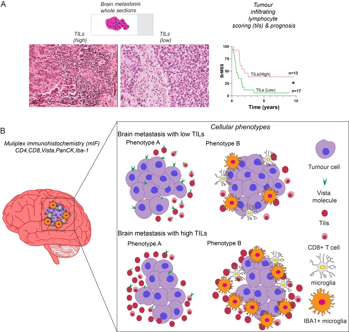
The heterogeneity of tumor infiltrating lymphocytes is not well characterized in brain metastasis. To address this, we performed a targeted analysis of immune cell subsets in brain metastasis tissues to test which immunosuppressive routes are involved in brain metastasis. We performed multiplex immunofluorescence (mIF), using commercially available validated antibodies on twenty formalin-fixed paraffin embedded whole sections. We quantitated the subsets of immune cells utilizing a targeted panel of proteins including PanCK, CD8, CD4, VISTA and Iba1, and analyzed an average of 15000 cells per sample. We classified tumours as either high (>30%) or low (<30%) tumour infiltrating lymphocytes (TILs) and found that increased TILs density correlated with survival. We next sought out to phenotype these TILs using mIF. The tumours with low TILs (n=9) had significantly higher expression of the immune-checkpoint molecule VISTA in tumor cells (p<0.01) as well as in their microenvironment (p<0.001). Contrastingly, the brain metastatic tumours with high TILs (n=8) displayed higher levels of activated microglia, as measured by Iba1 expression. Low TILs-tumours displayed CD8+ T-cells that co-express VISTA (p<0.01) significantly more compared to high TILs group, where CD8+ T-cells significantly co-express Iba1 (p<0.05). Interestingly, no definite phenotypes of CD4+ subsets were observed. These results were supported by RNA analysis of a publicly available, independent cohort. In conclusion, our work contributes to a growing understanding of the immune surveillance escape routes active in brain metastasis.
Keywords:
Brain Metastasis
; Immune Checkpoint
; Tumor Microenvironment
; T-cells
; TILs
Copyright: This open access article is published under a Creative Commons CC BY 4.0 license, which permit the free download, distribution, and reuse, provided that the author and preprint are cited in any reuse.


