
Submitted:
14 November 2018
Posted:
15 November 2018
You are already at the latest version
Abstract
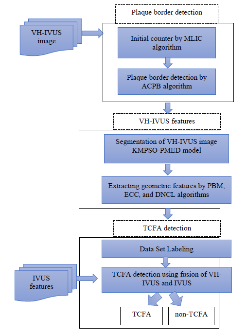
Virtual Histology- Intravascular Ultrasound (VH-IVUS) image is an available method for visualizing plaque component to detect thin cap fibroatheroma. Nevertheless, this imaging modality has considerable limitations to extract the plaque component features and identifying the TCFA plaque. The aim of this paper is to improve the identification of TCFA using fusion of IVUS and VH-IVUS images. In order to generate the automatic technique for reducing the human interaction, a new method namely Active Contour based Plaque Border Detection (ACPB) is proposed. In order to perform the pixel wise classification, hybrid of K-means algorithm with Particle Swarm Optimization and Plaque based Minimum Euclidean Distance (KMPSO-PMED) method is presented to classify the plaque region as well. Moreover, to obtain more significant information of imaging modalities, fusion of two different images consisting of VH-IVUS and IVUS is performed. Therefore, geometric features are extracted from the plaque region and combine with IVUS features. Furthermore, different group of plaque features are divided by means of the histopathological studies. SVM classifiers is applied to detect the TCFA and non-TCFA plaques. The proposed method is applied on 566 in-vivo IVUS and their matching VH-IVUS images obtained from 9 patients. The best result of SVM illustrates the accuracy rates of 99.41% for classification of TCFA plaque. The results prove that the highest accuracy is achieved by integrated features of IVUS and VH-IVUS images.
Keywords:
TCFA
; VH-IVUS image
; plaque border detection
; active counter method
; support vector machines.
Copyright: This open access article is published under a Creative Commons CC BY 4.0 license, which permit the free download, distribution, and reuse, provided that the author and preprint are cited in any reuse.


