
Submitted:
13 November 2026
Posted:
16 November 2026
You are already at the latest version
Abstract
Frontal sinus obliteration is a procedure usually reserved for select cases of refractory chronic rhinosinusitis, tumors and trauma management. These conditions are relatively rare in children, which makes the procedure even rarer in the pediatric population. Bioactive glass S53P4 is a biocompatible mineral allogenic material used in various applications in maxillofacial, orthopedic and head and neck surgery. Although its’ osteo-stimulating and antibacterial properties, as well as clinical outcomes are well documented in literature, limited data exists about its’ use in frontal sinus obliteration in children is scarce. We present two cases of successful frontal sinus obliteration with S53P4 in different clinical indications, where an inflammatory process and an osteoma have produced bone destruction between the frontal sinus, the orbit and the anterior crania fossa. Bioactive glass in both cases has proven to be easy to use and effective in achieving frontal sinus obliteration in a pediatric patient.
Keywords:
frontal sinus
; sinus obliteration
; bioactive glass
; S53P4
1. Introduction
Late development of the frontal sinus during childhood and adolescence and overall lower sinus disease prevalence in younger individuals makes frontal sinus pathology diagnosis rare in the pediatric population. Chronic sinusitis, tumors and frontal bone fractures, which constitute the most common causes for frontal sinus surgery, are much more often found in adults. Contemporary approach to frontal sinus surgery includes mainly endoscopic approaches, with external access being usually reserved for refractory or selected cases. Fontal sinus obliteration is usually considered a last resort treatment option for frontal pathology. Various materials for sinus obliteration have been used and described in literature. One of them is bioactive glass (S53P4), a synthetic mineral material that has been shown to be safe and effective in various applications. Additional aspects of S53P4 include an anti-bacterial and anti-infectious properties, as well as a very good biocompatibility with autogenous surrounding bone over time. Scientific data for frontal sinus obliteration in children is very limited. The authors have not identified any studies regarding sinus obliteration with bioactive glass in the pediatric population in Polish and international literature. This work is aimed at presenting two cases of successful pediatric frontal sinus obliteration with bioactive glass in which different frontal sinus pathology has led to the destruction of bone between the frontal sinus, orbit and anterior cranial fossa.
2. Case 1
A 11-yeard old boy was admitted to the Department of Pediatric Otolaryngology at the Medical University of Warsaw due to eyelid edema, facial pain and fever. His medical history involved adenoidectomy followed by several endoscopic sinus surgery procedures in the previous years performed due to refractory chronic rhinosinusitis with nasal polyps. Genetic studies revealed a biallelic CFTR (Cystic fibrosis transmembrane conductance regulator) mutation not causing cystic fibrosis. He had been previously diagnosed with chronic Pseudomonas aeruginosa colonization of the sinuses. At the moment of admission, he was 6 months after his most recent endoscopic sinus surgery with polypectomy.
Based on clinical evaluation and a computed tomography (CT) scan he was diagnosed with an orbital abscess originating from the right frontal sinus. The CT scan revealed neo-osteogenesis in the right frontal recess leading to complete radiologic obstruction of the drainage pathway of the right frontal sinus (Figure 1). The patient underwent abscess drainage from an upper brow incision with intravenous antibiotic therapy. The etiology of the abscess was found to be of Pseudomonas aeruginosa origin and the patient received intravenous ceftazidime. Due to a good response to the abovementioned treatment the patient was discharged home with a plan of subsequent endoscopic frontal sinus surgery to open and maintain drainage in the right frontal recess.
The patient was admitted to the Department again before the time of his planned revision surgery due to a recurrence of eyelid oedema and fever. This episode was successfully managed conservatively, as a CT scan performed on admission did not reveal abscess formation in the orbit, only tissue oedema resulting from frontal sinusitis. The patient was qualified for external approach frontal sinus surgery with obliteration. Given the history of bacterial colonization and chronic inflammation witch exacerbations bioactive glass (BAG) S53P4 was chosen as the proper material for sinus obliteration due to its’ reported antimicrobial and anti-inflammatory properties.
The surgery was performed from a medial brow incision, with a complete removal of the sinus mucosa and confirming the complete closure of the sinus drainage pathway by bone tissue. The sinus lumen was filled with S53P4 bioactive glass 0.5-0.8 mm granules with 0.9% saline (Figure 2). The anterior access foramen to the sinus approximately 5mm in diameter was covered and with a layer of thin absorbable gelatin sponge and the soft tissues closed. In the postoperative period the patient did not report any alarming symptoms and has been symptomless for a period of 32 months at the time of manuscript submission. A CT scan performed a year after surgery showed complete obliteration of the frontal sinus area by the BAG (Figure 3).
3. Case 2
The second patient was referred do the Department of Pediatric Otolaryngology after a head CT scan ordered by a neurologist due to reported ptosis of the left eye revealed a massive high-density lesion in the left frontal sinus. Additional findings in the CT scan included erosion of the posterior and anterior wall of the left frontal sinus and superior orbital wall in direct contact with the main lesion and a mucocele protruding from the lateral aspect of the frontal sinus into the left orbit (Figure 4). The last finding was found to result in a slight bulging of the left upper eyelid, initially mistaken for ptosis. Apart from the visible asymmetry in eyelid fissure the patient was symptomless.
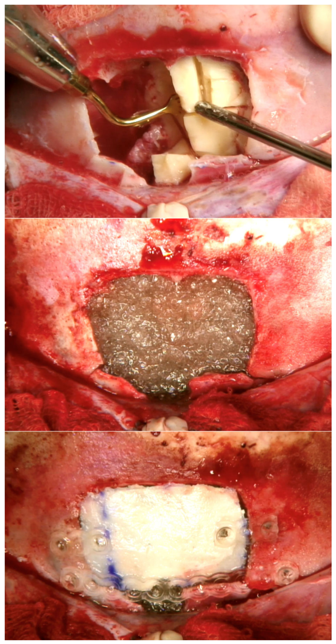
Additional radiological workup was ordered with an magnetic resonance imaging (MRI) scan, that established no involvement of the anterior cranial fossa dura. The patient was qualified for surgical removal of the bony lesion from an external approach through a coronal incision and sinus obliteration in a multidisciplinary team involving an otolaryngologist, neurosurgeon and maxillofacial surgeon. The anterior table of the frontal sinus was revealed to be thinned but mostly retained over the tumor, an osteoplastic flap was raised, and the lesion was removed completely after fragmentation. The origin of the tumor was found on the interfrontal septum, which led to opening of the right frontal sinus during the complete resection. The mucosal lining of the frontal sinuses was removed and S53P4 bioactive glass 0.5-0.8 mm granules with 0.9% saline were used to fill the sinuses. The exposed dura and periorbita were covered with a thin layer of absorbable gelatin sponge to prevent BAG granules shifting into the orbit and anterior cranial fossa. The osteoplastic flap was secured in place with absorbable osteosynthesis plate and screws (Figure 5). The patient received ceftriaxone for 5 days after surgery and was discharged in general and local good condition.
Histopathological examination of the lesion confirmed the diagnosis of osteoma. The patient is symptomless in a follow-up of 20 months at the moment of manuscript submission with good cosmetic effect.
4. Discussion
True refractory chronic rhinosinusitis (CRS) in children is very rare. Most cases of pediatric CRS resolve with intranasal corticosteroids (INCS), antibiotics and adenoidectomy (1-3), with intranasal steroids functional endoscopic sinus surgery (FESS) having been shown to be both safe and effective in cases not responding to the forementioned initial treatment (3).
Patients with chronic rhinosinusitis refractory to both conservative and surgical management are prone to development of neo-osteogenesis, which impacts FESS outcomes, with patients with a Pseudomonas aeruginosa colonization having additionally a higher incidence of neo-osteogenesis (4). Orbital complications are more common in acute rhinosinusitis and develop more frequently in children. An orbital abscess formed secondary to chronic rhinosinusitis is more likely to be found in the adult population (5). Although such a severe course of CRS is rare in children, it may be found in patients with secondary disease that develops at a younger age including mucocele formation and infection leading to sinus wall destruction (6).
Osteomas are relatively common benign tumors of the sinuses characterized by their slow growth and initial asymptomatic course that are mainly seen in adults (7, 8). The data regarding osteomas found in childhood and adolescence is scarce. A 2018 review by Badran et al revealed in the studied literature 16 cases of frontal osteomas in children with various surgical and reconstructive techniques used (9). Osteomas are considered “giant” if their diameter exceeds 30mm (10). A 2013 case series and review of literature by Cheng et al reported 45 cases of giant osteomas in the available literature, of which the youngest patient was 11 and seven were under the age of 18 (11).
The management techniques for frontal osteomas discussed in the literature are mainly based on studies on adults, of which some include teenagers in the analyzed population. Small osteoma management favoring endoscopic management and large tumors requiring more often external approach (12-14). Localization, size of the tumor and experience of the surgeon need to be taken into consideration when choosing the appropriate surgical approach and technique. Turri-Zanoni reviewed sixty cases of frontoethmoidal and orbital osteomas, most of which were removed with endoscopic or combined (endoscopic and external) approach, however only two patients in their group had anterior skull base involvement and the largest one was 41 mm in diameter (15). More recently Nguyen and Nadeau present a series of 10 cases of giant frontal osteomas in adults utilizing both endoscopic and external approaches without obliteration. The tumor size in their series ranges from 30 to 50 mm (13). Gotlib et al describes a series of larger osteomas in adults removed endoscopically (16). Farah et al reported a case of giant frontal osteoma with intracranial extension removed via open approach and cranialization of the frontal sinus (17).
A vast array of materials for sinus obliteration has been described for many years, especially in neurosurgical literature. The most commonly used material are the patient’s own fat (18) and bone (19). These autogenic materials are characterized by good biocompatibility, but their harvesting is linked to donor-site morbidity and limited availability. These issues have resulted in an increasing interest in allogenic materials for bone reconstruction and filling, that are freely available in needed quantities and do not need to be collected from donor sites. Such materials include gelatin sponge used with a pericranial flap (20), polymethyl methacrylate (21), hydroxyapatite cement (22) and beta-tricalcium phosphate putty (23), among others.
Bioactive glass S53P4 (BAG) is a ceramic material consisting of SiO2, Na2O, CaO, and P2O5, which has a well-documented history in successful obliteration of bone cavities in the head and neck region. It has been first proposed by Hench at al. in 1971 (24) and further studied as a material for sinus obliteration since the 1990s with very good clinical results (25, 26).
On molecular level S53P4 has been shown to have a stimulating effect on adjacent bone leading to migration and activation of osteogenic cells and acting as a scaffold for natural bone regeneration (27). The mineral structure of bioactive glass allows and facilitates natural hydroxyapatite generation (28). Histopathological examination of tissue samples harvested 10 years after obliteration showed natural progression towards bone formation in the region of obliteration (25).
One of the unique characteristics of BAG is its’ anti-microbial and anti-inflammatory capacity. Several studies have shown bactericidal and antibiofilm properties of bioactive glass which include local effectiveness against multi-drug-resistant strains of Staphylococci and Pseudomonas (29, 30). This makes BAG a potent candidate for application in the challenging environment of paranasal sinuses.
Clinical applications of BAG include maxillofacial and head and neck surgery, as well as orthopedic surgery. Studies by Peltola et al. including patients with chronic rhinosinusitis that failed previous endoscopic management and sinus obliteration with different material revealed a high success rate of S53P4 use in chronic inflammatory disease (25, 26). Aitasalo and Peltola have reported the successful use of bioactive glass granules and plates in bone defect reconstruction in the fronto-orbital region (31). BAG has been shown to be safe and useful in maxillary floor elevation (32) and mastoid obliteration in cholesteatoma surgery (33, 34). Recent findings in S53P4 use in dental implant surgery have shown further anti-inflammatory properties of the material (35). Outside of the head and neck very good clinical outcomes have been shown in diabetic foot osteomyelitis management (36) and in surgical osteomyelitis treatment (37) further highlighting the antibacterial properties of BAG contributing to the high success rate in its’ use in sites with chronic infection (38).
In our described case of severe CRS with a prominent bacterial colonization element, complete clinical resolution of the localized inflammatory process has been observed, contributing to the previous observations. The high biocompatibility and gradual transformation of the implanted material into tissue strongly resembling natural bone suggest a promising use in younger patients, although prospective studies on this topic are needed.
5. Conclusions
Our observations are in concurrence with the evidence suggesting that bioactive glass S54P4 is a safe, easy to use and effective material for frontal sinus obliteration in children including complicated chronic frontal sinusitis with bacterial colonization involvement. Further studies are mandated to establish the long-term effect of BAG frontal sinus obliteration in children, considering further facial growth and delayed recurrence of initial symptoms.
Author Contributions
Conceptualization, PK. and LZG.; writing—original draft preparation, PK.; writing—review and editing, PK.; supervision, LZG.; project administration, LZG.; funding acquisition, LZG. All authors have read and agreed to the published version of the manuscript.”
Funding
This research received no external funding.
Institutional Review Board Statement
Ethical review and approval were waived for this study due to retrospective nature of the study without medical intervention involvement in the study.
Informed Consent Statement
Informed consent was obtained from all subjects involved in the study.
Acknowledgments
During the preparation of this manuscript/study, the authors did not use AI tools.
Conflicts of Interest
The authors declare no conflicts of interest.
Abbreviations
The following abbreviations are used in this manuscript:
| CFTR | Cystic fibrosis transmembrane conductance regulator |
| CT | Computed tomography |
| BAG | Bioactive glass |
| MRI | Magnetic resonance imaging |
| CRS | Chronic rhinosinusitis |
| INCS | Intranasal corticosteroids |
| FESS | Functional endoscopic sinus surgery |
References
- Bettadahalli, V.; Chakravarti, A. Post-adenoidectomy quality of life in children with refractory chronic rhinosinusitis. J. Laryngol. Otol. 2017, 131(9), 773–8. [Google Scholar] [CrossRef] [PubMed]
- Adappa, N.D.; Coticchia, J.M. Management of refractory chronic rhinosinusitis in children. Am. J. Otolaryngol. 2006, 27(6), 384–9. [Google Scholar] [CrossRef] [PubMed]
- Fokkens, W.J.; Lund, V.J.; Hopkins, C.; Hellings, P.W.; Kern, R.; Reitsma, S.; et al. European Position Paper on Rhinosinusitis and Nasal Polyps 2020. Rhinology. 2020, 58 (Suppl S29), 1–464. [Google Scholar] [CrossRef] [PubMed]
- Huang, Z.; Hajjij, A.; Li, G.; Nayak, J.V.; Zhou, B.; Hwang, P.H. Clinical predictors of neo-osteogenesis in patients with chronic rhinosinusitis. Int Forum Allergy Rhinol. 2015, 5(4), 303–9. [Google Scholar] [CrossRef] [PubMed]
- Giusan, A.O.; Kubanova, A.A.; Uzdenova, R. Rhinosinusogenic orbital complications: the prevalence and principles of treatment. Vestn. Otorinolaringol. 2010, 4, 64–7. [Google Scholar] [PubMed]
- Sidam, S.; Gupta, V.; Kumar, A. Mucopyocele: An Entity Infrequently Reported as a Complication of Functional Endoscopic Sinus Surgery. Indian J Otolaryngol Head Neck Surg. 2022, 74 (Suppl 2), 1310–3. [Google Scholar] [CrossRef] [PubMed] [PubMed Central]
- Castelnuovo, P.; Valentini, V.; Giovannetti, F.; Bignami, M.; Cassoni, A.; Iannetti, G. Osteomas of the maxillofacial district: endoscopic surgery versus open surgery. J. Craniofac Surg. 2008, 19(6), 1446–52. [Google Scholar] [CrossRef] [PubMed]
- Chahed, H.; Hachicha, H.; Bachraoui, R.; Marrakchi, J.; Mediouni, A.; Zainine, R.; et al. Paranasal sinus osteomas: Diagnosis and treatment. Rev. Stomatol Chir. Maxillofac. Chir. Orale 2016, 117(5), 306–10. [Google Scholar] [CrossRef] [PubMed]
- Badran, K.W.; Suh, J.D.; Namiri, N.; Wrobel, B.; Ference, E.H. Pediatric Benign Paranasal Sinus Osteoneogenic Tumors: A Case Series and Systematic Review of Outcomes, Techniques, and a Multiportal Approach. Am. J. Rhinol. Allergy 2018, 32(6), 465–72. [Google Scholar] [CrossRef] [PubMed]
- Oleś, K.; Stręk, P.; Wiatr, M.; Składzień, J.; Tomik, J.; Morawska, A.; et al. Endoscopic treatment of giant skull base osteomas. Otolaryngol. Pol. 2011, 65(6), 410–3. [Google Scholar] [CrossRef] [PubMed]
- Cheng, K.J.; Wang, S.Q.; Lin, L. Giant osteomas of the ethmoid and frontal sinuses: Clinical characteristics and review of the literature. Oncol Lett. 2013, 5(5), 1724–30. [Google Scholar] [CrossRef] [PubMed] [PubMed Central]
- Strek, P.; Zagólski, O.; Składzień, J.; Kurzyński, M.; Dyduch, G. Osteomas of the paranasal sinuses: surgical treatment options. Med Sci Monit. 2007, 13(5), Cr244–50. [Google Scholar] [PubMed]
- Nguyen, S.; Nadeau, S. Giant Frontal Sinus Osteomas: Demographic, Clinical Presentation, and Management of 10 Cases. Am. J. Rhinol. Allergy 2019, 33(1), 36–43. [Google Scholar] [CrossRef] [PubMed]
- Strek, P.; Zagólski, O.; Składzień, J.; Oleś, K.; Konior, M.; Hydzik-Sobocińska, K.; et al. Endoscopic management of osteomas of the paranasal sinuses--own experience. Otolaryngol. Pol. 2007, 61(3), 260–4. [Google Scholar] [CrossRef] [PubMed]
- Turri-Zanoni, M.; Dallan, I.; Terranova, P.; Battaglia, P.; Karligkiotis, A.; Bignami, M.; et al. Frontoethmoidal and intraorbital osteomas: exploring the limits of the endoscopic approach. Arch. Otolaryngol. Head Neck Surg. 2012, 138(5), 498–504. [Google Scholar] [CrossRef] [PubMed]
- Gotlib, T.; Held-Ziółkowska, M.; Niemczyk, K. Frontal sinus and recess osteomas: an endonasal endoscopic approach. B-ent 2014, 10(2), 141–7. [Google Scholar] [PubMed]
- Farah, R.A.; Poletti, A.; Han, A.; Navarro, R. Giant frontal sinus osteoma and its potential consequences: illustrative case. J Neurosurg Case Lessons 2021, 1(21), Case21105. [Google Scholar] [CrossRef] [PubMed] [PubMed Central]
- Weber, R.; Draf, W.; Kahle, G.; Kind, M. Obliteration of the frontal sinus--state of the art and reflections on new materials. Rhinology. 1999, 37(1), 1–15. [Google Scholar] [PubMed]
- Ito, S.; Nagayama, K.; Iino, N.; Saito, I.; Takami, Y. Frontal sinus repair with free autologous bone grafts and fibrin glue. Surg. Neurol. 2003, 60(2), 155–8; discussion 8. [Google Scholar] [CrossRef] [PubMed]
- Guo, Y.; Fu, X.; Yin, W.; Jiang, Z.; Kuang, Y.; Wu, Z.; et al. A practical and economical method for frontal sinus reconstruction after frontal craniotomy: A single-center experience with 140 patients. Front Surg. 2022, 9, 919276. [Google Scholar] [CrossRef] [PubMed] [PubMed Central]
- Matsuura, J.; Otsuka, T.; Nakagawa, T.; Kai, K. Frontal Sinus Repair Using Polymethyl Methacrylate After Bifrontal Craniotomy. World Neurosurg. 2018. [Google Scholar] [CrossRef] [PubMed]
- Martinez, O.P.; Le, P.B.; Martinez, C.R.; Chen, E. Autologous Fat Versus Hydroxyapatite Cement for Use in Frontal Sinus Obliteration: Systematic Review and Meta-Analysis. Ann. Plast. Surg. 2023, 91(4), 497–502. [Google Scholar] [CrossRef] [PubMed]
- Moore, P.; Kam, J.K.T.; Castle-Kirszbaum, M.; Tan, D.; Wreghitt, S.; Lee, S.; et al. Frontal sinus obliteration with beta-tricalcium phosphate putty: case series with long-term radiological follow up. J. Laryngol. Otol. 2023, 137(2), 163–8. [Google Scholar] [CrossRef] [PubMed]
- Hench, L.L.; Splinter, R.J.; Allen, W.C.; Greenlee, T.K. Bonding mechanisms at the interface of ceramic prosthetic materials. J. Biomed. Mater. Res. 1971, 5(6), 117–41. [Google Scholar] [CrossRef]
- Peltola, M.; Aitasalo, K.; Suonpaa, J.; Varpula, M.; Yli-Urpo, A. Bioactive glass S53P4 in frontal sinus obliteration: a long-term clinical experience. Head Neck 2006, 28(9), 834–41. [Google Scholar] [CrossRef] [PubMed]
- Peltola, M.; Suonpaa, J.; Aitasalo, K.; Varpula, M.; Yli-Urpo, A.; Happonen, R.P. Obliteration of the frontal sinus cavity with bioactive glass. Head Neck 1998, 20(4), 315–9. [Google Scholar] [CrossRef] [PubMed]
- Virolainen, P.; Heikkilä, J.; Yli-Urpo, A.; Vuorio, E.; Aro, H.T. Histomorphometric and molecular biologic comparison of bioactive glass granules and autogenous bone grafts in augmentation of bone defect healing. J Biomed Mater Res. 1997, 35(1), 9–17. [Google Scholar] [CrossRef] [PubMed]
- Välimäki, V.V.; Aro, H.T. Molecular basis for action of bioactive glasses as bone graft substitute. Scand. J. Surg. 2006, 95(2), 95–102. [Google Scholar] [CrossRef] [PubMed]
- Drago, L.; Toscano, M.; Bottagisio, M. Recent Evidence on Bioactive Glass Antimicrobial and Antibiofilm Activity: A Mini-Review. Materials (Basel) 2018, 11(2). [Google Scholar] [CrossRef] [PubMed] [PubMed Central]
- Munukka, E.; Leppäranta, O.; Korkeamäki, M.; Vaahtio, M.; Peltola, T.; Zhang, D.; et al. Bactericidal effects of bioactive glasses on clinically important aerobic bacteria. J. Mater. Sci. Mater. Med. 2008, 19(1), 27–32. [Google Scholar] [CrossRef] [PubMed]
- Aitasalo, K.M.J.; Peltola, M.J. Bioactive glass hydroxyapatite in fronto-orbital defect reconstruction. Plast. Reconstr. Surg. 2007, 120(7), 1963–72. [Google Scholar] [CrossRef] [PubMed]
- Menezes, J.D.; Pereira, R.D.S.; Bonardi, J.P.; Griza, G.L.; Okamoto, R.; Hochuli-Vieira, E. Bioactive glass added to autogenous bone graft in maxillary sinus augmentation: a prospective histomorphometric, immunohistochemical, and bone graft resorption assessment. J Appl Oral Sci. 2018, 26, e20170296. [Google Scholar] [CrossRef] [PubMed] [PubMed Central]
- Kroon, V.J.; Mes, S.W.; Borggreven, P.A.; van de Langenberg, R.; Colnot, D.R.; Quak, J.J. Mastoid Obliteration Using S53P4 Bioactive Glass in Cholesteatoma Surgery: A 10-Year Single-Center Experience in 173 Adult Patients with Long-Term Magnetic Resonance Imaging Controlled Follow-up. Otol. Neurotol. 2022, 43(10), 1181–8. [Google Scholar] [CrossRef] [PubMed] [PubMed Central]
- Krol, B.; Cywka, K.B.; Skarzynska, M.B.; Skarzynski, P.H. Mastoid obliteration with S53P4 bioactive glass after canal wall down mastoidectomy: Preliminary results. Am. J. Otolaryngol. 2021, 42(2), 102895. [Google Scholar] [CrossRef] [PubMed]
- Barrak, F.N.; Li, S.; Mohammed, A.A.; Myant, C.; Jones, J.R. Anti-inflammatory properties of S53P4 bioactive glass implant material. J. Dent. 2022, 127, 104296. [Google Scholar] [CrossRef] [PubMed]
- Kastrin, M.; Urbančič Rovan, V.; Frangež, I. Possible Advantages of S53P4 Bioactive Glass in the Treatment of Septic Osteoarthritis of the First Metatarsophalangeal Joint in the Diabetic Foot. J. Clin. Med. 2021, 10(6), 1208. [Google Scholar] [CrossRef] [PubMed]
- Bigoni, M.; Turati, M.; Zanchi, N.; Lombardo, A.S.; Graci, J.; Omeljaniuk, R.J.; et al. Clinical applications of Bioactive glass S53P4 in bone infections: a systematic review. Eur. Rev. Med. Pharmacol. Sci. 2019, 23((2) Suppl, 240–51. [Google Scholar] [CrossRef] [PubMed]
- Profeta, A.C. Emerging developments in the use of bioactive glass for reconstruction of craniofacial bone. Br J Oral Maxillofac Surg. 2015, 53(8), 760–2. [Google Scholar] [CrossRef] [PubMed]
Figure 1.
complete opacification of the frontal sinuses with bone erosion between the right frontal sinus and the orbit (A) and neo-osteogenesis in the right frontal recess (B).
Figure 1.
complete opacification of the frontal sinuses with bone erosion between the right frontal sinus and the orbit (A) and neo-osteogenesis in the right frontal recess (B).
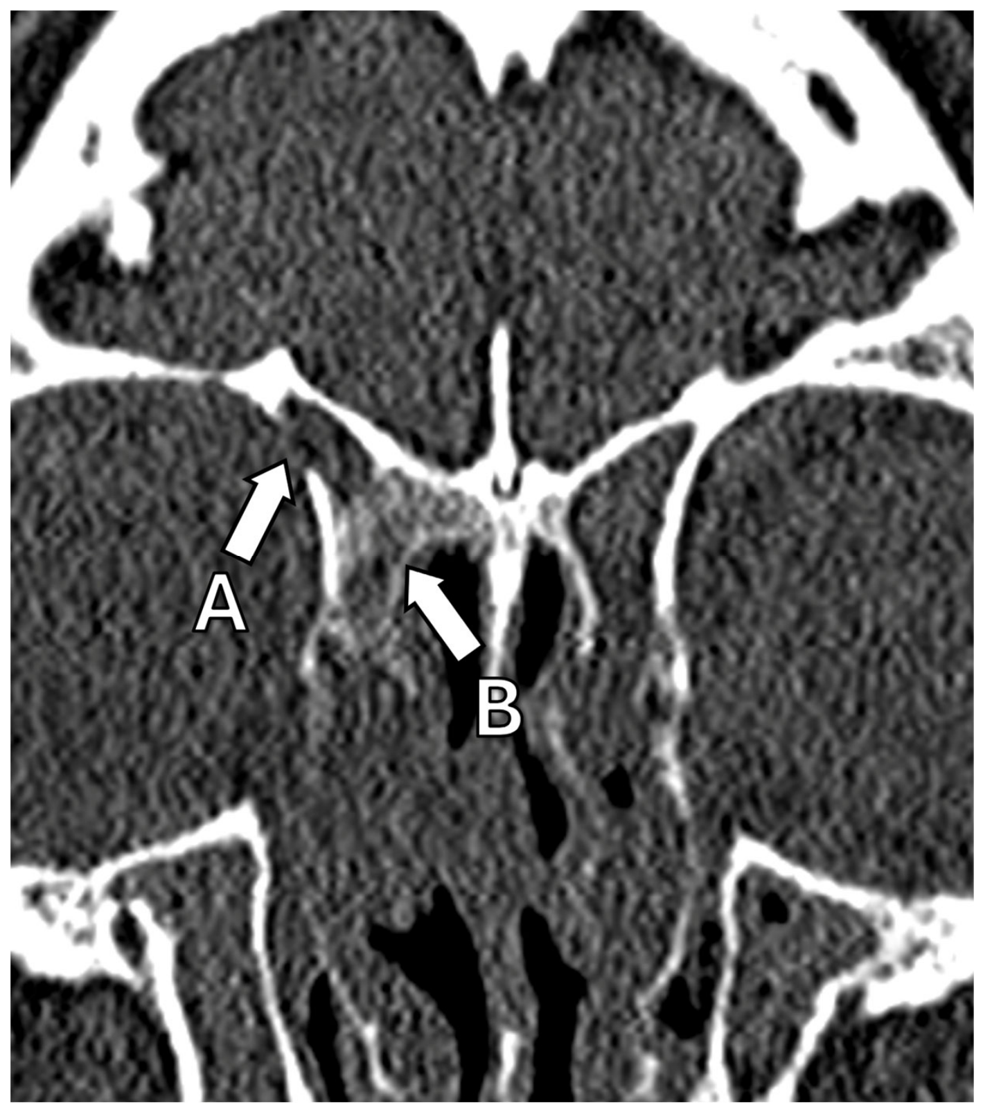
Figure 2.
bioactive glass filling the lumen of the frontal sinus.
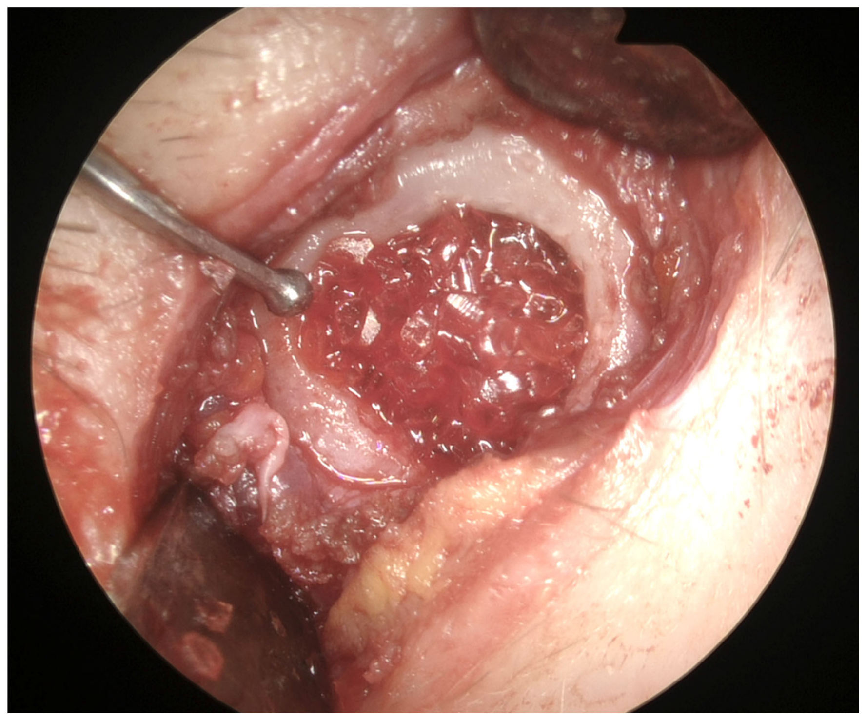
Figure 3.
CT scan performed a year after obliteration of the right frontal sinus with bioactive glass. Structure of the right frontal bone resembles natural bone.
Figure 3.
CT scan performed a year after obliteration of the right frontal sinus with bioactive glass. Structure of the right frontal bone resembles natural bone.
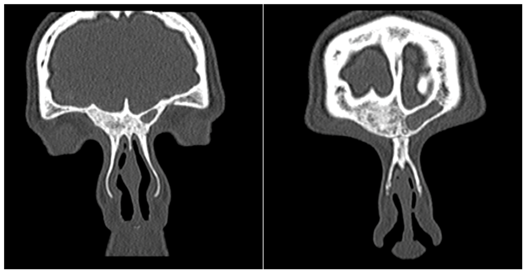
Figure 4.
On the left: frontal plane CT scans of the giant osteoma of the left frontal sinus with mucocele of the left orbit (A) and bone erosion to the anterior cranial fossa (B).
Figure 4.
On the left: frontal plane CT scans of the giant osteoma of the left frontal sinus with mucocele of the left orbit (A) and bone erosion to the anterior cranial fossa (B).
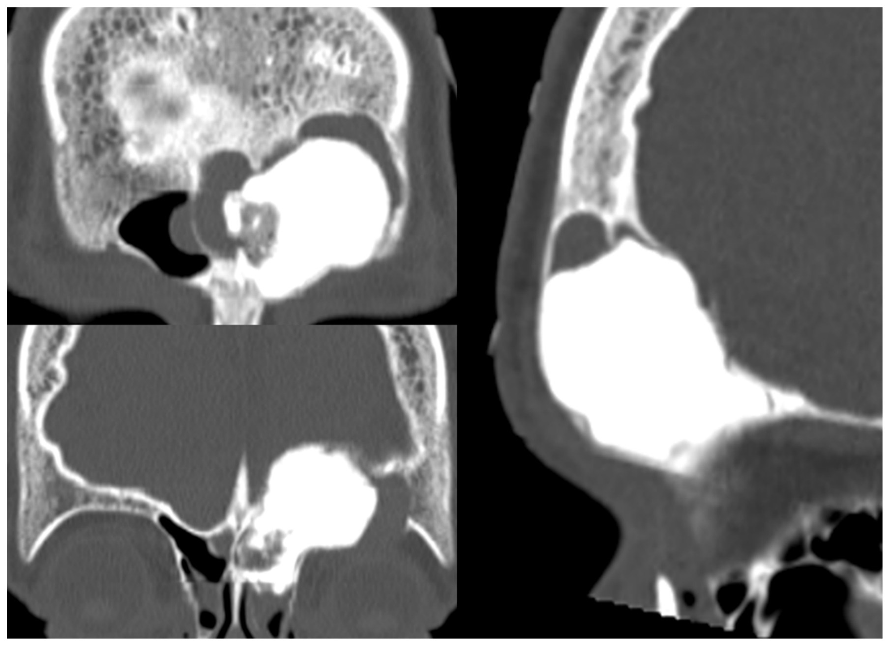
Figure 5.
Giant frontal osteoma removal with frontal obliteration. From the top: tumor fragmentation and removal, the sinus cavity filled with BAG granules, osteoplastic flap secured with absorbable plates.
Figure 5.
Giant frontal osteoma removal with frontal obliteration. From the top: tumor fragmentation and removal, the sinus cavity filled with BAG granules, osteoplastic flap secured with absorbable plates.
Disclaimer/Publisher’s Note: The statements, opinions and data contained in all publications are solely those of the individual author(s) and contributor(s) and not of MDPI and/or the editor(s). MDPI and/or the editor(s) disclaim responsibility for any injury to people or property resulting from any ideas, methods, instructions or products referred to in the content. |
© 2026 by the authors. Licensee MDPI, Basel, Switzerland. This article is an open access article distributed under the terms and conditions of the Creative Commons Attribution (CC BY) license (http://creativecommons.org/licenses/by/4.0/).
Copyright: This open access article is published under a Creative Commons CC BY 4.0 license, which permit the free download, distribution, and reuse, provided that the author and preprint are cited in any reuse.