
Submitted:
14 November 2026
Posted:
16 November 2026
You are already at the latest version
Abstract
Background: Spontaneous intracerebral hemorrhage (ICH) is associated with substantial long-term neurological disability. The clinical role of hydrogen-based nanobubble infusion as an adjunct to rehabilitation remains uncertain. Methods: A 55-year-old woman with hypertension and residual right hemiparesis after left basal ganglia hemorrhage received 29 adjunctive hydrogen–oxygen nanobubble (HHO) infusion sessions beginning approximately three months after stroke onset; intermittent nitric oxide (NO) supplementation was recorded during later sessions. Results: Right upper- and lower-limb strength changed from Medical Research Council (MRC) grade 2/5 to 3/5. Total cholesterol changed from 243 to 138 mg/dL, low-density lipoprotein cholesterol from 169 to 66 mg/dL, serum uric acid from 7.1 to 3.3 mg/dL, and erythrocyte sedimentation rate from 68 to 42 mm/h. After 19 sessions, follow-up computed tomography showed disappearance of the initial hematoma and residual chronic cerebral changes without new hemorrhage. A transient low-grade fever was reported during early sessions. Conclusions: These observations document a favorable clinical course during combined standard care, rehabilitation, and adjunctive treatment. A single uncontrolled case cannot determine whether HHO–NO contributed to recovery, hematoma clearance, metabolic changes, or treatment safety. Standardized formulation characterization and controlled clinical investigation are warranted.
Keywords:
intracerebral hemorrhage
; hydrogen–oxygen nanobubble
; molecular hydrogen
; neurological recovery
; hematoma resolution
1. Introduction
Spontaneous intracerebral hemorrhage (ICH) is a severe subtype of stroke associated with high mortality, persistent disability, and long-term dependence. According to the World Stroke Organization's global factsheet, ICH accounted for approximately 28.8% of incident strokes and a disproportionate share of stroke mortality and disability in 2021 [1]. Hypertension remains one of the most important modifiable risk factors. Contemporary guidance prioritizes timely diagnosis, blood pressure management, prevention of hematoma-related deterioration, and coordinated follow-up care [2,3]. However, recovery of neurological function after ICH remains highly variable, motivating investigation of supportive interventions that do not replace evidence-based treatment.
Neurological injury after ICH comprises primary tissue disruption and secondary injury associated with perihematomal edema, blood–brain barrier dysfunction, hemoglobin degradation, inflammation, and oxidative stress. Free iron generated during erythrocyte breakdown can promote lipid peroxidation and ferroptosis, while inflammatory signaling may exacerbate neurovascular damage [4,5]. These processes provide a biological rationale for studying antioxidant- and inflammation-related pathways, although a plausible mechanism does not imply clinical efficacy.
Functional recovery following ICH depends on lesion severity and location, endogenous repair, neuroplastic changes, and rehabilitation. Recent analyses have indicated that the timing of motor recovery after ICH may differ from that after ischemic stroke, with clinically relevant improvement continuing during the subacute period [6]. Rehabilitation is therefore an essential co-intervention that must be considered when interpreting clinical improvement during any adjunctive treatment [7].
Molecular hydrogen (H₂) has been investigated for possible effects on oxidative and inflammatory pathways. Foundational experimental work described selective antioxidant activity under certain conditions [8], whereas a later review highlighted the heterogeneity of diseases, formulations, treatment routes, and measured clinical outcomes [9]. In experimental ICH, hydrogen inhalation reduced selected measures of early brain injury and improved short-term neurological outcomes in mice [10,11]. Conversely, a rat study found reduced oxidative DNA damage markers without significant improvement in brain edema or functional outcomes [12]. Thus, the experimental literature offers mechanistic hypotheses but no consistent basis for assuming a functional benefit in patients.
Hydrogen–oxygen nanobubble (HHO) preparations represent a distinct, formulation-dependent approach to gas delivery. Findings from oral or inhaled hydrogen cannot be assumed to apply to intravenously administered nanobubble dispersions. Preliminary clinical reports of hydrogen-enriched intravenous fluid in cerebral ischemia and oxyhydrogen nanobubble infusion in chronic kidney disease provide limited context, but involve different populations and formulations [13,14]. Nitric oxide (NO), used intermittently in the protocol described here, also has complex dose- and context-dependent cerebrovascular effects [15]. Accordingly, independent contributions and safety of combined HHO–NO infusion remain unestablished in ICH.
Against this background, this report describes the clinical course of a woman with hypertensive ICH receiving adjunctive HHO infusion with intermittent NO supplementation alongside conventional medical care and rehabilitation. It documents the treatment sequence, changes in muscle strength, selected metabolic and inflammatory laboratory results, and serial computed tomography (CT) observations. The objective is to report temporal clinical observations and identify considerations for future research, rather than to establish causality or efficacy in a single case.
2. Case Presentation
2.1. Patient Information and Initial Assessment
A 55-year-old woman with long-standing hypertension experienced spontaneous intracerebral hemorrhage in late August 2025. Initial brain CT demonstrated hemorrhage in the left basal ganglia with surrounding edema and mass effect. She developed right hemiparesis without reported disturbance of speech or consciousness. Acute treatment was undertaken at another hospital as part of standard care.
Following discharge, conventional rehabilitation was continued, although significant right-sided weakness persisted. On 9 December 2025, she attended the clinic for additional supportive treatment. Examination documented MRC grade 2/5 muscle strength in the right upper and lower extremities. Vital signs and general physical examination were described in the source record as otherwise unremarkable, without numerical baseline values.
She denied smoking, alcohol consumption, and illicit drug use. The source record describes an unrestricted diet comprising three meals daily and no regular physical exercise after the stroke. Her documented medication regimen was candesartan 16 mg once daily, rosuvastatin 20 mg once daily, donepezil 5 mg once daily, allopurinol 300 mg once daily, Neulin once daily, and Neurosanbe once daily. These medications were reported to continue without major modification throughout observation.
2.2. Clinical Timeline

Table 1.
Documented clinical sequence and treatment milestones.
| Period / visit | Observation documented in clinical source |
|---|---|
| Late August 2025 | Onset of left basal ganglia ICH with edema and mass effect on initial CT; subsequent right hemiparesis. |
| 9 December 2025 | Clinic assessment approximately three months after onset; right upper- and lower-extremity MRC 2/5. |
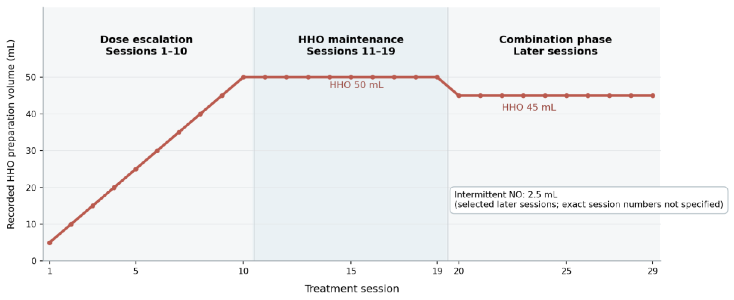
| Sessions 1–10 | HHO dose escalation from 5 to 50 mL, according to the source treatment graph. |
| Sessions 11–19 | HHO maintenance at 50 mL, according to the source treatment graph. |
| After session 19 | Follow-up CT described resolution of the initial hematoma with residual chronic changes. |
| Later sessions to session 29 | Intermittent NO supplementation recorded; the source figure displays HHO 45 mL + NO 2.5 mL in the combination phase. |
| After session 29 | MRC improved to 3/5 in right upper and lower extremities; follow-up laboratory trends reported. |
2.3. Therapeutic Intervention
The patient received adjunctive hydrogen-based nanobubble (HHO) infusion therapy over 29 sessions. Treatment began with a gradual increase in the recorded HHO preparation volume from 5 mL to 50 mL, followed by a maintenance phase. During later sessions, intermittent NO supplementation of 2.5 mL was recorded. The original treatment figure specifies a combination phase of HHO 45 mL plus NO 2.5 mL, whereas the narrative source refers to HHO maintenance at 50 mL. This difference is retained as a source-document discrepancy requiring confirmation against session-level records before submission.
These values represent recorded preparation volumes, not verified doses of dissolved hydrogen, oxygen, or nitric oxide. The available case material does not establish the gas concentrations, nanobubble size distribution, diluent composition, infusion rate, or other formulation-specific parameters needed to characterize actual exposure. No missing protocol parameters were inferred for this report.
Figure 1.
Recorded dose-escalation, maintenance, and combination phases of HHO and intermittent NO administration (redrawn from the original clinical draft). The graph reproduces the recorded HHO phases and indicates intermittent NO use; the exact NO session numbers were not provided. Confirm the combination-phase values against the original session-level record.
Figure 1.
Recorded dose-escalation, maintenance, and combination phases of HHO and intermittent NO administration (redrawn from the original clinical draft). The graph reproduces the recorded HHO phases and indicates intermittent NO use; the exact NO session numbers were not provided. Confirm the combination-phase values against the original session-level record.
2.4. Clinical, Laboratory, and Radiological Observations
Serial neurological examinations, laboratory tests, and CT were used in routine follow-up. The patient reported progressive improvement in overall well-being and right upper-limb function. She became able to actively elevate her right arm, although hand grip remained limited. Right upper- and lower-extremity MRC strength improved from 2/5 at the initial assessment to 3/5 after the 29-session treatment course.
Recorded laboratory values changed during observation: total cholesterol decreased from 243 to 138 mg/dL; LDL cholesterol from 169 to 66 mg/dL; serum uric acid from 7.1 to 3.3 mg/dL; and erythrocyte sedimentation rate (ESR) from 68 to 42 mm/h. Renal and hepatic function were described as being within acceptable limits in the original account, but their numerical results were not supplied for this manuscript.
After 19 sessions, follow-up CT was reported to show no residual visible hematoma, with mild cerebral atrophy, ex vacuo ventriculomegaly, and abnormalities described as chronic infarction affecting the left basal ganglia, insular region, and centrum semiovale. No recurrent hemorrhage or new acute intracranial lesion was reported. The radiology description is presented as recorded; the chronic infarction terminology should be compared with the original images and radiological reports.
Table 2.
Clinical and laboratory observations recorded at baseline and follow-up.
| Parameter | Baseline | Follow-up | Observed change |
|---|---|---|---|
| MRC, right arm | 2/5 | 3/5 after 29 sessions | +1 MRC grade |
| MRC, right leg | 2/5 | 3/5 after 29 sessions | +1 MRC grade |
| Total cholesterol (mg/dL) | 243 | 138 | −105 |
| LDL cholesterol (mg/dL) | 169 | 66 | −103 |
| Uric acid (mg/dL) | 7.1 | 3.3 | −3.8 |
| ESR (mm/h) | 68 | 42 | −26 |
| Brain CT | Left basal ganglia hematoma with edema and mass effect | After 19 sessions: hematoma no longer visible; chronic abnormalities | Descriptive imaging evolution |
Note. Laboratory observation dates and additional serial measurements were not provided. Follow-up values should not be interpreted as attributable to HHO–NO.
2.5. Safety Monitoring and Follow-up
A transient low-grade fever was documented during the first several treatment sessions and resolved spontaneously without specific intervention. The source report recorded no serious adverse events or neurological deterioration during the observation period. Fever characteristics, systematic adverse-event assessment, infusion-specific physiological monitoring, and long-term follow-up were not provided. These data do not permit a formal conclusion about the safety of HHO–NO infusion.
3. Discussion
3.1. Neurological and Functional Recovery
This case documents improvement in right upper- and lower-limb muscle strength from MRC 2/5 to 3/5 during 29 sessions of adjunctive HHO therapy, together with subjective improvement in the ability to raise the affected arm. The transition from MRC 2/5 to 3/5 reflects movement against gravity rather than gravity-eliminated movement, but does not by itself demonstrate restoration of independence in daily activities; residual grip weakness was still reported.
The trajectory of post-ICH recovery is heterogeneous. Hammerbeck et al. [6] reported differences in subacute motor recovery patterns between ICH and ischemic stroke, highlighting potential improvement beyond the earliest post-stroke interval. Rehabilitation also contributes through motor relearning and adaptive neuroplasticity, although its timing and intensity vary across clinical settings [7]. Because this patient had received conventional rehabilitation and standard medical management, the observed motor improvement must be considered alongside recovery that may occur independently of the adjunctive infusion.
Guideline-based stroke care emphasizes organized management and rehabilitation [2,3]. The INTERACT3 trial additionally demonstrated that structured acute-care management can improve outcomes following ICH [16]; while the trial's acute setting differs from this patient's later rehabilitation phase, it reinforces that conventional treatment is a substantial determinant of outcome. Thus, the temporal association between HHO administration and improved MRC grade is descriptive, not proof of an intervention-specific effect.
3.2. Secondary Brain Injury and the Biological Rationale for Hydrogen
Secondary injury following ICH includes toxic effects of blood degradation products, oxidative injury, inflammation, mitochondrial dysfunction, and impairment of the blood–brain barrier. Recent work has highlighted iron toxicity, ferroptosis, and endogenous hematoma clearance as potentially modifiable mechanisms [4,5]. However, such mechanisms were not directly measured in this patient and can only provide context for considering hydrogen-based interventions.
Hydrogen has been studied for its capacity to influence selected redox and inflammatory pathways [8,9]. In a mouse ICH model, Manaenko et al. [10] reported reduced early edema and improved short-term neurological outcomes following inhalation, and a subsequent study implicated attenuation of mast cell-related brain injury [11]. These results suggest biological plausibility but are restricted to specific experimental settings.
Not all results have been favorable. Takeuchi et al. [12] identified reduced oxidative DNA damage after hydrogen exposure in an ICH rat model without significant reductions in brain edema or improvements in functional outcomes. This distinction matters because an altered biomarker does not guarantee neurological benefit. Moreover, the animal studies used acute-phase hydrogen interventions, unlike the post-acute intravenous HHO protocol described here. Neither tissue oxidative damage nor inflammatory cytokines were measured in this case, so a biological mechanism for the observed recovery cannot be demonstrated.
3.3. Intravenous Hydrogen Preparations and Intermittent Nitric Oxide
The existing clinical evidence for intravenous hydrogen-containing preparations is limited. Nagatani et al. [14] described initial experience with hydrogen-enriched fluid in cerebral ischemia, whereas Indrajani et al. [13] reported exploratory clinical observations after intravenous oxyhydrogen nanobubble treatment in three patients with chronic kidney disease. These reports provide context for clinical use but differ in disease population and preparation characteristics; they do not validate an HHO–NO protocol for recovery after ICH.
The later sessions of the present course included intermittent NO supplementation, introducing an additional component whose independent effect cannot be determined. NO participates in vascular tone, endothelial signaling, and inflammatory regulation, but the therapeutic and potentially harmful effects of medical gases depend on route, concentration, exposure, and disease stage [15]. A documented volume in milliliters is insufficient to establish the dose of active gas. Consequently, the combined intervention should be regarded as investigational until formulation and administration details can be characterized and evaluated prospectively.
3.4. Interpretation of Metabolic and Inflammatory Changes
The documented reductions in total cholesterol (243 to 138 mg/dL), LDL cholesterol (169 to 66 mg/dL), uric acid (7.1 to 3.3 mg/dL), and ESR (68 to 42 mm/h) represent favorable trends in selected laboratory measurements during the observation period. However, the patient was also receiving rosuvastatin 20 mg and allopurinol 300 mg daily. These concurrent treatments directly affect the biochemical pathways corresponding to the observed laboratory outcomes.
In a STELLAR trial subgroup analysis, rosuvastatin 20 mg was associated with a substantial LDL cholesterol reduction in women with hypercholesterolemia [17]. Allopurinol and its metabolite oxypurinol inhibit xanthine oxidoreductase, which mediates uric acid production [18]. Accordingly, these medications are strong alternative explanations for the observed lipid and urate changes. Although ESR declined, it is a nonspecific marker that cannot independently demonstrate reduced cerebral inflammation or hydrogen-mediated antioxidant activity. The available data therefore do not establish a distinct metabolic or anti-inflammatory effect of HHO–NO.
3.5. Radiological Evolution and Natural Hematoma Clearance
Follow-up CT after 19 sessions no longer showed the initial hematoma, and no recurrent hemorrhage was described. Nevertheless, hematoma resorption is an expected biological process after ICH. Neuroimaging has a central role in diagnosis and longitudinal interpretation of hemorrhagic lesions [19], while endogenous erythrophagocytosis, clearance of hemoglobin breakdown products, and fluid drainage contribute to hematoma resolution [20]. The imaging observation therefore cannot be considered evidence that HHO accelerated clot removal.
The source also describes mild cerebral atrophy, ex vacuo ventriculomegaly, and areas of chronic infarction in the left basal ganglia, insular region, and centrum semiovale. These findings represent residual abnormalities rather than evidence of tissue regeneration or structural restoration. The term 'chronic infarction' should be reconciled with the original radiology documentation before a final diagnostic interpretation is made. No quantitative baseline and follow-up hematoma volumes were available to examine a treatment-related change in clearance rate.
3.6. Treatment Tolerability and Safety
The patient experienced transient low-grade fever in the early treatment phase, with spontaneous resolution, and no serious adverse event or neurological deterioration was recorded in the supplied account. These observations are important to report without assuming that the fever was either caused by or unrelated to the infusion. A single case with limited structured adverse-event and physiological monitoring cannot establish short- or long-term safety of intravenous HHO–NO.
The limited clinical literature on hydrogen-containing infusions cannot substitute for formulation-specific evaluation [13,14]. Particularly important missing details include hydrogen and oxygen content, NO preparation identity and concentration, nanobubble physicochemical characteristics, infusion rate, sterility, and session-level safety observations. Such gaps restrict both reproducibility and interpretation of possible adverse reactions.
3.7. Strengths, Limitations, and Future Implications
This report combines a documented sequence of 29 adjunctive treatment sessions with neurological findings, selected laboratory measures, and interval CT observations. Reporting both favorable outcomes and transient fever, together with concurrent medications and residual imaging abnormalities, provides a transparent description of the patient’s course.
Its limitations are substantial. The uncontrolled single-patient design precludes causal attribution. Standardized functional scores such as the Modified Rankin Scale, Barthel Index, or Fugl–Meyer Assessment were unavailable, and the frequency and intensity of rehabilitation were not quantified. Treatment effects are confounded by spontaneous recovery, antihypertensive treatment, rosuvastatin, allopurinol, and intermittent NO coadministration. The timing of laboratory samples, the source discrepancy between narrative and figure dosing, missing formulation characterization, and incomplete safety records further constrain interpretation.
Future studies should first define the HHO and NO preparations analytically and document their delivery and safety. Only subsequently can controlled prospective studies with standardized motor and disability measures, quantified rehabilitation exposure, serial imaging, and validated biomarkers determine whether any incremental effect exists beyond usual post-ICH care.
4. Conclusion
In this single-patient case report, right-sided muscle strength improved from MRC 2/5 to 3/5 over 29 adjunctive HHO treatment sessions following hypertensive intracerebral hemorrhage. Selected lipid, uric acid, and inflammatory laboratory measures also declined, and follow-up CT after 19 sessions showed resolution of the initial hematoma with persistent chronic abnormalities and no reported recurrent hemorrhage. These findings document a favorable course during ongoing rehabilitation and standard pharmacological management, but they do not demonstrate that HHO–NO caused neurological recovery, metabolic improvement, or accelerated hematoma clearance. The combined infusion protocol remains investigational, and the absence of recorded serious complications in one patient cannot establish safety. The value of this case lies in identifying clinically measurable outcomes and critical reporting requirements for future controlled studies, particularly standardized formulation characterization, independent functional assessment, and prospective adverse-event monitoring.
Author Contributions
Conceptualization, L.L. and E.E.S.; methodology, L.L. and E.E.S.; investigation, L.L. and E.E.S.; data curation, L.L.; clinical supervision, E.E.S. and S.; validation, E.E.S. and S.; writing—original draft preparation, L.L.; writing—review and editing, L.L., E.E.S., and S.; visualization, L.L.; supervision, E.E.S. and S. All authors have read and agreed to the final version of the manuscript and approved its submission for publication..
Institutional Review Board Statement
This retrospective single-patient case report was prepared in accordance with the ethical principles of the Declaration of Helsinki. All clinical information was obtained from medical records generated during routine patient care. No additional diagnostic procedures, therapeutic interventions, or investigations were performed specifically for research purposes.
Informed Consent Statement
Written informed consent was obtained from the patient for publication of anonymized clinical information. All patient-identifying information has been removed.
Data Availability Statement
Relevant clinical and laboratory data are presented in the manuscript. Additional anonymized data may be made available upon reasonable request, subject to patient consent and institutional regulations.
Acknowledgments
The authors express their gratitude to the Indonesian Molecular Innovation Foundation (IMI), Malang, Indonesia, and the healthcare professionals involved in patient care and clinical documentation for their support.
Conflicts of Interest
The authors declare that they have conflicts of interest.
Abbreviations
| Abbreviation | Definition |
| CT | Computed tomography |
| ESR | Erythrocyte sedimentation rate |
| H₂ | Molecular hydrogen |
| HHO | Hydrogen–oxygen preparation, as recorded in the clinical source |
| ICH | Intracerebral hemorrhage |
| LDL | Low-density lipoprotein |
| MRC | Medical Research Council muscle strength grading |
| NB-HHO | Hydrogen–oxygen nanobubble preparation |
| NO | Nitric oxide |
| ROS | Reactive oxygen species |
References
- Parry-Jones, A. R.; Krishnamurthi, R.; Ziai, W. C.; Shoamanesh, A.; Wu, S.; Martins, S. O.; Anderson, C. S. World Stroke Organization (WSO): Global intracerebral hemorrhage factsheet 2025. Int. J. Stroke 2025, 20(2), 145–150. [Google Scholar] [CrossRef]
- Greenberg, S. M.; Ziai, W. C.; Cordonnier, C.; Dowlatshahi, D.; Francis, B.; Goldstein, J. N.; Hemphill, J. C.; Johnson, R.; Keigher, K. M.; Mack, W. J.; Mocco, J.; Newton, E. J.; Ruff, I. M.; Sansing, L. H.; Schulman, S.; Selim, M. H.; Sheth, K. N.; Sprigg, N.; Sunnerhagen, K. S. 2022 guideline for the management of patients with spontaneous intracerebral hemorrhage: A guideline from the American Heart Association/American Stroke Association. Stroke 2022, 53(7), e282–e361. [Google Scholar] [CrossRef]
- Steiner, T.; Purrucker, J. C.; Aguiar de Sousa, D.; Apostolaki-Hansson, T.; Beck, J.; Christensen, H.; Cordonnier, C.; Downer, M. B.; Eilertsen, H.; Gartly, R.; Gerner, S. T.; Ho, L.; Holt Jahr, S.; Klijn, C. J. M.; Martinez-Majander, N.; Orav, K.; Petersson, J.; Raabe, A.; Sandset, E. C.; Al-Shahi Salman, R. European Stroke Organisation (ESO) and European Association of Neurosurgical Societies (EANS) guideline on stroke due to spontaneous intracerebral haemorrhage. Eur. Stroke J. 2025, 10(4), 1007–1086. [Google Scholar] [CrossRef]
- Chiang, P.-T.; Tsai, L.-K.; Tsai, H.-H. New targets in spontaneous intracerebral hemorrhage. Curr. Opin. Neurol. 2025, 38(1), 10–17. [Google Scholar] [CrossRef]
- Luo, Y.; Luo, M.; Chen, J.; Qiu, Y.; Xie, J.; Lei, R.; Zhou, Z.; Zhou, J. Ferroptosis associated with secondary brain damage induced by hypertensive underlying biological processes in intracerebral hemorrhage and therapeutic prospects. Eur. J. Med. Res. 2026, 31, 237. [Google Scholar] [CrossRef]
- Hammerbeck, U.; Balancy, P.; Gittins, M.; Parry-Jones, A. Differences in subacute motor recovery after intracerebral haemorrhage and ischaemic stroke: Analysis using the VISTA database cohort. J. Stroke Cerebrovasc. Dis. 2025, 34(5), 108266. [Google Scholar] [CrossRef]
- Marek, K.; Zielińska-Nowak, E.; Redlicka, J.; Starosta, M.; Miller, E. Intracranial hemorrhage—Is very early rehabilitation safe? A narrative review. J. Clin. Med. 2024, 13(13), 3776. [Google Scholar] [CrossRef]
- Ohsawa, I.; Ishikawa, M.; Takahashi, K.; Watanabe, M.; Nishimaki, K.; Yamagata, K.; Katsura, K.; Katayama, Y.; Asoh, S.; Ohta, S. Hydrogen acts as a therapeutic antioxidant by selectively reducing cytotoxic oxygen radicals. Nat. Med. 2007, 13(6), 688–694. [Google Scholar] [CrossRef]
- Johnsen, H. M.; Hiorth, M.; Klaveness, J. Molecular hydrogen therapy—A review on clinical studies and outcomes. Molecules 2023, 28(23), 7785. [Google Scholar] [CrossRef]
- Manaenko, A.; Lekic, T.; Ma, Q.; Ostrowski, R. P.; Zhang, J. H.; Tang, J. Hydrogen inhalation is neuroprotective and improves functional outcomes in mice after intracerebral hemorrhage. Acta Neurochir. Suppl. 2011, 111, 179–183. [Google Scholar] [CrossRef]
- Manaenko, A.; Lekic, T.; Ma, Q.; Zhang, J. H.; Tang, J. Hydrogen inhalation ameliorated mast cell-mediated brain injury after intracerebral hemorrhage in mice. Crit. Care Med. 2013, 41(5), 1266–1275. [Google Scholar] [CrossRef]
- Takeuchi, S.; Nagatani, K.; Otani, N.; Wada, K.; Mori, K. Hydrogen does not exert neuroprotective effects or improve functional outcomes in rats after intracerebral hemorrhage. Turk. Neurosurg. 2016, 26(6), 854–859. [Google Scholar] [CrossRef]
- Indrajani, O.; Hernowo, A. T.; Ekasari, C. P.; Yudhanata, R.; Sumitro, S. B. The effect of intravenous oxyhydrogen nanobubble on chronic kidney disease: Case series. Clin. Ther. 2025, 47(9), e13–e17. [Google Scholar] [CrossRef]
- Nagatani, K.; Nawashiro, H.; Takeuchi, S.; Tomura, S.; Otani, N.; Osada, H.; Wada, K.; Katoh, H.; Tsuzuki, N.; Mori, K. Safety of intravenous administration of hydrogen-enriched fluid in patients with acute cerebral ischemia: Initial clinical studies. Med. Gas. Res. 2013, 3, 13. [Google Scholar] [CrossRef]
- Cao, L.; Chen, C.; Pi, W.; Zhang, Y.; Xue, S.; Yong, V. W.; Xue, M. Exploring medical gas therapy in hemorrhagic stroke treatment: A narrative review. Nitric Oxide 2025, 156, 94–106. [Google Scholar] [CrossRef]
- Ma, L.; Hu, X.; Song, L.; Chen, X.; Ouyang, M.; Billot, L.; Li, Q.; Malavera, A.; Li, X.; Muñoz-Venturelli, P.; de Silva, A.; Thang, N. H.; Wahab, K. W.; Pandian, J. D.; Wasay, M.; Pontes-Neto, O. M.; Abanto, C.; Arauz, A.; Shi, H.; Anderson, C. S. The third Intensive Care Bundle with Blood Pressure Reduction in Acute Cerebral Haemorrhage Trial (INTERACT3): An international, stepped wedge cluster randomised controlled trial. The Lancet 2023, 402(10395), 27–40. [Google Scholar] [CrossRef]
- Welty, F. K.; Lewis, S. J.; Friday, K. E.; Cain, V. A.; Anzalone, D. A. A comparison of statin therapies in hypercholesterolemia in women: A subgroup analysis of the STELLAR study. J. Women’s Health 2016, 25(1), 50–56. [Google Scholar] [CrossRef]
- Sekine, M.; Okamoto, K.; Pai, E. F.; Nagata, K.; Ichida, K.; Hille, R.; Nishino, T. Allopurinol and oxypurinol differ in their strength and mechanisms of inhibition of xanthine oxidoreductase. J. Biol. Chem. 2023, 299(9), 105189. [Google Scholar] [CrossRef]
- Penckofer, M.; Kazmi, K. S.; Thon, J.; Tonetti, D. A.; Ries, C.; Rajagopalan, S. Neuro-imaging in intracerebral hemorrhage: Updates and knowledge gaps. Front. Neurosci. 2024, 18, 1408288. [Google Scholar] [CrossRef]
- Wan, S.; Ji, X.; Meng, R.; Li, M. Potential therapeutic targets and emerging strategies to promote hematoma resolution in intracerebral hemorrhage. Rev. De Neurol. 2025, 80(11), 46121. [Google Scholar] [CrossRef]
Disclaimer/Publisher’s Note: The statements, opinions and data contained in all publications are solely those of the individual author(s) and contributor(s) and not of MDPI and/or the editor(s). MDPI and/or the editor(s) disclaim responsibility for any injury to people or property resulting from any ideas, methods, instructions or products referred to in the content. |
© 2026 by the authors. Licensee MDPI, Basel, Switzerland. This article is an open access article distributed under the terms and conditions of the Creative Commons Attribution (CC BY) license (http://creativecommons.org/licenses/by/4.0/).
Copyright: This open access article is published under a Creative Commons CC BY 4.0 license, which permit the free download, distribution, and reuse, provided that the author and preprint are cited in any reuse.