

Submitted:
14 November 2026
Posted:
16 November 2026
You are already at the latest version
Abstract
Aims: To describe the diagnostic performance of CA125, HE4, and recorded ROMA scores against final histopathology in a 402-record regional cancer-hospital cohort, and to assess the implications of risk-group, stage, and hereditary-risk subgroup analyses.
Methods: This retrospective study examined 402 records dated 2020–2025 from a regional Thai cancer hospital, using the linked patient-level workbook and the source statistical report. The investigators confirmed that ethics approval permits using records from 2020–2026. Reported diagnostic estimates were compared with the workbook for data completeness and denominator consistency; unverified estimates were not treated as paired comparisons.
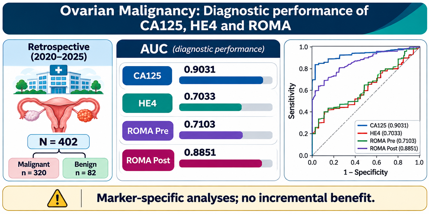
Results: Among 402 records, 320 were coded malignant and 82 benign. After parsing comma-grouped numeric values and values annotated “Repeated” as the recorded numeric result, marker availability was CA125 n=402, HE4 n=192, and ROMA Pre and Post n=190 each. Recomputed AUCs were 0.9031 for CA125, 0.7033 for HE4, 0.7103 for ROMA Pre, and 0.8851 for ROMA Post. These estimates are marker-specific and do not establish incremental diagnostic value in a paired cohort.
Conclusions: CA125 and recorded ROMA Post showed relatively high apparent discrimination in marker-specific analyses. Results are descriptive because the available marker cohorts differ, ROMA Pre/Post definitions are not independently validated, and subgroup stage coding requires adjudication. These findings do not demonstrate incremental diagnostic benefit.
Keywords:
CA125
; HE4
; ROMA
; ovarian malignancy
; diagnostic accuracy
; histopathology
Introduction
Preoperative evaluation of an adnexal mass requires differentiation of malignant from benign disease while limiting unnecessary referral and surgery. International consensus places expert ultrasound and structured risk assessment at the centre of triage [1]. The Ovarian-Adnexal Reporting and Data System (O-RADS) ultrasound system standardises lesion description and risk stratification [2,3]. Comparative analyses of O-RADS and International Ovarian Tumor Analysis (IOTA)-derived classification also emphasise the importance of the ultrasound framework [4].
Cancer Antigen 125 (CA125) remains an established circulating marker, while human epididymis protein 4 (HE4), the risk of ovarian malignancy algorithm (ROMA) and the risk of malignancy index (RMI) have been evaluated as complementary tests [5]. Their performance is not uniform across settings: a recent head-to-head comparison found differing trade-offs between sensitivity and specificity for biochemical markers and ultrasound-based assessment of different neoplasias in the adnexa (ADNEX) [6]. Other preoperative comparisons have similarly examined HE4, CA125, ROMA and the Copenhagen index [7], whereas evidence syntheses have evaluated models combining clinical, sonographic and biochemical information [8].
Thai clinical research has compared CA125, HE4, ROMA and RMI in women with ovarian lesions [9]. A separate Thai study evaluated HE4 and CA125 in relation to optimal primary surgery rather than the same diagnostic endpoint used here [10]. Prospective multicentre evidence indicates that performance depends on the clinical spectrum of women being evaluated [11]. Other studies comparing biochemical algorithms with clinical assessment have also reported context-dependent accuracy [12].
Diagnostic investigations have evaluated CA125, HE4, ROMA and CPH-I in primary ovarian cancer [13]. A 2026 diagnostic-accuracy study extended this comparison to preoperative ovarian tumour differentiation [14]. More recent work has examined whether serum markers complement O-RADS ultrasound [15]. A prospective Danish multicentre study reported limited incremental benefit from incorporating HE4 into the IOTA ADNEX model, with performance varying by clinical setting [16].
Beyond established tests, investigations have assessed HE4 and CA125 alongside matrix metalloproteinases [17], inflammatory indices [18] and different assay systems [19]. Multiplex biomarker panels may improve performance in particular study populations, but are not directly interchangeable with a two-marker clinical comparison [20]. In postmenopausal adnexal masses, HE4 performance has also been evaluated prospectively [21].
Non-malignant factors may influence marker concentrations: population-based analyses have linked HE4 to age and smoking, and have identified other determinants of CA125 [22]. HE4 has also been studied specifically in ovarian disease with elevated CA125 [23]. Distinguishing borderline from invasive ovarian tumours can present a different diagnostic problem from the benign–malignant comparison [24].
Alternative specimen matrices, including urine, have been investigated for CA125 and HE4 [25]. A pilot clinical report has considered measuring HE4 alongside CA125 [26]. Interpretation of CA125 in endometriomas highlights the relevance of benign-disease case mix [27]. These distinctions matter when extrapolating findings from a cancer-referral hospital to a broader adnexal-mass population.
External evaluations of O-RADS show that performance also depends on operators and implementation [28]. Interobserver studies of IOTA and O-RADS terminology illustrate the need for reproducible ultrasound descriptors [29]. Updated IOTA consensus terminology provides a contemporary basis for standardised sonographic assessment [30].
This study describes the diagnostic performance of CA125, HE4 and recorded ROMA scores in a histopathology-classified 402-record hospital cohort, and explores clinical subgroup results reported in the source statistical analysis. It does not establish the incremental value of HE4 beyond CA125 using a validated paired prediction model.
Materials and Methods
Study Design and Source Population
This retrospective study included 402 clinical records dated 2020–2025 (17 in 2020 and 385 in 2021–2025). Ethics Committee approval was issued under EC 013/2026 on 23 June 2026 for the ovarian biomarker research protocol. The approved protocol should be checked to confirm that its authorised retrospective data period includes all records analysed here. No records from 2026 were included.
Reference Standard, Index Tests and Data Completeness
The binary reference standard was the investigator-confirmed final histopathological classification (320 malignant and 82 benign). CA125, HE4, ROMA Pre and ROMA Post were extracted from the workbook. The labels ROMA Pre and ROMA Post are retained as recorded because their exact operational definitions have not been independently verified against assay documentation. RMI was not available as a numeric field.
Statistical Interpretation
Diagnostic accuracy was assessed against the recorded binary histopathology classification. Numeric laboratory fields were parsed by removing thousands separators and the text annotation “Repeated” while retaining the preceding measured value. Marker-specific AUCs were recomputed from the linked workbook. Sensitivity and specificity at the cut-offs shown in Table 2 were cross-checked with the corresponding marker-specific subsets. The 95% confidence intervals and subgroup analyses shown are those supplied in the statistical report and have not all been independently recalculated. No formal paired AUC comparison or incremental-value model was established.
Results
Cohort Composition and Completeness
The cohort comprised 402 records, with 320 coded malignant and 82 benign. After removing numeric grouping commas and the annotation “Repeated” without excluding those observations, CA125 was available for 402 records (320 malignant, 82 benign), HE4 for 192 (148 malignant, 44 benign), and recorded ROMA Pre and Post for 190 each (148 malignant, 42 benign). The RMI field was empty. Different marker-specific denominators preclude direct unpaired AUC comparisons.
Source-Reported Diagnostic Accuracy
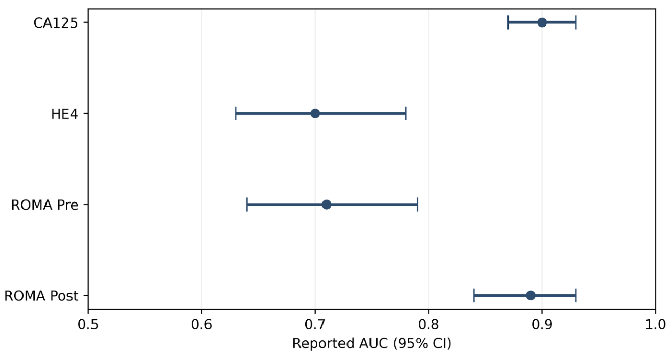
In marker-specific analyses, CA125 had an AUC of 0.9031 (n=402), recorded ROMA Post 0.8851 (n=190), ROMA Pre 0.7103 (n=190), and HE4 0.7033 (n=192). The source report rounded these to 0.90, 0.89, 0.71 and 0.70, respectively (Table 2). Figure 2 shows source-reported rounded AUCs and confidence intervals, whereas Figure 1 presents common complete-case ROC curves (n=165); these figures represent different analysis sets and must not be interpreted as paired comparisons of the entire cohort.
Figure 1.
Paired Receiver Operating Characteristic Curves for CA125, HE4 and Recorded ROMA Indices. Patient-level ROC curves reconstructed from the complete-case subset (n=165).
Figure 1.
Paired Receiver Operating Characteristic Curves for CA125, HE4 and Recorded ROMA Indices. Patient-level ROC curves reconstructed from the complete-case subset (n=165).
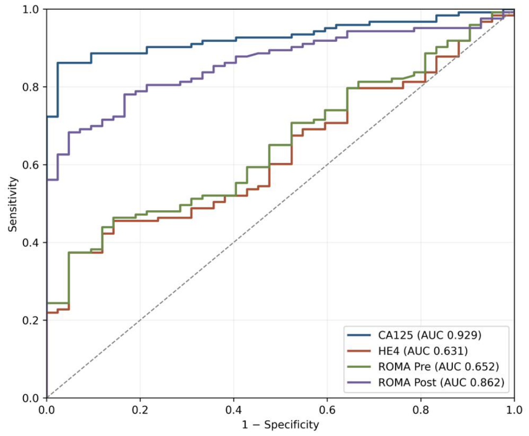
Figure 2.
Source-Reported Diagnostic Performance of CA125, HE4 and ROMA Indices. Points indicate source-reported AUCs and error bars indicate the corresponding reported 95% confidence intervals. Different marker-specific denominators limit direct comparison; HE4 numeric values were reconciled after parsing repeated-result annotations.
Figure 2.
Source-Reported Diagnostic Performance of CA125, HE4 and ROMA Indices. Points indicate source-reported AUCs and error bars indicate the corresponding reported 95% confidence intervals. Different marker-specific denominators limit direct comparison; HE4 numeric values were reconciled after parsing repeated-result annotations.
Risk-Group and Stage Analyses
The source report divided records into general (n=143) and high-risk (n=49) groups, but these 192 observations do not represent the full 402-record cohort, and their selection criteria were not specified. It reported CA125 AUCs of 0.90 and 0.91 in general and high-risk groups, respectively, and HE4 AUCs of 0.70 and 0.64. These comparisons are exploratory and imprecise, particularly for specificity in the high-risk subgroup. Selected subgroup estimates are summarised in Table 3.
The source report also presented early-stage (I–II; n=68) and advanced-stage (III–IV; n=168) analyses. CA125 AUC was reported as 0.60 in early disease and 1.00 in advanced disease, with HE4 AUCs of 0.40 and 0.76. However, the linked workbook contains inconsistencies between FIGO stage and stage-group labels; the extremely high advanced-stage CA125 AUC must not be treated as independently verified.
Exploratory Histopathology and Hereditary-Risk Comparisons
The source-reported CA125 median was 19.20 in benign records and 243.85 in malignant records (p<0.001 as reported). HE4 medians were 69.90 and 144.15, respectively (reported p<0.001). The report described a BRCA-associated difference in ROMA Post (p=0.0464), but other BRCA/HBOC marker comparisons were not clearly significant. These findings were not adjusted for multiplicity or confounders and were based on potentially inconsistent subgroup counts. The provenance of BRCA and HBOC classifications remains unverified. Group medians are summarised in Table 4.

Table 1.
Cohort and biomarker data completeness.
| Variable | Information Records | Notes |
|---|---|---|
| Total cohort | 402 | 320 malignant; 82 benign |
| 2020 records | 17 | Outside the designated five-year window |
| CA125 | 402 | 320 malignant; 82 benign; numeric values including annotated repeats |
| HE4 | 192 | 148 malignant; 44 benign; numeric values including annotated repeats |
| ROMA Pre | 190 | 148 malignant; 42 benign |
| ROMA Post | 190 | 148 malignant; 42 benign |
Note: Numeric results were parsed after removal of thousands separators and the annotation “Repeated” (the numeric prefix was retained). CA125 n=402, HE4 n=192, and ROMA Pre/Post n=190 each; the RMI field was blank. The pathology status was taken from the workbook coding, not independently verified against signed pathology reports.
Table 2.
Source-reported diagnostic performance in the 402-record analysis.
| Marker | Cut-off | Sensitivity % (95% CI) |
Specificity % (95% CI) |
AUC (95% CI) |
|---|---|---|---|---|
| CA125 | 58.88 | 79.69 (74.86–83.96) | 97.56 (91.47–99.70) | 0.90 (0.87–0.93) |
| HE4 | 123 | 54.73 (46.35–62.92) | 86.36 (72.65–94.83) | 0.70 (0.63–0.78) |
| ROMA Pre | 43.37 | 55.41 (47.02–63.57) | 85.71 (71.46–94.57) | 0.71 (0.64–0.79) |
| ROMA Post | 46.81 | 73.65 (65.78–80.54) | 95.24 (83.84–99.42) | 0.89 (0.84–0.93) |
Note: Transcribed from Result Record, Table 1. HE4 specificity corrected to 86.36% (38/44 benign records); assay/ROMA definitions and subgroup analyses still require confirmation. Cut-offs are not clinical recommendations.
Table 3.
Selected source-reported subgroup AUCs.
| Marker | General (n=143) |
High-risk (n=49) |
Early stage (n=68) |
Advanced stage (n=168) |
|---|---|---|---|---|
| CA125 | 0.90 | 0.91 | 0.60 | 1.00 |
| HE4 | 0.70 | 0.64 | 0.40 | 0.76 |
| ROMA Pre | 0.71 | 0.65 | 0.39 | 0.77 |
| ROMA Post | 0.88 | 0.88 | 0.51 | 0.96 |
Table 4.
Selected source-reported biomarker medians.
| Biomarker | Benign median (IQR) | Malignant median (IQR) | Reported p-value |
|---|---|---|---|
| CA125 | 19.20 (22.45) | 243.85 (751.25) | <0.001 |
| HE4 | 69.90 (54.10) | 144.15 (540.86) | <0.001 |
| ROMA Pre | 16.91 (26.24) | 53.56 (82.65) | <0.001 |
| ROMA Post | 20.14 (22.82) | 75.47 (52.92) | <0.001 |
Note: Data are presented as median (interquartile range, IQR). Between-group comparisons were assessed using the two-sided Mann–Whitney U test. Statistical significance was defined as p<0.05.
Discussion
This regional cancer-hospital study describes biomarker performance in a clinically selected population with a high proportion of malignancies. The source-reported AUCs suggest that substantial discrimination for CA125 and recorded ROMA Post, but they do not establish superiority or incremental value without matched-patient analyses and validated ROMA coding. Recent comparative evidence highlights the role of ultrasound-based assessment alongside serum markers [6,16].
After parsing values with grouping commas and the annotation “Repeated”, all 192 nonblank HE4 records were numeric. At the HE4 cut-off of 123, sensitivity was 54.73% (81/148) and specificity was 86.36% (38/44), correcting the 46.36% transcription error in the supplied statistical report. CA125 had 402 numeric results and ROMA Pre/Post each had 190. The available marker-specific subsets differ and are not directly interchangeable.
The apparent high performance of CA125 in advanced-stage disease may reflect a spectrum effect; the reported AUC of 1.00 is particularly vulnerable to coding or selection artefacts. Stage labels in the workbook require adjudication before stage-stratified claims. Hereditary-risk findings remain exploratory because BRCA testing methods, germline versus somatic status and the origin of HBOC coding are not established. Observational biomarker associations do not imply independent predictive effects.
Limitations include retrospective referral selection, differing marker-specific availability, uncertain operational definitions and timing of recorded ROMA Pre/Post, unresolved stage-group coding, and lack of external validation. Some confidence intervals and exploratory subgroup analyses are retained from the source report rather than independently reconstructed. The cohort is not representative of unselected patients with adnexal masses; these estimates should not be used alone to recommend diagnostic thresholds or treatment pathways.
Recent Thai prospective evidence provides a particularly relevant clinical comparator. In 110 women with adnexal masses, Tangjanyatham and Chaowawanit reported higher sensitivity for ADNEX than ROMA at the study-specific thresholds (91.9% versus 64.8%), but lower specificity (65.7% versus 86.3%). These findings illustrate a clinically important sensitivity–specificity trade-off; they cannot be used to infer the performance of ultrasound models in the present cohort, which lacks a validated linked imaging assessment. [36]
A multicentre external validation comparing ADNEX with ROMA in surgically treated patients further supports assessing diagnostic algorithms in their intended clinical populations rather than assuming that risk scores are interchangeable. This evidence also underscores why the present unverified ROMA Pre and ROMA Post fields should not be interpreted as validated menopausal-status-specific algorithms. [37]
A 2024 systematic review and meta-analysis of external validations of ADNEX highlights the need to consider between-study heterogeneity and transportability when interpreting risk prediction performance. Accordingly, the high malignancy proportion and incomplete marker availability in this cancer-referral cohort limit generalisation of the source-reported AUCs to unselected adnexal-mass populations. [38]
More recent prospective Danish multicentre evidence from general gynaecological settings, where malignancy prevalence was lower, found that ADNEX discriminated malignancy better than RMI or CA125 alone. This difference in clinical spectrum reinforces the importance of setting-specific validation and does not establish the comparative effectiveness of those models in our dataset. [39]
Conclusion
In this regional Thai cancer-hospital cohort, CA125 and recorded ROMA Post demonstrated relatively high apparent discrimination against recorded histopathology classification. HE4 specificity at the source cut-off was corrected to 86.36% after reconciling repeated-value annotations. Differences in marker availability and unresolved ROMA and stage definitions limit comparative inference. Independent clinical validation is required before applying these findings to triage decisions.
Author Contributions
CRediTs: Conceptualization, J.K. and S.W.; methodology and statistical analysis, J.K., T.P., K.S., and S.W.; investigation, J.K., S.W.; data curation, K.S.; writing—original draft preparation, J.K.; writing—review and editing, all authors; supervision, S.W. All authors have read and agreed to the published version of the manuscript. J.K.: Jiraon Karunpan, B.Sc. (MT), Clinical Pathology and Medical Technology Section, Ubon Ratchathani Cancer Hospital, Thailand, Email: mthcujiraon@gmail.com; K.S.: Krittaya Somakasetrin, MD, Gynecologic Oncology Division, Ubon Ratchathani Cancer Hospital, Thailand, Email: Noonoy04@gmail.com; T.P.: Thanita Parina, B.Sc., Research and Technology Assessment Section, Ubon Ratchathani Cancer Hospital, Thailand, Email: research.ubcc@gmail.com; S.W.: Surasak Wanram, Ph.D., Clinical Pathology, College of Medicine and Public Health, Ubon Ratchathani University, Ubon Ratchathani, Thailand, Email: Surasak.w@ubu.ac.th, ORCID iD: https://orcid.org/0000-0002-1866-0723.
Funding
This research received no specific funding.
Institutional Review Board Statement
This study was approved by the Ethics Committee of Ubon Ratchathani Cancer Hospital, Thailand (Protocol No. EC 013/2026; approval date: 23 June 2026).
Informed Consent Statement
The requirement for informed consent was waived by the Ethics Committee of Ubon Ratchathani Cancer Hospital, Thailand, in accordance with the approved retrospective study protocol.
Data Availability Statement
De-identified patient-level data and applicable assay documentation may be made available from the corresponding author subject to ethics and institutional requirements.
Acknowledgments
The authors would like to express their sincere gratitude to the Cancer Registry Unit and all healthcare professionals and staff of Ubon Ratchathani Cancer Hospital, Thailand, for their valuable assistance in data collection, medical record retrieval, and coordination throughout this study. Their support and cooperation were essential to the successful completion of this research.
Conflicts of Interest
All authors declare no conflicts of interest.
Declaration of AI-Assisted Tools: During the preparation of this manuscript, the authors used SciSpace for literature discovery and retrieval of relevant peer-reviewed publications and ChatGPT (OpenAI, accessed November 2026) for language editing, manuscript organisation, citation quality control, and technical formatting. All source verification, analyses, clinical interpretations and final submission decisions remain the responsibility of the authors.
Abbreviations
ADNEX = Assessment of Different Neoplasias in the adneXa
AUC = Area Under the Curve
BRCA1/BRCA2 = Breast Cancer Susceptibility Genes 1 and 2
CA-125 = Cancer Antigen 125
CSV = Comma-Separated Values
FIGO = International Federation of Gynecology and Obstetrics
HE4 = Human Epididymis Protein 4
HBOC = Hereditary Breast and Ovarian Cancer
HIS = Hospital Information System
IOTA = International Ovarian Tumor Analysis
LIS = Laboratory Information System
NLP = Natural Language Processing
O-RADS = Ovarian-Adnexal Reporting and Data System
RMI = Risk of Malignancy Index
ROC = Receiver Operating Characteristic
ROMA = Risk of Ovarian Malignancy Algorithm
US = Ultrasound
References
- Timmerman, D.; Planchamp, F.; Bourne, T.; et al. ESGO/ISUOG/IOTA/ESGE consensus statement on pre-operative diagnosis of ovarian tumors. Int. J. Gynecol. Cancer 2021, 31, 961–982. [Google Scholar] [CrossRef] [PubMed]
- Strachowski, L.M.; Jha, P.; Phillips, C.H.; et al. O-RADS US v2022: an update from the American College of Radiology Ovarian-Adnexal Reporting and Data System US Committee. Radiology 2023, 308, e230685. [Google Scholar] [CrossRef] [PubMed]
- Andreotti, R.F.; Timmerman, D.; Strachowski, L.M.; et al. O-RADS US risk stratification and management system: a consensus guideline from the ACR Ovarian-Adnexal Reporting and Data System Committee. Radiology 2020, 294, 168–185. [Google Scholar] [CrossRef] [PubMed]
- Vara, J.; Pagliuca, M.; Springer, S.; et al. O-RADS classification for ultrasound assessment of adnexal masses: agreement between IOTA lexicon and ADNEX model for assigning risk group. Diagnostics 2023, 13, 673. [Google Scholar] [CrossRef] [PubMed]
- Dochez, V.; Caillon, H.; Vaucel, E.; et al. Biomarkers and algorithms for diagnosis of ovarian cancer: CA125, HE4, RMI and ROMA, a review. J. Ovarian Res. 2019, 12, 28. [Google Scholar] [CrossRef] [PubMed]
- Spagnol, G.; Marchetti, M.; Carollo, M.; et al. Clinical utility and diagnostic accuracy of ROMA, RMI, ADNEX, HE4, and CA125 in the prediction of malignancy in adnexal masses. Cancers 2024, 16, 3790. [Google Scholar] [CrossRef] [PubMed]
- Carreras-Dieguez, N.; Glickman, A.; Munmany, M.; et al. Comparison of HE4, CA125, ROMA and CPH-I for preoperative assessment of adnexal tumors. Diagnostics 2022, 12, 226. [Google Scholar] [CrossRef] [PubMed]
- Davenport, C.F.; Rai, N.; Sharma, P.; et al. Diagnostic models combining clinical information, ultrasound and biochemical markers for ovarian cancer: Cochrane systematic review and meta-analysis. Cancers 2022, 14, 3621. [Google Scholar] [CrossRef] [PubMed]
- Oranratanaphan, S.; Wanishpongpan, S.; Termrungruanglert, W.; Triratanachat, S. Assessment of diagnostic values among CA-125, RMI, HE4, and ROMA for cancer prediction in women with nonfunctional ovarian cysts. Obstet. Gynecol. Int. 2018, 2018, 7821574. [Google Scholar] [CrossRef] [PubMed]
- Promchit, K.; Oranratanaphan, S. Human epididymis protein 4 (HE4) and cancer antigen 125 (CA125) for prediction of optimal primary surgery in non-mucinous epithelial ovarian cancer. Asian Pac. J. Cancer Prev. 2024, 25, 281–286. [Google Scholar] [CrossRef] [PubMed]
- Dochez, V.; Randet, M.; Renaudeau, C.; et al. Efficacy of HE4, CA125, risk of malignancy index and risk of ovarian malignancy index to detect ovarian cancer in women with presumed benign ovarian tumours: a prospective, multicentre trial. J. Clin. Med. 2019, 8, 1784. [Google Scholar] [CrossRef] [PubMed]
- Janas, L.; Stachowiak, G.; Glowacka, E.; et al. The use of CA125, human epididymis protein 4 (HE4), risk of ovarian malignancy algorithm (ROMA), risk of malignancy index (RMI) and subjective assessment (SA) in preoperative diagnosing of ovarian tumors. Ginekol. Pol. 2024, 95, 321–327. [Google Scholar] [CrossRef] [PubMed]
- Luo, H.J.; Hu, Z.D.; Cui, M.; et al. Diagnostic performance of CA125, HE4, ROMA, and CPH-I in identifying primary ovarian cancer. J. Obstet. Gynaecol. Res. 2023, 49, 998–1006. [Google Scholar] [CrossRef] [PubMed]
- Feng, W.; Zhang, C.; Meng, L.; et al. Application of serum CA125, HE4, ROMA, and CPH-I in the preoperative differentiation of ovarian tumors: a diagnostic accuracy study. J. Clin. Lab Anal. 2026, 40, e70319. [Google Scholar] [CrossRef] [PubMed]
- Gong, J.; Li, K.; Liu, Y.; Ren, M. Comparing and combining O-RADS ultrasound, serum CA125, and HE4 for the preoperative diagnosis of ovarian tumors. BMC Med. Imaging 2026, 26, 513. [Google Scholar] [CrossRef] [PubMed]
- Karlsen, N.S.; Høgdall, C.K.; Dreisler, E.; et al. Adding HE4 to the IOTA ADNEX model for differentiating adnexal masses: a Danish prospective, multicenter study. Gynecol. Oncol. 2026, 209, 111–119. [Google Scholar] [CrossRef] [PubMed]
- Kicman, A.; Gacuta, E.; Kulesza, M.; et al. Diagnostic utility of selected matrix metalloproteinases (MMP-2, MMP-3, MMP-11, MMP-26), HE4, CA125 and ROMA algorithm in diagnosis of ovarian cancer. Int. J. Mol. Sci. 2024, 25, 6265. [Google Scholar] [CrossRef] [PubMed]
- Song, L.; Qi, J.; Zhao, J.; et al. Diagnostic value of CA125, HE4, and systemic immune-inflammation index in the preoperative investigation of ovarian masses. Medicine 2023, 102, e35240. [Google Scholar] [CrossRef] [PubMed]
- Feng, P.; Chen, T.; Wischhusen, J.; et al. The diagnostic performance of the Mindray system in detecting CA125 and HE4 for patients with ovarian cancer. Transl. Cancer Res. 2024, 13, 4474–4484. [Google Scholar] [CrossRef] [PubMed]
- Leandersson, P.; Åkesson, A.; Hedenfalk, I.; Malander, S.; Borgfeldt, C. A multiplex biomarker assay improves the diagnostic performance of HE4 and CA125 in ovarian tumor patients. PLoS ONE 2020, 15, e0240418. [Google Scholar] [CrossRef] [PubMed]
- Gentry-Maharaj, A.; Burnell, M.; Dilley, J.; et al. Serum HE4 and diagnosis of ovarian cancer in postmenopausal women with adnexal masses. Am. J. Obstet. Gynecol. 2020, 222, 56.e1–56.e17. [Google Scholar] [CrossRef] [PubMed]
- Cramer, D.W.; Vitonis, A.F.; Sasamoto, N.; Yamamoto, H.; Fichorova, R.N. Epidemiologic and biologic correlates of serum HE4 and CA125 in women from the National Health and Nutritional Survey (NHANES). Gynecol. Oncol. 2021, 161, 282–290. [Google Scholar] [CrossRef] [PubMed]
- Li, W.; Wang, D. The differential diagnostic value and clinical significance of serum HE4 in ovarian disease with elevated CA125. Arch. Gynecol. Obstet. 2020, 301, 1219–1225. [Google Scholar] [CrossRef] [PubMed]
- Wang, Z.; Tao, X.; Ying, C. CPH-I and HE4 are more favorable than CA125 in differentiating borderline ovarian tumors from epithelial ovarian cancer at early stages. Dis. Markers 2019, 2019, 6241743. [Google Scholar] [CrossRef] [PubMed]
- Barr, C.E.; Njoku, K.; Owens, G.L.; Crosbie, E.J. Urine CA125 and HE4 for the detection of ovarian cancer in symptomatic women. Cancers 2023, 15, 1256. [Google Scholar] [CrossRef] [PubMed]
- Crawford, S.M.; Evans, C.; Shaw, A.; Barr, C.E.; Crosbie, E.J. Measuring HE4 alongside CA125 for ovarian cancer diagnosis: a pilot clinical study. BJOG 2024, 131, 1725–1726. [Google Scholar] [CrossRef] [PubMed]
- He, H.; Yang, X.; Li, Y.; Zhang, Y.; Ye, Q. CA125 outperforms HE4 and CA19-9 as a serum biomarker for preoperative diagnosis of ovarian endometriomas: a retrospective cohort study. Reprod. Sci. 2026, 33, 1149–1155. [Google Scholar] [CrossRef] [PubMed]
- Buranaworathitikul, P.; Wisanumahimachai, V.; Phoblap, N.; et al. Accuracy of O-RADS system in differentiating between benign and malignant adnexal masses assessed via external validation by inexperienced gynecologists. Cancers 2024, 16, 3820. [Google Scholar] [CrossRef] [PubMed]
- Antil, N.; Raghu, P.R.; Shen, L.; et al. Interobserver agreement between eight observers using IOTA simple rules and O-RADS lexicon descriptors for adnexal masses. Abdom. Radiol. (NY) 2022, 47, 3318–3326. [Google Scholar] [CrossRef] [PubMed]
- Timmerman, D.; Valentin, L.; Testa, A.C.; et al. Terms, definitions and measurements to describe the sonographic features of adnexal tumors: updated consensus opinion from the International Ovarian Tumor Analysis (IOTA) Group. Ultrasound Obstet. Gynecol. 2026, 67, 702–724. [Google Scholar] [CrossRef] [PubMed]
- Collins, G.S.; Moons, K.G.M.; Dhiman, P.; et al. TRIPOD+AI statement: updated guidance for reporting clinical prediction models that use regression or machine learning methods. BMJ 2024, 385, e078378. [Google Scholar] [CrossRef] [PubMed]
- Van Calster, B.; McLernon, D.J.; van Smeden, M.; et al. Calibration: the Achilles heel of predictive analytics. BMC Med. 2019, 17, 230. [Google Scholar] [CrossRef] [PubMed]
- Riley, R.D.; Archer, L.; Snell, K.I.E.; et al. Evaluation of clinical prediction models (part 2): how to undertake an external validation study. BMJ 2024, 384, e074820. [Google Scholar] [CrossRef] [PubMed]
- Sperrin, M.; Riley, R.D.; Collins, G.S.; Martin, G.P. Targeted validation: validating clinical prediction models in their intended population and setting. Diagn. Progn. Res. 2022, 6, 24. [Google Scholar] [CrossRef] [PubMed]
- Mansmann, U.; Ön, B.I. The validation of prediction models deserves more recognition. BMC Med. 2025, 23, 166. [Google Scholar] [CrossRef] [PubMed]
- Tangjanyatham, P.; Chaowawanit, W. Comparison of sensitivity for Risk of Ovarian Malignancy Algorithm (ROMA) and Assessment of Different NEoplasias in the adneXa (ADNEX) model for predicting ovarian cancer in a woman with adnexal masses. Int. J. Gynecol. Cancer 2025, 35(6), 101827. [Google Scholar] [CrossRef] [PubMed]
- Landolfo, C.; Ceusters, J.; Valentin, L.; et al. Comparison of the ADNEX and ROMA risk prediction models for the diagnosis of ovarian cancer: a multicentre external validation in patients who underwent surgery. Br. J. Cancer 2024, 130(6), 934–940. [Google Scholar] [CrossRef] [PubMed]
- Barreñada, L.; Ledger, A.; Dhiman, P.; et al. ADNEX risk prediction model for diagnosis of ovarian cancer: systematic review and meta-analysis of external validation studies. BMJ Med. 2024, 3(1), e000817. [Google Scholar] [CrossRef] [PubMed]
- Dueholm Hjorth, I.M.; Bech, B.H.; Ledger, A.; et al. Prospective external validation of ADNEX and RMI for estimating risk of malignancy in adnexal masses in general gynecological settings: Danish multicenter study. Ultrasound Obstet. Gynecol. 2026, 68(2), 256–269. [Google Scholar] [CrossRef] [PubMed]
Disclaimer/Publisher’s Note: The statements, opinions and data contained in all publications are solely those of the individual author(s) and contributor(s) and not of MDPI and/or the editor(s). MDPI and/or the editor(s) disclaim responsibility for any injury to people or property resulting from any ideas, methods, instructions or products referred to in the content. |
© 2026 by the authors. Licensee MDPI, Basel, Switzerland. This article is an open access article distributed under the terms and conditions of the Creative Commons Attribution (CC BY) license (http://creativecommons.org/licenses/by/4.0/).
Copyright: This open access article is published under a Creative Commons CC BY 4.0 license, which permit the free download, distribution, and reuse, provided that the author and preprint are cited in any reuse.