

Submitted:
13 November 2026
Posted:
16 November 2026
You are already at the latest version
Abstract
Background: Alzheimer’s disease (AD) is a progressive neurodegenerative disorder in which amyloid-β, tau pathology, synaptic dysfunction, and neuroinflammation interact across molecular, cellular, and network levels. Although these processes are well established individually, their reciprocal and stage-dependent relationships remain insufficiently integrated into a unified mechanistic framework. Age-related decline in proteostatic, metabolic, vascular, and immune resilience may further lower the threshold at which these interacting pathways become self-sustaining. This review therefore examines how protein aggregation, neuronal stress, and glial responses converge to shape disease progression. Summary: Amyloid-β oligomerization can disrupt synaptic signaling and calcium homeostasis, creating conditions that promote tau-directed kinase activation, tau hyperphosphorylation, mislocalization, and propagation. Pathological tau further compromises microtubule stability, axonal transport, synaptic function, and neuronal resilience. In parallel, microglial and astrocytic responses influence amyloid clearance, inflammatory signaling, blood-brain barrier integrity, and neuronal injury through pathways that include TREM2 and the NLRP3 inflammasome. These interactions generate feedback loops in which impaired clearance, proteinopathy, synaptic stress, and chronic inflammation can reinforce one another, while their relative contribution may vary across disease stages and individuals.Conclusion: Here we highlight AD as a dynamic mechanistic network rather than a fixed single-pathway cascade. The proposed framework distinguishes disease initiation from subsequent pathway dominance and amplification. Integrating amyloid, tau, synaptic, and neuroimmune processes may improve interpretation of disease heterogeneity, support multimodal biomarker strategies, and inform stage-matched therapeutic combinations. This framework also identifies causal direction, pathway dominance, and temporal transitions as priorities for future mechanistic and clinical research.
Keywords:
Alzheimer’s Disease
; amyloid-β
; tau
; neuroinflammation
; microglia
; synaptic dysfunction
1. Introduction
Alzheimer’s disease (AD) is a progressive neurodegenerative disorder that imposes an increasing clinical and societal burden as populations age, with prevalence projected to rise substantially over coming decades (Safiri et al., 2024; Frisoni et al., 2025; Zhong et al., 2025). Beyond progressive cognitive impairment, AD reduces functional independence and quality of life while placing substantial demands on caregivers and healthcare systems (The Lancet Global Health, 2024; Alzheimer’s Dement., 2024). These consequences reinforce the need to understand not only the individual pathological hallmarks of AD but also the mechanisms through which they interact to drive progression.
Clinically, AD is characterized by progressive cognitive decline, particularly impairments in memory and executive function. These manifestations are associated with the pathological accumulation of amyloid-β plaques and neurofibrillary tangles, which contribute to synaptic dysfunction, neuronal loss, and widespread neurodegeneration (Serrano-Pozo et al., 2011). Beyond memory impairment, declining executive function, including planning and decision-making, progressively reduces patient autonomy and limits adaptation to functional and lifestyle changes imposed by disease progression (Guarino et al., 2019; Rostamian et al., 2022). Biomarker-based examinations, including cerebrospinal fluid analysis and positron emission tomography (PET), can detect amyloid and tau pathology, thereby linking clinical manifestations to their underlying biological mechanisms (Frisoni et al., 2025). These clinicopathological relationships identify major components of AD but do not, by themselves, explain how their relative contributions evolve over time, why one pathological process amplifies another, or why patients with apparently similar amyloid burdens may follow different biological and clinical trajectories.
Despite decades of intensive research, the biological mechanisms underlying the initiation and progression of AD remain incompletely understood (Hardy and Selkoe, 2002). The classical neuropathological hallmarks of AD include extracellular deposits of amyloid-β (Aβ) peptides and intracellular neurofibrillary tangles composed of hyperphosphorylated tau protein (Serrano-Pozo et al., 2011). Aβ aggregation, particularly the formation of soluble oligomers, disrupts neuronal ionic and calcium homeostasis, alters synaptic signaling, and impairs dendritic function. These disturbances can activate tau-directed kinases and promote abnormal tau phosphorylation (Fani et al., 2021; Muralidar et al., 2020). Consequently, hyperphosphorylated tau dissociates from microtubules, reducing their stability and compromising cytoskeletal integrity, axonal transport, and neuronal communication. Mislocalized tau then accumulates intracellularly and forms neurofibrillary tangles, further intensifying synaptic dysfunction and neuronal injury (Muralidar et al., 2020). In parallel, persistent activation of microglia and astrocytes sustains the release of proinflammatory mediators, creating a chronic neuroinflammatory environment. This response interacts bidirectionally with Aβ and tau pathologies, establishing a self-perpetuating cycle that accelerates synaptic loss, neuronal death, and cognitive decline throughout disease progression (Leng and Edison, 2021; Roda et al., 2022).
Ageing is not merely the epidemiological context of AD but a biological modifier that progressively reduces neuronal and glial resilience to proteotoxic stress (López-Otín et al., 2023). Age-related deterioration of proteostatic systems, including autophagic and lysosomal clearance, may favor the persistence of misfolded Aβ and tau species, while mitochondrial dysfunction and oxidative stress can reduce the capacity of neurons to tolerate synaptic and metabolic demands (Nixon and Rubinsztein, 2024; D’Alessandro et al., 2025). In parallel, age-associated changes in innate and adaptive immunity may shift glial responses toward persistent inflammatory signaling, whereas vascular and blood-brain barrier dysfunction can further compromise metabolic support and clearance of pathological proteins (Franceschi et al., 2018; Cunha et al., 2024; Jang et al., 2026). These alterations do not constitute an independent disease pathway; rather, they may lower the threshold at which initially compensatory responses become maladaptive and allow amyloid, tau, synaptic, and neuroimmune mechanisms to enter self-reinforcing feedback states (López-Otín et al., 2023).
Within this age-related decline in biological resilience, glial responses provide a particularly important interface between proteinopathy and neuronal dysfunction. Microglia and astrocytes initially participate in homeostatic functions including aggregate clearance, synaptic maintenance, neurotransmitter regulation, and metabolic support, but persistent exposure to Aβ, pathological tau, and neuronal injury can shift these responses toward sustained inflammatory signaling (Heneka et al., 2015; Leng and Edison, 2021). Cytokines, complement signaling, reactive oxygen species, altered glutamate handling, and impaired ionic homeostasis can then intensify synaptic stress and neuronal injury, while accumulating protein aggregates and damaged neurons further stimulate glial activation (Hong et al., 2016; Machado and Carvalho, 2020; Ge et al., 2022). The important unresolved issue is therefore not whether amyloid, tau, and inflammation coexist, but how their reciprocal interactions change from compensatory to self-reinforcing states and how these transitions contribute to heterogeneous disease trajectories.
Accordingly, this integrated mechanistic review examines AD as an age- and stage-dependent dynamic network in which amyloid pathology, tau dysfunction, synaptic injury, and neuroinflammation assume different initiating, permissive, amplifying, and propagating roles across disease progression. Particular attention is given to mechanistic bridges linking Aβ oligomerization with calcium dysregulation and tau-directed kinase activity; tau mislocalization and propagation with synaptic and glial dysfunction; and TREM2-, NLRP3-, and astrocyte-associated responses with protein clearance, inflammatory amplification, and neuronal injury. By distinguishing pathway interaction from simple pathological coexistence, this review aims to explain how self-reinforcing loops and stage-dependent pathway dominance may contribute to biological heterogeneity and potentially inform biomarker-guided therapeutic strategies.
This mechanistic narrative review was informed by literature identified primarily through PubMed, supplemented by peer-reviewed journal and publisher platforms. Searches emphasized studies published between 2000 and 2026 and were last updated on September 2026 while earlier landmark articles were retained when essential to established concepts in AD pathogenesis. Search terms combined “Alzheimer disease” with “amyloid-β,” “tau,” “synaptic dysfunction,” “prion-like propagation,” “microglia,” “astrocytes,” “neuroinflammation,” “TREM2,” and “NLRP3 inflammasome.” Primary mechanistic studies, longitudinal human investigations, biomarker studies, and authoritative reviews were selected according to relevance, methodological quality, and contribution to an integrated mechanistic framework. Additional sources were identified by screening the reference lists of included publications. The review was narrative rather than systematic and was designed to synthesize mechanistic relationships across molecular, cellular, and clinical evidence.
2. Amyloid-β Pathology and Early Molecular Events
2.1. Amyloid Precursor Protein (APP) Processing and Generation of Aβ Peptides
The molecular basis of the amyloid hypothesis can be traced to 1984, when George Glenner and Caine Wong isolated and characterized amyloid-β (Aβ) from cerebral vascular amyloid deposits of affected individuals (Paroni et al., 2019). Notably, different Aβ isoforms—particularly Aβ40 and Aβ42—play distinct roles in this process, with Aβ42 showing a greater propensity for aggregation and neurotoxicity and therefore being more closely associated with disease progression (Selkoe et al., 2001; Saido, 2013). Pathological conditions may arise when alterations in APP processing, particularly those associated with mutations in APP or presenilin genes, create an imbalance in Aβ production that favors the generation of Aβ42. The amyloidogenic pathway begins with cleavage of APP by β-site APP-cleaving enzyme 1 (BACE1), generating soluble APPβ (sAPPβ) and a membrane-bound C-terminal fragment (C99). Subsequently, C99 is cleaved by the γ-secretase complex, resulting in the extracellular release of Aβ peptides (Hur, 2022). Following their release, soluble Aβ peptides progressively aggregate into oligomeric assemblies, which organize into fibrillar structures and eventually form mature amyloid plaques. This process is schematically illustrated in Figure 1.
Genetic alterations affecting APP and presenilin-dependent processing can shift Aβ production toward aggregation-prone species. Genetic studies identified APP, PSEN1, and PSEN2 as key genes responsible for autosomal dominant AD (Goate et al., 1991; Sherrington et al., 1995; Levy-Lahad et al., 1995). Their functions converge within the amyloidogenic pathway rather than acting independently. APP encodes the transmembrane precursor from which Aβ peptides are generated through cleavage by BACE1 and γ-secretase. PSEN1 and PSEN2 encode alternative catalytic subunits of γ-secretase that determine cleavage of the APP-derived C99 fragment (De Strooper et al., 1998; Wolfe et al., 1999). Pathogenic APP mutations may increase Aβ production or favor formation of aggregation-prone peptides, whereas PSEN1 and PSEN2 mutations commonly alter γ-secretase cleavage and increase the proportion of longer Aβ species, particularly Aβ42. These effects disturb the balance among Aβ isoforms and promote accumulation and extracellular plaque formation (Bettens et al., 2013).
2.2. Amyloid Plaque Formation and Oligomerization
The pathogenic significance of Aβ depends not only on plaque burden but on the abundance, conformation, and cellular localization of soluble aggregation intermediates. Aβ40 is the predominant isoform, whereas Aβ42 contains two additional hydrophobic C-terminal residues that increase its propensity for β-sheet formation and self-association (Jarrett et al., 1993; Saido, 2013). This greater aggregation tendency favors the formation of oligomeric and fibrillar assemblies. Importantly, soluble Aβ oligomers are considered especially neurotoxic because they disrupt synaptic signaling and plasticity before extensive plaque deposition becomes evident (Walsh et al., 2002). Thus, differences in peptide composition influence downstream toxicity primarily by altering the probability and properties of aggregation-prone Aβ assemblies.
Among these assemblies, soluble Aβ oligomers are particularly relevant because their neurotoxicity can emerge before extensive fibrillar or plaque deposition. Aβ monomers self-associate into structurally heterogeneous oligomers that expose hydrophobic surfaces and adopt conformations distinct from monomers and mature fibrils (Kayed et al., 2003; Haass and Selkoe, 2007). These properties promote interactions with neuronal membranes, where oligomers can increase membrane permeability, disturb calcium homeostasis, and alter receptor-mediated signaling (Demuro et al., 2005). Consequently, oligomer toxicity is not determined solely by total Aβ deposition, but also by the size, conformation, and cellular localization of soluble assemblies. Experimental studies demonstrate that naturally secreted Aβ oligomers inhibit hippocampal long-term potentiation, while Aβ dimers isolated directly from AD brains impair synaptic structure, plasticity, and memory (Walsh et al., 2002; Shankar et al., 2008). Oligomers may therefore compromise neuronal communication before extensive plaque accumulation becomes evident. This early synaptotoxicity helps explain why soluble Aβ concentrations often correlate more closely with cognitive impairment than plaque burden (Haass and Selkoe, 2007) and provides a mechanistic transition from amyloid aggregation to the synaptic dysfunction examined in the following section.
Interactions between Aβ species and neuronal membranes can facilitate conformational conversion, nucleation, and further aggregate assembly. Following APP cleavage, the C99 fragment remains embedded within the lipid bilayer through its hydrophobic transmembrane domain (Bokvist et al., 2004). This environment places the Aβ sequence in close contact with membrane lipids and, after γ-secretase cleavage, can locally concentrate released peptides at the membrane surface. Electrostatic interactions with polar lipid headgroups and hydrophobic interactions with the bilayer promote conformational transitions from disordered or α-helical states toward aggregation-prone β-sheet–rich structures (Matsuzaki, 2007; Valappil et al., 2022). Membrane composition further influences this process: binding to GM1 ganglioside can generate seed-competent Aβ conformations, whereas cholesterol-containing membranes markedly accelerate Aβ42 primary nucleation (Kakio et al., 2002; Habchi et al., 2018). By restricting peptide diffusion and increasing local Aβ concentration, the neuronal membrane acts as a heterogeneous nucleation interface that lowers the energetic barrier for self-assembly. Formed nuclei then recruit additional soluble peptides, promoting oligomer growth, fibril elongation, and plaque maturation. Thus, neuronal membranes are active molecular surfaces that can shape the initiation and progression of Aβ aggregation. These membrane-dependent nucleation events therefore generate biologically active assemblies that can simultaneously provide substrates for further seeding and increase neuronal and glial exposure to toxic Aβ species, linking aggregate formation to subsequent synaptic and inflammatory responses.
2.3. Prion-like Propagation of Amyloid-β Aggregates
Prion-like Aβ propagation depends on both the formation of aggregation-competent seeds and the failure of mechanisms that normally restrict their persistence and dissemination. Nucleation refers to the rate-limiting organization of soluble Aβ monomers into a stable aggregate capable of sustaining progressive fibril growth (Jarrett and Lansbury, 1993; Zampa et al., 2024). Because formation of this nucleus is energetically unfavorable, unseeded aggregation is typically preceded by a characteristic lag phase. Once a seed is present, this barrier is reduced: soluble monomers bind to fibril ends and undergo templated conformational conversion, adopting the β-sheet–rich arrangement of the pre-existing aggregate. Repeated recruitment and conversion promote fibril elongation, while fibril surfaces can catalyze secondary nucleation and generate additional aggregation-competent species (Cohen et al., 2013). Experimental inoculation studies demonstrate that preformed Aβ assemblies can accelerate amyloid deposition in susceptible hosts, supporting the biological relevance of seeding in vivo (Meyer-Luehmann et al., 2006). This process is described as prion-like because it involves self-templating protein misfolding; however, unlike classical prion diseases, this terminology does not imply natural person-to-person infectivity (Jucker and Walker, 2013). Throughout this review, prion-like therefore refers specifically to the intracerebral conformational templating, amplification, and propagation of pathological protein assemblies within an individual, rather than to natural contagious transmission.
Failure of proteostatic clearance allows aggregation-competent Aβ species to persist and sustain further seeding. Under physiological conditions, the ubiquitin–proteasome system (UPS) contributes to intracellular protein quality control by degrading soluble misfolded proteins, including intracellular Aβ species (Zhao and Yang, 2010). Extracellular Aβ is removed by multiple Aβ-degrading proteases and other brain-clearance pathways (Saido and Leissring, 2012). Ageing may progressively weaken several components of this clearance network. Reduced autophagic-lysosomal efficiency and proteasomal capacity can limit intracellular removal of damaged or aggregation-prone proteins, while age-related deterioration of vascular and interstitial clearance may reduce elimination of extracellular Aβ species (López-Otín et al., 2023; Nixon and Rubinsztein, 2024). Together, these changes may prolong seed persistence and increase opportunities for templated aggregation and dissemination. However, once stable oligomeric nuclei and fibrillar aggregates form, their compact β-sheet–rich organization limits proteolytic accessibility and favors persistence (Zampa et al., 2024). Aβ assemblies can impair proteasome activity, creating a feedback loop in which defective clearance promotes further protein accumulation (Tseng et al., 2008). Human kinetic studies indicate that reduced Aβ clearance, rather than increased production alone, contributes substantially to late-onset AD (Mawuenyega et al., 2010). Persistent aggregates remain available as templates that recruit soluble peptides and continually generate new seeds. Their prolonged survival increases opportunities for release, uptake, and movement through interconnected neural pathways, thereby linking age-associated loss of proteostatic resilience with the progressive spatial dissemination of amyloid pathology (Jucker and Walker, 2013).
3. Synaptic Dysfunction Induced by Aβ Oligomers
3.1. Aβ-PrPc Interaction and Synaptic Signaling
Soluble Aβ oligomers may disrupt postsynaptic signaling through PrPᶜ-associated receptor complexes that converge on mGluR5 Fyn, and NMDAR regulation. One proposed route involves binding to cellular prion protein (PrPᶜ), which can participate in postsynaptic complexes containing mGluR5, Fyn, and NMDARs (Um and Strittmatter, 2013; Haas and Strittmatter, 2016). This interaction provides a mechanistic route through which extracellular Aβ stress may be translated into altered receptor signaling and postsynaptic calcium dysregulation.
Within this proposed signaling complex, mGluR5 can activate Fyn, which phosphorylates the GluN2B subunit of NMDARs and enhances receptor signaling (Um and Strittmatter, 2013; Haas and Strittmatter, 2016). Within this molecular platform, mGluR5 functions as the proximal activator of Fyn kinase, a Src-family tyrosine kinase that directly phosphorylates the NR2B subunit (GluN2B) of NMDARs (Um and Strittmatter, 2013; Haas and Strittmatter, 2016). This phosphorylation enhances NMDAR channel open probability and prolongs calcium influx, converting a transient physiological signal into a sustained excitotoxic stimulus that exceeds the postsynaptic calcium-buffering capacity (Um et al., 2013; Zampa et al., 2024). The resulting calcium overload drives synaptic dysfunction and contributes to the early cognitive deficits characteristic of AD (Haas and Strittmatter, 2016).
3.2. Calcium Dysregulation and Excitotoxicity
Aβ-associated disruption of astrocytic glutamate handling shifts physiological calcium signaling toward persistent excitatory stress. Under physiological conditions, calcium influx plays a tightly regulated role in neuronal depolarization, synaptic transmission, and postsynaptic plasticity, preserving neural network homeostasis (Burgoyne et al., 2019; Ge et al., 2022). Astrocytes maintain this balance by expressing high levels of the excitatory amino acid transporters EAAT-1 and EAAT-2, which are responsible for clearing the majority of glutamate from the synaptic cleft and thereby preventing excessive glutamatergic stimulation (Fani et al., 2021). Aβ oligomers compromise this critical clearance mechanism by reducing the expression and function of these transporters, leading to uncontrolled astrocytic glutamate release and extracellular glutamate accumulation (Fani et al., 2021). The resulting neurotoxic surplus drives sustained activation of postsynaptic ionotropic glutamate receptors, particularly NMDARs, thereby initiating persistent excitatory stress that underlies downstream synaptic pathology. This represents an early convergence point between neuronal and glial pathology: impaired astrocytic glutamate regulation increases neuronal excitatory stress, while calcium-dependent neuronal injury can subsequently generate signals that further recruit inflammatory responses.
Sustained NMDAR activation converts transient calcium signaling into excitotoxic calcium overload and progressive synaptic failure. The resulting pathological calcium influx shifts calcium from its role as a second messenger of plasticity to a driver of synaptic pathology (Fani et al., 2021; Ge et al., 2022). This calcium overload activates calpains and calcineurin, which contribute to AMPA receptor internalization, dendritic spine collapse, and impaired synaptic plasticity (Burgoyne et al., 2019; Ge et al., 2022). Excessive intracellular calcium also promotes mitochondrial dysfunction and reactive oxygen species generation, further compromising synaptic energy metabolism and structural integrity (Fani et al., 2021; Burgoyne et al., 2019). Collectively, these interconnected events erode the functional and structural basis of synaptic transmission, establishing a direct mechanistic link between early calcium dysregulation and the progressive cognitive decline observed in AD (Ge et al., 2022).
3.3. Neuronal Injury and Early Cognitive Impairment
Persistent calcium overload progressively converts functional synaptic abnormalities into structural neuronal injury. Excessive intracellular calcium disrupts neuronal energy metabolism by impairing mitochondrial respiration and ATP synthesis, reducing the metabolic capacity required for synaptic maintenance (Ge et al., 2022; Burgoyne et al., 2019). These metabolic deficits are compounded by calcium-dependent activation of calpains, which cleave cytoskeletal and scaffolding proteins, leading to dendritic degeneration, spine retraction, and progressive simplification of neuronal morphology (Ge et al., 2022). Sustained mitochondrial calcium accumulation also triggers the opening of the permeability transition pore, releasing pro-apoptotic factors that engage caspase cascades and ultimately drive neuronal apoptosis (Burgoyne et al., 2019; Fani et al., 2021). Together, these events can shift initially functional synaptic abnormalities toward increasingly persistent structural neuronal injury, linking early synaptic stress with later neurodegenerative change. Synaptic failure therefore represents an early and clinically relevant component of the pathological transition associated with cognitive deterioration in AD (Ge et al., 2022).
Synaptic failure provides a mechanistic link between early Aβ toxicity, cognitive deterioration, and downstream tau pathology. Sustained calcium overload activates calcineurin and calpain, which deregulate tau-associated kinase signaling through at least two complementary routes. Calcium/calmodulin-dependent protein kinase II (CaMKII) becomes hyperactive under persistent calcium stimulation, directly phosphorylating tau at pathologically relevant residues. In parallel, calpain-mediated cleavage of the CDK5 regulatory subunit p35 to p25 generates a proteolytically stable fragment that hyperactivates CDK5, a major tau kinase responsible for abnormal tau hyperphosphorylation (Muralidar et al., 2020; Ge et al., 2022). Hyperphosphorylated tau dissociates from microtubules, leading to microtubule destabilization, impaired axonal transport, and progressive cytoskeletal collapse (Muralidar et al., 2020; Burgoyne et al., 2019). This tau-dependent cascade amplifies synaptic injury and accelerates cognitive decline, providing a mechanistic bridge between early Aβ-driven calcium dysregulation and the later neurodegenerative pathology characteristic of AD.
4. Tau Pathology and Neurofibrillary Degeneration
4.1. Physiological Role of Tau in Neuronal Homeostasis
Tau is a multifunctional neuronal protein whose physiological roles extend beyond microtubule stabilization to synaptic, nuclear, and RNA-related functions. Encoded by the MAPT gene, this protein is enriched in neurons and contributes to axonal organization and intracellular transport through its interaction with microtubules (Hong et al., 2025; Muralidar et al., 2020). These microtubules are formed by α/β-tubulin polymers and provide structural support and transport tracks, while tau regulates their organization and stability through its microtubule-binding domains (Alberts et al., 2015; Kadavath et al., 2015; Barbier et al., 2019). Excessive phosphorylation reduces tau affinity for microtubules, impairing microtubule organization and axonal transport while increasing the soluble tau pool available for mislocalization, misfolding, and aggregation (Alonso et al., 1994; Barbier et al., 2019). These loss-of-function and toxic gain-of-function mechanisms make tau detachment a critical step in neurodegeneration. Tau domains and their association with neuronal microtubules are illustrated in Figure 2.
Although the organization of the tau–microtubule complex remains incompletely understood, growing evidence indicates that tau functions beyond the axonal cytoskeleton and occupies several neuronal compartments. In addition to its predominant axonal localization, tau has been identified in pre- and postsynaptic regions, the nucleus, mitochondria, and the extracellular space (Alonso et al., 2024). Under pathological conditions, tau becomes hyperphosphorylated and redistributes toward somatodendritic and synaptic compartments, where it may contribute to synaptic dysfunction and neuronal injury (Alonso et al., 2024; Watamura et al., 2025). Aging-related oxidative stress and mitochondrial dysfunction may further modify tau-associated molecular networks, increasing neuronal vulnerability and reinforcing degenerative signaling (Younas et al., 2023; Xu et al., 2025). Beyond its interaction with microtubules, tau can bind DNA, RNA, and RNA-binding proteins and may participate in nuclear stability, transcriptional regulation, and mRNA metabolism. Tau mRNA can also undergo activity-dependent local translation within somatodendritic compartments, linking neuronal excitation to tau accumulation and hyperphosphorylation (Kobayashi et al., 2017; Alonso et al., 2024). These noncanonical functions may therefore contribute to molecular dysregulation across tauopathies, including AD.
4.2. Tau Hyperphosphorylation and Microtubule Destabilization
Aβ- and calcium-associated kinase dysregulation can shift tau phosphorylation beyond its physiological range, promoting detachment, mislocalization, and aggregation competence. Under physiological conditions, reversible phosphorylation regulates tau–microtubule interactions; however, excessive phosphorylation at multiple serine and threonine residues alters tau conformation and weakens its association with tubulin (Wang and Mandelkow, 2016; Guo et al., 2017). The resulting detachment destabilizes axonal microtubules, compromises cytoskeletal organization, and interferes with the transport of mitochondria, vesicles, and other cargoes required for neuronal homeostasis (Abdulkhaliq et al., 2026). Simultaneously, detached tau becomes redistributed from the axon toward the soma and dendrites, including postsynaptic compartments, where its abnormal presence can disrupt receptor organization and synaptic signaling (Hoover et al., 2010).
Mechanistically, these effects converge on dysregulated tau-directed kinase activity. Soluble Aβ oligomers activate pathways that increase phosphorylation at AD-associated tau epitopes, providing a mechanistic bridge between amyloid pathology and tau dysfunction (De Felice et al., 2008). Among the major kinases involved, GSK3β phosphorylates numerous proline-directed sites, whereas deregulated CDK5 activity, particularly following conversion of its regulatory subunit p35 to p25, can sustain tau phosphorylation and facilitate subsequent modification by GSK3β (Flaherty et al., 2000; Town et al., 2002; Li et al., 2006). Additional inputs from PKA, MARK, and JNK further modulate tau phosphorylation, with MARK-mediated modification of KXGS motifs particularly reducing tau affinity for microtubules (Drewes et al., 1997; Wang et al., 2013). Collectively, these kinase abnormalities translate Aβ-associated and calcium-dependent stress into tau hyperphosphorylation, microtubule detachment, and somatodendritic redistribution.
Hyperphosphorylation therefore creates both loss-of-function and gain-of-vulnerability conditions for tau pathology. Detachment from microtubules compromises cytoskeletal stability and axonal transport, while somatodendritic redistribution increases the pool of soluble tau available for conformational change and pathological self-association. These changes establish an aggregation-prone tau state that provides the mechanistic transition to the seed formation and propagation processes discussed in Section 4.3.
4.3. Neurofibrillary Tangle Formation and Prion-like Tau Propagation
Mislocalized hyperphosphorylated tau can undergo conformational conversion into oligomeric and fibrillar assemblies that nucleate further aggregation. Although tau is intrinsically disordered and soluble, pathological conditions can expose aggregation-prone sequences within its microtubule-binding region, allowing seed-competent conformers to emerge (Mirbaha et al., 2018). These initial nuclei overcome the energetic barrier limiting spontaneous assembly and initiate a nucleation-dependent process (Friedhoff et al., 1998). Once formed, tau seeds bind soluble monomers and template their conversion into a misfolded structure, enabling rapid elongation at the fibril ends. Repeated monomer recruitment produces paired helical and straight filaments with ordered cross-β cores, the principal structural components of neurofibrillary tangles in Alzheimer’s disease (Fitzpatrick et al., 2017). Fibril fragmentation can also generate additional seeding surfaces, thereby amplifying the number of aggregation-competent particles (Van Alstyne et al., 2025). This self-templating cycle resembles the prion-like Aβ propagation mechanism introduced in Section 2.3 because conformational information is transmitted from pathological tau to native tau; however, tau propagation is not considered naturally infectious between individuals. The progression from hyperphosphorylated soluble tau through oligomeric and fibrillar assemblies to neurofibrillary tangles is summarized in Figure 3.
Tau seeds can propagate pathological conformations through interconnected neuronal populations by a prion-like templating mechanism. Experimental inoculation of aggregated tau into mouse brains induces new inclusions beyond the injection site, showing that pathological assemblies can initiate and disseminate pathology (Clavaguera et al., 2009). Seeds released from affected neurons may enter extracellular space, be taken up by recipient cells, and travel anterogradely or retrogradely along neuronal processes (Wu et al., 2013). After reaching the cytoplasm, these assemblies recruit endogenous soluble tau and template its conversion into aggregation-competent conformers. Repetition of this cycle supports progressive trans-synaptic propagation through anatomically connected regions, including entorhinal–hippocampal pathways affected early in AD (de Calignon et al., 2012; Liu et al., 2012). This trans-synaptic spread extends the intracellular templating mechanism into anatomically connected neuronal networks.
Progressive tau aggregation links molecular proteinopathy to axonal failure, neuronal degeneration, and clinical progression. As hyperphosphorylated tau detaches from microtubules and accumulates in somatodendritic compartments, microtubule organization deteriorates and kinesin-dependent transport of vesicles and organelles becomes impaired (Stamer et al., 2002). These transport deficits deprive axons and synapses of support, while mislocalized tau disrupts postsynaptic signaling and weakens neuronal communication before extensive cell loss (Hoover et al., 2010). Continued accumulation of aggregation-prone tau and NFTs promotes synaptic failure, axonal degeneration, and ultimately neuronal death. Accordingly, regional NFT burden and neuronal loss correlate closely with the severity of cognitive dysfunction in AD (Giannakopoulos et al., 2003). Tau pathology also engages surrounding glial cells: extracellular tau species and damage-associated signals activate microglia and inflammatory pathways, including the NLRP3 inflammasome. Resulting cytokine signaling can further enhance tau phosphorylation, aggregation, and propagation, establishing a self-reinforcing cycle between proteinopathy and inflammation (Ising et al., 2019; Pascoal et al., 2021). This reciprocal amplification provides the mechanistic transition to the neuroinflammatory processes examined in Section 5.
5. Neuroinflammation and Glial Dysfunction in Alzheimer’s Disease
5.1. Microglial Activation and Disease-Associated Microglia (DAM)
Microglial responses in AD are heterogeneous and stage-dependent, spanning protective containment and maladaptive inflammatory states. Under physiological conditions, microglia continually survey the CNS parenchyma through motile processes, enabling rapid detection of molecular changes and timely remodeling of synaptic circuits (Butovsky and Weiner, 2018; Lin et al., 2025). During early AD stages, microglia exhibit enhanced phagocytic activity and debris clearance, contributing to the removal of Aβ aggregates and maintaining tissue homeostasis (Keren-Shaul et al., 2017; Leng and Edison, 2021). As disease progresses, sustained exposure to Aβ and neuroinflammatory signals can promote transitions toward disease-associated microglial states characterized by altered transcriptional programs, including APOE-related pathways, together with changes in cytokine production, oxidative signaling, and phagocytic function (Keren-Shaul et al., 2017; Heneka et al., 2015; Lin et al., 2025). This stage-dependent heterogeneity indicates that microglial responses are neither uniformly protective nor uniformly harmful, but rather context-specific adaptations to the changing pathological environment (Butovsky and Weiner, 2018; Leng and Edison, 2021).
In mouse models of AD, disease-associated microglia (DAM) exhibit TREM2-associated transcriptional and metabolic reprogramming, including downregulation of homeostatic programs and increased expression of genes such as Apoe, Lpl, Cst7, and Itgax (Keren-Shaul et al., 2017; Butovsky and Weiner, 2018). Human single-cell and single-nucleus studies identify partially overlapping but considerably more heterogeneous AD-associated microglial states; therefore, the canonical mouse DAM program should be viewed as an experimental framework rather than as a single universal phenotype of human AD microglia (Mathys et al., 2019). Early disease-associated responses may enhance aggregate containment and Aβ phagocytosis, whereas sustained proteinopathy and inflammatory stimulation can favor cytokine release, oxidative stress, synaptic disruption, and impaired clearance. DAM and related AD-associated microglial states should therefore be interpreted as a context- and stage-dependent spectrum rather than as a single uniformly protective or neurotoxic phenotype.
5.2. TREM2 Signaling in Alzheimer’s Disease
TREM2 is a central regulator of microglial sensing, metabolic adaptation, survival, and phagocytic responses during AD pathology. Expressed on the surface of microglia, TREM2 engages a range of ligands, including anionic lipids, apolipoproteins, and Aβ aggregates, and transmits intracellular signals through the adaptor proteins DAP12 and SYK (Wu and Cao, 2025; Wang and Zheng, 2026). This signaling axis supports microglial metabolic fitness by sustaining mTOR-dependent biosynthetic programs and autophagic flux, while also promoting cell survival under chronic pathological stress (Butovsky and Weiner, 2018; Wang and Zheng, 2026). Downstream of TREM2 activation, microglia upregulate phagocytic receptors and enzymes required for Aβ internalization and lysosomal degradation, thereby coupling receptor engagement to amyloid clearance (Keren-Shaul et al., 2017; Wu and Cao, 2025). TREM2 stimulation also licenses the transcriptional reprogramming toward the disease-associated microglial state, integrating clearance functions with the expression of genes involved in lipid metabolism and tissue remodeling (Butovsky and Weiner, 2018; Lin et al., 2025). Altered TREM2 signaling can shift microglial responses from effective pathological containment toward impaired clearance and persistent inflammatory dysfunction. Genetic variants in TREM2 are established risk factors for late-onset AD and can impair ligand binding and downstream signaling (Abdulkhalielag et al., 2026).
This loss of function disrupts microglial metabolic adaptation, reducing ATP availability and compromising the energy-demanding processes of chemotaxis, phagocytosis, and lysosomal degradation of Aβ (Butovsky and Weiner, 2018; Wang and Zheng, 2026). Impaired clearance allows amyloid accumulation to proceed unchecked, while the inability of TREM2-deficient microglia to resolve inflammatory stimuli drives chronic production of cytokines and reactive oxygen species (Heneka et al., 2015; Wu and Cao, 2025). These deficits convert microglia from competent responders into dysfunctional effectors that fail to contain plaques and instead sustain a damaging inflammatory niche (Keren-Shaul et al., 2017; Pilat et al., 2025). Therapeutically, strategies that enhance TREM2 signaling, through agonistic antibodies or genetic upregulation, are under investigation as a means to restore protective microglial function and slow disease progression (Wang and Zheng, 2026; Abdulkhaliq et al., 2026). Accordingly, TREM2 should be considered a context-dependent regulator of microglial state rather than a uniformly protective pathway; the consequences of altering its signaling may depend on amyloid burden, tau pathology, inflammatory state, and disease stage.
5.3. Astrocytic Reactivity and Blood-Brain Barrier Dysfunction
Reactive astrocytes participate actively in AD neuroinflammation through reciprocal communication with microglia and neurons. Under pathological conditions, sustained exposure to Aβ aggregates and pro-inflammatory mediators released by microglia drives astrocytes to undergo reactive astrogliosis, a process characterized by morphological hypertrophy, altered gene expression, and modified calcium signaling (Liddelow and Barres, 2017; Butovsky and Weiner, 2018). This reactive state is not a single uniform phenotype; rather, reactive astrocytes exhibit a spectrum of transcriptional signatures that depend on the nature and duration of the initiating stimulus (Liddelow and Barres, 2017; Lin et al., 2025). In early-stage responses, astrogliosis can be associated with barrier formation and tissue repair, whereas chronic or severe activation shifts this spectrum toward neurotoxic programs (Butovsky and Weiner, 2018; Heneka et al., 2015). Through bidirectional signaling with microglia, these astrocytic states feed into a self-sustaining inflammatory cycle that amplifies synaptic and neuronal dysfunction (Santos et al., 2026; Machado and Carvalho, 2020).
Astrocytic dysfunction links local glial inflammation to disruption of blood-brain barrier (BBB) homeostasis. Astrocytic endfeet, which form a polarized interface with cerebral endothelial cells, are essential for the induction and maintenance of tight-junction protein expression, transendothelial electrical resistance, and nutrient transport across the BBB (Liddelow and Barres, 2017; Machado and Carvalho, 2020). Upon transition to a reactive state, astrocytes lose this supportive capacity and begin to secrete vasoactive mediators, including vascular endothelial growth factor and matrix metalloproteinases, which degrade basement-membrane components and weaken endothelial junctional complexes (Heneka et al., 2015; Santos et al., 2026). These changes increase BBB permeability, allowing the paracellular leakage of plasma proteins such as albumin and fibrinogen into the CNS parenchyma (Fani et al., 2021; Lin et al., 2025). The extravasated serum proteins, in turn, activate both microglia and additional astrocytes, further perpetuating the inflammatory cascade and progressively eroding barrier integrity (Butovsky and Weiner, 2018; Machado and Carvalho, 2020).
Loss of barrier integrity may permit peripheral immune components to contribute more strongly to inflammatory signaling within the AD brain. Studies cited here describe CD8+ T lymphocytes in proximity to amyloid plaques and regions of neurodegeneration, where interactions with resident glial cells may further reinforce inflammatory signaling (Heneka et al., 2015; Butovsky and Weiner, 2018). Within the brain parenchyma, these infiltrating lymphocytes engage in direct interactions with microglial cells, triggering reciprocal activation and the release of interferon-gamma (IFN-γ) (Machado and Carvalho, 2020; Santos et al., 2026). Elevated IFN-γ levels further activate microglia, enhancing their production of pro-inflammatory cytokines and reactive oxygen species, while simultaneously exerting direct cytotoxic effects on neurons through the induction of caspase-dependent apoptosis (Heneka et al., 2015; Lin et al., 2025). This neuroimmune interface amplifies local inflammatory signaling and contributes to progressive neuronal damage, establishing a feed-forward loop in which central inflammation and peripheral immune involvement mutually reinforce each other (Butovsky and Weiner, 2018). The reciprocal interactions among microglia, reactive astrocytes, blood-brain barrier dysfunction, and peripheral immune components are summarized in Figure 4.
5.4. NLRP3 Inflammasome and Inflammatory Signaling Pathways
NLRP3 inflammasome activation in AD can be conceptualized as an NF-κB-dependent priming phase followed by danger-signal-driven inflammasome assembly and caspase-1 activation. During the priming phase, Aβ, pathological tau, neuronal damage-associated signals, and inflammatory cytokines can activate pattern-recognition and cytokine receptors that converge on NF-κB, increasing expression of NLRP3 and pro-IL-1β (Heneka et al., 2015; Zhang et al., 2020; Liang et al., 2022). A second activation signal is provided by cellular stressors such as potassium efflux, reactive oxygen species generation, and lysosomal damage. In AD, microglial uptake of fibrillar Aβ can destabilize lysosomes and promote lysosomal rupture, thereby providing an important activation signal for NLRP3 inflammasome assembly (Heneka et al., 2015; Butovsky and Weiner, 2018).
Following activation, NLRP3 oligomerizes with the adaptor ASC and recruits pro-caspase-1, forming the active inflammasome complex. Autocatalytic caspase-1 activation then promotes maturation of pro-IL-1β and pro-IL-18 into IL-1β and IL-18 (Liang et al., 2022). Caspase-1 also cleaves gasdermin D (GSDMD), generating pore-forming fragments that disrupt membrane integrity and promote pyroptosis. This inflammatory cell-death program releases cytokines and additional damage-associated molecular patterns, thereby amplifying local neuroinflammation (Zhang et al., 2020; Lin et al., 2025). The two-step priming and activation process of the NLRP3 inflammasome and its downstream inflammatory consequences are summarized in Figure 5.
Inflammasome-derived cytokines subsequently interact with broader inflammatory signaling networks. IL-1-family signaling can reinforce MyD88/NF-κB activity, whereas secondary cytokines such as IL-6 engage JAK/STAT signaling in microglia and astrocytes, extending inflammatory transcription beyond the initial inflammasome response (Heneka et al., 2015; Zhang et al., 2020; Lin et al., 2025). Reciprocal cytokine exchange between microglia and astrocytes can therefore sustain inflammation beyond the initiating Aβ- or tau-associated stimulus. Persistent inflammatory signaling may impair microglial Aβ clearance and converge on kinase pathways that promote tau hyperphosphorylation, while accumulating Aβ, pathological tau, and neuronal injury provide renewed priming and activation signals. The NLRP3-centered inflammatory network therefore functions as an amplifier linking proteinopathy, glial reactivity, synaptic dysfunction, and neuronal injury rather than as an isolated downstream pathway.
6. Age- and Stage-Dependent Pathogenic Network
When considered together, the mechanisms reviewed above suggest that AD is better represented as a dynamically coupled network than as a fixed sequence initiated and controlled by a single lesion (D’Errico et al., 2020; Pascoal et al., 2021). Aβ accumulation can provide an early permissive or initiating influence by promoting oligomer-mediated synaptic stress and calcium dysregulation (De Felice et al., 2008; Watamura et al., 2025). Increased intracellular calcium may subsequently enhance tau-directed kinase activity, contributing to tau hyperphosphorylation, mislocalization, and aggregation (Nizynski et al., 2017; Zheng et al., 2024; Van Alstyne et al., 2025), while Aβ-associated stress can also promote glial activation. However, the relative importance of these pathways is likely to shift as the disease progresses. Tau mislocalization and propagation can increasingly sustain synaptic and axonal dysfunction, while persistent microglial and astrocytic responses can become amplifying processes that impair aggregate clearance, reinforce tau phosphorylation, and maintain neuronal stress (Heneka et al., 2015; Ising et al., 2019; Pascoal et al., 2021). Thus, initiating mechanisms need not remain the dominant mechanisms, and downstream pathways may acquire partial autonomy once reciprocal feedback loops have been established.
The integrated network is organized around several reciprocal mechanistic motifs rather than a single unidirectional cascade. Aβ-associated synaptic and calcium stress can promote tau dysregulation, while pathological tau feeds back on synaptic function and activates glial responses (Heneka et al., 2015; Butovsky and Weiner, 2018; Liang et al., 2022; Lin et al., 2025). Microglia and astrocytes may initially support aggregate containment and tissue homeostasis, but persistent stimulation can shift these responses toward impaired Aβ clearance, cytokine signaling, oxidative stress, and inflammasome activation. In turn, accumulating protein aggregates, damaged synapses, and degenerating neurons provide renewed stimuli for glial activation, allowing inflammatory and neurodegenerative processes to persist even when the initiating influence becomes less dominant. Different mechanisms may therefore act as initiators, amplifiers, propagators, or downstream effectors at different stages and may vary in relative importance among patients. This distinction between pathway initiation and pathway dominance provides a mechanistic basis for disease heterogeneity and shifts the therapeutic question from identifying one universally dominant target toward determining which interacting pathways are active and modifiable at a particular disease stage (De Strooper and Karran, 2016; Long and Holtzman, 2019). The principal reciprocal interactions are summarized in Figure 6.
Biological ageing may lower the threshold at which these feedback loops become self-sustaining by weakening proteostasis, mitochondrial resilience, vascular integrity, and immune homeostasis. Age-related decline in autophagic-lysosomal and proteasomal function reduces the capacity to remove damaged organelles and aggregation-prone proteins, favoring persistence of Aβ and tau species (López-Otín et al., 2023; Nixon and Rubinsztein, 2024). In parallel, mitochondrial dysfunction, impaired mitophagy, and declining redox buffering increase energetic and oxidative vulnerability, reducing the ability of neurons to compensate for synaptic and calcium stress (D’Alessandro et al., 2025). Cellular senescence and inflammaging can further establish a chronic pro-inflammatory milieu, while ageing-associated transcriptional and metabolic changes may alter microglial surveillance, phagocytosis, and responsiveness to accumulating pathology (Franceschi et al., 2018; Antignano et al., 2023; Tanaka M. 2026). At the neurovascular level, progressive blood-brain barrier dysfunction and deterioration of perivascular and glymphatic clearance may compromise metabolic support and reduce removal of interstitial Aβ, tau, and other waste products (Cunha et al., 2024; Beschorner and Nedergaard, 2024). Importantly, ageing should not be interpreted as an additional pathway operating alongside amyloid, tau, synaptic, and inflammatory mechanisms. Rather, it acts as a background modifier of network stability, progressively reducing biological reserve and resilience and increasing the probability that initially compensatory responses cross into mutually reinforcing pathological states. The proposed age- and stage-dependent roles of these interacting mechanisms, together with corresponding human biomarker readouts and potential therapeutic strategies, are summarized in Table 1.
7. Discussion
The principal implication of this synthesis is not simply that Aβ, tau, synaptic dysfunction, and neuroinflammation interact, but that their mechanistic roles may change as AD evolves. Evidence considered across experimental, neuropathological, biomarker, and clinical studies is most consistent with a model in which an upstream process can become less dominant even as the downstream network it helped establish continues to propagate injury (Roda et al., 2022; Hardy, 2025). Aβ may therefore exert an important early permissive influence without remaining the principal determinant of later clinical progression (Chen et al., 2023; Suzuki et al., 2024), while tau propagation and sustained neuroimmune dysfunction may increasingly track or reinforce neuronal failure (Heneka et al., 2015; Leng and Edison, 2021). This distinction between initiation, amplification, and progression provides a more useful framework than treating amyloid, tau, and inflammation as competing single-cause hypotheses.
The relationship between amyloid and tau illustrates why AD cannot be adequately explained by a single dominant pathological cascade. Longitudinal PET studies indicate that Aβ accumulation begins early and may become substantial while cognition remains relatively preserved, particularly in individuals with positive amyloid but negative tau biomarkers (Knopman et al., 2021; Suzuki et al., 2024). By contrast, tau spread across vulnerable cortical regions shows stronger associations with clinical severity and cognitive decline than amyloid burden alone (Pontecorvo et al., 2017; Ossenkoppele et al., 2021). Longitudinal observations suggest that antecedent Aβ accumulation facilitates later tau deposition, whereas changes in tau mediate the relationship between initial amyloid burden and later cognitive deterioration (Hanseeuw et al., 2019). Accordingly, individuals presenting both amyloid and tau positivity generally decline more rapidly than those with isolated amyloid positivity (Sperling et al., 2019; Suzuki et al., 2024). These findings support a model in which Aβ creates conditions that favor disease initiation, while tau propagation more directly reflects the transition toward progressive neuronal injury and symptomatic disease (Chen et al., 2023). Their relationship is therefore sequential and complementary, but not adequately represented by an exclusively amyloid-driven cascade. Importantly, the evidence supporting this temporal relationship is not equivalent across experimental levels. Manipulation studies can test mechanistic dependence directly, whereas longitudinal PET and fluid-biomarker studies primarily establish temporal ordering and association. Integrating these evidence types is informative, but mechanistic causality should not be inferred solely from biomarker precedence.
Neuroinflammation may transform transient or locally restricted proteinopathy into a persistent, self-reinforcing neurodegenerative environment. Initially, microglial and astrocytic responses may support homeostasis by promoting aggregate clearance, limiting injury, and maintaining neuronal function. However, persistent exposure to Aβ and pathological tau can shift these responses toward glial reactivity and chronic inflammatory signaling (Heneka et al., 2015; Butovsky and Weiner, 2018). The resulting release of cytokines and inflammatory mediators aggravates synaptic dysfunction, promotes tau hyperphosphorylation and aggregation, and reduces the efficiency of Aβ clearance (Liang et al., 2022; Chen and Yu, 2023). In turn, accumulating protein aggregates, damaged synapses, and degenerating neurons provide additional stimuli for glial activation, allowing inflammation to persist even after the initiating pathological signal has become less dominant (Leng and Edison, 2021). Neuroinflammation therefore functions not merely as a secondary reaction, but as an amplifier connecting amyloid pathology, tau dysfunction, and neuronal injury. This reciprocal feedback may help explain how localized molecular alterations expand into a disease process, although the intensity and consequences of glial activation vary across individuals and stages of AD (Roda et al., 2022).
Taken together, the evidence supports a view of AD in which amyloid-β, tau, synaptic dysfunction, and neuroinflammation operate as interacting modules rather than as competing explanations of a single linear cascade. Several links within this network remain incompletely understood, including how membrane lipid homeostasis influences the transition from physiological protein processing to amyloidogenic and neuroinflammatory states. Altered cholesterol homeostasis may act as a cross-cutting modifier of these pathways rather than as an independent pathogenic axis. Cholesterol-dependent changes in the membrane environment may favor amyloidogenic APP processing, while cholesterol interaction with the C99 fragment may influence its subsequent processing and Aβ production (Ahmed et al., 2024; Barrett et al., 2012). Cholesterol can also accelerate Aβ aggregation (Habchi et al., 2018). In parallel, altered neuronal cholesterol handling may engage microglial lipid-processing responses and thereby intersect with neuroinflammatory signaling (Ahmed et al., 2024). Within the framework proposed here, cholesterol homeostasis may therefore contribute to the coupling of amyloidogenic and neuroimmune processes, although the direction, timing, cellular specificity, and clinical significance of these relationships remain to be established.
Interindividual variation in biological ageing may help determine when a compensatory molecular network crosses the threshold into self-sustaining neurodegeneration. Group-level biomarker models place Aβ accumulation before tau deposition, neurodegeneration, and cognitive decline; however, individuals do not necessarily follow a uniform sequence (Jack et al., 2024; Hanseeuw et al., 2019). Some amyloid-positive individuals remain stable for years, whereas others develop rapid tau accumulation and clinical deterioration (Knopman et al., 2021; Sperling et al., 2019). Tau follows heterogeneous spatial trajectories that may correspond to distinct phenotypes (Ossenkoppele et al., 2016; Vogel et al., 2021). Chronological age alone is unlikely to capture this variability. Rather, differences in biological ageing, reflected in the integrity of proteostatic, mitochondrial, vascular, and immune systems, may influence how long compensatory responses remain effective before pathological feedback becomes self-sustaining (López-Otín et al., 2023; Franceschi et al., 2018; Cunha et al., 2024). Genetic susceptibility, cognitive reserve, vascular comorbidity, and coexisting pathologies may further modify this ageing-dependent resilience. These overlapping processes also complicate causal interpretation: temporal precedence or biomarker correlation does not establish that one pathway directly drives another, particularly when measurements capture different biological compartments and timescales. Therefore, the proposed network should be interpreted as a dynamic and probabilistic model rather than a fixed cascade. Clarifying causal direction will require repeated multimodal measurements within individuals, combined with experimental approaches capable of testing how pathway dominance changes across biological contexts and disease stages.
Resolving the temporal organization of these interacting pathways will require longitudinal integration of molecular biomarkers and imaging approaches (Tanaka et al., 2025). No single marker captures the network because amyloid, tau, glial, synaptic, and neurodegenerative changes may become detectable at different stages. Amyloid PET and CSF or plasma Aβ42/Aβ40 ratios identify amyloid pathology, whereas tau PET and phosphorylated tau biomarkers provide information related to tau burden and clinical progression (Jack et al., 2024; Palmqvist et al., 2020). Neurofilament light, glial fibrillary acidic protein, structural MRI, and functional imaging can complement these measurements by reflecting neuronal injury, astrocytic responses, and network disruption. Repeated amyloid and tau PET assessments are valuable for distinguishing stable positivity from active pathological spread (Hanseeuw et al., 2019; Ossenkoppele et al., 2021). Beyond established biomarkers, proteomic and single-cell transcriptomic technologies can reveal stage-dependent molecular signatures and cell-specific responses that conventional imaging cannot resolve (Mathys et al., 2019; Johnson et al., 2020). Integrating these data through longitudinal studies may improve patient stratification, clarify temporal relationships among pathways, and distinguish coordinated progression from parallel biomarker changes (Battaglia and Tanaka, 2026). Such multimodal approaches may provide an important basis for testing predictions of the proposed network and identifying biologically meaningful windows for intervention.
A network-based interpretation of AD shifts the therapeutic question from identifying one optimal target toward determining which mechanisms are most active and modifiable at each disease stage. Anti-amyloid antibodies such as lecanemab and donanemab can modestly slow decline when administered early, but their effects remain limited and do not directly address tau pathology or neuroinflammation (Suzuki et al., 2024; Ono et al., 2026; Kim et al., 2025). Their use also requires monitoring for amyloid-related imaging abnormalities, including edema and microhemorrhage, emphasizing the need to balance efficacy against treatment risk (Noguchi-Shinohara et al., 2025; Shields et al., 2026). BBB transport also remains a translational constraint because effective CNS target engagement depends on reliable brain exposure and co-design of chemistry and brain-targeted delivery systems (Tanaka et al., 2026). As disease progresses, tau aggregation, glial reactivity, and neuronal injury may become increasingly self-sustaining and less tightly coupled to contemporaneous amyloid burden. Tau-directed immunotherapies and aggregation or kinase inhibitors could complement amyloid removal, while NLRP3-directed approaches and TREM2 modulation may reduce inflammatory amplification or restore beneficial microglial functions (Lin et al., 2025; Suzuki et al., 2024). Other pleiotropic metabolic approaches, including adiponectin receptor agonism, have shown preclinical effects on neuroinflammation, mitochondrial function, autophagy, and tau phosphorylation (Barbalho et al., 2025). Gene-based strategies may eventually permit longer-lasting modification of pathways involved in Aβ production, tau dysfunction, or immune regulation, although their safety and clinical durability remain uncertain (Hardy, 2025). Coordinated treatment should therefore not mean indiscriminate simultaneous targeting. Instead, biomarker-guided combinations should be matched to disease stage, pathway activity, and individual risk, potentially offering greater benefit than uniform single-target therapy (Long and Holtzman, 2019; Kim et al., 2025).
The principal strength of this review lies in integrating several traditionally compartmentalized mechanisms, but important limitations constrain the certainty of the proposed network. By interpreting Aβ, tau, synaptic injury, and glial responses as interacting processes, this approach connects evidence with biomarkers and therapeutic implications (Roda et al., 2022). However, as a narrative review, study selection was based on scientific relevance rather than a preregistered protocol, introducing selection and interpretive bias. The evidence is heterogeneous, combining cell and animal experiments with postmortem studies, cohorts, and clinical trials that differ in methods and populations. Mechanistic findings may not translate across experimental systems or disease stages. Longitudinal biomarker studies support temporal relationships, but associations among amyloid, tau, inflammation, and cognition cannot establish causality (Hanseeuw et al., 2019; Ossenkoppele et al., 2021). Biological heterogeneity and distinct tau trajectories limit the application of one network to every patient (Vogel et al., 2021). Finally, therapeutic implications rely on preclinical or early clinical evidence, while coordinated multitarget strategies remain insufficiently validated (Hardy, 2025). The proposed network should be considered an integrative framework requiring experimental and clinical testing.
Future research should test interactions among pathological pathways directly rather than continuing to examine each mechanism primarily in isolation. Patient stratification should combine amyloid and tau status with markers of neurodegeneration, glial activation, genetics, comorbidities, and cognitive reserve because individuals follow distinct trajectories (Jack et al., 2024; Vogel et al., 2021). Longitudinal cohorts integrating repeated PET, fluid biomarkers, imaging, and molecular profiling are needed to determine when pathway dominance changes and whether temporal patterns are consistent with hypothesized causal transitions. Causal inference will require complementary experimental perturbation and triangulation across study designs (Hanseeuw et al., 2019; Johnson et al., 2020). Experimental models should evaluate sequential and combined perturbation of amyloid, tau, and inflammation to identify interactions overlooked by single-pathway experiments. Biomarker-defined trials can test whether anti-amyloid therapy is most effective before extensive tau spread and whether tau- or inflammation-directed interventions provide greater benefit later (Long and Holtzman, 2019; Kim et al., 2025). Future trials should test whether biomarker-defined combinations matched to individual biological activity provide greater benefit than uniformly applied treatment strategies. Ultimately, validating these stage-specific interactions across diverse populations will determine whether the proposed network can move from an explanatory model toward a practical framework for precision treatment (Hardy, 2025).
Overall, the model predicts that pathway dominance changes across AD progression and that the mechanism most relevant to disease initiation may differ from the process that later sustains clinical decline. Testing this proposition will require longitudinal multimodal human studies integrated with experimental models that perturb multiple pathways sequentially. Such work could determine whether stage-specific network states are useful for patient stratification and rational combination therapy.
Conclusions
AD is best understood as a dynamic network in which amyloid-β, tau pathology, synaptic dysfunction, and neuroinflammation interact through stage-dependent and self-reinforcing mechanisms. Aβ may provide an important early permissive influence, whereas tau propagation, synaptic failure, and persistent glial dysfunction can acquire increasing importance as disease progresses. Biological ageing may further shape these transitions by progressively reducing proteostatic, metabolic, vascular, and immune resilience, thereby lowering the threshold at which initially compensatory responses become maladaptive and feedback loops become increasingly self-sustaining. This framework emphasizes that disease initiation and subsequent pathway dominance are not equivalent: the mechanism that first permits pathology to emerge may differ from the process that later sustains progression or represents the most relevant therapeutic target. Accordingly, clinical assessment should move beyond simply confirming the presence of AD pathology toward longitudinal multimodal characterization of the network that is currently most active within an individual. Integrating amyloid, tau, neuronal injury, synaptic, vascular, and glial biomarkers may therefore improve biological staging and help identify transitions in pathway dominance. Future longitudinal and interventional studies should test whether such network-informed stratification can guide treatment sequencing and rational combinations of disease-modifying therapies.
Author Contributions
Sophia Ferreira: Writing—original draft; Visualization. Pedro Henrique Amorim Klauss: Writing—original draft. Ashley Tang: Writing—review and editing. Rui Curi: Writing—original draft; Writing—review and editing. Raquel Batista Gritte: Writing—original draft: Visualization; Supervision. Masaru Tanaka: Conceptualization; Supervision; Writing—review and editing.
Funding
This research did not receive any specific grant from funding agencies in the public, commercial or not-for-profit sector.
Data Availability Statement
Data sharing is not applicable to this article as no new datasets were generated or analyzed during the current study.
Declaration of generative AI and AI-assisted technologies in the manuscript preparation process
During the preparation of this manuscript, the authors used ChatGPT (OpenAI) to assist with language refinement, manuscript organization, background literature exploration, and the initial conceptualization of schematic figures. All AI-assisted outputs were critically reviewed and edited by the authors, and literature-related information was verified against the original scientific sources where applicable. AI tools were not used as a substitute for scientific judgment, interpretation, or critical evaluation. The authors take full responsibility for the accuracy, integrity, scientific content, and final form of the manuscript.
Declaration of competing
The authors declare that they have no know competing financial interests or personal relationships that could have appeared to influence the work reported in this paper.
References
- Abdulkhaliq, AA; Alasiri, G; Kim, B; Khan, J; Ajoolabady, A; Yousof, SM; et al. TREM2 in neurodegeneration and diseases. Mol Psychiatry 2026, 31(7), 3978–3986. [Google Scholar] [CrossRef] [PubMed]
- Abdulkhaliq, AA; Kim, B; Almoghrabi, YM; Khan, J; Ajoolabady, A; Ren, J; et al. Amyloid-β and Tau in Alzheimer’s disease: pathogenesis, mechanisms, and interplay. Cell Death Dis. 2026, 17(1), 21. [Google Scholar] [CrossRef] [PubMed]
- Ahmed, H; Wang, Y; Griffiths, WJ; Levey, AI; Pikuleva, I; Liang, SH; et al. Brain cholesterol and Alzheimer’s disease: challenges and opportunities in probe and drug development. Brain 2024, 147(5), 1622–1635. [Google Scholar] [CrossRef] [PubMed]
- Alberts, B; Johnson, A; Lewis, J; Morgan, D; Raff, M; Roberts, K; et al. Molecular Biology of the Cell, 6th ed.; Garland Science: New York, 2015. [Google Scholar]
- Alonso, AC; Zaidi, T; Grundke-Iqbal, I; Iqbal, K. Role of abnormally phosphorylated tau in the breakdown of microtubules in Alzheimer disease. Proc Natl Acad Sci U S A 1994, 91(12), 5562–5566. [Google Scholar] [CrossRef] [PubMed]
- Alonso, ADC; El Idrissi, A; Candia, R; Morozova, V; Kleiman, FE. Tau: More than a microtubule-binding protein in neurons. Cytoskeleton (Hoboken) 2024, 81(1), 71–77. [Google Scholar] [CrossRef] [PubMed]
- Alzheimer’s Association. Alzheimer’s disease facts and figures. Alzheimers Dement. 2024, 20(5), 3708–3821. [Google Scholar] [CrossRef] [PubMed]
- Bagaria, J; Bagyinszky, E; An, SSA. Genetics, Functions, and Clinical Impact of Presenilin-1 (PSEN1) Gene. Int J Mol Sci. 2022, 23(18), 10970. [Google Scholar] [CrossRef] [PubMed]
- Barbalho, SM; Laurindo, LF; de Oliveira Zanuso, B; da Silva, RMS; Gallerani Caglioni, L; Nunes Junqueira de Moraes, VBF; et al. AdipoRon’s Impact on Alzheimer’s Disease-A Systematic Review and Meta-Analysis. Int J Mol Sci. 2025, 26(2), 484. [Google Scholar] [CrossRef] [PubMed]
- Barbier, P; Zejneli, O; Martinho, M; Lasorsa, A; Belle, V; Smet-Nocca, C; et al. Role of Tau as a Microtubule-Associated Protein: Structural and Functional Aspects. Front Aging Neurosci. 2019, 11, 204. [Google Scholar] [CrossRef] [PubMed]
- Barrett, PJ; Song, Y; Van Horn, WD; Hustedt, EJ; Schafer, JM; Hadziselimovic, A; et al. The amyloid precursor protein has a flexible transmembrane domain and binds cholesterol. Science 2012, 336(6085), 1168–1171. [Google Scholar] [CrossRef] [PubMed]
- Battaglia, S; Tanaka, M. Screen, Sample, Stratify: Biomarkers and Machine Learning Compress Dementia Pathways. Biomedicines 2026, 14(1), 159. [Google Scholar] [CrossRef] [PubMed]
- Benhelli-Mokrani, H; Mansuroglu, Z; Chauderlier, A; Albaud, B; Gentien, D; Sommer, S; et al. Genome-wide identification of genic and intergenic neuronal DNA regions bound by Tau protein under physiological and stress conditions. Nucleic Acids Res. 2018, 46(21), 11405–11422. [Google Scholar] [CrossRef] [PubMed]
- Beschorner, N; Xu, Y; Jucker, M; Ruiz-Riquelme, A. Amyloid-β Seeds in Alzheimer’s Disease: Research Challenges and Implications. J Neurochem. 2025, 169(11), e70267. [Google Scholar] [CrossRef] [PubMed]
- Bettens, K; Sleegers, K; Van Broeckhoven, C. Genetic insights in Alzheimer’s disease. Lancet Neurol. 2013, 12(1), 92–104. [Google Scholar] [CrossRef] [PubMed]
- Bokvist, M; Lindström, F; Watts, A; Gröbner, G. Two types of Alzheimer’s beta-amyloid (1-40) peptide membrane interactions: aggregation preventing transmembrane anchoring versus accelerated surface fibril formation. J Mol Biol. 2004, 335(4), 1039–1049. [Google Scholar] [CrossRef] [PubMed]
- Burgoyne, RD; Helassa, N; McCue, HV; Haynes, LP. Calcium Sensors in Neuronal Function and Dysfunction. Cold Spring Harb Perspect Biol. 2019, 11(5), a035154. [Google Scholar] [CrossRef] [PubMed]
- Busche, MA; Hyman, BT. Synergy between amyloid-β and tau in Alzheimer’s disease. Nat Neurosci. 2020, 23(10), 1183–1193. [Google Scholar] [CrossRef] [PubMed]
- Butovsky, O; Weiner, HL. Microglial signatures and their role in health and disease. Nat Rev Neurosci. 2018, 19(10), 622–635. [Google Scholar] [CrossRef] [PubMed]
- Cario, A; Berger, CL. Tau, microtubule dynamics, and axonal transport: New paradigms for neurodegenerative disease. Bioessays 2023, 45(8), e2200138. [Google Scholar] [CrossRef] [PubMed]
- Chen, GF; Xu, TH; Yan, Y; Zhou, YR; Jiang, Y; Melcher, K; et al. Amyloid beta: structure, biology and structure-based therapeutic development. Acta Pharmacol Sin. 2017, 38(9), 1205–1235. [Google Scholar] [CrossRef] [PubMed]
- Chen, S; Cao, Z; Nandi, A; Counts, N; Jiao, L; Prettner, K; et al. The global macroeconomic burden of Alzheimer’s disease and other dementias: estimates and projections for 152 countries or territories. Lancet Glob Health 2024, 12(9), e1534–e1543. [Google Scholar] [CrossRef] [PubMed]
- Chen, Y; Yu, Y. Tau and neuroinflammation in Alzheimer’s disease: interplay mechanisms and clinical translation. J Neuroinflammation 2023, 20(1), 165. [Google Scholar] [CrossRef] [PubMed]
- Clavaguera, F; Bolmont, T; Crowther, RA; Abramowski, D; Frank, S; Probst, A; et al. Transmission and spreading of tauopathy in transgenic mouse brain. Nat Cell Biol. 2009, 11(7), 909–913. [Google Scholar] [CrossRef] [PubMed]
- Cohen, SIA; Linse, S; Luheshi, LM; Hellstrand, E; White, DA; Rajah, L; et al. Proliferation of amyloid-β42 aggregates occurs through a secondary nucleation mechanism. Proc Natl Acad Sci U S A 2013, 110(24), 9758–9763. [Google Scholar] [CrossRef] [PubMed]
- Cunha, S; Bicker, J; Sereno, J; Falcão, A; Fortuna, A. Blood brain barrier dysfunction in healthy aging and dementia: Why, how, what for? Ageing Res Rev. 2024, 99, 102395. [Google Scholar] [CrossRef] [PubMed]
- D’Alessandro, MCB; Kanaan, S; Geller, M; Praticò, D; Daher, JPL. Mitochondrial dysfunction in Alzheimer’s disease. Ageing Res Rev. 2025, 107, 102713. [Google Scholar] [CrossRef] [PubMed]
- de Calignon, A; Polydoro, M; Suárez-Calvet, M; William, C; Adamowicz, DH; Kopeikina, KJ; et al. Propagation of tau pathology in a model of early Alzheimer’s disease. Neuron 2012, 73(4), 685–697. [Google Scholar] [CrossRef] [PubMed]
- De Felice, FG; Wu, D; Lambert, MP; Fernandez, SJ; Velasco, PT; Lacor, PN; et al. Alzheimer’s disease-type neuronal tau hyperphosphorylation induced by A beta oligomers. Neurobiol Aging 2008, 29(9), 1334–1347. [Google Scholar] [CrossRef] [PubMed]
- De Strooper, B; Saftig, P; Craessaerts, K; Vanderstichele, H; Guhde, G; Annaert, W; et al. Deficiency of presenilin-1 inhibits the normal cleavage of amyloid precursor protein. Nature 1998, 391(6665), 387–390. [Google Scholar] [CrossRef] [PubMed]
- Demuro, A; Mina, E; Kayed, R; Milton, SC; Parker, I; Glabe, CG. Calcium dysregulation and membrane disruption as a ubiquitous neurotoxic mechanism of soluble amyloid oligomers. J Biol Chem. 2005, 280(17), 17294–17300. [Google Scholar] [CrossRef] [PubMed]
- d’Errico, P; Meyer-Luehmann, M. Mechanisms of Pathogenic Tau and Aβ Protein Spreading in Alzheimer’s Disease. Front Aging Neurosci. 2020, 12, 265. [Google Scholar] [CrossRef] [PubMed]
- Drewes, G; Ebneth, A; Preuss, U; Mandelkow, EM; Mandelkow, E. MARK, a novel family of protein kinases that phosphorylate microtubule-associated proteins and trigger microtubule disruption. Cell 1997, 89(2), 297–308. [Google Scholar] [CrossRef] [PubMed]
- Fani, G; Mannini, B; Vecchi, G; Cascella, R; Cecchi, C; Dobson, CM; et al. Aβ Oligomers Dysregulate Calcium Homeostasis by Mechanosensitive Activation of AMPA and NMDA Receptors. ACS Chem Neurosci. 2021, 12(4), 766–781. [Google Scholar] [CrossRef] [PubMed]
- Fitzpatrick, AWP; Falcon, B; He, S; Murzin, AG; Murshudov, G; Garringer, HJ; et al. Cryo-EM structures of tau filaments from Alzheimer’s disease. Nature 2017, 547(7662), 185–190. [Google Scholar] [CrossRef] [PubMed]
- Flaherty, DB; Soria, JP; Tomasiewicz, HG; Wood, JG. Phosphorylation of human tau protein by microtubule-associated kinases: GSK3beta and cdk5 are key participants. J Neurosci Res. 2000, 62(3), 463–472. [Google Scholar] [CrossRef] [PubMed]
- Franceschi, C; Garagnani, P; Parini, P; Giuliani, C; Santoro, A. Inflammaging: a new immune-metabolic viewpoint for age-related diseases. Nat Rev Endocrinol. 2018, 14(10), 576–590. [Google Scholar] [CrossRef] [PubMed]
- Friedhoff, P; von Bergen, M; Mandelkow, EM; Davies, P; Mandelkow, E. A nucleated assembly mechanism of Alzheimer paired helical filaments. Proc Natl Acad Sci U S A 1998, 95(26), 15712–15717. [Google Scholar] [CrossRef] [PubMed]
- Frisoni, GB; Hansson, O; Nichols, E; Garibotto, V; Schindler, SE; van der Flier, WM; et al. New landscape of the diagnosis of Alzheimer’s disease. Lancet 2025, 406(10510), 1389–1407. [Google Scholar] [CrossRef] [PubMed]
- Ge, M; Zhang, J; Chen, S; Huang, Y; Chen, W; He, L; et al. Role of Calcium Homeostasis in Alzheimer’s Disease. Neuropsychiatr Dis Treat. 2022, 18, 487–498. [Google Scholar] [CrossRef] [PubMed]
- Geschwind, MD. Prion Diseases. Continuum (Minneap Minn) 2015, 21(6 Neuroinfectious Disease), 1612–1638. [Google Scholar] [CrossRef] [PubMed]
- Giannakopoulos, P; Herrmann, FR; Bussière, T; Bouras, C; Kövari, E; Perl, DP; et al. Tangle and neuron numbers, but not amyloid load, predict cognitive status in Alzheimer’s disease. Neurology 2003, 60(9), 1495–1500. [Google Scholar] [CrossRef] [PubMed]
- Goate, A; Chartier-Harlin, MC; Mullan, M; Brown, J; Crawford, F; Fidani, L; et al. Segregation of a missense mutation in the amyloid precursor protein gene with familial Alzheimer’s disease. Nature 1991, 349(6311), 704–706. [Google Scholar] [CrossRef] [PubMed]
- Grewal, A; Raikundalia, S; Zaia, J; Sethi, MK. Overview of Proteomic Analysis of Amyloid Plaques and Neurofibrillary Tangles in Alzheimer’s Disease. Biomolecules 2025, 15(9), 1310. [Google Scholar] [CrossRef] [PubMed]
- Guarino, A; Favieri, F; Boncompagni, I; Agostini, F; Cantone, M; Casagrande, M. Executive Functions in Alzheimer Disease: A Systematic Review. Front Aging Neurosci. 2019, 10, 437. [Google Scholar] [CrossRef] [PubMed]
- Guo, JL; Lee, VM. Seeding of normal Tau by pathological Tau conformers drives pathogenesis of Alzheimer-like tangles. J Biol Chem. 2011, 286(17), 15317–15331. [Google Scholar] [CrossRef] [PubMed]
- Guo, T; Noble, W; Hanger, DP. Roles of tau protein in health and disease. Acta Neuropathol. 2017, 133(5), 665–704. [Google Scholar] [CrossRef] [PubMed]
- Haass, C; Selkoe, DJ. Soluble protein oligomers in neurodegeneration: lessons from the Alzheimer’s amyloid beta-peptide. Nat Rev Mol Cell Biol. 2007, 8(2), 101–112. [Google Scholar] [CrossRef] [PubMed]
- Habchi, J; Chia, S; Galvagnion, C; Michaels, TCT; Bellaiche, MMJ; Ruggeri, FS; et al. Cholesterol catalyses Aβ42 aggregation through a heterogeneous nucleation pathway in the presence of lipid membranes. Nat Chem. 2018, 10(6), 673–683. [Google Scholar] [CrossRef] [PubMed]
- Hanseeuw, BJ; Betensky, RA; Jacobs, HIL; Schultz, AP; Sepulcre, J; Becker, JA; et al. Association of Amyloid and Tau With Cognition in Preclinical Alzheimer Disease: A Longitudinal Study. JAMA Neurol. 2019, 76(8), 915–924. [Google Scholar] [CrossRef] [PubMed]
- Hardy, J. Alzheimer’s Disease: Treatment Challenges for the Future. J Neurochem. 2025, 169(8), e70176. [Google Scholar] [CrossRef] [PubMed]
- Hardy, J; Selkoe, DJ. The amyloid hypothesis of Alzheimer’s disease: progress and problems on the road to therapeutics. Science 2002, 297(5580), 353–356. [Google Scholar] [CrossRef] [PubMed]
- He, Z; Guo, JL; McBride, JD; Narasimhan, S; Kim, H; Changolkar, L; et al. Amyloid-β plaques enhance Alzheimer’s brain tau-seeded pathologies by facilitating neuritic plaque tau aggregation. Nat Med. 2018, 24(1), 29–38. [Google Scholar] [CrossRef] [PubMed]
- Heneka, MT; Carson, MJ; El Khoury, J; Landreth, GE; Brosseron, F; Feinstein, DL; et al. Neuroinflammation in Alzheimer’s disease. Lancet Neurol. 2015, 14(4), 388–405. [Google Scholar] [CrossRef] [PubMed]
- Heneka, MT; Golenbock, DT; Latz, E. Innate immunity in Alzheimer’s disease. Nat Immunol. 2015, 16(3), 229–236. [Google Scholar] [CrossRef] [PubMed]
- Hong, S; Beja-Glasser, VF; Nfonoyim, BM; Frouin, A; Li, S; Ramakrishnan, S; et al. Complement and microglia mediate early synapse loss in Alzheimer mouse models. Science 2016, 352(6286), 712–716. [Google Scholar] [CrossRef] [PubMed]
- Hong, X; Huang, L; Lei, F; Li, T; Luo, Y; Zeng, M; et al. The Role and Pathogenesis of Tau Protein in Alzheimer’s Disease. Biomolecules 2025, 15(6), 824. [Google Scholar] [CrossRef] [PubMed]
- Hoover, BR; Reed, MN; Su, J; Penrod, RD; Kotilinek, LA; Grant, MK; et al. Tau mislocalization to dendritic spines mediates synaptic dysfunction independently of neurodegeneration. Neuron 2010, 68(6), 1067–1081. [Google Scholar] [CrossRef] [PubMed]
- Hur, JY. γ-Secretase in Alzheimer’s disease. Exp Mol Med. 2022, 54(4), 433–446. [Google Scholar] [CrossRef] [PubMed]
- Ising, C; Venegas, C; Zhang, S; Scheiblich, H; Schmidt, SV; Vieira-Saecker, A; et al. NLRP3 inflammasome activation drives tau pathology. Nature 2019, 575(7784), 669–673. [Google Scholar] [CrossRef] [PubMed]
- Jack, CR, Jr.; Andrews, JS; Beach, TG; Buracchio, T; Dunn, B; Graf, A; et al. Revised criteria for diagnosis and staging of Alzheimer’s disease: Alzheimer’s Association Workgroup. Alzheimers Dement. 2024, 20(8), 5143–5169. [Google Scholar] [CrossRef] [PubMed]
- Jang, IH; Niedernhofer, LJ; Robbins, PD; Camell, CD. The ageing immune system as a driver of systemic ageing. Nat Rev Immunol. 2026, 26(7), 489–506. [Google Scholar] [CrossRef] [PubMed]
- Jarrett, JT; Berger, EP; Lansbury, PT, Jr. The carboxy terminus of the beta amyloid protein is critical for the seeding of amyloid formation: implications for the pathogenesis of Alzheimer’s disease. Biochemistry 1993, 32(18), 4693–4697. [Google Scholar] [CrossRef] [PubMed]
- Jarrett, JT; Lansbury, PT, Jr. Seeding “one-dimensional crystallization” of amyloid: a pathogenic mechanism in Alzheimer’s disease and scrapie? Cell 1993, 73(6), 1055–1058. [Google Scholar] [CrossRef] [PubMed]
- Jaunmuktane, Z; Mead, S; Ellis, M; Wadsworth, JD; Nicoll, AJ; Kenny, J; et al. Evidence for human transmission of amyloid-β pathology and cerebral amyloid angiopathy. Nature 2015, 525(7568), 247–250. [Google Scholar] [CrossRef] [PubMed]
- Johnson, ECB; Dammer, EB; Duong, DM; Ping, L; Zhou, M; Yin, L; et al. Large-scale proteomic analysis of Alzheimer’s disease brain and cerebrospinal fluid reveals early changes in energy metabolism associated with microglia and astrocyte activation. Nat Med. 2020, 26(5), 769–780. [Google Scholar] [CrossRef] [PubMed]
- Jucker, M; Walker, LC. Self-propagation of pathogenic protein aggregates in neurodegenerative diseases. Nature 2013, 501(7465), 45–51. [Google Scholar] [CrossRef] [PubMed]
- Kadavath, H; Hofele, RV; Biernat, J; Kumar, S; Tepper, K; Urlaub, H; et al. Tau stabilizes microtubules by binding at the interface between tubulin heterodimers. Proc Natl Acad Sci U S A 2015, 112(24), 7501–7506. [Google Scholar] [CrossRef] [PubMed]
- Kakio, A; Nishimoto, S; Yanagisawa, K; Kozutsumi, Y; Matsuzaki, K. Interactions of amyloid beta-protein with various gangliosides in raft-like membranes: importance of GM1 ganglioside-bound form as an endogenous seed for Alzheimer amyloid. Biochemistry 2002, 41(23), 7385–7390. [Google Scholar] [CrossRef] [PubMed]
- Kayed, R; Head, E; Thompson, JL; McIntire, TM; Milton, SC; Cotman, CW; et al. Common structure of soluble amyloid oligomers implies common mechanism of pathogenesis. Science 2003, 300(5618), 486–489. [Google Scholar] [CrossRef] [PubMed]
- Kent, SA; Spires-Jones, TL; Durrant, CS. The physiological roles of tau and Aβ: implications for Alzheimer’s disease pathology and therapeutics. Acta Neuropathol. 2020, 140(4), 417–447. [Google Scholar] [CrossRef] [PubMed]
- Keren-Shaul, H; Spinrad, A; Weiner, A; Matcovitch-Natan, O; Dvir-Szternfeld, R; Ulland, TK; et al. A Unique Microglia Type Associated with Restricting Development of Alzheimer’s Disease. Cell 2017, 169(7), 1276–1290.e17. [Google Scholar] [CrossRef] [PubMed]
- Kim, BH; Kim, S; Nam, Y; Park, YH; Shin, SM; Moon, M. Second-generation anti-amyloid monoclonal antibodies for Alzheimer’s disease: current landscape and future perspectives. Transl Neurodegener. 2025, 14(1), 6. [Google Scholar] [CrossRef] [PubMed]
- Knopman, DS; Lundt, ES; Therneau, TM; Albertson, SM; Gunter, JL; Senjem, ML; et al. Association of Initial β-Amyloid Levels With Subsequent Flortaucipir Positron Emission Tomography Changes in Persons Without Cognitive Impairment. JAMA Neurol. 2021, 78(2), 217–228. [Google Scholar] [CrossRef] [PubMed]
- Knowles, TPJ; Vendruscolo, M; Dobson, CM. The amyloid state and its association with protein misfolding diseases. Nat Rev Mol Cell Biol. 2014, 15(6), 384–396. [Google Scholar] [CrossRef] [PubMed]
- Kobayashi, S; Tanaka, T; Soeda, Y; Almeida, OFX; Takashima, A. Local Somatodendritic Translation and Hyperphosphorylation of Tau Protein Triggered by AMPA and NMDA Receptor Stimulation. EBioMedicine 2017, 20, 120–126. [Google Scholar] [CrossRef] [PubMed]
- Krishnamurthy, HK; Jayaraman, V; Krishna, K; Wang, T; Bei, K; Changalath, C; et al. An overview of the genes and biomarkers in Alzheimer’s disease. Ageing Res Rev. 2025, 104, 102599. [Google Scholar] [CrossRef] [PubMed]
- Lacor, PN; Buniel, MC; Chang, L; Fernandez, SJ; Gong, Y; Viola, KL; et al. Synaptic targeting by Alzheimer’s-related amyloid beta oligomers. J Neurosci. 2004, 24(45), 10191–10200. [Google Scholar] [CrossRef] [PubMed]
- Lasagna-Reeves, CA; Castillo-Carranza, DL; Sengupta, U; Clos, AL; Jackson, GR; Kayed, R. Tau oligomers impair memory and induce synaptic and mitochondrial dysfunction in wild-type mice. Mol Neurodegener. 2011, 6, 39. [Google Scholar] [CrossRef] [PubMed]
- Laurén, J; Gimbel, DA; Nygaard, HB; Gilbert, JW; Strittmatter, SM. Cellular prion protein mediates impairment of synaptic plasticity by amyloid-beta oligomers. Nature 2009, 457(7233), 1128–1132. [Google Scholar] [CrossRef] [PubMed]
- Leng, F; Edison, P. Neuroinflammation and microglial activation in Alzheimer disease: where do we go from here? Nat Rev Neurol. 2021, 17(3), 157–172. [Google Scholar] [CrossRef] [PubMed]
- Levy-Lahad, E; Wasco, W; Poorkaj, P; Romano, DM; Oshima, J; Pettingell, WH; et al. Candidate gene for the chromosome 1 familial Alzheimer’s disease locus. Science 1995, 269(5226), 973–977. [Google Scholar] [CrossRef] [PubMed]
- Li, T; Hawkes, C; Qureshi, HY; Kar, S; Paudel, HK. Cyclin-dependent protein kinase 5 primes microtubule-associated protein tau site-specifically for glycogen synthase kinase 3beta. Biochemistry 2006, 45(10), 3134–3145. [Google Scholar] [CrossRef] [PubMed]
- Liang, T; Zhang, Y; Wu, S; Chen, Q; Wang, L. The Role of NLRP3 Inflammasome in Alzheimer’s Disease and Potential Therapeutic Targets. Front Pharmacol. 2022, 13, 845185. [Google Scholar] [CrossRef] [PubMed]
- Liddelow, SA; Barres, BA. Reactive Astrocytes: Production, Function, and Therapeutic Potential. Immunity 2017, 46(6), 957–967. [Google Scholar] [CrossRef] [PubMed]
- Liddelow, SA; Guttenplan, KA; Clarke, LE; Bennett, FC; Bohlen, CJ; Schirmer, L; et al. Neurotoxic reactive astrocytes are induced by activated microglia. Nature 2017, 541(7638), 481–487. [Google Scholar] [CrossRef] [PubMed]
- Lin, S; Zhan, Y; Wang, R; Pei, J. Decoding neuroinflammation in Alzheimer’s disease: a multi-omics and AI-driven perspective for precision medicine. Front Immunol. 2025, 16, 1616899. [Google Scholar] [CrossRef] [PubMed]
- Liu, L; Drouet, V; Wu, JW; Witter, MP; Small, SA; Clelland, C; et al. Trans-synaptic spread of tau pathology in vivo. PLoS One 2012, 7(2), e31302. [Google Scholar] [CrossRef] [PubMed]
- Long, JM; Holtzman, DM. Alzheimer Disease: An Update on Pathobiology and Treatment Strategies. Cell 2019, 179(2), 312–339. [Google Scholar] [CrossRef] [PubMed]
- López-Otín, C; Blasco, MA; Partridge, L; Serrano, M; Kroemer, G. Hallmarks of aging: An expanding universe. Cell 2023, 186(2), 243–278. [Google Scholar] [CrossRef] [PubMed]
- Loy, CT; Schofield, PR; Turner, AM; Kwok, JB. Genetics of dementia. Lancet 2014, 383(9919), 828–840. [Google Scholar] [CrossRef] [PubMed]
- Machado, APR; Carvalho, IO; Rocha Sobrinho, HM. Neuroinflamação na doença de Alzheimer. Rev Bras Militar Cienc. 2020, 6(14), 30–38. [Google Scholar] [CrossRef]
- Mathys, H; Davila-Velderrain, J; Peng, Z; Gao, F; Mohammadi, S; Young, JZ; et al. Single-cell transcriptomic analysis of Alzheimer’s disease. Nature 2019, 570(7761), 332–337. [Google Scholar] [CrossRef] [PubMed]
- Matos, M; Augusto, E; Oliveira, CR; Agostinho, P. Amyloid-beta peptide decreases glutamate uptake in cultured astrocytes: involvement of oxidative stress and mitogen-activated protein kinase cascades. Neuroscience 2008, 156(4), 898–910. [Google Scholar] [CrossRef] [PubMed]
- Matsuzaki, K. Physicochemical interactions of amyloid beta-peptide with lipid bilayers. Biochim Biophys Acta 2007, 1768(8), 1935–1942. [Google Scholar] [CrossRef] [PubMed]
- Mawuenyega, KG; Sigurdson, W; Ovod, V; Munsell, L; Kasten, T; Morris, JC; et al. Decreased clearance of CNS beta-amyloid in Alzheimer’s disease. Science 2010, 330(6012), 1774. [Google Scholar] [CrossRef] [PubMed]
- Mecca, AP; Chen, MK; O’Dell, RS; Naganawa, M; Toyonaga, T; Godek, TA; et al. In vivo measurement of widespread synaptic loss in Alzheimer’s disease with SV2A PET. Alzheimers Dement. 2020, 16(7), 974–982. [Google Scholar] [CrossRef] [PubMed]
- Meyer-Luehmann, M; Coomaraswamy, J; Bolmont, T; Kaeser, S; Schaefer, C; Kilger, E; et al. Exogenous induction of cerebral beta-amyloidogenesis is governed by agent and host. Science 2006, 313(5794), 1781–1784. [Google Scholar] [CrossRef] [PubMed]
- Mirbaha, H; Chen, D; Morazova, OA; Ruff, KM; Sharma, AM; Liu, X; et al. Inert and seed-competent tau monomers suggest structural origins of aggregation. Elife 2018, 7, e36584. [Google Scholar] [CrossRef] [PubMed]
- Muralidar, S; Ambi, SV; Sekaran, S; Thirumalai, D; Palaniappan, B. Role of tau protein in Alzheimer’s disease: The prime pathological player. Int J Biol Macromol. 2020, 163, 1599–1617. [Google Scholar] [CrossRef] [PubMed]
- Nixon, RA; Rubinsztein, DC. Mechanisms of autophagy-lysosome dysfunction in neurodegenerative diseases. Nat Rev Mol Cell Biol. 2024, 25(11), 926–946. [Google Scholar] [CrossRef] [PubMed]
- Nizynski, B; Dzwolak, W; Nieznanski, K. Amyloidogenesis of Tau protein. Protein Sci. 2017, 26(11), 2126–2150. [Google Scholar] [CrossRef] [PubMed]
- Ono, K; Noguchi-Shinohara, M; Watanabe-Nakayama, T. The Basis of Anti-Aβ Antibody Therapy: The Toxicity of Aβ Aggregates and the Mechanism of Action of Anti-Aβ Antibodies. Intern Med. 2026, 65(1), 41–45. [Google Scholar] [CrossRef] [PubMed]
- Ossenkoppele, R; Schonhaut, DR; Schöll, M; Lockhart, SN; Ayakta, N; Baker, SL; et al. Tau PET patterns mirror clinical and neuroanatomical variability in Alzheimer’s disease. Brain 2016, 139 Pt 5, 1551–1567. [Google Scholar] [CrossRef] [PubMed]
- Ossenkoppele, R; Smith, R; Mattsson-Carlgren, N; Groot, C; Leuzy, A; Strandberg, O; et al. Accuracy of Tau Positron Emission Tomography as a Prognostic Marker in Preclinical and Prodromal Alzheimer Disease: A Head-to-Head Comparison Against Amyloid Positron Emission Tomography and Magnetic Resonance Imaging. JAMA Neurol. 2021, 78(8), 961–971. [Google Scholar] [CrossRef] [PubMed]
- Palmqvist, S; Janelidze, S; Quiroz, YT; Zetterberg, H; Lopera, F; Stomrud, E; et al. Discriminative Accuracy of Plasma Phospho-tau217 for Alzheimer Disease vs Other Neurodegenerative Disorders. JAMA 2020, 324(8), 772–781. [Google Scholar] [CrossRef] [PubMed]
- Paroni, G; Bisceglia, P; Seripa, D. Understanding the Amyloid Hypothesis in Alzheimer’s Disease. J Alzheimers Dis. 2019, 68(2), 493–510. [Google Scholar] [CrossRef] [PubMed]
- Pascoal, TA; Benedet, AL; Ashton, NJ; Kang, MS; Therriault, J; Chamoun, M; et al. Microglial activation and tau propagate jointly across Braak stages. Nat Med. 2021, 27(9), 1592–1599. [Google Scholar] [CrossRef] [PubMed]
- Pilat, DJ; Le, H; Prokopenko, D; Lin, CJ; Eimer, WA; Quinti, L; et al. The gain-of-function TREM2-T96K mutation increases risk for Alzheimer’s disease by impairing microglial function. Neuron 2026, 114(1), 46–66.e13. [Google Scholar] [CrossRef] [PubMed]
- Pontecorvo, MJ; Devous, MD, Sr.; Navitsky, M; Lu, M; Salloway, S; Schaerf, FW; et al. Relationships between flortaucipir PET tau binding and amyloid burden, clinical diagnosis, age and cognition. Brain 2017, 140(3), 748–763. [Google Scholar] [CrossRef] [PubMed]
- Prusiner, SB. Prions. Proc Natl Acad Sci U S A 1998, 95(23), 13363–13383. [Google Scholar] [CrossRef] [PubMed]
- Roda, AR; Serra-Mir, G; Montoliu-Gaya, L; Tiessler, L; Villegas, S. Amyloid-beta peptide and tau protein crosstalk in Alzheimer’s disease. Neural Regen Res. 2022, 17(8), 1666–1674. [Google Scholar] [CrossRef] [PubMed]
- Rosenberg, RN; Lambracht-Washington, D; Yu, G; Xia, W. Genomics of Alzheimer Disease: A Review. JAMA Neurol. 2016, 73(7), 867–874. [Google Scholar] [CrossRef] [PubMed]
- Rostamian, S; le Cessie, S; Marijt, KA; Jukema, JW; Mooijaart, SP; van Buchem, MA; et al. Association of cognitive function with increased risk of cancer death and all-cause mortality: Longitudinal analysis, systematic review, and meta-analysis of prospective observational studies. PLoS One 2022, 17(1), e0261826. [Google Scholar] [CrossRef] [PubMed]
- Safiri, S; Ghaffari Jolfayi, A; Fazlollahi, A; Morsali, S; Sarkesh, A; Daei Sorkhabi, A; et al. Alzheimer’s disease: a comprehensive review of epidemiology, risk factors, symptoms diagnosis, management, caregiving, advanced treatments and associated challenges. Front Med (Lausanne) 2024, 11, 1474043. [Google Scholar] [CrossRef] [PubMed]
- Saido, T; Leissring, MA. Proteolytic degradation of amyloid β-protein. Cold Spring Harb Perspect Med. 2012, 2(6), a006379. [Google Scholar] [CrossRef] [PubMed]
- Saido, TC. Metabolism of amyloid β peptide and pathogenesis of Alzheimer’s disease. Proc Jpn Acad Ser B Phys Biol Sci. 2013, 89(7), 321–339. [Google Scholar] [CrossRef] [PubMed]
- Salcedo, C; Pozo Garcia, V; García-Adán, B; Ameen, AO; Gegelashvili, G; Waagepetersen, HS; et al. Increased glucose metabolism and impaired glutamate transport in human astrocytes are potential early triggers of abnormal extracellular glutamate accumulation in hiPSC-derived models of Alzheimer’s disease. J Neurochem. 2024, 168(5), 822–840. [Google Scholar] [CrossRef] [PubMed]
- Santos, LN; Silva, RB; Rabelo, CR; Araújo, DSS; Lima, DAS; Silva, ALL; et al. Neuroinflamação na doença de Alzheimer: mecanismos patogênicos e implicações terapêuticas. Brazilian Journal of Implantology and Health Sciences 2026, 8(3), 930–936. [Google Scholar] [CrossRef]
- Selkoe, DJ. Alzheimer’s disease: genes, proteins, and therapy. Physiol Rev. 2001, 81(2), 741–766. [Google Scholar] [CrossRef] [PubMed]
- Serrano-Pozo, A; Frosch, MP; Masliah, E; Hyman, BT. Neuropathological alterations in Alzheimer disease. Cold Spring Harb Perspect Med. 2011, 1(1), a006189. [Google Scholar] [CrossRef] [PubMed]
- Shankar, GM; Li, S; Mehta, TH; Garcia-Munoz, A; Shepardson, NE; Smith, I; et al. Amyloid-beta protein dimers isolated directly from Alzheimer’s brains impair synaptic plasticity and memory. Nat Med. 2008, 14(8), 837–842. [Google Scholar] [CrossRef] [PubMed]
- Sherrington, R; Rogaev, EI; Liang, Y; Rogaeva, EA; Levesque, G; Ikeda, M; et al. Cloning of a gene bearing missense mutations in early-onset familial Alzheimer’s disease. Nature 1995, 375(6534), 754–760. [Google Scholar] [CrossRef] [PubMed]
- Shields, LBE; Hust, HS; Cooper, GE; Kluthe, T; Hart, RN; Thaliath, AP; et al. Lecanemab over a two-year duration: Key insights from a regional specialty medical center. J Prev Alzheimers Dis. 2026, 13(3), 100489. [Google Scholar] [CrossRef] [PubMed]
- Sigurdson, CJ; Bartz, JC; Glatzel, M. Cellular and Molecular Mechanisms of Prion Disease. Annu Rev Pathol. 2019, 14, 497–516. [Google Scholar] [CrossRef] [PubMed]
- Sperling, RA; Mormino, EC; Schultz, AP; Betensky, RA; Papp, KV; Amariglio, RE; et al. The impact of amyloid-beta and tau on prospective cognitive decline in older individuals. Ann Neurol. 2019, 85(2), 181–193. [Google Scholar] [CrossRef] [PubMed]
- Stamer, K; Vogel, R; Thies, E; Mandelkow, E; Mandelkow, EM. Tau blocks traffic of organelles, neurofilaments, and APP vesicles in neurons and enhances oxidative stress. J Cell Biol. 2002, 156(6), 1051–1063. [Google Scholar] [CrossRef] [PubMed]
- Suárez-Calvet, M; Kleinberger, G; Araque Caballero, MÁ; Brendel, M; Rominger, A; Alcolea, D; et al. sTREM2 cerebrospinal fluid levels are a potential biomarker for microglia activity in early-stage Alzheimer’s disease and associate with neuronal injury markers. EMBO Mol Med. 2016, 8(5), 466–476. [Google Scholar] [CrossRef] [PubMed]
- Suzuki, N; Hatta, T; Ito, M; Kusakabe, KI. Anti-amyloid-β Antibodies and Anti-tau Therapies for Alzheimer’s Disease: Recent Advances and Perspectives. Chem Pharm Bull (Tokyo) 2024, 72(7), 602–609. [Google Scholar] [CrossRef] [PubMed]
- Tanaka, M; Araujo, AC; Valenti, VE; Guiguer, EL; Catharin, VCS; Gualhardi, CM; et al. From Polyphenols to Prodrugs: Bridging the Blood-Brain Barrier with Nanomedicine and Neurotherapeutics. Int J Mol Sci. 2026, 27(5), 2370. [Google Scholar] [CrossRef] [PubMed]
- Tanaka, M; Battaglia, S; Liloia, D. Navigating Neurodegeneration: Integrating Biomarkers, Neuroinflammation, and Imaging in Parkinson’s, Alzheimer’s, and Motor Neuron Disorders. Biomedicines 2025, 13(5), 1045. [Google Scholar] [CrossRef] [PubMed]
- Tanaka, M. Neurogenesis and Neuroinflammation in Dialogue: Mapping Gaps, Modulating Microglia, Rewiring Aging. Cells 2026, 15(1), 78. [Google Scholar] [CrossRef] [PubMed]
- Town, T; Zolton, J; Shaffner, R; Schnell, B; Crescentini, R; Wu, Y; et al. p35/Cdk5 pathway mediates soluble amyloid-beta peptide-induced tau phosphorylation in vitro. J Neurosci Res. 2002, 69(3), 362–372. [Google Scholar] [CrossRef] [PubMed]
- Tseng, BP; Green, KN; Chan, JL; Blurton-Jones, M; LaFerla, FM. Abeta inhibits the proteasome and enhances amyloid and tau accumulation. Neurobiol Aging 2008, 29(11), 1607–1618. [Google Scholar] [CrossRef] [PubMed]
- Tzioras, M; McGeachan, RI; Durrant, CS; Spires-Jones, TL. Synaptic degeneration in Alzheimer disease. Nat Rev Neurol. 2023, 19(1), 19–38. [Google Scholar] [CrossRef] [PubMed]
- Udagawa, T; Swanger, SA; Takeuchi, K; Kim, JH; Nalavadi, V; Shin, J; et al. Bidirectional control of mRNA translation and synaptic plasticity by the cytoplasmic polyadenylation complex. Mol Cell 2012, 47(2), 253–266. [Google Scholar] [CrossRef] [PubMed]
- Um, JW; Strittmatter, SM. Amyloid-β induced signaling by cellular prion protein and Fyn kinase in Alzheimer disease. Prion 2013, 7(1), 37–41. [Google Scholar] [CrossRef] [PubMed]
- Valappil, DK; Mini, NJ; Dilna, A; Nath, S. Membrane interaction to intercellular spread of pathology in Alzheimer’s disease. Front Neurosci. 2022, 16, 936897. [Google Scholar] [CrossRef] [PubMed]
- Van Alstyne, M; Pratt, J; Parker, R. Diverse influences on tau aggregation and implications for disease progression. Genes Dev. 2025, 39(9-10), 555–581. [Google Scholar] [CrossRef] [PubMed]
- Vogel, JW; Young, AL; Oxtoby, NP; Smith, R; Ossenkoppele, R; Strandberg, OT; et al. Four distinct trajectories of tau deposition identified in Alzheimer’s disease. Nat Med. 2021, 27(5), 871–881. [Google Scholar] [CrossRef] [PubMed]
- Walker, LC; Diamond, MI; Duff, KE; Hyman, BT. Mechanisms of protein seeding in neurodegenerative diseases. JAMA Neurol. 2013, 70(3), 304–310. [Google Scholar] [CrossRef] [PubMed]
- Walsh, DM; Klyubin, I; Fadeeva, JV; Cullen, WK; Anwyl, R; Wolfe, MS; et al. Naturally secreted oligomers of amyloid beta protein potently inhibit hippocampal long-term potentiation in vivo. Nature 2002, 416(6880), 535–539. [Google Scholar] [CrossRef] [PubMed]
- Wang, D; Fu, Q; Zhou, Y; Xu, B; Shi, Q; Igwe, B; et al. β2 adrenergic receptor, protein kinase A (PKA) and c-Jun N-terminal kinase (JNK) signaling pathways mediate tau pathology in Alzheimer disease models. J Biol Chem. 2013, 288(15), 10298–10307. [Google Scholar] [CrossRef] [PubMed]
- Wang, R; Reddy, PH. Role of Glutamate and NMDA Receptors in Alzheimer’s Disease. J Alzheimers Dis. 2017, 57(4), 1041–1048. [Google Scholar] [CrossRef] [PubMed]
- Wang, T; Liu, X; Wang, X; Hua, F; Yan, L. TREM2 and microglial immunity in Alzheimer’s disease: mechanisms, genetics, and therapeutic opportunities. Front Immunol. 2026, 17, 1739875. [Google Scholar] [CrossRef] [PubMed]
- Wang, Y; Mandelkow, E. Tau in physiology and pathology. Nat Rev Neurosci. 2016, 17(1), 5–21. [Google Scholar] [CrossRef] [PubMed]
- Watamura, N; Foiani, MS; Bez, S; Bourdenx, M; Santambrogio, A; Frodsham, C; et al. In vivo hyperphosphorylation of tau is associated with synaptic loss and behavioral abnormalities in the absence of tau seeds. Nat Neurosci. 2025, 28(2), 293–307. [Google Scholar] [CrossRef] [PubMed]
- Wolfe, MS; Xia, W; Ostaszewski, BL; Diehl, TS; Kimberly, WT; Selkoe, DJ. Two transmembrane aspartates in presenilin-1 required for presenilin endoproteolysis and gamma-secretase activity. Nature 1999, 398(6727), 513–517. [Google Scholar] [CrossRef] [PubMed]
- Wu, JW; Herman, M; Liu, L; Simoes, S; Acker, CM; Figueroa, H; et al. Small misfolded Tau species are internalized via bulk endocytosis and anterogradely and retrogradely transported in neurons. J Biol Chem. 2013, 288(3), 1856–1870. [Google Scholar] [CrossRef] [PubMed]
- Wu, Z; Yu, S; Tian, D; Cheng, L; Jing, J. Microglial TREM2 and cognitive impairment: insights from Alzheimer’s disease with implications for spinal cord injury and AI-assisted therapeutics. Front Cell Neurosci. 2025, 19, 1705069. [Google Scholar] [CrossRef] [PubMed]
- Xu, X; Pang, Y; Fan, X. Mitochondria in oxidative stress, inflammation and aging: from mechanisms to therapeutic advances. Signal Transduct Target Ther. 2025, 10(1), 190. [Google Scholar] [CrossRef] [PubMed]
- Younas, N; Saleem, T; Younas, A; Zerr, I. Nuclear face of Tau: an inside player in neurodegeneration. Acta Neuropathol Commun. 2023, 11(1), 196. [Google Scholar] [CrossRef] [PubMed]
- Zempel, H; Thies, E; Mandelkow, E; Mandelkow, EM. Abeta oligomers cause localized Ca(2+) elevation, missorting of endogenous Tau into dendrites, Tau phosphorylation, and destruction of microtubules and spines. J Neurosci. 2010, 30(36), 11938–11950. [Google Scholar] [CrossRef] [PubMed]
- Zhang, Y; Zhao, Y; Zhang, J; Yang, G. Mechanisms of NLRP3 Inflammasome Activation: Its Role in the Treatment of Alzheimer’s Disease. Neurochem Res. 2020, 45(11), 2560–2572. [Google Scholar] [CrossRef] [PubMed]
- Zhao, X; Yang, J. Amyloid-β peptide is a substrate of the human 20S proteasome. ACS Chem Neurosci. 2010, 1(10), 655–660. [Google Scholar] [CrossRef] [PubMed]
- Zheng, H; Sun, H; Cai, Q; Tai, HC. The Enigma of Tau Protein Aggregation: Mechanistic Insights and Future Challenges. Int J Mol Sci. 2024, 25(9), 4969. [Google Scholar] [CrossRef] [PubMed]
- Zhong, S; Xiao, C; Li, R; Lan, Y; Gong, C; Feng, C; et al. The global, regional, and national burdens of dementia in 204 countries and territories from 1990 to 2021: A trend analysis based on the Global Burden of Disease Study 2021. Medicine (Baltimore) 2025, 104(11), e41836. [Google Scholar] [CrossRef] [PubMed]
Figure 1.
Amyloidogenic processing of APP and formation of Aβ assemblies. (A) At the neuronal membrane, amyloid precursor protein (APP) undergoes sequential cleavage by β-secretase (BACE1), generating the C99 fragment and soluble APPβ (sAPPβ), followed by γ-secretase cleavage (with PSEN1/2 as the catalytic subunit), producing amyloid-β (Aβ) peptides, predominantly Aβ40 and Aβ42. (B) In the extracellular space, Aβ peptides aggregate into soluble oligomers—considered the primary neurotoxic species—which can further assemble into fibrils and ultimately deposit as insoluble amyloid-β plaques.
Figure 1.
Amyloidogenic processing of APP and formation of Aβ assemblies. (A) At the neuronal membrane, amyloid precursor protein (APP) undergoes sequential cleavage by β-secretase (BACE1), generating the C99 fragment and soluble APPβ (sAPPβ), followed by γ-secretase cleavage (with PSEN1/2 as the catalytic subunit), producing amyloid-β (Aβ) peptides, predominantly Aβ40 and Aβ42. (B) In the extracellular space, Aβ peptides aggregate into soluble oligomers—considered the primary neurotoxic species—which can further assemble into fibrils and ultimately deposit as insoluble amyloid-β plaques.
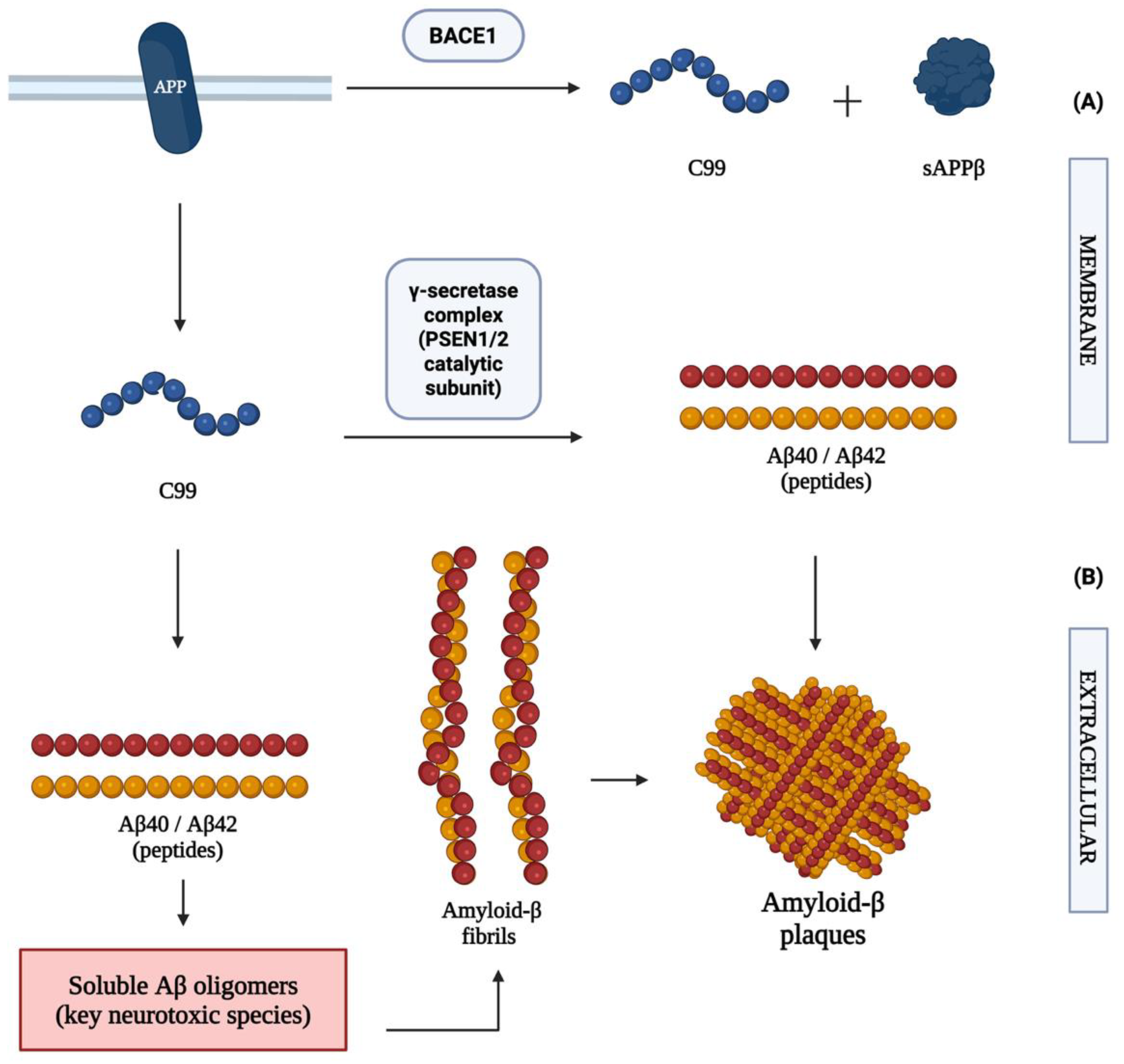
Figure 2.
Physiological organization and functional domains of Tau protein and its interaction with neuronal microtubules. (A) The MAPT gene encodes the Tau protein, a microtubule-associated protein predominantly expressed in neurons. Microtubules are dynamic cytoskeletal polymers composed of α- and β-tubulin heterodimers that play essential roles in intracellular transport, maintenance of neuronal morphology, and axonal stability. Tau contributes to the stabilization and organization of axonal microtubules, thereby preserving neuronal integrity. The physiological localization of Tau within healthy neuronal structures and axonal microtubule networks is illustrated on the left side of the figure. (B) Structural organization of Tau protein domains, including the N-terminal domain, proline-rich domain, C-terminal domain, and microtubule-binding repeat region (MTBR). (C) Physiological functions associated with each domain. The N-terminal domain interacts with cytoskeletal proteins and contributes to axonal stability, whereas the proline-rich domain contains multiple phosphorylation sites involved in Tau regulation. The C-terminal domain participates in structural modulation, while the MTBR mediates Tau binding to tubulin and promotes microtubule stabilization. Tau-mediated stabilization of microtubules is essential for efficient axonal transport and maintenance of neuronal architecture (Alberts et al., 2015; Kadavath et al., 2015; Muralidar et al., 2020; Hong et al., 2025).
Figure 2.
Physiological organization and functional domains of Tau protein and its interaction with neuronal microtubules. (A) The MAPT gene encodes the Tau protein, a microtubule-associated protein predominantly expressed in neurons. Microtubules are dynamic cytoskeletal polymers composed of α- and β-tubulin heterodimers that play essential roles in intracellular transport, maintenance of neuronal morphology, and axonal stability. Tau contributes to the stabilization and organization of axonal microtubules, thereby preserving neuronal integrity. The physiological localization of Tau within healthy neuronal structures and axonal microtubule networks is illustrated on the left side of the figure. (B) Structural organization of Tau protein domains, including the N-terminal domain, proline-rich domain, C-terminal domain, and microtubule-binding repeat region (MTBR). (C) Physiological functions associated with each domain. The N-terminal domain interacts with cytoskeletal proteins and contributes to axonal stability, whereas the proline-rich domain contains multiple phosphorylation sites involved in Tau regulation. The C-terminal domain participates in structural modulation, while the MTBR mediates Tau binding to tubulin and promotes microtubule stabilization. Tau-mediated stabilization of microtubules is essential for efficient axonal transport and maintenance of neuronal architecture (Alberts et al., 2015; Kadavath et al., 2015; Muralidar et al., 2020; Hong et al., 2025).
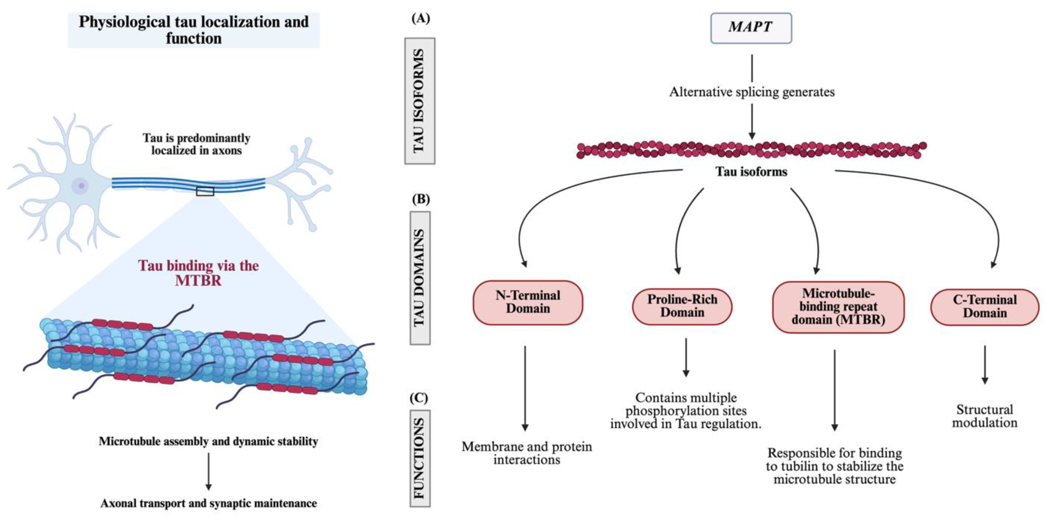
Figure 3.
Tau aggregation pathway and neurofibrillary tangle formation in Alzheimer’s disease. Hyperphosphorylation of Tau promotes its dissociation from neuronal microtubules, impairing microtubule stability and intracellular transport. Soluble hyperphosphorylated Tau monomers undergo conformational changes and progressively aggregate into oligomers and β-sheet–rich fibrillar structures through prion-like seeding mechanisms. These fibrils further accumulate into neurofibrillary tangles (NFTs), contributing to neuronal dysfunction, cytoskeletal destabilization, and neurodegeneration associated with AD. Amyloid-β accumulation further enhances Tau pathological aggregation and neuronal injury. Based on concepts described by D’Errico et al. (2020), De Felice et al. (2008), Zheng et al. (2024), and Van Alstyne et al. (2025).
Figure 3.
Tau aggregation pathway and neurofibrillary tangle formation in Alzheimer’s disease. Hyperphosphorylation of Tau promotes its dissociation from neuronal microtubules, impairing microtubule stability and intracellular transport. Soluble hyperphosphorylated Tau monomers undergo conformational changes and progressively aggregate into oligomers and β-sheet–rich fibrillar structures through prion-like seeding mechanisms. These fibrils further accumulate into neurofibrillary tangles (NFTs), contributing to neuronal dysfunction, cytoskeletal destabilization, and neurodegeneration associated with AD. Amyloid-β accumulation further enhances Tau pathological aggregation and neuronal injury. Based on concepts described by D’Errico et al. (2020), De Felice et al. (2008), Zheng et al. (2024), and Van Alstyne et al. (2025).
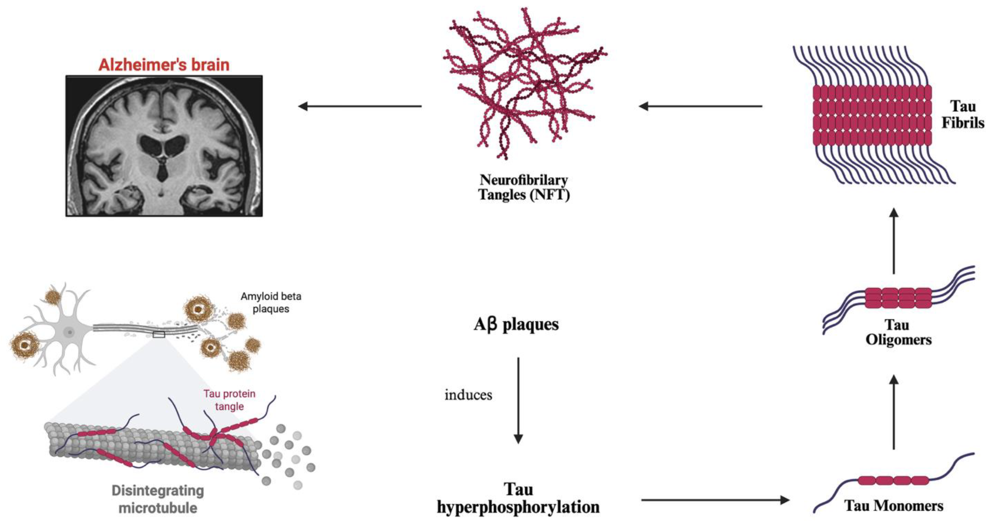
Figure 4.
Cellular dynamics and glial cross-talk involved in Alzheimer’s disease-associated neuroinflammation. This schematic illustrates the interaction between glial cells and peripheral immune components during neuroinflammatory progression in AD. Pathological hallmarks of AD, including amyloid-β (Aβ) plaques and Tau tangles, induce activation of homeostatic microglia, promoting their transition into disease-associated microglia (DAM) through a TREM2-dependent mechanism. Activated DAM stimulate reactive astrocytes, contributing to blood-brain barrier (BBB) dysfunction and increased permeability. BBB impairment facilitates infiltration of peripheral T cells into the central nervous system, where release of interferon-gamma (IFN-γ) further enhances DAM activation, establishing a self-perpetuating neuroinflammatory cycle associated with neuronal dysfunction and neurodegeneration. Based on concepts described by Heneka et al. (2015), Keren-Shaul et al. (2017), Machado and Carvalho (2020), and Wu and Cao (2025).
Figure 4.
Cellular dynamics and glial cross-talk involved in Alzheimer’s disease-associated neuroinflammation. This schematic illustrates the interaction between glial cells and peripheral immune components during neuroinflammatory progression in AD. Pathological hallmarks of AD, including amyloid-β (Aβ) plaques and Tau tangles, induce activation of homeostatic microglia, promoting their transition into disease-associated microglia (DAM) through a TREM2-dependent mechanism. Activated DAM stimulate reactive astrocytes, contributing to blood-brain barrier (BBB) dysfunction and increased permeability. BBB impairment facilitates infiltration of peripheral T cells into the central nervous system, where release of interferon-gamma (IFN-γ) further enhances DAM activation, establishing a self-perpetuating neuroinflammatory cycle associated with neuronal dysfunction and neurodegeneration. Based on concepts described by Heneka et al. (2015), Keren-Shaul et al. (2017), Machado and Carvalho (2020), and Wu and Cao (2025).
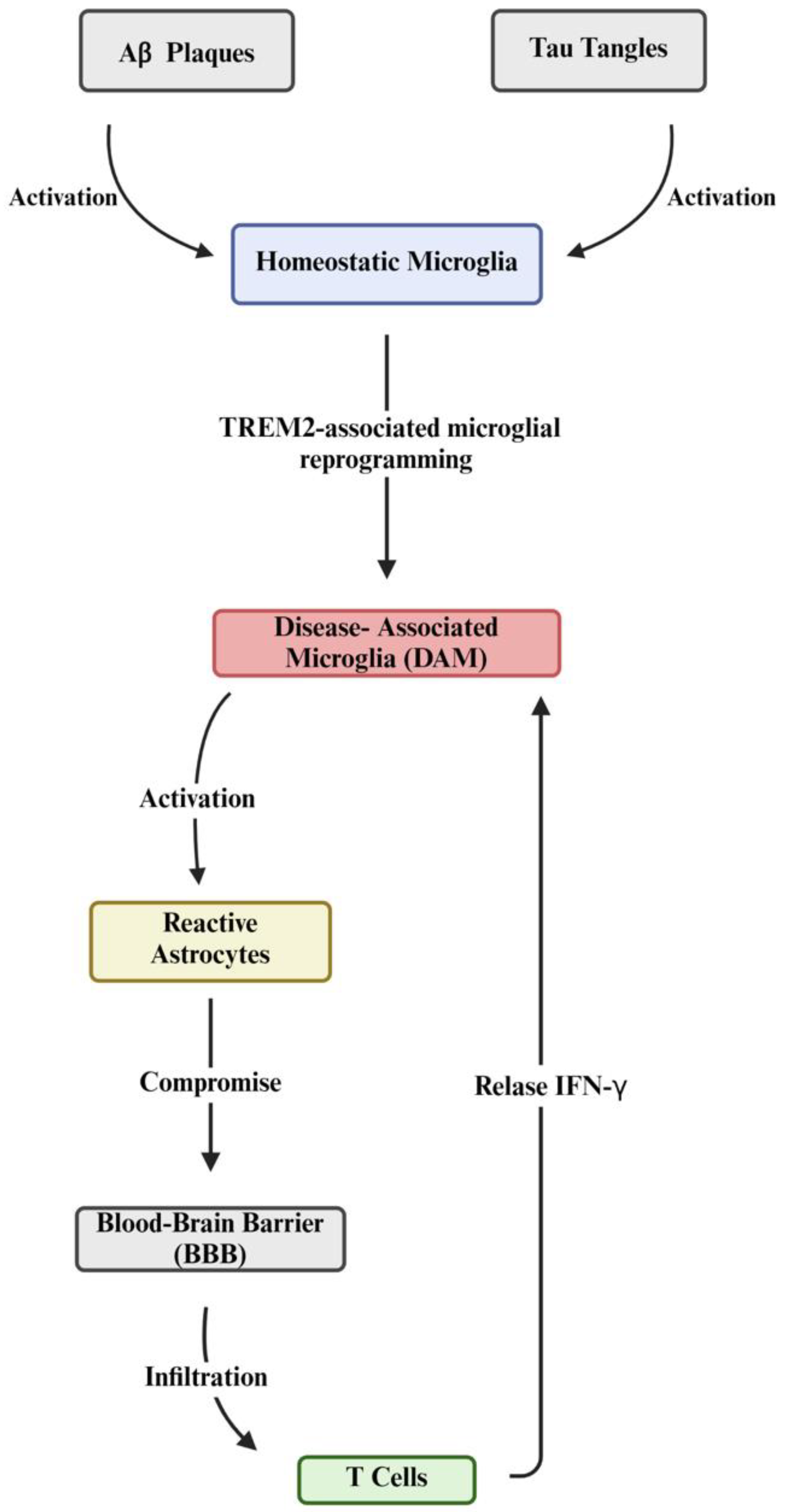
Figure 5.
Two-step activation and downstream amplification of NLRP3 inflammasome signaling in Alzheimer’s disease. NLRP3 inflammasome activation can be conceptualized as an NF-κB-dependent priming phase followed by a danger-signal-driven activation phase. Amyloid-β (Aβ), pathological tau, neuronal damage-associated molecular patterns (DAMPs), and inflammatory mediators can promote NF-κB signaling, increasing expression of NLRP3 and pro-IL-1β. Subsequent cellular stress, including lysosomal damage following Aβ uptake, potassium efflux, and reactive oxygen species (ROS) generation, promotes assembly of the NLRP3-ASC-pro-caspase-1 complex and activation of caspase-1. Caspase-1 processes pro-IL-1β and pro-IL-18 into mature IL-1β and IL-18 and cleaves gasdermin D (GSDMD), promoting membrane pore formation, pyroptosis, and release of additional DAMPs. Inflammasome-derived cytokines interact with broader NF-κB- and JAK/STAT-associated inflammatory networks in microglia and astrocytes, reinforcing neuroinflammation, synaptic dysfunction, neuronal injury, impaired Aβ clearance, and tau pathology. These reciprocal interactions can provide renewed priming and activation signals, establishing a self-reinforcing inflammatory loop in AD. Based on concepts described by Heneka et al. (2015), Zhang et al. (2020), Liang et al. (2022), and Lin et al. (2025).
Figure 5.
Two-step activation and downstream amplification of NLRP3 inflammasome signaling in Alzheimer’s disease. NLRP3 inflammasome activation can be conceptualized as an NF-κB-dependent priming phase followed by a danger-signal-driven activation phase. Amyloid-β (Aβ), pathological tau, neuronal damage-associated molecular patterns (DAMPs), and inflammatory mediators can promote NF-κB signaling, increasing expression of NLRP3 and pro-IL-1β. Subsequent cellular stress, including lysosomal damage following Aβ uptake, potassium efflux, and reactive oxygen species (ROS) generation, promotes assembly of the NLRP3-ASC-pro-caspase-1 complex and activation of caspase-1. Caspase-1 processes pro-IL-1β and pro-IL-18 into mature IL-1β and IL-18 and cleaves gasdermin D (GSDMD), promoting membrane pore formation, pyroptosis, and release of additional DAMPs. Inflammasome-derived cytokines interact with broader NF-κB- and JAK/STAT-associated inflammatory networks in microglia and astrocytes, reinforcing neuroinflammation, synaptic dysfunction, neuronal injury, impaired Aβ clearance, and tau pathology. These reciprocal interactions can provide renewed priming and activation signals, establishing a self-reinforcing inflammatory loop in AD. Based on concepts described by Heneka et al. (2015), Zhang et al. (2020), Liang et al. (2022), and Lin et al. (2025).
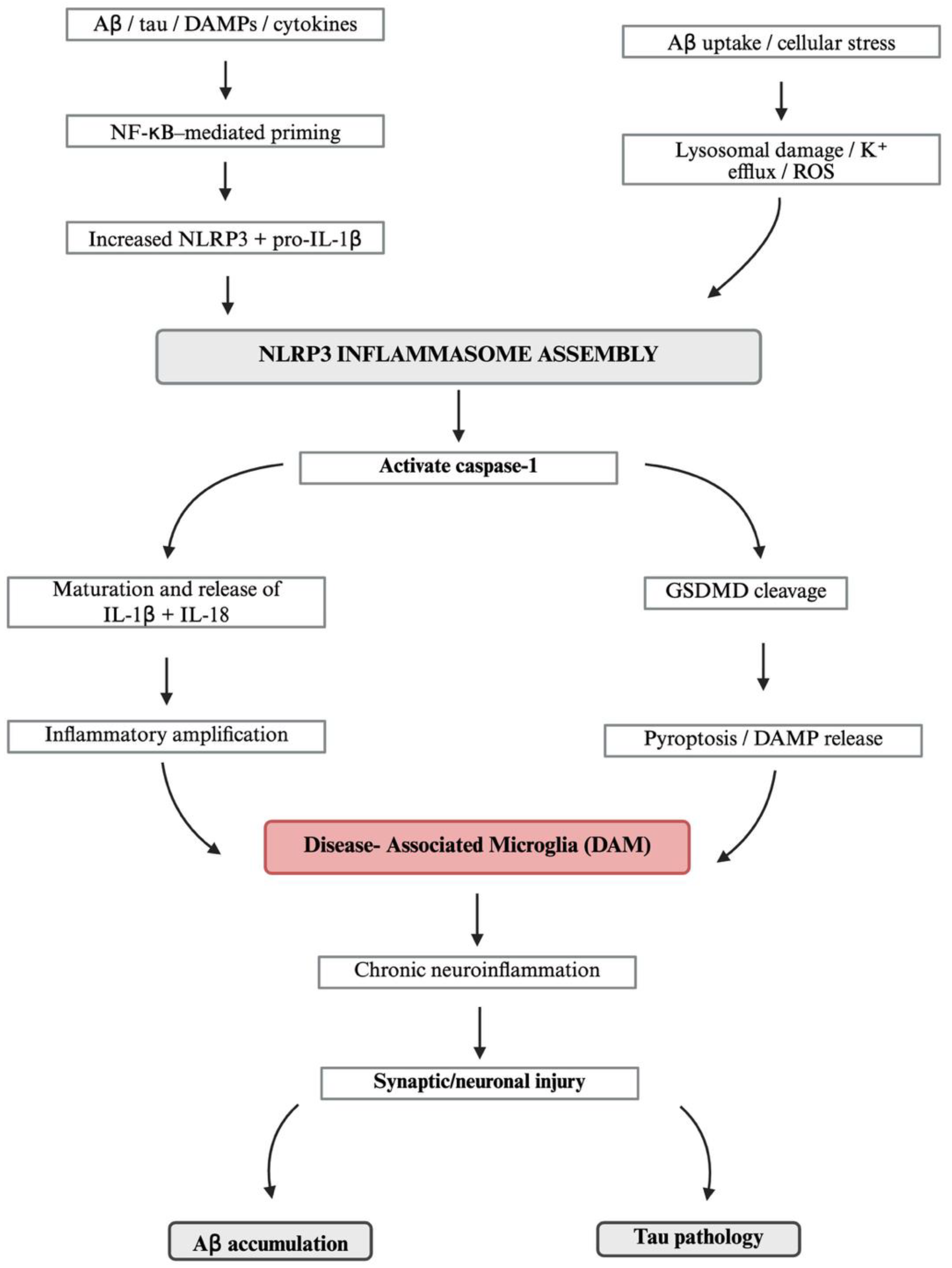
Figure 6.
Integrated, stage-dependent mechanistic network of Alzheimer’s disease progression. Amyloid-β accumulation and oligomerization may provide early permissive influences through synaptic dysfunction and calcium dysregulation, which can promote tau hyperphosphorylation and mislocalization. Amyloid aggregates also engage microglial NLRP3 signaling and disease-associated microglial responses, while reciprocal microglia–astrocyte interactions amplify neuroinflammatory signaling and may impair Aβ clearance. Tau dysfunction can progress toward seed formation and propagation, while persistent glial activation and neuronal injury generate additional feedback that may become partly autonomous from initiating amyloid-associated processes. The relative dominance of these mechanisms is proposed to vary across disease stages and individuals; therefore, the model represents an interacting network rather than a fixed linear cascade. Based on concepts described by Hardy and Selkoe (2002), De Felice et al. (2008), Heneka et al. (2015), Butovsky and Weiner (2018), D’Errico et al. (2020), Liang et al. (2022), Alonso et al. (2024), Zheng et al. (2024), Lin et al. (2025), Van Alstyne et al. (2025), and Watamura et al. (2025).
Figure 6.
Integrated, stage-dependent mechanistic network of Alzheimer’s disease progression. Amyloid-β accumulation and oligomerization may provide early permissive influences through synaptic dysfunction and calcium dysregulation, which can promote tau hyperphosphorylation and mislocalization. Amyloid aggregates also engage microglial NLRP3 signaling and disease-associated microglial responses, while reciprocal microglia–astrocyte interactions amplify neuroinflammatory signaling and may impair Aβ clearance. Tau dysfunction can progress toward seed formation and propagation, while persistent glial activation and neuronal injury generate additional feedback that may become partly autonomous from initiating amyloid-associated processes. The relative dominance of these mechanisms is proposed to vary across disease stages and individuals; therefore, the model represents an interacting network rather than a fixed linear cascade. Based on concepts described by Hardy and Selkoe (2002), De Felice et al. (2008), Heneka et al. (2015), Butovsky and Weiner (2018), D’Errico et al. (2020), Liang et al. (2022), Alonso et al. (2024), Zheng et al. (2024), Lin et al. (2025), Van Alstyne et al. (2025), and Watamura et al. (2025).
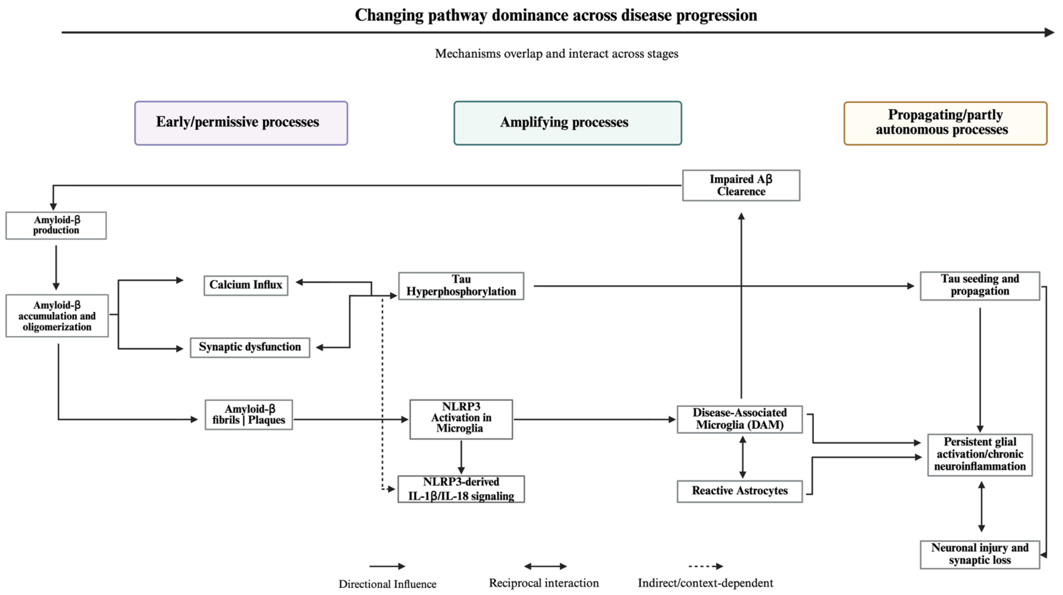

Table 1.
Integrated age- and stage-dependent pathogenic network in Alzheimer’s disease: mechanistic roles, human biomarker evidence, therapeutic opportunities, and representative references. Major mechanistic axes are organized according to their proposed early/permissive and subsequent amplifying or propagating functions within the integrated network presented in this review. These roles are overlapping, context dependent, and likely to vary among individuals and across disease stages; they should therefore not be interpreted as a fixed temporal sequence. Biological ageing is presented as a cross-cutting modifier of network resilience rather than as an independent Alzheimer disease pathway. Human biomarkers provide evidence of pathway-associated activity but do not, by themselves, establish causal direction or pathway dominance. Therapeutic approaches include established anti-amyloid treatments and emerging or experimental strategies targeting tau, synaptic dysfunction, microglial/TREM2 signaling, astrocytic and neurovascular dysfunction, and inflammasome-associated inflammation.
Table 1.
Integrated age- and stage-dependent pathogenic network in Alzheimer’s disease: mechanistic roles, human biomarker evidence, therapeutic opportunities, and representative references. Major mechanistic axes are organized according to their proposed early/permissive and subsequent amplifying or propagating functions within the integrated network presented in this review. These roles are overlapping, context dependent, and likely to vary among individuals and across disease stages; they should therefore not be interpreted as a fixed temporal sequence. Biological ageing is presented as a cross-cutting modifier of network resilience rather than as an independent Alzheimer disease pathway. Human biomarkers provide evidence of pathway-associated activity but do not, by themselves, establish causal direction or pathway dominance. Therapeutic approaches include established anti-amyloid treatments and emerging or experimental strategies targeting tau, synaptic dysfunction, microglial/TREM2 signaling, astrocytic and neurovascular dysfunction, and inflammasome-associated inflammation.
| Mechanistic axis or modifier | Early/permissive role | Amplifying or propagating role | Human biomarker evidence | Potential intervention | Representative references |
|---|---|---|---|---|---|
| Biological ageing and loss of resilience | Declining proteostatic, autophagic-lysosomal, mitochondrial, vascular, and immune resilience may lower the threshold for Aβ- and tau-associated cellular stress. | Impaired aggregate clearance, oxidative stress, inflammaging, and BBB dysfunction may facilitate persistence and coupling of proteinopathy, synaptic injury, and neuroinflammation. | No single validated biomarker captures biological resilience. Multimodal approaches combining glial, neuronal-injury, inflammatory, vascular, and molecular signatures are under investigation. | Restoration of proteostasis/autophagy, mitochondrial function, vascular integrity, and immune homeostasis; currently experimental rather than established AD-specific gerotherapeutic strategies. | López-Otín et al., 2023; Franceschi et al., 2018; Nixon & Rubinsztein, 2024; D’Alessandro et al., 2025. |
| Amyloid-β production, oligomerization, and seeding | Increased production or impaired clearance of aggregation-prone Aβ species can promote oligomer formation, membrane interactions, synaptic stress, and calcium dysregulation. | Secondary nucleation, prion-like seeding, impaired proteostatic clearance, and glial activation can sustain amyloid accumulation and reinforce downstream tau and inflammatory pathways. | Amyloid PET and CSF or plasma Aβ42/Aβ40 ratios provide evidence of amyloid pathology. Amyloid positivity commonly precedes extensive tau deposition and clinical deterioration. | Anti-Aβ monoclonal antibodies, including lecanemab and donanemab; additional approaches aimed at Aβ production, aggregation, or clearance remain under investigation. | Walsh et al., 2002; Haass & Selkoe, 2007; Jack et al., 2024; Palmqvist et al., 2020; Suzuki et al., 2024; Kim et al., 2025. |
| Synaptic dysfunction and calcium dysregulation | Soluble Aβ oligomers may alter PrPᶜ-mGluR5-Fyn-NMDAR signaling and astrocytic glutamate handling, producing early excitatory and calcium stress. | Persistent calcium overlood promotes mitochondrial dysfunction, oxidative stress, dendritic injury, and tau-directed kinase activation, linking synaptic dysfunction to structural neurodegeneration. | SV2A PET is an emerging in vivo measure of synaptic density; CSF synaptic proteins such as neurogranin and SNAP-25 provide complementary research readouts. NfL reflects downstream neuronal injury rather than synaptic dysfunction specifically. | Strategies preserving synaptic function, glutamate homeostasis, mitochondrial resilience, or calcium regulation; no established synapse-specific disease-modifying treatment for AD. | De Felice et al., 2008; Zempel et al., 2010; Burgoyne et al., 2019; Mecca et al., 2020; Tzioras et al., 2023. |
| Tau hyperphosphorylation, aggregation, and propagation | Aβ- and calcium-associated kinase dysregulation can promote tau hyperphosphorylation, microtubule detachment, and somatodendritic mislocalization. | Seed formation, templated propagation, axonal transport failure, and reciprocal glial activation can increasingly sustain neuronal dysfunction as disease progresses. | Tau PET and CSF or plasma phosphorylated tau provides complementary measures of tau pathology. Regional tau accumulation is more closely associated with clinical progression than amyloid burden alone. | Tau-directed immunotherapies, aggregation inhibitors, and modulation of tau-directed kinases; clinical efficacy remains under investigation. | Wang & Mandelkow, 2016; Hanseeuw et al., 2019; Ossenkoppele et al., 2021; Vogel et al., 2021; Pascoal et al., 2021. |
| Microglial/TREM2 axis | TREM2-associated microglial responses can support plaque containment, phagocytosis, lipid handling, survival, and metabolic adaptation during early pathology. | Persistent proteinopathy or impaired TREM2 signaling may contribute to dysfunctional microglial states, reduced clearance, cytokine release, oxidative stress, and reinforcement of tau and synaptic pathology. | CSF soluble TREM2 is an emerging marker of microglial/TREM2 activity and shows stage-dependent relationships with amyloid, neuronal injury, and disease progression; it does not by itself establish TREM2 pathway dominance. | TREM2 agonistic antibodies or other approaches that modulate TREM2-dependent microglial states; currently investigational and likely disease-stage dependent. | Keren-Shaul et al., 2017; Butovsky & Weiner, 2018; Suárez-Calvet et al., 2016; Wu & Cao, 2025; Wang & Zheng, 2026. |
| Astrocytic and neurovascular/BBB dysfunction | Astrocytes normally support glutamate uptake, neuronal metabolism, ion homeostasis, and BBB integrity; early reactive responses may also contribute to tissue protection. | Persistent astrocytic reactivity may impair glutamate handling, reinforce microglial inflammation, weaken BBB integrity, and facilitate peripheral inflammatory influences. | Plasma or CSF GFAP is an emerging marker of astrocytic reactivity; YKL-40 may provide complementary information. Vascular/permeability imaging and related measures can assess aspects of BBB dysfunction. | Restoration of astrocytic homeostasis, glutamate transport, neurovascular function, and BBB integrity; presently experimental rather than established disease-modifying therapy. | Liddelow & Barres, 2017; Heneka et al., 2015; Machado & Carvalho, 2020; Ankeny et al., 2024; Lin et al., 2025. |
| NLRP3 and downstream inflammatory signaling | Aβ, pathological tau, lysosomal injury, ROS, and other danger signals can prime and activate innate inflammatory pathways, including NLRP3 in microglia. | NLRP3-ASC-caspase-1 signaling, IL-1β/IL-18 release, gasdermin-D-mediated pyroptosis, and NF-κB/JAK-STAT crosstalk can establish self-reinforcing inflammatory loops that impair Aβ clearance and promote tau and neuronal injury. | Circulating or CSF inflammatory mediators such as IL-1β, IL-18, and IL-6 provide indirect evidence of inflammatory activity, but not clinically validated NLRP3-specific biomarker currently defines pathway dominance in individual patients. | NLRP3 inhibitors and modulation of inflammasome-associated cytokine or pyroptotic signaling; promising mechanistically but predominantly preclinical or early translational approaches. | Heneka et al., 2015; Ising et al., 2019; Zhang et al., 2020; Liang et al., 2022; Chen & Yu, 2023. |
Disclaimer/Publisher’s Note: The statements, opinions and data contained in all publications are solely those of the individual author(s) and contributor(s) and not of MDPI and/or the editor(s). MDPI and/or the editor(s) disclaim responsibility for any injury to people or property resulting from any ideas, methods, instructions or products referred to in the content. |
© 2026 by the authors. Licensee MDPI, Basel, Switzerland. This article is an open access article distributed under the terms and conditions of the Creative Commons Attribution (CC BY) license (http://creativecommons.org/licenses/by/4.0/).
Copyright: This open access article is published under a Creative Commons CC BY 4.0 license, which permit the free download, distribution, and reuse, provided that the author and preprint are cited in any reuse.