

Submitted:
13 November 2026
Posted:
16 November 2026
You are already at the latest version
Abstract
This narrative review outlines potential pathophysiological mechanisms in the mammalian heart in response to chronic infrasound exposure from technical installations. Recent research findings suggest that repeated exposure to infrasound can activate the heart's mechanosensitive PIEZO-1 channels. PIEZO-1 plays an important role in cardiac contractility via modulation of Ca²⁺ release and is involved in ROS homeostasis. The increasingly widespread sources of technical infrasound - which often exhibit high sound pressure intensities at infrasonic frequencies - should be reevaluated with regard to their impact on living organisms. Experimental overexpression of cardiac PIEZO-1 in mice serves as an example of potential effects of chronic infrasound exposure on the heart. Associated reactive oxygen species (ROS) generation reveals links to inflammatory pathways. Continued PIEZO-1 (over)stimulation appears to disrupt Ca²⁺ and ROS homeostasis in the long term, ultimately resulting in cardiomyopathy and arrhythmogenicity.
Keywords:
PIEZO-1 overstimulation
; infrasound
; mechanotransduction
; calcium homeostasis
; reactive oxygen species
1. Introduction
1.1. Technical Infrasound and Its Biological Effectiveness
In addition to natural sources, the environment is increasingly exposed to man-made sound waves of various frequencies, including growing levels of infrasound. Infrasound is defined as airborne sound below 20 Hz. Infrasound from technical sources typically occurs in the form of periodic pressure fluctuations that set large masses of air in motion, thereby generating high-energy pulses. The biological effects of infrasound signals are determined by an interplay of all characteristics of a sound (sound pressure, frequency, temporal structure, impulsivity, exposure time profile, and chronicity) [1]. For more information, also see examples of technical sources in reference [2]. Data centers and wind turbines have seen particularly strong expansion. In industrial wind turbines, an increase in rotor diameter results in a large increase in electrical output as well as in audible noise and IS. The effects of infrasound on living organisms must be re-evaluated in light of the recent findings by K. Mattsson et al., who showed that the infrasound pressure levels from modern wind turbines fall within a range of high intensity and have, therefore, been significantly underestimated to date [3]. Another key finding by Mattsson et al. is that dB(Z) values depend significantly on atmospheric conditions and range between 90 and 115 dB(Z) at a distance of several kilometers from a single modern wind turbine. Under inversion conditions, the sound pressure levels are expected to reach 120 dB(Z). Other important results include possible diurnal fluctuations of up to 20 dB(Z), the dependence on different layouts of the wind turbine installations, as well as a fundamental frequency in the very low-frequency infrasound range [3]. In modern wind turbines, this frequency, corresponding to the blade-pass frequency (BPF), currently ranges from 0.2 to 0.4 Hz, which facilitates long-range, wide propagation, and an increased risk of mechanical forces being transmitted into the viscoelastic tissue of organisms [1,4,5,6]. Furthermore, the authors emphasize the need for infrasound assessment in dB(Z) [3]. Figure 1 shows the unweighted dB(Z) rating for infrasound compared with the dB(A), dB(C) and dB(G) ratings. It follows, therefore, that in Figure 1 only the unweighted dB (Z) curve (red) can represent the infrasound that actually exists. The dB(A) curve shows a loss of 70 dB at 10 Hz. The G-weighting extends well into the infrasound range, but also involves a significant loss of the actual sound pressure level, especially in the important range below 10 Hz [7].
Infrasound from natural sources differs from that originating from technical sources primarily in terms of the duration of exposure, its stochastic structure, and the absence of impulsive characteristics or periodicity. Another source of prolonged exposure to infrasound that may pose a risk of harmful effects on the entire body and, consequently, on heart health arises when living along busy streets and during long car trips, train rides, and flights. Unlike exposure inside buildings, the latter are voluntary and should be taken into account particularly during sensitive life stages such as pregnancy, infancy, and childhood. Work-related exposure to infrasound in these areas should also be assessed. Occupational safety regulations usually prohibit access to workplaces with infrasound and vibration exposure for vulnerable groups such as pregnant women or children. Equivalent protection is not possible, as the infrasound generated by technical installations - such as modern wind turbines located in open areas - cannot be dampened and therefore can penetrate houses, roofs, and walls [8]. Depending on the size of the room and the wavelength, resonance effects can occur between structure-borne and airborne sound, which can lead to peaks and dips in sound pressure levels, particularly indoors, and can therefore cause additional impact [1,8].
1.2. Maintenance of Homeostasis
The endothelium plays a central role in maintaining homeostasis. It continuously receives mechanical and chemical stimuli from its environment and thus adapts, beat by beat, to the constantly changing flow conditions, including shear stress, cyclic stretching stress, and external stimuli that act upon it. Key factors in this process include the endothelial cell’s tensegrity structure [9], which adapts to changing flow conditions, and the various regulatory mechanisms of microcirculation that ensure laminar flow in the low-pressure region of the microcirculation [10]. According to Chien et al. [11], under pulsating shear stress and cyclic strain, there is merely a temporary molecular signal transduction via proinflammatory and proliferative signaling pathways, which are downregulated when laminar shear stress persists [11]. Mechanotransduction via endothelial PIEZO channels forms the central basis for how information about shear stress and cyclic stress controls the vital regulatory processes of endothelial cells such as proliferation, migration, production of biochemical substances via gene expression, blood pressure regulation, the course of inflammatory processes, coagulation balance, and embryonic growth [12,13]. Mechanotransduction is the crucial link between forces and biological response, a fundamental mechanism common to all organisms that serves to maintain structure, function, and communication not only in the endothelium but in all organs and tissues, including the heart. Ca2+ homeostasis and redox homeostasis depend on Ca2+ inflow and adequate Nitric oxide (NO) production, which are regulated according to demand [14]. At the heart of any homeostasis lies the demand-oriented regulation of biological and biochemical reactions [15].
In order to adapt to the constantly and rapidly changing conditions, such as current demand and fluctuating preload and afterload, the healthy heart possesses the intrinsic ability to immediately adjust its cardiac output. To achieve this, a precisely regulated ion flow adjusts the stroke volume to the prevailing hemodynamic conditions and the corresponding requirements [16,17]. The autonomic nervous system thereby influences contractility via hormones such as adrenaline and noradrenaline, which increase the concentration of free Ca²⁺ in the cytosol of cardiomyocytes for electromechanical coupling. The ventricular cardiomyocytes contract in a temporal sequence initiated by the cardiac conduction system.
According to the Frank-Starling mechanism, the pre-stretch of the ventricle at the end of diastole determines the subsequent isovolumetric force development - and thus the heart’s ejection fraction - a process that is mediated by PIEZO-1. In addition to maintaining Ca2+ and ROS homeostasis, it is important for heart health to prevent overstretching of the myocardial sarcomeres, as damage to the cardiac muscle fibers can occur even with a slight overstretching of the myocardial sarcomere length by more than 2.2 µm [16]. Compared with skeletal muscle cells, cardiomyocytes are significantly less stretchable; they exert high mechanical resistance to acute overstretching, which results in a low tolerance to stretch-induced damage. Unimpeded PIEZO-1 channel function is crucial for all of these processes. During excitation-contraction coupling, a dyad is a unit within a very tight space and is responsible for the rapid, calcium-induced calcium release (CICR) [16]. The objective of the research project is to evaluate the potential effects of chronic infrasound exposure on the heart. This will be done with a particular focus on Ca2+ and ROS homeostasis as well as energy metabolism.
2. PIEZO-1-Related Mechanisms
2.1. PIEZO-Channels
Mechanosensitive PIEZO channels are an essential element of force perception in all organisms. Recognizing their importance as a key sensory modality led to the awarding of the Nobel Prize in Medicine (2021). First described by Coste et al. (2010) [18], PIEZO channels sense forces in blood vessels and tissues, as well as external forces acting on the organism, such as air pressure, gravity, proprioception, touch, and environmental sound[16,19]. When the mechanically activated non-selective cation channels of PIEZO-1 and -2 are activated by mechanical forces, a signal transduction process is triggered, which in turn modulates various signaling pathways such as gene transcription and enzyme activity, including cell division, migration, and differentiation [13,20,21,22,23,24]. The resulting biological response depends on the specific cell type and manifests primarily as a Ca²⁺ influx, regardless of whether the mechanical stimulus originates from endogenous or external sources [24] . Figure 2 provides an overview of the distribution of PIEZO-1 and PIEZO-2 channels in various mammalian organs.
There is a spatial, biochemical, and functional link between the PIEZO-1 channel and the actin cytoskeleton via the Cadherin-β-Catenin-mechanotransduction complex [18,19,25]. These connections suggest a direct force transmission, known as the “force-from-filament” (FFF) model [24]. The intracellular tail of E-cadherin binds to catenin, anchoring the protein complex to the cell’s actin cytoskeleton, while the cytosolic tail of E-cadherin interacts with the cytosolic linker and the C-terminal domain (CTD) of PIEZO-1, also allowing reciprocal power transmission. In turn, the latter two are closely linked to lateral plug gates as well as to binding domains of the sarcoplasmic/ endoplasmic reticulum calcium ATPase (SERCA2) [24,25]. Cox et al.[26], a prominent representative for the “force-from-lipid” (FFL) model, states that changes in membrane tension are the key trigger for PIEZO-1 and PIEZO-2 activation. The respective proportions are under debate. Cox et al. also demonstrated that the cytoskeleton protects membrane-bound proteins, which apparently shields the activation of PIEZO-1 from excessive forces. This may be particularly relevant at higher frequencies. However, at lower frequencies, force transmission via the filaments is more direct. At low frequencies, Mazzag and Barakat stated that the cytoskeleton acts as a low-pass filter and is therefore apparently unable to provide adequate protection against infrasound frequencies as they decrease. In a computer-simulated analysis, based on the conductive structures’ actin stress fibers and microtubules, incoming impulses were found to converge increasingly as the transmission frequency decreases [5,6]. In their spatio-temporal network model [5] , this resulted in a theoretical threshold for a 1:1 transmission at 10-3 – 10-2 Hz for actin stress fibers and 10-5 – 10-4 Hz for microtubules. This suggests that stress fibers can transmit forces more effectively across a wider frequency range than microtubules. Additionally, the authors found that an impulsive signal is transmitted more readily than a steady one, and that, over the course of evolution, the physiological pressure fluctuations of the pulse wave do not mask the perception of incoming force impulses [5,6].
The required pressure to reach the lateral membrane tension (λ50 / T50) for a half-maximal activation of PIEZO-1 is around ≈ 1.4–4.5 Pascal [22] . This is consistent with the experimental results of Na et al., with 1,8 Pa as the needed stimulus [27]. Several authors consistently cite 25–30 mmHg, like Coste et al. [18] and Xiao [24], which also corresponds to this value. Converting pressures into sound pressure levels (SPL), 1,8 Pa corresponds to ~ 99 dB (Z).
Remarks: The Laplace equation establishes the link in electrophysiological experiments, e.g., the pressure-clamp technique, between the applied hydrostatic pressure (in mmHg) and the resulting lateral mechanical membrane tension. In this way, a pipette pressure of approximately 25–30 mmHg corresponds to a lateral membrane tension of about 1.4–4.5 Pascal. The membrane tension is directly proportional to the applied pressure and the membrane’s radius of curvature. If the membrane radius is known, a conversion can be performed using Laplace’s law, in which, according to Douguet & Honoré [28], stiffness and the pre-stressed cytoskeleton play a role [18], as well as the depth of the signal [5,6]. However, this is not a universally valid conversion, but rather an experiment-dependent relationship.
2.2. Endothelial Vascular Regulation via PIEZO-1
Intact regulation of vasodilation and vasoconstriction, vascular permeability, and immune signaling during an inflammatory response is of crucial importance for vascular health. The large surface area of the endothelium enables an efficient supply of nutrients, tailored to current requirements [10,12]; however, due to its size, it is also a more vulnerable target for internal and external disruptive factors. As part of endothelial vascular regulation, PIEZO-1 is activated, leading to the release of adenosine triphosphate (ATP), which in turn results in the phosphorylation of endothelial NO synthase and thus in corresponding NO production as well as demand-driven vasodilation [10,12]. Persistent changes in shear stress patterns, particularly oscillatory flows, are associated with reduced NO bioavailability, increased ROS generation, loss of endothelial integrity, increased endothelial apoptosis, higher lipoprotein oxidation rates, and chronic inflammation, as shown in Figure 3 [13].
To maintain the homeostasis of the redox system, the release of NO in the right quantity, in the right place, and at the right time is crucial [14,29,30]. This is only possible if the release is demand-driven. Non-demand-driven, i.e., externally induced signals, are therefore likely to disrupt the homeostasis that depends on it. Irregular and sustained VSMC activation, in turn, can lead to inadequate NO production and increased intracellular calcium and ROS production. Chronic exposure to infrasound may disrupt microvascular vasomotion through dysregulation of PIEZO-1 [30]. One example of an endogenous cause of impaired endothelial vascular regulation is chronic hypertension, which is shown to result in overstimulation of PIEZO-1 in VSMCs, as well as in the activation of inflammatory pathways, which in turn leads to remodeling of the small arteries, including thickening of the vessel walls [31,32].
2.3. The Role of PIEZO-1 in the Regulation of Ca²⁺ Homeostasis in the Heart
Any alteration in the finely orchestrated Ca²⁺ion fluxes – regardless of the cause – inevitably affects the cardiac contraction cycle and rhythmology. In their 2021 article, Jiang, F. et al.[16] show that in a mouse model, both a PIEZO-1 knockout model (KO mouse model) and a PIEZO-1 overexpression model (TG mouse model) lead to impaired Ca²⁺and ROS homeostasis and ultimately result in cardiomyopathy and heart failure. The essential mediating role of PIEZO-1 was demonstrated in the untreated control group (CO group), in which different levels of diastolic pre-stretching, e.g., 8 percent versus 15 percent, caused a corresponding rapid increase in Ca²⁺sparks, which was not observed in the PIEZO-1 KO cardiomyocytes. Using Yoda-1, an activator of PIEZO-1, it was demonstrated that PIEZO-1 mediates the adaptation of ejection fraction to the respective demand[16]. The overstimulation group (TG group) showed a different pattern compared with the control group; the Ca²⁺spark rate was significantly higher (4.6 ± 1.6 versus 0.7 ± 0.2 per 100 μm per second) and, at rest, a significantly increased spark rate, extending into the diastolic phase, was observed alongside a reduced Ca²⁺ content in the Sarcoplasmatic Reticulum (SR). The authors conclude that the reduced Ca²⁺ concentration in the SR is due to a substantial loss of Ca²⁺ from the SR’s Ca²⁺ store. Figure 4 shows the results for all groups: the KO group, the control group, and the TG group, together with the original description [16].
PIEZO-1, which is responsible for calcium-induced calcium release (CICR), mediates the Ca²⁺sparks triggered by pre-stretch in cardiomyocytes via a two-phase amplification mechanism. The influx of Ca²⁺ via L-type Ca²⁺ channels triggers the release of Ca²⁺ from RyRs; thereby, a small flow of Ca²⁺ ions leads to a 100- to 1,000-fold increase in the release of Ca²⁺ sparks [16]. For this reason, any dysfunction of PIEZO-1 must lead to a loss of the synchronization between ion flux and the action potential. In Jiang’s experimental model, both the deletion and the overexpression of cardiac PIEZO-1 channels led to end-stage cardiomyopathy. The TG group, which is of particular relevance in this context, showed, on echocardiography, an early increase in end-diastolic and end-systolic internal diameter as well as an increased left ventricular volume, suggesting the development of dilated cardiomyopathy[16]. The increased oxygen demand, as described by Laplace, resulting from increased wall tension could be compensated only temporarily by the Frank–Starling mechanism. Morphological changes in TG hearts progressed more rapidly than in KO hearts. Histological findings revealed advanced fibrosis and elevated markers of cardiac cell damage. All TG mice exhibited ventricular tachycardia, accompanied by arrhythmogenic Ca²⁺ sparks. Early signs of heart failure were observed. Taken together, these characterizations show that cardiac-specific overexpression of PIEZO-1 will demonstrably lead to severe heart failure and arrhythmias.
2.4. Under Pathological Mechanical Stress, PIEZO-1 Causes Inflammation
In the event of an acute (vascular) injury, inflammatory processes are triggered, resulting in the removal of damaged tissue and healing with restoration of function, including a new balance of PIEZO-1. Prolonged high stress, including stretching, leads to overstimulation of PIEZO-1, which enhances Ca²⁺influx and, via increased production of IL-6, activates the proliferation of cardiac fibroblasts [33,34,35]. Following damage to the heart, for example, as a result of a myocardial infarction (MI), the subsequent inflammatory response is decisive in determining whether the course of the disease leads to recovery or to cardiomyopathy [33,35,36]. Within the context of inflammatory and fibrotic homeostasis, the endothelial cytoskeleton is involved in all successive reaction chains and a complex interplay between temporally and spatially defined processes of mutual influence and activation of intravascular and extravascular factors [35]. Endothelial integrity is thereby the fundamental prerequisite. Various authors confirm that the adverse outcome of inflammatory processes in the damaged heart constitutes a key pathophysiological mechanism that contributes to progressive fibrosis and, consequently, to remodeling [35,36]. Relevant guidelines and recommendations on Angiotensin-1 (AT1) receptor blockade after heart injury aim to counteract PIEZO-1 overexpression and, consequently, remodeling [37]. Several recent studies have confirmed that PIEZO-1 plays a crucial role in opening the endothelial barrier and, therefore, in proper leukocyte diapedesis [35,36,38]. Ultimately, the authors argue that leukocyte-mediated interaction between shear stress and endothelial ICAM-1 cluster formation triggers the mechanical activation of endothelial PIEZO-1 [38]. Impaired PIEZO-1 function thus affects a very important phase of the inflammatory process, namely the leukocyte diapedesis.
2.5. The Energy Balance in the Heart Fails due to PIEZO-1 Disruption
In a failing human heart, compensatory mechanisms may initially adapt to mechanical stress; persistent mechanical stress then triggers an autonomic response involving PIEZO-1 overexpression. Together with electromechanical coupling, the SERCA2a pump accounts for the largest proportion of energy consumption, followed by the Na⁺/K⁺-ATPase, which maintains a sodium gradient for the NCX (a sodium-calcium exchanger), and in third place, the plasma membrane calcium ATPase (PMCA) [39,40,41]. Sustained signal transduction under mechanical stress leads to gradual exhaustion, particularly of the SERCA2a pump, resulting in elevated intracellular Ca²⁺ levels and reduced Ca²⁺ available in the SR for electromechanical coupling. Increasing Ca²⁺overload can gradually damage the mitochondria, as they have only a limited compensatory capacity as an additional buffer for Ca²⁺ [30,42]. Uncontrolled activation of the ryanodine receptor is therefore associated with an increased diastolic SR Ca² leak, which can trigger arrhythmias during the diastolic phase [43]. The heart’s energy consumption is related to the pressure-frequency product, contractility and myocardial wall tension. The heart’s attempt to counteract a decreasing stroke volume is accompanied by adrenergic stimulation which, whilst it may temporarily increase contractility, also leads to increasingly inefficient oxygen utilization and a further rise in ROS. According to Laplace’s law, as myocardial dilatation increases, a higher wall tension, accompanied by higher oxygen consumption, is required to maintain the same ventricular pressure [44]. Oxygen supply and demand drift apart [42]. Chronic heart failure, regardless of the cause, is typically associated with increased fibrosis, which affects electrical conduction, as well as a verifiable decrease in the mRNA expression of SERCA 2a. Additionally, there is a decrease in connexin43, a gap-junction protein crucial for the transmission of electrical impulses via gap junctions and the regulation of smooth muscle tone, which can typically be observed in endogenously caused heart failure. This has been demonstrated for both humans and rodents [45]. A reduction in SERCA2a and connexin43 leads to a further increase in arrhythmias and a decrease in the ejection fraction. In a rat model of heart failure, an upregulation of PIEZO-1 was observed between the 4th and 8th week following the onset of myocardial infarction (MI) under conditions of persistently high blood pressure. As already mentioned, the results indicated that Angiotensin-II caused a significant increase in PIEZO-1 density at the mRNA level, mediated by the angiotensin-1 receptor and gene expression, called the Erk1/2 signaling pathway [37].
3. Evidence from Experimental Studies with Chronic Infrasound Exposure
3.1. Historic Studies in the 1980`s
A large number of rigorous studies, dating back to the 1980s, demonstrated the harmful effects of infrasound on the heart, using high-intensity infrasound levels. In their findings, different researchers consistently concluded early damage signs in force generation of the heart, higher arrhythmogenicity, histopathological changes, damage to mitochondrial and endothelial integrity, oxidative stress, and inflammation pathways. The historical paper from Alekseev et al. (1983) [46] can be assessed as one of the earliest foundational papers for the highly damaging potential of low-frequency sound with SPL over 100 dB[Z]. The authors emphasized that the human body has no shield against infrasound and pointed to possible resonance effects, especially from the upper thorax. Infrasound could potentially disrupt human health, given that it involves frequencies similar to those produced by the human body [1]. In using frequencies from 10 to 15 Hz, the authors found decreased arterial diameters and damaged cardiomyocytes. Further examples from early research include Gordeladze et al. (1986) [47]. The group microscopically examined the effects of infrasound at a frequency of 8 Hz and intensity of 120 dB on white rats and guinea pigs. After 3 hours of exposure, they noticed pallor and swelling of the left and right ventricular walls with small-point hemorrhages in the pericardium, mitochondrial swelling, destruction of outer membranes, and endothelial swelling. Further observations included an early decrease in redox enzymes, damage to the SR, fragmented myofibrils, cardiomyocytes with granular dystrophy after 5 days of impact, swollen mitochondria with fragmented cristae, increasing destruction of cardiomyocytes, accumulation of red blood cells, endothelial swelling with destroyed mitochondria, and accumulation of erythrocytes in the lumen of dilated capillaries. Karpova NI et al. [48] confirmed early body reactions in the circulatory and respiratory systems, as well as autonomic symptoms. The assessment of this study is limited because the original text in Russian is not freely accessible; therefore, no conclusions can be drawn about the used sound pressure levels or frequencies in the infrasound range. It is noteworthy, however, that the authors assumed the existence of effects, such as force transmission or reflexes that are independent of auditory perception and can lead to detectable effects within a short period of time.
3.2. Experimental Studies After 2000
Pei et al. investigated the effects of infrasound exposure on calcium influx via the L-type Ca2+ channels in cardiomyocytes from rat ventricles in a setting with 5 Hz and 130 dB(Z). This led to a significant increase in intracellular calcium concentration in cardiomyocytes, as well as a significant downregulation of L-type calcium channels, associated with decreased mRNA expression of key L-type calcium channel subunits and the SERCA2 protein [49](2009). In a subsequent study, Pei et al. found increased oxidative stress in cardiomyocytes, downregulation of key antioxidant enzyme activities (including catalase, glutathione peroxidase, superoxide dismutase, and superoxide dismutase2), and reduced cell viability. Their results showed that PPAR-γ, which plays an important role in fat oxidation in rat cardiomyocytes, decreased depending on the duration of infrasound exposure, leading to a significant increase in oxidative stress [50].
Lousinha performed a 28-day exposure of Wistar rats to infrasound at 120 dB(Z) with a broadband infrasound signal below 20 Hz and found that the coronary vessel wall-to-perivascular tissue ratio indicated that the perivascular connective tissue shifted significantly in favor of wall thickness, while the lumen-to-wall ratio shifted at the expense of vessel diameter, suggesting either reduced blood flow or thickening and remodeling of the vessel wall. In a corticosteroid-pretreated group, while perivascular fibrosis decreased due to the blocking of inflammatory signaling cascades, the lumen-to-wall ratio worsened [51]. The same group confirmed similar results regarding cardiac fibrosis in the atrium of the heart[34] (2020). 36 rats, with an equal number serving as untreated controls, were exposed to infrasound at 120 dB (pseudo-random waveform, frequency of 2–20 Hz for 1,6, and 12 weeks). The treated group showed significant increases in interstitial atrial fibrosis as well as a decrease in connexin43 [34]. A decrease in connexin43, as a determining factor, has been shown to play a significant role in arrhythmogenicity.
Chaban et al. experimentally demonstrated the contractile behavior of isolated myocardial tissue to 100, 110, and 120 dB(Z) infrasound exposure, applying a sinusoidal signal at 16 Hz for one hour. They observed a significant decrease in contractility of almost 9% at 100 dB(Z), 11% at 110 dB(Z), and 18% at 120 dB(Z). This implies possible mechanosensitive signaling pathways beyond 100 dB(Z) [52].
3.3. Experimental Studies with Low Frequency Noise (LFN)
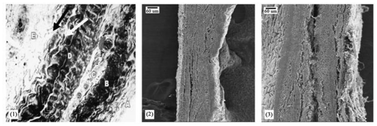
Research on the symptomatology of occupationally exposed groups with LFN (20 -200 Hz) in the 1980s showed wall thickening of vessels and an increase in collagen and elastin [53]. According to the authors, animal studies showed abnormal amounts of fibrosis/collagen in the trachea, lungs, and pleura; damaged (sheared) tracheal and bronchial cilia; and fused actin-based microvilli of tracheal and bronchial brush cells. A significant thickening of the pericardium was observed as a cardiac-specific alteration for infrasound exposure in occupationally exposed persons to LFN [53] and in persons living in the vicinity of large wind turbine plants [54]. Figure 5. shows pericardial wall thickening in chronic occupationally exposed persons in the eighties.
The authors Antunes et al. report on a series of experiments describing the effects of increasing intracardial fibrosis in isolated heart tissue of Wistar rats through prolonged exposure (three months) to low-frequency sound (LFN) (below 90 Hz) [55]. In another paper, the same group performed an immunohistochemical evaluation of cardiac connexin43 in rats exposed to low-frequency noise, focusing on the verifiable significant reduction in connexin43 [56].
3.4. Findings in Humans
The evidence in humans is limited at present. In a study in healthy volunteers, no acute physical reactions, no ECG or EEG changes were observed after 30 minutes of impact with infrasound exposure. In the setting, a 3 - 18 Hz, 85 -105 dB(Z), sinusoidal signal was used for 30 min [57]. Compared to the control group, a greater degree of discomfort, as well as increased pressure in the head and chest area, was observed. The intervals between exertion and examination are not recognizable [57]. A recent retrospective study by Christian Vahl et al. [58] (2026) demonstrated in a cohort exposed to higher levels of infrasound a significant increase in the incidence of new cases of heart failure and clinically serious cardiac arrhythmias compared to a cohort with low exposure. Although the results were significant, this observational study cannot establish a causal link on its own, but it is one of the first studies based on reliable data, featuring significant findings from a realistic environment.
Two studies about the brain should be mentioned to demonstrate that exposure to infrasound can lead to early changes in brain functions, possibly also influencing cardiovascular functions, specifically via autonomic brain centers. Weichenberger et al. exposed participants to a 12 Hz infrasound signal 2 dB below their individual hearing thresholds and at an SPL between 110 and 124 dB(Z). FMRI revealed increased connectivity in the anterior cingulate cortex, the right amygdala, and the superior frontal gyrus [59]. According to the authors, overactivation - specifically in the region of the right amygdala - can, inter alia, influence the cardiovascular system, including blood pressure, heart rate, the respiratory system, and cortisol production. Forlim et al. [60] found that prolonged exposure (for 4 weeks, 8 hours per night, 6 Hz, sound pressure level 80–90 dB infrasound) caused measurable changes in three functional networks of the brain. Unlike in the Weichenberger study, the fMRI scans were conducted not during, but before and immediately after the 4-week exposure. In their conclusions, the authors state that prolonged exposure to infrasound affects functional connectivity (FC) within the resting-state ventral default-mode network (vDMN), the sensorimotor network (SMN), and the executive control network (ECN), which are associated with cognitive and sensory processing. Notably, the auditory network itself was unaffected.
4. Limitations
One limitation of mouse-model experiments is the transferability of results to humans. For ethical reasons, experiments involving pulsatile acoustic signals in humans are only possible for very short periods and within a very limited scope and high intensities of sound pressure are ruled out in humans. These factors severely limit experimental research in humans on chronic effects following infrasound exposure. Therefore, studies on mammals - such as the mouse models are used, whose molecular foundations are among the best understood and which yield reliable data based on established baseline parameters. In short-term trials, however, subacute or chronic effects cannot be expected. Acute changes in the ECG couldn’t be seen till day 14 in the study of Jiang [16] and also in Krahe [57]. Under chronic exposure, a gradual shift in homeostasis towards Ca²⁺, ROS, and energy imbalance may be assumed. The genetically modified overexpression model (TG model) can be utilized as a comparison model to evaluate the potential effects of exogenous overstimulation of PIEZO-1 on the heart under chronic exposure to technical infrasound. Although this comparison certainly has its limitations, it nevertheless appears justified in light of the known course of endogenous PIEZO-1 overstimulation and the comparable pathophysiological mechanisms that play a role in both exogenous and endogenous mechanical stress.
There are further uncertainties regarding the potential for reversibility or possible adaptive mechanisms. The vast majority of signals in the environment and nature are not sinusoidal and have a bipolar signature. Because signal shape also influences biological significance[1] , former studies with sinusoidal character can have limitations in terms of their informative value. Future experiments should use harmonic sound signals that most closely resemble reality. In the experiments, the used frequencies changed over a wide range; nevertheless, they showed results that can be attributed to mechanotransduction processes. One possible explanation is the high degree of transferability via actin structures, as described in [5,6].
5. Proposals
Since exposure to infrasound from technical sources can be assumed to cause a gradual shift in homeostasis, experiments should focus on potential early signs. These could include, for example, the modulation of microcirculatory properties or experimental data revealing early disturbances in inflammatory responses within the context of incipient endothelial dysfunction. In their 2014 paper, Pathak et al. [61] confirm that mechanical forces, acting via PIEZO-1 activation, influence the differentiation of neural stem cells into either neurons or astrocytes. Overstimulation of PIEZO-1 could potentially lead to disruptions in early brain development. It should be noted that the results of the authors Forlim et al. [60] occurred following exposure to 80–90 dB(Z). Further research should deepen our understanding of the complexity of signaling pathways in the brain; a key aspect of this would be to gain a better understanding of the potential effects of resonance phenomena. Apart from the necessary distance - which requires re-evaluation - potential therapeutic approaches could include AT1 blockade to counteract overstimulation of PIEZO1 as well as the use of rosiglitazone as an agonist to stabilize PPAR-γ [50].
6. Conclusion and Future Directions
The studies presented highlight the dual nature of PIEZO-1 function. While its normal activity is vital for survival, its overactivation or downregulation can trigger severe pathological cascades. The focus of this review was on the heart as the target organ. The vital importance of intact PIEZO-1 function for the heart has been described in detail. PIEZO-1-mediated pathology is primarily based on three processes that occur in the heart: disturbance of Ca2+ homeostasis, ROS production, and energy balance. Additional factors include, e.g., reduced expression of mRNA for connexin43 and SERCA2a protein under sustained mechanical load. There is a possible impact on autonomic centers of the brain, which also may affect cardiovascular functions.
The reviewed literature paints a consistent picture of the potential harm of infrasound at high-intensity exposure levels. There is consistent evidence that, starting at approximately 1,8 Pa, which corresponds to about 100 dB(Z), PIEZO-1 can be triggered in a manner that is not dependent on demand. Potential detrimental outcomes include heart failure and cardiac arrhythmias. It should be noted that environmental factors affect the entire organism, also during sensitive developmental stages. Several studies point to a lower threshold for potential cellular interactions with infrasound, particularly with regard to functional and structural changes in the brain. Future studies also should aim to determine more precise data on critical infrasound levels and exposure times, as well as to establish new criteria for at-risk groups. In summary, a re-evaluation of high-intensity infrasound is necessary.
Author Contributions
Conceptualization, U.B.-S. and C.L.; methodology, U.B.-S.; investigation, U.B.-S., M.D., J.Z. and A.P.; writing—original draft preparation, U.B.-S.; writing—review and editing, U.B.-S., M.D., J.Z., A.P. and C.L.; visualization, U.B.-S.; supervision, C.L. All authors have read and agreed to the published version of the manuscript.
Funding
This research received no external funding.
Institutional Review Board Statement
Not applicable.
Informed Consent Statement
Not applicable.
Data Availability Statement
No new data were created or analyzed in this study.
Conflicts of Interest
The authors declare no conflicts of interest.
Abbreviations
The following abbreviations are used in this manuscript:
| LFN | Low-frequency noise |
| SPL | Sound pressure level |
| BPF | Blade-pass frequency |
| ATP | Adenosine triphosphate |
| CICR | Calcium-induced calcium release |
| KO | Knockout |
| TG | Transgenic |
| NO | Nitric oxide |
| ROS | Reactive oxygen species |
| SR | Sarcoplasmic reticulum |
| SERCA2 | Sarcoplasmic/endoplasmic reticulum calcium ATPase 2 |
| VSMC | Vascular smooth muscle cell |
| VAD | Vibroacoustic disease |
References
- Persinger, M.A. Infrasound, Human Health, and Adaptation: An Integrative Overview of Recondite Hazards in a Complex Environment.. [CrossRef]
- Dastan, M.; Dyminski Parente Ribeiro, E.; Bellut-Staeck, U.; Zhou, J.; Lehmann, C. Infrasound and Human Health: Mechanisms, Effects, and Applications. Applied Sciences 2026, 16, 1553. [CrossRef]
- Mattsson, K.; Eriksson, G.; Persson, L.; Chilo, J.; Tatar, K. Efficient Finite Difference Modeling of Infrasound Propagation in Realistic 3D Domains: Validation with Wind Turbine Measurements. Appl. Acoust. 2026, 243, 111156. [CrossRef]
- Vanderkooy, J.; Mann, R. Measuring Wind Turbine Coherent Infrasound. In Proceedings of the 6th International Conference on Wind Turbine Noise (WTN2015), Glasgow; 2015; pp. 20–23.
- Mazzag, B.; Barakat, A.I. The Effect of Noisy Flow on Endothelial Cell Mechanotransduction: A Computational Study. Ann. Biomed. Eng. 2011, 39, 911–921. [CrossRef]
- Mazzag, B.; Gouget, C.L.M.; Hwang, Y.; Barakat, A.I. Mechanical Force Transmission via the Cytoskeleton in Vascular Endothelial Cells. In Endothelial Cytoskeleton; CRC Press, 2013; pp. 91–115.
- Eulitz, C.; Zobel, P.; Ost, L.; Möhler, U.; Schröder, M.; Rechtsanwälte, S. Ermittlung Und Bewertung Tieffrequenter Geräusche in Der Umgebung von Wohnbebauung Abschlussbericht Von.
- Univ.-Prof. Dr.-Ing. Detlef Krahé, D.S. Machbarkeitsstudie zu Wirkungen von Infraschall | Umweltbundesamt Available online: https://www.umweltbundesamt.de/publikationen/machbarkeitsstudie-zu-wirkungen-von-infraschall (accessed on 16 October 2026).
- Ingber, D.E.; Wang, N.; Stamenović, D. Tensegrity. Cellular Biophysics, and the Mechanics of Living Systems. Reports on Progress in Physics 2014, 77, 046603. [CrossRef]
- Sperandio, M.; Brandes, R. Mikrozirkulation. In Physiologie des Menschen: mit Pathophysiologie; Ralf Brandes, F.L.R.F.S., Ed.; Springer: Berlin, Heidelberg, 2019; pp. 241–256.
- Chien, S. Mechanotransduction and Endothelial Cell Homeostasis: The Wisdom of the Cell. In Proceedings of the American Journal of Physiology - Heart and Circulatory Physiology; March 2007; Vol. 292.
- Laurindo, F.R.M.; Liberman, M.; Fernandes, D.C.; Leite, P.F. Endothelium-Dependent Vasodilation: Nitric Oxide and Other Mediators. Endothelium and Cardiovascular Diseases: Vascular Biology and Clinical Syndromes 2026, 49–65. [CrossRef]
- Fernandes, D. C., Araujo, T. L., Laurindo, F. R., & Tanaka, L. Y. (2026). Hemodynamic forces in the endothelium: from mechanotransduction to implications on development of atherosclerosis. In Endothelium and Cardiovascular Diseases (pp. 37-47). Academic Press.
- Wink, D.A.; Mitchell, J.B. Chemical Biology of Nitric Oxide: Insights into Regulatory, Cytotoxic, and Cytoprotective Mechanisms of Nitric Oxide. Free Radic. Biol. Med. 1998, 25, 434–456. [CrossRef]
- Jin, C.; Su, S.; Yu, S.; Zhang, Y.; Chen, K.; Xiang, M.; Ma, H. Essential Roles of PIEZO1 in Mammalian Cardiovascular System: From Development to Diseases. Cells 2024, 13.
- Jiang, F.; Yin, K.; Wu, K.; Zhang, M.; Wang, S.; Cheng, H.; Zhou, Z.; Xiao, B. The Mechanosensitive Piezo1 Channel Mediates Heart Mechano-Chemo Transduction.. [CrossRef]
- Daut, J. Herzmechanik. In Physiologie des Menschen: mit Pathophysiologie; Springer, 2019; pp. 165–186.
- Coste, B.; Mathur, J.; Schmidt, M.; Earley, T.J.; Ranade, S.; Petrus, M.J.; Dubin, A.E.; Patapoutian, A. Piezo1 and Piezo2 Are Essential Components of Distinct Mechanically Activated Cation Channels. Science (1979). 2010, 330, 55–60. [CrossRef]
- Fang, X.-Z.; Zhou, T.; Xu, J.-Q.; Wang, Y.-X.; Sun, M.-M.; He, Y.-J.; Pan, S.-W.; Xiong, W.; Peng, Z.-K.; Gao, X.-H.; et al. Structure, Kinetic Properties and Biological Function of Mechanosensitive Piezo Channels. Cell Biosci. 2021, 11, 13. [CrossRef]
- Zhao, Q.; Wu, K.; Geng, J.; Chi, S.; Wang, Y.; Zhi, P.; Zhang, M.; Xiao, B. Ion Permeation and Mechanotransduction Mechanisms of Mechanosensitive Piezo Channels. Neuron 2016, 89, 1248–1263. [CrossRef]
- Ellefsen, K.L.; Holt, J.R.; Chang, A.C.; Nourse, J.L.; Arulmoli, J.; Mekhdjian, A.H.; Abuwarda, H.; Tombola, F.; Flanagan, L.A.; Dunn, A.R.; et al. Myosin-II Mediated Traction Forces Evoke Localized Piezo1-Dependent Ca2+ Flickers. Commun. Biol. 2019, 2, 298. [CrossRef]
- Lewis, A.H.; Grandl, J. Mechanical Sensitivity of Piezo1 Ion Channels Can Be Tuned by Cellular Membrane Tension. Elife 2015, 4. [CrossRef]
- Shi, Z.; Graber, Z.T.; Baumgart, T.; Stone, H.A.; Cohen, A.E. Cell Membranes Resist Flow. Cell 2018, 175, 1769-1779.e13. [CrossRef]
- Xiao, B. Mechanisms of Mechanotransduction and Physiological Roles of PIEZO Channels. Nat. Rev. Mol. Cell Biol. 2024, 25, 886–903.
- Wang, J.; Jiang, J.; Yang, X.; Zhou, G.; Wang, L.; Xiao, B. Tethering Piezo Channels to the Actin Cytoskeleton for Mechanogating via the Cadherin-β-Catenin Mechanotransduction Complex. Cell Rep. 2022, 38, 110342. [CrossRef]
- Cox, C.D.; Bae, C.; Ziegler, L.; Hartley, S.; Nikolova-Krstevski, V.; Rohde, P.R.; Ng, C.-A.; Sachs, F.; Gottlieb, P.A.; Martinac, B. Removal of the Mechanoprotective Influence of the Cytoskeleton Reveals PIEZO1 Is Gated by Bilayer Tension. Nat. Commun. 2016, 7, 10366. [CrossRef]
- Na, S.; Collin, O.; Chowdhury, F.; Tay, B.; Ouyang, M.; Wang, Y.; Wang, N. Rapid Signal Transduction in Living Cells Is a Unique Feature of Mechanotransduction. Proceedings of the National Academy of Sciences 2008, 105, 6626–6631. [CrossRef]
- Douguet, D.; Honoré, E. Mammalian Mechanoelectrical Transduction: Structure and Function of Force-Gated Ion Channels. Cell 2019, 179, 340–354. [CrossRef]
- Espey, M.G.; Miranda, K.M.; Thomas, D.D.; Xavier, S.; Citrin, D.; Vitek, M.P.; Wink, D.A. A Chemical Perspective on the Interplay Between NO, Reactive Oxygen Species, and Reactive Nitrogen Oxide Species. Ann. N. Y. Acad. Sci. 2002, 962, 195–206. TB04068.X. [CrossRef]
- Dastan, M.; Bellut-Staeck, U.; Zhou, J.; Lehmann, C. Infrasound Affects Vasomotion via PIEZO1 Dysregulation. 2026. [CrossRef]
- Retailleau, K.; Duprat, F.; Arhatte, M.; Demolombe, S.; Patel, A.; Honoré, E.; Ranade, S.S.; Peyronnet, R.M.; Martins, J.R.; Jodar, M.; et al. Piezo1 in Smooth Muscle Cells Is Involved in Hypertension-Dependent Arterial Remodeling Article Piezo1 in Smooth Muscle Cells Is Involved in Hypertension-Dependent Arterial Remodeling. Cell Rep. 2015, 13. [CrossRef]
- Richardson, J.; Kotevski, A.; Poole, K. From Stretch to Deflection: The Importance of Context in the Activation of Mammalian, Mechanically Activated Ion Channels. FEBS J. 2022, 289, 4447–4469.
- Liu, H.; Hu, J.; Zheng, Q.; Feng, X.; Zhan, F.; Wang, X.; Xu, G.; Hua, F. Piezo1 Channels as Force Sensors in Mechanical Force-Related Chronic Inflammation. Front. Immunol. 2022, 13, 816149.
- Lousinha, A.; Pereira, G.; Borrecho, G.; Brito, J.; Oliveira de Carvalho, A.; Freitas, D.; Oliveira, P.; Maria, M.J.; Antunes, E. Atrial Fibrosis and Decreased Connexin 43 in Rat Hearts after Exposure to High-Intensity Infrasound. Exp. Mol. Pathol. 2020, 114, 104409. [CrossRef]
- Suthahar, N.; Meijers, W.C.; Silljé, H.H.W.; de Boer, R.A. From Inflammation to Fibrosis—Molecular and Cellular Mechanisms of Myocardial Tissue Remodelling and Perspectives on Differential Treatment Opportunities. Curr. Heart Fail. Rep. 2017, 14, 235–250.
- Kong, P.; Christia, P.; Frangogiannis, N.G. The Pathogenesis of Cardiac Fibrosis. Cellular and Molecular Life Sciences 2014, 71, 549–574.
- Liang, J.; Huang, B.; Yuan, G.; Chen, Y.; Liang, F.; Zeng, H.; Zheng, S.; Cao, L.; Geng, D.; Zhou, S. Stretch-Activated Channel Piezo1 Is up-Regulated in Failure Heart and Cardiomyocyte Stimulated by AngII; 2017; Vol. 9.
- Wang, S.; Wang, B.; Shi, Y.; Möller, T.; Stegmeyer, R.I.; Strilic, B.; Li, T.; Yuan, Z.; Wang, C.; Wettschureck, N.; et al. Mechanosensation by Endothelial PIEZO1 Is Required for Leukocyte Diapedesis. Blood 2022, 140, 171–183. [CrossRef]
- Deussen, A. Herzstoffwechsel Und Koronardurchblutung. 2010, 565–571. [CrossRef]
- Tran, K.; Loiselle, D.S.; Crampin, E.J. Regulation of Cardiac Cellular Bioenergetics: Mechanisms and Consequences. Physiol. Rep. 2015, 3, e12464. [CrossRef]
- Doenst, T.; Nguyen, T.D.; Abel, E.D. Cardiac Metabolism in Heart Failure: Implications beyond ATP Production. Circ. Res. 2013, 113, 709–724. [CrossRef]
- Contreras, L.; Drago, I.; Zampese, E.; Pozzan, T. Mitochondria: The Calcium Connection. Biochim. Biophys. Acta Bioenerg. 2010, 1797, 607–618.
- Terentyev, D.; Györke, I.; Belevych, A.E.; Terentyeva, R.; Sridhar, A.; Nishijima, Y.; De Blanco, E.C.; Khanna, S.; Sen, C.K.; Cardounel, A.J.; et al. Redox Modification of Ryanodine Receptors Contributes to Sarcoplasmic Reticulum Ca2+ Leak in Chronic Heart Failure. Circ. Res. 2008, 103, 1466–1472. [CrossRef]
- Brandes, Ralf.; Lang, Florian.; Schmidt, R.F. Physiologie Des Menschen. Mit Pathophysiologie; Springer, 2019; ISBN 9783662564684.
- Kawase, Y.; Hajjar, R.J. The Cardiac Sarcoplasmic/Endoplasmic Reticulum Calcium ATPase: A Potent Target for Cardiovascular Diseases. Nat. Clin. Pract. Cardiovasc. Med. 2008, 5, 554–565.
- Alekseev, S. V; Glinchikov, V. V; Usenko, V.R. Myocardial Ischemia in Rats Exposed to Infrasound. Gig. Tr. Prof. Zabol. 1983, 34–38.
- Gordeladze, A.S.; Glinchikov, V. V; Usenko, V.R. Experimental Myocardial Ischemia Caused by Infrasound. Gig. Tr. Prof. Zabol. 1986, 30–33.
- Karpova, N.I.; Alekseev, S. V; Erokhin, V.N.; Kadyskina, E.N.; Reutov, O. V. Early Body Reaction to Low-Frequency Acoustic Oscillations. Gig. Tr. Prof. Zabol. 1979, 16–19.
- Pei, Z.; Zhuang, Z.; Xiao, P.; Chen, J.; Sang, H.; Ren, J.; Wu, Z.; Yan, G. Influence of Infrasound Exposure on the Whole L-Type Calcium Currents in Rat Ventricular Myocytes. Cardiovasc. Toxicol. 2009, 9, 70–77. [CrossRef]
- Pei, Z.; Meng, R.; Zhuang, Z.; Zhao, Y.; Liu, F.; Zhu, M.Z.; Li, R. Cardiac Peroxisome Proliferator-Activated Receptor-γ Expression Is Modulated by Oxidative Stress in Acutely Infrasound-Exposed Cardiomyocytes. Cardiovasc. Toxicol. 2013, 13, 307–315. [CrossRef]
- Lousinha, A.; R. Oliveira, M.J.; Borrecho, G.; Brito, J.; Oliveira, P.; Oliveira de Carvalho, A.; Freitas, D.; P. Águas, A.; Antunes, E. Infrasound Induces Coronary Perivascular Fibrosis in Rats. Cardiovascular Pathology 2018, 37, 39–44. [CrossRef]
- Chaban, R.; Ghazy, A.; Georgiade, E.; Stumpf, N.; Vahl, C.F. Negative Effect of High-Level Infrasound on Human Myocardial Contractility: In-Vitro Controlled Experiment. Noise Health 2021, 23, 57–66. [CrossRef]
- Alves-Pereira, M.; Castelo Branco, N.A.A. Vibroacoustic Disease: Biological Effects of Infrasound and Low-Frequency Noise Explained by Mechanotransduction Cellular Signalling. Prog. Biophys. Mol. Biol. 2007, 93, 256–279. [CrossRef]
- Castelo Branco, N.; Alves-Pereira, M.; Pimenta, A.; Ferreira, J. Low Frequency Noise-Induced Pathology: Contributions Provided by the Portuguese Wind Turbine Case 2015.
- Antunes, E.; Borrecho, G.; Oliveira, P.; Alves De Matos, A.P.; Brito, J.; Águas, A.; Martins, J.; Santos, D. Original Article Effects of Low-Frequency Noise on Cardiac Collagen and Cardiomyocyte Ultrastructure: An Immunohistochemical and Electron Microscopy Study; 2013; Vol. 6;.
- Antunes, E.; Borrecho, G.; Oliveira, P.; Brito, J.; Águas, A.; Martins, J.; Santos, D. Immunohistochemical Evaluation of Cardiac Connexin43 in Rats Exposed to Low-Frequency Noise; 2013; Vol. 6;.
- Krahé, D.; Di Loro, A.A.; Müller, U.; Elmenhorst, E.M.; De Gioannis, R.; Schmitt, S.; Schreckenberg, D. Lärmwirkungen von Infraschallimmissionen. Umwelt-Bundesamt, Texte 2020, 163.
- Vahl, C.; Dietz, O. Significantly Increased Incidence of Heart Failure and Cardiac Arrhythmias in Municipalities with Substantial Wind Energy Expansion. In Proceedings of the INNERE MEDIZIN; SPRINGER MEDIZIN VERLAG GmbH Heidelbergerplatz 3, Berlin, GERMANY, 2026; Vol. 67, pp. S169–S169.
- Weichenberger, M.; Bauer, M.; Kühler, R.; Hensel, J.; Forlim, C.G.; Ihlenfeld, A.; Ittermann, B.; Rgen Gallinat, J.; Koch, C.; Kühn, S. Altered Cortical and Subcortical Connectivity Due to Infrasound Administered near the Hearing Threshold – Evidence from fMRI. 2017. [CrossRef]
- Forlim, C.G.; Ascone, L.; Koch, C.; Kühn, S. Resting State Network Changes Induced by Experimental Inaudible Infrasound Exposure and Associations with Self-Reported Noise Sensitivity and Annoyance. Sci. Rep. 2024, 14. [CrossRef]
- Pathak, M.M.; Nourse, J.L.; Tran, T.; Hwe, J.; Arulmoli, J.; Le, D.T.T.; Bernardis, E.; Flanagan, L.A.; Tombola, F. Stretch-Activated Ion Channel Piezo1 Directs Lineage Choice in Human Neural Stem Cells. Proc. Natl. Acad. Sci. U. S. A. 2014, 111, 16148–16153. [CrossRef]
Figure 1.
Typical frequency weighting curves in dBA, dBC, dBG and dBZ, modified after the original source [7].
Figure 1.
Typical frequency weighting curves in dBA, dBC, dBG and dBZ, modified after the original source [7].
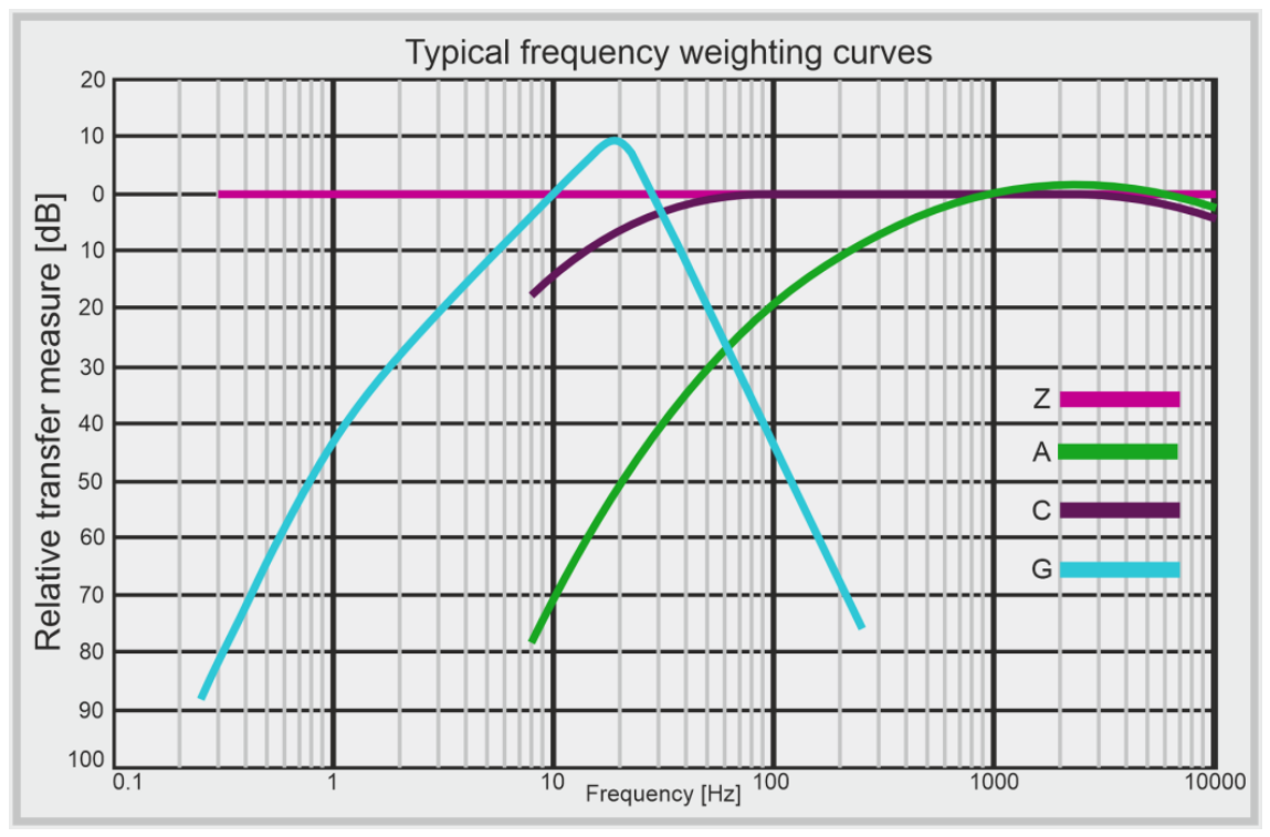
Figure 2.
Distribution of Piezo channels. Modified from Fang et al. Cell Biosci (2021) [19].
Figure 2.
Distribution of Piezo channels. Modified from Fang et al. Cell Biosci (2021) [19].
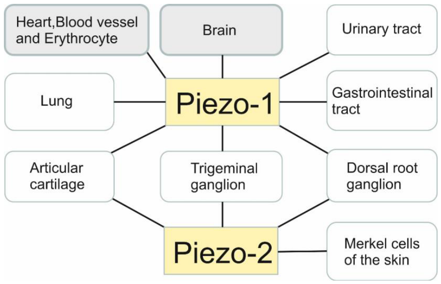
Figure 3.
Different effects of laminar and oscillatory shear on cell function and atherosclerosis. The dotted lines represent the endothelial cell cytoskeleton. Laminar and oscillatory shear forces are recognized in endothelial cells by mechanosensors, and the mechanosignals initiate signaling cascades that regulate the production of vasoactive factors and the balance between these factors. While laminar shear stimulates the production of atheroprotective factors, oscillatory shear stimulates the production of atherogenic factors, and the balance of these factors determines the vessel’s tendency to stay healthy or to develop atherogenic plaques. PGI2, prostacyclin; TM, thrombomodulin; TGFb, Transforming Growth Factor beta; PDGF, Platelet-Derived Growth Factor; ET-1, Endothelin-1; BMP4, Bone Morphogenetic Protein 4. With permission [License ID: 1750882-1] Fernandes, CD et al. [13].
Figure 3.
Different effects of laminar and oscillatory shear on cell function and atherosclerosis. The dotted lines represent the endothelial cell cytoskeleton. Laminar and oscillatory shear forces are recognized in endothelial cells by mechanosensors, and the mechanosignals initiate signaling cascades that regulate the production of vasoactive factors and the balance between these factors. While laminar shear stimulates the production of atheroprotective factors, oscillatory shear stimulates the production of atherogenic factors, and the balance of these factors determines the vessel’s tendency to stay healthy or to develop atherogenic plaques. PGI2, prostacyclin; TM, thrombomodulin; TGFb, Transforming Growth Factor beta; PDGF, Platelet-Derived Growth Factor; ET-1, Endothelin-1; BMP4, Bone Morphogenetic Protein 4. With permission [License ID: 1750882-1] Fernandes, CD et al. [13].
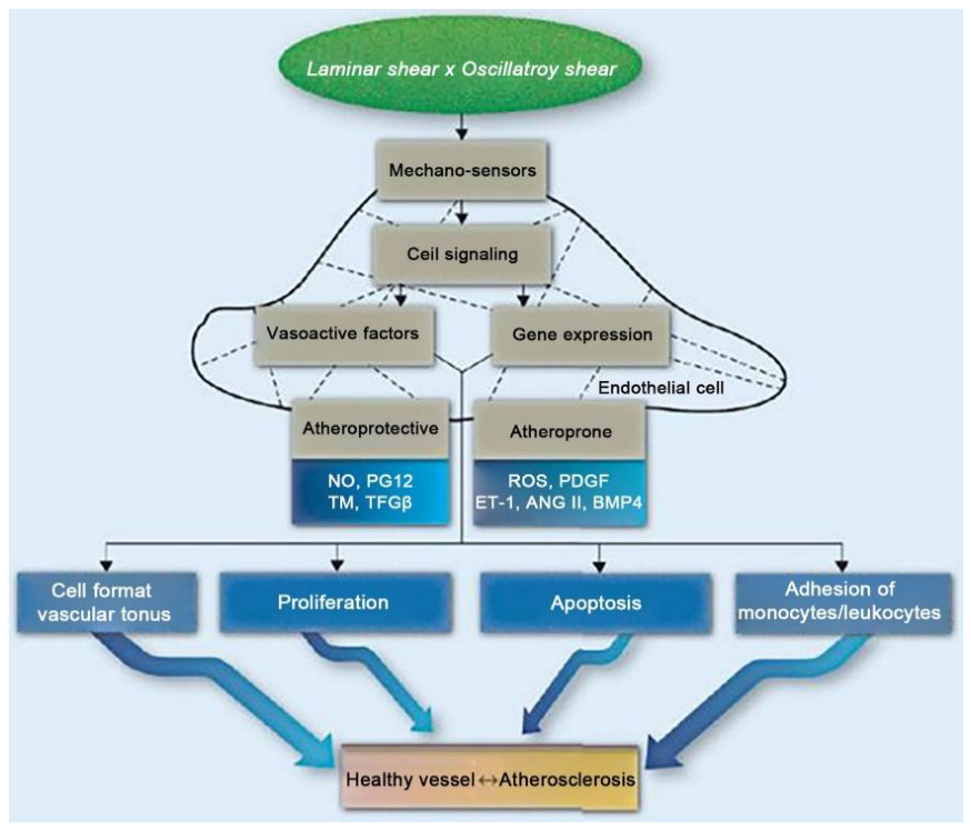
Figure 4.
Piezo1 mediates homeostatic Ca²⁺signaling. a, d Representative fluorescence surface plots of spontaneous Ca²⁺sparks of the indicated cardiomyocytes. b, e Histogram analysis of the Ca²⁺spark frequency of the indicated cardiomyocytes. The peak amplitudes are labeled above the fit. c, f Scatter plot of Ca² sparks of cardiomyocytes with the indicated genotypes: 23 Ctrl cells and 26 KO cells from KO (n = 5) mice and their littermate control (n = 5) mice in b, c; 29 Ctrl cells and 47 TG cells from TG (n = 3) mice and their littermate control (n = 3) mice in e, f. Unpaired Student’s t-test, two-sided. Values are mean ± SEM. g Representative fluorescence surface plot of spontaneous Ca²⁺waves of the indicated cardiomyocytes. h Proportion of the indicated cardiomyocytes with or without Ca²waves, 31 Ctrl cells and 30 TG cells from TG (n =3) mice and their littermate control (n = 3) mice]. i Scatter plot of frequency of Ca²⁺waves of the indicated cardiomyocytes [31 Ctrl cells and 30 TG cells from TG (n = 3) mice and their littermate control (n = 3) mice]. Unpaired Student’s t-test, two-sided. Values are mean ± SEM. Original source: Jiang, F et al. [16]. With permission.
Figure 4.
Piezo1 mediates homeostatic Ca²⁺signaling. a, d Representative fluorescence surface plots of spontaneous Ca²⁺sparks of the indicated cardiomyocytes. b, e Histogram analysis of the Ca²⁺spark frequency of the indicated cardiomyocytes. The peak amplitudes are labeled above the fit. c, f Scatter plot of Ca² sparks of cardiomyocytes with the indicated genotypes: 23 Ctrl cells and 26 KO cells from KO (n = 5) mice and their littermate control (n = 5) mice in b, c; 29 Ctrl cells and 47 TG cells from TG (n = 3) mice and their littermate control (n = 3) mice in e, f. Unpaired Student’s t-test, two-sided. Values are mean ± SEM. g Representative fluorescence surface plot of spontaneous Ca²⁺waves of the indicated cardiomyocytes. h Proportion of the indicated cardiomyocytes with or without Ca²waves, 31 Ctrl cells and 30 TG cells from TG (n =3) mice and their littermate control (n = 3) mice]. i Scatter plot of frequency of Ca²⁺waves of the indicated cardiomyocytes [31 Ctrl cells and 30 TG cells from TG (n = 3) mice and their littermate control (n = 3) mice]. Unpaired Student’s t-test, two-sided. Values are mean ± SEM. Original source: Jiang, F et al. [16]. With permission.
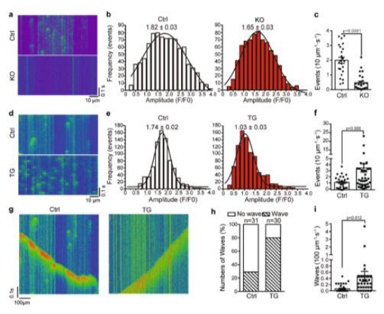
Figure 5.
Light microscopy (100×) - VAD patient pericardium, with pericardial sac on right. Five (instead of the normal three) layers are identifiable: (A) mesothelial, (B) internal fibrosa, (C) loose tissue, (D) external fibrosa, and (E) epipericardium. The loose tissue is rich in vessels. No inflammatory cellularity was identified in any of the five layers. In both fibrous layers, wavy collagen bundles are visible; however, the wavelength of fibers in layer B (internal fibrosa) is smaller than that in layer D (external fibrosa). Taking together the increased amount of collagen bundles, in wavy, accordion-like arrangements, with different orientations in relation to each other, and with more than one elastic fiber accompanying the bundles at seemingly perpendicular angles (seen through electron microscopy, not shown), seems to suggest a pneumatic-like structure, designed to absorb abnormally large external forces. Similarly, this functional arrangement also explains why there is no diastolic dysfunction, despite the thickened pericardial walls. (2) SEM of non-VAD patient pericardium. Normal three layers are visible: mesothelium (white arrow), fibrosa (black arrow), and epipericardium. (3) SEM of VAD patient pericardium. Fibrosa has split into two halves (arrows) that sandwich a newly formed layer of loose tissue (L). With permission [53].
Figure 5.
Light microscopy (100×) - VAD patient pericardium, with pericardial sac on right. Five (instead of the normal three) layers are identifiable: (A) mesothelial, (B) internal fibrosa, (C) loose tissue, (D) external fibrosa, and (E) epipericardium. The loose tissue is rich in vessels. No inflammatory cellularity was identified in any of the five layers. In both fibrous layers, wavy collagen bundles are visible; however, the wavelength of fibers in layer B (internal fibrosa) is smaller than that in layer D (external fibrosa). Taking together the increased amount of collagen bundles, in wavy, accordion-like arrangements, with different orientations in relation to each other, and with more than one elastic fiber accompanying the bundles at seemingly perpendicular angles (seen through electron microscopy, not shown), seems to suggest a pneumatic-like structure, designed to absorb abnormally large external forces. Similarly, this functional arrangement also explains why there is no diastolic dysfunction, despite the thickened pericardial walls. (2) SEM of non-VAD patient pericardium. Normal three layers are visible: mesothelium (white arrow), fibrosa (black arrow), and epipericardium. (3) SEM of VAD patient pericardium. Fibrosa has split into two halves (arrows) that sandwich a newly formed layer of loose tissue (L). With permission [53].
Disclaimer/Publisher’s Note: The statements, opinions and data contained in all publications are solely those of the individual author(s) and contributor(s) and not of MDPI and/or the editor(s). MDPI and/or the editor(s) disclaim responsibility for any injury to people or property resulting from any ideas, methods, instructions or products referred to in the content. |
© 2026 by the authors. Licensee MDPI, Basel, Switzerland. This article is an open access article distributed under the terms and conditions of the Creative Commons Attribution (CC BY) license (http://creativecommons.org/licenses/by/4.0/).
Copyright: This open access article is published under a Creative Commons CC BY 4.0 license, which permit the free download, distribution, and reuse, provided that the author and preprint are cited in any reuse.