

Submitted:
12 November 2026
Posted:
16 November 2026
You are already at the latest version
Abstract
Complete thoracic spinal cord injury (SCI) causes severe motor and sensory impairment. Epidural electrical stimulation (EES) may facilitate activation of spinal motor networks, although clinical experience with 32-contact electrode arrays remains limited. This prospective case series evaluated intraoperative electromyography (EMG)-guided electrode placement, postoperative stimulation programming, and functional outcomes in four participants (two males and two females, aged 18–42 years) with thoracic SCI classified as AIS A at baseline. Participants underwent implantation of a 32-contact epidural array targeting the lumbosacral enlargement at T11–T12, followed by eight weeks of individualized EES programming and neurorehabilitation. Intraoperative EMG mapping guided electrode positioning and demonstrated preferential recruitment of proximal muscles by rostral contacts and distal muscles by caudal contacts, with mixed responses from intermediate contacts. Asymmetric responses in two participants were addressed through lead repositioning. After eight weeks, mean Lower Extremity Motor Score increased from 0 to 18.1 ± 1.5; mean light-touch score increased from 51.6 ± 1.0 to 62.1 ± 1.0, and mean pin-prick score increased from 52.2 ± 1.0 to 61.8 ± 1.0. Three participants achieved a Modified Ashworth Scale score of 0, while one regained voluntary anal contraction and deep anal pressure and was reclassified as AIS C. All participants stood for 30–45 minutes using a walker and demonstrated stepping with minimal assistance. These findings support the feasibility of physiology-guided 32-contact EES combined with rehabilitation, without establishing comparative efficacy over lower-density arrays. Larger controlled studies are needed to establish safety and durability.
Keywords:
spinal cord injury
; epidural spinal cord stimulation
; neurorehabilitation
; motor mapping
; neuroplasticity
1. Introduction
Spinal cord injury (SCI) is one of the most destructive neurological injuries, resulting in permanent motor, sensory and autonomic dysfunction and significantly impacting quality of life. Globally, more than 750,000 new cases of traumatic SCI are reported annually, with thoracic injuries accounting for a substantial proportion of cases. Individuals with complete thoracic SCI, classified as ASIA Impairment Scale Grade A, experience total loss of voluntary motor and sensory function below the level of injury and have historically been considered to have minimal potential for neurological recovery [1,2,3].
Conventional rehabilitation strategies, including physiotherapy, occupational therapy, and locomotor training, primarily aim to preserve residual function, prevent secondary complications, and improve independence in activities of daily living [5]. While such approaches may yield partial benefits in individuals with incomplete SCI, meaningful recovery in complete SCI has long been regarded as unlikely due to the loss of supraspinal input to spinal motor networks. As a result, therapeutic innovation in this population has traditionally focused on compensation rather than restoration of function [4,6].
Over the past two decades, advances in neurophysiology have challenged this pessimistic view by demonstrating that the spinal cord possesses intrinsic circuitry capable of creating coordinated motor output independent of brain input. Lumbar–sacral central pattern generators (CPGs) can produce rhythmic, task-specific locomotor activity when appropriately modulated [7,8,9]. In the preclinical studies, epidural spinal cord stimulation (EES) has been found to re-engage the dormant spinal networks and restore stepping and standing in animal models of complete SCI [10,11,12] via task-dependent motor activation (TDMA).
These insights gained in animal models have since radically informed human research. Clinical studies have shown that EES can be used to facilitate voluntary movement, standing with body weight and assisted stepping in people with chronic SCI, including those with complete motor and sensory loss. [13]. Importantly, these effects are not solely attributable to direct muscle activation but appear to involve modulation of spinal excitability, facilitation of residual descending pathways, and activity-dependent plasticity within spinal networks [14]. These observations suggest that complete SCI may represent a state of functional disconnection rather than irreversible loss of motor capacity [15].
Despite these advances, outcomes following EES remain highly variable across individuals with SCI [15]. Although all critical determinants of therapeutic success are still unknown, the accuracy of epidural electrode placement and the ability to selectively recruit functionally relevant spinal segments appear to play a significant role [16,17]. One of the key obstacles is the absence of universal protocols for selecting electrode configurations and stimulation settings. To this time, different approaches have been used to optimize stimulation protocols. This leads to an inability to compare the results and suboptimal programming in the following studies, and as a result, limited functional carryover. Intra-operative electrophysiological mapping using electromyography (EMG) provides a unique opportunity to identify optimal electrode positioning by visualizing real-time muscle responses and ensuring bilateral, segment-specific recruitment [18,19,20].
Recent technological developments have enabled the use of high-density, multichannel epidural electrode arrays, allowing more refined spatial control of stimulation [21]. However, systematic clinical evaluations of high-density epidural stimulation after SCI remain limited. In particular, the role of high-density electrode arrays in individuals with accomplish thoracic SCI has not been comprehensively examined. Furthermore, the use of such arrays during surgery to guide mapping is under-explored, and few studies have combined detailed intraoperative mapping with subsequent programming and intensive neurorehabilitation to evaluate outcomes in this population [22,23].
The current study aims to fill these gaps by assessing the feasibility of a structured intra- and post-operative mapping protocol with a 32-contact epidural electrode array in people with chronic complete thoracic SCI. We hypothesized that optimized motor recruitment, by accurately mapping the target area and then precisely positioning the electrodes, individualized programming of stimulation parameters, and structured, motor-specific rehabilitation would improve segment-specific motor, sensory, and autonomic function. By focusing exclusively on individuals with complete thoracic SCI, this study provides important insights into the restorative potential of EES in a population traditionally considered beyond the reach of functional recovery.
2. Materials and Methods
This study was conducted as a prospective pilot interventional case series evaluating the feasibility and physiological effects of epidural spinal cord stimulation using a 32-contact electrode array combined with structured neurorehabilitation in individuals with chronic complete thoracic spinal cord injury.
Ethical approval was obtained from the Institutional Review Board of NuRaX Clinics, Baku, Azerbaijan and EGE Hospital, Baku, Azerbaijan (Date- 10th Jan 2025 and Approval number- EPS-NUX-27L-035). The study was conducted in accordance with the Declaration of Helsinki (2013 revision). Written informed consent was obtained from all participants prior to enrollment. The study was not registered in any Clinical Trials Registry.
- A)
- Subject Details
A total of four participants (two males and two females), aged 18–42 years, were enrolled in this study. All participants were diagnosed with complete sensorimotor SCI and classified as American Spinal Cord Injury Association Impairment Scale Grade A (ASIA-A). Written informed consent was obtained from all participants prior to enrollment.
All participants had complete paraplegia with injury levels ranging from T2 to T10. Two participants sustained SCI due to road traffic accidents and had been paraplegic for 6 months –7 years. One participant sustained a spinal cord injury due to a fall from height and had been paraplegic for two years, while another sustained injury due to a gunshot wound and had been paraplegic for six months at the time of enrollment. Although six months post-injury represents a relatively early chronic stage, this participant demonstrated stable neurological status and consistent motor and sensory assessments across repeated evaluations before intervention, indicating a plateau in spontaneous recovery. Based on this clinical stability and suitability for protocol requirements, the participant was considered appropriate for inclusion to assess intervention-related outcomes within the study timeframe. The participant screening, enrollment, and follow-up process is summarized in Table 1, while Table 2 presents the CARE guideline reporting checklist applied to the study. The demographic and baseline clinical characteristics of the four participants, including age, sex, injury mechanism, neurological level, and functional assessments, are presented in Table 3. A total of six individuals were screened for eligibility, of whom four were enrolled and completed the intervention and follow-up assessments.
- B)
- Neuro-Rehabilitation (Motor Training) Before Epidural Stimulation Implantation
Following the diagnosis of SCI, all participants had previously undergone rehabilitation training, including locomotor therapy, at various centres. After enrollment at our institution, participants underwent comprehensive baseline neurological assessment, including detailed sensory and motor evaluations. Prior to the implantation of the epidural electrical stimulation system, we asked all participants to complete 18 motor-training sessions over 8 weeks to understand the effects of rehabilitation with and without Epidural stimulation for our data analysis. About 30 minutes of lower-extremity stretching, 30 minutes of balance training, and task-specific strengthening exercises made up each session. Body-weight support was used for all activities, and trainer and physiotherapist help was given as needed (Table 4).
- C)
- Surgical Procedure for Epidural Stimulation Device Implantation
All participants underwent preoperative whole-spine magnetic resonance imaging (MRI) and comprehensive laboratory investigations. MRI findings were used to identify the damaged spinal segments as well as intact spinal regions responsible for lower-extremity motor control. Surgical planning proceeded only when these target regions were confirmed to be structurally preserved. Under general anaesthesia, participants were positioned in the prone position using a Jackson surgical table and Mayfield head clamp. Inhalational anaesthetic agents and neuromuscular blocking agents were deliberately avoided to allow intraoperative neuromuscular electrophysiological monitoring during epidural electrical stimulation testing. A midline posterior incision was made extending from T11 to L2, guided by fluoroscopy and anatomical landmarks (Figure 1A). The paraspinal muscles were dissected through the lumbodorsal fascia to expose the spinous processes. Vertebral levels were confirmed intraoperatively using fluoroscopy. A subperiosteal dissection was performed from the spinous processes to the laminae and laterally toward the facet joints. The T11–T12 and L1–L2 interspinous ligaments were removed using a rongeur, while facet joints were preserved to maintain spinal stability. The ligamentum flavum was carefully dissected and separated from the underlying dura mater. A 32-contact epidural electrode array (CoverEdge™ 32, Boston Scientific) was positioned over the lumbosacral spinal cord enlargement at the T11–T12 vertebral level, with intraoperative fluoroscopic confirmation of electrode placement (Figure 1B). Intraoperative electromyography (EMG) recordings were obtained bilaterally from the medial hamstring (MH), rectus femoris (RF), medial gastrocnemius (MG), and tibialis anterior (TA) muscles to assess asymmetry in responses and recruitment of the muscles. Stimulation parameters were systematically varied, including voltage (0–10 mA), frequency (0.5–5 Hz), and pulse width (120–450 µs), to map evoked motor responses.
- D)
- Postoperative EMG Mapping and Programming workflow
Postoperative stimulation mapping began after a 3-day recovery period. EMG recordings were obtained in supine, sitting, standing, and stepping conditions. The stimulation parameters were programmed through an iterative physiological process that did not involve trial-and-error experimentation.
Programming followed a structured, stepwise sequence:
- Single-contact testing to establish segmental recruitment thresholds
- Rostro-caudal grouping to define proximal vs distal muscle activation patterns
- Task-specific program development (standing, stepping, knee locking, spasticity suppression)
- Program consolidation, where task-specific programs were merged into a stable combined configuration
Each parameter modification was carried out with real-time EMG monitoring and function checking. The programming was based on symmetry, muscle selectivity, participant comfort and function achievement.
- E)
- Spasticity- Specific Programming Protocol
Participants with Modified Ashworth Scale (MAS) ≥2 received a dedicated resting stimulation program. This program used combined rostral and caudal contacts with low amplitudes (2.0–4.5 mA), higher frequencies (60–70 Hz), and longer pulse widths (340–390 μs). Stimulation was applied for 6–8 hours per day for 5 consecutive days. Spasticity scores were reassessed after 5 days. Severe spasticity in all three subjects was managed through reduction to MAS 0-1. This program was used intermittently by the subjects during the research (Table 5).
- F)
- EMG Latency Analysis
The response latency of the EMG was considered to be > two standard deviations above baseline noise from the stimulation artefact. Latencies were measured during stimulation at a constant pulse rate rather than stepping. For each muscle group, the mean latencies were computed and compared between the intraoperative and postoperative periods (Table 6).
- G)
- Neurorehabilitation Protocol
Pre-implantation Rehabilitation (Baseline Stability Phase)
All patients underwent 18 rehabilitation training sessions within 8 weeks before the surgery to achieve neurological stability and functionality. Each session was 60 minutes and consisted of:
- Passive and active-assisted stretching,
- Sitting and standing balance training,
- Assisted lower-limb movement with body-weight support.
No participant demonstrated independent standing or stepping during this phase.
Post-implantation Rehabilitation
Post-implantation, patients went through physical therapy 5 days a week for 8 weeks, which amounted to about 2 hours a day of:
- Body-weight-supported treadmill stepping (BWSTS),
- Over-ground standing and stepping,
- Trunk stabilization and lower-limb strengthening.
Step training was conducted at 40-60% body weight unloading, which was gradually decreased depending on tolerability and stability (Table 4).
- H)
- Outcome Measures
The following measures were assessed:
- Motor function: Lower Extremity Motor Score (LEMS) (Ditunno et al., 1994)
- Sensory function: Light touch and pin-prick scores (ISNCSCI, 2020)
- Autonomic function: Voluntary anal contraction (VAC) and deep anal pressure (DAP)
- Spasticity: Modified Ashworth Scale (Bohannon & Smith, 1987)
- I)
- Epidural Electrode Configuration and Programming Parameters:
All participants went back to the rehab centre following implantation to be systemically programmed. Several trial-and-error techniques have been employed to determine the best electrode contact section, amplitude, width of the pulse and frequency of stimulation. Due to the large number of possible programming combinations, extensive contact testing was required. The programming was done step-by-step, with continuous monitoring of the results, adjusting the contacts, amplitudes, pulse width and frequency. A variety of stimulation programs had to be used for various functional tasks. Even in the same patient, different standing and stepping patterns required different contact configurations and stimulation parameters. Programs were created initially for individual tasks. After approximately 2-3 weeks, these programs were gradually combined into a single integrated program that enabled independent stepping with an assistive device like a walker. Development of a stable combined program typically required 4-5 weeks, with variability across patients. While contact selection, pulse width, and amplitude varied significantly between patients and tasks, stimulation frequency showed greater consistency. The greatest functional benefit was observed within a frequency range of 35-60 Hz, which was preferentially selected during programming. In below A–D demonstrated electrodes configurations with pulse width, amplitudes and frequency differences in different tasks specifics including one specific programming’s for spasticity.
- J)
- Statistical Analysis
All statistical analyses were performed using IBM SPSS Statistics (version 26.0). Data normality was assessed using the Shapiro–Wilk test. For normally distributed variables, paired-sample t-tests were applied to compare pre- and post-intervention values. For non-normally distributed data, the Wilcoxon signed-rank test was used. Statistical significance was defined as p < 0.05. Continuous variables are reported as mean ± standard deviation (SD).
3. Results
3.1. Epidural Stimulation–Evoked Motor Responses
Epidural stimulation–evoked controlled motor responses were evaluated two weeks post-surgery. After lead implantation, all participants were provided with three days of rest, after which electrode configuration and stimulation mapping were initiated in the supine position. During the first week, participants underwent low-amplitude (from 2.5 mA- 5.0 mA) stimulation mapping to assess bilateral lower-limb muscle responses. Three (P1, P2, P4) of the four participants exhibited severe spasticity, which was significantly reduced following individualized electrode programming. For these participants, a resting stimulation protocol or spasticity programming was applied for five days, during which rostral and caudal contacts were activated within a single program using stimulation amplitudes of 2.0 mA to 3.5 mA, frequencies of 60–70 Hz, and pulse widths of 340–390 µs, adjusted according to participants' comfort (Figure 2D). After one week, all participants returned to the neurorehabilitation centre, where electrode_mapping and physical training were resumed. Blood pressure was monitored daily before and after stimulation for safety assessment. Mapping and locomotor training were conducted twice daily, consisting of one-hour morning and one-hour afternoon sessions. Electrode mapping was performed, and muscle responses were recorded during each session using surface EMG in all participants.
3.2. Epidural Stimulation–Evoked EMG Responses and Intraoperative Mapping
Intraoperative neurophysiological mapping elicited reproducible, short-latency EMG responses in all participants. Single pulse stimulation through each or combination of the electrode contacts resulted in a focal single peak EMG in the RF, medial hamstrings (MH), TA, and MG. Contacts on the rostrum tended to recruit the more proximal musculature, while contacts at the tail area recruited the more distal muscle areas. All participants reached bilateral symmetry of activation after lead optimization during surgery. Lead repositioning was performed when unilateral dominance or insufficient distal muscle recruitment was observed. The resulting short-latency, single-peak EMG responses were consistent with direct dorsal root or motor root recruitment and were used to guide final electrode positioning (Figure 3A–C).
3.3. Postoperative EMG Mapping and Latency Analysis
A standardized single-pulse stimulation pattern was used for postoperative EMG mapping such that direct comparisons could be made with intraoperative mapping. Compared to the intraoperative responses, the postoperative responses were more complex temporally, as evidenced by broader, multi-peak responses but with similar onset latencies. The mean onset latency for the agonist muscles was 10.17 ± 0.87 ms after surgery and 10.42 ± 0.57 ms intraoperatively; for the antagonist muscles, it was 11.04 ± 0.56 ms after surgery and 11.56 ± 1.04 ms intraoperatively. The differences were not significantly different. The preservation of response latency, together with the increment of the complexity of the waveforms, indicates a switch from focal root activation to engagement of the polysynaptic spinal network after the programming and training after surgery.
3.4. Contact-Specific Muscle Recruitment Across the Epidural Array
The systematic testing of the 32-electrode epidural electrode array demonstrated that there was a uniform rostral-caudal arrangement for the recruitment of muscle fibres among subjects. Stimulation of the rostral contact groups primarily activated proximal muscle fibres responsible for hip flexion and stabilization while mid contact groups controlled knee flexion/extension and caudal contacts controlled ankle movements. This segment-specific recruitment pattern provided the physiological basis for developing task-specific stimulation programs, including standing, stepping, knee locking, and spasticity suppression (Figure 4). The contact-specific muscle recruitment map is presented in Figure 4A–D
3.5. Epidural Stimulation–Evoked Controlled Motor Responses at Two Weeks Post-Surgery
After two weeks of EES programming combined with neurorehabilitation, two out of the four participants (P2, P3) demonstrated Volitional step-like bilateral lower-limb movements while standing inside parallel bars with body-weight–support harness assistance. During stepping, bilateral lower-limb muscle activity was recorded simultaneously using surface electromyography (EMG). EES-evoked controlled motor responses while stepping differed in waveform morphology, peak amplitude, and number of peaks between intraoperative and postoperative recordings. However, latencies from stimulus pulse to motor response onset did not differ significantly between intraoperative and postoperative recordings. Mean latencies for agonist muscles were 10.42 ± 0.57 ms intraoperatively and 10.17 ± 0.87 ms postoperatively. For antagonist muscles, mean latencies were 11.56 ± 1.04 ms intraoperatively and 11.04 ± 0.56 ms postoperatively. These differences were not statistically significant across time points.
3.6. Epidural Stimulation Evoked Motor-Controlled Responses at 5 Weeks Post-Surgery
Within the period between the second and fifth postoperative weeks, there was definite functional improvement of EES and neurorehabilitation. The correct setting of the parameters of the stimulation, along with exercises and locomotor training, led to gradual progress in all patients. EES-evoked motor responses and assistance to stepping under epidural stimulation patterns appeared in two patients in the first two postoperative weeks (P1, P3), whereas the rest of the patients (P2, P4) showed motor responses to stepping from the third postoperative week. By the fifth postoperative week, all patients managed to stand without any help, but using a walker with better postural alignment than during the previous sessions. Within the same period, all patients managed to stand without any help but using a walker for 30-45 minutes and began stepping using a platform walker. In addition to two hours of daily EES programming with stepping pattern generation and bilateral knee locking practice, participants underwent trunk stability and range-of-motion exercises. The mean number of step cycles recorded per participant was 30.26 ± 4.00, as assessed using surface EMG. Latencies from stimulation pulses to motor responses were calculated for both agonist and antagonist muscles.
3.7. Epidural Stimulations Evoked Motor-Controlled Responses at 8 Weeks Post-Surgery
At eight weeks post-surgery, all participants were tested using different range of stimulation amplitudes, frequencies, and pulse widths to identify the optimal “sweet spot” for stimulation mapping. Three participants (P1, P2, P4) demonstrated motor responses at lower amplitudes (1.5 mA to 15.5 mA), frequencies of 10–60 Hz, and pulse widths of 180–450 µs. One participant (P3) demonstrated robust motor responses at 3.0–4.0 mA, 30–40 Hz, and 210–270 µs, with consistent voluntary stepping patterns. At the end stage, stimulation was done on the participant by using one program, which was chosen after the earlier muscle and segment mapping was completed. The beginning of the stimulation started by stimulating the small segments of electrodes, which included rostral, medial, and caudal contacts, to activate the nerves selectively. After combining all the small segments, it helped in the formation of a bigger electrode system that would help in bilateral lower limb stepping pattern using a platform walker. The progression of stimulation-enabled functional recovery during the postoperative rehabilitation period is illustrated in Figure 5A–C
3.8. EMG During Functional Stepping
During assisted stepping, EMG recordings demonstrated alternating bilateral activation of lower-limb muscles synchronized with step cycles. Reciprocal burst patterns were observed between tibialis anterior and medial gastrocnemius muscles, corresponding to swing and stance phases of gait, respectively. These EMG patterns were observed only during active stimulation and were reproducible across sessions. Step counts reported elsewhere represent continuous bilateral step cycles achieved during the best session for each participant at the five-week postoperative time point. Functional EMG recordings obtained during stepping were not used for latency analysis (Figure 6).
Individual participant changes in motor and sensory outcomes following the eight-week intervention are presented in Figure 7A–C. Figure 7A illustrates Lower Extremity Motor Scores (LEMS), Figure 7B presents light-touch sensory scores, and Figure 7C shows pin-prick sensory scores before and after the intervention.
5. Discussion
We determined the feasibility and physiological effects of using intraoperative EMG to position the electrode array and postoperative muscle-response programming techniques in patients with complete thoracic traumatic spinal cord injury (tSCI) in this pilot clinical series. We noted consistent segment-specific motor recruitment and functional gains following stimulation during the procedure, while the participants were also provided with structured postoperative programming and intensive neurorehabilitation, resulting in electro physiologically guided motor recruitment with functional gains in the segments targeted for stimulation.
Importantly, this study was not designed to prove efficacy, but rather feasibility and physiological plausibility. Conclusions should thus be evaluated in light of a small, heterogeneous group and the lack of a control group.
5.1. Segment-Specific Recruitment and Functional Organization of the Lumbosacral Cord
Intraoperative and postoperative EMG mapping demonstrated a reproducible rostro-caudal organization of muscle recruitment across the epidural array. Rostral contacts preferentially activated proximal lower-limb musculature, mid-array contacts modulated knee flexion and extension, and caudal contacts recruited distal muscles involved in ankle control [5,8,10]. This pattern is consistent with the known anatomical organization of motor pools within the human lumbosacral enlargement and supports the value of physiology-guided electrode placement for optimizing functional recruitment [18]. The ability to identify and exploit this segmental organization was central to the development of task-specific stimulation programs for standing, stepping, knee locking, and spasticity suppression. While the present study does not compare electrode arrays of differing contact density, the expanded programming flexibility afforded by the 32-contact array facilitated fine-grained modulation of motor output tailored to individual participants and task demands [11,13,16].
5.2. Evolution of EMG Responses and Engagement of Spinal Networks
An important electrophysiological finding was the transition from a focal single-peak EMG response during intraoperative mapping to a broader, multi-peak response during postoperative stimulation in standardized conditions. This transformation took place without any substantial increase in EMG onset latency, indicating that conduction timing did not change significantly, but rather that motor responses became more complex in time (10,17). This pattern does not actually measure synaptic plasticity, but rather an engagement of spinal network circuitry. The findings suggest that the effect of the surgical programming on the spinal excitability and interneuronal integration may be used to modulate motor output under conditions of stimulation, supporting the interpretation that task-specific training following surgery may be used to modulate the excitability of the spinal circuits and the way in which they interact with each other [18,19].
5.3. Importance of Intraoperative Electrophysiological Feedback
Intraoperative EMG feedback during the implantation proved to be crucial in attaining bilateral symmetry and proper recruitment of the distal muscles. Multiple patients had leads positioned in abnormal activation patterns, which were corrected intraoperatively by repositioning the leads to the correct activation pattern and ensuring good functional outcome, demonstrating how important the electrode location is for this functional outcome. The results highlight the need for intraoperative neurophysiological mapping as a useful tool to aid in optimizing lead placement, especially in people with complete thoracic injury, where there is a lack of descending input [20].
5.4. Stimulation-Enabled Functional Outcomes
With active epidural stimulation and aids, participants were able to show improvements in postural control, sustained standing, and stepping alternately between lower-limb bilateral EMGs. These were functional behaviors that were task dependent and repeatable within a session. Importantly, stepping and standing were performed under stimulation-enabled conditions, and is not indicative of independent ambulation over ground. Better LEMS and sensory improvements were noted during the intervention period, but no one converted in AIS grade beyond stimulation-enabled conditions. Thus, these results are not due to spontaneous neurological recovery, but instead to functional improvements resulting from neuromodulation and training [21].
5.5. Interaction Between Epidural Stimulation and Rehabilitation
Participants were all given intensive, task-specific rehabilitation along with epidural stimulation. The relative effect of stimulation and training was not separated due to the lack of a rehabilitation-only control group. Together, functional improvements were seen that are consistent with the notion that epidural stimulation acts as a modulation to the excitability of the spinal circuits and improves the responsiveness to sensory input and volitional effort during training [21,22].
5.6. Role of Preserved T11–L1 and Conus Integrity
All participants met preoperative imaging criteria demonstrating preserved T11–L1 spinal cord anatomy, including integrity of the conus medullaris. Although this study did not include participants with conus disruption for comparison, the consistent physiological recruitment observed supports the importance of this region for effective engagement of lumbosacral motor networks. These findings inform future patient selection strategies and underscore the relevance of anatomical integrity for epidural stimulation–enabled recovery [23].
Notably, the functional gains observed in this study were consistent and emerged early following intervention. Although this study was not designed for direct comparison with previously published cohorts, the observed outcomes align with and extend prior reports of Epidural stimulation-enabled functional recovery in complete thoracic SCI. These findings suggest the integrated, physiology-guided approach used here may influence the extent and pattern of functional improvement.
The superior outcomes achieved here are likely attributable to the integrated, physiology-guided approach, combining:
- (i)
- confirmation of intact T11–L1/conus anatomy,
- (ii)
- intraoperative electrophysiological mapping to ensure accurate rostro caudal electrode placement, and
- (iii)
- adaptive, participant-specific stimulation programming paired with intensive neurorehabilitation.
Collectively, these findings extend previous reports of functional improvement with epidural electrical stimulation in complete thoracic SCI and emphasize the critical role of patient selection and segment specific neuromodulation. In our data, we have identified a trend that long-term preservation of spinal networks, specifically those in the T11–L1 area and conus medullaris, relates to functional improvements observed in conjunction with structured rehabilitation.
6. Limitations
It is important to note that this study has several limitations, such as the small sample size, the heterogeneous injury chronicity and etiology, the lack of the presence of a control group, and the lack of blinding of the assessor. No specific measures of synaptic plasticity or network reorganization were measured, and the findings of EMG were analyzed descriptively. Therefore, mechanistic interpretation is only speculative and needs to be substantiated in more comprehensive and controlled investigations.
Clinical Implications and Future Directions
These are important limitations but the uniformity of physiological recruitment and stimulation-induced functional behaviour among all participants confirms the potential viability of a physiology-guided approach for epidural stimulation in complete thoracic SCI. Future studies should include larger numbers of patients, uniform programming algorithms, rehabilitation-only controls, and follow-up to determine the long-term effects of functional gains. Therapeutic outcomes can be further optimized by integration of adaptive (closed-loop) stimulation strategies.
7. Conclusion
This research shows that accurate real-time intraoperative positioning and mapping of the muscle response of a 32-contact epidural electrode array allows for highly selective, segment-specific activation of spinal motor circuits in individuals with complete thoracic SCI. This correlated with clinically relevant motor and postural functions that were reproducible and measurable with objective electrophysiological mapping and standardized clinical outcome measures, and that were enhanced by individualized epidural stimulation programming and intensive, task-specific neurorehabilitation. Participants showed improvements in voluntary motor activation, postural control and modulation of spasticity; functional improvements were seen most often under conditions of assistance and task dependence, suggesting that this is likely to be due to activation of spinal networks by neuromodulation, and not to spontaneous recovery. Although this study does not aim to prove causal efficacy, the fact that the physiological recruitment of a segment is similar to the observed changes in function justifies the biological plausibility of this strategy.
Although limited by sample size, the consistency of physiological recruitment patterns and functional trends across participants highlights the translational potential of this approach. Larger, longitudinal, multicenter trials are warranted to confirm durability of functional gains, refine patient selection, particularly with respect to conus and lumbosacral segment integrity and establish standardized protocols for broader clinical adoption.
Ethical Approval
Approved by NuRaX Clinics and EGE Hospital, Baku, Azerbaijan, Ethics Committee (Date- 10th Jan 2025 and Approval number- EPS-NUX-27L-035).
References
- American Spinal Injury Association. International Standards for Neurological Classification of Spinal Cord Injury (ISNCSCI) [Internet]. Rev. 2020. Available online: https://asia-spinalinjury.org/international-standards/.
- Angeli, C.A.; Boakye, M.; Morton, R.A.; Vogt, J.; Benton, K.; Chen, Y.; et al. Altering spinal cord excitability enables voluntary movements after chronic complete paralysis in humans. Brain 2014, 137, 1394–1409. [Google Scholar] [CrossRef] [PubMed]
- Bach-y-Rita, P.; Kercel, S.W. Sensory substitution and the human–machine interface. Trends Cogn. Sci. 2003, 7, 541–546. [Google Scholar] [CrossRef] [PubMed]
- Bohannon, R.W.; Smith, M.B. Interrater reliability of a Modified Ashworth Scale of muscle spasticity. Phys. Ther. 1987, 67, 206–207. [Google Scholar] [CrossRef] [PubMed]
- Courtine, G.; Gerasimenko, Y.; van den Brand, R.; Yew, A.; Musienko, P.; Zhong, H.; et al. Transformation of nonfunctional spinal circuits into functional states after the loss of brain input. Nat. Neurosci. 2009, 12, 1333–1341. [Google Scholar] [CrossRef] [PubMed]
- Ditunno, J.F.; Young, W.; Donovan, W.H.; Creasey, G. The international standards booklet for neurological and functional classification of spinal cord injury. Paraplegia 1994, 32, 70–80. [Google Scholar] [CrossRef] [PubMed]
- Edgerton, V.R.; Tillakaratne, N.J.; Bigbee, A.J.; de Leon, R.D.; Roy, R.R. Plasticity of the spinal neural circuitry after injury. Annu Rev. Neurosci. 2008, 27, 145–167. [Google Scholar] [CrossRef] [PubMed]
- Eisdorfer, J.T.; Smit, R.D.; Keefe, K.M.; Lemay, M.A.; Smith, G.M.; Spence, A.J. Epidural electrical stimulation: A review of plasticity mechanisms that are hypothesized to underlie enhanced recovery from spinal cord injury with stimulation. Front Mol. Neurosci. 2020, 13, 163. [Google Scholar] [CrossRef] [PubMed]
- Fong, A.J.; Roy, R.R.; Ichiyama, R.M.; Lavrov, I.; Courtine, G.; Gerasimenko, Y.; et al. Recovery of control of posture and locomotion after a spinal cord injury: Solutions staring us in the face. Prog. Brain Res. 2009, 175, 393–418. [Google Scholar]
- Gad, P.; Gerasimenko, Y.; Zdunowski, S.; Sayenko, D.; Haakana, P.; Turner, A.; et al. Weight bearing over-ground stepping in an exoskeleton with non-invasive spinal cord neuromodulation after motor complete paraplegia. Front Neurosci. 2017, 11, 333. [Google Scholar] [CrossRef] [PubMed]
- Gill, M.L.; Grahn, P.J.; Calvert, J.S.; Linde, M.B.; Lavrov, I.A.; Strommen, J.A.; et al. Neuromodulation of lumbosacral spinal networks enables independent stepping after complete paraplegia. Nat. Med. 2018, 24, 1677–1682. [Google Scholar] [CrossRef] [PubMed]
- Grillner, S. Neurobiological bases of rhythmic motor acts in vertebrates. Science 1985, 228, 143–149. [Google Scholar] [CrossRef] [PubMed]
- Harkema, S.; Gerasimenko, Y.; Hodes, J.; Burdick, J.; Angeli, C.; Chen, Y.; et al. Effect of epidural stimulation of the lumbosacral spinal cord on voluntary movement, standing, and assisted stepping after motor complete paraplegia: A case study. Lancet 2011, 377, 1938–1947. [Google Scholar] [CrossRef] [PubMed]
- Singh, A.; Tetreault, L.; Kalsi-Ryan, S.; Nouri, A.; Fehlings, M.G. Global prevalence and incidence of traumatic spinal cord injury. Clin. Epidemiol. 2014, 6, 309–331. [Google Scholar] [CrossRef] [PubMed]
- Gad, P.N.; Kreydin, E.; Zhong, H.; Edgerton, V.R. Enabling respiratory control after severe chronic tetraplegia: An exploratory case study. J. Neurophysiol. 2020, 124, 2023–2034. [Google Scholar] [CrossRef] [PubMed]
- Taccola, G.; Sayenko, D.; Gad, P.; Gerasimenko, Y.; Edgerton, V.R. And yet it moves: Recovery of volitional control after spinal cord injury. Prog. Neurobiol. 2018, 160, 64–81. [Google Scholar] [CrossRef] [PubMed]
- Angeli, C.A.; Boakye, M.; Morton, R.A.; Vogt, J.; Benton, K.; Chen, Y.; et al. Recovery of over-ground walking after chronic motor complete spinal cord injury. N Engl. J. Med. 2018, 379, 1244–1250. [Google Scholar] [CrossRef] [PubMed]
- Harkema, S.; Gerasimenko, Y.; Hodes, J.; Burdick, J.; Angeli, C.; Chen, Y.; et al. Effect of epidural stimulation of the lumbosacral spinal cord on voluntary movement, standing, and assisted stepping after motor complete paraplegia: A case study. Lancet 2011, 377, 1938–1947. [Google Scholar] [CrossRef] [PubMed]
- Angeli, C.A.; Edgerton, V.R.; Gerasimenko, Y.P.; Harkema, S.J. Altering spinal cord excitability enables voluntary movements after chronic complete paralysis in humans. Brain 2014, 137, 1394–1409. [Google Scholar] [CrossRef] [PubMed]
- Gad, P.N.; Gerasimenko, Y.; Zdunowski, S.; Sayenko, D.; Haakana, P.; Turner, A.; et al. Iron "ElectriRx" man: Overground stepping in an exoskeleton combined with noninvasive spinal cord stimulation after paralysis. Conf. Proc. IEEE Eng. Med. Biol. Soc. 2015, 2015, 1124–1127. [Google Scholar] [CrossRef] [PubMed]
- Kaura, S.; Zafar, S.; Majumdar, P. Restoration of Sitting Balance and Bladder Control via Epidural Stimulation in SCI: A Clinical Study. J. Neonatal Surg. [Internet] 2025J, 14(32S). Available online: https://www.jneonatalsurg.com/index.php/jns/article/view/7439.
- Urban, L.; Thornton, M.; Ingraham, K.; Dale, E.; Zhong, H.; Phelps, P.; et al. Formation of a novel supraspinal-spinal connectome that relearns the same motor task after complete paralysis. J. Neurophysiol. Epub ahead of print. 2023. [Google Scholar] [CrossRef]
- Eisdorfer, J.T.; Smit, R.D.; Keefe, K.M.; Lemay, M.A.; Smith, G.M.; Spence, A.J. Epidural electrical stimulation: A review of plasticity mechanisms that are hypothesized to underlie enhanced recovery from spinal cord injury with stimulation. Front Mol. Neurosci. 2020, 13, 163. [Google Scholar] [CrossRef] [PubMed]
Figure 1.
Surgical details and epidural array placement on table. (A) A midline posterior incision was made extending from T11 to L2, guided by fluoroscopy and anatomical landmarks. The paraspinal muscles were dissected through the lumbodorsal fascia to expose the spinous processes. (B) Invasive spinal cord surgery and 32-contact array placement at the T11-12 level.
Figure 1.
Surgical details and epidural array placement on table. (A) A midline posterior incision was made extending from T11 to L2, guided by fluoroscopy and anatomical landmarks. The paraspinal muscles were dissected through the lumbodorsal fascia to expose the spinous processes. (B) Invasive spinal cord surgery and 32-contact array placement at the T11-12 level.
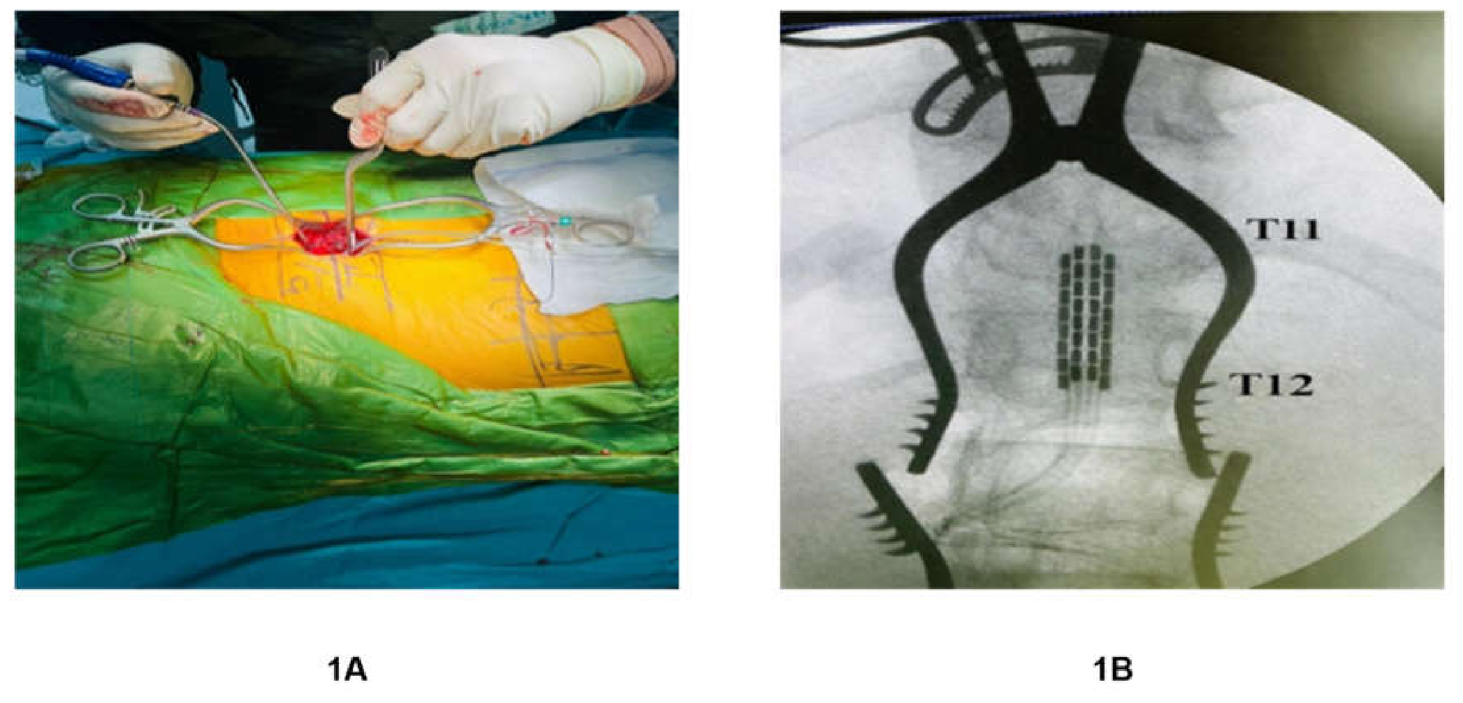
Figure 2.
Representative stimulation configurations from Participant 2 illustrating task-specific programming evolution. (A) Right knee locking configuration, (B) Left knee locking configuration, (C) Final integrated stepping program combining multiple contact groups, (D) Spasticity suppression program. Configurations evolved over approximately three weeks from multiple task-specific programs to a single stable combined program.
Figure 2.
Representative stimulation configurations from Participant 2 illustrating task-specific programming evolution. (A) Right knee locking configuration, (B) Left knee locking configuration, (C) Final integrated stepping program combining multiple contact groups, (D) Spasticity suppression program. Configurations evolved over approximately three weeks from multiple task-specific programs to a single stable combined program.
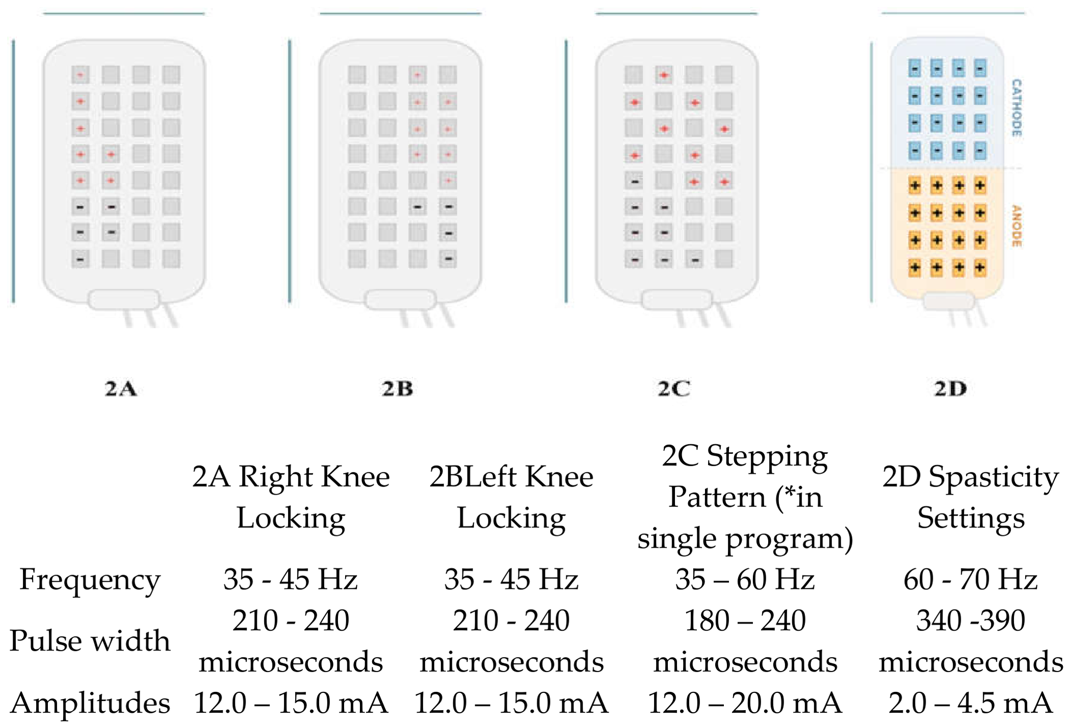
Figure 3.
Schematic representation of epidural electrode positioning and functional segmental recruitment. (A) Diagrammatic illustration showing approximate nerve root coverage (L1–S3) when the electrode array is positioned at the T11–T12 vertebral level. This diagram is schematic and not derived from participant-specific root tracing. (B)Representative intraoperative EMG responses recorded during single pulse stimulation showing short latency, single peak responses consistent with direct root recruitment. (C) Functional interpretation of rostro-caudal contact activation: rostral contacts preferentially recruited proximal musculature, mid-array contacts modulated knee control, and caudal contacts facilitated ankle and plantar flexor activation.
Figure 3.
Schematic representation of epidural electrode positioning and functional segmental recruitment. (A) Diagrammatic illustration showing approximate nerve root coverage (L1–S3) when the electrode array is positioned at the T11–T12 vertebral level. This diagram is schematic and not derived from participant-specific root tracing. (B)Representative intraoperative EMG responses recorded during single pulse stimulation showing short latency, single peak responses consistent with direct root recruitment. (C) Functional interpretation of rostro-caudal contact activation: rostral contacts preferentially recruited proximal musculature, mid-array contacts modulated knee control, and caudal contacts facilitated ankle and plantar flexor activation.
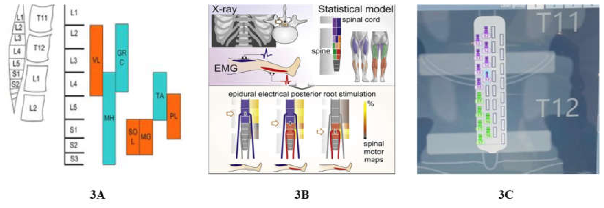
Figure 4.
Contact-specific muscle recruitment map- Schematic representation of contact-specific muscle recruitment across the 32-Contact epidural array, illustrating rostro-caudal functional organization. Model of segmental innervation and the spinal cord: (A) The vertebral body and intervertebral disk heights define the straight-line anatomical model of the spine. (B) Lumbosacral spinal cord straight-line model in accordance with the spine model. (C) The opacity of the corresponding colours represents the segmental innervation probabilities (0%–100%) of Add, RF, TA, SM, ST, BF, Gast, and Sol. (D) Distribution of tested vertebral cathode locations; counts per bin (upper, middle, or lower third of the vertebral body or intervertebral disc).
Figure 4.
Contact-specific muscle recruitment map- Schematic representation of contact-specific muscle recruitment across the 32-Contact epidural array, illustrating rostro-caudal functional organization. Model of segmental innervation and the spinal cord: (A) The vertebral body and intervertebral disk heights define the straight-line anatomical model of the spine. (B) Lumbosacral spinal cord straight-line model in accordance with the spine model. (C) The opacity of the corresponding colours represents the segmental innervation probabilities (0%–100%) of Add, RF, TA, SM, ST, BF, Gast, and Sol. (D) Distribution of tested vertebral cathode locations; counts per bin (upper, middle, or lower third of the vertebral body or intervertebral disc).
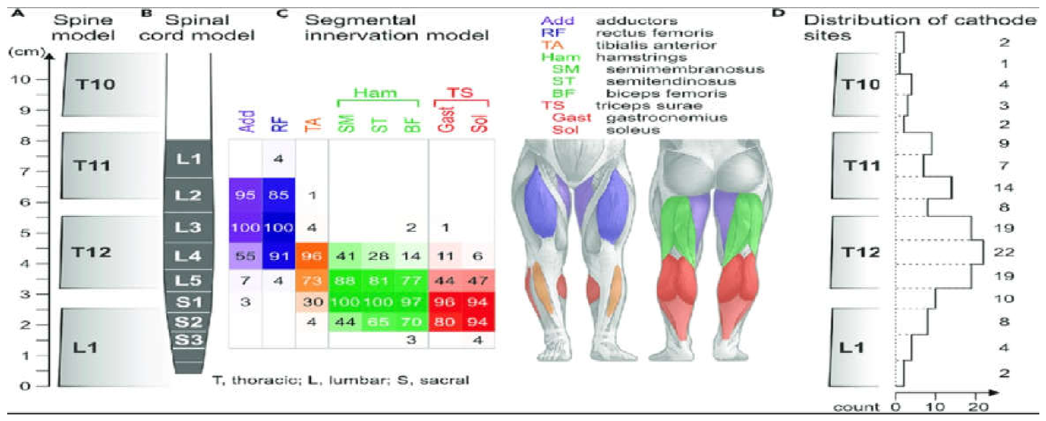
Figure 5.
(A) Illustrates a participant able to initiate stepping after two weeks of training with harness support. (B) demonstrates a participant after five weeks of training maintaining postural stability using a platform walker without harness support. (C) shows a participant after eight weeks of EES training maintaining balance using a standard walker without external support, with bilateral knee locking.
Figure 5.
(A) Illustrates a participant able to initiate stepping after two weeks of training with harness support. (B) demonstrates a participant after five weeks of training maintaining postural stability using a platform walker without harness support. (C) shows a participant after eight weeks of EES training maintaining balance using a standard walker without external support, with bilateral knee locking.
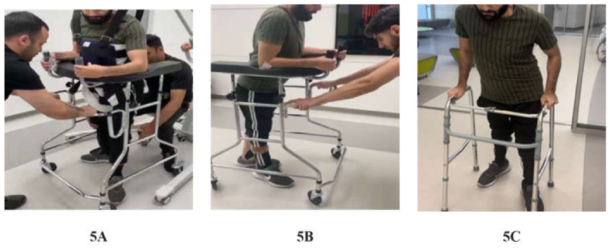
Figure 6.
Joint motions and electromyography (EMG) activity during active treadmill walking in a person with motor-complete spinal cord damage, both with and without continuous 30 Hz lumbar spinal cord stimulation (A and B). EMG activity from the right (R) and left (L) quadriceps (Q), hamstrings (Ham), tibialis anterior (TA), and triceps surae (TS) is shown for ten gait cycles, beginning with right foot contact. Phases of stance are shown by black bars. The stance and swing stages of one example gait cycle are indicated by vertical dashed lines. 2.0 km/h treadmill belt speed; no body weight support; sporadic left foot placement help during stimulation-off condition; stimulation intensity: 27 V; subject 2.
Figure 6.
Joint motions and electromyography (EMG) activity during active treadmill walking in a person with motor-complete spinal cord damage, both with and without continuous 30 Hz lumbar spinal cord stimulation (A and B). EMG activity from the right (R) and left (L) quadriceps (Q), hamstrings (Ham), tibialis anterior (TA), and triceps surae (TS) is shown for ten gait cycles, beginning with right foot contact. Phases of stance are shown by black bars. The stance and swing stages of one example gait cycle are indicated by vertical dashed lines. 2.0 km/h treadmill belt speed; no body weight support; sporadic left foot placement help during stimulation-off condition; stimulation intensity: 27 V; subject 2.
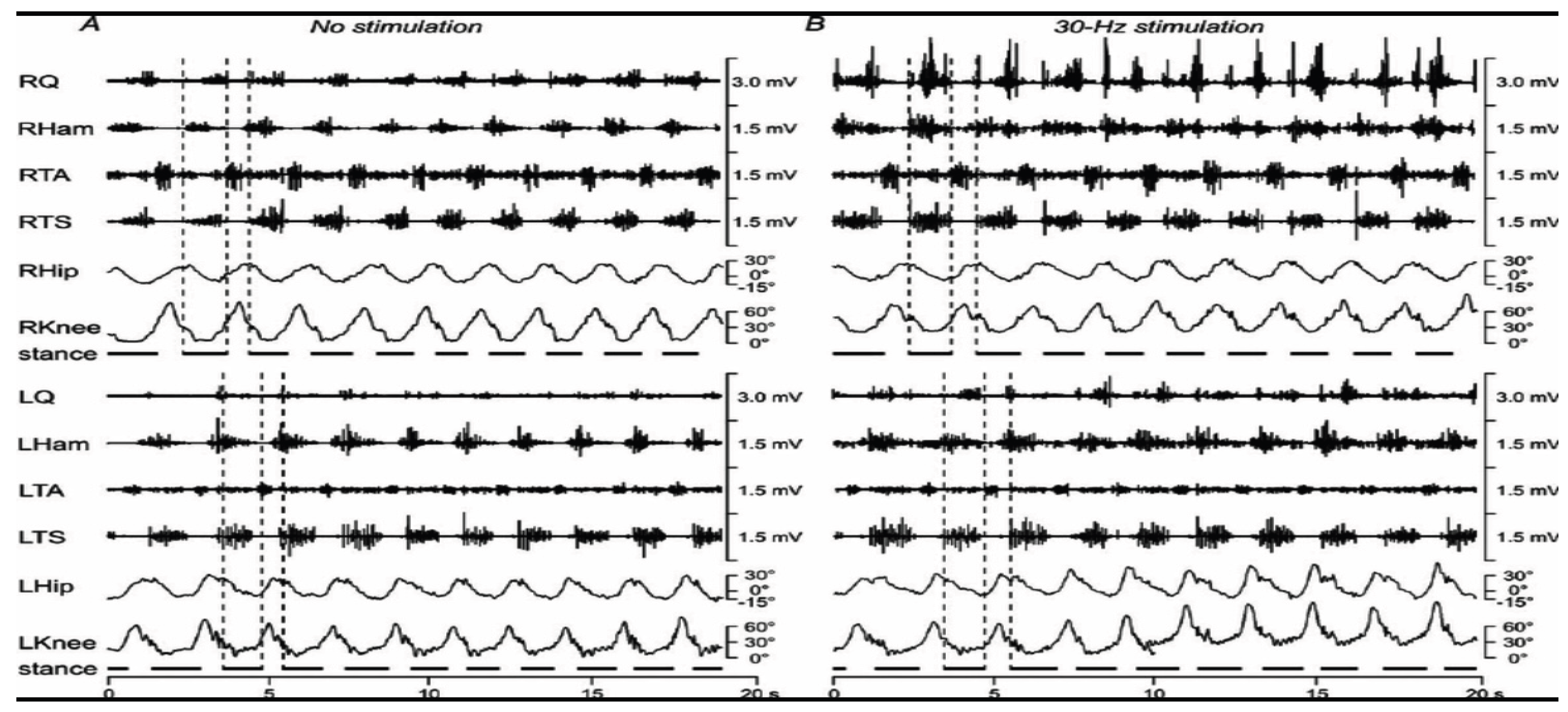
Figure 7.
Motor and sensory outcomes before and after epidural electrical stimulation (EES) combined with neurorehabilitation. (A) Individual participant Lower Extremity Motor Scores (LEMS). (B) Light-touch sensory scores before and after EES. (C) Pin-prick sensory scores before and after EES. The graphs illustrate participant-level changes in motor and sensory outcomes following the intervention.
Figure 7.
Motor and sensory outcomes before and after epidural electrical stimulation (EES) combined with neurorehabilitation. (A) Individual participant Lower Extremity Motor Scores (LEMS). (B) Light-touch sensory scores before and after EES. (C) Pin-prick sensory scores before and after EES. The graphs illustrate participant-level changes in motor and sensory outcomes following the intervention.
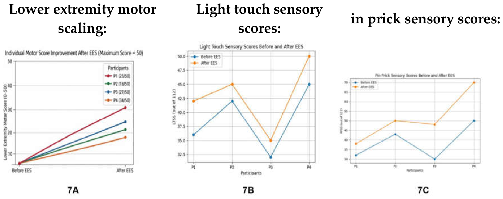

Table 1.
-Study Participant Flow Diagram.
| Stage | Participants (n) | Notes/Exclusions |
|---|---|---|
| Screened for eligibility | 6 |
|
| Excluded | 2 | Not eligible (n=1); Declined participation (n=1) |
| Enrolled | 4 |
|
| Pre-implant rehabilitation (8 weeks) | 4 |
|
| Epidural electrode implantation | 4 |
|
| Postoperative EES + rehabilitation (8 weeks) | 4 | -We have improvements in motor recovery with sensory improvements. |
| Completed follow-up | 4 | All participants completed 8 weeks of rehab with mapping the device. |
| Included in analysis | 4 | - |
Table 2.
CARE Guidelines.
| Section | Item | Description | Where Addressed in Manuscript |
|---|---|---|---|
| Title | 1 | Identify the article as a case report or case series | Title |
| Keywords | 2 | Include key elements of the case | Keywords section |
| Abstract | 3 | Structured summary including background, case presentation, and outcomes | Abstract |
| Introduction | 4 | Background information and rationale for the study | Introduction |
| Participant Information | 5 | Demographics, medical history, and clinical characteristics | Methods – Subject Details |
| Clinical Findings | 6 | Relevant physical examination findings | Baseline neurological assessment |
| Timeline | 7 | Chronological description of events and interventions | Study timeline/protocol |
| Diagnostic Assessment | 8 | Diagnostic tests, imaging, and classification (e.g., AIS grading) | Methods |
| Therapeutic Intervention | 9 | Description of surgical procedure, stimulation programming, and rehabilitation | Methods – Surgical procedure and rehabilitation protocol |
| Follow-up and Outcomes | 10 | Outcomes after intervention including functional and neurological changes | Results |
| Discussion | 11 | Strengths, limitations, and interpretation of findings | Discussion |
| Participant Perspective | 12 | Patient experience (optional) | Not included / optional |
| Informed Consent | 13 | Confirmation that informed consent was obtained | Ethics statement |
Table 3.
Participant demographics were as follows.
| P1 | P2 | P3 | P4 | |
|---|---|---|---|---|
| Age | 30 | 18 | 42 | 35 |
| Gender | Male | Male | Female | Female |
| Mode of Injury | Road Traffic Accident | Road Traffic Accident | Fall from Height | Gunshot Injury |
| Post-injury months | 12 | 84 | 24 | 6 |
| WISCI Scores | 0 | 0 | 0 | 0 |
| Voluntary control of lower muscles | None | None | None | None |
| AIS | A | A | A | A |
| Level of Injury | T2-3 | T5-6 | T6-7 | T9-10 |
| Light Touch Sensory Scale | 36 | 42 | 32 | 45 |
| Pin-Prick Sensory Scale | 32 | 43 | 30 | 50 |
| Voluntary Anal Contraction Scale | No | No | No | No |
| Modified Ashworth Scale for spasticity | 09 | 07 | 02 | 08 |
Table 4.
Rehabilitation Dose and Content.
| Phase | Duration | Frequency | Session Length | Key Comments |
|---|---|---|---|---|
| Pre- Implant | 8 weeks | 2-3/week | 60 min | Stretching, balance, assisted movement |
| Post- Implant | 8 weeks | 5/week | 2 hours/day | BWSTS, Standing, stepping, trunk control |
Table 5.
Postoperative Stimulation Parameter ranges by task.
| Task | Frequency (Hz) | Pulse width (PW) | Amplitude (mA) |
|---|---|---|---|
| Standing/ Knee Lock | 35-45 | 210-240 | 12-15 |
| Stepping | 35-60 | 180-240 | 12-20 |
| Spasticity suppression | 60-70 | 340-390 | 2-4.5 |
Table 6.
EMG Mapping Interpretation Framework.
| EMG Feature | Interpretation |
|---|---|
| Single peak, short latency | Direct root recruitment |
| Multi-peak waveform | Polysynaptic network engagement |
| Bilateral symmetry | Optimal midline lead placement |
| Distal muscle activation | Effective caudal segment engagement |
Disclaimer/Publisher’s Note: The statements, opinions and data contained in all publications are solely those of the individual author(s) and contributor(s) and not of MDPI and/or the editor(s). MDPI and/or the editor(s) disclaim responsibility for any injury to people or property resulting from any ideas, methods, instructions or products referred to in the content. |
© 2026 by the authors. Licensee MDPI, Basel, Switzerland. This article is an open access article distributed under the terms and conditions of the Creative Commons Attribution (CC BY) license (http://creativecommons.org/licenses/by/4.0/).
Copyright: This open access article is published under a Creative Commons CC BY 4.0 license, which permit the free download, distribution, and reuse, provided that the author and preprint are cited in any reuse.