

Submitted:
12 November 2026
Posted:
13 November 2026
You are already at the latest version
Abstract
Alzheimer’s disease (AD) is no longer viewed simply as a proteinopathy driven by amyloid-β (Aβ) and tau. It also involves neuroimmune dysregulation, blood-brain barrier (BBB) and neurovascular unit (NVU) dysfunction, gut-brain axis perturbation, and peripheral metabolic imbalance. Dietary lipids are biologically distinct molecules that can influence these systems through membrane remodeling, lipid-mediator generation, receptor signaling, metabolic support, and central-peripheral crosstalk. Clinical studies of n-3 polyunsaturated fatty acids (PUFAs) and related lipid interventions have nevertheless produced inconsistent or negative results, which may reflect both genuinely limited efficacy in some settings and heterogeneity in lipid species, disease stage, formulation, exposure, and host background. We therefore place the proposed 4D framework within a broader systems-physiology concept of allostatic integrity: the capacity of interacting neural, glial, vascular, immune, and metabolic systems to adapt to repeated perturbations and return toward functional stability. We further use “biological memory” as an operational concept for persistent molecular and cellular state changes—such as epigenetic, metabolic, lipidomic, and glial-state remodeling—that can alter responses to subsequent challenges. The 4D framework comprises Disease stage, Dominant driver pathway, Delivery and formulation, and Data-guided stratification. It is intended as a hypothesis-generating research framework for trial design and stratification, not as a validated clinical algorithm. The 4D framework has not yet been prospectively validated, and its prospective utility remains to be tested.
Keywords:
Alzheimer’s disease
; dietary lipids
; neuroimmune microenvironment
; blood-brain barrier
; gut-brain axis
; lipidomics
; precision nutrition
1. Introduction
Alzheimer’s disease (AD) is the most common cause of dementia worldwide. Amyloid-β (Aβ) deposition and tau pathology are the most recognized pathological hallmarks of AD. They remain the main targets for disease-modifying therapy [1,2]. However, current anti-Aβ and anti-tau treatments show only modest clinical benefits. There is also substantial variability in treatment response among individuals. These observations suggest that AD cannot be understood as a single, uniform brain disorder driven only by protein aggregation [3]. Recent studies from human and animal models show that multiple factors jointly shape the onset and progression of AD. These factors include neuroimmune dysregulation [4], impairment of the blood-brain barrier (BBB) and neurovascular unit (NVU), systemic metabolic reprogramming, and the coupling between central and peripheral inflammatory networks [5,6,7]. Therefore, AD is better understood as a biologically heterogeneous and systemic neurodegenerative disorder [8].
Within this broader conceptual framework, the role of dietary lipids needs to be reconsidered. In this review, “dietary lipids” refers to biologically distinct lipid classes or lipid-derived nutrients obtained through diet or supplementation, including long-chain n-3 PUFAs (especially DHA and EPA), their phospholipid/triglyceride/ethyl-ester forms, and lipid-derived mediators discussed in relation to these precursors [9,10,11,12]; these entities should not be treated as mechanistically interchangeable. Beyond serving as energy substrates and membrane components, selected lipids may influence membrane organization, inflammatory resolution, metabolic support, and gut-periphery-brain signaling [13,14,15]. However, inconsistent and negative clinical findings may reflect a combination of limited efficacy for some interventions and context-dependent effects related to molecular species, disease stage, formulation, exposure, and host background [16].
Accordingly, this review does not treat dietary lipids as a single nutritional exposure; instead, it examines them through the lens of neuroimmune microenvironment remodeling across the course of AD. We focus on three major issues: how stage-specific neuroimmune and neurovascular states define windows of opportunity for lipid intervention, through which mechanistic interfaces dietary lipids influence the AD microenvironment, and why current clinical findings remain inconsistent [6,17]. On this basis, we propose a 4D precision nutrition framework to organize recurrent sources of heterogeneity in the literature and to inform the design of next-generation translational studies [17,18]. This narrative review synthesizes representative recent evidence on disease-stage stratification, lipid metabolism, glial responses, BBB transport, and neurovascular interactions in AD. For clarity and consistency, we use the following operational definitions throughout this review. “Precision nutrition” refers to the use of individual-level biological, dietary, and clinical information to guide the selection, timing, and monitoring of nutritional interventions; it is distinct from personalized supplementation, nutrigenomics, nutrigenetics, and precision medicine. “Preclinical AD” refers to cognitively unimpaired individuals with AD biomarker positivity. “MCI due to AD” refers to MCI with biomarker or pathological evidence supporting an AD etiology; because MCI is etiologically heterogeneous, findings from unselected MCI cohorts are not interpreted as AD-specific. “Clinical AD” refers to clinically diagnosed AD dementia, and “late AD” refers to moderate-to-severe dementia, which is discussed primarily in relation to supportive care, feasibility, and safety. The proposed 4D framework is intended primarily for research on biomarker-defined AD populations and is not intended for unselected MCI cohorts or broader dementia-risk populations without biomarker confirmation. In this review, we distinguish among dietary patterns and food-based exposures, conventional oral DHA/EPA supplements, mixed fish-oil products, medical-food or multinutrient formulations, lipid-derived mediators, lipidomic biomarkers, and experimental delivery systems; these categories differ substantially in composition, bioavailability, and level of evidence, and findings from one should not be generalized to another.
This narrative review was designed to synthesize representative evidence concerning dietary lipids and the neuroimmune microenvironment across the AD continuum. Literature searches were conducted in PubMed/MEDLINE, Web of Science, and Scopus using combinations of terms related to AD, preclinical AD, MCI, omega-3, DHA, EPA, dietary lipids, lipidomics, specialized pro-resolving mediators, microglia, astrocytes, BBB, neurovascular unit, and gut-brain axis. We prioritized peer-reviewed articles published from approximately 2000 through September 2026, while also including selected earlier landmark studies. Priority was given to randomized clinical trials, human cohort studies, biomarker and imaging studies, meta-analyses, and well-characterized experimental studies when human evidence was limited. Because this is a narrative rather than a systematic review, the search was not intended to be exhaustive. Contradictory findings and negative trials were intentionally included. Mechanistic findings derived from animal or in vitro models are identified as preclinical unless supported by direct human evidence.
2. The Neuroimmune Microenvironment of AD Across the Disease Continuum and Lipid Intervention Windows
2.1. AD as a Dynamic Continuum
AD is not a simple linear sequence of Aβ deposition, tau spread, and cognitive decline. Rather, it is a dynamic continuum, and the stage boundaries discussed throughout this review are proposed biological windows rather than fixed temporal landmarks. In this continuum, local brain homeostatic vulnerability progressively evolves into central-peripheral coupled dysfunction [19,20]. In the early phase, abnormalities may manifest as subtle increases in microenvironmental fragility. These include soluble Aβ accumulation, shifts in glial surveillance states, and declining NVU stability [21,22]. As the disease advances, several changes become integrated into the pathological network. These include BBB breakdown, enhanced peripheral inflammatory input, and systemic metabolic imbalance. Eventually, this produces a self-amplifying loop of chronic neuroinflammation and neurovascular injury [20,23].
Human studies support this stratified view. Fluid biomarkers such as glial fibrillary acidic protein (GFAP), soluble triggering receptor expressed on myeloid cells 2 (sTREM2), chitinase-3-like protein 1 (YKL-40), and neurofilament light chain (NFL) suggest that glial reactivity and neurodegeneration-related changes can be detected during preclinical or prodromal stages [24,25]. Magnetic resonance imaging (MRI) studies further indicate that BBB water exchange abnormalities are already detectable in mild cognitive impairment (MCI) [26]. Single-cell and spatial omics studies have added resolution. They reveal state transitions in glial and vascular-associated cells across different pathological stages [27,28]. It should be emphasized, however, that stage boundaries remain blurred at the population level. The temporal position of BBB disruption, the critical timing of glial state transitions, and the specific contribution of peripheral immune infiltration all still lack sufficient direct longitudinal evidence [29,30]. Disease-stage stratification is therefore better understood as a biological organizing framework. It helps interpret heterogeneity in AD. It is not a rigid clinical staging system [20]. Within this framework, the key question for lipid intervention is no longer whether it is “generally effective”. Instead, the question is whether it acts on the right pathological interface at the right stage (Figure 1). In this review, “glial priming” refers to a sensitized cellular state in which prior or ongoing pathological exposure alters baseline transcriptional/metabolic programs and increases or qualitatively changes responsiveness to subsequent stimuli; “reduced glial response threshold” is used descriptively for this increased responsiveness rather than as a directly measured universal threshold.
2.2. Allostatic Integrity and Biological Memory as a Unifying Systems Framework
To provide a theoretical foundation for the proposed 4D framework, we draw upon the concepts of allostatic integrity and biological memory, adapted from systems biology and neuroendocrine stress research [31,32]. Allostasis refers to the process by which the brain and peripheral organs collectively maintain stability (homeostasis) through active physiological regulation in response to changing environmental demands [33]. In the healthy brain, multiple systems—neuroimmune, neurovascular, metabolic, and gut–brain—operate within adaptive ranges, preserving what can be termed allostatic integrity: the capacity to mount and terminate appropriate responses to homeostatic challenges while maintaining functional reserve [31,34,35].
AD progression can be understood as a progressive erosion of allostatic integrity across these interconnected systems. Early-stage pathology (preclinical and MCI) represents a phase of allostatic load—the cumulative wear-and-tear from repeated or chronic challenges, including oxidative stress, glial activation, and metabolic dysregulation [32]. At this stage, adaptive responses remain partially functional, but response thresholds are reduced and homeostatic recovery becomes less efficient. With disease progression, the systems begin to lose their ability to return to baseline after perturbation, a state of allostatic overload, characterized by chronic neuroinflammation, persistent BBB dysfunction, and sustained peripheral immune input [33].
Biological memory further explains why previous exposures and interventions may shape current and future responsiveness. In the context of AD, biological memory encompasses both pathological memory (the lasting effects of prior inflammatory episodes, oxidative damage, or metabolic insults) and therapeutic memory (the persistent benefits or residual adaptations induced by earlier interventions) [31]. These changes may include DNA methylation and histone modifications, persistent shifts in glial transcriptional states, lipidomic remodeling of membrane phospholipids, and stable changes in mitochondrial and metabolic function. In research settings, they could be assessed through longitudinal epigenetic profiling, single-cell transcriptomics, lipidomic analysis, and repeated glial biomarker measurements, although standardized approaches for quantifying biological memory in human AD remain to be established. This concept suggests that when an intervention is delivered may be as important as what is delivered: early-stage interventions that preserve allostatic integrity may have sustained effects beyond the immediate period of treatment, whereas late-stage interventions, when homeostatic reserves are exhausted, may have limited efficacy regardless of the biological potency of the agent [17,31].
Within this theoretical lens, dietary lipids are not simply “nutrients” that provide substrate or “drugs” that inhibit a single target. Rather, they are modulators of allostatic integrity. They can influence membrane composition, inflammatory resolution, BBB function, and metabolic–gut–brain communication in ways that either support or undermine the brain’s capacity to maintain homeostasis across multiple scales [9,11,36]. A lipid intervention that is effective at preserving allostatic resilience in the preclinical stage may be less impactful when the same pathway is already overwhelmed in late-stage disease. Conversely, the apparent inconsistency of clinical trial results may be partially explained by differences in the allostatic state of study populations at enrollment.
This allostatic integrity framework provides the underlying systems-level logic for the 4D precision nutrition approach proposed in this review. The first dimension (Disease stage) captures the shifting balance of allostatic load and reserve. The second dimension (Dominant driver pathway) identifies which physiological system is under the greatest allostatic strain. The third dimension (Delivery and formulation) addresses whether sufficient biological signal can be delivered to restore system function. The fourth dimension (Data-guided stratification) uses biomarkers to estimate individual allostatic states and predict likely responses. In this way, the 4D framework is elevated from a descriptive checklist to a systems biology-guided approach for organizing lipid intervention research in AD.
2.3. Proposed Biological Windows for Lipid Intervention
The following stage assignments should be interpreted as proposed biological windows rather than a fixed temporal sequence. AD trajectories overlap substantially, and biomarker-defined states may not align with chronological or clinical categories. In a preclinical or high-risk state, covert shifts may include altered glial surveillance, membrane-lipid disequilibrium, and early BBB/NVU vulnerability [7,37]. At this stage, the most rational goals of lipid intervention are to preserve membrane lipid homeostasis, support barrier resilience, and maintain inflammatory resolution capacity, with biomarker rather than short-term cognitive endpoints [11,38]. MCI may represent one potentially informative—but not uniquely privileged—window because measurable glial and vascular abnormalities can coexist with residual neuronal and synaptic reserve [24,37]. These propositions require longitudinal and interventional validation and should not be interpreted as established treatment timing rules [39].
Once clinical AD is established, several changes become more pronounced. These include chronic microglial activation, reactive astrocytosis, tau-associated network spread, and synaptic loss. BBB and NVU impairment and persistent peripheral inflammatory input further aggravate pathology [40,41]. At this point, lipid intervention is more realistically positioned as part of a multimodal therapeutic strategy. Its likely role is to dampen the neuroinflammation–neurovascular injury loop. It also supports membrane and energy metabolic homeostasis. It is not meant to reverse existing structural damage [10,42]. In late-stage AD, several factors further constrain responsiveness. These include widespread neuronal loss, reduced barrier transport capacity, and poor nutritional-metabolic reserve. Lipid intervention in this context should mainly be considered a supportive strategy. For example, it may improve nutritional tolerance, assist energy metabolism, or buffer part of the inflammatory burden [7,43]. Overall, disease-stage stratification strongly argues against the continued use of a single paradigm. In that old paradigm, one lipid is uniformly supplemented and overall cognitive change is used as the sole outcome. Dominant pathology, biological plasticity, and delivery conditions differ markedly across stages. Accordingly, intervention goals and endpoints should also differ (Table 1).
3. Key Mechanistic Interfaces Through Which Dietary Lipids Remodel the AD Neuroimmune Microenvironment
To avoid reducing diverse dietary lipids to generic “anti-inflammatory nutrients”, a more useful approach is to understand their effects through several key mechanistic interfaces. Broadly speaking, preclinical and MCI stages are characterized more by homeostatic fragility, insufficient inflammatory resolution, and BBB and NVU dysfunction. Clinical and late stages are dominated more by chronic inflammatory amplification, neurovascular uncoupling, and failure of metabolic support. Consequently, the same class of lipids may act on different pathological interfaces at different stages. They do not exert a fixed and uniform effect.
3.1. Interface 1: Membrane Lipid Composition and Receptor Signaling
The membrane lipid environment directly influences receptor clustering, conformational stability, and downstream signaling efficiency. Although both DHA and EPA are n-3 PUFAs, they differ in membrane incorporation and functional emphasis. DHA is more readily incorporated into neuronal membrane phospholipids. It influences membrane fluidity and lipid raft organization. EPA more strongly competes with arachidonic acid pathways. It may have a greater impact on systemic inflammatory tone [14,38]. These distinctions suggest that different dietary lipids are not biologically interchangeable in AD [47].
In the AD context, activation thresholds and signaling outputs of receptors depend on specific membrane microenvironments. These receptors include Toll-like receptor 4 (TLR4), CD36, and triggering receptor expressed on myeloid cells 2 (TREM2) on microglia and brain endothelial cells [9,10,24]. Altered membrane lipid composition may therefore influence inflammatory initiation, phagocytic efficiency, and clearance of pathological debris [11,12]. Some early effects of lipid intervention may thus reset receptor signaling platforms, rather than immediately lowering cytokine levels.
3.2. Interface 2: Inflammatory Resolution and Glial State Regulation
Microglia and astrocytes are central effectors of the AD neuroimmune microenvironment [48]. Recent single-cell studies indicate that AD-associated glia cannot be adequately described by a binary “resting and activated” model. Instead, they occupy a continuum of states that includes disease-associated microglia (DAM), interferon-responsive microglia, and lipid droplet-accumulating microglia (LDAM), among others [21,27]. It has been shown that during early amyloid pathology, pro-inflammatory and anti-inflammatory microglial subgroups are co-regulated by common upstream regulators. This indicates that microglial heterogeneity is organized through coordinated transcriptional programs. It is not driven by independent polarization states [49,50]. In this context, the key question is not whether a lipid is simply “anti-inflammatory”. Instead, the question is whether it can help shift glial cells from a high-inflammatory, low-phagocytic, metabolically dysregulated state toward one that is more permissive to inflammatory resolution and pathological clearance [11,51].
One central mechanism at this interface involves specialized pro-resolving mediators (SPMs). DHA and EPA serve as precursors of resolvins, maresins, and protectins [11,52]. Unlike traditional anti-inflammatory frameworks that emphasize suppression of inflammation, SPMs support the active termination of inflammatory responses. They also promote the clearance of debris and restoration of tissue homeostasis [52]. In human microglial models, maresin 1 derived from DHA has been shown to reduce the inflammatory gene expression changes induced by Aβ42, although this evidence is derived from in vitro human microglial models and requires in vivo confirmation [11]. This suggests that a major problem in AD may not be excessive inflammation alone, but insufficient execution of resolution programs. It should be noted that “pro-resolving” describes a biological mechanism of actively terminating inflammation, and should not be equated with clinically established anti-inflammatory efficacy in AD. Direct evidence that SPM-based interventions produce clinical benefit in AD patients remains limited. It should also be noted that SPMs are rapidly metabolized and technically challenging to measure in biological fluids, which may partly explain the limited human evidence.
Emerging transcriptomic and functional evidence suggests that TREM2 contributes to the regulation of microglial activation states in AD. It may exert stage-dependent effects during disease progression [24,53]. Lipid droplet dysregulation represents another important link between glial dysfunction and systemic metabolic disturbance. Lipid droplet-accumulating microglia often exhibit impaired phagocytosis, increased reactive oxygen species (ROS), and reduced metabolic flexibility [9,51]. ANGPTL4-mediated accumulation of microglial lipid droplets suggests that peripheral metabolic abnormalities such as obesity may influence brain glial states through disordered lipid handling [51]. Likewise, in astrocytes, nodes related to fatty acid oxidation have been proposed as candidate links between lipid metabolic dysfunction and reactive transformation. These include downregulated carnitine palmitoyltransferase 1A (CPT1A) [10,12]. At present, however, direct evidence that SPM supplementation or lipid droplet remodeling improves glial states in human AD remains limited. This evidence is derived mainly from in vitro and animal models. This mechanistic interface should therefore still be viewed primarily as a translationally relevant working hypothesis (Figure 2), which uses microglia as a representative cellular interface. It is not a fully established human pathway [11].
Astrocytes form a complementary lipid-sensitive arm of this neuroimmune network. They synthesize and redistribute cholesterol and APOE-containing lipoproteins, take up neuron-derived fatty acids, store neutral lipids in lipid droplets, and provide metabolic substrates that support neuronal and synaptic function. Perturbation of fatty-acid oxidation, cholesterol trafficking, or lipid-droplet turnover can therefore couple metabolic stress to reactive astrocyte programs. APOE ε4 is particularly relevant: experimental work shows altered astrocytic lipid-droplet composition, turnover, and susceptibility to lipid peroxidation, while AD-associated sulfatide/myelin-lipid disturbances can induce astrogliosis independently of TREM2-mediated microglial activation [54]. Astrocytes also regulate BBB/NVU function through endfeet-endothelial interactions, ion and water homeostasis, trophic support, and inflammatory signaling. Their crosstalk with microglia is bidirectional: microglial cytokines and complement-related signals can reshape astrocyte states, whereas astrocyte-derived lipoproteins, cytokines, chemokines, and metabolites can alter microglial phagocytosis and inflammatory tone. Consequently, lipid intervention should be evaluated at the level of coordinated microglia-astrocyte-NVU state transitions rather than microglia alone. Much of the astrocyte-related evidence discussed here is derived from preclinical models (in vitro and animal studies), and direct human in vivo validation remains limited [10,12,54].
3.3. Interface 3: BBB and NVU Integrity and Brain Delivery Accessibility
The BBB and the NVU are essential for maintaining brain homeostasis and restricting peripheral inflammatory influx. The NVU is composed of brain microvascular endothelial cells, pericytes, astrocytic endfeet, and extracellular matrix [5,7]. In AD, BBB and NVU dysfunction is not only a major amplifier of pathology. It is also a biological filter that determines whether lipid interventions can achieve effective brain exposure [20,46]. In other words, even if a lipid reaches measurable concentrations in the periphery, its central effect may remain limited. This occurs if it fails to cross the barrier and enter target cells.
Available evidence suggests that DHA and related n-3 PUFAs may influence brain endothelial function indirectly. They may do so by improving membrane fluidity, lowering inflammatory sensitivity, and supporting tight junction stability, although this evidence is largely derived from animal models and in vitro endothelial cell studies [55]. Pericyte injury is increasingly viewed as an early driver of barrier dysfunction in AD. SPMs may also exert barrier-restorative and vasculoprotective effects [7]. However, this interface is also one of the most overlooked translational bottlenecks. Brain endothelial transport dysfunction and oxidative stress may arise early in AD. They reduce trans-barrier lipid efficiency [46,56]. In the context of apolipoprotein E ε4 (APOE ε4), DHA loading, transport, and brain utilization may be further compromised [44,54,57]. However, evidence that APOE ε4 modifies lipid transport and brain lipid homeostasis should be distinguished from evidence that APOE ε4 predicts a clinically meaningful response to a specific DHA, EPA, fish-oil, or multinutrient formulation. The former is supported by convergent mechanistic and observational evidence, whereas the latter remains inconsistent and insufficient for genotype-specific prescribing. Future studies should therefore evaluate not only how much lipid is supplemented. They should also evaluate whether it reaches the target system in a biologically meaningful form.
3.4. Interface 4: Metabolic-Gut-Brain Axis Coupling
The AD neuroimmune microenvironment is not a closed intracerebral system. It is continuously shaped by signals originating from the gut, liver, adipose tissue, and peripheral immune system [36,58]. Dietary lipids may alter brain-related biology indirectly. They do so by reshaping the gut microbiota and its metabolic outputs. Short-chain fatty acids (SCFAs) are among the most studied relevant metabolites. Their levels are influenced jointly by dietary fat and fiber. They may affect BBB stability and microglial maturation indirectly. They do this by modulating gut barrier integrity, immune differentiation, and systemic inflammation [36,59].
At the same time, metabolic comorbidities such as obesity, insulin resistance, and fatty liver disease alter lipid absorption, distribution, and inflammatory output. They thereby affect glial states in the brain and responsiveness to lipid intervention [45,51]. These factors should not be treated as peripheral confounders only. They may directly determine the direction and magnitude of intervention response. It must also be acknowledged that causal evidence for direct effects of gut-derived lipid metabolites on the AD brain microenvironment remains limited. Most human studies are correlational (Tier II observational evidence) rather than interventional [36,58]; the mechanistic chain from gut metabolites to brain glial states is largely supported by preclinical models. A more balanced interpretation is therefore that dietary lipids in AD do not simply “nourish the brain”. Instead, they participate in a continuous regulatory chain. This chain links the gut, peripheral metabolism, BBB and NVU, and brain-resident glia [41]. Through a cascade of serial effects linking the gut microbiota, peripheral immunity, and the BBB interface, dietary lipids can also indirectly remodel the intracerebral microenvironment (Figure 3).
3.5. Mechanistic Integration: From Membrane Remodeling to Systemic Crosstalk
The effects of dietary lipids in AD can be understood as a layered network. The first layer is membrane remodeling. This determines basic effects on membrane fluidity, lipid raft structure, and receptor organization. The second layer is receptor and transcriptional network modulation. This influences state transitions in glia and vascular cells. The third layer is lipid mediator generation. This particularly involves the conversion of DHA and EPA into SPMs. This may influence whether inflammation remains chronic or proceeds toward resolution. The fourth layer is systemic crosstalk. This encompasses gut microbiota, peripheral lipid handling, metabolic comorbidities, and BBB integrity. All this shapes the ultimate brain response [8,14,36]. The evidence supporting these layers is not uniform. Some components are supported by extensive model-based work. Direct validation in humans remains sparse. Accordingly, this framework should currently be regarded as a biologically informative integrative model. It is not a fully established pharmacological pathway map. The evidence supporting these four layers is derived from a mix of preclinical (animal and in vitro) and human observational studies, with direct interventional human validation remaining sparse for most mechanisms discussed.
4. How Multi-Omics Supports Lipid-Related Stratification and Target Discovery in AD
The value of multi-omics in AD lipid research lies not merely in increasing data dimensionality. It lies in helping to identify disease heterogeneity, critical cellular states, and patient subgroups that are more likely to benefit from intervention [60,61]. In other words, multi-omics should serve the questions of who may benefit, at what stage, and through which mechanism. It should not become an end in itself.
4.1. Single-Cell and Spatial Omics: Identifying Target Cells and Lesion Niches
Traditional lipid studies have often relied on bulk tissue analysis. This approach cannot determine in which cell types or lesion environments lipid changes occur. Single-cell and single-nucleus transcriptomics have improved this resolution substantially.In microglia, programs related to lipid handling, phagocytosis, and inflammation are prominently reconfigured in disease-associated subpopulations. In astrocytes, fatty acid oxidation pathways also show AD state-dependent alterations [12,21,27]. Spatial technologies further suggest that lipid remodeling is not uniformly distributed throughout the brain. It often clusters around Aβ plaques, white matter lesions, or specific cortical layers [62,63,64]. For precision lipid intervention, therefore, it is equally important to know which cell types are targeted and which pathological niches are reached.
4.2. Fluid and Tissue Lipidomics: From Risk Signals to Stratification Biomarkers
Whereas single-cell and spatial approaches are particularly useful for mechanistic localization, fluid and tissue lipidomics are closer to clinical translation. Existing studies indicate that specific neutral lipid, ceramide, and sphingomyelin patterns in cerebrospinal fluid (CSF) are associated with the risk of progression from MCI to AD [24,65]. For instance, specific CSF ceramide and sphingomyelin species, such as Cer (d18:1/16:0) and SM (d18:1/16:0), have been linked to the risk of progression from MCI to AD [65]. However, it should be noted that these lipidomic biomarkers have not yet been validated in independent, large-scale cohorts and should currently be considered exploratory rather than established stratification tools. Plasma lysophosphatidylcholine (lysoPC) species and DHA and EPA-related lipid modules may also correlate with early phosphorylated tau (p-tau) levels and AD risk [57,66]. For example, specific plasma lysoPC species, including lysoPC 16:0 and lysoPC 18:0, have been associated with early p-tau181 levels [66].Tissue lipidomics showing focal lipid remodeling provides pathological context for these fluid findings [62,63]. Thus, the most translationally promising readouts may not be single lipid molecules. Rather, they may be lipid feature modules that jointly reflect membrane metabolism, inflammatory status, myelin homeostasis, and peripheral metabolic background.
4.3. Integrative Multi-Omics for Precision Lipid Intervention Stratification
The real significance of multi-omics integration lies in combining genetic background, cellular states, lipid features, spatial niches, vascular measures, and clinical phenotypes into testable stratification models [60,61]. AD lipid biology provides concrete examples. CSF neutral-lipid patterns have been associated with progression from MCI to AD [65]; lysophosphatidylcholine species have been linked to early p-tau181 [66]; mass-spectrometry imaging demonstrates plaque-associated spatial lipid remodeling in mouse and human brain [62]; APOE ε4 is associated with distinct cerebrovascular lipid profiles [57] and with ACSL1-positive, lipid-droplet-rich microglial states in human AD tissue [51]. These features can be integrated with astrocyte fatty-acid oxidation/reactivity programs, GFAP or sTREM2, BBB imaging, and APOE genotype. Thus, a useful AD-specific workflow would test whether genotype-lipid modules co-localize with particular glial states and lesion niches, and whether those combinations predict target engagement or response to a defined lipid formulation. Such models should be prespecified and externally validated rather than used only for post-hoc subgroup discovery (Figure 4).
5. Biological and Design-Related Sources of Inconsistency in Clinical Evidence
Although extensive preclinical work and some randomized controlled trials (RCTs) suggest neurobiological effects of n-3 PUFAs and related lipids, the overall clinical evidence remains inconclusive and includes well-conducted negative studies [38,39]. A negative result may reflect inadequate exposure, stage or endpoint mismatch, but it may also indicate that the tested lipid has little or no clinically meaningful effect in that population. These alternatives must be distinguished rather than assuming design mismatch whenever efficacy is not observed. [16,18].
First, intervention molecules are not equivalent. DHA and EPA differ mechanistically. Mixed fish oil products, pure DHA, pure EPA, and multinutrient formulations each carry distinct biological properties [14,39]. If efficacy is judged mainly by daily dose, true molecule-specific effects may be obscured. They may be misinterpreted as unstable results. Second, insufficient central exposure is likely an underappreciated limitation. Molecular form and carrier system influence absorption, distribution, and brain uptake routes. An increase in peripheral levels does not necessarily mean that effective target tissue exposure has been achieved in the brain [44,55]. Third, mixing disease stages can substantially dilute signal detection. Individuals in preclinical or MCI stages are more likely to benefit at the level of membrane homeostasis, inflammatory resolution, and BBB support. Patients with established dementia may already have severe neuronal loss and barrier dysfunction. These limit the reversibility of damage by nutritional supplementation alone [20,39]. Fourth, inadequate participant stratification is another major reason for inconsistent findings. APOE ε4 status, baseline omega-3 status, and metabolic conditions such as obesity, insulin resistance, and fatty liver disease may all alter lipid absorption, distribution, and biological response [14,44,61].
A further recurrent problem is mismatch between endpoints and mechanism level (Table 2), but this should be treated as one possible explanation rather than a default explanation for failure. Earlier biological effects may appear in membrane composition, SPM generation, vascular measures, or atrophy rates before cognitive scales change [15,16]. Conversely, demonstration of target engagement without clinical benefit would argue for limited downstream efficacy. Future studies should therefore be designed to distinguish exposure failure, target-engagement failure, mechanism failure, and true lack of clinical efficacy. The inconsistency of omega-3 and other lipid-intervention trials should not be attributed solely to inadequate stratification. Disease stage, formulation, dose, duration, adherence, background diet, and brain exposure may contribute to heterogeneous results. However, negative findings may also indicate that a given intervention has limited or no clinically meaningful effect in the tested population. Future trials should therefore be designed not only to identify responsive subgroups, but also to falsify mechanistic hypotheses and determine settings in which lipid interventions are unlikely to be beneficial.
6. A 4D Precision Nutrition Framework for Dietary Lipids in AD
Based on the mechanistic and translational evidence discussed above, we propose a 4D precision nutrition framework to guide AD lipid research and intervention design. Importantly, this framework is not intended to establish a new disease classification system, nor is it a direct clinical prescribing algorithm. It has not yet been prospectively validated and should be viewed as a hypothesis-generating research tool for trial design and stratification, rather than a clinically validated precision-nutrition algorithm or prescribing framework [17]. Rather, it organizes recurrent sources of heterogeneity in existing evidence in a way that can improve study design and participant stratification [18,41]. The four dimensions are: Disease stage; Dominant driver pathway; Delivery and formulation; Data-guided stratification (Table 3).

Table 3.
Major sources of inconsistency in AD lipid intervention trials and implications for next-generation study design.
Table 3.
Major sources of inconsistency in AD lipid intervention trials and implications for next-generation study design.
| Source of heterogeneity | Specific issue | Impact on trial results | Suggested improvement |
|---|---|---|---|
| Intervention molecule heterogeneity [14,15,16,38,39] |
DHA, EPA, mixed fish oil, and multinutrient formulas are not equivalent | Dilutes true molecule-specific effects | Report DHA, EPA, and composite formulations separately |
| Formulation and delivery differences [14,44,55] |
TG, EE, PL, lysoPC, intranasal delivery, etc., may differ in brain exposure | Peripheral increase does not equal effective brain exposure | Incorporate pharmacokinetics and brain delivery verification |
| Disease-stage mixing [1,2,13,18,41] |
Preclinical, MCI, and dementia-stage participants are enrolled together | Different levels of plasticity dilute average effect | Stratify or enrich enrollment by disease stage |
| Genetic background [14,41,44,57,67] |
APOE ε4 affects brain omega-3 metabolism and cerebrovascular lipid profiles | Same intervention may act differently across genotypes | Prespecify APOE-stratified analyses |
| Baseline nutritional and metabolic status [16,19,41,45] |
Baseline omega-3 status, obesity, insulin resistance, fatty liver, etc. differ | Alters absorption, distribution, and immunometabolic response | Measure red blood cell omega-3 index and metabolic phenotype |
| Endpoint mismatch [15,16,18,39] |
MMSE/ADAS-cog used as sole determinant of success | May miss biological intermediate changes | Combine cognitive, imaging, fluid, and lipidomic endpoints |
| Insufficient mechanistic validation [11,29,46,55] |
BBB status, SPM generation, or brain exposure not assessed | Cannot distinguish “mechanism failure” from “exposure failure” | Include mechanistic intermediate endpoints |
| AD biological confirmation [24,39] |
Unselected MCI or clinical dementia cohorts may include non-AD etiologies | Dilutes AD-specific effects and increases biological heterogeneity | Prespecify AD biomarker confirmation where feasible |
| Adherence, duration, and background diet [15,16,39] |
Capsule burden, GI symptoms, low compliance, short follow-up, and variable background diets | Insufficient exposure and inability to detect slow effects | Monitor adherence objectively, justify duration, and measure baseline diet |
| Sex, frailty, comorbidity, and medication use [45,68] |
Sex differences, sarcopenia, vascular disease, renal/hepatic dysfunction, and co-medications (anticoagulants, antiplatelets, lipid-lowering drugs) | Alters metabolism, safety, competing risks, and response | Prespecify subgroup analyses and document co-medications |
| Product quality, oxidation, and socioeconomic access [38,55] |
DHA/EPA ratio, chemical form, oxidation, storage, contaminants, and unequal access to lipidomics, APOE testing, MRI, or CSF | Variable biological activity and limited generalizability | Report product composition and stability; use tiered, scalable assessment strategies |
| Intervention duration and follow-up length [15,16] |
Short follow-up in many trials may fail to capture slow disease-modifying effects | Insufficient duration to detect effects on neurodegeneration | Prespecify duration based on expected mechanism and use extended follow-up where feasible |
Note: MMSE, Mini-Mental State Examination; ADAS-cog, Alzheimer’s Disease Assessment Scale–Cognitive Subscale. A negative trial can reflect design/exposure mismatch, but it can also reflect limited biological or clinical efficacy. Mechanistic intermediate endpoints are needed to distinguish these possibilities. Additional sources of heterogeneity include AD biomarker confirmation, adherence and intervention duration, background diet, sex, frailty, comorbidity, medication use, product quality, and socioeconomic access to advanced assessments.
Table 4.
Practical operationalization of the 4D precision nutrition framework for dietary lipid intervention studies in AD.
Table 4.
Practical operationalization of the 4D precision nutrition framework for dietary lipid intervention studies in AD.
| 4D Dimension | Core Question | Operational Variables or Stratification Markers | Recommended Primary Endpoints | Trial Design Implication |
|---|---|---|---|---|
| Disease stage [1,2,13,15, 18,41] |
When should intervention occur? | Preclinical high risk, MCI, mild AD, late AD | Preclinical: biomarkers; MCI: biomarkers plus cognitive trajectories; Clinical AD: slowing of progression; Late AD: supportive measures | Design stage-specific trials rather than pooling all stages |
| Dominant driver pathway [3,5,12,19,29, 30,37,46] |
What is the current dominant pathological interface? | Inflammatory markers, BBB and NVU imaging, metabolic phenotype, gut-related phenotype | Mechanism-aligned endpoints corresponding to the dominant pathway | Define the dominant subtype first, then match the lipid strategy |
| Delivery and formulation [14,44,55,57] |
Can effective brain exposure be achieved? | Molecular form, carrier, stability, APOE background, surrogate markers of brain exposure | Pharmacokinetics, plasma and red blood cell omega-3 index, peripheral exposure measures and exploratory central target-engagement proxies | Specify formulation and document delivery parameters |
| Data-guided stratification [18,27,36,41, 44,57] |
Who is most likely to benefit? | APOE, baseline omega-3 status, lipidomic profiles, GFAP, sTREM2, MRI, selected microbiome data | Subgroup effects, mechanistic response profiles | Prespecify stratification and enrichment rather than post-hoc exploration |
Note: This table is intended as a research design tool and does not constitute clinical prescribing recommendations. There is overlap between dimensions. For example, APOE ε4 influences both stratification, brain delivery, and stage-dependent responsiveness.
6.1. Disease Stage: First Ask “When to Intervene”
The first dimension emphasizes timing. The stages described here are proposed biological windows that are not strictly sequential at the individual level. In preclinical high-risk states, intervention should primarily aim to preserve membrane lipid homeostasis, reduce low-grade inflammatory background, and support BBB and NVU resilience. Short-term cognitive benefits should not be overstated [22,26]. In MCI, where neural networks retain partial plasticity, glial states remain modifiable, and barrier abnormalities begin to emerge, mechanism-matched lipid interventions may be especially worth prioritizing [15,20]. In clinical AD, by contrast, lipid strategies are more appropriately viewed as progression-slowing or supportive adjuncts. They are not reversal therapies.
6.2. Dominant Driver Pathway: Then Ask “What Is Most Worth Targeting Now?”
The second dimension does not assume that inflammatory, cerebrovascular, metabolic, and gut-brain processes are mutually exclusive. We define a “dominant driver pathway” operationally as the pathway with the strongest convergent evidence of current activity and plausible modifiability in an individual or prespecified subgroup, relative to the other measured pathways. Identification should therefore use a composite rather than a single biomarker. An inflammation-dominant profile could require concordant elevation of markers such as GFAP/sTREM2 and inflammatory proteomic signatures; a BBB/NVU-dominant profile could incorporate dynamic contrast-enhanced MRI or BBB water-exchange measures together with vascular injury markers; a metabolic-dominant profile could incorporate insulin resistance, hepatic/adipose metabolic phenotype, omega-3 index, and lipidomic modules; and a gut-related profile would require reproducible microbiome/metabolite features plus evidence of systemic coupling. It should be noted that GFAP and sTREM2 are also elevated in non-AD neurological conditions, and AD biomarker confirmation therefore remains essential for correct classification. Because overlap is expected, participants may receive probabilistic pathway scores rather than categorical labels. The proposed cutoffs and weighting schemes must be derived and validated prospectively before they can guide intervention selection. [17,36,69].
The “dominant driver pathway” should not be interpreted as a fixed or mutually exclusive patient subtype. Rather, it refers to the biological interface that appears most prominent, measurable, and potentially modifiable at a given disease stage. Individuals may show overlapping inflammatory, neurovascular, metabolic, and gut-related abnormalities, and pathway dominance may change over time. In research settings, classification should ideally rely on prespecified multimodal criteria rather than a single biomarker. For early-phase studies, pathway assignment may be based on prespecified composite scores or enrichment criteria. In larger datasets, unsupervised clustering, latent-class analysis, or multivariable prediction models may be used to identify overlapping biological profiles. These approaches should be prospectively specified and externally validated; they are not currently suitable for routine clinical classification. For illustrative purposes only, a research protocol might prespecify an inflammation-dominant profile as plasma GFAP and CSF sTREM2 both above the cohort median, combined with at least one elevated inflammatory proteomic marker. A composite z-score could then be calculated by summing standardized values across these markers. These thresholds are illustrative and would need to be derived and validated within each specific study population [60].
6.3. Delivery and Formulation: Ensure That Mechanistic Rationale Translates Into Effective Brain Exposure
The third dimension emphasizes that intervention intensity cannot be defined by nominal dose alone. Chemical form, carrier structure, stability, and route of administration all influence absorption and BBB crossing [55]. Different formulations may differ substantially in central bioavailability. Such differences may be further magnified in APOE ε4 carriers [57]. Future trials should therefore incorporate pharmacokinetic information, surrogate measures of brain exposure, or delivery-related intermediate endpoints whenever possible. They should not report peripheral intake dose alone. In human studies, “brain delivery verification” should be interpreted cautiously. Plasma and red-blood-cell omega-3 indices measure peripheral exposure and adherence, but they do not demonstrate uptake into specific brain cell populations. CSF measures, when feasible, may provide a closer but still imperfect approximation of central exposure. CSF omega-3 measures are also influenced by CSF dynamics, choroid plexus function, and spinal cord contribution, which further limits their interpretation as direct indicators of brain target engagement. Imaging or downstream molecular markers are indirect indicators of target engagement rather than direct evidence of lipid incorporation into microglia, astrocytes, neurons, endothelial cells, or pericytes (Table 5).
6.4. Data-Guided Stratification: Define Participants by Data Rather than Average Labels
The fourth-dimension stresses that precision nutrition begins with stratification, not with averaging. APOE genotype, baseline omega-3 status, lipidomic profiles, GFAP, sTREM2, BBB and NVU imaging features, and selected metabolic and microbiome data may all serve as candidate tools for identifying subgroups more likely to benefit [44,48,60,67]. APOE-stratified analyses should be prespecified in future trials, but should be interpreted as hypothesis-testing rather than as a basis for routine genotype-guided supplementation. In resource-limited multicenter studies, a pragmatic first step may be basic stratification by APOE ε4 status and disease stage. More advanced centers may layer in lipidomic and imaging markers to improve subgroup resolution. The essential point is not that all studies must include a full omics panel. Rather, stratification logic should be moved upstream into enrollment and endpoint planning.
Once operationalized, the 4D framework may guide trial design in the following way. Disease stage determines the most appropriate primary endpoint type. Dominant driver pathway informs intervention selection. Delivery and formulation require inclusion of central exposure-related intermediate measures. Data-guided stratification supports enrichment or prespecified subgroup analysis to improve signal detection efficiency. At present, this framework still lacks sufficient direct validation from prospective trials. Thus, this framework is best seen as a hypothesis-generating tool for trial design and stratification, not as a clinically validated precision-nutrition algorithm or prescribing framework [17]. A practical challenge is that some dimensions require data (e.g., lipidomic profiles, advanced imaging) that may not be available in all research settings. Simplified versions of the framework may be needed for smaller-scale or resource-limited studies.
The 4D framework is related to, but distinct from, existing multidomain prevention and precision-medicine approaches. Multidomain prevention programs generally combine lifestyle, vascular, cognitive, and social interventions, whereas precision-medicine frameworks seek to characterize heterogeneity in disease biology and treatment response. The 4D model does not replace either approach. Its specific contribution is to organize lipid-focused intervention research across four linked questions: when in the AD continuum an intervention is considered; which biological interface is most relevant; whether the selected lipid formulation can plausibly achieve adequate exposure; and which participant characteristics may modify response. Thus, the framework is lipid-specific, exposure-aware, and intended to connect biological stratification with practical intervention design.
7. Safety, Feasibility, and Implementation Considerations
Although dietary lipid interventions are often perceived as low risk, safety and feasibility depend on the specific product, dose, population, and co-medications. In older adults with AD, relevant considerations include gastrointestinal adverse effects, pill burden, adherence, product oxidation and quality control, and variability in DHA/EPA composition across products. Strategies for monitoring adherence in nutrition trials include red-blood-cell omega-3 index and plasma phospholipid fatty-acid profiling [15,16]. These measures should be prespecified and interpreted alongside self-reported adherence, which is often overestimated. Particular caution may be warranted in individuals receiving anticoagulant or antiplatelet therapy, those with elevated bleeding risk, frailty, renal or hepatic dysfunction, or complex multimorbidity. Although clinically important bleeding attributable to usual omega-3 supplementation appears uncommon, a dose-dependent relationship has been suggested, and caution is warranted at high doses or when combined with antithrombotic therapy [44]. This caution applies primarily to high-dose EPA and DHA combined, rather than to usual dietary intake or low-dose supplementation. Sex-related differences, background diet, body composition, and metabolic status may also affect exposure and tolerability. The implementation of a precision-nutrition approach raises practical and equity-related issues. APOE genotyping, lipidomics, advanced neuroimaging, and CSF sampling may not be accessible or appropriate in routine settings. Therefore, future studies should distinguish between high-intensity research phenotyping and scalable clinical measures, such as dietary assessment, medication review, vascular-risk profiling, and peripheral omega-3 indices. At present, the 4D framework is best regarded as a research-design framework rather than a routine clinical decision tool. Although AD predominantly affects older adults, pediatric and young adult populations may be relevant in specific contexts, such as inherited forms of AD or early-onset familial AD. Safety and dosing data in these populations remain extremely limited, and any nutritional intervention should be considered only within a research framework.
8. Conclusions and Future Directions
Dietary lipids in AD should not be viewed as universally effective nutritional therapeutics. They are heterogeneous exposures that may modulate neuroimmune, neurovascular, metabolic, and gut-brain networks, but the magnitude and clinical relevance of these effects remain uncertain. The proposed 4D framework is therefore a hypothesis-generating tool for trial design and stratification, not a clinically validated precision-nutrition algorithm or prescribing framework.
The long-standing inconsistency in clinical findings has multiple possible explanations. Heterogeneity in lipid species, exposure, disease stage, and participant biology may dilute context-dependent effects, but some negative trials may also reflect genuinely limited efficacy. Future work should explicitly distinguish these possibilities by combining clinical outcomes with pharmacokinetic, target-engagement, imaging, and molecular intermediate endpoints.
Four directions deserve priority in future research. First, prospective studies should test prespecified stratification approaches incorporating disease stage, APOE status, baseline nutritional/metabolic state, and selected neuroimmune or vascular markers. Second, formulations and delivery routes should be compared with explicit assessment of exposure and target engagement. Third, lipid strategies should be evaluated within multimodal intervention frameworks rather than as isolated prescriptions. Fourth, standardized reporting of lipid product composition—including DHA/EPA ratio, chemical form, and oxidation status—should be adopted in future trials to improve comparability and reproducibility. The 4D framework may organize these studies, but its predictive value, thresholds, feasibility, and clinical utility require prospective validation. The immediate need is therefore not to assume that more supplementation will succeed, but to conduct falsifiable, mechanism-informed trials capable of identifying both responsive subgroups and settings in which lipid intervention is ineffective. Prospective validation of the 4D framework in adequately powered, biomarker-stratified trials will be essential before any clinical application can be considered.
AI Usage Declaration
Figures were conceptualized by the authors and prepared using standardized graphical elements. AI-assisted tools (ChatGPT V5.6) were used for refinement. All figures were subsequently reviewed and finalized by the authors, with corrections made in Adobe Photoshop CS6.
Author Contributions
Conceptualization, F.X.; methodology, F.X., J.W., P.D., J.D., and S.C.; software, F.X., J.W., P.D., J.D. and S.C.; validation, J.W. and F.X.; formal analysis, J.W.; investigation, F.X. resources, F.X., J.W., P.D., J.D. and S.C.; data curation, J.W. and F.X.; writing—original draft preparation, J.W. and F.X.; writing—review and editing, F.X., and J.W.; visualization, J.W.; supervision, F.X.; project administration, F.X.; funding acquisition, F.X. All authors have read and agreed to the published version of the manuscript.
Funding
This research was funded by the Ningbo Summit Advancement Discipline of Biological Engineering, Zhejiang Wanli University (DZS2026009, to F.X.), the Scientific Research Fund of Zhejiang Wanli University (SC1032511980070, to F.X.), the Dedicated Funding for the Pathology and Pharmacology Laboratory for Innovative Research (1119540506, to F.X.), the funding from the Ministry of Industry and Information Technology of the People’s Republic of China (20231023, to F.X.), and the Zhejiang Wanli Education Group Scientific & Technological Innovation and Achievement Transformation Fund (JT2680100001).
Data Availability Statement
No new data were created or analyzed in this study. Data sharing is not applicable to this article.
Conflicts of Interest
The authors declare no conflicts of interest.
Abbreviations
| ACSL1 | Acyl-CoA synthetase long-chain family |
| AD | Alzheimer’s disease |
| ADAS-cog | Alzheimer’s Disease Assessment Scale-Cognitive Subscale |
| ANGPTL4 | Angiopoietin-like protein 4 |
| APOE | Apolipoprotein E |
| APOE ε4 | Apolipoprotein E ε4 allele |
| Aβ | Amyloid-beta |
| BBB | Blood-brain barrier |
| cGAS | Cyclic GMP-AMP synthase |
| CPT1A | Carnitine palmitoyltransferase 1A |
| CSF | Cerebrospinal fluid |
| DAM | Disease-associated microglia |
| DHA | Docosahexaenoic acid |
| EE | Ethyl ester |
| EPA | Eicosapentaenoic acid |
| FAO | Fatty acid oxidation |
| GFAP | Glial fibrillary acidic protein |
| HDAC | Histone deacetylase |
| LDAM | Lipid droplet-accumulating microglia |
| LXR | Liver X receptor |
| lysoPC | Lysophosphatidylcholine |
| MAPK | Mitogen-activated protein kinase |
| MaR1 | Maresin 1 |
| MCTs | Medium-chain triglycerides |
| MCI | Mild cognitive impairment |
| MMSE | Mini-Mental State Examination |
| MRI | Magnetic resonance imaging |
| MUFAs | Monounsaturated fatty acids |
| NF-κB | Nuclear factor kappa B |
| NFL | Neurofilament light chain |
| NLRP3 | NOD-like receptor family pyrin domain-containing protein 3 |
| n-3 PUFAs | Omega-3 polyunsaturated fatty acids |
| NVU | Neurovascular unit |
| PD1 | Protectin D1 |
| PL | Phospholipid |
| PPAR | Peroxisome proliferator-activated receptor |
| p-tau | Phosphorylated tau |
| RBC | Red blood cell |
| RCT | Randomized controlled trial |
| ROS | Reactive oxygen species |
| RvD1 | Resolvin D1 |
| RvE1 | Resolvin E1 |
| SCFAs | Short-chain fatty acids |
| SFAs | Saturated fatty acids |
| SPMs | Specialized pro-resolving mediators |
| STING | Stimulator of interferon genes |
| sTREM2 | Soluble triggering receptor expressed on myeloid cells 2 |
| tau | Microtubule-associated protein tau |
| TG | Triglyceride |
| TLR4 | Toll-like receptor 4 |
References
- Jie, W.; Chaofan, G.; Liyang, L.; Yi, T. Advancement of disease-modifying therapy of Alzheimer’s disease: from the perspective of new revised criteria for diagnosis and staging of Alzheimer’s disease. Med. Plus 2025, 2, 100112. [Google Scholar] [CrossRef]
- Zhang, Y.; Chen, J.; Li, Y.; Jiao, B.; Luo, S. Disease-modifying therapies for Alzheimer’s disease: Clinical trial progress and opportunity. Ageing Res. Rev. 2025, 103, 102595. [Google Scholar] [CrossRef] [PubMed]
- Zhang, S.; Gao, Y.; Zhao, Y.; Huang, T.Y.; Zheng, Q.; Wang, X. Peripheral and central neuroimmune mechanisms in Alzheimer’s disease pathogenesis. Mol. Neurodegener. 2025, 20, 22. [Google Scholar] [CrossRef] [PubMed]
- Wang, X.; Rong, Z.; Xue, F. Multi-Dimensional Transcriptomics Reveals the Prominent Role of Neuroinflammation in Alzheimer’s Disease. Int. J. Mol. Sci. 2026, 27. [Google Scholar] [CrossRef] [PubMed]
- Wang, H.; Qiao, Y.; Lu, H.; Bo, X.; Chen, F.; Pu, N.; Zhou, Y.; Cheng, Q. Central integration mechanisms of neurovascular unit dysfunction and novel synergistic therapeutic strategies. Neurobiol. Dis. 2026, 218, 107224. [Google Scholar] [CrossRef] [PubMed]
- Topalis, V.; Voros, C.; Ziaka, M. Targeting Inflammation in Alzheimer’s Disease: Insights Into Pathophysiology and Therapeutic Avenues-A Comprehensive Review. J. Geriatr. Psychiatry Neurol. 2026, 39, 115–144. [Google Scholar] [CrossRef] [PubMed]
- Divecha, Y.A.; Rampes, S.; Tromp, S.; Boyanova, S.T.; Fleckney, A.; Fidanboylu, M.; Thomas, S.A. The microcirculation, the blood-brain barrier, and the neurovascular unit in health and Alzheimer disease: The aberrant pericyte is a central player. Pharmacol. Rev. 2025, 77, 100052. [Google Scholar] [CrossRef] [PubMed]
- Gopalakrishna, P.K.; Che Mohd Nassir, C.M.N.; Anandan, S.; Hussan, F.; Dandala, K.C.R.; Sura, S.; Jagadeesan, S.; Mohd Moklas, M.A.; Vishnumukkala, T.; Hein, Z.M. The Immuno-Glial Connectome in Alzheimer’s Disease: Integrating Central and Peripheral Inflammatory Networks. Cell Mol. Neurobiol. 2026, 46, 31. [Google Scholar] [CrossRef] [PubMed]
- Mallick, K.; Paul, S.; Banerjee, S.; Banerjee, S. Lipid Droplets and Neurodegeneration. Neuroscience 2024, 549, 13–23. [Google Scholar] [CrossRef] [PubMed]
- Zhang, X.; Chen, C.; Liu, Y. Navigating the metabolic maze: anomalies in fatty acid and cholesterol processes in Alzheimer’s astrocytes. Alzheimers Res. Ther. 2024, 16, 63. [Google Scholar] [CrossRef] [PubMed]
- Wang, Y.; Zhang, X.; Biverstål, H.; Bazan, N.G.; Tan, S.; Li, N.; Ohshima, M.; Schultzberg, M.; Li, X. Pro-resolving lipid mediator reduces amyloid-β42-induced gene expression in human monocyte-derived microglia. Neural Regen. Res. 2025, 20, 873–886. [Google Scholar] [CrossRef] [PubMed]
- Ju, Y.; Li, S.; Kong, X.; Zhao, Q. Exploring fatty acid metabolism in Alzheimer’s disease: the key role of CPT1A. Sci. Rep. 2024, 14, 31483. [Google Scholar] [CrossRef] [PubMed]
- Xia, J.; Yang, L.; Huang, C.; Deng, S.; Yang, Z.; Zhang, Y.; Zhang, C.; Song, C. Omega-3 Polyunsaturated Fatty Acid Eicosapentaenoic Acid or Docosahexaenoic Acid Improved Ageing-Associated Cognitive Decline by Regulating Glial Polarization. Mar. Drugs 2023, 21. [Google Scholar] [CrossRef] [PubMed]
- Zhang, X.; Yuan, T.; Chen, X.; Liu, X.; Hu, J.; Liu, Z. Effects of DHA on cognitive dysfunction in aging and Alzheimer’s disease: The mediating roles of ApoE. Prog. Lipid Res. 2024, 93, 101256. [Google Scholar] [CrossRef] [PubMed]
- Deshmukh, G.V.; Niaz, H.; Bai, R.; Kim, D.H.; Kim, J.W.; Asghar, J.; Ramzan, T.; Maqbool, M.; Abushalha, N.B.; Arif, S.; et al. The Role of Omega-3 Fatty Acid Supplementation in Slowing Cognitive Decline Among Elderly Patients With Alzheimer’s Disease: A Systematic Review of Randomized Controlled Trials. Cureus 2024, 16, e73390. [Google Scholar] [CrossRef] [PubMed]
- Castellanos-Perilla, N.; Borda, M.G.; Aarsland, D.; Barreto, G.E. An analysis of omega-3 clinical trials and a call for personalized supplementation for dementia prevention. Expert Rev. Neurother. 2024, 24, 313–324. [Google Scholar] [CrossRef] [PubMed]
- Tuigunov, D.; Sinyavskiy, Y.; Nurgozhin, T.; Zholdassova, Z.; Smagul, G.; Omarov, Y.; Dolmatova, O.; Yeshmanova, A.; Omarova, I. Precision Nutrition and Gut-Brain Axis Modulation in the Prevention of Neurodegenerative Diseases. Nutrients 2025, 17. [Google Scholar] [CrossRef] [PubMed]
- Pang, M.; Gabelle, A.; Saha-Chaudhuri, P.; Huijbers, W.; Gafson, A.; Matthews, P.M.; Tian, L.; Rubino, I.; Hughes, R.; de Moor, C.; et al. Precision medicine analysis of heterogeneity in individual-level treatment response to amyloid beta removal in early Alzheimer’s disease. Alzheimers Dement 2024, 20, 1102–1111. [Google Scholar] [CrossRef] [PubMed]
- Lutshumba, J.; Nikolajczyk, B.S.; Bachstetter, A.D. Dysregulation of Systemic Immunity in Aging and Dementia. Front Cell Neurosci. 2021, 15, 652111. [Google Scholar] [CrossRef] [PubMed]
- Wang, L.; Han, L.; Liu, S. Dysfunction of the neurovascular unit as a temporal driver in Alzheimer’s pathogenesis. Transl. Neurodegener. 2026, 15. [Google Scholar] [CrossRef] [PubMed]
- Zhou, Y.; Glass, C.K. Microglia networks within the tapestry of alzheimer’s disease through spatial transcriptomics. Mol. Neurodegener. 2025, 20, 102. [Google Scholar] [CrossRef] [PubMed]
- Fernández-Matarrubia, M.; Valera-Barrero, A.; Renuncio-García, M.; Aguilella, M.; Lage, C.; López-García, S.; Ocejo-Vinyals, J.G.; Martínez-Dubarbie, F.; Molfetta, G.D.; Pozueta-Cantudo, A.; et al. Early microglial and astrocyte reactivity in preclinical Alzheimer’s disease. Alzheimers Dement 2025, 21, e70502. [Google Scholar] [CrossRef] [PubMed]
- Ghosh, S.; Bhaskar, R.; Singh, K.K.; Sharma, S.; Yarlagadda, B.; Sinha, J.K.; Han, S.S. Neuroimmune Dysregulation and AI-Driven Therapeutic Strategies in Alzheimer’s Disease. Cell Mol. Neurobiol. 2025, 46, 17. [Google Scholar] [CrossRef] [PubMed]
- Li, T.R.; Lyu, D.Y.; Liu, F.Q. Cerebrospinal Fluid sTREM2 in Alzheimer’s Disease Is Associated with Both Amyloid and Tau Pathologies but not with Cognitive Status. J. Alzheimers Dis. 2022, 90, 1123–1138. [Google Scholar] [CrossRef] [PubMed]
- Lee, S.Y.; Diaz, V.E.; Emanuel, O.M.; Matusz, E.F.; Webb, J.; Chan, B.; Lago, A.L.; Wang, W.E.; DeSimone, J.; Rojas, J.C.; et al. Moderating effects of plasma glial fibrillary acidic protein along the Alzheimer’s disease continuum. Alzheimers Dement 2025, 21, e70626. [Google Scholar] [CrossRef] [PubMed]
- Padrela, B.E.; Tecelão, S.; Kirsebom, B.E.; Geier, O.; Tranfa, M.; Masserini, F.; Sneve, M.H.; Slivka, M.; Falch, E.S.; Pålhaugen, L.; et al. Blood-brain barrier water exchange in relation to amyloid, cognition and cerebrovascular burden. Neuroimage Clin. 2026, 49, 103926. [Google Scholar] [CrossRef] [PubMed]
- He, Y.; Lu, W.; Zhou, X.; Mu, J.; Shen, W. Unraveling Alzheimer’s disease: insights from single-cell sequencing and spatial transcriptomic. Front Neurol. 2024, 15, 1515981. [Google Scholar] [CrossRef] [PubMed]
- Zhang, Y.; Qian, X.H.; Zhuang, H.; Zhang, W.; Hu, J.; Zhao, Y.; Li, Y.; Jin, W.; Xu, C.; Meng, Z.; et al. Dual association patterns between microglial activation and neuronal health in Alzheimer’s disease: a whole-brain MRSI/PET study. Alzheimers Dement 2026, 22, e71229. [Google Scholar] [CrossRef] [PubMed]
- Neyra Chauca, J.M.; Robles Martinez, G.G. Histological and Functional Breakdown of the Blood-Brain Barrier in Alzheimer’s Disease: A Multifactorial Intersection. Neurol. Int. 2025, 17. [Google Scholar] [CrossRef] [PubMed]
- Jiang, F.; Wang, N.; Meng, Q. From APOE to cellular changes: Unraveling the multidimensional mechanisms of blood-brain barrier damage in Alzheimer’s disease. J. Alzheimers Dis. 2025, 107, 421–438. [Google Scholar] [CrossRef] [PubMed]
- McEwen, B.S.; Wingfield, J.C. What is in a name? Integrating homeostasis, allostasis and stress. Horm. Behav. 2010, 57, 105–111. [Google Scholar] [CrossRef] [PubMed]
- McEwen, B.S. Stress, adaptation, and disease. Allostasis and allostatic load. Ann. N Y Acad. Sci. 1998, 840, 33–44. [Google Scholar] [CrossRef] [PubMed]
- Picard, M.; McEwen, B.S. Psychological Stress and Mitochondria: A Conceptual Framework. Psychosom. Med. 2018, 80, 126–140. [Google Scholar] [CrossRef] [PubMed]
- Ruesga Mundo, S. Hierarchical-circular model of biological memory: a multilevel hypothesis for pathogenesis and allostatic integrity in Alzheimer’s disease and related dementias. Front Dement 2026, 5, 1841647. [Google Scholar] [CrossRef] [PubMed]
- Ruesga Mundo, S. Neuroinflammation as a failure of allostatic integrity: a hierarchical-circular model of biological memory for pathogenesis in neurodegenerative and neuropsychiatric diseases. Front Syst. Neurosci. 2026, 20, 1888548. [Google Scholar] [CrossRef] [PubMed]
- Mustafa, M.A.; Vadia, N.; Varma, P.; Al-Shaker, H.; Mohanty, B.; Dhyani, A.; Kaur, I.; Chauhan, A.S.; Garg, G. The Gut-Brain Axis in Alzheimer’s Disease: Exploring Microbial Influences and Therapeutic Strategies. Mol. Neurobiol. 2025, 63, 151. [Google Scholar] [CrossRef] [PubMed]
- Han, J.; Zhang, Z.; Zhang, P.; Yu, Q.; Cheng, Q.; Lu, Z.; Zong, S. The roles of microglia and astrocytes in neuroinflammation of Alzheimer’s disease. Front Neurosci. 2025, 19, 1575453. [Google Scholar] [CrossRef] [PubMed]
- Dyall, S.C.; Plourde, M. Omega-3 polyunsaturated fatty acids in neurodegenerative disorders: Mixed designs = mixed results. Prog. Lipid Res. 2025, 100, 101356. [Google Scholar] [CrossRef] [PubMed]
- Calderon Martinez, E.; Zachariah Saji, S.; Salazar Ore, J.V.; Borges-Sosa, O.A.; Srinivas, S.; Mareddy, N.S.R.; Manzoor, T.; Di Vanna, M.; Al Shanableh, Y.; Taneja, R.; et al. The effects of omega-3, DHA, EPA, Souvenaid® in Alzheimer’s disease: A systematic review and meta-analysis. Neuropsychopharmacol. Rep. 2024, 44, 545–556. [Google Scholar] [CrossRef] [PubMed]
- LeVasseur, H.; Zhou, B. The immune response within Alzheimer’s disease: from the lense of peripheral monocyte-derived macrophages. J. Neuroimmunol. 2025, 407, 578711. [Google Scholar] [CrossRef] [PubMed]
- Norwitz, N.G.; Saif, N.; Ariza, I.E.; Isaacson, R.S. Precision Nutrition for Alzheimer’s Prevention in ApoE4 Carriers. Nutrients 2021, 13. [Google Scholar] [CrossRef] [PubMed]
- Yang, C.; Pan, R.Y.; Guan, F.; Yuan, Z. Lactate metabolism in neurodegenerative diseases. Neural Regen. Res. 2024, 19, 69–74. [Google Scholar] [CrossRef] [PubMed]
- Alhattab, M.; Moorthy, L.S.; Patel, D.; Franco, C.M.M.; Puri, M. Oleaginous Microbial Lipids’ Potential in the Prevention and Treatment of Neurological Disorders. Mar. Drugs 2024, 22. [Google Scholar] [CrossRef] [PubMed]
- Ebright, B.; Duro, M.V.; Chen, K.; Louie, S.; Yassine, H.N. Effects of APOE4 on omega-3 brain metabolism across the lifespan. Trends Endocrinol. Metab. 2024, 35, 745–757. [Google Scholar] [CrossRef] [PubMed]
- Singhaarachchi, P.H.; Antal, P.; Calon, F.; Culmsee, C.; Delpech, J.C.; Feldotto, M.; Geertsema, J.; Hoeksema, E.E.; Korosi, A.; Layé, S.; et al. Aging, sex, metabolic and life experience factors: Contributions to neuro-inflammaging in Alzheimer’s disease research. Neurosci. Biobehav Rev. 2024, 162, 105724. [Google Scholar] [CrossRef] [PubMed]
- Yue, Q.; Leng, X.; Xie, N.; Zhang, Z.; Yang, D.; Hoi, M.P.M. Endothelial Dysfunctions in Blood-Brain Barrier Breakdown in Alzheimer’s Disease: From Mechanisms to Potential Therapies. CNS Neurosci. Ther. 2024, 30, e70079. [Google Scholar] [CrossRef] [PubMed]
- Bazinet, R.P.; Layé, S. Polyunsaturated fatty acids and their metabolites in brain function and disease. Nat. Rev. Neurosci. 2014, 15, 771–785. [Google Scholar] [CrossRef] [PubMed]
- Dias, D.; Socodato, R. Beyond Amyloid and Tau: The Critical Role of Microglia in Alzheimer’s Disease Therapeutics. Biomedicines 2025, 13. [Google Scholar] [CrossRef] [PubMed]
- Zhou, Y.; Huang, Y.; Fan, Y.; Xue, F. Co-regulation of microglial subgroups in Alzheimer’s amyloid pathology: Implications for diagnosis and drug development. PLoS ONE 2025, 20, e0337741. [Google Scholar] [CrossRef] [PubMed]
- Hu, X.; Ma, Y.N.; Xia, Y. Association between abnormal lipid metabolism and Alzheimer’s disease: New research has revealed significant findings on the APOE4 genotype in microglia. Biosci. Trends 2024, 18, 195–197. [Google Scholar] [CrossRef] [PubMed]
- Li, N.; Wang, X.; Lin, R.; Yang, F.; Chang, H.C.; Gu, X.; Shu, J.; Liu, G.; Yu, Y.; Wei, W.; et al. ANGPTL4-mediated microglial lipid droplet accumulation: Bridging Alzheimer’s disease and obesity. Neurobiol. Dis. 2024, 203, 106741. [Google Scholar] [CrossRef] [PubMed]
- See, V.H.L.; Mas, E.; Prescott, S.L.; Beilin, L.J.; Burrows, S.; Barden, A.E.; Huang, R.C.; Mori, T.A. Effects of postnatal omega-3 fatty acid supplementation on offspring pro-resolving mediators of inflammation at 6 months and 5 years of age: A double blind, randomized controlled clinical trial. Prostaglandins Leukot. Essent. Fat. Acids 2017, 126, 126–132. [Google Scholar] [CrossRef] [PubMed]
- Xue, F.; Du, H. TREM2 Mediates Microglial Anti-Inflammatory Activations in Alzheimer’s Disease: Lessons Learned from Transcriptomics. Cells 2021, 10. [Google Scholar] [CrossRef] [PubMed]
- He, S.; Mittra, N.; Bao, H.; Bhattacharjee, A.; Dodds, S.G.; Dupree, J.L.; Han, X. Sulfatide deficiency-induced astrogliosis and myelin lipid dyshomeostasis are independent of TREM2-mediated microglial activation. Nat. Commun. 2026, 17, 338. [Google Scholar] [CrossRef] [PubMed]
- Otaegui, L.; Urgin, T.; Zaiter, T.; Zussy, C.; Vitalis, M.; Pellequer, Y.; Acar, N.; Vigor, C.; Galano, J.M.; Durand, T.; et al. Nose-to-brain delivery of DHA-loaded nanoemulsions: A promising approach against Alzheimer’s disease. Int. J. Pharm. 2025, 670, 125125. [Google Scholar] [CrossRef] [PubMed]
- Matsuo, K.; Nshihara, H. Rebuilding insight into the pathophysiology of Alzheimer’s disease through new blood-brain barrier models. Neural Regen. Res. 2024, 19, 1954–1960. [Google Scholar] [CrossRef] [PubMed]
- Inoue, Y.; Wang, H.; Heckman, M.G.; Ren, Y.; White, L.J.; Lu, W.; Wang, P.; Tcw, J.; Koga, S.; Alnobani, A.; et al. Impact of APOE on cerebrovascular lipid profile in Alzheimer’s disease. Acta Neuropathol. 2025, 150, 39. [Google Scholar] [CrossRef] [PubMed]
- Tassibekova, G.; Zholdassova, M.; Novosolova, N.; Malm, T.; Giniatullin, R.; Kustubayeva, A. More than Dysbiosis: Imbalance in Humoral and Neuronal Bidirectional Crosstalk Between Gut and Brain in Alzheimer’s Disease. Int. J. Mol. Sci. 2025, 27. [Google Scholar] [CrossRef] [PubMed]
- Ngah, W.Z.W.; Ahmad, H.F.; Ankasha, S.J.; Makpol, S.; Tooyama, I. Dietary Strategies to Mitigate Alzheimer’s Disease: Insights into Antioxidant Vitamin Intake and Supplementation with Microbiota-Gut-Brain Axis Cross-Talk. Antioxidants 2024, 13. [Google Scholar] [CrossRef] [PubMed]
- İş, Ö.; Min, Y.; Wang, X.; Oatman, S.R.; Abraham Daniel, A.; Ertekin-Taner, N. Multi Layered Omics Approaches Reveal Glia Specific Alterations in Alzheimer’s Disease: A Systematic Review and Future Prospects. Glia 2025, 73, 539–573. [Google Scholar] [CrossRef] [PubMed]
- Waheed, A.; Ghaffar, M.; Mustafa, S.; Abbas, A.; Khan, S.; Waheed, A.; Naz, H. Nutrigenomics and neurological disorders: exploring diet-brain interactions for cognitive health. Neurogenetics 2024, 26, 10. [Google Scholar] [CrossRef] [PubMed]
- Huang, H.X.; Inglese, P.; Tang, J.; Yagoubi, R.; Correia, G.D.S.; Horneffer-van der Sluis, V.M.; Camuzeaux, S.; Wu, V.; Kopanitsa, M.V.; Willumsen, N.; et al. Mass spectrometry imaging highlights dynamic patterns of lipid co-expression with Aβ plaques in mouse and human brains. J. Neurochem 2024, 168, 1193–1214. [Google Scholar] [CrossRef] [PubMed]
- Moreno-Rodriguez, M.; Perez, S.E.; Martinez-Gardeazabal, J.; Manuel, I.; Malek-Ahmadi, M.; Rodriguez-Puertas, R.; Mufson, E.J. Frontal Cortex Lipid Alterations During the Onset of Alzheimer’s Disease. J. Alzheimers Dis. 2024, 98, 1515–1532. [Google Scholar] [CrossRef] [PubMed]
- Gouilly, D.; Vrillon, A.; Bertrand, E.; Goubeaud, M.; Catala, H.; Germain, J.; Ainaoui, N.; Rafiq, M.; Nogueira, L.; Mouton-Liger, F.; et al. Translocator protein (TSPO) genotype does not change cerebrospinal fluid levels of glial activation, axonal and synaptic damage markers in early Alzheimer’s disease. Neuroimage Clin. 2024, 43, 103626. [Google Scholar] [CrossRef] [PubMed]
- Dakterzada, F.; Jové, M.; Huerto, R.; Carnes, A.; Sol, J.; Pamplona, R.; Piñol-Ripoll, G. Cerebrospinal fluid neutral lipids predict progression from mild cognitive impairment to Alzheimer’s disease. Geroscience 2024, 46, 683–696. [Google Scholar] [CrossRef] [PubMed]
- Kalia, V.; Reyes-Dumeyer, D.; Dubey, S.; Udhani, H.; Nandakumar, R.; Lee, A.J.; Lantigua, R.; Medrano, M.; Rivera, D.; Honig, L.S.; et al. Lysophosphatidylcholines are associated with amyloidosis in early stages of Alzheimer’s disease. Nat. Aging 2026, 6, 221–234. [Google Scholar] [CrossRef] [PubMed]
- Golden, L.R.; Siano, D.S.; Stephens, I.O.; MacLean, S.M.; Saito, K.; Nolt, G.L.; Funnell, J.L.; Pallerla, A.V.; Lee, S.; Smith, C.; et al. APOE4 to APOE2 allelic switching in mice improves Alzheimer’s disease-related metabolic signatures, neuropathology and cognition. Nat. Neurosci. 2025, 28, 2461–2475. [Google Scholar] [CrossRef] [PubMed]
- Walsh, C.; Fogel, A.; Schalkamp, A.K.; Mabiala, V.; Shariati, B.; Sandor, C.; Ryten, M.; Nilforooshan, R.; Barnaghi, P. Temporal comorbidity patterns in Alzheimer’s disease and vascular dementia: A population-based observational study in UK Biobank. Alzheimers Dement (Amst) 2026, 18, e70265. [Google Scholar] [CrossRef] [PubMed]
- Guo, J.L.; Braun, D.; Fitzgerald, G.A.; Hsieh, Y.T.; Rougé, L.; Litvinchuk, A.; Steffek, M.; Propson, N.E.; Heffner, C.M.; Discenza, C.; et al. Decreased lipidated ApoE-receptor interactions confer protection against pathogenicity of ApoE and its lipid cargoes in lysosomes. Cell 2025, 188, 187–206.e126. [Google Scholar] [CrossRef] [PubMed]
Figure 1.
Overlapping biological states across the AD continuum and proposed windows for lipid-intervention research. The horizontal progression is conceptual rather than a fixed temporal sequence. Preclinical susceptibility, MCI, clinical AD, and late AD can overlap biologically. Symbols indicate changes in glial reactivity, membrane-lipid homeostasis, BBB/NVU function, peripheral coupling, synaptic integrity, and tissue loss. Shaded windows represent hypothesized periods of biological plasticity and are not established treatment windows. The left-to-right layout is used for visual clarity only and should not be interpreted as a strict temporal or causal sequence.
Figure 1.
Overlapping biological states across the AD continuum and proposed windows for lipid-intervention research. The horizontal progression is conceptual rather than a fixed temporal sequence. Preclinical susceptibility, MCI, clinical AD, and late AD can overlap biologically. Symbols indicate changes in glial reactivity, membrane-lipid homeostasis, BBB/NVU function, peripheral coupling, synaptic integrity, and tissue loss. Shaded windows represent hypothesized periods of biological plasticity and are not established treatment windows. The left-to-right layout is used for visual clarity only and should not be interpreted as a strict temporal or causal sequence.
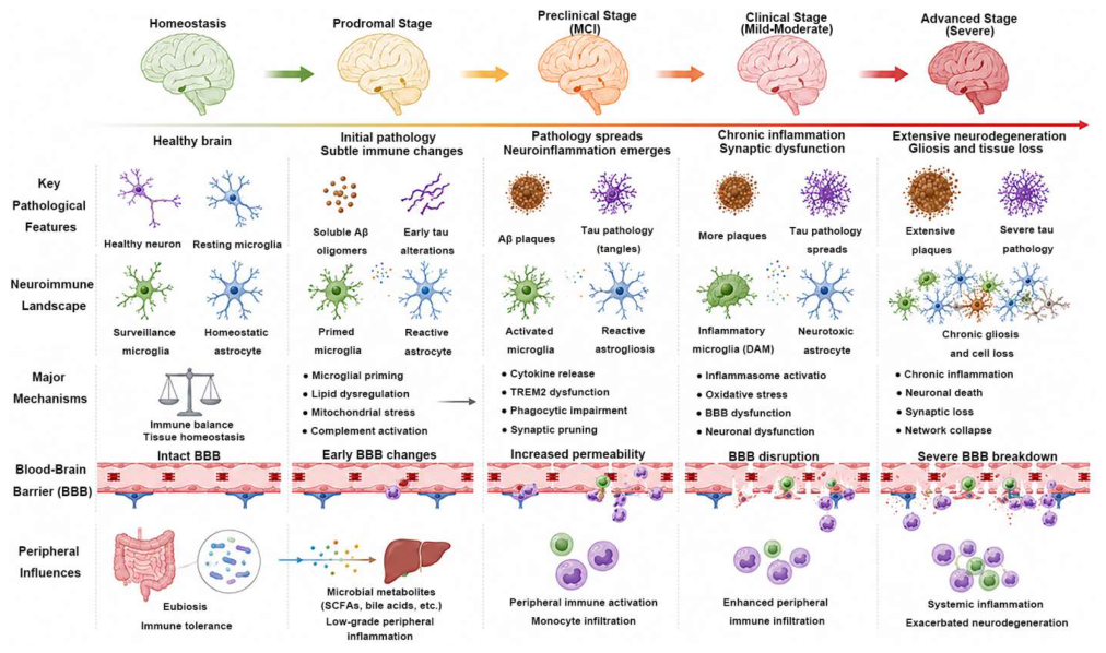
Figure 2.
Lipid-sensitive glial and neurovascular interfaces in AD. The microglial axis illustrates injurious programs involving NF-κB/MAPK, NLRP3, cGAS-STING, complement, mitochondrial ROS, and lipid-droplet accumulation, contrasted with pro-resolving programs involving PPAR/LXR, TREM2, mitochondrial support, and SPMs (RvD1, RvE1, PD1, MaR1). Astrocyte lipid handling, reactive-state regulation, metabolic support, microglia–astrocyte crosstalk, and BBB/NVU interactions are also relevant. Arrows denote proposed regulatory relationships rather than proven causal sequences in human AD. Solid arrows indicate pathways supported by direct human evidence, including human intervention, biomarker, or imaging studies. Dashed arrows indicate mechanisms supported predominantly by animal or in vitro studies. Dotted arrows indicate hypothesized or emerging links requiring further validation. Arrow style denotes the level of evidence rather than the magnitude or certainty of effect.
Figure 2.
Lipid-sensitive glial and neurovascular interfaces in AD. The microglial axis illustrates injurious programs involving NF-κB/MAPK, NLRP3, cGAS-STING, complement, mitochondrial ROS, and lipid-droplet accumulation, contrasted with pro-resolving programs involving PPAR/LXR, TREM2, mitochondrial support, and SPMs (RvD1, RvE1, PD1, MaR1). Astrocyte lipid handling, reactive-state regulation, metabolic support, microglia–astrocyte crosstalk, and BBB/NVU interactions are also relevant. Arrows denote proposed regulatory relationships rather than proven causal sequences in human AD. Solid arrows indicate pathways supported by direct human evidence, including human intervention, biomarker, or imaging studies. Dashed arrows indicate mechanisms supported predominantly by animal or in vitro studies. Dotted arrows indicate hypothesized or emerging links requiring further validation. Arrow style denotes the level of evidence rather than the magnitude or certainty of effect.
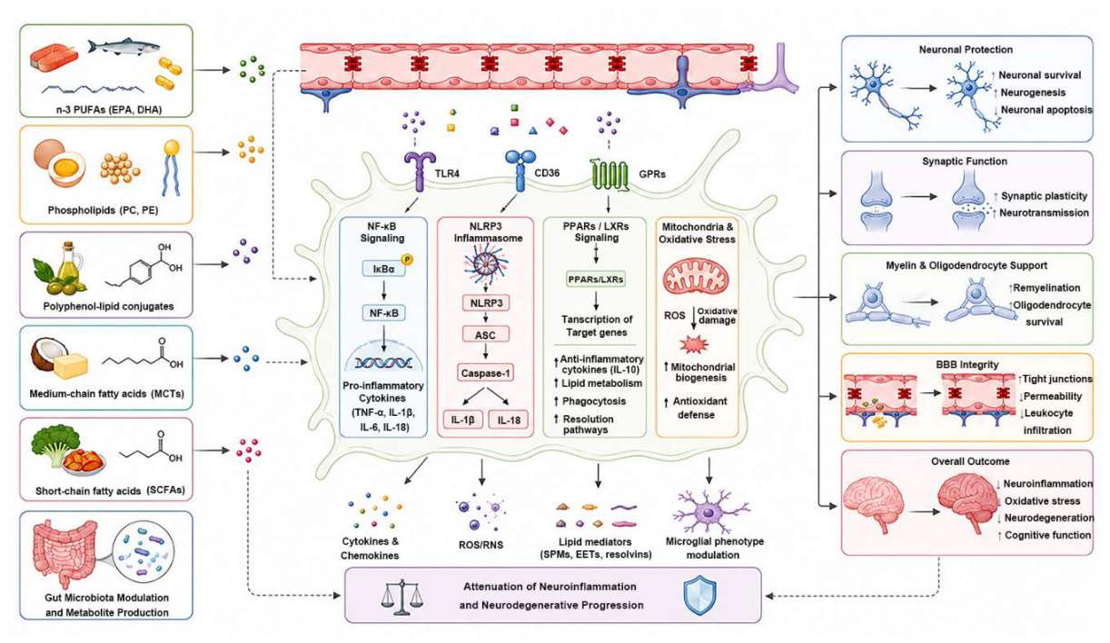
Figure 3.
Dietary lipids shape the AD microenvironment through the gut-periphery-brain axis. Dietary lipids and lipid-related nutrients may alter gut microbial composition and metabolism, affecting the generation of metabolites including SCFAs, bile acid derivatives, indole derivatives, N-acylethanolamines, lysophospholipids, and sphingolipid-related molecules. These signals can influence intestinal barrier integrity, systemic immune tone, liver and adipose metabolism, and ultimately BBB function, modulating microglial activation, mitochondrial function, synaptic stability, neuronal survival, and BBB integrity. Solid arrows indicate pathways supported by direct human evidence. Dashed arrows indicate mechanisms supported predominantly by preclinical models. Dotted arrows indicate hypothesized or emerging links that require further validation.
Figure 3.
Dietary lipids shape the AD microenvironment through the gut-periphery-brain axis. Dietary lipids and lipid-related nutrients may alter gut microbial composition and metabolism, affecting the generation of metabolites including SCFAs, bile acid derivatives, indole derivatives, N-acylethanolamines, lysophospholipids, and sphingolipid-related molecules. These signals can influence intestinal barrier integrity, systemic immune tone, liver and adipose metabolism, and ultimately BBB function, modulating microglial activation, mitochondrial function, synaptic stability, neuronal survival, and BBB integrity. Solid arrows indicate pathways supported by direct human evidence. Dashed arrows indicate mechanisms supported predominantly by preclinical models. Dotted arrows indicate hypothesized or emerging links that require further validation.
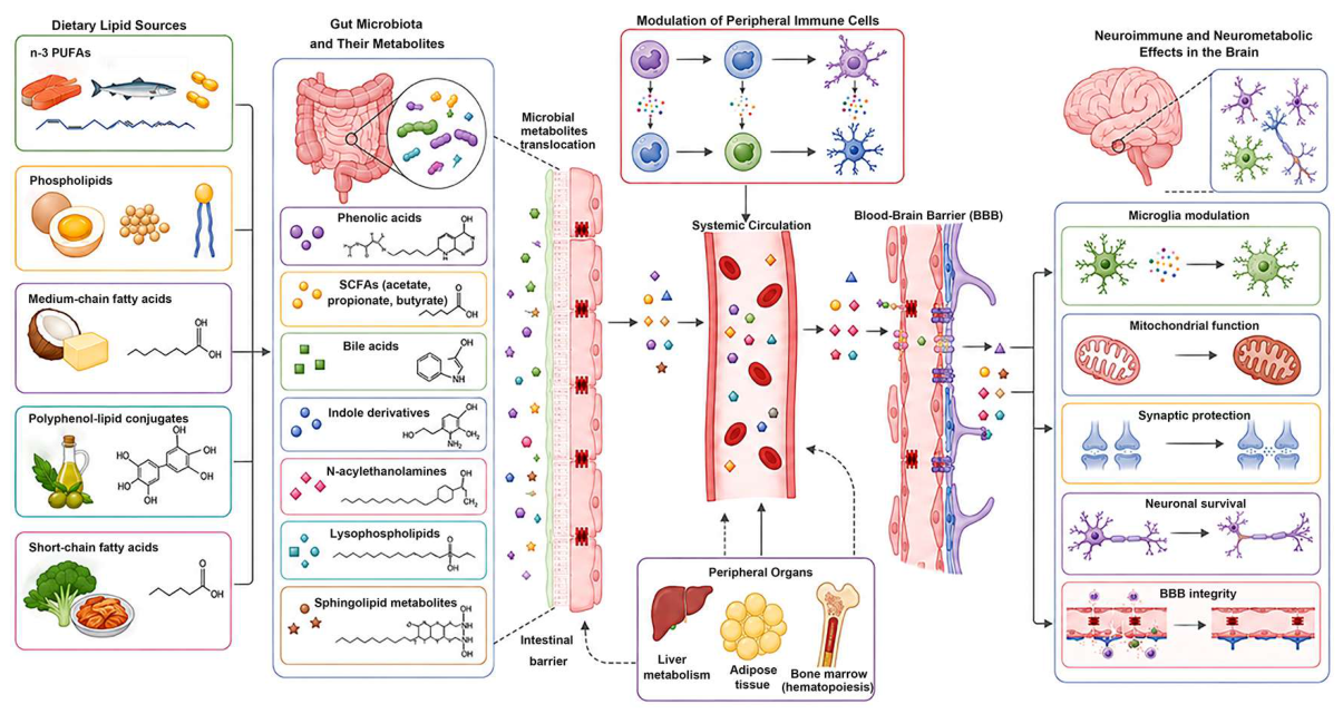
Figure 4.
Multi-omics integration strategy for lipid-related stratification and target discovery in AD. The workflow includes cohort design, biospecimen collection, multi-layer data generation (single-cell/spatial transcriptomics, lipidomics, metabolomics, proteomics, imaging, genotype), quality control, integrative analysis, biological validation, and translational output. Integration aims to identify biologically meaningful subgroups, vulnerable cellular states, candidate mechanisms, and response-related biomarkers for precision nutrition.
Figure 4.
Multi-omics integration strategy for lipid-related stratification and target discovery in AD. The workflow includes cohort design, biospecimen collection, multi-layer data generation (single-cell/spatial transcriptomics, lipidomics, metabolomics, proteomics, imaging, genotype), quality control, integrative analysis, biological validation, and translational output. Integration aims to identify biologically meaningful subgroups, vulnerable cellular states, candidate mechanisms, and response-related biomarkers for precision nutrition.
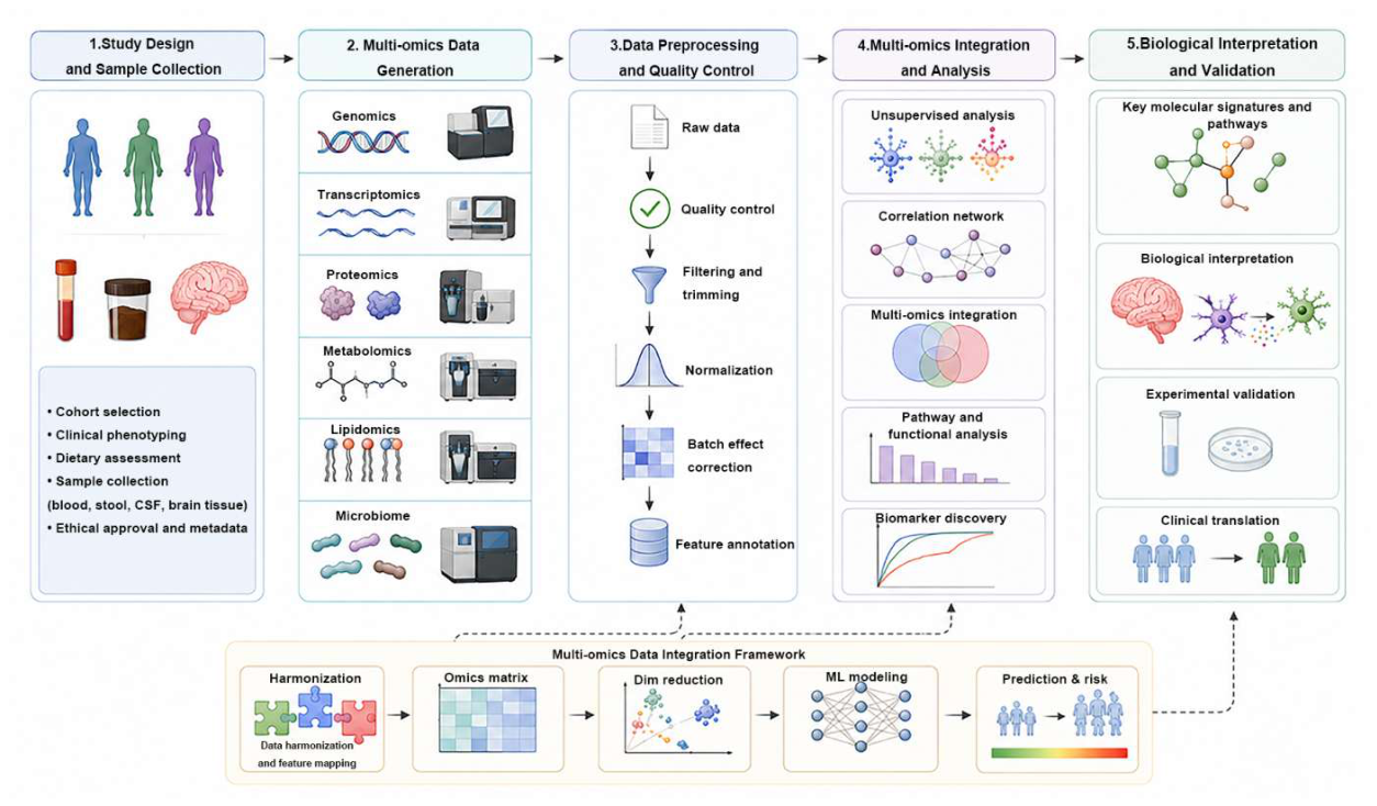
Table 1.
Neuroimmune characteristics across the AD continuum and potential stage-specific research endpoints for lipid intervention studies.
Table 1.
Neuroimmune characteristics across the AD continuum and potential stage-specific research endpoints for lipid intervention studies.
| Disease Stage | Major Neuroimmune and NVU Features | Peripheral Coupling Characteristics | Primary Goals of Lipid Intervention | More Suitable Endpoints | Evidence Level |
|---|---|---|---|---|---|
| Preclinical or High-Risk Stage | Glial priming, fragile membrane lipid homeostasis, reduced resolution capacity, early NVU and BBB vulnerability | Peripheral inflammatory and metabolic signals begin to amplify central susceptibility | Preserve membrane lipid homeostasis, support inflammatory resolution, strengthen BBB and NVU resilience | GFAP, sTREM2, early BBB imaging, lipidomic changes | Human observational and mechanistic studies [3,19,29,30,37,44,45] |
| MCI Stage | Glial program remodeling, more detectable BBB dysfunction, reduced neurovascular coupling | Aggravated central-peripheral immune and metabolic amplification | Promote pro-resolving pathways, support brain delivery, slow pathological amplification | Fluid biomarkers plus MRI and BBB measures plus cognitive trajectories | Human and preclinical mixed evidence [5,38,39,41,46] |
| Clinical AD Stage | Chronic neuroinflammation, self-sustaining injury, synaptic loss, NVU damage | Persistent systemic metabolic imbalance and peripheral inflammatory input | Adjunctive intervention to support membrane stability, energy metabolism, and inflammatory buffering | Atrophy rate, functional scales, nutritional status | Limited clinical evidence [2,15,16,38,39] |
| Late AD Stage | Widespread neuronal loss, glial support failure, increased brain delivery constraints | Low nutritional and metabolic reserve | Supportive nutrition and improved tolerability | Body weight, nutritional status, caregiver burden, tolerability | Mainly inferential [2,13,41] |
Note: MCI, mild cognitive impairment; GFAP, glial fibrillary acidic protein; sTREM2, soluble triggering receptor expressed on myeloid cells 2; BBB, blood–brain barrier; NVU, neurovascular unit. The table emphasizes potential intervention windows and research-oriented endpoints rather than fixed staging criteria. Most molecular, glial, BBB-related, and neurovascular measures listed here are exploratory endpoints and should not be interpreted as validated nutritional-response biomarkers or surrogate clinical endpoints.
Table 2.
Evidence hierarchy for major claims regarding dietary lipids and the neuroimmune microenvironment in Alzheimer’s disease.
Table 2.
Evidence hierarchy for major claims regarding dietary lipids and the neuroimmune microenvironment in Alzheimer’s disease.
| Major claim | Evidence level | Representative evidence type | Number of studies / aggregate sample size | Interpretation and limitation |
|---|---|---|---|---|
| Oral DHA/EPA supplementation improves cognition in established AD dementia [15,16,39] |
Limited/ inconsistent human interventional evidence |
RCTs and meta-analyses |
3 RCTs and meta-analyses cited in this review; several thousand participants in total | Clinical benefit has not been consistently demonstrated; null effects may reflect limited efficacy as well as heterogeneity |
| Higher dietary fish/omega-3 exposure is associated with lower dementia risk [13,14,44] |
Observational human evidence | Prospective cohorts | 3 studies cited in this review; several thousand participants | Associations are vulnerable to residual confounding and do not prove causality |
| DHA/EPA alter plasma/RBC fatty-acid composition [15,16] |
Direct human pharmacodynamic evidence | Supplementation studies | 2 studies cited in this review; hundreds of participants | Peripheral target engagement does not establish exposure in specific brain cell types |
| APOE ε4 modifies cerebral/peripheral lipid handling [44,57,67] |
Human genetic, biomarker, and mechanistic evidence | Genetic and imaging/biomarker studies | 3 studies cited in this review; several thousand participants | Evidence for differential lipid metabolism is stronger than evidence for genotype-specific treatment benefit |
| SPMs promote resolution of neuroinflammation in AD [11,52] |
Predominantly preclinical/mechanistic evidence | Animal models, cell culture, limited human biomarker evidence | 2 preclinical studies cited; limited human data | “Pro-resolving” does not establish clinical anti-inflammatory efficacy in AD |
| TREM2, PPAR/LXR, complement pruning, lipid-droplet remodeling mediate dietary-lipid effects [9,10,51,53] |
Mainly preclinical or hypothesis-generating | Cell and animal studies | 4 preclinical studies cited; no direct human data | Direct causal evidence in humans remains limited |
| Dietary lipids improve BBB integrity/NVU function in AD [7,46,55] |
Mixed evidence; mainly preclinical | Experimental models, limited human vascular associations | 3 experimental studies cited; limited human data | Clinical relevance and nutritional responsiveness remain uncertain |
| Gut microbiota mediates lipid effects on AD pathology [36,58,59] |
Predominantly preclinical and associative human evidence | Animal studies, microbiome association studies | 3 reviews cited; no primary interventional data | Causality and clinically actionable microbial signatures remain unvalidated |
| 4D framework can guide lipid intervention selection [17,18] |
Conceptual/hypothesis-generating | Integrative framework | Conceptual framework; no empirical studies yet | Requires prospective validation before clinical use |
Note: Evidence categories reflect the level of support for the specific claim rather than the overall importance of the biological pathway. Direct human evidence does not necessarily establish causality or clinical efficacy.
Table 5.
Lipid formulations and evidence for brain penetration in humans.
| Formulation | Description | Evidence for brain penetration in humans | Limitations |
|---|---|---|---|
| Triglyceride (TG) | Natural fish-oil form; DHA/EPA bound to glycerol backbone [14] |
Indirect evidence via plasma and RBC omega-3 indices; brain penetration inferred but not directly measured | Peripheral exposure does not establish central target engagement |
| Ethyl ester (EE) | Common supplement form; DHA/EPA esterified to ethanol [14] |
Similar to TG; some studies suggest slightly lower bioavailability than TG form | Brain penetration not directly assessed |
| Phospholipid (PL) | DHA/EPA bound to phospholipids (e.g., krill oil) [3] |
Preclinical evidence suggests improved brain uptake; human data limited | Human brain penetration evidence remains sparse |
| LysoPC | Lysophosphatidylcholine-bound DHA [55] |
Preclinical studies suggest enhanced brain delivery; human evidence limited | No direct human brain penetration data |
| Nanoemulsion / intranasal | Experimental delivery systems designed to bypass BBB [55] |
Preclinical proof-of-concept; human data not yet available | Not yet tested in human AD trials |
Note: Direct evidence for brain penetration in humans remains limited for all formulation categories. Peripheral omega-3 indices (plasma, RBC) measure exposure and adherence but do not establish uptake into specific brain cell populations.
Disclaimer/Publisher’s Note: The statements, opinions and data contained in all publications are solely those of the individual author(s) and contributor(s) and not of MDPI and/or the editor(s). MDPI and/or the editor(s) disclaim responsibility for any injury to people or property resulting from any ideas, methods, instructions or products referred to in the content. |
© 2026 by the authors. Licensee MDPI, Basel, Switzerland. This article is an open access article distributed under the terms and conditions of the Creative Commons Attribution (CC BY) license (http://creativecommons.org/licenses/by/4.0/).
Copyright: This open access article is published under a Creative Commons CC BY 4.0 license, which permit the free download, distribution, and reuse, provided that the author and preprint are cited in any reuse.