
Submitted:
12 November 2026
Posted:
13 November 2026
You are already at the latest version
Abstract
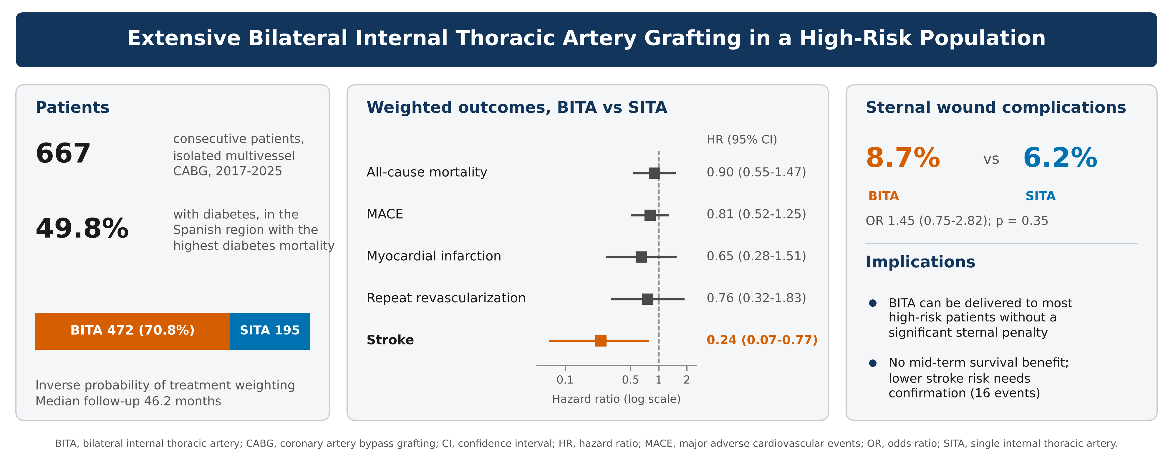
Background/Objectives: Concern about sternal wound complications limits bilateral internal thoracic artery (BITA) grafting, particularly in patients with diabetes and obesity. We evaluated the mid-term results of a program of extensive BITA grafting in the Canary Islands, the Spanish region with the highest diabetes-related mortality. Methods: We retrospectively analyzed 667 consecutive patients undergoing non-emergent, first-time, isolated multivessel coronary artery bypass grafting between 2017 and 2025; 472 (70.8%) received BITA and 195 (29.2%) single internal thoracic artery (SITA) grafting. Outcomes were compared using inverse probability of treatment weighting. Results: Half of the patients (49.8%) had diabetes. Median follow-up was 46.2 months. After weighting, BITA and SITA grafting did not differ in all-cause mortality (hazard ratio [HR] 0.90; 95% confidence interval [CI] 0.55–1.47; p = 0.68), myocardial infarction (HR 0.65; 95% CI 0.28–1.51), repeat revascularization (HR 0.76; 95% CI 0.32–1.83) or major adverse cardiovascular events (HR 0.81; 95% CI 0.52–1.25). Stroke was less frequent after BITA grafting (HR 0.24; 95% CI 0.07–0.77; p = 0.016). Sternal wound complications occurred in 8.7% vs. 6.2% of patients (odds ratio 1.45; 95% CI 0.75–2.82; p = 0.35), with no excess among patients with diabetes (8.8% vs. 8.7%). Conclusions: In a population with a high burden of diabetes, BITA grafting was delivered to 70.8% of unselected patients without a significant excess of sternal wound complications. It did not improve mid-term survival, but stroke was less frequent after BITA grafting, a hypothesis-generating finding that requires confirmation.
Keywords:
coronary artery bypass grafting
; bilateral internal thoracic artery
; diabetes mellitus
; sternal wound infection
; stroke
; inverse probability of treatment weighting
; propensity score
1. Introduction
Coronary artery bypass grafting (CABG) remains the preferred revascularization strategy for multivessel or left main coronary disease, particularly with diabetes [1,2,3,4,5]. Bilateral internal thoracic artery (BITA) grafting is the most established form of multiple arterial grafting, and observational series and meta-analyses have repeatedly associated it with better long-term survival than single internal thoracic artery (SITA) grafting [6,7,8,9]; against a SITA combined with a radial artery, an advantage appears only beyond the first decade [10]. Randomized evidence has been less definitive: in the Arterial Revascularization Trial (ART), all-cause mortality was similar at 5, 10 and 15 years [11,12,13], although crossover and frequent radial artery use in the control arm narrowed the separation between strategies [11,12].
All-cause mortality may be an insensitive endpoint, because non-cardiovascular causes overtake cardiovascular causes of late death after CABG [14]. Conduit strategy may also influence neurological risk, since in situ bilateral grafting can reduce manipulation of the ascending aorta, a major source of embolic stroke [15,16,17]. These potential benefits must be weighed against sternal wound complications, particularly with diabetes, obesity or chronic pulmonary disease [18,19,20,21,22], which are associated with worse long-term survival [23].
The Canary Islands have the highest diabetes-related mortality of any Spanish region and a higher prevalence of diabetes and obesity than the national average [24]. In 2017 our institution, the regional cardiovascular referral center, initiated a program of extensive BITA use, avoiding bilateral harvesting in patients with at least 2 of chronic obstructive pulmonary disease, insulin-dependent diabetes and body mass index above 40 kg/m2, with the final decision left to the operating surgeon. We evaluated whether a BITA-based strategy could be delivered to most of an unselected population in this setting and whether it improved mid-term outcomes.
2. Materials and Methods
The Ethics Committee of Hospital Universitario de Gran Canaria Doctor Negrín approved the study (approval number 2026-438-1, October 29, 2026) and waived individual informed consent given the retrospective design and the use of anonymized data. The study was registered at ClinicalTrials.gov (NCT07796750), was conducted in accordance with the Declaration of Helsinki and is reported according to the STROBE statement.
2.1. Study Population
We included all patients who underwent non-emergent, first-time, isolated CABG for multivessel disease, left main disease or both at our institution between 2017 and 2025: 667 consecutive patients, of whom 472 received BITA and 195 SITA grafting. Data were extracted from a prospectively maintained institutional database after a structured quality review.
2.2. Surgical Technique
All procedures were performed through median sternotomy, off-pump or with normothermic cardiopulmonary bypass (CPB), single aortic cross-clamping and intermittent antegrade cold blood cardioplegia, according to surgeon preference. Both internal thoracic arteries were harvested skeletonized. The in situ left internal thoracic artery grafted the anterior wall; the right internal thoracic artery was used as a composite Y- or T-graft, typically for sequential grafting of the lateral and posterior territories. Saphenous vein was used as a free graft with proximal aortic anastomosis or as a composite graft. Transit-time flow was measured in all cases.
2.3. Outcomes
The primary endpoint was all-cause mortality. Secondary time-to-event endpoints were myocardial infarction, stroke, repeat revascularization and major adverse cardiovascular events (MACE: death, stroke, myocardial infarction or repeat revascularization, whichever occurred first). Cause of death was classified from the clinical record as cardiovascular (cardiogenic shock, acute coronary syndrome, graft failure, end-stage heart failure, pulmonary embolism, infective endocarditis or death during a cardiac procedure), non-cardiovascular or undetermined. Events were recorded as diagnosed in routine practice and were not adjudicated. Stroke timing was categorized a priori as within 30 days, 31 days to 1 year, and beyond 1 year. A sternal wound complication (SWC) was any sternal wound problem requiring negative-pressure therapy or surgical intervention.
2.4. Statistical Analysis
Analyses were performed by the institutional Research Unit with R version 4.6.1 (R Foundation for Statistical Computing). Tests were 2-sided, with significance at p < 0.05. Quantitative variables, all of which departed from normality on the Kolmogorov–Smirnov test, are presented as mean ± SD or median (interquartile range) and were compared with the Mann–Whitney U test; categorical variables were compared with the Fisher exact test. Balance was assessed with standardized mean differences (SMD), with an absolute value above 0.10 indicating imbalance. Completeness of follow-up was quantified with the Clark completeness index.
The probability of receiving BITA was estimated by multivariable logistic regression [25,26] with stepwise (backward-forward) selection from the characteristics in Table 1. The retained covariates were creatinine clearance, age, dyspnea, cerebrovascular disease and CPB (all variance inflation factors < 5; area under the curve, 0.80). Inverse probability of treatment weights (IPTW) were derived from this model (Figure 1).
Survival was estimated with the Kaplan–Meier method and compared with the log-rank test. Hazard ratios (HR) with 95% CI were obtained by Cox regression, unweighted and weighted, and the proportional hazards assumption was tested with Schoenfeld residuals. Cardiovascular mortality was analyzed among patients who died of a recorded cause. The association between BITA and SWC was assessed with the Fisher exact test and the odds ratio (OR).
3. Results
3.1. Patients and Follow-Up
BITA grafting was used in 472 of 667 patients (70.8%). The cohort carried a heavy cardiometabolic burden (diabetes 49.8%, hypertension 89.7%, mean body mass index 27.6 ± 4.4 kg/m2) but low operative risk (median EuroSCORE II, 1.55 [0.99–2.85]) (Table 1). CPB was used in 327 patients (49.0%). Median follow-up was 46.2 months (interquartile range, 23.2–76.9; maximum, 107.7); vital status was ascertained in every patient, and the completeness index was 90.3%.
Before weighting, the only substantial imbalance was CPB use, which was far less frequent in the BITA group (absolute SMD 1.24). After weighting, the 5 model covariates were balanced, with a small residual imbalance in age (SMD 0.12) (Figure 1); left main disease (SMD 0.02 to 0.32) and body mass index (0.15 to 0.26), which were not in the model, became imbalanced (Table 1).
3.2. All-Cause and Cardiovascular Mortality
During follow-up, 133 patients (19.9%) died: 93 (19.7%) after BITA and 40 (20.5%) after SITA grafting; 30-day mortality was 1.1%. Mortality did not differ before (HR 0.84; 95% CI 0.58–1.23; p = 0.37) or after weighting (HR 0.90; 95% CI 0.55–1.47; p = 0.68) (Table 2). Weighted survival at 12, 60 and 96 months was 95.6%, 81.3% and 71.5% after BITA and 93.3%, 76.2% and 70.2% after SITA grafting (Figure 2).
A cause of death was recorded in 120 patients and was cardiovascular in 17 (8 of 83 BITA, 9 of 37 SITA). Cardiovascular death was less frequent after BITA grafting before weighting (HR 0.36; 95% CI 0.14–0.94) but not after (HR 0.66; 95% CI 0.20–2.14; p = 0.49). Because the proportional hazards assumption was rejected (weighted p < 0.001), these estimates are descriptive: SITA deaths clustered in the first months, whereas BITA deaths accrued progressively (Supplementary Figure S2).
3.3. Myocardial Infarction, Repeat Revascularization and MACE
Myocardial infarction occurred in 45 patients, repeat revascularization in 41 and MACE in 178 (26.9% BITA, 26.2% SITA). None differed significantly before or after weighting (Table 2; Supplementary Figure S1).
3.4. Stroke
Stroke occurred in 16 patients: 8 (1.7%) after BITA and 8 (4.1%) after SITA grafting. Four strokes occurred within 30 days (1 BITA [0.2%] vs. 3 SITA [1.5%]), 1 SITA stroke between 31 days and 1 year, and 11 beyond 1 year (Table 3). The difference was borderline before weighting (HR 0.38; 95% CI 0.14–1.02; p = 0.055) and significant after weighting (HR 0.24; 95% CI 0.07–0.77; p = 0.016). Weighted freedom from stroke at 96 months was 97.5% after BITA and 90.3% after SITA grafting (Figure 3).
3.5. Sternal Wound Complications
SWC occurred in 53 patients (7.9%): 41 (8.7%) after BITA and 12 (6.2%) after SITA grafting (OR 1.45; 95% CI 0.75–2.82; p = 0.35). Among patients with diabetes, SWC rates were almost identical (21 of 240 [8.8%] vs. 8 of 92 [8.7%]; OR 1.01; 95% CI 0.43–2.36). Exploratory subgroup comparisons by diabetes and body mass index are given in Supplementary Table S1 and Supplementary Figure S3.
4. Discussion
In an unselected population with a heavy cardiometabolic burden, in which half of the patients had diabetes, a program of extensive BITA grafting reached 70.8% of consecutive patients without a significant excess of SWC, and with no excess at all among patients with diabetes. Over mid-term follow-up it conferred no survival or MACE advantage over SITA grafting; stroke was the only endpoint that differed.
4.1. Feasibility
The only prospective attempt to systematize bilateral grafting to its limit, the CATHEXIS registry, reached 88.5% BITA use but was stopped after 226 patients because sternal complications (5.5%) and graft failure (3%) exceeded those of a matched cohort [27]. Our program differed in that a few prespecified wound-risk criteria excluded the highest-risk patients and the final decision remained with the surgeon. Together, the 2 experiences suggest that the safety of a BITA program depends on the threshold at which it is systematized, and that deliberately incomplete penetration may make it sustainable in a population at high sternal risk.
4.2. Survival and Cause of Death
The absence of a survival benefit is consistent with ART [13] and with the limited power of a mid-term cohort. Cardiovascular deaths after SITA grafting concentrated in the first year, which may reflect a more vulnerable profile among patients selected for SITA rather than protection by bilateral grafting. Still, all-cause mortality is an incomplete endpoint for a conduit effect: only 17 of 120 deaths with a recorded cause were cardiovascular, and bilateral grafting has no plausible mechanism to prevent the non-cardiovascular causes that dominate late mortality [14]. Even in a randomized trial this attenuates the observable effect and the power to detect it, so separating cardiovascular from non-cardiovascular death should give a more informative assessment.
4.3. Stroke
A mechanistic explanation is available for the lower stroke rate: in situ bilateral grafting may reduce proximal aortic anastomoses, and minimizing aortic manipulation reduces perioperative embolic stroke [15,16]. The 30-day rate (0.2% vs. 1.5%) places part of the difference in the window in which this mechanism is most plausible. However, only 16 strokes occurred, stroke was a secondary endpoint without correction for multiplicity, 11 strokes occurred after the first year, when atrial fibrillation and progressive atherosclerosis predominate, and the database did not record aortic manipulation, so BITA grafting should not be regarded as a surrogate for anaortic CABG.
4.4. Sternal Wound Complications
The nonsignificant excess of SWC after BITA grafting is consistent with the literature. In our meta-analysis of propensity-matched studies, BITA increased deep sternal wound infection (relative risk, 1.66; 95% CI 1.41–1.95), less so with skeletonized harvesting (relative risk, 1.37; 95% CI 1.04–1.79) [9], the technique used in all our patients and associated with fewer infections than pedicled harvesting [28]. In ART, SWC were more frequent after BITA grafting (3.5% vs. 1.9%) [11] and, in a post hoc analysis, were independently associated with 10-year mortality (HR 1.81; 95% CI 1.30–2.54), even after excluding the first 6 months [29]. Within the trial that failed to show a survival benefit, bilateral grafting therefore increased a complication that shortens survival.
4.5. Randomized and Causal-Inference Evidence
Crossover and radial artery use in ART are usually invoked to explain its discrepancy with observational data, including our own [9,10]. That explanation may be incomplete: an instrumental-variable analysis of Medicare beneficiaries substantially attenuated the apparent advantage of multiarterial grafting [30]. Propensity weighting cannot remove unmeasured confounding, so part of the historical survival advantage may reflect which patients are offered the strategy, a caution that applies equally to our stroke finding. Neither SWC nor cause of death is reported in the 15-year ART analysis [13]; this is the gap that real-world cohorts can help to fill.
4.6. Limitations
This was a retrospective single-center study without randomization, so residual and unmeasured confounding are possible. The propensity model was parsimonious and stepwise; it balanced its own covariates, but left main disease and body mass index became imbalanced after weighting, so the weighted estimates mainly address confounding by CPB use. Event counts were small, particularly for stroke and cardiovascular death. The cause-of-death analysis was restricted to patients who died, the cause was undetermined in 13, and proportional hazards were violated. Endpoints were not adjudicated, and aortic manipulation and proximal anastomoses were not recorded. The findings arise from a Southern European cohort and may not generalize [24].
5. Conclusions
Extensive BITA grafting in an unselected population at high sternal risk was not associated with better mid-term survival or fewer MACE, nor with a significant increase in SWC, but stroke was less frequent after weighting. Future studies should distinguish cardiovascular from non-cardiovascular death and early from late stroke, and should record aortic manipulation and sternal wound outcomes.
Supplementary Materials
The following supporting information can be downloaded at the website of this paper posted on Preprints.org, Table S1: Sternal wound complications, overall and within the prespecified subgroups; Figure S1: Kaplan–Meier estimates of event-free survival for the secondary endpoints; Figure S2: Kaplan–Meier estimates of freedom from cardiovascular death; Figure S3: Sternal wound complications, overall and within the prespecified subgroups.
Author Contributions
Conceptualization, J.I.J.D.R. and S.U.; methodology, S.U. and C.P.R.C.; software, J.M.G.M.; validation, J.M.G.M.; formal analysis, J.M.G.M.; investigation, J.I.J.D.R., L.D.B., A.J.E.B., A.M.P., A.T.C. and M.S.H.; resources, F.P.T.; data curation, J.I.J.D.R. and L.D.B.; writing—original draft preparation, J.I.J.D.R.; writing—review and editing, S.U., C.P.R.C., J.M.G.M., L.D.B., A.J.E.B., A.M.P., A.T.C., M.S.H. and F.P.T.; supervision, S.U., C.P.R.C. and F.P.T. All authors have read and agreed to the published version of the manuscript.
Funding
This research received no external funding.
Institutional Review Board Statement
The study was conducted in accordance with the Declaration of Helsinki and approved by the Ethics Committee of Hospital Universitario de Gran Canaria Doctor Negrín (approval number 2026-438-1, date of approval 29 October 2026).
Informed Consent Statement
Patient consent was waived by the Ethics Committee because of the retrospective design of the study and the use of anonymized data.
Data Availability Statement
The data presented in this study are available on reasonable request from the corresponding author. The data are not publicly available because of institutional data protection regulations.
Acknowledgments
During the preparation of this manuscript, the authors used Claude (Anthropic; Claude Opus 5.5) for language editing, manuscript formatting and the formatting of references. The authors have reviewed and edited the output and take full responsibility for the content of this publication.
Conflicts of Interest
The authors declare no conflicts of interest.
Abbreviations
The following abbreviations are used in this manuscript:
| ART | Arterial Revascularization Trial |
| BITA | Bilateral internal thoracic artery |
| CABG | Coronary artery bypass grafting |
| CI | Confidence interval |
| CPB | Cardiopulmonary bypass |
| HR | Hazard ratio |
| IPTW | Inverse probability of treatment weighting |
| MACE | Major adverse cardiovascular events |
| OR | Odds ratio |
| SITA | Single internal thoracic artery |
| SMD | Standardized mean difference |
| SWC | Sternal wound complication |
References
- Lawton, J.S.; Tamis-Holland, J.E.; Bangalore, S.; et al. 2021 ACC/AHA/SCAI guideline for coronary artery revascularization. J. Am. Coll. Cardiol. 2022, 79, e21–e129. [Google Scholar] [CrossRef]
- Neumann, F.J.; Sousa-Uva, M.; Ahlsson, A.; et al. 2018 ESC/EACTS guidelines on myocardial revascularization. Eur. Heart J. 2019, 40, 87–165. [Google Scholar] [CrossRef]
- Farkouh, M.E.; Domanski, M.; Sleeper, L.A.; et al. Strategies for multivessel revascularization in patients with diabetes. N. Engl. J. Med. 2012, 367, 2375–2384. [Google Scholar] [CrossRef]
- Urso, S.; Sadaba, R.; González-Martín, J.M.; et al. Coronary surgery provides better survival than drug-eluting stent: a pooled meta-analysis of Kaplan-Meier-derived individual patient data. J. Thorac. Cardiovasc. Surg. 2024, 167, 2138–2146.e3. [Google Scholar] [CrossRef]
- Urso, S.; Sadaba, R.; Nogales, E.; et al. Major cardiovascular events at 5 years in surgical versus percutaneous revascularization for left main stem disease. Interact. Cardiovasc. Thorac. Surg. 2021, 32, 530–536. [Google Scholar] [CrossRef]
- Taggart, D.P.; D’Amico, R.; Altman, D.G. Effect of arterial revascularisation on survival. Lancet 2001, 358, 870–875. [Google Scholar] [CrossRef]
- Yi, G.; Shine, B.; Rehman, S.M.; et al. Effect of bilateral internal mammary artery grafts on long-term survival. Circulation 2014, 130, 539–545. [Google Scholar] [CrossRef]
- Buttar, S.N.; Yan, T.D.; Taggart, D.P.; et al. Long-term and short-term outcomes of bilateral versus left internal mammary artery grafting: a meta-analysis. Heart 2017, 103, 1419–1426. [Google Scholar] [CrossRef]
- Urso, S.; Nogales, E.; González, J.M.; et al. Bilateral internal thoracic artery versus single internal thoracic artery: a meta-analysis of propensity score-matched observational studies. Interact. Cardiovasc. Thorac. Surg. 2019, 29, 163–172. [Google Scholar] [CrossRef]
- Urso, S.; Sadaba, R.; González Martín, J.M.; et al. Bilateral internal thoracic artery versus single internal thoracic artery plus radial artery: a double meta-analytic approach. J. Thorac. Cardiovasc. Surg. 2024, 167, 183–195.e3. [Google Scholar] [CrossRef]
- Taggart, D.P.; Altman, D.G.; Gray, A.M.; et al. Randomized trial of bilateral versus single internal-thoracic-artery grafts. N. Engl. J. Med. 2016, 375, 2540–2549. [Google Scholar] [CrossRef]
- Taggart, D.P.; Benedetto, U.; Gerry, S.; et al. Bilateral versus single internal-thoracic-artery grafts at 10 years. N. Engl. J. Med. 2019, 380, 437–446. [Google Scholar] [CrossRef]
- Taggart, D.P.; Gaudino, M.; Gerry, S.; et al. Long-term outcomes following bilateral vs single internal thoracic artery grafts: a secondary analysis of a randomized clinical trial. JAMA Cardiol. Online ahead of print. 2026. [Google Scholar] [CrossRef]
- Butt, J.H.; Sørensen, R.; Bäck, C.; et al. Short- and long-term cause of death in patients undergoing isolated coronary artery bypass grafting. J. Thorac. Cardiovasc. Surg. 2018, 156, 54–60.e4. [Google Scholar] [CrossRef]
- Gaudino, M.; Angiolillo, D.J.; Di Franco, A.; et al. Stroke after coronary artery bypass grafting and percutaneous coronary intervention. J. Am. Heart Assoc. 2019, 8, e013032. [Google Scholar] [CrossRef]
- Zhao, D.F.; Edelman, J.J.; Seco, M.; et al. Coronary artery bypass grafting with and without manipulation of the ascending aorta. J. Am. Coll. Cardiol. 2017, 69, 924–936. [Google Scholar] [CrossRef]
- Head, S.J.; Milojevic, M.; Daemen, J.; et al. Stroke rates following surgical versus percutaneous coronary revascularization. J. Am. Coll. Cardiol. 2018, 72, 386–398. [Google Scholar] [CrossRef]
- Aldea, G.S.; Bakaeen, F.G.; Pal, J.; et al. The Society of Thoracic Surgeons clinical practice guidelines on arterial conduits for coronary artery bypass grafting. Ann. Thorac. Surg. 2016, 101, 801–809. [Google Scholar] [CrossRef]
- Gaudino, M.; Taggart, D.; Suma, H.; et al. The choice of conduits in coronary artery bypass surgery. J. Am. Coll. Cardiol. 2015, 66, 1729–1737. [Google Scholar] [CrossRef]
- Lazar, H.L. The risk of mediastinitis and deep sternal wound infections with single and bilateral, pedicled and skeletonized internal thoracic arteries. Ann. Cardiothorac. Surg. 2018, 7, 663–672. [Google Scholar] [CrossRef]
- Deo, S.V.; Shah, I.K.; Dunlay, S.M.; et al. Bilateral internal thoracic artery harvest and deep sternal wound infection in diabetic patients. Ann. Thorac. Surg. 2013, 95, 862–869. [Google Scholar] [CrossRef]
- Ohira, S.; Miyata, H.; Yamazaki, S.; et al. Deep sternal wound infection after bilateral internal thoracic artery grafting. J. Thorac. Cardiovasc. Surg. 2019, 157, 166–173.e1. [Google Scholar] [CrossRef]
- Perezgrovas-Olaria, R.; Audisio, K.; Cancelli, G.; et al. Deep sternal wound infection and mortality in cardiac surgery: a meta-analysis. Ann. Thorac. Surg. 2023, 115, 272–280. [Google Scholar] [CrossRef]
- Marcelino-Rodríguez, I.; Elosua, R.; Rodríguez Pérez, M.C.; et al. On the problem of type 2 diabetes-related mortality in the Canary Islands, Spain. Diabetes Res. Clin. Pract. 2016, 111, 74–82. [Google Scholar] [CrossRef]
- Austin, P.C. An introduction to propensity score methods for reducing the effects of confounding in observational studies. Multivar. Behav. Res. 2011, 46, 399–424. [Google Scholar] [CrossRef]
- Austin, P.C.; Stuart, E.A. Moving towards best practice when using inverse probability of treatment weighting. Stat. Med. 2015, 34, 3661–3679. [Google Scholar] [CrossRef]
- Gaudino, M.; Glieca, F.; Luciani, N.; et al. Systematic bilateral internal mammary artery grafting: lessons learned from the CATHolic University EXtensive BIMA Grafting Study. Eur. J. Cardiothorac. Surg. 2018, 54, 702–707. [Google Scholar] [CrossRef]
- Sá, M.P.B.; Ferraz, P.E.; Escobar, R.R.; et al. Skeletonized versus pedicled internal thoracic artery and risk of sternal wound infection after coronary bypass surgery. Interact. Cardiovasc. Thorac. Surg. 2013, 16, 849–857. [Google Scholar] [CrossRef]
- Gaudino, M.; Audisio, K.; Rahouma, M.; et al. Association between sternal wound complications and 10-year mortality following coronary artery bypass grafting. J. Thorac. Cardiovasc. Surg. 2023, 166, 532–539.e4. [Google Scholar] [CrossRef]
- Schaffer, J.M.; Shih, E.; Squiers, J.J.; et al. Multiarterial grafting and survival after coronary artery bypass grafting: an instrumental variable analysis. J. Am. Coll. Cardiol. 2026, 87, 3414–3428. [Google Scholar]
Figure 1.
Covariate balance before and after weighting. Absolute standardized mean differences of the propensity score and of the 5 covariates in the propensity model, before weighting (gray) and after inverse probability of treatment weighting (orange). The dashed line marks the 0.10 imbalance threshold. Abbreviations: IPTW, inverse probability of treatment weighting; NYHA, New York Heart Association.
Figure 1.
Covariate balance before and after weighting. Absolute standardized mean differences of the propensity score and of the 5 covariates in the propensity model, before weighting (gray) and after inverse probability of treatment weighting (orange). The dashed line marks the 0.10 imbalance threshold. Abbreviations: IPTW, inverse probability of treatment weighting; NYHA, New York Heart Association.
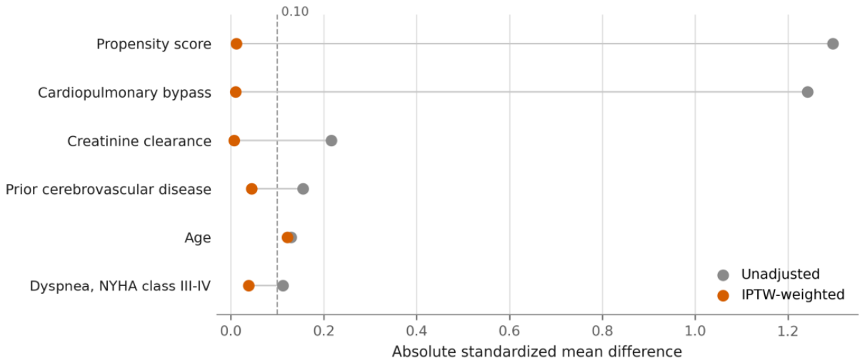
Figure 2.
All-cause survival. Kaplan–Meier estimates of all-cause survival by conduit strategy, (A) unweighted and (B) after inverse probability of treatment weighting, with numbers at risk (weighted in panel B). Tick marks denote censored observations. The vertical axis starts at 50%. Abbreviations: BITA, bilateral internal thoracic artery; HR, hazard ratio; IPTW, inverse probability of treatment weighting; SITA, single internal thoracic artery.
Figure 2.
All-cause survival. Kaplan–Meier estimates of all-cause survival by conduit strategy, (A) unweighted and (B) after inverse probability of treatment weighting, with numbers at risk (weighted in panel B). Tick marks denote censored observations. The vertical axis starts at 50%. Abbreviations: BITA, bilateral internal thoracic artery; HR, hazard ratio; IPTW, inverse probability of treatment weighting; SITA, single internal thoracic artery.
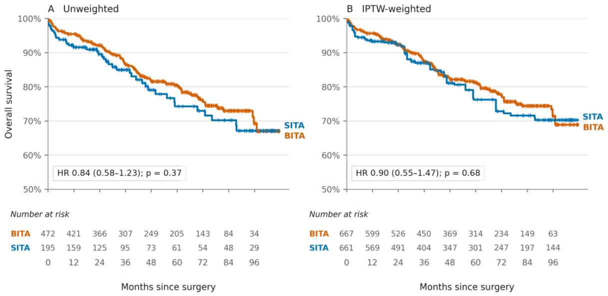
Figure 3.
Freedom from stroke. Kaplan–Meier estimates of freedom from stroke by conduit strategy, (A) unweighted and (B) after inverse probability of treatment weighting, with numbers at risk (weighted in panel B). Tick marks denote censored observations. The vertical axis starts at 85%. Abbreviations: BITA, bilateral internal thoracic artery; HR, hazard ratio; IPTW, inverse probability of treatment weighting; SITA, single internal thoracic artery.
Figure 3.
Freedom from stroke. Kaplan–Meier estimates of freedom from stroke by conduit strategy, (A) unweighted and (B) after inverse probability of treatment weighting, with numbers at risk (weighted in panel B). Tick marks denote censored observations. The vertical axis starts at 85%. Abbreviations: BITA, bilateral internal thoracic artery; HR, hazard ratio; IPTW, inverse probability of treatment weighting; SITA, single internal thoracic artery.
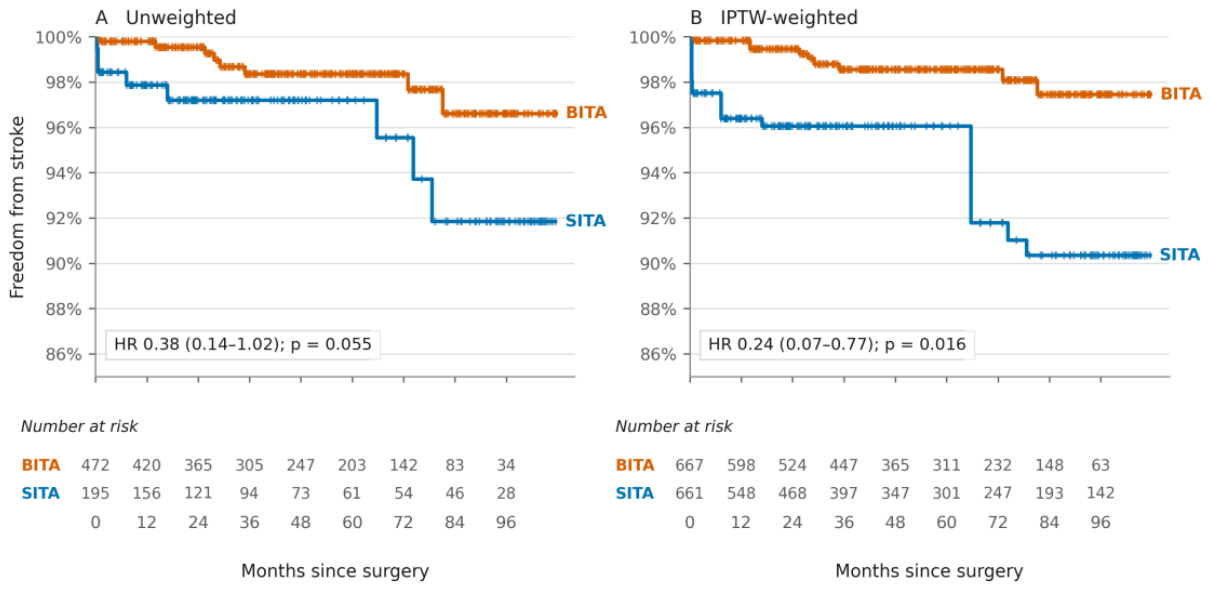

Table 1.
Baseline characteristics of the study population.
| Characteristic | BITA (n = 472) | SITA (n = 195) | p-Value | Unweighted SMD | Weighted SMD |
|---|---|---|---|---|---|
| Age, y | 65.5 ± 8.6 | 66.6 ± 8.4 | 0.12 | −0.13 | +0.12 |
| Female sex | 74 (15.7) | 28 (14.4) | 0.72 | +0.04 | +0.20 |
| EuroSCORE II | 1.50 (0.95–2.80) | 1.63 (1.01–2.94) | 0.37 | −0.07 | +0.06 |
| Creatinine clearance, mL/min | 80.6 ± 34.3 | 73.3 ± 33.5 | 0.04 | +0.22 | −0.01 |
| Body mass index, kg/m2 | 27.7 ± 4.3 | 27.1 ± 4.5 | 0.01 | +0.15 | +0.26 |
| Diabetes mellitus | 240 (50.8) | 92 (47.2) | 0.40 | +0.07 | +0.18 |
| Chronic obstructive pulmonary disease | 55 (11.7) | 21 (10.8) | 0.79 | +0.03 | +0.09 |
| Current smoker | 103 (21.8) | 36 (18.5) | 0.35 | +0.08 | +0.05 |
| CCS class 3–4 angina | 208 (44.1) | 74 (37.9) | 0.17 | +0.12 | +0.07 |
| Dyspnea, NYHA class III-IV | 72 (15.3) | 38 (19.5) | 0.21 | −0.11 | +0.04 |
| LVEF ≥ 50% | 338 (71.6) | 147 (75.4) | 0.34 | −0.09 | +0.17 |
| LVEF 30%-49% | 107 (22.7) | 39 (20.0) | 0.47 | +0.07 | −0.11 |
| LVEF < 30% | 27 (5.7) | 9 (4.6) | 0.71 | +0.05 | −0.12 |
| Hypertension | 422 (89.4) | 176 (90.3) | 0.78 | −0.03 | +0.03 |
| Peripheral vascular disease | 78 (16.5) | 21 (10.8) | 0.07 | +0.17 | +0.15 |
| Prior cerebrovascular disease | 30 (6.4) | 6 (3.1) | 0.09 | +0.16 | +0.04 |
| Preoperative atrial fibrillation | 25 (5.3) | 9 (4.6) | 0.85 | +0.03 | +0.13 |
| Left main disease | 256 (54.2) | 104 (53.3) | 0.87 | +0.02 | +0.32 |
| Three-vessel disease | 389 (82.4) | 163 (83.6) | 0.82 | −0.03 | +0.05 |
| Cardiopulmonary bypass | 160 (33.9) | 167 (85.6) | <0.001 | −1.24 | −0.01 |
Values are mean ± SD, median (interquartile range) or n (%). Standardized mean differences are positive when the value is higher in the BITA group; an absolute value above 0.10 indicates imbalance. Propensity model covariates: age, creatinine clearance, dyspnea, prior cerebrovascular disease and cardiopulmonary bypass. Abbreviations: BITA, bilateral internal thoracic artery; CCS, Canadian Cardiovascular Society; LVEF, left ventricular ejection fraction; NYHA, New York Heart Association; SITA, single internal thoracic artery; SMD, standardized mean difference.
Table 2.
Clinical outcomes, unweighted and after inverse probability of treatment weighting.
| Outcome | BITA (n = 472) | SITA (n = 195) | Unweighted HR (95% CI); p-Value | Weighted HR (95% CI); p-Value |
|---|---|---|---|---|
| All-cause mortality | 93 (19.7) | 40 (20.5) | 0.84 (0.58–1.23); 0.37 | 0.90 (0.55–1.47); 0.68 |
| Stroke | 8 (1.7) | 8 (4.1) | 0.38 (0.14–1.02); 0.055 | 0.24 (0.07–0.77); 0.016 |
| Myocardial infarction | 34 (7.2) | 11 (5.6) | 1.07 (0.54–2.11); 0.85 | 0.65 (0.28–1.51); 0.31 |
| Repeat revascularization | 31 (6.6) | 10 (5.1) | 1.10 (0.54–2.24); 0.80 | 0.76 (0.32–1.83); 0.54 |
| MACE | 127 (26.9) | 51 (26.2) | 0.90 (0.65–1.24); 0.52 | 0.81 (0.52–1.25); 0.33 |
| Cardiovascular deatha | 8/83 (9.6) | 9/37 (24.3) | 0.36 (0.14–0.94); 0.037 | 0.66 (0.20–2.14); 0.49 |
Values are n (%). aRestricted to the 120 patients who died with a recorded cause of death; the proportional hazards assumption was not met, so these estimates are descriptive. Abbreviations: BITA, bilateral internal thoracic artery; CI, confidence interval; HR, hazard ratio; MACE, major adverse cardiovascular events; SITA, single internal thoracic artery.
Table 3.
Timing of the first stroke.
| Time of First Stroke | BITA (n = 472) | SITA (n = 195) |
|---|---|---|
| ≤ 30 days | 1 (0.2) | 3 (1.5) |
| 31 days to 1 year | 0 (0.0) | 1 (0.5) |
| > 1 year | 7 (1.5) | 4 (2.1) |
| Any stroke during follow-up | 8 (1.7) | 8 (4.1) |
Values are n (%). Abbreviations: BITA, bilateral internal thoracic artery; SITA, single internal thoracic artery.
Disclaimer/Publisher’s Note: The statements, opinions and data contained in all publications are solely those of the individual author(s) and contributor(s) and not of MDPI and/or the editor(s). MDPI and/or the editor(s) disclaim responsibility for any injury to people or property resulting from any ideas, methods, instructions or products referred to in the content. |
© 2026 by the authors. Licensee MDPI, Basel, Switzerland. This article is an open access article distributed under the terms and conditions of the Creative Commons Attribution (CC BY) license.
Copyright: This open access article is published under a Creative Commons CC BY 4.0 license, which permit the free download, distribution, and reuse, provided that the author and preprint are cited in any reuse.