

Submitted:
11 November 2026
Posted:
12 November 2026
You are already at the latest version
Abstract
The conventional etiologic list for pediatric pleural effusion is a management taxonomy in etiologic clothing: its categories are bounded where treatment changes rather than where physiology changes, so mechanistic repairs keep failing. Effusion in children is a shared endpoint of mechanisms as distinct as parapneumonic inflammation, cardiac failure, hypoalbuminemia, malignant pleural involvement, thoracic duct disruption and trauma, yet most pediatric evidence comes from single-mechanism series and the list assigns each episode one label. We argue that single-label classification misrepresents the sign on three grounds. First, the four mechanistic routes — elevated hydrostatic pressure, reduced oncotic pressure, increased capillary permeability, impaired lymphatic clearance — are neither mutually exclusive nor independent: a permeability lesion rescales rather than adds to any concurrent pressure abnormality, and central venous pressure acts on both formation and clearance. Second, the transudate–exudate partition describes the capillary barrier, not the disease: an effusion is the time integral of formation minus clearance, so a single sample measures a stock asked to report a flow, and the inverse problem is underdetermined in principle; the partition was also derived in adults and requires fluid often never obtained in children. Third, effusions documented by imaging alone carry no mechanistic evidence, so complete classification is achievable only by imputation. We review pleural fluid homeostasis and its developmental features, Light's criteria and their pediatric limits, and the infectious and non-infectious mechanisms, then audit what the literature supports about the relative weight of each cause: published series are not comparable, and no multi-center pediatric series has characterized the full spectrum with a classification admitting simultaneous mechanisms and an explicit indeterminate category. We close by proposing — as a framework for evaluation, not a validated instrument — a three-level scheme whose mechanism axis is named for physiological route, admits simultaneous routes and an indeterminate category, carries an overlapping etiologic axis, and records fluid phenotypes as tags alongside mechanism rather than competing with it.
Keywords:
pleural effusion
; child
; etiology
; empyema
; chylothorax
; exudates and transudates
; classification
Introduction
Pleural effusion is not a diagnosis. It is a sign produced whenever pleural fluid formation exceeds the substantial reserve of lymphatic clearance, and the conditions capable of producing it span almost every pediatric subspecialty. The list of those conditions is where the trouble starts. It looks etiologic and is not: its categories are bounded where management changes, not where physiology changes, and they move when therapeutic convention moves while the physiology stays put. That diagnosis—the conventional list is a management taxonomy in etiologic clothing—is the organizing claim of this review and explains why mechanistic repairs to the list have repeatedly failed; the argument is built in Section 3 and Section 5 and stated in Section 3.8.
Because the sign is shared, the literature describing it is fragmented along service lines: respiratory and infectious-disease series describe parapneumonic effusion and empyema, cardiac-surgical series post-operative chylothorax and persistent serous drainage, oncology series malignant and treatment-related effusion, nephrology series effusion as one compartment of nephrotic edema. Each literature is internally coherent. Together they do not add up to a spectrum, because each was assembled under inclusion criteria that presuppose the mechanism it reports.
Figure 1.
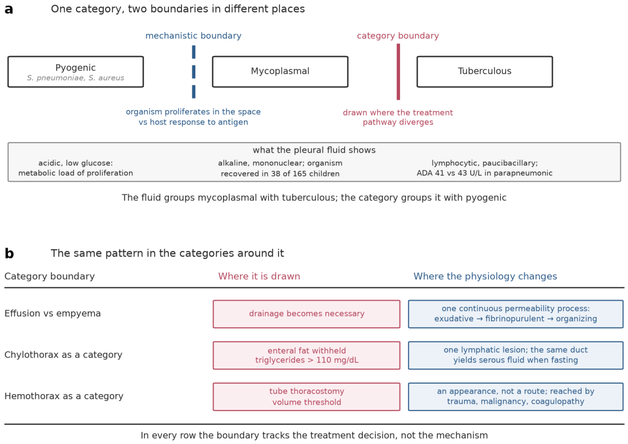
The categories of the conventional etiologic list are bounded where treatment changes, not where physiology changes. (a) Within pneumonia-associated effusion the two boundaries fall in different places. The category boundary separates tuberculous effusion from the rest, because antituberculous therapy starts at the outset; the mechanistic boundary separates pyogenic infection from the rest, because only there is the fluid mainly a product of bacterial proliferation within the space. Mycoplasmal effusion lies between them: counted with pyogenic infection, it behaves like tuberculous effusion, the organism being recovered from the fluid in 38 of 165 children with mycoplasmal pneumonia and effusion (Section 3.7). Fluid values are from the sources cited in Section 3.7 and Section 3.8 and come from different cohorts rather than a head-to-head comparison; adenosine deaminase medians are 41 U/L in tuberculous and 43 U/L in parapneumonic fluid in mixed-age series, and the mycoplasmal pH is from a pediatric intensive-care series. (b) The same mismatch in the categories around it, with thresholds as cited in Section 3.7 and Section 4.4. None of these rows is an error: each boundary does the job clinical vocabulary exists to do. The difficulty is that the same vocabulary is then used to count (Section 3.8).
Figure 1.
The categories of the conventional etiologic list are bounded where treatment changes, not where physiology changes. (a) Within pneumonia-associated effusion the two boundaries fall in different places. The category boundary separates tuberculous effusion from the rest, because antituberculous therapy starts at the outset; the mechanistic boundary separates pyogenic infection from the rest, because only there is the fluid mainly a product of bacterial proliferation within the space. Mycoplasmal effusion lies between them: counted with pyogenic infection, it behaves like tuberculous effusion, the organism being recovered from the fluid in 38 of 165 children with mycoplasmal pneumonia and effusion (Section 3.7). Fluid values are from the sources cited in Section 3.7 and Section 3.8 and come from different cohorts rather than a head-to-head comparison; adenosine deaminase medians are 41 U/L in tuberculous and 43 U/L in parapneumonic fluid in mixed-age series, and the mycoplasmal pH is from a pediatric intensive-care series. (b) The same mismatch in the categories around it, with thresholds as cited in Section 3.7 and Section 4.4. None of these rows is an error: each boundary does the job clinical vocabulary exists to do. The difficulty is that the same vocabulary is then used to count (Section 3.8).
Two features of the pediatric setting complicate synthesis. Mechanistic classification rests on pleural fluid biochemistry, yet diagnostic thoracentesis is performed less often in children than in adults, and a large share of pediatric effusions are documented by imaging and never sampled. And the transudate–exudate partition on which mechanistic reasoning depends describes the state of the capillary barrier rather than the identity of the disease: a child may satisfy exudative criteria on a sample to which a transudative driver also contributed.
This review takes the mechanism rather than the diagnostic label as its organizing unit, reporting what the cited series found in their own settings rather than generalizing across them. Its scope is the etiologic spectrum and its classification; management of established pleural infection appears only where it bears on how cases are ascertained and categorized, and readers seeking management guidance are referred to the society guidance cited in Section 3.
Evidence Sources
The literature underlying this review was assembled by searching OpenAlex across 34 topic areas spanning the etiologic spectrum, parapneumonic effusion and empyema, specific pathogens, malignant and lymphatic effusion, cardiac and renal mechanisms, trauma, neonatal effusion, diagnostic criteria and imaging, and post-2020 epidemiology, restricted to records published from 2008 onward. Of 686 de-duplicated records retrieved, 615 carried abstracts and were screened for relevance to pediatric pleural effusion; 174 were selected against section-level quotas and 169 resolved to verifiable bibliographic metadata, of which 117 are cited here.
To position this review against its neighbours, the same search infrastructure was applied to the review literature itself. Fifty-five comparable pediatric pleural-effusion reviews published since 2010 were retrieved and screened: fifty concern a single mechanism, most often parapneumonic effusion and empyema (29) or chylothorax and lymphatic disease (16), and two are multi-mechanism overviews. None attempts the full etiologic spectrum with a classification permitting simultaneous mechanisms; none proposes or evaluates a taxonomy beyond the staging of empyema; two discuss two mechanisms in one patient; one discusses effusions never sampled; three examine whether proportions across series can be compared (Figure 2). These counts rest on a structured screen of titles and abstracts by a single assessor, so they describe how this literature presents itself rather than proving that no such review exists. The closest published work is not a review but the 492-child series in Section 5.3 [1], which describes the spectrum under mutually exclusive subgroups and sampling-dependent entry. On that reading this review is complementary rather than a first: the evidence it synthesizes is published and largely single-mechanism, and what it adds is the audit of why those series cannot be stacked and a recording scheme built to carry what they discard.
This is a narrative review, not a systematic one. No protocol was registered, screening was performed by a single assessor, no risk-of-bias assessment was undertaken, and grey literature and non-English records were not systematically sought. Statements in this review about what the pediatric literature does not contain should therefore be read as claims about this assembled evidence base rather than as exhaustive claims about the published literature. Every reference cited here was retrieved through the CrossRef API rather than written from memory, and each digital object identifier in the reference list was independently confirmed to resolve and to return the title of the work it is attached to.
1. Pleural Fluid Homeostasis and the Mechanistic Basis of Effusion
1.1. Normal Turnover and the Reserve Capacity of Lymphatic Clearance
The pleural space couples lung to chest wall and holds a small fluid volume in continuous turnover, steady state balancing capillary entry against lymphatic drainage from the most dependent parietal pleura. Starling forces — the hydrostatic and oncotic pressures of capillary and pleural space—govern entry; the metabolically active mesothelium both produces and reabsorbs [2].
Exit is anatomically distinct. Stomata and large lacunae, exclusive to pleural and peritoneal mesothelia, couple to lymph formation and propulsion driven by spontaneous lymphatic muscle contraction and extrinsic motion [3]. Lymphatic capillaries take up fluid through open button-like junctions, collecting vessels sealing with tight zipper-like junctions [4], so uptake and transport fail independently. The anatomical asymmetry decides what each surface removes. Filtration is one passive mechanism from the parietal interstitium, removal several, pleural lymphatics dominant and alone matching a changing drainage need [5]. Only the parietal surface has stomata into submesothelial lacunae, clearing by bulk flow, protein and cells unsieved; visceral uptake is a pressure gradient plus active solute transport, moving neither leukocyte nor fibrin strand [3,6]. Propulsion has two independent sources: spontaneous lymphatic muscle contraction, the lymphangion a pump with its own preload, afterload, rate and contractility [7], and extrinsic diaphragmatic compression modulated by cardiogenic and respiratory swings [8]. They fail differently: sedation, paralysis or a splinted diaphragm leaves an intact vessel unprimed; contractile or valve failure leaves the pump unable to raise lymph against outflow pressure [7].
That pathway carries substantial reserve, working against a gradient. The pleural space is subatmospheric—about −5 cmH2O at end-expiration at right-atrial height in humans, falling further on inspiration [9]—so the lymphatic lumen must hold lower pressure still, and that gradient is actively generated: blocking diaphragmatic lymphatic contractility cuts pleural fluid egress by about 40% in animal preparations [10]. Clearance being actively maintained, its failure needs no change in filtration. Positive-pressure ventilation acts against exit on several terms at once—raising mean pleural pressure, removing the excursions supplying extrinsic pumping, and raising the central venous pressure the duct empties into: in anesthetized pigs, 15 cmH2O positive end-expiratory pressure reduced thoracic duct flow when it drained against central venous pressure but increased it against atmospheric pressure, while spontaneous breathing raised flow in both [11,12]. Outflow pressure, not ventilation, closes the exit. This review thus predicts mechanical ventilation selectively loads the lymphatic route rather than raising filtration, so a ventilated child’s effusion is mechanistically distinct from the same finding in a spontaneously breathing child—a prediction resting on animal preparations under Section 1.3’s rule; no pediatric study here measured pleural pressure or lymphatic flow under such ventilation. A narrative review puts reabsorption at about 28 times production [2]—animal and adult work, not pediatric, reported without an interval. Formation is always present, so an effusion is never simply its presence: formation must exceed the reserve, or clearance be impaired, or both [2]. No pediatric study here measures either directly.
1.2. The Four Mechanistic Routes
Four routes produce a net gain of pleural fluid, differing in whether they act on the pressures across an intact barrier or on the barrier itself. Listing them in parallel, as this section and Figure 3 do, is a convenience of exposition and is physiologically misleading in two respects that matter for everything that follows.
First, they are not equivalent in what they must overcome. Clearance holds a reserve of the order of tens of times resting production (Section 1.1) [2]. A route raising filtration must raise it by that order before fluid accumulates; a route disabling clearance need only remove the reserve, after which ever-present filtration suffices alone. Pressure abnormalities work against the reserve, lymphatic failure on it, so modest lymphatic impairment should reach effusion more efficiently than severe pressure derangement—a prediction no pediatric study in this evidence base has tested.
Second, these routes interact. The Starling relation is multiplicative: flow equals conductance times pressure difference. A permeability lesion raises conductance, so an abnormality yielding no effusion across an intact barrier may yield one across a damaged one. The air-blood barrier shows this: large gradients drive minimal flow while the interstitial matrix stays intact, small gradients high flow once degraded [14]. Routes rescale each other rather than adding increments, so co-occurrence matters clinically and single labeling discards information physiology treats as interactive. The hydrostatic route has two forms on different surfaces. Fluid enters across the parietal pleura down a net filtering gradient, the visceral surface normally absorptive [5]. Raised left atrial pressure acts indirectly, flooding lung interstitium, which empties across the visceral surface. In sheep, volume loading drove liquid of lung-interstitial protein concentration across it, 23 to 29% of edema cleared [15]; after oleic acid injury, pleural liquid accumulated over subpleural edema with no visceral pleural injury [16]. The space is an overflow sink, not a primary filtration site. Raised systemic venous pressure acts differently. Among 37 adults with heart failure, mean wedge pressure was 24.1 mmHg with effusion versus 17.2 mmHg without (p < 0.001), right atrial pressure unchanged [17]; among 27 patients with chronic right atrial or pulmonary arterial hypertension and normal wedge pressure, none had an effusion, including four with right atrial pressures above 20 mmHg [18]. Systemic venous hypertension thus reaches the space without filtration; it loads the lymphatic exit, which is why the two are often recorded together.
Higher hydrostatic and lower oncotic pressure raise filtration without pleural injury [2]. Pediatric hydrostatic causes are usually cardiac; pediatric heart failure is classified by hemodynamic abnormality—excessive preload, excessive afterload, abnormal rhythm, decreased contractility—not by adult-style contractile lesion [19]. Oncotically, 23 of 69 nephrotic children developed effusion, correlating with hypoalbuminemia severity (p = 0.000, r = 0.437) [20]; yet the mechanism is not purely oncotic—edema may reflect compensatory sodium/water retention after hypovolemia or primary renal sodium retention [21].
The oncotic route is quantitatively weakest. Colloid osmotic pressure opposes filtration only as the barrier reflects protein, so the oncotic term is a reflection coefficient times a transbarrier protein gradient, not albumin alone. Two buffers blunt its fall: subglycocalyx protein, not bulk interstitial fluid, opposes filtration, and interstitial protein washes down as filtration rises, eroding its gradient [22]. Falling albumin thus yields sub-proportional filtration rise, which must still exhaust clearance reserve before fluid is retained. The lesion raising hydraulic conductance also lowers the reflection coefficient [22]—stripping oncotic leverage while amplifying the hydrostatic term.
Increased permeability affects the barrier itself: flow disturbances damaging pleural layers generate an exudate, with inflammatory and immune reactions shaping its composition [2]. Mesothelial cells express a distinct tight-junction repertoire governing paracellular permeability, so the exudative route is change in a defined structure, not inflammation as metaphor [23]. The mesothelium does not simply fail: challenged, it releases chemokines directing phagocytes to its apical surface and permits bulk protein movement into the space, organizing its own breach [24]. Exudates show raised procoagulant activity (mainly tissue factor) and depressed fibrinolysis from plasminogen activator inhibitors 1 and 2, inhibitor 1 up to 913-fold normal pooled plasma; cardiac-failure transudates reverse this, favoring fibrin clearance [25]. A barrier-deficient space also loses capacity to clear deposited fibrin. Since protein-rich fluid exits mainly via stomata in the dependent parietal pleura [6], fibrin partitioning separates fluid from its drainage site, raising conductance and removing reserve from within the space. Matrix integrity outweighs gradient magnitude; safety factors against accumulation vanish once matrix degrades [14]. Mediators stay compartmentalized: interleukin-6 in pleural infection runs roughly 5000-fold higher in pleural fluid than matched serum (median 72,752 vs. 15 pg/mL) [26].
Its two conductances open separately. Liquid enters across the parietal pleura down a net filtering gradient [6], so the systemic capillary wall is one, the mesothelial tight-junction repertoire the other [23]. Interferon-gamma and tumor necrosis factor destabilize endothelial junctions synergistically [27], and the mesothelium secretes interleukin-8 under macrophage-derived tumor necrosis factor-alpha and interleukin-1-beta [28]. A circulating cytokine profile therefore raises filtration with no organism in the space.
Impaired lymphatic drainage, the only route yielding effusion without added filtration, hides two lesions. First, the conduit is patent but clearance fails—stomatal exit blocked by fibrin or tumor, a weak pump, or raised venous-end outflow pressure. That last follows a two-slope relation: flow resists back-pressure to a breakpoint, then fails. In unanesthetized adult sheep, flow was independent of outflow pressure until it rose well above venous pressure, then fell linearly, ceasing at 25.6 ± 4.2 mmHg; volume loading to 351 ± 54% of control shifted neither breakpoint nor cessation pressure [29]. Fetal preparations show the same pattern lower: flow fell above 5.1 mmHg, stopping at 18 mmHg in one series [30], at 11.5 ± 0.6 mmHg in another, where a 1 mmHg venous rise near normal cut flow by 12.7 ± 1.2% [31]. Cessation pressure is thus preparation-dependent, unmeasured in children at any age. Second, the duct is disrupted and lymph enters directly—a breach within the lymphatic circuit, grouped here only because the fluid is lymph [32]. Co-occurrence is explicit after cardiac surgery: chylothorax attributed to duct damage, accessory vessel disruption, or venous pressure exceeding duct pressure—lymphatic and hydrostatic routes as alternatives for one fluid [33].
Central venous pressure acts on both sides of the balance, but not equally: the duct drains into systemic veins, so raised central venous pressure impairs clearance downstream while adding little to filtration, which is why isolated systemic venous hypertension does not by itself produce an effusion. Elevated post-surgical central venous pressure independently predicted chylothorax and persistent serous effusion (OR 1.076 per unit, p = 0.046), with univentricular hearts and assist-device use [34]. In Fontan physiology, venous return staying pressure-dependent throughout, effusion is expected, not exceptional. Such effusion reflects one hemodynamic variable driving two routes, not two mechanisms in a patient. Asking whether it is hydrostatic or lymphatic poses a question physiology cannot answer—a stronger objection to single labeling than bookkeeping. That is why Section 6.2’s mechanism axis is multi-label by construction, not a concession to uncertainty.
The coupling is nonlinear. The two-slope form, in adult [29] and fetal [30] preparations alike, holds clearance reserve intact, then surrenders it over a narrow range rather than eroding it in proportion to central venous pressure. Hence moderate systemic venous hypertension produces no effusion [18], while a circulation pushed beyond the breakpoint, as cardiac surgery routinely produces, reliably does.
The transudate/exudate dichotomy partly fits: hydrostatic or oncotic changes cause transudates without structural pleural damage, while increased filtration or decreased reabsorption through processes damaging the pleural layers cause exudates [2], reflecting barrier integrity, not the disease behind it. Light’s criteria, its operational form, have high sensitivity but limited specificity, and procedural risk or patient factors may preclude sampling [35]. No pediatric validation study appears here; pediatric parapneumonic thresholds remain descriptive, not validated discriminants [36].
Figure 3.
Pleural fluid is in continuous turnover across two serosal surfaces, and an effusion is a failure of that balance rather than the appearance of filtration. (a) Liquid filters from the parietal side into a space held at roughly −5 cmH2O at end-expiration at the height of the right atrium [9], and leaves chiefly through stomata in the dependent parietal pleura; the lymphatic lumen must hold a pressure more subatmospheric still for drainage to proceed, which is why blocking intrinsic contractility of diaphragmatic lymphatics reduces pleural fluid egress by about 40% in animal preparations [9,10]. (b) A route acting by raising filtration must overcome the clearance reserve before fluid accumulates; raised pulmonary venous pressure reaches the space through the lung rather than across the parietal surface. (c) A route acting on the exit needs no rise in filtration at all. Routes of direct entry across a breached boundary are not shown; see Section 6.2. Fluid character and representative causes are given in Table 1.
Figure 3.
Pleural fluid is in continuous turnover across two serosal surfaces, and an effusion is a failure of that balance rather than the appearance of filtration. (a) Liquid filters from the parietal side into a space held at roughly −5 cmH2O at end-expiration at the height of the right atrium [9], and leaves chiefly through stomata in the dependent parietal pleura; the lymphatic lumen must hold a pressure more subatmospheric still for drainage to proceed, which is why blocking intrinsic contractility of diaphragmatic lymphatics reduces pleural fluid egress by about 40% in animal preparations [9,10]. (b) A route acting by raising filtration must overcome the clearance reserve before fluid accumulates; raised pulmonary venous pressure reaches the space through the lung rather than across the parietal surface. (c) A route acting on the exit needs no rise in filtration at all. Routes of direct entry across a breached boundary are not shown; see Section 6.2. Fluid character and representative causes are given in Table 1.
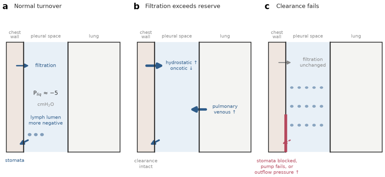

Table 1.
The four mechanistic routes to pleural effusion, the fluid character most often observed, and representative pediatric causes.
Table 1.
The four mechanistic routes to pleural effusion, the fluid character most often observed, and representative pediatric causes.
| Mechanistic route | Capillary barrier | Fluid character most often observed | Representative pediatric causes |
| Increased hydrostatic pressure (pulmonary venous) | Intact | Transudate; frequently exudative under diuretic therapy | Left heart failure; left-sided obstructive lesions; fluid overload in renal failure |
| Decreased oncotic pressure | Intact | Transudate | Nephrotic syndrome; protein-losing enteropathy; malnutrition; critical illness |
| Increased capillary permeability | Disrupted | Exudate | Parapneumonic infection; tuberculosis; malignancy; connective tissue disease |
| Impaired lymphatic clearance or direct duct leak | Intact; outflow obstructed, pump failing, or duct disrupted | Chylous when enteral fat is being absorbed; serous or exudative when fasting | Systemic venous hypertension (Fontan physiology, post-cardiotomy, superior vena cava obstruction); congenital or surgical thoracic duct injury; mediastinal mass; lymphatic malformation |
Cardiac disease appears on two rows rather than one: raised pulmonary venous pressure reaches the pleural space by filtration through the lung, whereas raised systemic venous pressure acts on lymphatic outflow (Section 1.2). The mapping from route to fluid character is a tendency, not a rule: the exceptions in column three are common enough in pediatric practice that fluid character alone does not identify the route. The table covers accumulation across an intact pleural boundary; direct entry by trauma, iatrogenic injury or transdiaphragmatic passage is outside it (Section 6.2).
Fluid phenotypes (purulent, hemorrhagic, chylous) describe appearance and composition, not accumulation route; recorded alongside mechanism, not as mechanisms themselves (Section 6).
1.3. Developmental Differences Bearing on Mechanism
The routes themselves are not pediatric. Starling’s relation, the pleural barrier and lymphatic exit anatomy are mammalian physiology; nothing here proposes a mechanism in children that adults lack. Adult or mixed-age evidence records where the work was done, not a limit on where the physiology applies, and such sources serve as mechanistic evidence without qualification.
Developmental specificity is quantitative, in two kinds kept apart here. A physiological term’s magnitude—clearance reserve, chest wall compliance, an analyte’s reference interval—may shift with age without changing the relation it enters. A measurement property—threshold, cutoff, sensitivity, reported proportion—reflects the population sampled and instrument used; it is not physiology at all, and does not transport across age, era, or setting (Section 2.2, Section 3.4 and Section 5.2). An age qualifier flags a non-transportable number; its absence a claim about mechanism that does transport.
Three developmental features alter how much these routes deliver in children; the evidence for each is unequal.
Diaphragmatic lymphatic anatomy, the principal exit route, is asymmetric: mouse pleural surfaces show mostly vessel-like LYVE1-positive configurations with scanty stomata, peritoneal surfaces abundant lacy-ladder structures with numerous stomata-like pores, and peripheral-rim contraction with cardiogenic and respiratory swings modulates drainage from both cavities [8]. No equivalent morphometry exists for human infants or children, so claims that a smaller, faster-cycling thorax alters extrinsic pumping of that drainage extrapolate from murine anatomy.
The neonatal circulation differs further. Congenital chylothorax, the leading cause of perinatal pleural effusion, has unknown etiology in most cases [37]; causes may be developmental, not mechanical, since FLT4 encodes vascular endothelial growth factor receptor 3, required for lymphangiogenesis, and signaling-impairing variants disrupt lymphatic development [38]. This exemplifies a class: germline mutations in at least 20 genes converging on VEGFR-3 via RAS/MAPK and PI3K/AKT underlie primary lymphedema, lymphatic malformation, and chylous effusion [39]; primary intestinal lymphangiectasia is the same lesion presenting enterically [40]. Developmental lymphatic failure lowers exit-path conductance before, and independent of, any filtration defect. In the Section 4.1 congenital heart surgery cohort, chylous and persistent serous effusions showed distinct risk profiles, univentricular hearts among them [34]; circulations with structurally elevated systemic venous pressure load the lymphatic route structurally, the hydrostatic route only when left-sided filling pressures also rise.
Relative pleural surface area is the weakest-supported of the three: no study here indexes it to body mass or circulating volume across childhood. A given volume fills more thoracic capacity in an infant than an adolescent, implying an earlier clinical threshold at identical mechanism and rate—inferred from body size, not measured in children. Documented instead: compressive consequence dominates neonatal presentation, congenital chylothorax manifesting through pressure effects including pulmonary hypoplasia as well as through protein and lymph loss [41].
1.4. Phenotype Is a Property of the Fluid, Not a Mechanism
The categories dominating bedside reporting—purulent, hemorrhagic, chylous—classify the aspirate, not the process producing it, and each is reachable by more than one of the four routes.
Purulence marks stage, not etiology: pleural infection progresses through exudative, fibrin-deposition-and-pus, and organizing stages [42]; empyema denotes a parapneumonic exudate once bacteria are detected or surrogates such as low pH or leukocytosis appear [43]. Tuberculous effusion combines true pleural infection with an effusive hypersensitivity reaction compartmentalized within the space—one exudate, two processes [44].
Those stages [42] are generated, not observed; the generator explains the sequence’s direction. The exudative stage is a pure permeability lesion: fluid crosses inflamed membrane, stays drainable, fibrinolysis still clearing deposits. The fibrinopurulent stage starts when that balance inverts [25]; neutrophil influx adds extracellular DNA and citrullinated histones, an order of magnitude higher in parapneumonic than malignant or transudative fluid, inversely correlated with pH [45]. Pleural plasminogen activator inhibitor 1 alone independently predicted sonographic septation presence and severity across 214 pleural infection samples [46]. The organizing stage is a repair lesion, not a holdover: mesothelium failing to re-epithelialize leaves fibrous adhesions between serosal surfaces [47]. Each stage supplies the next’s substrate; stage predicts drainage need, the organism does not.
Chyle is defined biochemically, by triglycerides above 110 mg/dL with cholesterol below 200 mg/dL, the same phenotype arising from trauma including iatrogenic surgical injury, from malignancy, from primary lymphatic disorders, and from heart failure [32]. Pediatric hemothorax follows blunt and penetrating mechanisms [48], yet is listed among pediatric etiologies alongside hematolymphoid malignancy, heart failure and hypoalbuminemia despite being an appearance rather than a cause [49].
Phenotype and mechanism dissociate within one etiologic label too. Among 54 children with parapneumonic effusion, infants showed mononuclear predominance with higher glucose and lower protein, older children polymorphonuclear predominance, age correlating with the polymorphonuclear fraction (rho = 0.509) and with protein (rho = 0.622, both p < 0.001) [36]. The pleural space is also a distinct immunological compartment: in 30 children with effusion after congenital heart surgery, pleural interleukin-6, interleukin-8, interleukin-10 and tumor necrosis factor exceeded plasma, interferon-γ and interleukin-17A lower [50]—an exudative signature generated in situ within an effusion convention labels cardiac and transudative.
One structural cause makes the dissociation systematic. An effusion is a stock, the routes flows: accumulation equals the time integral of formation minus clearance. Sampled fluid carries the summed history of every acting route, weighted by duration and concurrent mesothelial and clearance activity. Two consequences follow. A single sample cannot recover the flows producing it: many combinations integrate to the same stock, underdetermined in principle, not just practice, which no refinement of fluid chemistry fixes. The mesothelium being metabolically active and clearance selective for protein and cells [2,6], fluid is modified while it sits, not passively recorded. Fluid appearance thus reports what occurred in the space, aggregated over time and mechanisms: useful diagnostically, underdetermined mechanistically. So the transudate-exudate call belongs at the phenotype level, not the mechanism axis (Section 6.2)—not because it is imprecise and sharpenable, but because it measures a stock while being asked to report a flow. These four routes cover accumulation across an intact pleural boundary, excluding direct entry across a breached one (traumatic or iatrogenic injury, transdiaphragmatic passage) and fluid forming against a non-expanding lung; Section 6.2 covers both. Following sections remap the pediatric etiologic literature onto these routes.
2. Classifying Effusion: Light’s Criteria and Their Limits in Children
2.1. What the Transudate–Exudate Partition Was Built to Do
The transudate–exudate dichotomy is mechanistic before it is biochemical. Pleural fluid volume reflects Starling filtration against lymphatic drainage, whose reabsorptive reserve is reported at roughly 28 times production. Altered hydrostatic or oncotic pressure gives a transudate, sparing the pleural layers; disturbed filtration or impaired reabsorption gives an exudate through processes that damage them [2]. Light’s criteria recover that distinction from paired fluid and serum total protein and lactate dehydrogenase (LDH). Derived in adults, they call an effusion exudative if any of three thresholds is exceeded [51]: fluid/serum protein >0.5, fluid/serum LDH >0.6, or fluid LDH above two-thirds of the serum reference upper limit.
Being disjunctive, the rule is sensitive but not specific [35], and errs one way: transudates are called exudates more often than the reverse. Diuretics are the usual explanation, concentrating protein and LDH in a hydrostatic effusion until it crosses the ratio thresholds before sampling. The effect is established in adults, unmeasured in children. Diuretic exposure is near-universal in the pediatric conditions producing hydrostatic and oncotic effusions, and titrating it is central to managing childhood nephrotic syndrome [21]. The call was never terminal: one stage before etiology-directed testing, and preanalytical handling affects the values [52].
2.2. Pediatric-Specific Problems
No study assembled here derives or validates Light’s criteria in children against an independent etiologic reference standard, and no multi-center pediatric series reports their operating characteristics. The closest evidence is unidirectional: of 54 children with parapneumonic effusion, most samples met conventional exudate thresholds (white cell count >1000/µL, total protein >3 g/dL, LDH >200 U/L) [36]. Lacking transudates, it confirms expected behavior in an already exudative population but estimates no specificity.
Age-dependence of the analytes is the more serious objection. In that cohort, age correlated with polymorphonuclear percentage (rho = 0.509, p < 0.001) and total protein (rho = 0.622, p < 0.001), inversely with glucose (rho = −0.274, p = 0.043); infants showed mononuclear predominance, higher glucose and lower protein than older children [36]. Fixed thresholds spanning infancy to adolescence meet a shifting reference distribution. A second analyte behaves likewise: in a mixed-age cohort stratified at 55 years, adenosine deaminase (ADA) tracked age (r = −0.621) more strongly than any other variable examined, prompting age-stratified cutoffs [53]. Serum LDH reference intervals exceed adult ones in infants and young children, so the third criterion moves with the laboratory’s age-specific range. That follows from routine pediatric laboratory practice, not any pleural study, and no pediatric pleural series here has quantified it.
Sampling is the third problem, structural rather than analytic. Pediatric guidance reserves thoracentesis for effusions over a quarter of the hemithorax in a respiratory-compromised child, for large effusions over half, or for any mediastinal shift; a quarter or less gets antibiotics alone [43]. In a German nationwide cohort of children needing drainage or with effusion past seven days, 45% of 645 cases began non-invasively [54]; small-volume blunt traumatic hemothoraces are likewise observed without tube thoracostomy [48]. Pediatric respiratory units surveyed in four Central European countries agreed on initial diagnostics but not on the volume threshold triggering an invasive procedure [55]. Biochemistry therefore reaches a minority of pediatric effusions, selected for size and physiological compromise.
2.3. Proposed Alternatives and Adjuncts
Proposed substitutes for the paired-sample rule are all adult-derived: the serum-to-pleural-fluid albumin gradient, applied near a 1.2 g/dL threshold to rescue diuretic-treated transudates; cholesterol-based rules using pleural cholesterol with or without pleural LDH; and “blood-free” combinations classifying from pleural analytes alone. The albumin gradient still needs a simultaneous serum sample, correcting diuretic misclassification rather than sparing venepuncture; the other two drop the paired venous sample, the advantage most relevant to children. In the literature reviewed here, none has been evaluated in children or compared against Light’s criteria in a pediatric study.
ADA has the largest biomarker evidence base, and shows why a fluid threshold carries no etiologic label. A 40 U/L cutoff in a lymphocytic exudate stays pragmatic where tuberculosis prevalence is high [56]. Where prevalence falls, specificity collapses: 40 IU/L gave sensitivity 1.00 and specificity 0.61, 70 IU/L restored 0.93 and 0.93, and of 54 patients in the 40–70 IU/L band, 55.6% had malignant effusion and 5.6% tuberculosis [57]. A multi-center analysis of 276 exudative effusions put the optimal local threshold at ≥36 U/L (AUC 0.852) [58]. The cutoff belongs to the population sampled, not the analyte.
The one pediatric comparison performs worse, and blames childhood case mix. In 25 children and adolescents with confirmed pleural tuberculosis versus 68 with parapneumonic effusion, ADA at 40 U/L gave 88% sensitivity but 31% specificity and 46% accuracy; an LDH/ADA ratio of 8.3 did best, at 79% sensitivity, specificity and accuracy [59]. Parapneumonic fluid is itself ADA-rich in mixed-age series: median ADA 43 U/L versus 41 U/L in tuberculous effusion, separable mainly by LDH (2542 versus 449 U/L) [60]. Whether the ratio adds anything beyond ADA is contested; two mixed-age cohorts showed no incremental value by net reclassification or integrated discrimination [61], and neither reports an age breakdown, leaving the pediatric case untested outside that single series. Other adjuncts share the ceiling: angiopoietin-2 separates exudates from transudates (P = 0.001) but not malignant from benign [62]; calprotectin, TNF-alpha and IL-1 are not recommended for routine use [43]; quantitative ultrasound echogenicity analysis stays investigational, proposed because procedural risk limits sampling [35].
2.4. Biochemistry Classifies the Fluid, Not the Cause
Every rule above labels a sample, not a disease process, and that mapping is not one-to-one. Tuberculous effusion is clearest: two mechanisms feed one sample, and the fluid is paucibacillary enough that nucleic acid amplification on it averages about 44% sensitivity in adult series (Section 3.6) [44,63].
Co-occurring mechanisms are routine in pediatric series. In the congenital heart surgery cohort of Section 4.1, chylous and persistent serous effusions arose with barely overlapping risk profiles, putting lymphatic and hemodynamic mechanisms in one population and, plausibly, one patient [34]. Heart failure is itself a recognized non-traumatic cause of chylothorax, setting lymphatic fluid downstream of a hemodynamic lesion [32]. In nephrotic syndrome, hypoalbuminemia correlated with effusion across 69 children (r = 0.437) but only 23 developed one [20], and the sodium and water handling behind it differs between underfill and overfill physiology [21].
A parallel problem arises where no fluid exists to classify. Detection has outpaced sampling: of 81 children with confirmed community-acquired pneumonia, ultrasound found parapneumonic effusion in 17 and radiography 11 of those same 17 [64], and reported prevalence of effusion complicating pneumonia rises from 14% on radiography to 44% on ultrasound [65]. Each gain in imaging sensitivity enlarges the population of effusions that are documented, clinically real, and permanently unclassifiable by Light’s criteria or any successor. No multi-center pediatric series reports what fraction of imaged effusions undergo thoracentesis, so the denominator for every fluid-based scheme in children is unknown.
3. Infectious Mechanisms
Infection is the most studied mechanism in children, though whether it causes the largest share of pediatric effusions is beyond available ascertainment (Section 5.2). “Infectious effusion” is not a mechanism but at least four pathophysiologies sharing only an infectious antecedent: neutrophilic exudation from bacterial pleural invasion, lymphocytic exudation from delayed hypersensitivity, immune-mediated effusion with few recoverable organisms, and fluid around necrotizing parenchyma.
3.1. Staging Grades Severity, Not Cause, and the Two Staging Axes Do Not Agree
The simple/complicated/empyema convention grades severity, not cause, along two axes needing different evidence. Parapneumonic effusion is an exudate; “empyema” applies when bacteria, or surrogates such as low pH or pleural leukocytosis, are detected. Classification usually uses the four-grade Jaffe system, scored from pleural ultrasonography alone; pleural pH below 7.1, glucose below 40 mg/dL and lactate dehydrogenase above 1000 IU/mL are separately associated with empyema and require fluid [43]. The axes are not interchangeable. German surveillance of 645 children, 45% initially managed non-invasively, staged every case from reported sonography [54]—assignable only because grading rested on imaging, not biochemistry. Drainage-cohort distributions are conditioned on the decision to drain, not the source population.
Upstream, the denominator is unstable: the proportion of pneumonia complicated by effusion depends on ascertainment modality—40% of patients with pneumonia in mixed-age practice [66], and 14% on chest radiograph rising to 44% on ultrasound, many resolving on antibiotics alone [65]. Of 160 North East England children with radiologically confirmed pneumonia, 40 (25%) developed empyema, strongly associated with bacterial infection (OR 3.34, 97.5% credible interval 1.70–5.14) [67]. Parapneumonic effusion is called the most common cause of pediatric pleural effusion [36,43], yet no cited series reports cause distribution in an unselected pediatric population (Section 5). Stage belongs on a severity descriptor, not the mechanism axis; an effusion managed without a tap is indeterminate, not infectious by default.
3.2. One Infectious Label Covers Several Pleural Processes
Within bacterial infection the process ranges from exudation across an inflamed membrane to fluid around liquefying lung, at an intensity varying independently of the organism recovered; necrotizing pneumonia sits at the severe end, coupling both mechanisms. In a Boston series of 80 children over 15 years, 69 (86%) had pleural effusion, mean pH 7.08; cultures were positive in 48%, Streptococcus pneumoniae predominating; cases rose from 12 in 1993–1996 to 40 in 2001–2004 [68]. Blood culture with culture and molecular testing of pleural fluid yields a microbiologic diagnosis in up to 50% of cases [69]; S. pneumoniae, Staphylococcus aureus, Mycoplasma pneumoniae, and co-infection are implicated [70]. Among 124 Utah children with culture-confirmed pneumococcal pneumonia, radiographic necrosis rose from 13% in 1997–2000 to 33% in 2001–2006, serotype 3 associated with necrosis (OR 14.67, 95% CI 3.39–86.25) [71].
Host response varies on top of organism. An inflammatory rebound—C-reactive protein re-rising by at least 50 mg/L after an equal initial fall—occurred in 38% of 53 French children with pleural infection, and in 56% of S. pyogenes cases versus 30% each for S. pneumoniae and S. aureus [72]. One label per case records this as a single entry; naming the route, and allowing more than one, keeps the parenchymal component visible beside the pleural.
3.3. Attributed Bacteriology Is a Property of Era, Country, and Vaccine Program
Bacteriologic proportions move with vaccine schedule, country, period, and detection method, so pooled figures describe programs rather than mechanism and should not be collapsed. Across Asian, European, and US series the prevalent organisms in pediatric effusion/empyema are S. pneumoniae (21–56%), S. pyogenes (19–27%), and S. aureus (7–31%), with methicillin resistance reported in Australia (4%) and the United States (10%) but not Europe [43]. Pneumonia and empyema can also move in opposite directions: after PCV7, pneumonia admissions in English children under 15 fell 19% while empyema fell 22% [73], whereas in the United States pneumonia hospitalizations in children under 2 fell 33% yet empyema-complicated pneumonia rose 2.01-fold [75]; the divergence resolved after PCV13 in Scotland and Madrid [76,77]. German surveillance documents pleural-compartment replacement most directly: among 584 children with bacteriologically confirmed effusion/empyema (2010–2018), serotype 3 rose from 25.4% to 62.7% of serotyped cases, those cases younger (median 3.2 versus 5.6 years) and more often needing intensive care (89.6% versus 73.8%) [78]. Serotype 3 caused 26% of pediatric invasive pneumococcal disease in Portugal despite >95% vaccine uptake [79].
Non-pneumococcal organisms have their own geography. At one Houston center, 2001–2009, 117 children had non-ventilator-associated S. aureus pneumonia, MRSA causing 74% and the USA300 clone 92% of MRSA isolates; presentation was empyema in 72, lung abscess in 15 [81]. Methicillin resistance affected 31.3% of S. aureus in the German susceptibility-tested subset [82]. Group A streptococcus ranks second or third wherever molecular testing is applied: 16% of Canadian parapneumonic fluids by PCR [83], 11% of pathogen-positive Zurich cases [84]. Partitioning first by organism yields a label that expires, and that differs between children of identical pleural physiology born a decade or a border apart.
3.4. Attributed Etiology Is a Measurement of Laboratory Practice
The organism recorded against an effusion is a function of the test ordered, by a margin that dominates any epidemiological signal (Table 2). In 56 Ontario children with parapneumonic effusion sampled 2009–2011, real-time PCR detected a pathogen in 82% versus 25% for culture, S. pneumoniae in 62% versus 14% [83]: same children, fluid, and denominator; different assay. A pleural fluid S. pneumoniae PCR assay at one US children’s hospital raised pathogen detection from 38.9% to 73.4% and pneumococcal detection from 14.1% to 45.6%, cut median time to optimal antibiotics from 10.0 to 4.9 days, and changed neither stay length nor readmission [85].
Yield varies by platform: 45–84% sensitivity for S. pneumoniae-specific PCR, 50–69% for 16S rDNA amplification and sequencing [43]. Molecular methods recover serotype from culture-negative material [86] and ascertained 30% of pediatric invasive pneumococcal disease in Portugal in 2015–2018 [79]. Detection does not reliably alter prescribing: in 1402 German children, diagnosing S. pneumoniae, S. pyogenes, or S. aureus prompted a narrow-spectrum beta-lactam switch in only 9.4%, 18.9%, and 5.2% [82]. Molecular testing has shifted the apparent distribution toward S. pneumoniae and S. pyogenes and away from the unknown category, while the proportion of effusions never yielding fluid is untouched.
Where more organisms are sought, more are found together and sole causation is harder to assert. Among 846 children hospitalized with community-acquired pneumonia, a pathogen was identified in 70.1%: respiratory syncytial virus 22.9%, human rhinovirus 22.1%, M. pneumoniae 15.8%; co-infection in 34.6%, of which 71.3% were mixed viral-bacterial [87]. Viral co-infection in 18 of 68 tested children with S. aureus pneumonia was associated with respiratory failure (72% versus 24%) [81], and viral-bacterial interaction is proposed to contribute to necrotizing pneumonia [69]. A pathogen-discovery protocol in undiagnosed pediatric parapneumonic effusions recovered three novel torque teno mini virus isolates replicating in alveolar epithelial cells, raising but not establishing a pathogenic role [88]. No pediatric series here reports the frequency of fungal effusion. What remains after testing is large and is not a diagnosis: blood and pleural fluid culture detected a pathogen in only 25% of Canadian children with parapneumonic effusion [83], cultures are positive in 56% of pleural infection cases in mixed-age practice [65], and a scoping review of 127 pediatric empyema studies since 2000 called causative serotypes a persisting gap [89]. Attribution matters beyond bookkeeping: pathogen and host factors determine which childhood pneumonias end fatally [90]. “Infection, organism unknown” records what was looked for; treating it as a mechanism imports laboratory policy into the taxonomy.
The five comparisons span effusion detection, pathogen attribution, and species attribution, and the smallest moves the reported figure by half again. Two series differing only in instrument will differ in result by more than most epidemiological changes this literature reports—the concrete form of the problem Section 5 audits: a published number is a joint property of the children and the apparatus, and only one is usually described.
3.5. Severity and Mechanism Are Confounded in *M. pneumoniae* Effusion
Effusion marks a worse M. pneumoniae phenotype, making its frequency a severity statistic as much as a mechanistic one. The organism causes 10–40% of pediatric community-acquired pneumonia, cycling epidemically every 3–7 years [91]. Pleural involvement is common but not dominant. In the US EPIC study, 8% of 2254 children hospitalized with radiographically confirmed pneumonia were PCR-positive; of these 26% had effusion, 4% of isolates were macrolide-resistant, 28% had a co-pathogen [92]. Pediatric series report effusion in 16% in Korea [93] and 17% in northern Taiwan [94]. Roughly 20–25% of adults develop parapneumonic effusion, reportedly similar in children; that fluid is mononuclear-predominant with adenosine deaminase elevated as in tuberculous effusion, separable, the source holds, by the degree of elevation plus serum markers [95].
The severity signal is consistent: effusion accompanies longer fever and admission, higher inflammatory markers, and poorer response to stepwise treatment [93], and independently predicted poor macrolide response in Taiwan (OR 14.3, 95% CI 4.9–42.0) [94]. Organism in the fluid marks a worse course still, 15 versus 11 hospital days and necrotizing pneumonia in 23.7% versus 7.9% [96]. Resistance confounds this further. It is geographically stratified, arises from 23S rRNA domain V mutations at positions 2063 and 2064, and reaches 80–90% in Asian countries [97]. Pediatric rates run 10.3% in Jeju, Korea, 2010–2015, where frequent effusion was the only distinguishing feature [98]; 53% in Taipei, 2010–2019, identical across intensive-care and non-intensive-care groups (53% versus 53%, p = 0.986), with alkaline effusion (pH > 7.7), lymphocyte-predominant in 18 of 22 analyzed [99]; and 78.6% in Shanghai in late 2023 [100]. Whether resistance drives effusion is unresolved: two Chinese series link them [101,102], a Korean series found no difference in resistant-strain proportion with and without effusion [93], a Chinese one none by disease severity [103].
The 2023–2024 global resurgence after COVID-19 restrictions rebounded late, unlike respiratory syncytial virus and pneumococcus [104]. Rising counts of associated effusion admit two explanations separable in principle, more pneumonia at unchanged risk per case versus unchanged burden at higher risk per case, but the cited literature cannot distinguish them: resurgence series report positivity among tested children, or case counts and testing volumes, not population-denominated incidence [105,106]; effusion-proportion series are numerator-only cohorts of hospitalized children with M. pneumoniae pneumonia [93,94,100]; and current tests cannot separate infection from carriage [104]. None reports incidence against a population denominator before and after 2020. The label carries severity, testing intensity, and resistance epidemiology alongside whatever mechanism produced the fluid.
3.6. One Exudate, Two Processes: Tuberculous Effusion and the Limits of the Fluid Phenotype
Tuberculous effusion defeats single-label classification from inside one sample. Pathogenically dual, it combines true pleural infection with a compartmentalized effusive hypersensitivity reaction and spans complete resolution through loculation, pleural thickening, and frank empyema [44]. Most disease is paucibacillary, coexisting with and progressing to pulmonary tuberculosis at high rates [107]; a negative acid-fast smear, absent granulomas, and failure to culture Mycobacterium tuberculosis do not exclude it [108].
Physiology resolves that paucibacillary difficulty: the response to mycobacterial antigen generates the fluid, not proliferation in the space. Intrapleural tuberculin purified protein derivative effuses the sensitized animal [109], so antigen without a replicating population suffices. In humans the reaction is locally regulated, pleural B cells controlling interferon-gamma production by resident T and NK cells [110], and its effector consequence is the permeability lesion of Section 1.2 reached by another trigger: mediators generated in situ raise conductance across an uninvaded mesothelium. The account is not one of pure hypersensitivity. Understanding has moved to direct pleural infection followed by an immunological cascade [111], which is why the organism is recoverable at all and why one mechanism spans effusions that resolve and effusions that loculate or suppurate [44].
The organism is often absent from the compartment it is causing. An adult Italian series recovered it from 17.3% of pleural fluid against 63% of pleural biopsy cultures [112]; Xpert MTB/RIF on pleural fluid averages approximately 44% sensitivity in adults, making tissue the preferred specimen [63]. Both figures predate Xpert MTB/RIF Ultra, now the WHO-recommended first-line assay, and no pediatric pleural-fluid Ultra accuracy study was identified here. The fluid-phenotype surrogate filling that gap does not transport. Adenosine deaminase above 40 U/L in a lymphocytic effusion is the most accessible route in high-prevalence areas [56], and in one mixed-age Indian series all 56 tuberculous effusions exceeded it versus 2.5% of non-tuberculous effusions [113]. But the threshold shifts with population: it correlates negatively with age (r = −0.621) [53]; a multicenter Malaysian analysis of 276 exudative effusions found an optimal cutoff of ≥36 U/L [58]; and parapneumonic effusion itself runs high, median 43 U/L against 41 U/L in tuberculous effusion, with lactate dehydrogenase discriminating (2542 versus 449 U/L) [60]. In the only pediatric accuracy study identified here, 25 confirmed pleural tuberculosis versus 68 parapneumonic effusions at a Brazilian center, 2001–2018, adenosine deaminase at 40 U/L gave 88% sensitivity but 31% specificity and 46% accuracy, while a lactate dehydrogenase/adenosine deaminase ratio of 8.3 gave 79% sensitivity, specificity, and accuracy [59]. No multi-center pediatric series reports smear and culture yield from pleural fluid against a histologic reference standard.
Two conclusions follow. A lymphocytic exudate with high adenosine deaminase fits more than one mechanism and more than one organism, which argues for demoting fluid characterization to a tag rather than a classifying axis. Where one sample encodes infection and hypersensitivity together, the mechanism axis must record both; choosing discards evidence.
3.7. The Hard Discrimination Is Not Pus from Water: It Is Tuberculous from Mycoplasmal Effusion
Bedside practice treats purulent versus serous as the difficult distinction, but imaging settles it: ultrasonography grades the effusion from anechoic fluid to multiple thick septations with solid components and guides the choice between conservative and invasive management without obtaining fluid [43]. What the fluid cannot do is separate the two infectious effusions that most resemble each other in the pleural space. The scope is narrow: a tuberculosis clinician does not find these two hard to tell apart and is right not to, since contact history, tuberculin or interferon-γ release testing, radiographic pattern, tempo, and effusion size separate them in most children. The argument concerns the fluid alone, what the specimen an etiologic label nominally rests on can support, which is what bears on classification: a transudate-exudate call or an adenosine deaminase threshold interrogates the fluid, not the history.
They resemble each other because in neither is the effusion mainly a product of pleural invasion. The qualification is necessary: among 165 children with mycoplasmal pneumonia and effusion requiring thoracocentesis, M. pneumoniae was detectable in the fluid of 38 (23%), who fared worse (Section 3.5) [96]. The organism reaches the space in a minority, as in tuberculous effusion; what distinguishes both from pneumococcal empyema is that a host response, not proliferation within the space, generates the bulk of the fluid. In tuberculous pleuritis that response is compartmentalized hypersensitivity atop pleural infection [44], locally regulated [110], leaving the organism scarce and tissue outperforming fluid (Section 3.6) [63]. Mycoplasmal effusion is likewise held immune-mediated rather than invasive on an explicitly unresolved mechanism: proposed accounts run from delayed hypersensitivity to the organism [114] to the T-cell-mediated epithelial injury, immune-complex-mediated vascular injury, and antibody-mediated responses advanced for mycoplasmal extrapulmonary disease generally [115].
Sterile immune effusion shares that compartmentalized effector arm [110], the mesothelium participating secretorily rather than as a membrane [28]. The antigen differs: tuberculous pleuritis retains a persisting local one, so its exudate can organize into loculation, thickening or empyema [44], whereas in serositis the antigen is systemic or unidentified and the membrane a bystander, predicting resolution once the stimulus is removed [116]. The fluid does not carry that distinction.
They separate on a physiological rather than microbiological variable: whether a replicating population occupies the compartment. Organisms proliferating there impose a continuing metabolic load, read in the values associated with empyema, pH below 7.1 and glucose below 40 mg/dL [43]. Where the organism lies outside the space or is scanty within it, antigen rather than replication is the stimulus, so conductance rises without that load and the fluid need not acidify. Pleural pH and glucose would then read where the organisms are rather than borrowed severity, explaining why mycoplasmal fluid was alkaline in the series above while pyogenic empyema is acidic. No study in this evidence base tests that interpretation directly.
The convergence is not only mechanistic. Mycoplasmal fluid resembles tuberculous fluid in mononuclear predominance and adenosine deaminase, separable only by degree of elevation with serum inflammatory markers [95]: in a pediatric intensive-care series the mycoplasmal fluid was alkaline (pH above 7.7) and lymphocyte-predominant in 18 of 22 analyzed [99], while tuberculous fluid is separately described as lymphocyte-predominant with median adenosine deaminase 41 U/L in mixed-age series [60]—different cohorts, so a pattern across series rather than a head-to-head comparison. The best-evidenced marker thus separates by magnitude rather than kind, and that magnitude is population-specific. Nor is total adenosine deaminase mycobacterium-specific: the diagnostic elevation is carried by the ADA2 isoenzyme of monocyte-macrophage origin, and the ADA1-to-total ratio correctly reclassified lymphocytic effusions as non-tuberculous where total ADA alone did not [117,118]. No pediatric pleural series identified here reports fractionation. The pediatric accuracy study above derived an optimal cutoff of 125 U/L, at which sensitivity was 84%, specificity 65%, accuracy 70% and AUC 0.67—recalibration alone does not rescue the analyte in children [59].
What would separate them is a difference in kind of immune response rather than magnitude, a proposition the evidence here reaches without establishing. Tuberculous pleuritis is Th1 and macrophage-driven by consistent description. Pediatric pleural fluid is in any case a distinct immunological compartment, not a plasma filtrate: among 30 children with effusion after congenital heart surgery, interleukin-6, interleukin-8, interleukin-10 and tumor necrosis factor exceeded plasma levels while interferon-γ, GM-CSF and interleukin-17A fell below them, and Th1, Th17 and memory effector cytotoxic T cells were enriched in fluid relative to blood [50]. Its two-sided conclusion matters here: some cytokines appear locally produced within the pleural space, others to reflect the systemic response, and the enrichment of both Th1 and Th17 in a non-infectious post-surgical effusion is a caution against reading either signal as organism-specific. Severe and refractory mycoplasmal pneumonia is itself described as an excessive Th1- and Th17-weighted response [97], so a scheme assigning Th1 to tuberculous effusion and Th2 to mycoplasmal is unlikely to survive measurement; if a mediator-level discriminator exists it is more likely quantitative, or a ratio, than opposing polarities. No study identified here reports paired pleural-fluid immune profiling across mycoplasmal and tuberculous effusion in children, which is what would settle it.
That divergence of alkaline mycoplasmal fluid [99] from the pH below 7.2 defining complicated parapneumonic fluid [43] deserves more than it has been given. Pleural pH is measured wherever a tap is done and costs nothing extra. Whether it separates mycoplasmal from tuberculous effusion in children is untested here, a cheaper question than cytokine profiling that no one appears to have asked.
The stakes are asymmetric, so the discrimination matters more than its frequency suggests. Misreading tuberculous effusion as mycoplasmal delays antituberculous therapy in a child whose disease coexists with and progresses to pulmonary tuberculosis [44]; the reverse commits a child to months of hepatotoxic therapy for an illness that would have resolved. The same fluid invites both. Section 3.8 takes up the consequence: these effusions converge on mechanism and on fluid chemistry, yet nomenclature places one inside the parapneumonic category and the other outside.
3.8. What Counts as Parapneumonic
Parapneumonic effusion, the term organizing most of this literature, runs through the pediatric and adult pleural literature undefined. The usage is not merely loose but internally inconsistent in a way that matters for counting causes.
Two conventions run in parallel. In the pleural fluid biomarker literature, tuberculous and parapneumonic effusion are mutually exclusive groups: a series of 618 effusions was partitioned into tuberculous, parapneumonic and malignant as three disjoint categories [60], and adenosine deaminase studies are framed explicitly as differentiating the first from the second. On that convention tuberculous pleuritis is not parapneumonic, though it arises in the setting of pulmonary infection. The M. pneumoniae literature holds the opposite: effusion complicating mycoplasmal pneumonia is routinely named mycoplasmal parapneumonic effusion, abbreviated MPPE [95], and applied in clinical series without comment [119].
Both cannot be right, and their disagreement is no edge case. These two effusions form furthest from the direct bacterial invasion the term was coined to describe: one a locally regulated delayed-type hypersensitivity response to mycobacterial antigen with the organism scanty in the fluid [110], the other immune-mediated rather than invasive on an explicitly unresolved mechanism [114] spanning several proposed forms of immune injury [115] (Section 3.6 and Section 3.7). By mechanism they belong together, both apart from pneumococcal empyema.
The fluid agrees with the mechanism and not the nomenclature. The two converge on mononuclear predominance and on adenosine deaminase (Section 3.7), and the term’s own core case separates from neither: parapneumonic fluid in mixed-age series runs a median 43 U/L against the tuberculous 41 U/L, distinguished mainly by lactate dehydrogenase (2542 versus 449 U/L) [60]. The marker most used to exclude an effusion from the category thus fails to. These values come from different cohorts, a pattern across series rather than a head-to-head comparison, and the pediatric data are thinner than the adult; but mechanism and fluid chemistry point one way and current usage the other. The split does not approximate the physiology roughly. It is uncorrelated with it.
Three definitions are available, and the literature does not choose. The anatomical reading admits any effusion adjacent to and temporally associated with pneumonia, whatever the organism and route, tuberculous, fungal and mycoplasmal included. The mechanistic reading denotes effusion produced by contiguous spread of infection and the inflammatory response to it across the visceral pleura, admitting pyogenic bacteria and invasive fungal disease in the immunocompromised host, excluding hypersensitivity phenomena rather than infections of the pleural space. The pragmatic reading, governing practice, means effusion accompanying acute bacterial pneumonia that may progress to empyema, excluding tuberculous effusion because its management pathway differs entirely.
Each reading is defensible; using all three without saying which is not. The costs are concrete. A reported parapneumonic proportion is not comparable between a series that counted tuberculous effusions and one that did not, and in high-burden settings that choice alone moves the figure substantially. A mycoplasmal effusion counted as parapneumonic takes on a therapeutic implication it does not share, possible progression to empyema requiring drainage. And effusion complicating fungal pneumonia, rare but described in immunocompromised children, has no settled home.
One variable does track the split: treatment pathway. Tuberculous effusion sits outside the category because its pathway diverges from the outset, mycoplasmal effusion inside because initially it does not. Neighboring categories follow suit: effusion divides from empyema where drainage becomes necessary (Section 3.1); chylothorax is a category because enteral fat determines whether the fluid is chylous and withholding it is the first intervention (Section 4.4); hemothorax is bounded by a volume at which tube thoracostomy is indicated, below which small traumatic collections are observed [48]. Each boundary sits where a management decision changes, not where a mechanism does.
That is not a defect. A category built on a management threshold does what clinical vocabulary exists to do, telling a clinician which lever to reach for, and nothing here recommends abandoning it at the bedside. The difficulty is that the same vocabulary is then used to count, and a category optimized to guide action cannot carve a population, its boundary moving with therapeutic convention rather than with the thing counted. Treatment thresholds are revised; the physiology is not. This drives the failures audited in Section 5: the conventional etiologic list is a management taxonomy in etiologic clothing, which is why mechanistic repairs keep failing. The remedy is to stop asking one vocabulary to serve both registers—keep parapneumonic for the bedside, and count on mechanism.
This review states rather than implies its own choice: parapneumonic in the pragmatic sense, effusion accompanying acute pneumonia with the potential to progress to empyema, with tuberculous effusion treated separately throughout. For the classification proposed in Section 6 the question is better dissolved than answered. Recording mechanism, etiology and fluid phenotype separately makes tuberculous effusion a permeability-route effusion with a mycobacterial etiologic tag and a lymphocyte-predominant exudative phenotype, and mycoplasmal effusion a permeability-route effusion with a different tag. Neither need be forced into or out of a category the field has never bounded. The recommendation for primary studies is immediate: state whether tuberculous, mycoplasmal and fungal effusions entered the parapneumonic count, since at present a reader cannot tell.
4. Non-Infectious Mechanisms
Non-infectious causes are where single-label classification fails most visibly. Each subsection names one failure mode and its evidence; the last states what the scheme requires.
4.1. One Circulation, Three Mechanisms
Cardiac disease shows the problem at its purest: one physiology raises systemic venous pressure, overwhelms lymphatic return, and depletes serum protein in the same child at once. A one-label convention records whichever mechanism was sought first.
Pediatric heart failure converges on a few categories: excessive preload, excessive afterload, abnormal rhythm, decreased contractility. Remodeling raises filling pressures and produces pulmonary or systemic edema. Failure develops in 15% to 25% of children with structural heart disease [19]. Effusion is the thoracic expression of that pressure, not a pleural disease.
Surgery splits the output into two phenotypes whose risk profiles barely overlap. Of 745 congenital heart operations, chylothorax followed 68 (9.1%), serous effusion persisting beyond seven days 125 (16.8%). The serous phenotype tracked aortic arch involvement (OR 3.982), univentricular hearts (OR 2.644) and higher postoperative central venous pressure (OR 1.076), read as cardiac stress; chylothorax tracked trisomy 21 (OR 5.548) [34]. Nor is that space immunologically inert: in 30 children after cardiac surgery, pleural IL-6, IL-8, IL-10 and TNF exceeded paired plasma, indicating local cytokine production [50].
Post-cardiotomy effusion is not heart failure with a thoracic sign; three processes separate within it. First, postoperative central venous pressure, independently associated with persistent serous effusion (OR 1.076 per unit) [34]. Second, the pericardium. It converts a fluid or fibrotic lesion into elevated, equalized venous and ventricular diastolic pressures with heightened ventricular interdependence and no myocardial disease [120]; effusion accompanies pericardial disease [121]. Third, permeability: that same pleural excess of interleukin-6, interleukin-8, interleukin-10 and tumor necrosis factor over plasma in 30 children [50]. Arch involvement and cross-clamp time above 86 minutes [34] index surgical burden, not ventricular performance. The Starling relation being multiplicative, permeability rescales the pressure term, so effusion should follow cardiotomy at a lower filling pressure than in an unoperated circulation.
Fontan physiology assembles three routes in one patient, and the first is not what its label suggests. Venous pressure acts on lymphatic outflow, not filtration (Section 1.2): after extracardiac Fontan, 34 of 67 children (51%) developed effusion; fenestration lowered the odds (OR 0.3), warfarin raised them (OR 3.4), postoperative pulmonary artery pressure above 13 mmHg tracked higher frequency (p = 0.002) [122]. Lymphatic: chylothorax incidence after pediatric cardiac surgery was 2.1% overall, 8.3% after Glenn, 11.8% after total cavopulmonary connection [123]; of 21 postoperative cases, Fontan (47.6%) and bidirectional Glenn (23.8%) predominated [124]. Proposed mechanisms are duct damage, disrupted accessory lymphatics, and systemic venous pressure exceeding thoracic duct pressure. Chyle loss then depletes volume, nutrition, electrolytes and immunity [33]. Oncotic: high-dose diuretics after Fontan, in 11 treated children against 18 historical controls, cut chest drainage and eliminated the albumin infusion 6 of the 18 controls required [125]; Fontan-associated protein-losing enteropathy adds enteric albumin loss. One child may in principle meet all three criteria at once, a co-occurrence single-label conventions cannot record. Fixed, these derangements become Fontan failure with end-organ dysfunction, for which transplantation is considered after remediable causes are addressed [126].
One structural fact explains the convergence. Lacking a subpulmonary ventricle, this circulation drives all pulmonary blood flow with systemic venous pressure, so high venous pressure and low cardiac output define it rather than complicate it [127]. Central venous pressure stays chronically beyond the thoracic duct breakpoint. Drainage depends on the duct-to-vein gradient, so central drainage fails as lymph production rises. Chylothorax, protein-losing enteropathy and plastic bronchitis then express one pressure, not three diseases [128]. Fenestration tests that: decompressing the systemic venous circuit lowered effusion odds (OR 0.3) [122].
4.2. Mechanism or Severity Marker: Hypoalbuminemia
Pediatric evidence cannot establish whether reduced oncotic pressure drives the effusion or merely accompanies the illness that does. An oncotic label resting on a low albumin asserts more than the data support.
Direct evidence is thin and single-center. Of 69 children with nephrotic syndrome, 23 had effusion and 39 severe hypoalbuminemia, correlating significantly but moderately (p = 0.000, r = 0.437). Those already transfused with albumin, malnourished or chronically ill were excluded, so the estimate applies to an uncomplicated population [20]. Nephrology contests the oncotic reading before it reaches the pleura. Edema in childhood nephrotic syndrome divides between compensatory sodium and water retention triggered by hypovolemia (underfill, typical of minimal change disease) and primary pathophysiologic retention (overfill, in secondary forms). The distinction determines whether diuretics are safe [21].
Protein-losing enteropathy looks like the cleanest test of the oncotic route and fails it instructively. Enteric loss lowers albumin without proteinuria or a primary sodium-retaining stimulus, so the underfill-overfill ambiguity does not arise. But in its commonest pediatric setting that loss comes from the same lymphatic and venous derangement which independently suffices to produce effusion. Of 42 patients with post-Fontan protein-losing enteropathy, survival was worse when mean Fontan pressure exceeded 15 mmHg [129]. An oncotic label here counts one lesion twice.
Neither position states what must accompany hypoalbuminemia for fluid to appear. Of 152 adults screened on admission plus 20 found retrospectively, 3 of 21 with serum albumin at or below 2.0 g/dL had an effusion, and none with albumin below 3.5 g/dL had an effusion unexplained after review [130]; across 39 reported congenital-analbuminemia cases the phenotype is mild, offset by elevated non-albumin proteins and circulatory adaptation [131]. Depletion alone does not fill the pleural space: it needs a positive sodium balance raising capillary hydrostatic pressure, a permeability lesion, or failed clearance—and underfill and overfill differ in exactly that. Underfill (plasma volume contracted, renin–angiotensin–aldosterone activated) gives the oncotic deficit without the hydrostatic drive, predicting effusion only at extreme depletion; overfill (filtered plasmin activating the epithelial sodium channel, sodium retained primarily [21,132]) gives both, predicting effusion at a higher albumin. The concentration at which fluid appears is thus a property not of albumin but of the rest of the Starling set, and no threshold holds across patients differing in those four. The pediatric literature fits: severe hypoalbuminemia with effusion is typically reported alongside a concurrent circulatory or inflammatory lesion [133], and in frequently relapsing nephrotic syndrome an effusion persisting into remission has required chemical pleurodesis—which an oncotic reading does not predict [134].
4.2A. the Kidney Reaches the Pleura by More than One Route
Section 4 treats the kidney as a source of albumin loss; it reaches the pleura by three further routes. Volume: when filtration cannot match intake, retained sodium and water raise central venous and capillary hydrostatic pressure. That is the hydrostatic route via a renal rather than cardiac lesion; early fluid overload in critically ill children tracks acute kidney injury and intensive care mortality [135]. Uremia, whose route histology settles: of 14 patients on chronic dialysis the fluid was serosanguineous or hemorrhagic in 10 and exudative by protein and lactate dehydrogenase, with chronic fibrinous pleuritis on biopsy or autopsy in every one [136]. Uremic pleuritis is permeability, not oncotic, and common where sought. Of 100 hospitalized adults on long-term hemodialysis, effusion incidence was 21%, uremic pleurisy accounting for 16 cases [137]; of 76 chronic hemodialysis patients with effusion, 23.7% [138]. Mechanical: peritoneal dialysate crosses diaphragmatic defects into the pleural space in 50 of 3,195 patients on continuous ambulatory peritoneal dialysis (1.6%), 88% right-sided [139]. A child in renal failure may carry all three while serum albumin, the only variable the current text attends to, stays normal.
4.3. One Label, Four Routes: Malignancy
One etiologic label can conceal four mechanisms. The tumor may occupy the pleura, obstruct lymphatic drainage, generate effusion through its treatment, or lower plasma oncotic pressure through the systemic illness it causes; malignant effusion is written the same way each time.
Cytology and histology establish direct involvement, though lineage assignment is unreliable. Across 49 effusion specimens from acute lymphoblastic leukemia or lymphoblastic lymphoma, cytologic-histologic concordance was 95.2% for T-ALL/LBL but 0% for B-lineage, which rested on only two histologically confirmed cases [140]. An adult medical-thoracoscopy series reported malignant effusion in up to 20% of patients with non-Hodgkin lymphoma, thoracoscopic biopsy proving diagnostic in 9 of 10 such patients in whom thoracentesis or blind biopsy had failed [141]. Lymphoma, T-cell lymphoblastic leukemia, germ cell tumor and neurogenic tumor are the malignancies named in pediatric effusion [49]; no series in this evidence base gives their relative frequency against a defined denominator. T-cell ALL with leukemic effusion usually accompanies a mediastinal mass or lymphadenopathy, though isolated effusion without either is described [49]. Hemorrhagic fluid prompts the recommendation that it raise suspicion for lymphoma [142].
Second, mechanical: mediastinal or lymphatic obstruction by tumor mass, as in a reported 18-year-old with T-cell lymphoblastic lymphoma presenting with effusion, mediastinal mass and pericardial effusion, diagnosed only by thoracoscopic biopsy with immunophenotyping [143]. Chylous effusion from duct compression belongs here, malignancy being a recognized non-traumatic cause of chylothorax [32]. Third, iatrogenic: constrictive pericarditis after chest irradiation produces heart failure through reduced preload [19], and central venous thrombosis is identified as a cause of postoperative chylothorax responsive to thrombus lysis [144].
The fourth is systemic and the one most often left off the list. Cancer cachexia is defined by progressive involuntary weight loss whose features include hypoalbuminemia, arising from anorexia, mechanical interference with the gastrointestinal tract by the tumor, the effects of surgery, chemotherapy and radiotherapy, and altered intermediary metabolism [145]. A child with malignancy may therefore carry reduced plasma oncotic pressure generated by the illness and its treatment rather than by anything at the pleura: the oncotic route of Section 1.2 reached by an oncologic path. One label can thus denote permeability, lymphatic obstruction, or oncotic mechanism, and nothing in it distinguishes them.
Direct pleural involvement is no single-mechanism explanation either. Malignant effusion is understood to arise from increased vascular permeability driven by tumor-derived mediators, impaired lymphatic drainage through obstruction of the parietal stomata and mediastinal nodes, and local inflammatory response to tumor. The fluid is commonly exudative, occasionally carrying a transudative component where a concurrent systemic contributor exists [146]. The most clearly “etiologic” of effusion labels is itself multi-route. Two children with lymphoma, one with pleural infiltration and one with duct compression, share a diagnosis and nothing else; a third with neither, whose effusion follows cachectic hypoalbuminemia, shares the label with both.
4.4. The Phenotype Is Not the Mechanism: Chyle and Blood
Chylothorax and hemothorax describe fluid, not route of formation, so listing them among etiologies mixes two categories in one column.
Chylothorax, defined biochemically in Section 1.4, follows traumatic duct injury, including iatrogenic surgical injury, or non-traumatic causes: malignancy, lymphatic disorders, heart failure [32]. Surgery dominates the pediatric case mix: of 34 children with chylothorax or chylous-like disease, 31 (91%) followed surgery, 2 (6%) were idiopathic, 1 (3%) congenital [147]. Of 282 infants after congenital heart surgery, 26 (9.2%) developed chylothorax, attributed variously to duct injury, venous or lymphatic congestion, central vein thrombosis, or diffuse mediastinal lymphatic injury [144]. Four attributions, one label.
Why the fluid is chylous is a statement about the gut, not the pleura. Long-chain fatty acids are re-esterified in the enterocyte and exported as chylomicrons into intestinal lacteals, whereas medium-chain fatty acids pass largely into the portal vein [148,149]. Chyle exists only while long-chain fat is absorbed, which is why a medium-chain triglyceride diet empties a duct it cannot repair [144]. Two consequences follow for the phenotype tag. A duct lesion sampled during fasting or on parenteral nutrition yields serous fluid at an unchanged mechanism, so the triglyceride criterion reports feeding state as much as lymphatic integrity [38]. And a chylous tag localizes the lesion to the thoracic duct or central conducting lymphatics, not the lymphatic route at large: obstructed stomatal drainage impairs clearance without producing chyle. Congenital chylothorax is the most common cause of perinatal effusion, etiology unknown in the majority [37].
Imaging converts phenotype into mechanism. Dynamic contrast-enhanced MR lymphangiography, injecting gadolinium-based contrast into groin lymph nodes, extended assessment to the central conducting lymphatics, thoracic duct and cisterna chyli, which peripheral lymphangiography cannot visualize. It avoids the radiation and paradoxical embolization risk limiting fluoroscopic intranodal lymphangiography in children with left-to-right shunts, shows blockage or leak, and guides embolization [150]. It turns “chylothorax” from a fluid description into a located anatomic lesion. Adoption lags: of 17 congenital heart centers, 15 performed surgical thoracic duct ligation but only 7 obtained diagnostic lymphatic imaging beforehand [151].
The compartment fails elsewhere, the pleura being only its most visible outlet. Where central lymphatic flow is deranged rather than interrupted, lymph decompresses into whichever epithelium it reaches: retrograde flow from the thoracic duct toward lung parenchyma produces the casts of plastic bronchitis, shown in 16 of 18 patients with surgically corrected congenital heart disease and abolished by embolization or duct stenting [152]; abnormal hepatoduodenal communications deliver liver lymph into the duodenum in protein-losing enteropathy, found in 7 of 8 patients with congenital heart disease and elevated central venous pressure [153]. One compartment failing at three outlets makes the enteric albumin loss of Fontan-associated protein-losing enteropathy (Section 4.1) a consequence of the lymphatic route, not an independent oncotic one.
Bloody fluid repeats the pattern. In traumatic hemothorax, blood from a disrupted vessel fills the space rather than filtrate. Of 46 children with hemothorax, 23 blunt and 23 penetrating, 91.3% of penetrating but only 30.4% of blunt injuries received tube thoracostomy; observed patients showed no radiographic progression and none developed delayed empyema [48]. The pediatric thorax modifies detection: non-ossified, elastic ribs deform substantially without fracturing, so significant intrathoracic injury can be overlooked [154]. A hemorrhagic effusion without a mechanical history is not a hemothorax and should redirect investigation, most often toward malignancy [142]. Coagulopathy and anticoagulation are a recognized third route to bloody fluid, independent of mechanical injury and tumor, though no pediatric series in this evidence base quantifies their contribution.
4.5. Mechanisms That Supervene: Immune and Inflammatory Effusion
Inflammatory effusion creates a temporal difficulty: the mechanism operating on day one may not be the one operating on day seven, so any assignment at presentation is provisional.
It supervenes rather than replaces because of where it acts. An immune process acts on the conductance term of the Starling relation. Conductance multiplies whatever pressure difference is already present [2,14], so it rescales the operating route instead of substituting for it. It engages the other side of the balance at once. Stomatal drainage removes cells and protein as well as water, and its share of clearance rises as the intrapleural load rises [6], so the pathway asked to compensate is the one an inflammatory exudate loads. The mechanism present is amplified while the reserve against it is drawn down.
Serositis is the pleural expression of a systemic process, which separates it from infection-driven permeability. In pleural infection the stimulus lies inside the space and the mediator gradient points outward, interleukin-6 running approximately 5000-fold above matched serum [26]. In serositis the stimulus circulates and the membrane is a target surface, so pleural involvement is seldom isolated. Of 310 adults with systemic lupus erythematosus, 69 serositis episodes occurred in 37 patients, 44% of them pleural, 92% of those patients having active lupus elsewhere [155]. The effector is still assembled locally: pleural fluid-to-serum C3 ratios in lupus effusion were lower than in non-lupus effusion (median 0.29 versus 0.52) [156], so complement is consumed within the compartment rather than filtered into it. An immune lesion can also arrive by the lymphatic route rather than the pleural capillary: massive chylothorax is reported as the presenting manifestation of systemic lupus [157]. Vasculitis takes the shortest route, the systemic capillary bed of the parietal pleura being both filtering surface [6] and site of the lesion [158].
Three sterile variants extend the mechanism. Post-cardiotomy, Dressler-type serositis follows mechanical injury after a delay. The anti-heart antibody account does not by itself explain current observations, whereas interleukin-1-directed agents and colchicine reduce recurrence in recurrent pericarditis and NLRP3 inflammasome activation is implicated in human pericardial tissue [159]. Drug-induced serositis is identified by temporal association, pleural fluid eosinophilia and resolution on withdrawal rather than by any positive marker [160]. Monogenic autoinflammatory syndromes make the point genetically: a defined inborn lesion in innate immune regulation produces recurrent serositis with no infection and no circulatory abnormality [161]. Macrophage activation syndrome names the driver. Interferon-gamma rises when systemic juvenile idiopathic arthritis is complicated by macrophage activation syndrome but not in active disease, Kawasaki disease or bacterial pneumonia, which share elevated interleukin-6 and interleukin-10 [162]; interferon-gamma destabilizes endothelial junctions, synergistically with tumor necrosis factor [27].
The post-cardiac-surgical effusion of Section 1.2 is where this matters most: that fluid carries higher interleukin-6, interleukin-8, interleukin-10 and tumor necrosis factor than plasma, with Th1, Th17 and memory effector cytotoxic T cells enriched relative to blood [50]. A third route runs alongside the hydrostatic and the lymphatic, and it is immune.
The supervening case bears directly on classification. Inflammatory rebound, a C-reactive protein reincrease of at least 50 mg/L after an initial decrease of at least 50 mg/L, occurred in 20 of 53 children (38%) with bacterial pleural infection, more often with Streptococcus pyogenes (56%) than with S. pneumoniae or S. aureus (30% each) [72]. The etiologic label is unchanged; the dominant mechanism is not.
The evidence base is thinner than the phenomenon warrants, though not empty. In the United Kingdom JSLE Cohort Study, 480 children with juvenile-onset systemic lupus erythematosus were grouped by pulmonary involvement. Pulmonary manifestations occurred in 24.8% overall, 22.7% at diagnosis and 22.4% at five years, the commonest being pulmonary serositis; involvement tracked higher disease-activity scores and constitutional, musculoskeletal, gastrointestinal and hematological involvement [163]. Neither that cohort nor any pediatric series identified here reports how many serositis episodes produced an effusion large enough to detect or sample. The mechanism axis needs a quantity one level finer than the rheumatology literature records, and remaining pediatric connective tissue disease evidence specific to effusion is confined to case reports.
4.6. A Window Where Etiology Is Often Never Established: Fetal and Neonatal Effusion
Neonatal effusion is the case an explicit indeterminate category exists for. Its dominant cause is developmental rather than acquired, etiology is frequently never established, and intervention often precedes any chance to classify.
Congenital chylothorax is the most common form of neonatal pleural effusion [164]. It may progress to non-immune hydrops fetalis, whose manifestations arise from pressure effects including pulmonary hypoplasia and from loss of protein and lymphatic fluid [41]. Outcome tracks etiology. Of 60 pregnancies with non-immune hydrops fetalis and intrauterine effusion, 39 (65%) were liveborn but 25 died within days of birth [165]. Of 55 fetuses undergoing fetal intervention an etiology was identified in 33 (60%), and all 11 presenting as congenital chylothorax survived with favorable outcome [166].
Two features of this window defeat criteria developed for older children. A neonate with hydrops fetalis and chylothorax carried a maternally inherited FLT4 variant affecting VEGFR-3, a receptor tyrosine kinase required for lymphangiogenesis. Pleural triglycerides reached 625 mg/dL only after enteral feeds began [38]: the biochemical criterion for chyle depends on feeding status, as Section 4.4 sets out, and the dependence is most consequential in the neonate, where feeds may not yet have begun. Antenatal intervention also modifies presentation before classification is possible, thoraco-amniotic shunting having a reported survival of up to 85% in non-hydropic fetuses [167] and intrauterine thoracocentesis having been used for isolated fetal effusion [168].
Indeterminate is not a concession here but the accurate description of a large share of these cases.
4.7. What This Section Hands to the Scheme
Four requirements follow. Mechanism assignment must permit more than one route per patient. The post-Fontan child with elevated venous pressure, lymphatic failure and hypoalbuminemia satisfies three, the first two being one physiology recorded at two points on the same pathway rather than two independent lesions. It must sit apart from the etiologic diagnosis, since one malignancy reaches the pleura four ways, one of which, cachectic hypoalbuminemia, does not involve the pleura at all. It must be revisable, since an inflammatory mechanism can supervene on an infectious one. And it must include indeterminate. The adjudicating fluid biochemistry is incompletely discriminating, Light’s criteria offering high sensitivity with limited specificity [35], and in the neonate it depends on enteral intake at the time of sampling [38]. The fluid phenotype, chylous, hemorrhagic, purulent, transudative or exudative, belongs on its own axis, narrowing the mechanism list without standing in for it.
5. What Is Actually Known About the Relative Weight of Each Cause
Earlier sections argued that several distinct mechanisms generate pleural effusion in children and can operate at once. How often does each operate? This section audits the evidence an answer would need rather than answering. The pediatric literature reports cause-specific proportions almost exclusively within mechanism-restricted cohorts, so they cannot be stacked into a spectrum.
5.1. What Pediatric Series Actually Report
Two features outweigh any figure in the table. No row gives the distribution of causes across unselected pediatric effusions; every row gives one cause’s frequency within a cohort already defined by that cause or its parent condition. And the library’s only pooled synthesis, a scoping review of 127 pediatric empyema studies, found that heterogeneity in inclusion criteria, pharmacological and surgical approach, and setting prevented firm conclusions even for the one mechanism examined [89].
5.2. Why These Proportions Are Not Comparable
Case ascertainment differs by modality, and the modality fixes the number. In reviews of pleural infection not restricted to children, radiograph shows effusion in 14% of pneumonia cases and ultrasound in 44% [65]; another puts it at 40% of pneumonia patients [66]. Fluid-sampling series are narrower still: thoracentesis is selective, and procedural risk and patient factors limit who is sampled [35]. Code-based series, including the whole-population Scottish and Australian analyses in the table, capture whatever the coder recorded [74,76].
Denominators differ in kind, not only size (Figure 4). [67] reports 25% empyema among children with radiologically confirmed pneumonia; [92], 26% effusion among M. pneumoniae PCR-positive children hospitalized with pneumonia; [34], 16.8% persistent serous effusion among cardiac operations; [84], pneumococcus in 90 of 147 sampled effusions (61%), or 79% of the 114 with any pathogen identified. The four percentages share no reference population and no arithmetic relationship.
Referral pattern compounds both problems. The Scandinavian and Swiss cohorts are tertiary pulmonary and children’s hospital series [84,169]; the chylothorax series come from cardiac surgical and intensive care populations [123,144,147]; the fetal series from fetal-intervention practices [166,170]. A review of pediatric necrotizing pneumonia states the general problem: its case series are retrospective, usually single-center, and may not represent patients elsewhere [69].
Inclusion criteria are themselves single-mechanism. The German surveillance cohorts require parapneumonic effusion or empyema [54,78,82]; the trauma cohorts, hemothorax [48,172]; the cardiac cohorts, an operation [34]. A child whose effusion is at once parapneumonic and hypo-oncotic, or postoperative and lymphatic, enters one cohort and is counted once. Co-occurring mechanisms are therefore structurally invisible to the present evidence base, not merely under-reported in it.
5.3. The Gap
No multi-center pediatric series in this evidence base has characterized the full etiologic spectrum of pleural effusion using a classification that permits simultaneous mechanisms plus an explicit indeterminate category; the multi-center datasets that exist are mechanism-restricted by design [54,82,172]. Three studies come closer. A single-center series of 492 children over 29 years: parapneumonic effusion accounted for 77.4%, tuberculous pleurisy fell and malignant effusion doubled across three decades, and a causative organism was identified in 34.6% of parapneumonic cases with microbiological data [1]. Its ascertainment was not spectrum-wide in the required sense—entry demanded pleural sampling, so effusions too small to sample are absent by construction, and traumatic and postoperative effusions were excluded—and its mutually exclusive subgroups cannot record two mechanisms at once, though its residual categories are an indeterminate category in substance. A single-center fetal cohort used a multi-modality etiologic work-up and left 22 of 55 cases unexplained, again indeterminate in substance if not in name, yet confined to non-immune hydrops fetalis and one institution [166]. A single-center PICU series partitioned 34 chylothoraces into postsurgical, idiopathic, and congenital—mechanism-oriented but one center, one mechanism [147]. The biochemical substrate a full classification needs is also thinly characterized in children: the largest pediatric series with a full exudate biomarker panel from infancy to adolescence has 54 patients [36], and immunological characterization rests on 30 post-cardiac-surgery patients [50].
5.4. Post-2020 Epidemiology, and the Detection Problem
Non-pharmaceutical interventions broadly suppressed respiratory infection, including the pneumococcal and M. pneumoniae infections that account for the effusions most often studied in children; release was followed by resurgence. Across 112 studies and 308 985 participants, pooled respiratory syncytial virus prevalence among hospitalized children with lower respiratory tract infection was 25.60% pre-pandemic, 5.03% during lockdown, and 42.02% after measures relaxed [173]. Human metapneumovirus, absent in spring 2020 in Western Australia, returned at three-fold the 2017–2019 incidence overall and more than four-fold at ages 1–4 years, with viral diversity collapsing to a single subtype [174]. Respiratory syncytial virus was undetectable in Iranian samples across three post-2020 collection windows [175]. M. pneumoniae followed a distinct, delayed trajectory unlike respiratory syncytial virus and pneumococcus [104].
Whether any of this changed the etiologic mix of pediatric effusion cannot be read off these curves, because testing and recording changed with them. In a Danish cohort of 1 790 464 children, testing frequency rose across the study period and an increasing share of those tested were healthy children without chronic disease [106]. One center’s new pleural-fluid pneumococcal PCR assay raised pathogen detection from 38.9% to 73.4% with no change in the underlying disease [85]. A serotype review of complicated pneumococcal pneumonia attributes part of the observed 1990–2012 rise to higher suspicion and better assays rather than to transmission [80], and M. pneumoniae assays still cannot separate infection from carriage [104]. No study in the library validates pleural-effusion case ascertainment against the administrative codes used in the whole-population analyses. No study in this evidence base reports the positive predictive value of an effusion or empyema discharge code against a reference standard in a multi-center pediatric series. Trend estimates built on those codes [73,74,75,76,176] therefore carry an unquantified mixture of change in disease and change in recording.
6. Toward a Mechanism-Based Classification
6.1. Three Failure Modes of Single-Label Etiologic Classification
The conventional list (parapneumonic, malignant, cardiac, renal, chylous, traumatic) treats pleural effusion as a disease with one cause. Three failures recur.
The first is co-occurrence. Effusion followed the Fontan operation in 51% of 67 children, more often with raised postoperative pulmonary artery pressure [122]. Under the rule set out in Section 6.2 that pressure is recorded on the lymphatic route, since it obstructs duct outflow rather than driving filtration, with hydrostatic added only where left-sided filling pressures are themselves raised. Chylous and persistent serous effusions in that population implicate lymphatic disruption (Section 4.1) [34]. In 30 children after cardiac surgery, IL-6, IL-8, IL-10 and TNF were significantly higher in pleural fluid than paired plasma, indicating local production [50]. Tuberculous effusion combines true pleural infection with compartmentalized hypersensitivity (Section 3.4) [44]. Hypoalbuminemia correlated with effusion in 69 nephrotic children (23 of 69 affected; r = 0.437) [20], yet underfill and overfill physiology differ in predominant edema mechanism, and the distinction alters diuretic safety [21]. One label records the mechanism seen first; the second vanishes from every dataset built on it.
The second is category mixing: phenotypes and etiologies on one list. Chylothorax is a phenotype whose causes span trauma, malignancy, lymphatic disorders and heart failure [32]; of 34 pediatric cases, 91% were post-surgical and 6% idiopathic [147]. Empyema is a stage descriptor for an exudative parapneumonic effusion once bacteria or surrogates such as low pH or leukocytosis appear, and is graded in practice by pleural ultrasound rather than cause [43]. Hemorrhagic fluid presented non-Hodgkin lymphoma in a child [142]; isolated malignant effusion without mediastinal mass or lymphadenopathy presented T-cell acute lymphoblastic leukemia [49]. Listing “chylothorax” beside “malignancy” mixes two kinds of statement.
The third is forced assignment with no mechanistic evidence in the record. Light’s criteria require thoracentesis, which procedural risk and patient factors limit [35]; effusions of a quarter of the hemithorax or less usually receive antibiotics alone, yielding no fluid [43]. Microbiological attribution stays incomplete even with fluid: cultures are positive in only 56% of pleural infection cases [65], and pleural fluid pneumococcal PCR raised detection in complicated pneumonia from 38.9% to 73.4% [85]. Detection is itself modality-dependent. Ultrasound found parapneumonic effusion in 17 of 81 children with confirmed pneumonia, radiography in 11 [64]. MRI detected lung abscess in 72.7% of 33 children against 27.3% by radiograph, with higher sensitivity for empyema but no reported rate [177]. In the youngest, cause is often never established: unknown in most congenital chylothorax [37], identified in only 33 of 55 fetuses with non-immune hydrops [166]. Pediatric heart failure has no accepted universal classification, one reason its incidence and prevalence are unknown [19].
These failures are not only authorial habits; they are built into the terminology generating the data. The codes available for a pleural collection are distributed across blocks and mix unlike members. Fluid phenotype carries its own codes—J94.0 chylous effusion and J94.2 hemothorax in J90–J94, Other diseases of the pleura, and J86.0 and J86.9, pyothorax with and without fistula, in the separate suppurative block J85–J86. Etiology carries others, in J91.0 malignant pleural effusion, A15.6 tuberculous pleurisy and S27.1 traumatic hemothorax. J91.8 cross-references effusion in conditions classified elsewhere, and J90, pleural effusion not elsewhere classified, is the residual with J94.8 for other specified pleural conditions. An episode is coded to one of these at a time, so phenotype and etiology compete for the same slot: the second failure, as a property of the code set rather than an authorial choice. Nothing in the structure expresses two concurrent mechanisms, which is the first. And J90 is what an effusion receives when no more specific code fits, so a record carrying no mechanistic evidence is coded as unspecified rather than as indeterminate—the distinction the third failure turns on.
This constrains what the field can count, not how it codes. Three series in Table 3 and every whole-population incidence estimate in Section 5 rest on administrative codes, and one of them spans ICD-9 and ICD-10, so a rise in empyema coding is a rise in how often a collection was coded as empyema, not necessarily a rise in pleural infection. The scheme below is not an alternative code set, and nothing here argues ICD should have been built differently; it was built to count deaths and reimbursements, not to carve mechanisms. A terminology built for one purpose and used as the record for another passes its structure to every estimate drawn from it.
Figure 5.
The three-level scheme as a recording procedure. (a) What is entered for a single episode. The mechanism level is named for physiological route rather than for the transudate-exudate partition and is multi-label, assigned by the presence of marker evidence rather than by precedence between mechanisms, so an episode carrying two or more routes is labelled mixed by derivation rather than by a separate category. The etiologic level is a second multi-label axis whose proportions need not sum to 100%. Fluid phenotypes are tags on the sampled fluid, the transudative-exudative call among them; an unsampled effusion carries no tag, which is a different statement from carrying an unknown mechanism. (b) Two episodes on which the single-label convention and this scheme disagree, drawn from the arguments of Section 1.2 and Section 6.2. In the Fontan circulation, systemic venous hypertension acts on lymphatic outflow rather than on filtration, so the route recorded is lymphatic and hydrostatic is added only where left-sided filling pressures are themselves raised. A small parapneumonic effusion managed without drainage yields no fluid, so its route is recorded as indeterminate and no phenotype tag is entered; the indeterminate count is a measurement of the evidence base, whereas a forced label would be a measurement of the classifier (Section 6.2). The scheme is proposed as a structure for field discussion, not as a validated instrument.
Figure 5.
The three-level scheme as a recording procedure. (a) What is entered for a single episode. The mechanism level is named for physiological route rather than for the transudate-exudate partition and is multi-label, assigned by the presence of marker evidence rather than by precedence between mechanisms, so an episode carrying two or more routes is labelled mixed by derivation rather than by a separate category. The etiologic level is a second multi-label axis whose proportions need not sum to 100%. Fluid phenotypes are tags on the sampled fluid, the transudative-exudative call among them; an unsampled effusion carries no tag, which is a different statement from carrying an unknown mechanism. (b) Two episodes on which the single-label convention and this scheme disagree, drawn from the arguments of Section 1.2 and Section 6.2. In the Fontan circulation, systemic venous hypertension acts on lymphatic outflow rather than on filtration, so the route recorded is lymphatic and hydrostatic is added only where left-sided filling pressures are themselves raised. A small parapneumonic effusion managed without drainage yields no fluid, so its route is recorded as indeterminate and no phenotype tag is entered; the indeterminate count is a measurement of the evidence base, whereas a forced label would be a measurement of the classifier (Section 6.2). The scheme is proposed as a structure for field discussion, not as a validated instrument.
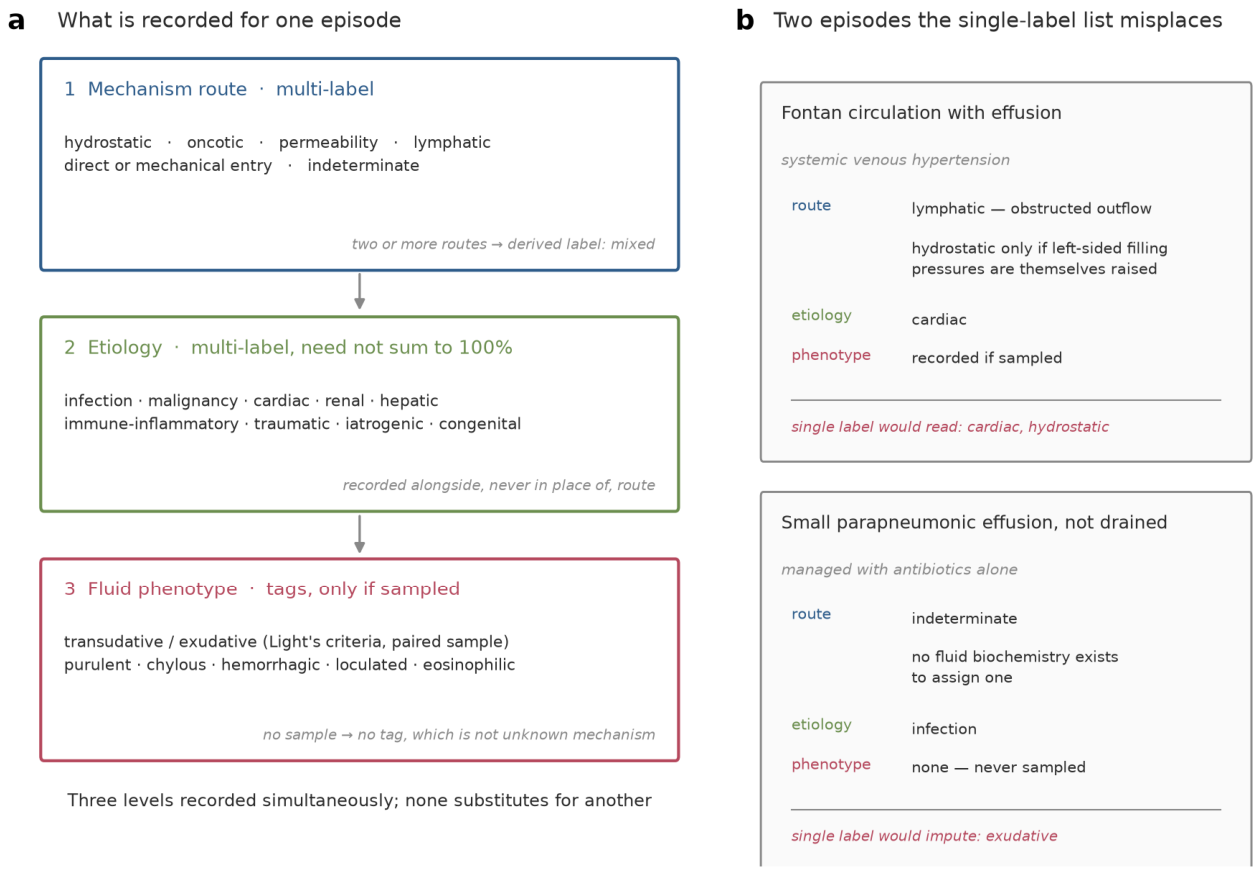
6.2. A Proposed Framework
We propose, for field discussion rather than as a validated instrument, recording pediatric pleural effusion on three simultaneous levels.
The mechanism level carries the four routes of Section 1.2 (hydrostatic, oncotic, permeability, lymphatic), a slot for direct or mechanical entry, and an explicit indeterminate category. Hydrostatic needs one qualifier at recording. Raised pulmonary venous and raised systemic venous pressure are both loosely hydrostatic but reach the pleural space differently: the first by filtration through the lung, the second by obstructing lymphatic outflow (Section 1.2). A child with a Fontan circulation is therefore recorded as lymphatic, with hydrostatic added only where left-sided filling pressures are themselves raised. The axis avoids transudate-exudate vocabulary, which describes the capillary barrier at sampling rather than the route of accumulation (Section 1.4 and Section 2.4). Lymphatic sits apart from permeability for two reasons: pleural drainage depends on anatomically specialized diaphragmatic lymphatics with stomata and spontaneous contractile segments [8], and chyle leak carries its own biochemical signature [32]. Four routes are not exhaustive. Direct entry across a breached boundary (traumatic or iatrogenic injury to chest wall, vessel, duct or diaphragm; transdiaphragmatic passage from the peritoneum) accumulates fluid without any of the four pressure or clearance abnormalities. Effusion against a non-expanding lung likewise reflects abnormal pleural pressure rather than altered filtration. Hence the fifth slot, though no pediatric series in this evidence base quantifies its contribution, so the field rather than this literature would set its boundaries. Deriving mixed from multi-label assignment, rather than offering it as a sixth category, removes the ambiguity of schemes providing both. The etiologic level is a second overlapping multi-label axis, permitting more than one assignment per episode: infection, malignancy, cardiac, renal, hepatic, immune-inflammatory, traumatic, iatrogenic, congenital.
The third level holds fluid-phenotype tags, recorded alongside mechanism rather than competing with it: transudative or exudative by Light’s criteria where paired samples exist, plus purulent, chylous, hemorrhagic, loculated or septated, and eosinophilic.
Three design decisions follow. Presence rather than precedence: an episode may carry two routes with none nominated dominant, since the post-surgical and tuberculous examples show two mechanisms at once [44,50]. An explicit indeterminate category: requiring complete classification imputes mechanism to records holding no fluid biochemistry, a substantial proportion wherever small effusions go undrained [35,43]. Phenotypes as tags: the mapping is many-to-many, since chylous fluid arises from trauma, malignancy, lymphatic disorders and heart failure [32], while one mechanism spans phenotypes from self-resolving lymphocytic effusion to frank empyema [44].
6.3. What Validation Would Require
Validation would require prospective cohorts with protocolized sampling reporting the proportion of episodes yielding no fluid. No multi-center pediatric series has reported the distribution of mechanism routes under systematic sampling. Existing infrastructure has addressed treatment, not classification, as in the six-hospital trial of intrapleural tPA with dornase alfa in 97 children [178]. It would also require multi-rater reliability testing on the ambiguous assignments, where disagreement will concentrate. No published pediatric study reports inter-rater agreement for mechanism assignment. Surveys instead document centers agreeing on initial diagnostic procedures while showing complete lack of agreement on the indications and effusion volume thresholds for invasive procedures [55], and wide variability in chylothorax management across 17 congenital heart programs [151].
Three boundary cases need adjudication first. Hypoalbuminemia with concurrent infection: is the oncotic contribution recorded when an exudative process is also documented [20,21]? Structural cardiac malformation without clinical failure: does malformation alone justify a transudative label, given that only 15% to 25% of children with structural heart disease develop heart failure [19]? Elevated myocardial injury markers in systemic inflammation: how is cardiac separated from inflammatory mechanism, since markedly elevated IL-6 accompanies multisystem inflammatory syndrome in children, in which hepatosplenomegaly was recorded in 10.5% and ascites in 42.1% [179]?
6.4. A Minimum Reporting Set
Neither the classification nor its validation is a precondition for improving the literature now. Most failures audited in Section 5 arose from information the investigators held and did not report, and most items below cost nothing to state; item 9 is the exception, since outcome stratified by mechanism requires follow-up that may not have been collected. We propose a minimum set for any pediatric study reporting a proportion of pleural effusion by cause.
1. State the ascertainment mode: administrative coding, clinician notification, imaging, fluid sampling, or cohort enrolment. It determines which effusions could have been seen at all (Section 5.2, Table 3, Figure 4).
2. State the denominator explicitly, naming the population the percentage is of. Proportions of sampled effusions, of children with pneumonia, and of all hospitalizations are three quantities routinely presented as one (Section 5.2).
3. Report the proportion of episodes never yielding fluid, and classify those separately rather than across etiologic categories (Section 2.4 and Section 6.1).
4. State whether tuberculous, mycoplasmal and fungal effusions were counted as parapneumonic. The term has no agreed boundary, and the choice is not recoverable from published reports (Section 3.7).
5. Report the diagnostic menu applied: which organisms were sought, by which assays, in what proportion of episodes. Attributed etiology follows the tests ordered, and an unsought organism is indistinguishable from an absent one (Section 3.4).
6. State the vaccine schedule and uptake during the study. Bacteriology tracks the immunization programme, and a serotype distribution without vaccine context cannot be compared across eras or countries (Section 3.3).
7. State the imaging modality and whether it was applied systematically. Detection is modality-dependent, and a series using ultrasound is not counting the same effusions as one using radiography (Section 2.4 and Section 3.4).
8. Where more than one mechanism was documented in one episode, report that rather than resolving it to a single label, even if the study’s own classification requires one (Section 4.1 and Section 6.1).
9. Where follow-up was obtained, report outcome stratified by mechanism rather than by etiologic label alone, naming the endpoint: imaging residue, pulmonary function, growth, or symptom burden. What an effusion leaves behind is a property of the route that formed it, and the current literature cannot be interrogated that way (Section 6.5).
None of the first eight items requires a new measurement, a new instrument, or agreement on the classification proposed here; the ninth requires follow-up many series already perform and do not report this way. Items 1 to 3 alone would make the Table 3 comparison possible, and the first two would tell a reader whether two proportions are the same kind of number.
6.5. Mechanism Predicts the Sequela, and the Evidence for That Is Uneven
A classification earns its place by carrying information the labels it replaces do not. The clearest candidate is prognosis: what an effusion leaves behind is a property of how it formed, not of which organism was named, and the routes of Section 1.2 differ in what they can damage.
Permeability is the only route injuring the pleura itself, so the only one whose natural history includes a structural residue. Tuberculous effusion spans complete resolution to loculation, pleural thickening, or frank empyema [44], and the pyogenic form passes through fibrin deposition and organization before resolving. Treated outcomes are nonetheless good. Across 127 studies and 10,896 children, 78 died (0.7%), mortality higher in Asia and Africa [89]. Of 139 children followed after a 14-day antibiotic regimen guided by pleural tap, 134 (96%) recovered fully [84]. Even necrotizing pneumonia, the severe end of this route, achieves good clinical, radiographic and functional recovery over the five to six months after diagnosis [69]. The residue is usually transient, but this is the route where a residue is possible at all.
The lymphatic route leaves no pleural scar, carrying instead a nutritional and immunological cost, since what is lost is chyle rather than transudate. Its sequelae track leak duration and management. A medium-chain triglyceride diet alone resolved 17 of 24 postoperative chylothoraces within ten days, and only one relapsed after conversion back to regular feeds [144]. Another series reported fasting with parenteral nutrition resolving 86.5% of cases, though it states 51% in its results and 86.5% in its conclusion without reconciling the two, and the 3.7% referred for surgery is reported for the arm receiving fasting, parenteral nutrition and octreotide together [123]. These are durations and nutritional exposures, not structural lesions: a different harm on a different clock.
The hydrostatic route is not symmetrical with the oncotic one. Sustained systemic venous hypertension remodels the clearance pathway it obstructs: in the Fontan circulation it drives lymphatic congestion and abnormal central lymphatic flow, now imaged directly and treated by embolization or duct decompression [128]. That residue is lymphatic, not pleural. Neither hydrostatic nor oncotic damages the pleura at all. Their prognosis is that of the circulation or kidney producing them, so the effusion marks an underlying state rather than an injury with its own sequela, and the one quantity belonging to it is how long it persists and what was done to it. The direct and mechanical route is bounded by the injury and the volume retained. Fetal and congenital presentations carry an outcome determined largely by the underlying cause rather than the fluid [166].
Renal disease, however, does not reach the pleura only by the oncotic route: uremic pleuritis is fibrinous pleuritis [136], so a child with a renal label may carry a structural residue the oncotic reading does not predict.
Two things follow. First, a reason to record mechanism independent of the epistemic argument in Section 6.1: a parent asking what will be left behind asks a question the mechanism axis can begin to answer and the etiologic label cannot, since the same label spans routes with different residues and the same route spans labels. Second, this review cannot complete the answer. The words sequelae and lung function do not appear in the pediatric evidence assembled here in any systematic form. Two records touch pulmonary function at all, and no multi-center pediatric series here reports structured long-term follow-up stratified by mechanism, with spirometry, imaging residue, growth, or symptom burden as endpoints. That evidence is distributed as the rest of the literature is: concentrated on the pyogenic permeability route and postoperative chylothorax, thin for tuberculous and mycoplasmal effusion, effectively absent for hydrostatic and oncotic routes. Mechanism-stratified outcome reporting is therefore the single addition that would make the proposed axis prognostically useful rather than merely descriptively honest, and it is item 9 above.
6.6. Research Priorities
1. A prospective multi-center pediatric cohort with protocolized pleural fluid sampling should report the distribution of mechanism routes and the proportion of episodes not sampled, since current characterization depends on a frequently omitted procedure [35,43].
2. Inter-rater reliability of mechanism assignment, including mixed and indeterminate categories, should be measured directly, because no pediatric agreement statistic for this task has been published.
3. Reporting of parapneumonic effusion should be harmonized across ultrasound grade, fluid biochemistry and molecular microbiology. Heterogeneous inclusion criteria across 127 pediatric empyema studies prevented firm conclusions [89], and molecular testing materially changes pathogen attribution [85].
4. Indications and thresholds for lymphatic imaging require definition, since diagnostic lymphatic imaging preceded surgical thoracic duct ligation in only 7 of 15 centers performing it [151] despite the availability of dynamic contrast-enhanced MR lymphangiography in children [150].
5. Candidate mechanistic biomarkers should be tested for discriminating mechanism rather than severity, given that pleural IL-6 in infection exceeds matched serum approximately 5000-fold and correlates with effusion size, pH and glucose [26].
6.7. Clinical Bottom Line
In a child with a pleural effusion, the useful initial question is which mechanisms are plausibly contributing and which are supported by evidence actually in the record, not which single etiologic label fits best. Where fluid has not been sampled, record the mechanism as indeterminate rather than inferring it from clinical context, because the inference is not recoverable later by anyone reading the record.
Funding
This work was supported by the Beijing Science and Technology Nova Program Interdisciplinary Project (20230484439) and the Beijing High-Level Public Health Technical Talents Project (2023-02-33). The funder had no role in study design, data collection, data analysis, data interpretation, or writing of the report. Use of AI tools. AI tools were used for language editing and for assistance with code generation and modification; the author takes full responsibility for the content.
Conflicts of Interest
The author declares no conflict of interest.
References
- Ütine GE, Özçelik Uu, Kiper N, et al. Pediatric pleural effusions: etiological evaluation in 492 patients over 29 years. Turk J Pediatr. 2009;51(3):214-219. [CrossRef]
- Ferreiro L, Toubes ME, Suárez-Antelo J, Rodríguez-Núñez N, Valdés L. Clinical overview of the physiology and pathophysiology of pleural fluid movement: a narrative review. ERJ Open Res. 2024;10(5):00050-2024. [CrossRef]
- Negrini D, Solari E, Marcozzi C, Moriondo A. The Thoracic Lymphatic Vasculature: Interorgan Strategies to Achieve an Optimal Tissue Function. Comprehensive Physiology. 2026;16(2):e70143. [CrossRef]
- Zhang F, Zarkada G, Yi S, et al. Lymphatic Endothelial Cell Junctions: Molecular Regulation in Physiology and Diseases. Front. Physiol. 2020;11. [CrossRef]
- Negrini D, Moriondo A. Pleural function and lymphatics. Acta Physiologica. 2012;207(2):244-259. [CrossRef]
- Zocchi L. Physiology and pathophysiology of pleural fluid turnover. Eur Respir J. 2002;20(6):1545-1558. [CrossRef]
- Scallan JP, Zawieja SD, Castorena-Gonzalez JA, et al. Lymphatic pumping: mechanics, mechanisms and malfunction. The Journal of Physiology. 2016;594(20):5749-5768. [CrossRef]
- Negrini D. Morphological, Mechanical and Hydrodynamic Aspects of Diaphragmatic Lymphatics. Biology. 2022;11(12):1803. [CrossRef]
- Solari E, Marcozzi C, Ottaviani C, et al. Draining the Pleural Space: Lymphatic Vessels Facing the Most Challenging Task. Biology. 2022;11(3):419. [CrossRef]
- Negrini D, Del Fabbro M. Subatmospheric pressure in the rabbit pleural lymphatic network. The Journal of Physiology. 1999;520(3):761-769. [CrossRef]
- Lattuada M, Hedenstierna G. Abdominal lymph flow in an endotoxin sepsis model: Influence of spontaneous breathing and mechanical ventilation*. Critical Care Medicine. 2006;34(11):2792-2798. [CrossRef]
- Pilon RN, Bittar DA. The Effect of Positive End-expiratory Pressure on Thoracic-duct Lymph Flow during Controlled Ventilation in Anesthetized Dogs. Anesthesiology. 1973;39(6):607-611. [CrossRef]
- Woolverton WC, Brigham KL, Staub NC. Effect of positive pressure breathing on lung lymph flow and water content in sheep. Circulation Research. 1978;42(4):550-557. [CrossRef]
- Beretta E, Romanò F, Sancini G, et al. Pulmonary Interstitial Matrix and Lung Fluid Balance From Normal to the Acutely Injured Lung. Front. Physiol. 2021;12. [CrossRef]
- Broaddus VC, Wiener-Kronish JP, Staub NC. Clearance of lung edema into the pleural space of volume-loaded anesthetized sheep. Journal of Applied Physiology. 1990;68(6):2623-2630. [CrossRef]
- Wiener-Kronish JP, Broaddus VC, Albertine KH, et al. Relationship of pleural effusions to increased permeability pulmonary edema in anesthetized sheep. J. Clin. Invest. 1988;82(4):1422-1429. [CrossRef]
- Wiener-Kronish JP, Matthay MA, Callen PW, et al. Relationship of pleural effusions to pulmonary hemodynamics in patients with congestive heart failure. Am Rev Respir Dis. 1985;132(6):1253-1256. PMID:3907444.
- Wiener-Kronish JP, Goldstein R, Matthay RA, et al. Lack of Association of Pleural Effusion with Chronic Pulmonary Arterial and Right Atrial Hypertension. Chest. 1987;92(6):967-970. [CrossRef]
- Madriago E, Silberbach M. Heart Failure in Infants and Children. Pediatrics In Review. 2010;31(1):4-12. [CrossRef]
- Perinandika T, Rachmadi D, Dwiyatnaningrum F. A Study of Hypoalbuminemia and Pleural Effusionin Pediatric Nephrotic Syndrome. AMJ. 2017;4(2):188-191. [CrossRef]
- Ellis D. Pathophysiology, Evaluation, and Management of Edema in Childhood Nephrotic Syndrome. Front. Pediatr. 2016;3. [CrossRef]
- Levick JR, Michel CC. Microvascular fluid exchange and the revised Starling principle. Cardiovascular Research. 2010;87(2):198-210. [CrossRef]
- Markov AG, Amasheh S. Tight junction physiology of pleural mesothelium. Front. Physiol. 2014;5. [CrossRef]
- Antony VB. Immunological mechanisms in pleural disease. Eur Respir J. 2003;21(3):539-544. [CrossRef]
- Idell S, Girard W, Koenig KB, et al. Abnormalities of Pathways of Fibrin Turnover in the Human Pleural Space. American Review of Respiratory Disease. 1991;144(1):187-194. [CrossRef]
- Argyriou A, Robbins A, Scott R, et al. The role of interleukin-6 signalling in pleural infection: observational and genetic analyses. eBioMedicine. 2025;119:105887. [CrossRef]
- Ng CT, Fong LY, Tan JJ, et al. Endothelial barrier disruptive effect of IFN-Ƴ and TNF-α: Synergism of pro-inflammatory cytokines. Cytokine. 2025;190:156922. [CrossRef]
- Park JS, Kim YS, Jee YK, et al. Interleukin-8 Production in Tuberculous Pleurisy: Role of Mesothelial Cells Stimulated by Cytokine Network Involving Tumour Necrosis Factor-α and Interleukin-1β. Scand J Immunol. 2003;57(5):463-469. [CrossRef]
- Brace RA, Valenzuela GJ. Effects of outflow pressure and vascular volume loading on thoracic duct lymph flow in adult sheep. American Journal of Physiology-Regulatory, Integrative and Comparative Physiology. 1990;258(1):R240-R244. [CrossRef]
- Gest AL, Bair DK, Vander Straten MC. The Effect of Outflow Pressure upon Thoracic Duct Lymph Flow Rate in Fetal Sheep. Pediatr Res. 1992;32(5):585-588. [CrossRef]
- Brace RA. Effects of outflow pressure on fetal lymph flow. American Journal of Obstetrics and Gynecology. 1989;160(2):494-497. [CrossRef]
- Bhatnagar M, Fisher A, Ramsaroop S, Carter A, Pippard B. Chylothorax: pathophysiology, diagnosis, and management—a comprehensive review. J Thorac Dis. 2024;16(2):1645-1661. [CrossRef]
- Ahmed MA. Post-Operative Chylothorax in Children Undergoing Congenital Heart Surgery. Cureus. 2021. [CrossRef]
- Raatz A, Schöber M, Zant R, et al. Risk factors for chylothorax and persistent serous effusions after congenital heart surgery. European Journal of Cardio-Thoracic Surgery. 2019;56(6):1162-1169. [CrossRef]
- Marchi G. Exploring pleural effusion characterisation with quantitative thoracic ultrasound imaging: a viewpoint on the investigational role of pixel-based echogenicity analysis in transudate and exudate differentiation. Breathe. 2025;21(4):250282. [CrossRef]
- Santotoribio JD, Nuñez-Jurado D, Rubio-Prieto JL, Guerrero JM, Corral-Pérez J, Fernández-Alba JJ. Pleural Fluid Biomarkers of Pediatric Parapneumonic Effusion. Diagnostics. 2025;15(9):1086. [CrossRef]
- Bagur Krishnamurthy M, Malhotra A. Congenital chylothorax: current perspectives and trends. RRN. 2017;Volume 7:53-63. [CrossRef]
- Rustom H, Koiala S, Al-Janabi F, Wolf L, Nanda VC. Neonate With Hydrops Fetalis, Chylothorax, and an FLT4 Variant: A Favorable Outcome. Cureus. 2026. [CrossRef]
- Brouillard P, Boon L, Vikkula M. Genetics of lymphatic anomalies. J. Clin. Invest. 2014;124(3):898-904. [CrossRef]
- He T, Yuan Y, Zhou Q, Gao J, Wang D. Primary Small Intestine Lymphatic Dilation Syndrome: A Case Report. . 2026. [CrossRef]
- Brock WW, Bradshaw WT. Congenital Chylothorax. Advances in Neonatal Care. 2016;16(2):114-123. [CrossRef]
- Yang W, Zhang B, Zhang ZM. Infectious pleural effusion status and treatment progress. J. Thorac. Dis. 2017;9(11):4690-4699. [CrossRef]
- Forster J, Paul P, Liese J. Current Management of Pediatric Parapneumonic Pleural Effusions and Pleural Empyema. Pediatric Infectious Disease Journal. 2023;42(11):e407-e410. [CrossRef]
- Shaw JA, Diacon AH, Koegelenberg CF. Tuberculous pleural effusion. Respirology. 2019;24(10):962-971. [CrossRef]
- Twaddell SH, Gibson PG, Grainge C, et al. Parapneumonic Effusions Are Characterized by Elevated Levels of Neutrophil Extracellular Traps. Chest. 2021;160(5):1645-1655. [CrossRef]
- Bedawi EO, Kanellakis NI, Corcoran JP, et al. The Biological Role of Pleural Fluid PAI-1 and Sonographic Septations in Pleural Infection: Analysis of a Prospectively Collected Clinical Outcome Study. American Journal of Respiratory and Critical Care Medicine. 2023;207(6):731-739. [CrossRef]
- Mutsaers SE. The mesothelial cell. The International Journal of Biochemistry & Cell Biology. 2004;36(1):9-16. [CrossRef]
- Choi P, Farmakis S, Desmarais T, Keller M. Management and outcomes of traumatic hemothorax in children. J Emerg Trauma Shock. 2015;8(2):83. [CrossRef]
- Garlapati S, Tambolkar S, Verma S, Pathak NV, Patil MG. Isolated Malignant Pleural Effusion in a Child: Unusual Presentation of Acute Leukemia. Cureus. 2024. [CrossRef]
- Flögel L, Kaiser E, Hans MC, et al. Immunological characterization of pleural effusions in pediatric patients. Front. Immunol. 2024;15:1506073. [CrossRef]
- Light RW, Macgregor MI, Luchsinger PC, Ball WC. Pleural Effusions: The Diagnostic Separation of Transudates and Exudates. Annals of Internal Medicine. 1972;77(4):507-513. [CrossRef]
- Milevoj Kopcinovic L, Culej J. Pleural, peritoneal and pericardial effusions—a biochemical approach. Biochem Med. 2014:123-137. [CrossRef]
- Tay TR, Tee A. Factors affecting pleural fluid adenosine deaminase level and the implication on the diagnosis of tuberculous pleural effusion: a retrospective cohort study. BMC Infect Dis. 2013;13(1):546. [CrossRef]
- Segerer FJ, Seeger K, Maier A, et al. Therapy of 645 children with parapneumonic effusion and empyema—A German nationwide surveillance study. Pediatric Pulmonology. 2016;52(4):540-547. [CrossRef]
- Hafen GM, Grenzbach A, Moeller A, Rochat MK. Lack of concordance in parapneumonic effusion management in children in central Europe. Pediatric Pulmonology. 2015;51(4):411-417. [CrossRef]
- Zhai K, Lu Y, Shi HZ. Tuberculous pleural effusion. J. Thorac. Dis. 2016;8(7):E486-E494. [CrossRef]
- Kim HW, Kim KH, Shin AY, et al. Investigating the appropriate adenosine deaminase cutoff value for the diagnosis of tuberculous pleural effusion in a country with decreasing TB burden. Sci Rep. 2022;12(1):7586. [CrossRef]
- Shien TK, Mohamad Jailaini MF, Azmel AA, Munusamy V, Alaga A, Abdul Hamid MF. Diagnostic utility of pleural fluid adenosine deaminase in tuberculous pleural effusion in patients with exudative effusion: a retrospective multicenter study (ADATPE). J Thorac Dis. 2026;18(3):216-216. [CrossRef]
- Lima Vieira1 J, Foschiera1 L, Schutz Ferreira1 IC, Coutinho Baldoto Gava Chakr1,2 V. Performance of the quantification of adenosine deaminase and determination of the lactate dehydrogenase/adenosine deaminase ratio for the diagnosis of pleural tuberculosis in children and adolescents. J Bras Pneumol. 2021:e20200558. [CrossRef]
- Zhao T, Zhang J, Zhang X, Wang C. Clinical significance of pleural fluid lactate dehydrogenase/adenosine deaminase ratio in the diagnosis of tuberculous pleural effusion. BMC Pulm Med. 2024;24(1):241. [CrossRef]
- Yan Z, Wen JX, Wang H, et al. Diagnostic accuracy of pleural fluid lactate dehydrogenase to adenosine deaminase ratio for tuberculous pleural effusion: an analysis of two cohorts. BMC Pulm Med. 2022;22(1):428. [CrossRef]
- Taş D, Köseler A. Diagnostic value of angiopoietin-2 in the differentiation of malignant pleural effusions. Journal of Surgery and Medicine. 2019. [CrossRef]
- Christopher DJ, Schumacher SG, Michael JS, et al. Performance of Xpert MTB/RIF on pleural tissue for the diagnosis of pleural tuberculosis. Eur Respir J. 2013;42(5):1427-1429. [CrossRef]
- Reali F, Sferrazza Papa GF, Carlucci P, et al. Can Lung Ultrasound Replace Chest Radiography for the Diagnosis of Pneumonia in Hospitalized Children?. Respiration. 2014;88(2):112-115. [CrossRef]
- Sundaralingam A, Banka R, Rahman NM. Management of Pleural Infection. Pulm Ther. 2020;7(1):59-74. [CrossRef]
- Girdhar A, Shujaat A, Bajwa A. Management of Infectious Processes of the Pleural Space: A Review. Pulmonary Medicine. 2012;2012:1-10. [CrossRef]
- Elemraid MA, Thomas MF, Blain AP, et al. Risk factors for the development of pleural empyema in children. Pediatric Pulmonology. 2014;50(7):721-726. [CrossRef]
- Sawicki GS, Lu FL, Valim C, Cleveland RH, Colin AA. Necrotising pneumonia is an increasingly detected complication of pneumonia in children. Eur Respir J. 2008;31(6):1285-1291. [CrossRef]
- Masters IB, Isles AF, Grimwood K. Necrotizing pneumonia: an emerging problem in children?. Pneumonia. 2017;9(1):11. [CrossRef]
- Chen Y, Li L, Wang C, Zhang Y, Zhou Y. Necrotizing Pneumonia in Children: Early Recognition and Management. JCM. 2023;12(6):2256. [CrossRef]
- Bender JM, Ampofo K, Korgenski K, et al. Pneumococcal Necrotizing Pneumonia in Utah: Does Serotype Matter?. CLIN INFECT DIS. 2008;46(9):1346-1352. [CrossRef]
- Poirault C, Hadchouel A, Roy C, et al. Inflammatory rebound and postinfectious inflammatory response in children with pleural infection: A single-center retrospective study. Pediatric Pulmonology. 2024;59(4):974-981. [CrossRef]
- Koshy E, Murray J, Bottle A, Sharland M, Saxena S. Impact of the seven-valent pneumococcal conjugate vaccination (PCV7) programme on childhood hospital admissions for bacterial pneumonia and empyema in England: national time-trends study, 1997-2008. Thorax. 2010;65(9):770-774. [CrossRef]
- Strachan RE, Snelling TL, Jaffé A. Increased paediatric hospitalizations for empyema in Australia after introduction of the 7-valent pneumococcal conjugate vaccine. Bull. World Health Organ. 2012;91(3):167-173. [CrossRef]
- Grijalva CG, Nuorti JP, Zhu Y, Griffin MR. Increasing Incidence of Empyema Complicating Childhood Community-Acquired Pneumonia in the United States. CLIN INFECT DIS. 2010;50(6):805-813. [CrossRef]
- Nath S, Thomas M, Spencer D, Turner S. Has the incidence of empyema in Scottish children continued to increase beyond 2005?. Arch Dis Child. 2014;100(3):255-258. [CrossRef]
- Picazo J, Ruiz-Contreras J, Casado-Flores J, et al. Impact of Introduction of Conjugate Vaccines in the Vaccination Schedule on the Incidence of Pediatric Invasive Pneumococcal Disease Requiring Hospitalization in Madrid 2007 to 2011. Pediatric Infectious Disease Journal. 2013;32(6):656-661. [CrossRef]
- Goettler D, Streng A, Kemmling D, et al. Increase in Streptococcus pneumoniae serotype 3 associated parapneumonic pleural effusion/empyema after the introduction of PCV13 in Germany. Vaccine. 2020;38(3):570-577. [CrossRef]
- Silva-Costa C, Gomes-Silva J, Prados L, Ramirez M, Melo-Cristino J. Pediatric Invasive Pneumococcal Disease Three Years after PCV13 Introduction in the National Immunization Plan—The Continued Importance of Serotype 3. Microorganisms. 2021;9(7):1428. [CrossRef]
- Fletcher MA, Schmitt HJ, Syrochkina M, Sylvester G. Pneumococcal empyema and complicated pneumonias: global trends in incidence, prevalence, and serotype epidemiology. Eur J Clin Microbiol Infect Dis. 2014;33(6):879-910. [CrossRef]
- Carrillo-Marquez MA, Hulten KG, Hammerman W, Lamberth L, Mason EO, Kaplan SL. Staphylococcus aureus Pneumonia in Children in the Era of Community-acquired Methicillin-resistance at Texas Children’s Hospital. Pediatric Infectious Disease Journal. 2011;30(7):545-550. [CrossRef]
- Forster J, Piazza G, Goettler D, et al. Empiric Antibiotic Therapy in 1402 Children With Parapneumonic Effusion/Pleural Empyema in Germany: A Long-term Surveillance Study. Pediatric Infectious Disease Journal. 2024;43(7):651-656. [CrossRef]
- Pernica JM, Moldovan I, Chan F, Slinger R. Real-Time Polymerase Chain Reaction for Microbiological Diagnosis of Parapneumonic Effusions in Canadian Children. Canadian Journal of Infectious Diseases and Medical Microbiology. 2014;25(3):151-154. [CrossRef]
- Meyer Sauteur PM, Burkhard A, Moehrlen U, et al. Pleural Tap-Guided Antimicrobial Treatment for Pneumonia with Parapneumonic Effusion or Pleural Empyema in Children: A Single-Center Cohort Study. JCM. 2019;8(5):698. [CrossRef]
- Ho EC, Olson KE, Butler M, et al. Clinical Impact of Pleural Fluid Streptococcus pneumoniae Polymerase Chain Reaction Testing in Children With Complicated Pneumonia. Clinical Infectious Diseases. 2024;79(6):1487-1494. [CrossRef]
- Azzari C, Moriondo M, Indolfi G, et al. Realtime PCR Is More Sensitive than Multiplex PCR for Diagnosis and Serotyping in Children with Culture Negative Pneumococcal Invasive Disease. PLoS ONE. 2010;5(2):e9282. [CrossRef]
- Jiang W, Wu M, Zhou J, et al. Etiologic spectrum and occurrence of coinfections in children hospitalized with community-acquired pneumonia. BMC Infect Dis. 2017;17(1):787. [CrossRef]
- Galmès J, Li Y, Rajoharison A, et al. Potential implication of new torque teno mini viruses in parapneumonic empyema in children. Eur Respir J. 2012;42(2):470-479. [CrossRef]
- Buonsenso D, Cusenza F, Passadore L, et al. Parapneumonic empyema in children: a scoping review of the literature. Ital J Pediatr. 2024;50(1):136. [CrossRef]
- Dembele BPP, Kamigaki T, Dapat C, et al. Aetiology and risks factors associated with the fatal outcomes of childhood pneumonia among hospitalised children in the Philippines from 2008 to 2016: a case series study. BMJ Open. 2019;9(3):e026895. [CrossRef]
- Youn YS, Lee KY. Mycoplasma pneumoniae pneumonia in children. Korean J Pediatr. 2012;55(2):42. [CrossRef]
- Kutty PK, Jain S, Taylor TH, et al. Mycoplasma pneumoniae Among Children Hospitalized With Community-acquired Pneumonia. Clinical Infectious Diseases. 2018;68(1):5-12. [CrossRef]
- Kim SH, Lee E, Song ES, Lee YY. Clinical Significance of Pleural Effusion in Mycoplasma pneumoniae Pneumonia in Children. Pathogens. 2021;10(9):1075. [CrossRef]
- Yen MH, Yan DC, Wang CJ, et al. The clinical significance of and the factors associated with macrolide resistance and poor macrolide response in pediatric Mycoplasma pneumoniae infection: A retrospective study. Journal of Microbiology, Immunology and Infection. 2023;56(3):634-640. [CrossRef]
- Kim CH, Lee J. Mycoplasma pneumoniae Pleural Effusion in Adults. JCM. 2022;11(5):1281. [CrossRef]
- Li S, Zhou J, He J, et al. Clinical profiles of Mycoplasma pneumoniae pneumonia in children with different pleural effusion patterns: a retrospective study. BMC Infect Dis. 2024;24(1):919. [CrossRef]
- Yang HJ, Song DJ, Shim JY. Mechanism of resistance acquisition and treatment of macrolide-resistant Mycoplasma pneumoniae pneumonia in children. Korean J Pediatr. 2017;60(6):167. [CrossRef]
- Kim YJ, Shin KS, Lee KH, Kim YR, Choi JH. Clinical Characteristics of Macrolide-Resistant Mycoplasma pneumoniae from Children in Jeju. J Korean Med Sci. 2017;32(10):1642. [CrossRef]
- Lee KL, Lee CM, Yang TL, et al. Severe Mycoplasma pneumoniae pneumonia requiring intensive care in children, 2010–2019. Journal of the Formosan Medical Association. 2021;120(1):281-291. [CrossRef]
- Han S, Fang Q, Wu X, et al. Clinical features associated with macrolides resistance of Mycoplasma pneumoniae pneumonia in children: a single-center analysis. BMC Infect Dis. 2025;25(1):854. [CrossRef]
- Jiang Tt, Sun L, Wang Ty, et al. The clinical significance of macrolide resistance in pediatric Mycoplasma pneumoniae infection during COVID-19 pandemic. Front. Cell. Infect. Microbiol. 2023;13:1181402. [CrossRef]
- Li W, Gao W, Xiong X, Tang X, Li A. Association analysis of Mycoplasma pneumoniae 23S rRNA gene mutation with refractory Mycoplasma pneumoniae pneumonia in children. PLoS One. 2026;21(1):e0341580. [CrossRef]
- Liu S, Zhang L, Dai L, Li J, Li D. Prediction model for severe mycoplasma pneumoniae pneumonia and analysis of macrolide-resistance in children: a case-control study. Ital J Pediatr. 2025;51(1):180. [CrossRef]
- de Groot RC, Streng BM, Bont LJ, Meyer Sauteur PM, van Rossum AM. Resurgence of Mycoplasma pneumoniae infections in children: emerging challenges and opportunities. Current Opinion in Infectious Diseases. 2025;38(5):468-476. [CrossRef]
- Yue Y, Wu D, Zeng Q, et al. Changes in children respiratory infections pre and post COVID-19 pandemic. Front. Cell. Infect. Microbiol. 2025;15:1549497. [CrossRef]
- Jensen A, Stensballe LG. Impact of COVID-19 on testing, positive cases, patient characteristics, and hospital contacts for respiratory syncytial virus, influenza, and pneumococcus in Danish children. Journal of Infection and Public Health. 2025;18(2):102660. [CrossRef]
- McNally E, Ross C, Gleeson LE. The tuberculous pleural effusion. Breathe. 2023;19(4):230143. [CrossRef]
- Mehta PK, Raj A, Singh N, Khuller GK. Diagnosis of extrapulmonary tuberculosis by PCR. FEMS Immunol Med Microbiol. 2012;66(1):20-36. [CrossRef]
- Allen JC, Apicella MA. Experimental Pleural Effusion as a Manifestation of Delayed Hypersensitivity to Tuberculin PPD. The Journal of Immunology. 1968;101(3):481-487. [CrossRef]
- Schierloh P, Landoni V, Balboa L, et al. Human pleural B-cells regulate IFN-γ production by local T-cells and NK cells in aMycobacterium tuberculosis-induced delayed hypersensitivity reaction. Clinical Science. 2014;127(6):391-403. [CrossRef]
- Vorster MJ, Allwood B, Diacon AH, et al. Tuberculous pleural effusions: advances and controversies. J Thorac Dis. 2015;7(6):981-91. [CrossRef]
- Casalini AG, Mori PA, Majori M, et al. Pleural tuberculosis: medical thoracoscopy greatly increases the diagnostic accuracy. ERJ Open Res. 2018;4(1):00046-2017. [CrossRef]
- Gupta B. Role of Adenosine Deaminase Estimation in Differentiation of Tuberculous and Non-tuberculous Exudative Pleural Effusions. J Clin Med Res. 2010. [CrossRef]
- Izumikawa K. Clinical Features of Severe or Fatal Mycoplasma pneumoniae Pneumonia. Front. Microbiol. 2016;7. [CrossRef]
- Meyer Sauteur PM, Ramien ML, Theiler M. Mycoplasma pneumoniae: Extrapulmonary Manifestations in Children With Focus on Mucocutaneous Disease. Pediatric Infectious Disease Journal. 2025;44(12):e454-e458. [CrossRef]
- Zhang Cx, Yin L, Mao Yy, Zhou Zy, Shen Jy, Zhou W. Sjogren Syndrome Presenting as Edema and Extensive Pleural Effusion: A Pediatric Case Report. . 2022. [CrossRef]
- Pérez-Rodriguez E, Castro DJ. The use of adenosine deaminase and adenosine deaminase isoenzymes in the diagnosis of tuberculous pleuritis. Current Opinion in Pulmonary Medicine. 2000;6(4):259-266. [CrossRef]
- Jiménez Castro D, Díaz Nuevo G, Pérez-Rodríguez E, et al. Diagnostic value of adenosine deaminase in nontuberculous lymphocytic pleural effusions. Eur Respir J. 2003;21(2):220-224. [CrossRef]
- Cha SI, Shin KM, Jeon KN, et al. Clinical relevance and characteristics of pleural effusion in patients with Mycoplasma pneumoniae pneumonia. Scandinavian Journal of Infectious Diseases. 2012;44(10):793-797. [CrossRef]
- Hoit BD. Pathophysiology of the Pericardium. Progress in Cardiovascular Diseases. 2017;59(4):341-348. [CrossRef]
- Weiss JM, Spodick DH. Association of Left Pleural Effusion with Pericardial Disease. N Engl J Med. 1983;308(12):696-697. [CrossRef]
- Bolukcu A, Topcu AC, Harmandar B, Karaci AR, Sasmazel A, Aydemir NA. Risk factors for pleural effusion after extracardiac Fontan procedure assessment of the commonest Fontan complication. mtd. 2022:3. [CrossRef]
- Christofe NM, Pessotti CFX, Paiva L, Jatene IB. Incidence and Treatment of Chylothorax in Children Undergoing Corrective Surgery for Congenital Heart Diseases. Braz J Cardiovasc Surg. 2017. [CrossRef]
- Le TKV, Nguyen TKL, Doan VP. Characteristics of chylothorax following pediatric congenital heart surgery at Cho Ray Hospital. VJCTS. 2026;56. [CrossRef]
- FUJITA T, KAWASAKI S, MATSUSHITA S, et al. Effect of Regular Medication Using Super High Dose Diuretics for Pleural Effusions at Early Stage After the Fontan Procedure. Juntendo Medical Journal. 2014;60(1):35-42. [CrossRef]
- Rajab TK, Jaggers J. Transplantation in Fontan failure: The final stage. JTCVS Open. 2020;3:154-159. [CrossRef]
- Van De Bruaene A, Claessen G, Salaets T, et al. Late Fontan Circulatory Failure. What Drives Systemic Venous Congestion and Low Cardiac Output in Adult Fontan Patients?. Front. Cardiovasc. Med. 2022;9. [CrossRef]
- Cánovas-Galindo C, Segura-Rodríguez D, Ladrón-Abia R, et al. The Lymphatic System in the Fontan Circulation: Pathophysiology, Imaging and Therapeutic Interventions. Pediatr Cardiol. 2026. [CrossRef]
- John AS, Johnson JA, Khan M, et al. Clinical Outcomes and Improved Survival in Patients With Protein-Losing Enteropathy After the Fontan Operation. Journal of the American College of Cardiology. 2014;64(1):54-62. [CrossRef]
- Eid AA, Keddissi JI, Kinasewitz GT. Hypoalbuminemia as a Cause of Pleural Effusions. Chest. 1999;115(4):1066-1069. [CrossRef]
- Koot BP, Houwen R, Pot DJ, et al. Congenital analbuminaemia: biochemical and clinical implications. A case report and literature review. Eur J Pediatr. 2004. [CrossRef]
- Svenningsen P, Bistrup C, Friis UG, et al. Plasmin in Nephrotic Urine Activates the Epithelial Sodium Channel. Journal of the American Society of Nephrology. 2009;20(2):299-310. [CrossRef]
- Wulandary ME, Sofandi AA. Severe hypoalbuminemia in patient with pulmonary edema and pleural effusion: a case report. Int J Res Med Sci. 2024;12(12):4701-4704. [CrossRef]
- Abhinay A, Singh A, Rajniti P, Mishra OP. Response to Chemical Pleurodesis in Children with Frequent Relapsing Nephrotic Syndrome Having Refractory Unilateral Pleural Effusion. Saudi Journal of Kidney Diseases and Transplantation. 2022;33(5):726-729. [CrossRef]
- Li Y, Wang J, Bai Z, et al. Early fluid overload is associated with acute kidney injury and PICU mortality in critically ill children. Eur J Pediatr. 2015;175(1):39-48. [CrossRef]
- BERGER HW, RAMMOHAN G, NEFF MS, et al. Uremic Pleural Effusion. Ann Intern Med. 1975;82(3):362-364. [CrossRef]
- Jarratt MJ, Sahn SA. Pleural Effusions in Hospitalized Patients Receiving Long-term Hemodialysis. Chest. 1995;108(2):470-474. [CrossRef]
- Rashid-Farokhi F, Pourdowlat G, Nikoonia MR, et al. Uremic pleuritis in chronic hemodialysis patients. Hemodialysis International. 2012;17(1):94-100. [CrossRef]
- Nomoto Y, Suga T, Nakajima K, et al. Acute Hydrothorax in Continuous Ambulatory Peritoneal Dialysis—A Collaborative Study of 161 Centers. Am J Nephrol. 2008:363-367. [CrossRef]
- Li J, Zhang W, Wang W, et al. Forty-nine cases of acute lymphoblastic leukaemia/lymphoma in pleural and pericardial effusions: A cytological-histological correlation. Cytopathology. 2017;29(2):172-178. [CrossRef]
- Wang Z, Wu Y, Xu L, et al. Diagnostic value of medical thoracoscopy in malignant pleural effusion induced by non-Hodgkin’s lymphoma. Oncol Lett. 2017. [CrossRef]
- Shrestha S, Basnet R, Mathema S, Acharya R, Nepal A. A Child with Non-Hodgkin Lymphoma Presenting With Haemorrhagic Pleural Effusion. J. Kathmandu Med. Coll. 2015;3(3):126-128. [CrossRef]
- He XL, Yu F, Guo T, et al. T-cell lymphoblastic lymphoma presenting with pleural effusion: A case report. Respiratory Medicine Case Reports. 2014;12:55-58. [CrossRef]
- Biewer ES, Zürn C, Arnold R, et al. Chylothorax after surgery on congenital heart disease in newborns and infants -risk factors and efficacy of MCT-diet. J Cardiothorac Surg. 2010;5(1):127. [CrossRef]
- August DA, Huhmann MB. A.S.P.E.N. Clinical Guidelines: Nutrition Support Therapy During Adult Anticancer Treatment and in Hematopoietic Cell Transplantation. J Parenter Enteral Nutr. 2009;33(5):472-500. [CrossRef]
- Gonnelli F, Hassan W, Bonifazi M, et al. Malignant pleural effusion: current understanding and therapeutic approach. Respir Res. 2024;25(1). [CrossRef]
- Hermon M, Tenner E, Burda G, Strohmaier W, Schlager G, Golej J. Chylothorax and Chylous-Like Diseases in Children: Clinical Management. Front. Pediatr. 2019;7:258. [CrossRef]
- Bernier-Latmani J, Petrova TV. Intestinal lymphatic vasculature: structure, mechanisms and functions. Nat Rev Gastroenterol Hepatol. 2017;14(9):510-526. [CrossRef]
- Sigalet DL, Winkelaar GB, Smith LJ. Determination of the Route of Medium-Chain and Long-Chain Fatty Acid Absorption by Direct Measurement in the Rat. J Parenter Enteral Nutr. 1997;21(5):275-278. [CrossRef]
- Chavhan GB, Amaral JG, Temple M, Itkin M. MR Lymphangiography in Children: Technique and Potential Applications. RadioGraphics. 2017;37(6):1775-1790. [CrossRef]
- Adamson GT, Winder MM, Catton KG, et al. Current practices for refractory chylothorax following congenital heart surgery. Cardiol Young. 2023;34(5):1058-1062. [CrossRef]
- Dori Y, Keller MS, Rome JJ, et al. Percutaneous Lymphatic Embolization of Abnormal Pulmonary Lymphatic Flow as Treatment of Plastic Bronchitis in Patients With Congenital Heart Disease. Circulation. 2016;133(12):1160-1170. [CrossRef]
- Itkin M, Piccoli DA, Nadolski G, et al. Protein-Losing Enteropathy in Patients With Congenital Heart Disease. Journal of the American College of Cardiology. 2017;69(24):2929-2937. [CrossRef]
- Özdülger A. Thoracic trauma in children. Turk Gogus Kalp Dama. 2024;32(Supplementum 1):21-28. [CrossRef]
- Man BL, Mok CC. Serositis related to systemic lupus erythematosus: prevalence and outcome. Lupus. 2005;14(10):822-826. [CrossRef]
- Choi BY, Yoon MJ, Shin K, et al. Characteristics of pleural effusions in systemic lupus erythematosus: differential diagnosis of lupus pleuritis. Lupus. 2014;24(3):321-326. [CrossRef]
- Corredor-Orlandelli D, Arévalo-Romero A, Reyes C, Arango D. Massive Right Chylothorax Secondary to a Severe Systemic Lupus Erythematosus Flare With Secondary Evans Syndrome: A Case Report and Literature Review. Clin Med Insights Case Rep. 2023;16:11795476231186735. [CrossRef]
- Rai DK, Gupta VB, Bharti S, et al. Granulomatosis with polyangiitis presenting as exudative pleural effusion: A rare case report. Lung India. 2023;40(2):177-179. [CrossRef]
- Sethi Y, Moinuddin A, Mahajan K, et al. Dressler syndrome and post-cardiac injury syndrome: an autoinflammatory framework for reappraising a seventy-year-old diagnosis: a state-of-the-art review. Front. Immunol. 2026;17. [CrossRef]
- Huggins JT, Sahn SA. Drug-induced pleural disease. Clinics in Chest Medicine. 2004;25(1):141-153. [CrossRef]
- Caso F, Rigante D, Vitale A, et al. Monogenic Autoinflammatory Syndromes: State of the Art on Genetic, Clinical, and Therapeutic Issues. International Journal of Rheumatology. 2013;2013:1-15. [CrossRef]
- Lu M, Teng L, Xu Y, et al. Performance of interferon-gamma levels may lead to earlier diagnosing macrophage activation syndrome complicating systemic juvenile idiopathic arthritis. Pediatr Rheumatol. 2023;21(1). [CrossRef]
- Faleye A, Mahmood K, Al-Abadi E, et al. Exploring the prevalence of pulmonary involvement in juvenile-onset systemic lupus erythematosus: Data from the UK JSLE Cohort Study. Reumatología Clínica (English Edition). 2025;21(9):501975. [CrossRef]
- Kaur TP, Narang GS, S. A. Neonatal chylothorax. Int J Contemp Pediatr. 2019;6(3):1406. [CrossRef]
- Cömert HS, Kader Ş, Osmanağaoğlu MA, et al. Prenatal and Postnatal Management of Intrauterine Pleural Effusions Associated with Nonimmune Hydrops Fetalis. Am J Perinatol. 2020;39(13):1405-1409. [CrossRef]
- Wu W, Ma G, Chang T, Lee M, Lin W, Chen M. Outcome and etiology of fetal pleural effusion, fetal ascites and hydrops fetalis after fetal intervention: retrospective observational cohort from a single institution. Ultrasound in Obstet & Gyne. 2024;63(4):536-543. [CrossRef]
- Nugroho N, Andi Ade Wijaya Ramlan . Anesthetic Considerations in a Neonate Undergoing Thoracoscopic Removal of a Thoraco-Amniotic Shunt Following Prenatal Management of Fetal Pleural Effusion: A Case Report. Proceedings Book of ICE on IMERI. 2026;9(-):86-91. [CrossRef]
- Kuo YL, Chan TF. Treatment of unilateral fetal pleural effusion by intrauterine thoracocentesis. Taiwanese Journal of Obstetrics and Gynecology. 2012;51(2):303-304. [CrossRef]
- Yu D, Buchvald F, Brandt B, Nielsen KG. Seventeen-year study shows rise in parapneumonic effusion and empyema with higher treatment failure after chest tube drainage. Acta Paediatrica. 2013;103(1):93-99. [CrossRef]
- Jeong BD, Won HS, Lee MY, Shim JY, Lee PR, Kim A. Perinatal outcomes of fetal pleural effusion following thoracoamniotic shunting. Prenat Diagn. 2015;35(13):1365-1370. [CrossRef]
- Shah A, Gurnani S. Clinical profile of Tuberculous Pleural Effusion in Children. Pediatric Oncall. 2022;19(1). [CrossRef]
- Goodman LF, Martino AM, Schomberg J, et al. Percutaneous pigtail catheter versus chest tube for the treatment of pediatric traumatic hemothorax: An Eastern Association for the Surgery of Trauma multicenter study. J Trauma Acute Care Surg. 2025;99(6):850-858. [CrossRef]
- Leija-Martínez JJ, Esparza-Miranda LA, Rivera-Alfaro G, Noyola DE. Impact of Nonpharmaceutical Interventions during the COVID-19 Pandemic on the Prevalence of Respiratory Syncytial Virus in Hospitalized Children with Lower Respiratory Tract Infections: A Systematic Review and Meta-Analysis. Viruses. 2024;16(3):429. [CrossRef]
- Foley DA, Sikazwe CT, Minney-Smith CA, et al. An Unusual Resurgence of Human Metapneumovirus in Western Australia Following the Reduction of Non-Pharmaceutical Interventions to Prevent SARS-CoV-2 Transmission. Viruses. 2022;14(10):2135. [CrossRef]
- Yavarian J, Shatizadeh Malekshahi S, Faraji-Zonouz M, et al. Impact of COVID-19 on the changing pattern of human orthopneumovirus (respiratory syncytial virus) infection in Iran. BMC Infect Dis. 2023;23(1):591. [CrossRef]
- Grijalva CG, Zhu Y, Nuorti JP, Griffin MR. Emergence of parapneumonic empyema in the USA. Thorax. 2011;66(8):663-668. [CrossRef]
- Konietzke P, Mueller J, Wuennemann F, et al. The value of chest magnetic resonance imaging compared to chest radiographs with and without additional lung ultrasound in children with complicated pneumonia. PLoS ONE. 2020;15(3):e0230252. [CrossRef]
- Livingston MH, Mahant S, Connolly B, et al. Effectiveness of Intrapleural Tissue Plasminogen Activator and Dornase Alfa vs Tissue Plasminogen Activator Alone in Children with Pleural Empyema. JAMA Pediatr. 2020;174(4):332. [CrossRef]
- Lazova S, Alexandrova T, Gorelyova-Stefanova N, Atanasov K, Tzotcheva I, Velikova T. Liver Involvement in Children with COVID-19 and Multisystem Inflammatory Syndrome: A Single-Center Bulgarian Observational Study. Microorganisms. 2021;9(9):1958. [CrossRef]
Figure 2.
Published pediatric pleural-effusion reviews concentrate on single mechanisms and seldom address classification. (a) Topic distribution of 55 comparable reviews retrieved from OpenAlex and screened for scope. (b) Number of those reviews that address each of the moves this review makes; bars at zero are marked with a stub. Screening was of titles and abstracts by a single assessor, so panel b describes how this literature presents itself rather than establishing that no such review exists.
Figure 2.
Published pediatric pleural-effusion reviews concentrate on single mechanisms and seldom address classification. (a) Topic distribution of 55 comparable reviews retrieved from OpenAlex and screened for scope. (b) Number of those reviews that address each of the moves this review makes; bars at zero are marked with a stub. Screening was of titles and abstracts by a single assessor, so panel b describes how this literature presents itself rather than establishing that no such review exists.
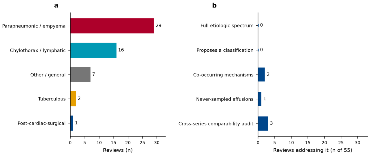
Figure 4.
Each pediatric series in Table 3 pairs its own ascertainment mode with its own denominator. Each point is one of the 25 series; colour encodes ascertainment mode. The 25 series fall into 17 distinct ascertainment-by-denominator combinations: 12 combinations hold a single series, and the 5 combinations shared by two or more series hold series that still differ in country, period, or case definition. A reported percentage is defined jointly by its column and its row, which is why the proportions in Table 3 cannot be pooled or ranked against one another. Ascertainment modes are not freely combinable with denominators: a whole-population denominator presupposes administrative capture, and a sampled-effusion denominator presupposes thoracentesis, so the figure maps the combinations that occur rather than a complete grid of those that could.
Figure 4.
Each pediatric series in Table 3 pairs its own ascertainment mode with its own denominator. Each point is one of the 25 series; colour encodes ascertainment mode. The 25 series fall into 17 distinct ascertainment-by-denominator combinations: 12 combinations hold a single series, and the 5 combinations shared by two or more series hold series that still differ in country, period, or case definition. A reported percentage is defined jointly by its column and its row, which is why the proportions in Table 3 cannot be pooled or ranked against one another. Ascertainment modes are not freely combinable with denominators: a whole-population denominator presupposes administrative capture, and a sampled-effusion denominator presupposes thoracentesis, so the figure maps the combinations that occur rather than a complete grid of those that could.
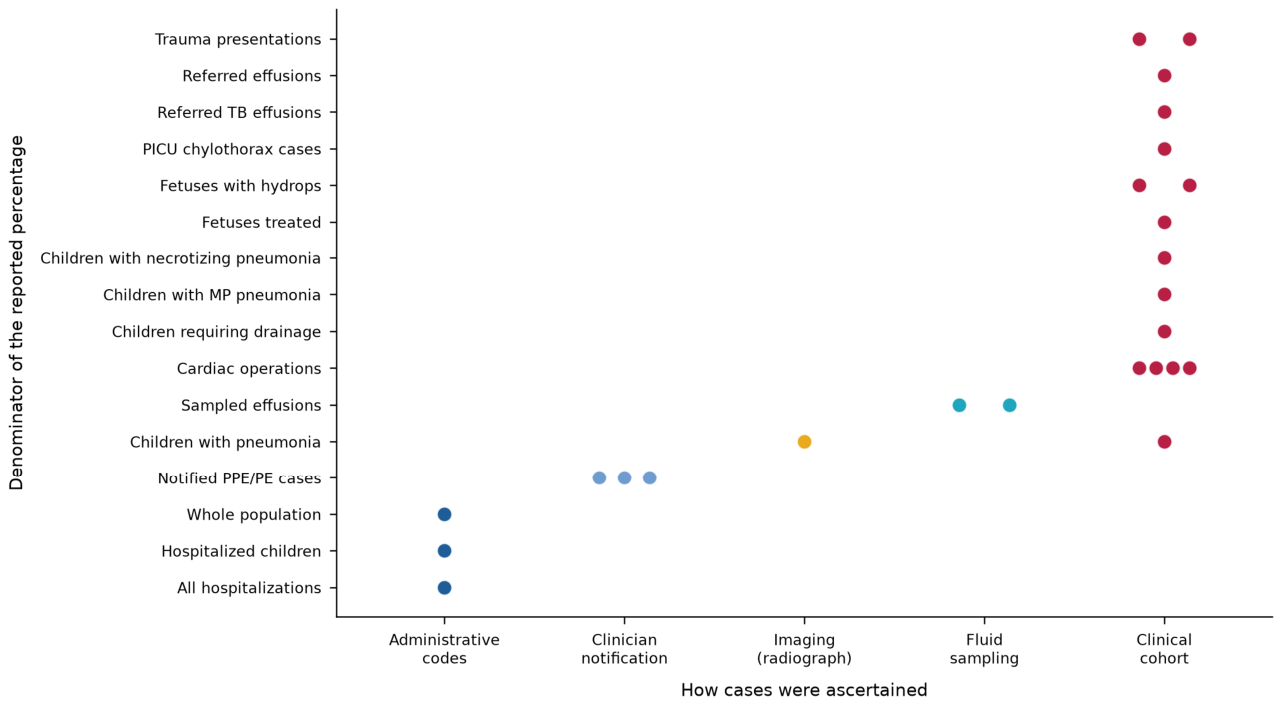
Table 2.
The same quantity measured with two instruments in the same population.
| What is being counted | Instrument A | Instrument B | Value with A | Value with B | Ratio | Setting | Source |
| Detection of effusion | Chest radiograph | Lung ultrasound | 11 of 81 | 17 of 81 | 1.5× | Children with confirmed pneumonia, same cohort; the 11 are a subset of the 17 | [64] |
| Pathogen in pleural fluid | Culture | Real-time PCR | 25% | 82% | 3.3× | 56 Ontario children, same fluid, same denominator | [83] |
| S. pneumoniae in pleural fluid | Culture | Real-time PCR | 14% | 62% | 4.4× | Same cohort as above | [83] |
| Pathogen detection | Before adding PCR | After adding PCR | 38.9% | 73.4% | 1.9× | One US children’s hospital, before and after | [85] |
| Pneumococcal detection | Before adding PCR | After adding PCR | 14.1% | 45.6% | 3.2× | Same hospital, same period comparison | [85] |
Each row holds the clinical question and the population constant and changes only the detection instrument; the ratio is the factor by which the reported figure moves. None of these differences is an epidemiological change. Ratios are computed from the values shown.
Table 3.
Pediatric series reporting a cause-specific proportion of pleural effusion, with the ascertainment mode and denominator of each.
Table 3.
Pediatric series reporting a cause-specific proportion of pleural effusion, with the ascertainment mode and denominator of each.
| Study | Country | Years | n | Ascertainment | Denominator | Population / setting | What the series reports |
| [68] | USA | 1990–2005 | 80 | Clinical cohort | Children with necrotizing pneumonia | Children hospitalized with necrotizing pneumonia, single center | Pleural effusion in 69 (86%); cultures positive in 38 (48%), S. pneumoniae predominant |
| [75] | USA | 1996–2007 | NR | Administrative codes | Hospitalized children | Children <5 y, Nationwide Inpatient Sample | Empyema complicating pneumonia rose 2.01-fold (3.5→7.0/100 000) in <2 y; staphylococcal empyema rose 4.08-fold |
| [144] | Germany | 2000–2006 | 282 | Clinical cohort | Cardiac operations | Newborns/infants after congenital heart surgery | Chylothorax in 26 (9.2%) |
| [74] | Australia | 1998–2010 | NR | Administrative codes | All hospitalizations | Hospitalizations, age 0–19 y, ICD-10 principal diagnosis | Pneumonia admissions IRR 0.78; empyema admissions IRR 1.35 after PCV7 |
| [169] | Denmark | 1993–2010 | 100 | Clinical cohort | Referred effusions | Tertiary pediatric pulmonary center | Incidence 0.5→2.6/100 000; S. pneumoniae most common; surgery beyond chest tube in 50% |
| [67] | UK | NR | 160 | Clinical cohort | Children with pneumonia | Children ≤16 y, radiologically confirmed pneumonia, prospective | Empyema in 40 (25%); bacterial infection OR 3.34 |
| [83] | Canada | 2009–2011 | 56 | Fluid sampling | Sampled effusions | Pleural fluid from children admitted with CAP and effusion | Pathogen in 82% by PCR vs 25% by culture; S. pneumoniae 62% (PCR) vs 14% (culture); S. pyogenes 16%; no S. aureus |
| [76] | UK (Scotland) | 1981–2013 | 217 + 323 admissions | Administrative codes | Whole population | Whole-population admissions, ICD-9/ICD-10 empyema codes | 9 cases/million/year (1981–2005) vs 47 (2006–2013); IRR 0.78 for 2010–2013 vs 2006–2009 |
| [48] | USA | 2007–2012 | 46 | Clinical cohort | Trauma presentations | Level-1 pediatric trauma center, hemothorax | 23 blunt / 23 penetrating; 91.3% vs 30.4% tube thoracostomy |
| [170] | South Korea | 1999–2012 | 68 | Clinical cohort | Fetuses treated | Fetuses with massive pleural effusion undergoing thoracoamniotic shunting | 50/65 (76.9%) hydropic; overall survival 75.4% |
| [54] | Germany | 2010–2013 | 645 | Clinician notification | Notified PPE/PE cases | Nationwide surveillance, PPE/PE <18 y | Median age 5 y; initial therapy invasive in 347 (55%), non-invasive in 282 (45%) |
| [123] | Brazil | 2004–2014 | NR | Clinical cohort | Cardiac operations | Children after corrective congenital heart surgery | Chylothorax 2.1% overall; 8.3% after Glenn, 11.8% after total cavopulmonary connection |
| [92] | USA | 2010–2012 | 2254 | Imaging (radiograph) | Children with pneumonia | Children hospitalized with radiographic pneumonia (EPIC) | 182 (8%) M. pneumoniae PCR-positive, of whom 26% had pleural effusion |
| [84] | Switzerland | 2001–2015 | 147 | Fluid sampling | Sampled effusions | Children ≤16 y with PPE/PE and diagnostic pleural tap, single center | Pathogen in 114 (78%): S. pneumoniae 79%, S. pyogenes 11%, S. aureus 6% |
| [147] | Austria | 1999–2012 | 34 | Clinical cohort | PICU chylothorax cases | PICU admissions with chylothorax/chylous-like disease | 31 (91%) postsurgical, 2 (6%) idiopathic, 1 (3%) congenital |
| [34] | Germany | 2014–2016 | 745 operations (667 patients) | Clinical cohort | Cardiac operations | Congenital heart surgery | Chylothorax 68 (9.1%); persistent serous effusion >7 days 125 (16.8%) |
| [78] | Germany | 2010–2018 | 584 | Clinician notification | Notified PPE/PE cases | Nationwide surveillance, bacteriologically confirmed PPE/PE | S. pneumoniae in 256 (43.8%); of 114 serotyped, serotype 3 42.1%, serotype 1 25.4%, 7F 12.3%, 19A 7.9% |
| [165] | Turkey | NR | 60 | Clinical cohort | Fetuses with hydrops | Non-immune hydrops fetalis with intrauterine pleural effusion | 39 (65%) liveborn; 25 died within days; chylothorax in 4 |
| [93] | South Korea | NR | 150 | Clinical cohort | Children with MP pneumonia | Children with M. pneumoniae pneumonia, tertiary hospital | Pleural effusion in 24 (16.0%) |
| [171] | India | 5 years | 76 | Clinical cohort | Referred TB effusions | Children referred with tuberculous pleural effusion | Right-sided 56.6%, left 40.8%, bilateral 2.6%; mean pleural ADA 107.6 ± 115.7 IU/L |
| [85] | USA | 2016–2023 | 149 pre / 79 post | Clinical cohort | Children requiring drainage | Children with complicated CAP requiring drainage, single center, pre/post S. pneumoniae PCR | Pathogen detected 38.9% → 73.4%; S. pneumoniae 14.1% → 45.6% |
| [82] | Germany | 2010–2018 | 1402 | Clinician notification | Notified PPE/PE cases | Nationwide surveillance, PPE/PE | Species identified: S. pneumoniae 192, S. pyogenes 111, S. aureus 38; methicillin resistance 10/32 (31.3%) |
| [166] | Taiwan | 2012–2021 | 55 | Clinical cohort | Fetuses with hydrops | Fetuses with non-immune hydrops fetalis undergoing intervention | Etiology identified in 33 (60%); 11 presented as congenital chylothorax |
| [172] | USA | 2010–2022 | 548 | Clinical cohort | Trauma presentations | Pediatric traumatic hemothorax, 41 trauma centers | 477 chest tube, 71 pigtail; failure 17.6% vs 12.6% |
| [124] | Vietnam | 2017–2026 | 21 | Clinical cohort | Cardiac operations | Chylothorax after pediatric congenital heart surgery | Fontan 47.6%, bidirectional Glenn 23.8%; right-sided 61.9% |
The table assembles records reporting a cause-specific proportion within a defined clinical cohort, excluding biomarker-only and single-case reports. It is curated, not exhaustive. NR marks cells the source abstract omits. The ascertainment and denominator columns record how each series found its cases and what its percentages are a fraction of, the two variables that defeat pooling (Section 5.2); across 25 series they take five and sixteen distinct values respectively.
Disclaimer/Publisher’s Note: The statements, opinions and data contained in all publications are solely those of the individual author(s) and contributor(s) and not of MDPI and/or the editor(s). MDPI and/or the editor(s) disclaim responsibility for any injury to people or property resulting from any ideas, methods, instructions or products referred to in the content. |
© 2026 by the authors. Licensee MDPI, Basel, Switzerland. This article is an open access article distributed under the terms and conditions of the Creative Commons Attribution (CC BY) license (http://creativecommons.org/licenses/by/4.0/).
Copyright: This open access article is published under a Creative Commons CC BY 4.0 license, which permit the free download, distribution, and reuse, provided that the author and preprint are cited in any reuse.