
Submitted:
11 November 2026
Posted:
12 November 2026
You are already at the latest version
Abstract
Keratoconus is increasingly recognised in children and adolescents, and early corneal cross-linking can halt its progression, but it remains unclear whether all children or only those with risk factors should be screened. Selected school-based, clinic-based and other population studies have reported prevalence estimates of approximately 0.4–2.7%, although differences in sampling, age distributions and diagnostic definitions limit direct comparisons. First-degree relatives show a high yield of keratoconus on corneal tomographic assessment, and Down syndrome is associated with increased prevalence, although the main registry estimate is not specific to children. Refractive astigmatism and its progression provide practical referral indicators, whereas atopy and eye rubbing do not currently define sufficiently narrow high-yield groups. The proportion of paediatric cases without recognised risk factors remains unknown. Test accuracy in unselected children, the natural history of screen-detected suspect disease and the comparative cost-effectiveness of screening strategies are also uncertain. Current evidence supports active case-finding in defined high-risk groups and corneal tomographic assessment in children with refractive warning signs. Universal screening should be evaluated in selected high-prevalence settings.
Keywords:
adolescents
; children
; corneal cross-linking
; corneal tomography
; health policy
; keratoconus
; risk factors
; screening
1. Introduction
Keratoconus was long regarded as an uncommon disease of young adults, usually diagnosed when spectacle correction became inadequate and frequently managed with corneal transplantation. This view has changed for three reasons. First, evidence from nationwide registration and corneal imaging studies has shown that the disease is considerably more common than clinic-based estimates suggested: a Dutch nationwide registration study reported a prevalence of about 1 in 375 [1], while a birth-cohort study using Scheimpflug-based corneal tomography identified keratoconus in about 1 in 84 young adults [2]. Second, keratoconus in children is more often advanced at presentation and progresses faster than in adults [3,4]. Third, corneal cross-linking (CXL) can stabilise paediatric keratoconus over the long term [5,6].
Taken together, these findings provide a rationale for earlier detection of keratoconus in childhood. The 2026 Global Consensus on Keratoconus and Ectatic Diseases and the ERN-EYE clinical consensus statement set out recommendations for the diagnosis, treatment and follow-up of affected children [7,8]. A scoping review of paediatric keratoconus epidemiology also highlighted the need for school-based screening research [9]. The appropriate target population, however, remains uncertain: it is not known whether corneal imaging should be offered universally or restricted to children with recognised risk factors.
The choice of screening strategy has implications for service capacity and case detection. Universal screening would entail imaging large numbers of children and providing specialist assessment for those with abnormal results. A risk-based approach would require fewer examinations, but could fail to identify children without recognised risk factors. The comparison therefore depends on screening yield, test performance, the number of missed cases, potential benefits and harms, and programme costs [10,11]. This review examines these considerations and their implications for current clinical practice.
2. Methods
We conducted a critical narrative review of evidence relevant to screening for paediatric keratoconus. PubMed was searched on 31 October 2026 for publications addressing epidemiology, early detection, diagnostic assessment, risk factors and management in children and adolescents. The search combined Medical Subject Headings (MeSH) with free-text terms for keratoconus and the paediatric population. The search string was:
("Keratoconus"[Mesh] OR keratoconus[tiab]) AND ("Child"[Mesh] OR "Adolescent"[Mesh] OR "Infant"[Mesh] OR child*[tiab] OR adolescen*[tiab] OR pediatric*[tiab] OR paediatric*[tiab] OR juvenile[tiab] OR teen*[tiab]).
The search retrieved 2,869 records. No limits were applied for publication date, language or publication type. Publications were considered if they addressed prevalence, screening yield, diagnostic methods, risk factors, disease progression, treatment, screening economics or screening policy in relation to paediatric or adolescent keratoconus. We gave particular consideration to studies of children and adolescents, school-based and population-based cohorts, corneal topography or tomography, and consensus statements on paediatric keratoconus. Adult, mixed-age and clinical-cohort studies were included when they informed diagnostic performance, epidemiological interpretation or screening principles; their applicability to children was considered separately.
Study selection was based on the relevance of each publication to the review objectives. We did not retain a formal count of records assessed at the title/abstract and full-text stages or document individual reasons for exclusion. The search and selection process was therefore not designed to identify exhaustively all eligible studies. Screening was not performed independently by two reviewers, and no formal risk-of-bias assessment or quantitative synthesis was undertaken.
Generative AI was used as an auxiliary tool to assist with summarizing selected publications and preparing preliminary syntheses of reported findings. The AI-generated material was treated as provisional and was not considered an independent source of scientific evidence. The authors verified the relevant information against the original publications and retained responsibility for the critical appraisal, interpretation, and final synthesis of the literature.
3. Rationale for Screening Children
3.1. Prevalence of Keratoconus in Children and Adolescents
Table 1 presents selected studies of keratoconus prevalence or screening yield in children and adolescents. Estimates ranged from 0.4% in a Chicago school-age vision clinic after recalculation with revised Scheimpflug-tomography criteria in 2026 [12,13] and 0.5% among high-school students in Trinidad [15], to 0.52% in New Zealand [14], 1.6% in Omani schoolchildren [16], 1.7% in Kenyan high-school students [17] and 2.7% in adolescents in Abu Dhabi [18]. A prevalence of 4.79% was reported in a Saudi sample recruited from non-ophthalmic emergency departments and including participants aged 6–21 years; this estimate is not directly comparable with school-based screening studies [19]. In New Zealand, prevalence was 2.25% among Māori students, compared with 0.52% in the overall sample [14].
In a nationwide cross-sectional study of 579,946 Israeli candidates for military service aged 16–19.9 years, keratoconus was recorded in 0.16% (164 per 100,000) [20]. Case identification was based on ophthalmological assessment at the military draft board rather than systematic corneal imaging, which may have resulted in lower case ascertainment than in school surveys using topography or tomography. The study does not, however, establish the extent of any underdiagnosis. In the Kenyan school survey, 88.2% of students diagnosed with keratoconus had been unaware of their condition [17].
3.2. Disease Course and Treatment in Children
Paediatric keratoconus is often more advanced at diagnosis than adult disease [3]. A meta-analysis of natural-history studies including 11,529 eyes found that younger age and steeper baseline maximum keratometry predicted faster steepening; the modelled 12-month steepening was approximately 0.8 D lower for every 10-year increase in age [4]. CXL can stabilise paediatric keratoconus over the long term [5,6], although the evidence for screening benefit remains indirect. The 2026 Global Consensus Edition 2 supports corneal tomographic assessment in children aged 6–18 years with astigmatism greater than 2.0 D on subjective refraction. For patients younger than 18 years with confirmed keratoconus, at least 86% of panellists supported considering CXL at diagnosis without waiting for documented progression; this recommendation does not apply to eyes classified as keratoconus suspects [7]. The ERN-EYE consensus similarly recommends corneal tomographic assessment for recent-onset corneal astigmatism and advises early assessment in children with a family history, with the timing individualised to age and family history [8].
Paediatric keratoconus meets several Wilson–Jungner criteria: the condition is clinically important, an early stage can be recognised, and treatment is available [10]. Evidence is less complete for the other criteria, particularly the suitability of the screening test, the definition of the target population and the balance of benefits, harms and costs. These unresolved issues are central to the choice between universal and risk-based screening.
The studies were selected to represent different settings and diagnostic approaches; the table is not exhaustive. Definitions of suspect keratoconus differed between studies.
4. Evidence Relevant to Universal Screening
4.1. Variability of Prevalence Estimates
The prevalence estimates in Table 1 are not directly comparable. Differences in case definitions, imaging methods, age distributions and population characteristics are likely to account for some of the variation. Studies have used different indices and thresholds, with grading sometimes dependent on examiner judgement. In an adult population-based cohort, the prevalence changed substantially when alternative keratoconus definitions were applied to the same participants [21]. Imaging methods also varied, from Placido-disc topography and retinoscopy to Scheimpflug tomography. In addition, estimates from cohorts spanning childhood and adulthood cannot be assumed to represent paediatric prevalence. Ethnicity and consanguinity may also contribute to geographic differences [14,16,22].
In paediatric screening, the distinction between normal variation, suspected keratoconus and definite disease is important. Suspect status was assigned to 19.8% of adolescents in the Abu Dhabi study, compared with 2.7% diagnosed with keratoconus [18]; the corresponding figures in Oman were 5.0% and 1.6% [16]. Reanalysis of the Chicago dataset using revised posterior-elevation thresholds changed the estimated prevalence from 1:334 to 1:251 for keratoconus and from 1:669 to 1:100 for suspect status [13]. Such differences illustrate the influence of diagnostic definitions on prevalence estimates. The 2026 Global Consensus Edition 2 updates diagnostic and progression criteria and broadens the approach to tomographic assessment, but does not provide universally validated age-specific normative thresholds for paediatric screening [7]. The ERN-EYE statement likewise notes the difficulty of diagnosing subclinical disease and the need to interpret multimodal imaging findings [8].
4.2. Screening Tests in Unselected Children
Universal screening requires a first-line test that is inexpensive, quick, acceptable and sufficiently accurate in an unselected population. Scheimpflug-based corneal tomography provides detailed assessment of anterior and posterior corneal shape and pachymetric distribution, but it requires equipment, cooperation and adequate scan quality. In the Abu Dhabi school study, 251 of 839 eyes (30%) were excluded because of insufficient scan quality [18]. In children with Down syndrome, corneal tomographic scans of sufficient quality were obtained in at least one eye in 75.9% in one paediatric hospital series [23]. Retinoscopy showed sensitivity of 97.7% and specificity of 79.9% in a selected clinical sample of 123 patients aged 10–30 years (245 eyes) [24]. These are eye-level estimates from a referred clinical sample, not validated child-level estimates from an unselected school population. In the Kenyan school study, individual measures such as visual acuity, refraction, astigmatism and keratometry had limited discriminatory ability; a combination of scissors reflex, maximum anterior corneal curvature greater than 46.5 D or refractive astigmatism greater than 2.50 D on objective refraction (autorefraction and retinoscopy) was proposed as a referral rule [25]. Because this threshold was derived from objective rather than subjective refraction, it is not directly interchangeable with the subjective-refraction criterion of the Global Consensus [7]. These data were also obtained in a mixed-age sample (13–25 years), and the performance of this rule in younger children remains uncertain.
These diagnostic estimates should not be extrapolated to school screening. Positive and negative predictive values vary with disease prevalence, and the retinoscopy results were obtained in referred patients aged 10–30 years and analysed at the eye level [24]. They have not been validated in unselected children using the child, rather than the eye, as the unit of analysis. The current data therefore do not support a reliable estimate of the number of children who would need to be screened or referred to detect one case. This question requires prospective accuracy studies in school populations.
4.3. Costs and Capacity
Economic evaluations of keratoconus have mainly modelled CXL rather than the screening pathway itself [26]. A 2025 proof-of-concept Markov model evaluated screening at age 15 followed by CXL for screen-positive children against usual care in the New Zealand context. It used a lifetime horizon, a societal perspective and a 3% discount rate. The reported incremental cost-effectiveness ratio was −NZ$1,278 per QALY gained (95% CI −1,418 to −1,139), indicating modelled cost savings with additional QALYs. Results were most sensitive to test specificity, prevalence at screening, discounting, the probability of progression, adherence and CXL efficacy [27]. These results are model-based rather than derived from an implemented screening programme, and their transferability to settings with lower prevalence, different costs or different service pathways is uncertain. No retrieved economic evaluation directly compared universal with risk-based paediatric screening.
5. Evidence Relevant to Risk-Based Screening
Risk-based case-finding depends on the ability to identify groups with a higher prevalence of disease than the general population. The findings reviewed here concern three distinct types of information: groups with a high diagnostic yield, notably first-degree relatives and children with Down syndrome; clinical findings that may prompt referral, particularly significant or increasing refractive astigmatism and an abnormal retinoscopic reflex; and risk modifiers, including atopy, allergic eye disease and eye rubbing. The latter may raise clinical suspicion but do not, on current evidence, identify a sufficiently restricted population for screening.
5.1. High-Yield Groups
First-degree relatives. Among 183 children aged 6–18 years who were first-degree relatives of patients, 17.5% had keratoconus confirmed by Scheimpflug-based corneal tomography, rising from 11.5–15.5% below age 11 to 25.5% at 16–18 years [28]. This corresponds to approximately one affected child for every six examined and supports planned corneal tomographic assessment in this group.
Down syndrome. The evidence indicates increased keratoconus prevalence in people with Down syndrome, but estimates vary markedly across selected studies. A review identified reported proportions from 0% to 71%, with generally low-quality and heterogeneous evidence [29]. A Norwegian national patient-registry study found keratoconus in 5.5% of people with Down syndrome; however, the mean age of affected individuals was 37.1 years, so this estimate is not a paediatric prevalence estimate [30]. A pilot screening programme in Irish adolescents with Down syndrome reported keratoconus detected by corneal tomography and requiring treatment in 15% of eyes at the first visit [31]. Feasibility is an important limitation: corneal tomographic scans of sufficient quality could not be obtained in approximately one quarter of children in a separate paediatric series [23].
Children with refractive warning signs. In a Colombian referral-centre cohort, 9.4% of 426 patients younger than 18 years referred for refractive astigmatism above 2 D, oblique astigmatism or myopia above 5 D had keratoconus confirmed using Scheimpflug-based corneal tomography (Pentacam) [32]. This is a selected referral population and should not be interpreted as the yield of screening all children with these refractive features. In a nationwide Israeli study of 896,377 adolescents aged 16–20 years, keratoconus prevalence increased with increasing cylinder power; the prevalence reached 2.3% at 2.00–<3.00 D, 9.5% at 3.00–<5.00 D and 17.4% at ≥5.00 D among adolescents undergoing topography or tomography when indicated by refractive findings. Cylinder power alone had moderate discrimination (AUC 0.752); the study supports astigmatism as a referral trigger, not as a stand-alone diagnostic test [33].
5.2. Risk Modifiers with Limited Discriminatory Value
Atopy and allergic eye disease. Atopy and ocular allergy are frequently associated with keratoconus; however, an association does not necessarily define a high-yield screening group. In the Colombian refractive referral cohort, 85% of children with keratoconus were atopic [32]; because the cohort was selected on refractive abnormalities, this proportion cannot establish the sensitivity of atopy as a population screening criterion. In a propensity-matched cohort of more than 348,000 patients aged 5–25 years, allergic conjunctivitis was associated with a higher incidence of keratoconus (hazard ratio 1.6), but the absolute incidence was 0.06% compared with 0.03% [34]. In a series of 651 patients with vernal keratoconjunctivitis (mean age 11.5 years; range 7–23), 5 patients (0.77%) had topographic keratoconus and 4 (0.61%) were classified as suspects [35]. These findings suggest that allergy may modify the risk of keratoconus, but they do not show that allergy alone defines an efficient screening population.
Eye rubbing. Eye rubbing was independently associated with keratoconus in Omani schoolchildren (OR 12.89, 95% CI 3.45–70.12), although the confidence interval was wide [16]. In the Trinidad study, 87.4% refers not to all 432 students but to 180 of the 206 participants classified as at risk; it therefore represents the frequency in a subgroup rather than the prevalence of eye rubbing in the study population [15]. Overall, 47.7% of participants were classified as at risk [15]. Eye rubbing remains a relevant behavioural risk marker and a target for counselling, but its high frequency within the at-risk subgroup and the broad composite risk definition limit its ability to identify a small, high-yield group.
Family history. First-degree relatives have a high screening yield, but family history may capture only a minority of affected children. It was reported in 7.5% of children with keratoconus in the Colombian referral cohort [32]. This estimate is subject to referral and ascertainment bias and should not be interpreted as the sensitivity of family-history-based screening in the general population.
5.3. Proportion of Cases Without Recognised Risk Factors
The proportion of children with keratoconus who have no recognised risk factor is central to the comparison of universal and risk-based screening. If few cases occur in children without identifiable risk factors, targeted screening may detect most cases with fewer examinations. If this proportion is substantial, a targeted strategy will miss affected children. No study identified in this review measured this proportion in an unselected paediatric population. Clinic-based series are unsuitable for this purpose because referral patterns favour children with recognised risk factors, while school surveys rarely report risk-factor status for each case. In one referral cohort, atopy was reported in 85% of affected children and family history in 7.5% [32]. The latter figure is affected by referral and ascertainment bias and cannot be interpreted as the sensitivity of family-history-based screening in the general population. Population-based studies with standardised assessment of risk factors are needed to address this question.

Table 2.
Yield, frequency and practical value of selected risk factors and clinical indicators for paediatric keratoconus.
Table 2.
Yield, frequency and practical value of selected risk factors and clinical indicators for paediatric keratoconus.
| Risk factor | Evidence of yield | Frequency/availability in relevant population or subgroup | Practical value for targeting |
| First-degree relative | 17.5% keratoconus on Scheimpflug-based corneal tomography in relatives aged 6–18 [28] | Low | High yield in identified relatives; population-level coverage remains uncertain |
| Down syndrome | 0–71% across heterogeneous studies; 5.5% in Norwegian national registry (all ages); 15% of eyes in Irish adolescent pilot [29,30,31] | Very low | High yield; imaging feasibility limited [23] |
| Refractive astigmatism ≥2 D, oblique or rising astigmatism, high myopia | 9.4% in referred children [32]; strong gradient in nationwide cohort aged 16–20 years [33] | Moderate | Good: detected in routine vision testing |
| Allergic conjunctivitis, atopy | HR 1.6, absolute incidence 0.06% vs 0.03% [34] | High | Weak: low absolute risk |
| Vernal keratoconjunctivitis | 0.77% keratoconus detected by corneal topography [35] | Low | Weak in that series |
| Eye rubbing | OR 12.89 in Oman (95% CI 3.45–70.12) [16] | 87.4% among the Trinidad subgroup classified as at risk (not the full school sample) [15] | Weak: does not narrow the population |
| Parental consanguinity | OR 14.74 in Oman (wide CI) [16] | Varies by region | Potentially useful where common; data sparse |
| Ethnicity / region | Population-specific; elevated estimates reported in selected Middle Eastern cohorts [22,36] | Population-specific | May help identify settings for evaluating universal screening rather than individual children |
HR, hazard ratio; OR, odds ratio; CI, confidence interval.
6. Critical Appraisal
6.1. Over-Labelling and Overtreatment
Screening may cause harm if abnormal findings are not interpreted appropriately. The 2026 Global Consensus supports consideration of CXL at diagnosis in patients younger than 18 years with confirmed keratoconus, even in the absence of documented progression; this recommendation does not apply to eyes classified as suspects [7]. The distinction between definite disease and suspect or subclinical findings is therefore essential. Suspect status was reported in 19.8% of adolescents in Abu Dhabi [18] and 1.0% of children in the revised Chicago analysis [13]. If screening programmes do not distinguish these categories, children may undergo repeated imaging and families may experience avoidable anxiety; inappropriate treatment is also a concern. The natural history of suspect or subclinical disease detected through screening has not been adequately characterised. Estimates from referred patients [4] may not reflect the course of disease detected before symptoms develop.
6.2. Lack of Outcome Data
No randomised or controlled study has compared visual outcomes, keratoplasty rates or quality of life in screened and unscreened children. The case for screening rests on separate strands of indirect evidence: prevalence estimates, the ability to detect disease, CXL outcomes in clinically identified patients and economic modelling. These data do not establish that screening itself improves patient outcomes. The Irish pilot programme for adolescents with Down syndrome reported diagnostic yield and treatment rates, but did not provide long-term outcome or cost data [31].
6.3. Equity
Targeting based on family history or parent-reported allergy may be less effective in families with limited health literacy or poor access to care. In New Zealand, keratoconus was associated with Māori ethnicity and lower school decile [14], populations that may also encounter barriers to specialist assessment. School-based programmes in communities with a high disease prevalence could improve access, whereas strategies based solely on questionnaires may perpetuate existing inequalities. Equity is recognised as an important component of screening policy [11], but has received little attention in keratoconus screening research. The UK National Screening Committee did not assess a proposal submitted during its 2017 annual call for screening children and young adults with Down syndrome because targeted screening of high-risk groups fell outside the remit of its population-screening process [37]. This distinction is relevant when applying population-screening criteria to clinical case-finding.
6.4. Summary Against Screening Criteria
Under the Wilson–Jungner criteria and the consolidated screening principles [10,11], paediatric keratoconus meets several requirements: it is an important health condition, an early stage can be identified, and effective treatment is available. Important uncertainties remain regarding the performance and acceptability of screening tests in unselected children, the threshold for treating subclinical disease, programme costs and the balance of benefits and harms. These uncertainties are particularly relevant to universal screening, which would expose many children with a low pre-test probability to testing, and less so to clinical assessment of groups in whom the expected diagnostic yield is higher.
6.5. Limitations of This Review
This narrative review has limitations. The evidence was identified primarily through PubMed and selected consensus documents; the review did not use a protocolised eligibility process, duplicate independent screening, formal risk-of-bias assessment or quantitative synthesis. Although the search retrieved 2,869 records, the numbers screened, excluded and included were not recorded, and a PRISMA flow diagram therefore cannot be provided. The included studies differed in age range, setting, imaging method and diagnostic criteria; some involved mixed-age cohorts or selected clinical populations. The review cannot, therefore, establish the comparative effectiveness of universal and risk-based screening.
7. Proposed Approach to Case-Finding
On the basis of the evidence reviewed, we propose a level-based approach to case-finding in which the intensity of assessment is matched to the strength of the available evidence (Figure 1). This proposal is intended to inform clinical discussion and should not be regarded as a formal guideline.
- Level 1: planned corneal tomographic assessment in defined high-yield groups. Offer or arrange corneal tomography for first-degree relatives of patients with keratoconus, with timing guided by age and family history; the ERN-EYE consensus describes early examinations around 8–13 years and regular assessment particularly when several family members are affected [8,28]. Children with Down syndrome should have an individualised pathway for corneal assessment, taking account of cooperation and feasibility. Current evidence supports heightened clinical vigilance and consideration of tomographic screening, but does not establish a universally accepted screening interval for all children with Down syndrome [23,30,31].
- Level 2: imaging guided by refractive referral indicators. In routine paediatric and school vision assessment, refer children aged 6–18 years with astigmatism greater than 2 D on subjective refraction, recent-onset or increasing refractive astigmatism, oblique or against-the-rule progressive myopic astigmatism, a scissors reflex on retinoscopy, or reduced best-corrected visual acuity without another cause [7,8,25,32,33]. These indicators justify referral for corneal imaging, with tomographic assessment where clinically indicated, but are not diagnostic criteria. The refractive thresholds should be interpreted in the context of the relevant consensus and clinical assessment, rather than as independently validated population-screening cut-offs.
- Level 3: universal screening only as evaluated programmes in selected high-prevalence settings. Where local, population-based surveys using corneal topography or tomography demonstrate a materially elevated prevalence, school-based screening may be reasonable to evaluate, but no evidence-based prevalence threshold has been established. Pilot programmes should prospectively report participation, scan quality, referral completion, confirmed diagnoses, treatment, harms, equity and costs [14,17,18].
- Across all levels: use explicit and reproducible imaging criteria, including tomographic criteria when tomography is performed; report definite keratoconus separately from suspect/subclinical status; and base CXL decisions on confirmed disease and the applicable clinical consensus, not on a screening-positive or suspect label alone [7,8].
Children with atopy or allergic eye disease should receive appropriate allergy treatment and advice to avoid eye rubbing. These factors may justify a lower threshold for corneal imaging when accompanied by refractive change, symptoms or other risk indicators. On their own, however, they do not identify a sufficiently restricted group for targeted screening [15,34,35].
8. Research Agenda
Further studies should address the following questions:
- Population-based surveys in unselected children from low- and moderate-prevalence regions, including Europe, should use a standardised corneal imaging protocol, agreed case definitions and systematic assessment of risk factors. Such studies should report not only prevalence and risk-factor frequencies but also the proportion of affected children without recognised risk factors.
- Prospective diagnostic-accuracy studies of low-cost first-line approaches, including refractive indicators, retinoscopy and portable topography, in unselected school populations, using corneal tomography as the reference standard. The role of artificial intelligence should be evaluated within these studies rather than assumed to be established.
- Longitudinal studies of children with screen-detected keratoconus or suspect findings to define the natural history and determine the proportion who subsequently require CXL.
- Decision-analytic comparisons of universal, risk-based and level-based strategies using local cost data and explicit threshold analyses.
9. Conclusions
Paediatric keratoconus is an important and treatable cause of visual morbidity, and the available evidence supports active case-finding. Planned corneal assessment is justified in first-degree relatives; children with Down syndrome warrant an individualised assessment pathway; and refractive abnormalities should prompt imaging when clinically indicated. The evidence does not currently support universal screening outside populations in which a high prevalence has been demonstrated. The main unresolved issues are the proportion of cases that would be missed by targeted strategies, the accuracy of first-line tests in unselected children, the course of screen-detected subclinical disease and the cost-effectiveness of alternative approaches. Any school-based universal screening programme should therefore be introduced as an evaluated pilot with prespecified clinical, operational and economic outcomes.
Author Contributions
Conceptualization, J.D.-S.; methodology, J.D.-S.; investigation, J.D.-S. and T.C.; formal analysis, A.D.; resources, A.D.; writing—original draft preparation, J.D.-S., T.C., R.R. and A.D.; writing—review and editing, J.D.-S., T.C., R.R. and A.D.; supervision, R.R. All authors have read and agreed to the published version of the manuscript.
Funding
This research was funded by the Minister of Science and Higher Education of Poland (formerly the Minister of Education and Science) from the state budget under the commissioned task “Lublin Digital Union – Use of Digital Solutions and Artificial Intelligence in Medicine – a research project”, grant number MEiN/2023/DPI/2194. The APC was funded by the same grant.
Institutional Review Board Statement
Not applicable.
Informed Consent Statement
Not applicable.
Data Availability Statement
No new data were created or analysed in this study. Data sharing is not applicable to this article.
Acknowledgments
During the preparation of this manuscript, the authors used Claude Opus 5.5 (Anthropic, San Francisco, CA, USA) to assist with summarizing selected publications and preparing preliminary syntheses of the reported findings. The authors reviewed and edited the generated material, verified the scientific information against the original publications, and take full responsibility for the content of this publication.
Conflicts of Interest
The authors declare no conflicts of interest.
References
- Godefrooij, D.A.; de Wit, G.A.; Uiterwaal, C.S.; Imhof, S.M.; Wisse, R.P.L. Age-specific incidence and prevalence of keratoconus: A nationwide registration study. Am. J. Ophthalmol. 2017, 175, 169–172. [Google Scholar] [CrossRef] [PubMed]
- Chan, E.; Chong, E.W.; Lingham, G.; Stevenson, L.J.; Sanfilippo, P.G.; Hewitt, A.W.; et al. Prevalence of keratoconus based on Scheimpflug imaging: The Raine Study. Ophthalmology 2021, 128, 515–521. [Google Scholar] [CrossRef] [PubMed]
- 3.; Mukhtar, S.; Ambati, B.K. Pediatric keratoconus: A review of the literature. Int. Ophthalmol. 2018, 38, 2257–2266. [Google Scholar] [CrossRef] [PubMed]
- Ferdi, A.C.; Nguyen, V.; Gore, D.M.; Allan, B.D.; Rozema, J.J.; Watson, S.L. Keratoconus natural progression: A systematic review and meta-analysis of 11 529 eyes. Ophthalmology 2019, 126, 935–945. [Google Scholar] [CrossRef] [PubMed]
- Godefrooij, D.A.; Soeters, N.; Imhof, S.M.; Wisse, R.P.L. Corneal cross-linking for pediatric keratoconus: Long-term results. Cornea 2016, 35, 954–958. [Google Scholar] [CrossRef] [PubMed]
- Kobashi, H.; Hieda, O.; Itoi, M.; Kamiya, K.; Kato, N.; Shimazaki, J.; et al. Corneal cross-linking for paediatric keratoconus: A systematic review and meta-analysis. J. Clin. Med. 2021, 10, 2626. [Google Scholar] [CrossRef] [PubMed]
- Gomes, J.A.P.; Hafezi, F.; Ambrósio, R., Jr.; Henriquez, M.A.; Campos, M.; Kinoshita, S.; et al. Global Consensus on Keratoconus and Ectatic Diseases—Edition 2. Cornea 2026, 45, 888–908. [Google Scholar] [CrossRef] [PubMed]
- Buzzonetti, L.; Mazzotta, C.; Iester, M.; Pauklin, M.; Boehringer, D.; Zemaitiene, R.; et al. Paediatric keratoconus: An ERN-EYE clinical consensus statement on diagnosis, treatment and follow-up care. Ophthalmol. Ther. 2026, 15, 2825–2861. [Google Scholar] [CrossRef] [PubMed]
- Hansen, L.O.; Garcia, R.; Cresta, F.B.; Torricelli, A.A.M.; Bechara, S.J. Pediatric keratoconus epidemiology: A systematic scoping review. Int. Ophthalmol. 2024, 44, 69. [Google Scholar] [CrossRef] [PubMed]
- Wilson, J.M.G.; Jungner, G. Principles and Practice of Screening for Disease . In Public Health Papers; World Health Organization: Geneva, Switzerland, 1968; Volume No. 34. [Google Scholar]
- Dobrow, M.J.; Hagens, V.; Chafe, R.; Sullivan, T.; Rabeneck, L. Consolidated principles for screening based on a systematic review and consensus process. CMAJ 2018, 190, E422–E429. [Google Scholar] [CrossRef] [PubMed]
- Harthan, J.S.; Gelles, J.D.; Block, S.S.; Tullo, W.; Morgenstern, A.S.; Su, B.; et al. Prevalence of keratoconus based on Scheimpflug corneal tomography metrics in a pediatric population from a Chicago-based school age vision clinic. Eye Contact Lens 2024, 50, 121–125. [Google Scholar] [CrossRef] [PubMed]
- Belin, M.W.; Harthan, J.S.; Eiden, S.B.; Morgenstern, A.S.; Tullo, W.; Gelles, J.D.; et al. Revised calculation of the prevalence of keratoconus based on updated Scheimpflug corneal tomography metrics in a pediatric population from a Chicago-based school age vision clinic. Eye Contact Lens 2026, 52, 103–104. [Google Scholar] [CrossRef] [PubMed]
- Papali'i-Curtin, A.T.; Cox, R.; Ma, T.; Woods, L.; Covello, A.; Hall, R.C. Keratoconus prevalence among high school students in New Zealand. Cornea 2019, 38, 1382–1389. [Google Scholar] [CrossRef] [PubMed]
- Ezinne, N.E.; Phagoo, S.; Roopnarinesingh, A.; Kwarteng, M.A. Prevalence of keratoconus and associated risk factors among high school students in Couva, Trinidad: A cross-sectional study. Vision 2025, 9, 89. [Google Scholar] [CrossRef] [PubMed]
- Mohamed, Z.; Noushad, B.; Vankudre, G.S.; Ayyappan, J.; Alkaabi, A. Prevalence and risk factors of keratoconus among school children in Oman: A population-based study. BMC Ophthalmol. 2026, 26, 616. [Google Scholar] [CrossRef] [PubMed]
- Rashid, Z.A.; Mashige, K.P.; Moodley, V.R. Prevalence and demographic profile of keratoconus among high school students in Kenya. Int. Ophthalmol. 2025, 45, 21. [Google Scholar] [CrossRef] [PubMed]
- Armstrong, B.K.; Smith, S.D.; Romac Coc, I.; Agarwal, P.; Mustapha, N.; Navon, S. Screening for keratoconus in a high-risk adolescent population. Ophthalmic Epidemiol. 2021, 28, 191–197. [Google Scholar] [CrossRef] [PubMed]
- Torres Netto, E.A.; Al-Otaibi, W.M.; Hafezi, N.L.; Kling, S.; Al-Farhan, H.M.; Randleman, J.B.; et al. Prevalence of keratoconus in paediatric patients in Riyadh, Saudi Arabia. Br. J. Ophthalmol. 2018, 102, 1436–1441. [Google Scholar] [CrossRef] [PubMed]
- Eliasi, E.; Bez, M.; Megreli, J.; Avramovich, E.; Fischer, N.; Barak, A.; et al. The association between keratoconus and body mass index: A population-based cross-sectional study among half a million adolescents. Am. J. Ophthalmol. 2021, 224, 200–206. [Google Scholar] [CrossRef] [PubMed]
- Shabani, H.; van Dooren, B.T.H.; Meester-Smoor, M.A.; Geerards, A.A.J.; Klaver, C.C.W.; Ramdas, W.D. Different keratoconus definitions can lead to substantial prevalence disparities in population-based studies. Sci. Rep. 2025, 15, 3483. [Google Scholar] [CrossRef] [PubMed]
- Khattak, A.; Altalhi, A.; Alotaibi, A.B.; Khattak, A.M. Prevalence of keratoconus in the young Eastern population of Saudi Arabia. Cureus 2024, 16, e55692. [Google Scholar] [CrossRef] [PubMed]
- Smith, C.G.; Edwards Mayhew, R.G.; Singh, J.K.; Patnaik, J.L.; Wagner, B.D.; Wise, R.E.; et al. Screening corneal tomography for the diagnosis of keratoconus in pediatric patients with trisomy 21. Am. J. Ophthalmol. 2025, 277, 349–355. [Google Scholar] [CrossRef] [PubMed]
- Al-Mahrouqi, H.; Bani Oraba, S.; Al-Habsi, S.; Mundemkattil, N.; Babu, J.; Panchatcharam, S.M.; et al. Retinoscopy as a screening tool for keratoconus. Cornea 2019, 38, 442–445. [Google Scholar] [CrossRef] [PubMed]
- Rashid, Z.A.; Mashige, K.P.; Moodley, V.R. Validity of keratoconus screening tests. Clin. Exp. Optom. 2026, 109, 195–204. [Google Scholar] [CrossRef] [PubMed]
- Salmon, H.A.; Chalk, D.; Stein, K.; Frost, N.A. Cost effectiveness of collagen crosslinking for progressive keratoconus in the UK NHS. Eye 2015, 29, 1504–1511. [Google Scholar] [CrossRef] [PubMed]
- Hong, C.Y.; Hong, S.C.; Wilson, G. Screening plus corneal cross-linking for keratoconus is cost-effective for New Zealand: A proof-of-concept Markov analysis. Clin. Exp. Ophthalmol. 2025, 53, 1125–1135. [Google Scholar] [CrossRef] [PubMed]
- Awwad, S.T.; Yehia, M.Y.; Mehanna, C.J.; Abdul Fattah, M.; Saad, A.; Hatoum, A.K.; et al. Tomographic and refractive characteristics of pediatric first-degree relatives of keratoconus patients. Am. J. Ophthalmol. 2019, 205, 186–194. [Google Scholar] [CrossRef] [PubMed]
- Kristianslund, O.; Drolsum, L. Prevalence of keratoconus in persons with Down syndrome: A review. BMJ Open Ophthalmol. 2021, 6, e000754. [Google Scholar] [CrossRef] [PubMed]
- Kristianslund, O.; Drolsum, L. Prevalence of keratoconus in persons with Down syndrome in a national registry in Norway. JAMA Netw. Open 2021, 4, e210814. [Google Scholar] [CrossRef] [PubMed]
- Power, B.; Stephenson, K.A.J.; Malata, D.; Murphy, C.; Quill, B.; McGrane, F.; et al. Pilot programme for keratoconus screening and management in adolescents with Down syndrome. Diagnostics 2025, 15, 683. [Google Scholar] [CrossRef] [PubMed]
- Sarria Calderón, N.A.; Martínez Córdoba, C.J.; Pinedo Agudelo, J.A.; Rosas Apraez, J.A.; Revelo Álvarez, M.L. Keratoconus frequency and associated risk factors among patients younger than 18 years with significant refractive errors. Cornea 2024, 43, 585–590. [Google Scholar] [CrossRef] [PubMed]
- Safir, M.; Nitzan, I.; Hanina, Y.; et al. Keratoconus prevalence in astigmatic adolescents: Findings from a nationwide screening setting. Eye 2025, 39, 2958–2962. [Google Scholar] [CrossRef] [PubMed]
- Lishinsky-Fischer, N.; Erdinest, N.; Buhbut, O.; Gur, Z. Incidence of keratoconus following allergic conjunctivitis: A multicenter retrospective cohort study using a U.S. healthcare network database. Cont. Lens Anterior Eye 2026, 49, 102569. [Google Scholar] [CrossRef] [PubMed]
- Caputo, R.; Versaci, F.; Pucci, N.; de Libero, C.; Danti, G.; De Masi, S.; et al. Very low prevalence of keratoconus in a large series of vernal keratoconjunctivitis patients. Am. J. Ophthalmol. 2016, 172, 64–71. [Google Scholar] [CrossRef] [PubMed]
- Abou Said, A.G.; Gispets, J.; Shneor, E. Strategies for early keratoconus diagnosis: A narrative review of evaluating affordable and effective detection techniques. J. Clin. Med. 2025, 14, 460. [Google Scholar] [CrossRef] [PubMed]
- 37. UK National Screening Committee. 2017 Annual Call Topic Proposals. Available online: https://www.gov.uk/government/publications/uk-nsc-previous-annual-call-submissions/2017-annual-call-topic-proposals (accessed on 30 October 2026).
Figure 1.
Proposed level-based approach to keratoconus case-finding in children and adolescents.
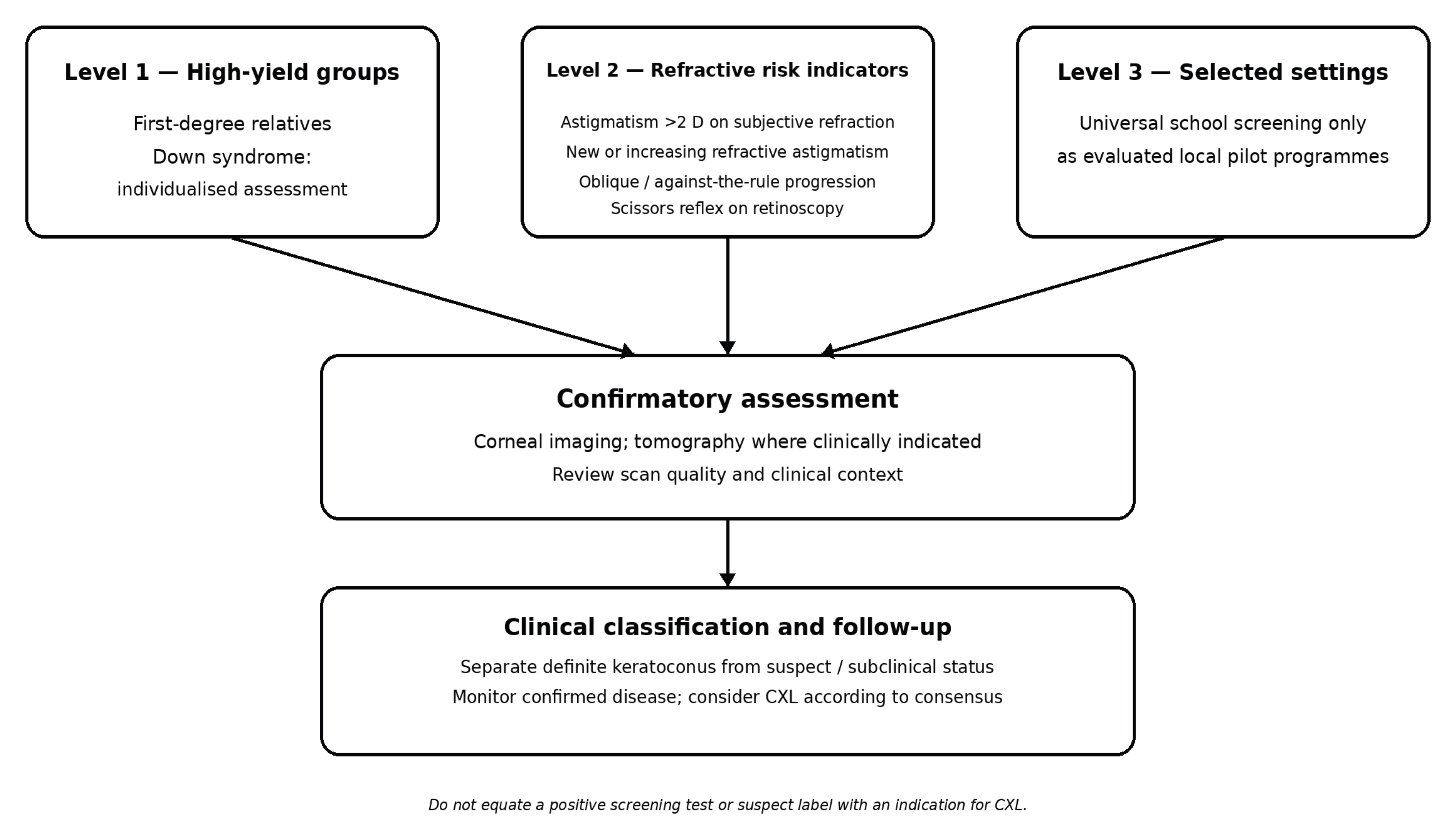
Table 1.
Selected studies reporting keratoconus prevalence or screening yield in children and adolescents.
Table 1.
Selected studies reporting keratoconus prevalence or screening yield in children and adolescents.
| Study | Setting, country | N | Age (years) | Method | Keratoconus | Suspects | Comment |
| Papali'i-Curtin 2019 [14] | High schools, New Zealand | 1,916 | Mean 14.6 | Scheimpflug-based corneal tomography (Pentacam; Belin/Ambrósio display) | 0.52% (1 in 191) | – | 2.25% (1 in 45) in Māori students; associated with atopy and lower school decile |
| Harthan 2024; recalculation 2026 [12,13] | School-age vision clinic, Chicago, USA | 2,007 | 3–18 | Scheimpflug-based corneal tomography (Pentacam); revised posterior-elevation thresholds | 0.40% (1 in 251) | 1.0% (1 in 100) | Original estimate: 6 KC (1 in 334) and 3 suspects (1 in 669). Revised 2026 criteria: 8 KC and 20 suspects; combined prevalence 1 in 72. Clinic-based, not a population sample. |
| Ezinne 2025 [15] | High schools, Trinidad | 432 | 12–17 | Clinical examination with corneal topography | 0.5% (2 of 432) | – | 47.7% met the study’s composite at-risk definition; eye rubbing was reported by 87.4% of this at-risk subgroup (180/206), not all students. |
| Mohamed 2026 [16] | Government schools, Oman | 733 | 9–18 | Auto-kerato-refractometry, retinoscopy and corneal topography; KC classification required ≥2 of 4 criteria, including refractive profile | 1.6% (2.2% at 13–18 y) | 5.0% | 12 cases (1.6%; all in age group 13–18 years); 2.2% in this older subgroup. 37 suspects (5.0%). |
| Rashid 2025 [17] | High schools, Nairobi, Kenya | 3,051 | 13–25 (mean 17.4) | Corneal topography at school (EyeSys/Medmont); referral for confirmatory MS-39 AS-OCT imaging | 1.7% (95% CI 1.2–2.2) | 3.8% | 88.2% of KC cases were unaware; mixed-age cohort (13–25 years), so prevalence specifically in those aged ≤18 years is not reported separately. |
| Armstrong 2021 [18] | Secondary schools, Abu Dhabi, UAE | 339 analysed (420 imaged) | 10–19 | Scheimpflug–Placido corneal tomography (Galilei) | 2.7% (95% CI 1.2–5.0) | 19.8% | High-risk adolescent school sample; 251/839 eyes excluded for poor scan quality. |
| Torres Netto 2018 [19] | Non-ophthalmic emergency departments, Riyadh, Saudi Arabia | 522 | 6–21 (mean 16.8) | Scheimpflug-based corneal tomography (Pentacam HR); two masked corneal specialists | 4.79% (95% CI 2.96–6.62) | Not reported | Non-ophthalmic emergency-department sample; 54.6% were aged 19–21 years. KC was not distinguished from other ectasias such as pellucid marginal degeneration. |
| Eliasi 2021 [20] | Military-service candidates, Israel (nationwide) | 579,946 | 16–19.9 | Ophthalmological examination at military draft board | 0.16% (164 per 100,000) | – | Ophthalmological examination at the military draft board; Jewish candidates only. Diagnosed cases were identified through clinical examination, not systematic corneal imaging. |
Disclaimer/Publisher’s Note: The statements, opinions and data contained in all publications are solely those of the individual author(s) and contributor(s) and not of MDPI and/or the editor(s). MDPI and/or the editor(s) disclaim responsibility for any injury to people or property resulting from any ideas, methods, instructions or products referred to in the content. |
© 2026 by the authors. Licensee MDPI, Basel, Switzerland. This article is an open access article distributed under the terms and conditions of the Creative Commons Attribution (CC BY) license.
Copyright: This open access article is published under a Creative Commons CC BY 4.0 license, which permit the free download, distribution, and reuse, provided that the author and preprint are cited in any reuse.