

Submitted:
10 November 2026
Posted:
12 November 2026
You are already at the latest version
Abstract
Background. Haemophilus influenzae (Hi) is a common commensal of the pediatric respiratory tract and a lower-respiratory pathogen frequently detected in Henoch–Schönlein purpura (HSP, IgA vasculitis). Pathogenicity is largely capsule-determined, but the relationship between Hi capsular type and disease phenotype in children remains unclear. Methods. Detection rates were analyzed in a single-center cohort of 11,017 children who underwent bacterial nucleic-acid testing (HSP 2,596; other respiratory diseases 8,382; 39 with other respiratory diagnoses not entered into the comparisons), with 39 concurrently examined healthy children tested by the same assay as an external reference group. Hi-positive specimens were capsular-typed by an in-house two-step qPCR (bexA screening, then Region II multiplex, a–f). Associations were estimated by logistic regression with complementary age-confounding control and immune-status/testing-intensity sensitivity analyses; pathogenic potential was profiled in 44 reference genomes. Results. Hi detection was higher in HSP than in other respiratory diseases (57.0% vs 11.4%; adjusted OR 9.8, 95%CI 8.7–11.1) and healthy children (12.8%), consistent across age-handling methods and immune-normal subsets (ORs 7.8–9.8), and HSP-specific rather than severity-related. Capsular type spectra differed: HSP was relatively enriched for encapsulated type c (~50% vs 75–80% NTHi; OR 2.68–3.87). In pneumonia, NTHi patients were younger (median 3 vs 8 years) and more pneumonia-prone (74% vs 18%). Genomic pathogenic potential stratified by capsule; NTHi retained a rich virulence-factor repertoire. Conclusions. Respiratory Hi is highly detected in pediatric pneumonia and HSP, with distinct capsular type spectra and clear type–phenotype associations, consistent with capsule-dependent disease tropism. These associations are hypothesis-generating and require mechanistic validation.
Keywords:
Haemophilus influenzae
; capsular type
; non-typeable
; children
; Henoch–Schönlein purpura
; pneumonia
; molecular epidemiology
1. Introduction
Haemophilus influenzae is an obligate colonizer of the human nasopharynx that can persist asymptomatically as part of the upper-respiratory flora [1,2], but can also descend to cause pneumonia and even invasive infection.[3,4,5] Its principal virulence determinant is the polysaccharide capsule, on which basis it is divided into six capsular types (a–f; historically called serotypes) and non-typeable (NTHi) strains.[3] Following widespread use of the type b conjugate vaccine, invasive Hib strains have declined sharply, and NTHi together with other encapsulated types now constitute the majority of pediatric respiratory Hi [6,7,8].
In 16S rRNA (V3–V4) amplicon profiling of respiratory specimens within our cohort, Haemophilus was enriched in both pneumonia and Henoch–Schönlein purpura (HSP). HSP is the most common IgA-mediated small-vessel vasculitis of childhood and frequently follows respiratory infection [9,10]; and because Hi is a common respiratory mucosal organism capable of stimulating mucosal IgA responses, its high detection in HSP raises the possibility of a triggering relationship. Because different capsular types may correspond to distinct pathogenic mechanisms, the capsular type found in each disease and its associated clinical phenotype are of interest.
Commercial type-specific antisera for slide-agglutination serotyping are available (e.g., Statens Serum Institut); however, agglutination requires a viable cultured isolate and is prone to cross-reaction and non-typeability, and was not applicable to our largely culture-independent, nucleic-acid–positive specimens. Molecular (PCR-based) capsular typing is therefore widely used but is not available as a single standardized commercial IVD kit, so laboratories implement published PCR protocols with in-house primers (bexA to determine encapsulation, then type-specific primers to distinguish a–f).[11,12] We therefore developed an in-house PCR typing assay with self-designed primers for specimens testing Hi-positive by nucleic-acid detection, following published methods, systematically performed typing and disease-association analyses, and used pathogenic-potential analysis of reference genomes to interpret the type–mechanism relationship.
2. Materials and Methods
Terminology. Throughout this paper we use “capsular type” to refer to the molecular classification based on the cap locus (a–f) and bexA encapsulation status, reserving “serotype” for contexts involving traditional slide-agglutination serotyping or vaccine-related nomenclature. “Non-typeable” (NTHi) denotes strains lacking a detectable cap locus by our assay.
Study population and data source. Records came from a single-center clinical database at Beijing Children’s Hospital, Capital Medical University (a tertiary pediatric referral and teaching hospital, Beijing, China), covering pediatric respiratory hospitalization episodes over 2017–2023 (source A); the capsular-typing specimens (Hi-positive and residual specimens) were from June 2021 to May 2023; the database contained clinical diagnoses, Hi detection and capsular typing. Residual respiratory specimens were collected into a commercial nucleic-acid preservation/transport medium (Ningbo HEALTH Gene Technology Co., Ltd., Ningbo, China)—a nuclease-inhibiting buffered solution containing protein stabilizers and RNase/DNase inhibitors that preserves nucleic acids in the specimen—and stored at −80 °C before nucleic-acid testing. Analytic cohort (denominator definition, critical): because untested patients cannot be counted as Hi-negative, all detection-rate analyses were restricted to the 11,017 episodes with completed bacterial nucleic-acid (“pneumonia bacteria”) testing: 2,596 with HSP, 8,382 with other respiratory diseases (pneumonia, bronchitis, etc.); the remaining 39 tested episodes carried other respiratory diagnoses (pulmonary tuberculosis or tracheitis) and were not entered into either comparison group. Within HSP, patients were divided by extent of involvement into skin-only and composite/systemic (abdominal/joint/renal types and their combinations); nephritis was merged into the composite subtype (an a priori clinical judgment—HSP nephritis generally reflects protracted, systemic disease rather than an independent entity; see Results 3.5). Capsular typing was completed for 245 cases from two specimen streams. The primary stream comprised 160 specimens that tested H. influenzae–positive on the clinical isothermal-amplification chip assay and were subsequently capsular-typed. The second stream comprised 85 residual specimens that had been submitted over the same period for testing of other respiratory pathogens; in these, 16S rRNA (V3–V4) sequencing showed a relatively high abundance of the genus Haemophilus and of the H. influenzae / H. haemolyticus clade (by exact ASV comparison; see Methods), prompting capsular typing. For caliber consistency the primary typing analysis used the 160 clinical-assay–positive specimens, with all 245 used for sensitivity validation. Capsular-type clinical-phenotype analysis of Hi-positive respiratory cases was based on a curated series with complete clinical data (source B, n=108; blood counts, length of stay, etc.). Sources A and B share the same identifier system (admission/outpatient number = medical-record number): matching source B’s 105 unique identifiers against source A medical-record numbers linked 89 (85%) to source A, and among the 90 comparable cases the capsular type assignments were fully concordant (90/90), confirming that source B is a clinically more detailed subset of source A, used to characterize each type’s clinical phenotype at the individual level; it is presented separately because it provides bedside indices (blood counts, length of stay) not available in source A. Healthy controls (source D): concurrently examined healthy children (n=39) tested for Hi by the same multiplex respiratory-pathogen PCR; age was taken as recorded in their microbiome-matching file (median 6 years). They serve as a non-diseased reference and, given the small size and screening-population nature, are used only for a descriptive three-group comparison of detection rates, not in the primary regression.
Ethics. The study was approved by the Medical Ethics Committee of Beijing Children’s Hospital, Capital Medical University, under three protocols covering its respective components: [2021]-E-196-Y (rapid molecular diagnosis of pathogens in pediatric respiratory infectious diseases; approved 23 November 2021), [2023]-E-008-R (epidemiology of respiratory pathogen infections in children; approved 31 January 2023), and [2022]-E-153-Y-01 (microbiome analysis and mechanistic study in pediatric respiratory infectious diseases; amendment approved 23 May 2023). For the retrospective analyses of de-identified clinical records, the requirement for individual informed consent was waived by the committee; written informed consent was obtained from guardians of the concurrently examined healthy children.
Hi detection and in-house typing. For the microbiome stream, 16S rRNA (V3–V4) reads were denoised into exact amplicon sequence variants (ASVs) and assigned against a Haemophilus reference set. Because the V3–V4 region cannot reliably separate H. influenzae from its close relative H. haemolyticus—in a panel of 38 H. haemolyticus genomes, 24% were identical to H. influenzae over this region and ~50% fell within the discrimination margin—reads were assigned to the “H. influenzae / H. haemolyticus clade” (H. influenzae including biogroup aegyptius; H. haemolyticus not excluded) rather than claimed as species-level H. influenzae. This clade-level limitation applies equally to all groups and therefore does not create between-group differences; capsular type was determined separately by PCR (below), independent of 16S. In the clinical stream, respiratory specimens were judged H. influenzae–positive/negative by a multiplex isothermal-amplification microfluidic chip assay (RTisochip, CapitalBio/CapitalBiotech Co., Ltd., Beijing, China) used in routine clinical testing. Hi-positive specimens were capsular-typed with an in-house assay adapted primarily from the real-time PCR approach of Wroblewski et al.[13], with reference to other published capsular-typing methods [11,12]: a two-step real-time quantitative PCR (qPCR) in which, first, the capsule-transport gene bexA determined encapsulation (positive = encapsulated; negative = non-typeable, NTHi) and, second, a multiplex real-time qPCR targeting Region II capsular type-specific genes distinguished the six capsular types a–f. Following the adopted protocol [13] and Maaroufi et al.[12], bexA (rather than bexB) served as the encapsulation marker. A known limitation is that bexA sequence variation in types e and f can cause false-negative encapsulation calls [14]; the true proportion of encapsulated strains may therefore be slightly higher than reported, which would if anything strengthen the observed HSP–encapsulated association (see Section 4). This two-step approach covers all six capsular types a–f and identifies unencapsulated strains, with typing of reference and clinical strains showing high concordance with conventional slide agglutination.[13] Specimens with indeterminate amplification—neither consistent with NTHi nor assignable to a capsular type-specific target—were recorded as typing failures. Capsular types are denoted in lowercase a–f (e.g., capsular type c corresponds to serotype c).
Statistical analysis. Because age distributions differed markedly and barely overlapped between HSP and other respiratory diseases (median 8.6 vs 2.7–4.3 years), single-regression adjustment risks extrapolation; the primary association was therefore addressed with multiple complementary methods for age confounding: (i) logistic regression adjusting for age + sex; (ii) Mantel–Haenszel pooling of ORs across six age bands; (iii) ±0.5-year + same-sex caliper matching (1:2, without replacement); (iv) mycoplasma pneumonia—naturally age-comparable—as a same-age pathogen control; (v) subdividing respiratory controls by disease spectrum and severity into non-pneumonia respiratory infection, non-severe pneumonia and severe pneumonia, each caliper-matched to HSP (index) by age (±1 year) and sex (1:1, without replacement), with concurrently examined healthy children (n=39) as a descriptive third control. All reported tests were two-sided, and P<0.05 was considered statistically significant. Immune status was constructed from database fields for immunodeficiency/autoimmunity/oncology-chemotherapy (“none” judged by exact match to avoid misclassifying true deficiencies such as agammaglobulinemia); it was both entered as a covariate and used in an “immune-normal only” sensitivity analysis. We did not classify HSP patients receiving immunosuppressive therapy (e.g., corticosteroids for nephritis or severe abdominal involvement) as “immune abnormal” unless they also carried a pre-existing immunodeficiency or autoimmune diagnosis distinct from HSP itself, to avoid conflating disease-related treatment with baseline immune status. This distinction is imperfect because database fields do not always distinguish “HSP treatment” from “underlying immune condition”; the immune-normal sensitivity analysis (OR 9.36) should be read as excluding patients with documented non-HSP immune conditions rather than as a perfectly pure immunocompetent stratum. To assess testing bias, length of stay/number of admissions were entered as testing-intensity proxies and detection was examined across testing-propensity strata. Categorical variables were compared by Fisher’s exact test, continuous variables by Mann–Whitney U; 95%CIs for encapsulated-strain proportions used the Woolf–logit method. Two-sided P<0.05 was significant. For comparisons with limited sample size, negative results were phrased as “difference not detected” rather than “no difference/equivalent.” Analyses used Python (pandas/statsmodels/scipy).
Genome source and capsular typing. Complete-level assemblies of H. influenzae were retrieved from NCBI RefSeq to establish the real capsular type distribution; from these a balanced reference set covering all capsular types (44 strains: b 10, e 9, a 6, f 5, c 2 + NTHi 12, with NTHi spanning invasive/mucosal/carriage sources) was drawn for virulence-factor and pathogenic-potential analysis. Capsular types were determined in silico by hicap [15]: Prodigal was used to identify all ORFs in each assembly, which were compared with the hicap reference database to identify the six capsular loci cap-a to cap-f; assemblies without a cap locus were classified NTHi. MLST sequence types (ST) were derived to verify concordance between capsular type and clonal lineage.
Virulence-factor panel and pathogenic-potential score. Reference protein sequences for recognized H. influenzae virulence factors were obtained from UniProt and screened for presence/absence in each genome by tblastn (hitA was excluded for lack of a qualifying reference sequence, giving a final matrix of 15 virulence factors: adhesins hia/hsf/hmw1A/hmw2A/hap/hifA/hifB, IgA protease, heme uptake hxuA/hgpB, lipooligosaccharide modification lic1A/lgtC, sialylation siaB, outer-membrane proteins ompP2/ompP5). A pathogenic-potential score combining capsular weight and virulence-factor tier was computed and binned into High/Moderate/Lower by heuristic cut-points. Isolation-source metadata were used to classify strains as invasive (blood/CSF/joint/spinal fluid or other sterile sites, or systemic infection such as bacteremia, meningitis, BPF, petechiae), mucosal (non-invasive, e.g., respiratory/sputum/middle ear) or carriage, for capsular type–pathogenicity cross-analysis.
Boundaries of the genomic analysis. This pathogenic-potential score is a heuristic associational index driven by qualitative literature ranking, not a clinically validated diagnostic model; the capsular-weight and virulence-factor tier cut-points are heuristic. Gene “presence” does not equal “expression” (many are phase-variable). Moreover, encapsulated reference strains (e.g., classic Hib laboratory strains) are largely deposited as type strains and generally lack clinical-source metadata, whereas NTHi mostly derive from COPD/otitis-media surveillance with complete metadata, so the invasive/carriage metadata cross-analysis carries sampling bias and its inferences are background rather than local clinical evidence.
3. Results
3.1. Respiratory Hi Detection Is Markedly Elevated in Children with HSP
Denominator. Detection rates are computed only in tested patients (n=11,017), because untested episodes cannot be counted as Hi-negative. In this population, HSP Hi detection was 57.0% (n=2,596), significantly higher than 11.4% (n=8,382) in other respiratory diseases. To add a non-diseased reference, we included concurrently examined healthy children as a third comparison group (n=39, same PCR assay, median age 6 years): their Hi detection was 12.8%, close to the respiratory-disease controls (11.4%), whereas HSP was markedly higher than the healthy group (57.0% vs 12.8%, Fisher OR 9.03, P<0.001). The elevated HSP detection thus held against both diseased and healthy comparators. Note that we measured Hi detection (colonization/carriage) in respiratory specimens; an elevated detection rate reflects enrichment of Hi in the HSP respiratory tract and does not imply that Hi causes HSP; all ORs below are measures of association, not causation.

Table 1.
Sample counts by group (source A database).
| Group | Tested (denominator) | Hi-positive | Detection rate | PCR-typed |
| Henoch–Schönlein purpura (all) | 2,596 | 1,481 | 57.0% | 49 |
| └ skin-only subtype | 291 | 168 | 57.7% | 5 |
| └ composite/systemic subtype | 2,305 | 1,313 | 57.0% | 44 |
| Other respiratory diseases (control) | 8,382 | 956 | 11.4% | 111 |
| └ mycoplasma (MP) pneumonia | 1,382 | 192 | 13.9% | 1 |
| └ viral pneumonia | 826 | 93 | 11.3% | 13 |
| └ bacterial-only pneumonia | 4,871 | 493 | 10.1% | 40 |
| Other diagnoses (pulmonary TB / tracheitis) | 39 | 1 | 2.6% | — |
| Total (source A tested) | 11,017 | 2,438 | 22.1% | 160 |
| Healthy controls (source D; reference, not in total) | 39 | 5 | 12.8% | — |
Note: the detection-rate denominator is patients who completed bacterial nucleic-acid testing. “PCR-typed” denotes clinical PCR-positive specimens with completed capsular typing (HSP 49; control, all respiratory disease, 111, of which the pneumonia subset is 54 and the pathogen subgroups MP/viral/bacterial sum to this 54). Including microbiome-sequencing and all typing routes gives 71 typed HSP cases and 245 in total (used for typing-caliber sensitivity, see 3.2). Source B (108 cases, a clinically detailed subset used for capsular type clinical phenotype) was confirmed by medical-record-number matching to be a subset of source A (concordant typing), and is excluded from this table’s denominator counts to avoid double-counting.
Age- and sex-matched controls stratified by disease spectrum and severity. To avoid treating “other respiratory diseases” as a single coarse group, we further subdivided the respiratory controls by nature and severity into non-pneumonia respiratory infection, non-severe pneumonia, and severe pneumonia, and caliper-matched each to HSP (index) by age (±1 year) and sex (matched pairs were balanced for age and sex, balance tests P>0.2). After matching, HSP Hi detection was significantly higher than every control group, with an OR gradient increasing as control disease worsened (see Figure 1, Supplementary Table S4):
Table 2.
HSP versus age- and sex-matched controls, by disease spectrum and severity.
| Control group (age–sex matched) | Pairs | HSP Hi% | Control Hi% | OR (95%CI) |
| Non-pneumonia respiratory infection | 828 | 50.7% | 17.6% | 4.81 (3.84–6.02) |
| Non-severe pneumonia | 2,109 | 54.8% | 14.6% | 7.09 (6.12–8.23) |
| Severe pneumonia | 1,523 | 53.5% | 8.5% | 12.33 (10.04–15.16) |
| Healthy controls (unmatched, descriptive) | n=39 | 57.0% | 12.8% | 9.03 (3.52–23.17) |
Notably, the more severe the control disease, the lower its own Hi detection (non-pneumonia 17.6% → non-severe pneumonia 14.6% → severe pneumonia 8.5%), so the OR for HSP versus severe pneumonia is the largest (12.3). This pattern—lower detection in more severe controls, yet larger HSP-versus-control ORs—is consistent with an HSP-specific enrichment rather than a correlate of disease severity or hospitalization.
Age distributions differed and showed limited overlap between groups (HSP median 8.6 years vs 2.7–4.3 years for respiratory disease), so multiple complementary methods were used to address age confounding, with highly consistent results (Supplementary Table S2):
- logistic adjustment for age + sex: OR 9.82 (95%CI 8.70–11.07, P<0.001);
- age-band stratification + Mantel–Haenszel pooling: OR 9.44—within every age band above 3 years, HSP detection was 5–8× that of pneumonia, independent of modeling assumptions;
- ±0.5-year + same-sex caliper matching (1,927 HSP to 3,833 pneumonia): OR 8.69;
- same-age pathogen control (mycoplasma pneumonia, median 7.3 years, naturally comparable to HSP): adjusted OR 7.81.
Immune-status confounding. Immune background was imbalanced between groups—immune abnormality was 0.5% in HSP versus 14.4% in controls (mostly oncology/chemotherapy, immunodeficiency). Immunocompromised children in fact had lower Hi detection (OR 0.55, P<0.001), so the presence of immunocompromised patients in the control group biases toward inflating rather than masking this association. Adjusting for immune status gave OR 9.18; restricting to immune-normal children (the cleanest control) gave OR 9.36, both consistent with the primary analysis.
Testing-bias statement. The testing proportion was higher in HSP than in respiratory disease (44% vs 12.7%), and the two groups’ testing-propensity distributions were highly separated; the ORs above should therefore be read as the association “between tested HSP children and tested respiratory children,” with absolute values not extrapolated to all children. Nonetheless, after adjusting for length-of-stay/admissions testing-intensity proxies the primary OR was unchanged (Supplementary Table S3 and Table S4), and within the higher-propensity, more comparable stratum HSP detection remained far above controls (direction consistent with the primary analysis), indicating the association is not merely an artifact of differential testing.
3.2. Hi Capsular Type Composition Differs Between HSP and Pneumonia
Comparison of typed positive specimens showed distinct capsular type spectra between HSP and other respiratory diseases (Figure 2a). To keep detection and typing calibers consistent, the primary analysis included only clinically PCR-positive, typed cases: in HSP Hi+ cases NTHi and capsular type c were roughly equal (NTHi 21 vs type c 19, i.e., 52% NTHi within this NTHi–type-c contrast), whereas other respiratory diseases were NTHi-predominant (NTHi 71 vs type c 24, i.e., 75% NTHi), a significant compositional difference (Fisher OR 2.68, P=0.015; NTHi versus type c, n=135). Including all typing routes (with microbiome-sequencing hits; 245 cases total) widened the difference (HSP 50% vs non-HSP 80% NTHi, OR 3.87, P<0.001), indicating that “HSP enrichment for encapsulated type c” is robust under both typing calibers. The overall typing composition was NTHi 162, type c 63, typing failure 28, type b 2; encapsulated types were predominantly c with b rare, consistent with the overall post-Hib-vaccine landscape.[7,16,17]
3.3. Different Hi Types Correspond to Different Clinical Phenotypes in Pneumonia
Among Hi-positive, typed respiratory cases (source B, n=108; mixed respiratory diagnoses, of which 64 were confirmed pneumonia), NTHi and capsular type c showed two distinct clinical phenotypes (Figure 1b,c):
- Age at onset: NTHi patients were significantly younger (median 3 vs 8 years for type c, Mann–Whitney P<0.001);
- Proportion diagnosed with pneumonia (i.e., the fraction of each type’s patients whose final diagnosis was pneumonia): NTHi 74% (57/77) far exceeded type c 18% (4/22) (Fisher OR 0.08, 95%CI 0.02–0.26, P<0.001); because the two types are naturally age-separated (median 3 vs 8 years), we adjusted for age and sex by logistic regression, and the type effect attenuated but remained significant (OR 0.15, 95%CI 0.04–0.59, P=0.007; see Discussion). As a consistency check, we also caliper-matched type-c to NTHi cases (±1 year, same sex; because the age overlap is narrow, 17 of 22 type-c cases matched, forming 17 pairs with fully balanced age): the matched pneumonia proportion was 18% (type c) vs 53% (NTHi) (OR 0.19), an effect size consistent with the adjusted estimate and directionally robust, not reaching significance (P=0.071) only because matching sharply reduced the sample (n=34). We therefore use regression adjustment as the primary analysis for this group (retaining all cases, higher power), with the matched subcohort as directional corroboration. The typing-failure group is small (n=9) and heterogeneous; its phenotype (33% pneumonia) is reported in Supplementary Material and is not interpreted as an independent phenotype group in the main text.
Thus NTHi is more often seen in younger, pneumonia-predominant patients, whereas capsular type c is more often seen at school age with lower pneumonia propensity—an association between type and clinical phenotype.
3.4. Genomic Features and Pathogenic Potential of Capsular Types
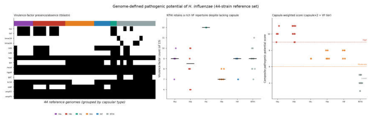
Analysis of 44 reference genomes (Figure 2d, Figure 3) showed that Hi pathogenic potential stratifies by capsular type: types a (score 10.0) and b (10.2) highest (High), c/e/f intermediate (7.0–7.6, Moderate), NTHi lowest (4.2, Lower)—a gradient driven mainly by the presence and type of capsule.
One observation from the virulence-factor profile is that NTHi, though unencapsulated, does not have a low virulence-factor count (mean 9.0). This comparison is severely limited by strain numbers—most capsular types had only a few strains, and Hic only 2 (mean 12.0, not statistically meaningful); between-type differences in virulence-factor count are therefore not quantified and offered only qualitatively. On this basis we frame “high virulence-factor load + no capsule” explicitly as a discussion-level mechanistic hypothesis (see Section 4), not a study conclusion: it suggests NTHi may cause mucosal disease via adhesion (hia/hsf/hmw, hif pili) and immune evasion (IgA protease, lipooligosaccharide modification), but a “high virulence-factor count” does not equal “greater virulence” (weighting, expression and host background were not considered), and there is no corresponding validation in the present cohort. Genomic metadata show NTHi clones are highly diverse (287 strains, 263 with an assigned ST spanning 125 STs), mostly from mucosal specimens, consistent with this hypothesis and usable as background context.
Mapping this back to the clinic as published background (not direct evidence from local strains): pneumonia is NTHi-predominant and HSP enriched for capsular type c, suggesting the two diseases may differ at the type level, with mechanistic implications left to discussion and future work.
3.5. HSP Clinical Subtypes Show No Difference in Hi Detection (After Testing-Bias Adjustment)
Among tested HSP patients, cases were divided by extent of involvement into skin-only (n=291) and composite/systemic (abdominal/joint/renal types and combinations, n=2,305). Nephritis was not set as a separate group: HSP nephritis generally reflects protracted, systemic disease rather than an independent entity and is mechanistically similar to other systemic types, so it was merged into the composite subtype (an a priori judgment based on clinical course, not derived from a statistical difference).
Among tested HSP patients, composite/systemic and skin-only subtypes had similar Hi detection (57.0% vs 57.7%; age- and sex-adjusted OR 0.96; OR 1.01, 95%CI 0.78–1.30, P=0.95 after further adjusting for testing intensity), with no significant subtype difference. This negative result did not depend on how nephritis was grouped—merging into composite, leaving it separate, or excluding it gave ORs of 1.01, 0.86 and 0.89 respectively, all non-significant (Supplementary Table S3).
Of note: a “higher detection in the composite subtype” (OR 1.3) was previously seen under a whole-database denominator, but disappeared once the denominator was restricted to tested patients. This reflects a testing bias: composite/more-severe patients are more likely to be tested, so using the whole population as the denominator inflates their apparent detection rate—illustrating why the denominator definition matters.
Subtype capsular type composition cannot be assessed: among typed HSP Hi+ cases with a defined subtype (all typing routes), 54 were composite/systemic but only 4 were skin-only—a sample size too small for any between-group comparison or compositional inference. We therefore do not report the skin-only subtype’s capsular type distribution in the main text (see Supplementary Material for raw counts without interpretation). This is an “unable to assess” due to limited power, not evidence that the two subtypes have identical capsular type composition.
3.6. Hi Detection and Capsular Type Composition of Pneumonia by Pathogen Background
Subdividing the broad “other respiratory disease” control by pathogen (tested patients): mycoplasma (MP) pneumonia (n=1,382) Hi detection 13.9%, viral pneumonia (n=826) 11.3%, bacterial-only pneumonia (no MP, no virus, n=4,871) 10.1%—all far below HSP’s 57.0%. In capsular type composition, every pneumonia type was NTHi-predominant among the few typed cases (viral pneumonia NTHi 9/13 typed, bacterial-only NTHi 27/40 typed), consistent with “pneumonia overall is NTHi-predominant,” with only HSP skewing toward capsular type c. Typed cases within pathogen subgroups were very few (MP pneumonia only 1, viral pneumonia 13), so subgroup capsular type composition is descriptive only, not for statistical inference. In addition, MP pneumonia (median age 7.3 years) is naturally age-comparable to HSP, and using it as a same-age control the HSP Hi-enrichment conclusion still holds (see 3.1).
4. Discussion
Based on a large clinical database and systematic in-house typing of Hi-positive specimens, this study yields a coherent set of findings: (i) among tested patients, HSP Hi detection is markedly higher than in other respiratory diseases and also higher than in concurrently examined healthy children (12.8%; ORs consistently 7.8–9.8 across multiple age-handling methods and after immune-status adjustment), indicating the enrichment holds against both diseased and healthy comparators; (ii) the capsular type spectra differ—HSP is relatively enriched for encapsulated type c while other respiratory diseases are NTHi-predominant (OR 2.68–3.87, significant under both typing calibers); (iii) in clinically detailed pneumonia cases, types correspond to distinct phenotypes (NTHi younger, higher pneumonia propensity). Analysis of published reference genomes (not validated on local strains) suggests pathogenic potential broadly stratifies by capsule; from this we advance the “high virulence-factor load + no capsule” mechanistic hypothesis for NTHi’s mucosal tendency, but this is limited by very small strain numbers (most types a few strains, Hic only 2; virulence-factor counts qualitative only), and a high count does not equal greater virulence—so it stands as a testable candidate explanation, not a study conclusion. Notably, HSP subtype (skin-only vs composite/systemic) Hi detection did not differ once the denominator was corrected; a previous apparent difference under the whole-database denominator was shown to be a testing-bias artifact, underscoring that denominator definition is critical for such observational associations.
Mechanistically, Hi is a major mucosal colonizer capable of inducing strong mucosal IgA responses; as HSP is an IgA-mediated vasculitis, the candidate pathway “Hi mucosal burden → IgA immune activation → IgA immune-complex deposition” is biologically plausible. Other respiratory diseases (especially younger-age pneumonia) are dominated by mucosally invasive NTHi [17,18,19,20], reflecting a different, directly mucosal-infection route.[21] We emphasize that HSP enrichment for encapsulated type c has large effect size and is significant under both typing calibers, making it the most robust of the type–disease associations; subtype-level compositional differences within HSP could not be assessed owing to limited typed samples.
The IgA-pathway and other mechanistic narratives above are candidate hypotheses consistent with the present data, used to interpret the observed associations rather than claimed as proven.
Several limitations should be noted. First, this is a single-center, observational, cross-sectional study: Hi nucleic-acid detection reflects respiratory carriage or infection, and all associations are reported at the detection level without causal or temporal inference. Second, testing was selective (performed more often in HSP than in respiratory-disease patients); estimates therefore apply to tested patients, although the association remained consistent across age-stratified, matched, testing-intensity-adjusted and immune-restricted analyses. Third, capsular typing was completed for a modest subset (245 cases, 160 at PCR caliber), so subtype-level and pathogen-subgroup compositions are descriptive only. Fourth, the bexA-based assay may miss bexA-variant encapsulated strains [14]; the true proportion of encapsulated strains may be slightly higher than reported, and such misclassification would if anything strengthen the observed HSP enrichment for encapsulated strains. Finally, the genomic analysis used public reference genomes as interpretive background and was not validated on local strains. Finally, species-level resolution of H. influenzae from 16S V3–V4 is inherently limited: this region cannot reliably distinguish H. influenzae from H. haemolyticus, so microbiome-stream assignments are reported at the “H. influenzae / H. haemolyticus clade” level; because capsular typing was performed by PCR independently of 16S and this clade-level ambiguity applies equally across groups, it does not bias the between-group comparisons.
5. Conclusions
Respiratory Hi is highly detected in both pneumonia and HSP in children, but the two diseases differ in capsular type spectrum and type is significantly associated with clinical phenotype; combined with the published genomic pathogenic-potential background, this suggests capsular status may distinguish the respiratory disease outcome of Hi. Using in-house typing on a large clinical sample, this study systematically characterizes Hi capsular types and characterizes the associations among capsular type, disease and clinical phenotype. These findings are associational and based on the study’s typing and clinical data.
As a hypothesis-generating study, the value of this work lies in proposing and clinically supporting the picture of “capsule-dependent disease tropism,” pointing the direction for subsequent research. For the field at large, whole-genome sequencing is a natural extension for characterizing Hi variant strains and local strain lineages, and may further resolve the molecular mechanisms behind capsular types in the future; but we emphasize that the typing distribution and disease associations reported here already stand without such subsequent work. We hope these findings provide new associational evidence and testable mechanistic hypotheses for the clinical understanding of pediatric Hi infection and HSP.
Supplementary Materials
The following supporting information can be downloaded at the website of this paper posted on Preprints.org, Effect-size summaries are provided in the Supplementary Material: Table S1 (tested-only Hi detection, capsular type composition, and subtype differences), Table S2 (multi-method age handling and immune adjustment), Table S3 (typing-caliber, nephritis-grouping, and testing-bias sensitivity analyses), and Table S4 (age- and sex-matched control comparisons by disease severity; source data for Table 2), all with ORs and 95%CIs.
Author Contributions
Jing Hao: specimen collection, data analysis. Hao Chen: specimen collection, detection-method development, data analysis. Xiaotong Wang: specimen collection, data analysis. Deze Li: specimen collection. Chen Shen: conceptualization, drafting of the manuscript, data review and verification. All authors participated in manuscript review, and read and approved the final manuscript.
Funding
This work was supported by the Beijing Science and Technology Nova Program Interdisciplinary Project (grant no. 20230484439).
Institutional Review Board Statement
The study was approved by the Medical Ethics Committee of Beijing Children’s Hospital, Capital Medical University, under three protocols covering its respective components: [2021]-E-196-Y (approved 23 November 2021), [2023]-E-008-R (approved 31 January 2023), and [2022]-E-153-Y-01 (amendment approved 23 May 2023). For the retrospective analyses of de-identified clinical records, the requirement for individual informed consent was waived by the committee; written informed consent was obtained from guardians of the concurrently examined healthy children. All procedures were performed in accordance with the Declaration of Helsinki.
Informed Consent Statement
Not applicable — no individually identifiable data are presented.
Data Availability Statement
The de-identified aggregate data supporting the conclusions are included in the Supplementary Material (Table S1–S4 and Supplementary Notes 1–2). Individual-level clinical records are not publicly available owing to patient-privacy and institutional restrictions, but are available from the corresponding author on reasonable request and with institutional approval. Reference genomes are publicly available from NCBI RefSeq.
Acknowledgments
We thank Haiqing Mou (CapitalBiotech Co., Ltd., Beijing, China) for his contribution to detection-method development.
Conflicts of Interest
The authors declare that they have no competing interests.
References
- Zhu, H.; Wang, A.; Tong, J.; Yuan, L.; Gao, W.; Shi, W.; et al. Nasopharyngeal carriage and antimicrobial susceptibility of Haemophilus influenzae among children younger than 5 years of age in Beijing, China. BMC Microbiol. 2015, 15, 6. [Google Scholar] [CrossRef]
- McMahon, F.; Ware, R.S.; Grimwood, K.; Atack, J.M. Haemophilus influenzae and pneumococci: co-colonization, interactions, cooperation and competition. Pediatr. Pulmonol. 2025, 60(1), e27318. [Google Scholar] [CrossRef]
- Van Eldere, J.; Slack, M.P.E.; Ladhani, S.; Cripps, A.W. Non-typeable Haemophilus influenzae, an under-recognised pathogen. Lancet Infect. Dis. 2014, 14(12), 1281–1292. [Google Scholar] [CrossRef]
- Slack, M.P.E. The evidence for non-typeable Haemophilus influenzae as a causative agent of childhood pneumonia. Pneumonia (Nathan) 2017, 9, 9. [Google Scholar] [CrossRef]
- Pneumonia Etiology Research for Child Health (PERCH) Study Group. Causes of severe pneumonia requiring hospital admission in children without HIV infection from Africa and Asia: the PERCH multi-country case-control study. Lancet 2019, 394(10200), 757–779. [Google Scholar] [CrossRef]
- Slack, M.P.E. Long term impact of conjugate vaccines on Haemophilus influenzae meningitis: narrative review. Microorganisms 2021, 9(5), 886. [Google Scholar] [CrossRef]
- Tsang, R.S.W.; Ulanova, M. The changing epidemiology of invasive Haemophilus influenzae disease: emergence and global presence of serotype a strains that may require a new vaccine for control. Vaccine 2017, 35(33), 4270–4275. [Google Scholar] [CrossRef]
- McTaggart, L.R.; Cronin, K.; Seo, C.Y.; Wilson, S.; Patel, S.N.; Kus, J.V. Increased incidence of invasive Haemophilus influenzae disease driven by non-type b isolates in Ontario, Canada, 2014 to 2018. Microbiol. Spectr. 2021, 9(2), e0080321. [Google Scholar] [CrossRef]
- Xu, L.; Li, Y.; Wu, X. IgA vasculitis update: epidemiology, pathogenesis, and biomarkers. Front Immunol. 2022, 13, 921864. [Google Scholar] [CrossRef]
- Wang, B.; Xiang, W.; Zhu, Z.; Shen, H. The influence of respiratory infections on Henoch-Schönlein purpura in children. BMC Infect. Dis. 2025, 25(1), 118. [Google Scholar] [CrossRef]
- Falla, T.J.; Crook, D.W.; Brophy, L.N.; et al. PCR for capsular typing of Haemophilus influenzae. J. Clin. Microbiol. 1994, 32(10), 2382–2386. [Google Scholar] [CrossRef]
- Maaroufi, Y.; De Bruyne, J.M.; Heymans, C.; Crokaert, F. Real-time PCR for determining capsular serotypes of Haemophilus influenzae. J. Clin. Microbiol. 2007, 45(7), 2305–2308. [Google Scholar] [CrossRef]
- Wroblewski, D.; Halse, T.A.; Hayes, J.; Kohlerschmidt, D.; Musser, K.A. Utilization of a real-time PCR approach for Haemophilus influenzae serotype determination as an alternative to the slide agglutination test. Mol. Cell Probes 2013, 27(2), 86–89. [Google Scholar] [CrossRef]
- Sam, I.C.; Smith, M. Failure to detect capsule gene bexA in Haemophilus influenzae types e and f by real-time PCR due to sequence variation within probe binding sites. J. Med. Microbiol. 2005, 54 Pt 5, 453–455. [Google Scholar] [CrossRef]
- Watts, S.C.; Holt, K.E. hicap: in silico serotyping of the Haemophilus influenzae capsule locus. J. Clin. Microbiol. 2019, 57(6), e00190-19. [Google Scholar] [CrossRef]
- Frankel, C.; Robinson, J.; Khan, S.; Alghounaim, M.; McDonald, J.; Lopez, A.; et al. A Pediatric Investigators Collaborative Network on Infections in Children (PICNIC) multi-centre Canadian descriptive analysis of Haemophilus influenzae bacteremia in children: emerging serotypes. Can. Commun. Dis. Rep. 2023, 49(9), 368–374. [Google Scholar] [CrossRef]
- Dong, Q.; Shi, W.; Cheng, X.; Chen, C.; Meng, Q.; Yao, K.; Qian, S. Widespread of non-typeable Haemophilus influenzae with high genetic diversity after two decades use of Hib vaccine in China. J. Clin. Lab Anal. 2020, 34(4), e23145. [Google Scholar] [CrossRef]
- Wen, S.; Mai, Y.; Chen, X.; Xiao, K.; Lin, Y.; Xu, Z.; et al. Molecular epidemiology and antibiotic resistance analysis of non-typeable Haemophilus influenzae (NTHi) in Guangzhou: a representative city of southern China. Antibiotics 2023, 12(4), 656. [Google Scholar] [CrossRef]
- Yuan, M.; Ma, M.; Jiang, H.; Fan, M.; Sun, Y.; Zhou, B.; et al. Characterization of serotypes and molecular drug resistance patterns of Haemophilus influenzae in Kunming children. Pol. J. Microbiol. 2023, 72(2), 125–131. [Google Scholar] [CrossRef]
- Zhou, Y.; Wang, Y.; Cheng, J.; Zhao, X.; Liang, Y.; Wu, J. Molecular epidemiology and antimicrobial resistance of Haemophilus influenzae in Guiyang, Guizhou, China. Front Public Health 2022, 10, 947051. [Google Scholar] [CrossRef]
- Balta, V.A.; Davis, B.; DeByle, C.K.; Desnoyers, C.; Gotcsik, M.; Westley, B.; et al. Clinical course and outcomes of invasive Haemophilus influenzae infections among children at two hospitals in Alaska. J. Pediatr. 2026, 288, 114799. [Google Scholar] [CrossRef]
Figure 1.
Age- and sex-matched controls stratified by disease spectrum and severity.(a). Within-pair Hi detection for HSP (matched) versus each control (non-pneumonia respiratory infection, non-severe pneumonia, severe pneumonia; healthy controls shown as a dashed line, unmatched and descriptive only, n=39); (b) forest plot of matched HSP-versus-control ORs for Hi detection (log scale, 95%CI). Healthy controls are not included in the forest plot because they were not age-sex matched and serve only as a non-diseased descriptive reference. The more severe the control disease, the lower its own detection and the larger the HSP OR (severe pneumonia 12.3), indicating an HSP-specific enrichment.
Figure 1.
Age- and sex-matched controls stratified by disease spectrum and severity.(a). Within-pair Hi detection for HSP (matched) versus each control (non-pneumonia respiratory infection, non-severe pneumonia, severe pneumonia; healthy controls shown as a dashed line, unmatched and descriptive only, n=39); (b) forest plot of matched HSP-versus-control ORs for Hi detection (log scale, 95%CI). Healthy controls are not included in the forest plot because they were not age-sex matched and serve only as a non-diseased descriptive reference. The more severe the control disease, the lower its own detection and the larger the HSP OR (severe pneumonia 12.3), indicating an HSP-specific enrichment.
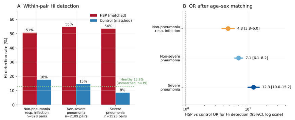
Figure 2.
Disease capsular type spectrum, clinical phenotype and genomic pathogenic potential of respiratory Hi in children. (a) Hi capsular type composition in HSP vs other respiratory diseases (NTHi vs capsular type c; all typing routes Fisher OR=3.87, P<0.001; PCR-positive caliber OR=2.68, P=0.015, see 3.2); (b) age distribution of NTHi vs type c pneumonia patients (box + scatter, Mann–Whitney P<0.001); (c) pneumonia proportion by type (NTHi vs type c OR=0.08, P<0.001); (d) virulence-factor count per capsular type (15 total), color indicating pathogenic-potential tier (High/Moderate/Lower), with NTHi virulence-factor–rich despite lacking a capsule. Numbers above bars indicate reference-genome sample size (type a n=6, b n=10, c n=2, e n=9, f n=5, NTHi n=12). The type c score is based on only 2 strains and should be interpreted with caution; between-type differences are qualitative and do not constitute a study conclusion.
Figure 2.
Disease capsular type spectrum, clinical phenotype and genomic pathogenic potential of respiratory Hi in children. (a) Hi capsular type composition in HSP vs other respiratory diseases (NTHi vs capsular type c; all typing routes Fisher OR=3.87, P<0.001; PCR-positive caliber OR=2.68, P=0.015, see 3.2); (b) age distribution of NTHi vs type c pneumonia patients (box + scatter, Mann–Whitney P<0.001); (c) pneumonia proportion by type (NTHi vs type c OR=0.08, P<0.001); (d) virulence-factor count per capsular type (15 total), color indicating pathogenic-potential tier (High/Moderate/Lower), with NTHi virulence-factor–rich despite lacking a capsule. Numbers above bars indicate reference-genome sample size (type a n=6, b n=10, c n=2, e n=9, f n=5, NTHi n=12). The type c score is based on only 2 strains and should be interpreted with caution; between-type differences are qualitative and do not constitute a study conclusion.
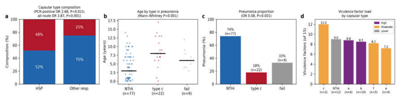
Figure 3.
Reference-genome virulence-factor profile of Haemophilus influenzae capsular types (44 genomes). The in-figure term “genome-defined pathogenic potential” denotes a heuristic, literature-based associational index computed from virulence-factor presence and capsule weighting; it is not a clinically validated measure of pathogenicity. Left: presence/absence matrix of 15 virulence factors (tblastn) grouped by capsular type; middle: virulence-factor count per capsular type, with NTHi retaining a rich repertoire despite lacking a capsule; right: capsule-weighted composite pathogenic-potential score (High/Moderate/Lower tiers). Sample sizes: type a n=6, b n=10, c n=2, e n=9, f n=5, NTHi n=12. Type c (n=2) is underpowered for quantitative comparison; the pathogenic-potential score is a heuristic associational index, not a clinically validated model.
Figure 3.
Reference-genome virulence-factor profile of Haemophilus influenzae capsular types (44 genomes). The in-figure term “genome-defined pathogenic potential” denotes a heuristic, literature-based associational index computed from virulence-factor presence and capsule weighting; it is not a clinically validated measure of pathogenicity. Left: presence/absence matrix of 15 virulence factors (tblastn) grouped by capsular type; middle: virulence-factor count per capsular type, with NTHi retaining a rich repertoire despite lacking a capsule; right: capsule-weighted composite pathogenic-potential score (High/Moderate/Lower tiers). Sample sizes: type a n=6, b n=10, c n=2, e n=9, f n=5, NTHi n=12. Type c (n=2) is underpowered for quantitative comparison; the pathogenic-potential score is a heuristic associational index, not a clinically validated model.
Disclaimer/Publisher’s Note: The statements, opinions and data contained in all publications are solely those of the individual author(s) and contributor(s) and not of MDPI and/or the editor(s). MDPI and/or the editor(s) disclaim responsibility for any injury to people or property resulting from any ideas, methods, instructions or products referred to in the content. |
© 2026 by the authors. Licensee MDPI, Basel, Switzerland. This article is an open access article distributed under the terms and conditions of the Creative Commons Attribution (CC BY) license.
Copyright: This open access article is published under a Creative Commons CC BY 4.0 license, which permit the free download, distribution, and reuse, provided that the author and preprint are cited in any reuse.