

Submitted:
09 November 2026
Posted:
11 November 2026
You are already at the latest version
Abstract
Background: Cell therapy for chronic ischaemic cardiomyopathy remains limited by poor myocardial retention and uncertain regenerative efficacy. We evaluated the effects of delayed intramyocardial transplantation of autologous cardiac atrial stem cells in a large-animal model of established chronic myocardial infarction. Methods: Myocardial infarction was induced in Göttingen minipigs by two-hour left anterior descending coronary artery occlusion. After two months of post-infarction remodeling, animals were randomly allocated to intramyocardial transplantation of autologous cardiac atrial stem cells or control treatment. Cardiac magnetic resonance imaging and three-dimensional electromechanical mapping were performed before treatment and two months thereafter. Histological and immunofluorescence analyses assessed persistence and phenotype of transplanted cells. Results: At follow-up, left ventricular ejection fraction was preserved in treated animals but declined in controls (55 ± 9% versus 41 ± 7%; p = 0.0002). Treatment attenuated adverse ventricular remodeling, with preservation of end-systolic volume and regional wall thickening. Late gadolinium enhancement-defined infarct mass decreased from 6.2 ± 2.0 g to 4.0 ± 1.0 g in treated animals and was significantly lower than in controls at follow-up (p = 0.04). Electromechanical mapping demonstrated improved myocardial electrical viability and regional mechanical function. Transplanted cells persisted within the injured myocardium and exhibited cardiomyocyte-associated structural features. Conclusions: Delayed intramyocardial transplantation of autologous cardiac atrial stem cells attenuated adverse remodeling and was associated with improved regional myocardial structure and function in established chronic ischaemic cardiomyopathy.
Keywords:
cardiac atrial stem cells
; chronic ischaemic cardiomyopathy
; myocardial infarction
; cardiac regeneration
; adverse ventricular remodelling
1. Introduction
Ischemic cardiomyopathy remains a major cause of heart failure and is characterized by progressive left ventricular dysfunction and adverse ventricular remodeling following myocardial infarction. Despite major advances in reperfusion strategies, pharmacological treatment, and device therapy, loss of viable myocardium may initiate a progressive process of ventricular dilatation, increased wall stress, and declining systolic function. Current therapies can slow this progression but do not directly restore the myocardial tissue lost after infarction. Strategies capable of modifying established post-infarction remodeling therefore remain an important therapeutic goal [1,2,3]. The pathophysiology and clinical relevance of adverse post-infarction remodeling remain central to the development of new therapeutic strategies.
Over the past two decades, a wide range of cell populations has been investigated for myocardial repair. Initial clinical studies mainly focused on bone marrow-derived mononuclear cells and reported modest improvements in left ventricular function. However, subsequent randomized trials and meta-analyses demonstrated modest and heterogeneous effects, with limited evidence for consistent long-term modification of ventricular remodeling [4,5,6]. Similar limitations emerged with mesenchymal stromal cells. Although these cells may exert beneficial paracrine and immunomodulatory effects, convincing evidence for durable cardiomyogenic differentiation and structural replacement of injured myocardium has remained limited [7,8,9]. Poor myocardial retention and limited survival after delivery have further restricted the capacity of extracardiac cell populations to achieve sustained structural repair [10].
These observations stimulated interest in resident cardiac progenitor populations, which were considered biologically more closely related to the target organ and potentially more capable of surviving within the myocardial microenvironment. However, the initial enthusiasm surrounding several cardiac-derived cell populations was subsequently tempered by experimental evidence demonstrating limited de novo cardiomyocyte formation. Genetic lineage-tracing studies showed that endogenous c-kit-positive cells contribute minimally to cardiomyocyte renewal after myocardial injury [11]. Likewise, the clinical and experimental experience with cardiosphere-derived cells suggested potential effects on scar characteristics and regional myocardial structure without establishing extensive cardiomyocyte replacement as the predominant mechanism of repair [12,13]. These findings shifted the field away from the expectation of extensive cardiomyocyte replacement toward the broader question of whether cell therapy can durably influence the progression of post-infarction ventricular remodeling.
An important limitation of the experimental literature is that cell therapy has frequently been evaluated during the acute or early post-infarction phase. This environment is biologically distinct from established ischemic cardiomyopathy, in which inflammatory wound healing has evolved into mature scar formation, altered extracellular matrix composition, changes in ventricular geometry, and chronic impairment of regional contractile function [1,3]. In contrast, patients with ischemic heart failure generally present after these structural changes have already developed. Whether a cardiac progenitor cell therapy can durably modify this established disease process when treatment is initiated after chronic post-infarction remodeling has become established therefore remains a clinically relevant question.
Cardiac atrial appendage-derived stem cells (CASCs) represent a distinct population of cardiac progenitor cells isolated from the atrial appendage based on high aldehyde dehydrogenase activity. This approach identifies a CD34-positive, CD45-negative cell population with substantial ex vivo expansion capacity and cardiomyogenic differentiation potential [14]. Previous experimental work further demonstrated myocardial engraftment and acquisition of cardiomyocyte-associated structural characteristics following transplantation [15]. These observations provide a biological rationale for investigating whether CASCs can influence the chronically remodeled myocardium rather than solely the early response to acute ischemic injury.
Accordingly, the present study investigated the effects of intramyocardial transplantation of autologous CASCs in a large-animal model of established chronic ischemic cardiomyopathy. Göttingen minipigs underwent experimental myocardial infarction and were allowed to develop chronic post-infarction remodeling for 2 months before treatment. Using serial cardiac magnetic resonance imaging, electromechanical mapping, and histological analysis, we assessed the effects of CASC transplantation on global and regional left ventricular function, ventricular remodeling, infarct size, and the persistence and structural phenotype of transplanted cells. We hypothesized that CASC transplantation, when administered after the establishment of chronic ischemic cardiomyopathy, could preserve ventricular function and attenuate progressive adverse remodeling.
We previously demonstrated the feasibility and therapeutic potential of CASC transplantation in an acute large-animal myocardial infarction model [15]. The present study was designed to determine whether these effects could be reproduced when treatment was delayed until chronic post-infarction remodeling had become established.
2. Results
2.1. Establishment of Chronic Ischemic Cardiomyopathy and Baseline Characteristics
Following myocardial infarction, animals were allowed to recover for 2 months before treatment to permit the development of established post-infarction ventricular remodeling. At the time of treatment, both groups showed evidence of chronic ischemic myocardial injury, with reduced myocardial viability and impaired regional mechanical function in infarct-related segments. Baseline characteristics at the time of treatment are summarized in Table 1.
There were no relevant differences between the MI-CASC and MI groups in left ventricular function or structural parameters before treatment. The subsequent analyses therefore assessed changes in ventricular function, remodeling, myocardial viability, and infarct size during the 2-month follow-up after CASC transplantation or sham treatment.
2.2. CASC Transplantation Preserved Left Ventricular Systolic Function
At the end of the 2-month follow-up period, left ventricular ejection fraction was preserved in CASC-treated animals, whereas a marked deterioration was observed in untreated MI controls. LVEF in the MI-CASC group was 54 ± 15% before treatment and 55 ± 9% at follow-up. In contrast, LVEF declined from 60 ± 4% to 41 ± 7% in untreated MI animals, resulting in a significant between-group difference at follow-up (p = 0.0002) (Figure 1).
Thus, CASC transplantation prevented the progressive deterioration in global left ventricular systolic function observed during the chronic post-infarction phase in untreated animals.
2.3. CASC Transplantation Preserved Regional Contractile Function and Attenuated Adverse Ventricular Remodeling
Regional myocardial function was evaluated by CMR-derived wall thickening in infarct-related segments. In untreated MI animals, wall thickening declined markedly from 78 ± 29% at baseline to 35 ± 27% at follow-up (p <0.001). In contrast, wall thickening was relatively preserved in CASC-treated animals, decreasing from 84 ± 27% to 67 ± 23% over the same period (p=0.01 between groups at follow-up, Figure 2A). Wall thickening in non-infarct-related segments remained stable during follow-up in both groups.
The preservation of regional contractile function was accompanied by attenuation of adverse ventricular remodeling. In CASC-treated animals, LVESV decreased from 21 ± 4 mL before treatment to 16 ± 5 mL at follow-up, whereas LVESV increased from 16 ± 3 mL to 28 ± 8 mL in untreated MI animals (p = 0.003 between groups at follow-up) (Figure 2B).
Together, these findings indicate that CASC transplantation preserved contractile performance within chronically injured myocardial regions and limited the progression of adverse left ventricular remodeling.
Electromechanical Mapping Demonstrated Improved Myocardial Viability and Regional Mechanical Function.
Three-dimensional electromechanical mapping was used to assess myocardial viability and regional mechanical function in both groups. At the time of treatment, reduced unipolar voltage and low local linear shortening were present within infarct-related LAD perfusion territories, consistent with electrically and mechanically impaired myocardium.
At 2 months after treatment, electromechanical maps showed improved unipolar voltage and local linear shortening in CASC-treated animals, whereas both parameters showed further deterioration in untreated MI animals (Figure 3). These findings were consistent with the CMR-derived preservation of regional contractile function and support a beneficial effect of CASC transplantation on the chronically injured myocardium.
2.4. CASC Transplantation Reduced LGE-Defined Infarct Size
LGE-CMR demonstrated a significant reduction in infarct size in CASC-treated animals over the 2-month follow-up period. Infarct mass decreased from 6.2 ± 2.0 g before treatment to 4.0 ± 1.0 g at follow-up (p = 0.04). In contrast, no significant reduction was observed in untreated MI animals, in which infarct mass changed from 5.5 ± 1.1 g to 6.1 ± 1.8 g (Figure 4).
Thus, the preservation of ventricular function and attenuation of remodeling after CASC transplantation were accompanied by a reduction in LGE-defined infarct burden.
2.5. Exploratory Estimation of Myocardial Regeneration and Functional Cell Retention
Late gadolinium enhancement (LGE) cardiac magnetic resonance imaging was used to assess changes in infarct burden following CASC transplantation. LGE-CMR is a well-established in vivo surrogate for infarct size and, in the chronic setting, predominantly reflects replacement fibrosis and expansion of the extracellular space.[16,17]
Infarct mass decreased by an average of approximately 2.5 g per animal between the pre-treatment and 2-month follow-up examinations.
Because changes in LGE-defined infarct mass cannot be interpreted as a direct measure of de novo cardiomyocyte formation, this analysis was intended only to provide an exploratory order-of-magnitude estimate of the potential contribution of transplanted cells to structural myocardial repair. LGE reflects differences in gadolinium distribution within the extracellular compartment and therefore cannot distinguish newly formed myocardium from changes in replacement fibrosis, extracellular matrix composition, or other forms of structural remodeling.16-18
For the purpose of this deliberately conservative exploratory model, only 20% of the observed reduction in infarct mass was attributed to potential newly formed myocardium. Based on a mean infarct mass reduction of 2.5 g, this corresponds to an estimated 0.5 g of newly formed myocardial tissue. Assuming that 75% of this tissue mass represents cardiomyocytes, the estimated cardiomyocyte mass would be 0.375 g.
Using an assumed average cardiomyocyte mass of 3 × 10⁻⁸ g per cell, this corresponds to approximately 12.5 million cardiomyocyte-equivalent cells. Animals received an average of 83 million CASCs, of which 98% expressed cardiac troponin T at the end of follow-up. Relative to the estimated 81.3 million cardiomyocyte-lineage cells, the calculated contribution of newly formed cardiomyocyte-equivalent tissue corresponds to approximately 15% of the transplanted cell population.
This calculation should be interpreted as an exploratory upper-bound estimate rather than a direct measurement of newly generated cardiomyocytes or cell retention. LGE-CMR cannot distinguish de novo myocardial formation from changes in fibrosis, extracellular matrix composition, or other forms of structural remodeling. Nevertheless, the order of magnitude of this estimate is compatible with the substantial myocardial persistence observed histologically and is broadly consistent with the degree of cell retention previously observed in our acute minipig model.[15]
2.6. Transplanted CASCs Persisted Within the Injured Myocardium and Displayed a Cardiomyocyte-Associated Structural Phenotype
Histological and immunofluorescence analyses performed 2 months after intramyocardial delivery confirmed persistence of transplanted CASCs within the chronically injured myocardium. GFP-positive donor-derived cells were predominantly localized within the border zone between viable and infarcted myocardium (Figure 5A).
Widespread co-expression of GFP and cardiac troponin T was observed, with donor-derived cells displaying an organized sarcomeric cTnT staining pattern (Figure 5B). Quantitative analysis demonstrated that 98 ± 1% of GFP-positive cells expressed cardiac troponin T.
In addition, connexin-43 was detected at interfaces between GFP-positive donor-derived cells and adjacent host cardiomyocytes (Figure 5C). Together, these findings demonstrate long-term persistence of transplanted CASCs within chronically injured myocardium and provide structural evidence consistent with acquisition of a cardiomyocyte-associated phenotype and integration with adjacent host tissue.
3. Discussion
3.1. Principal Findings
The present study shows that intramyocardial transplantation of autologous cardiac atrial appendage-derived stem cells (CASCs), administered after chronic post-infarction remodeling had become established, preserved left ventricular systolic function, attenuated adverse ventricular remodeling, and reduced infarct size in a clinically relevant large-animal model of ischemic cardiomyopathy. Importantly, these effects were observed when treatment was delayed until 2 months after myocardial infarction, distinguishing the present study from our previous work in an acute myocardial infarction model [15]. In addition, transplanted cells persisted within the chronically injured myocardium and acquired structural characteristics consistent with a cardiomyocyte-associated phenotype.
3.2. Cell Therapy in Established Ischemic Cardiomyopathy
The timing of cell delivery is likely to be an important determinant of therapeutic efficacy. The biological environment of the acutely infarcted myocardium differs substantially from that of established ischemic cardiomyopathy. During the early post-infarction phase, inflammation, extracellular matrix turnover, infarct healing, and ventricular remodeling are actively evolving. In contrast, chronic ischemic cardiomyopathy is characterized by mature scar formation, persistent alterations in ventricular geometry, and chronic regional contractile dysfunction [1,3]. These differences are clinically relevant because patients with ischemic heart failure generally present after these structural changes have become established. Whether a cell-based intervention can still modify the progression of ventricular remodeling at this stage therefore represents a more demanding and clinically relevant therapeutic question than intervention during the acute phase after myocardial infarction.
This distinction is clinically relevant. Patients considered for regenerative treatment of ischemic heart failure generally present after myocardial scar formation and adverse ventricular remodeling have already developed. Demonstration of efficacy in this setting therefore addresses a therapeutic question that differs fundamentally from acute cardioprotection or enhancement of early infarct healing. The present findings suggest that the chronically injured myocardium remains amenable to therapeutic modification and that cell-based interventions may retain biological activity after structural remodeling has become established.
3.3. Effects on Ventricular Remodeling
The principal therapeutic effect observed in the present study was a coordinated attenuation of post-infarction ventricular remodeling rather than an isolated change in a single functional parameter. CASC-treated animals showed preservation of left ventricular systolic function together with attenuation of progressive ventricular dilatation and a reduction in infarct size. This pattern is particularly relevant because post-infarction remodeling is a progressive process in which changes in ventricular volume, wall stress, and contractile function are closely interrelated [1].
Interpretation of changes in left ventricular ejection fraction alone can be problematic, as this parameter is influenced by both ventricular loading conditions and changes in chamber geometry. The parallel assessment of ventricular function, volumes, and infarct size by serial cardiac magnetic resonance imaging therefore provides a more comprehensive evaluation of the remodeling process. The concordant preservation of systolic function, limitation of ventricular expansion, and reduction in late gadolinium enhancement supports an effect of CASC transplantation on the progression of chronic post-infarction remodeling.
Importantly, however, a reduction in late gadolinium enhancement cannot by itself be interpreted as direct proof of replacement of scar tissue by newly formed myocardium. Changes in tissue composition, fibrosis, extracellular matrix characteristics, or viable myocardium within the infarct border zone may also influence LGE measurements. The infarct-size findings should therefore be interpreted in conjunction with the functional, electromechanical, and histological observations rather than as isolated evidence of myocardial regeneration.
3.4. Persistence and Structural Phenotype of Transplanted CASCs
A major limitation of many previously investigated cell populations has been poor persistence within the myocardium after delivery [10]. The identification of GFP-positive donor-derived cells 2 months after transplantation demonstrates that CASCs can persist within the chronically injured myocardial environment over the duration of the present study. These cells were predominantly observed within the infarct border zone, a region characterized by the transition between viable and scarred myocardium and of particular importance for post-infarction ventricular mechanics.
The majority of GFP-positive cells expressed cardiac troponin T and displayed an organized staining pattern compatible with sarcomeric organization. In addition, connexin-43 was detected at interfaces between donor-derived cells and adjacent host cardiomyocytes. Collectively, these observations support acquisition of a cardiomyocyte-associated structural phenotype and interaction of transplanted cells with the surrounding myocardial tissue [14,15].
Nevertheless, these findings should not be interpreted as definitive proof that de novo cardiomyocyte formation was solely responsible for the observed functional benefit. Expression of cardiac proteins and structural proximity to host myocardium do not establish mature electromechanical integration or quantify the relative contribution of direct cardiomyogenesis. Paracrine signaling, modulation of extracellular matrix remodeling, effects on local myocardial survival, or a combination of these mechanisms may have contributed to the observed response. The present data therefore support a multifactorial mechanism of action, with long-term cellular persistence and acquisition of cardiac structural characteristics providing a plausible component of the therapeutic effect.
3.5. CASCs in the Context of Cardiac Cell Therapy
The results of the present study should be considered within the broader experience with cardiac cell therapy. Earlier approaches using extracardiac cell populations, particularly bone marrow-derived cells and mesenchymal stromal cells, frequently produced signals of biological activity but generally resulted in modest and inconsistent effects on global ventricular function and adverse remodeling [4,5,6,7,8,9]. Limited myocardial retention and the absence of robust cardiomyogenic differentiation have been proposed as important factors underlying these findings [10,14].
Cardiac-derived cell populations were developed in part to address these limitations by using cells with a closer biological relationship to the target organ. However, experience with different cardiac progenitor populations has also demonstrated that cardiac origin alone does not guarantee extensive cardiomyocyte replacement or durable functional benefit. Genetic lineage-tracing studies have shown that endogenous c-kit-positive cells contribute minimally to cardiomyocyte renewal after myocardial injury [11]. Similarly, studies with cardiosphere-derived cells suggested potentially favourable effects on scar characteristics without establishing extensive cardiomyocyte replacement as the principal mechanism of repair [12,13].
CASCs represent a distinct atrial appendage-derived cell population selected on the basis of high aldehyde dehydrogenase activity. Previous experimental studies demonstrated their cardiac differentiation capacity, myocardial persistence, and therapeutic activity following myocardial injury [14,15]. The present study extends these observations by showing that therapeutic effects can also be observed when treatment is delayed until chronic post-infarction remodeling has developed. The principal advance of the present work therefore lies less in proposing a new mechanism of cardiac regeneration than in demonstrating that a cardiac-derived cell therapy can influence the progression of established ischemic cardiomyopathy in a clinically relevant large-animal model.
3.6. Clinical Implications
Although extrapolation from a preclinical large-animal model to patients requires caution, the present findings have several implications for the further clinical development of cardiac cell therapy. Previous clinical studies with a variety of cell products have generally established the feasibility and acceptable safety of intramyocardial cell delivery, including transendocardial administration in patients with chronic ischemic cardiomyopathy. However, therapeutic effects have remained heterogeneous, emphasizing the importance of the cell product, patient population, timing of treatment, delivery strategy, and selection of appropriate endpoints [19,20,21].
The present results provide a rationale for evaluating CASCs in patients with established ischemic cardiomyopathy rather than restricting regenerative interventions to the acute phase after myocardial infarction. This distinction is clinically important because the intended patient population for cell-based treatment will generally already have developed myocardial scar formation and adverse ventricular remodeling. The delayed-treatment design used in the present study therefore addresses a therapeutic setting that closely resembles the substrate encountered in patients with chronic ischemic heart failure.
The therapeutic objective should not necessarily be defined as complete replacement of the myocardium lost after infarction. A clinically meaningful objective may instead be attenuation of progressive ventricular remodeling and preservation of myocardial function. This concept is supported by previous clinical cell-therapy studies in chronic ischemic cardiomyopathy, in which changes in scar size, ventricular geometry, LV volumes, and functional status have been explored alongside safety endpoints [8,9,21].
Taken together, the present study provides a biological and preclinical efficacy rationale for first-in-human evaluation of CASCs in established ischemic cardiomyopathy. The demonstration of favourable effects in a delayed-treatment, clinically relevant large-animal model, together with the experience gained from previous catheter-based cell-therapy trials in chronic ischemic heart disease, supports the further clinical evaluation of this approach [8,19,20].
3.7. Study Limitations
Several limitations of the present study should be acknowledged. First, the number of animals was necessarily limited, as is inherent to complex large-animal studies. Although the Göttingen minipig provides a relevant model for studying post-infarction ventricular remodeling, the findings cannot be directly extrapolated to patients with ischemic cardiomyopathy.
Second, treatment was administered 2 months after myocardial infarction. This time point was selected on the basis of previous characterization of the adult Göttingen minipig model, in which an 8-week interval after LAD occlusion/reperfusion was associated with established post-infarction left ventricular dysfunction, adverse remodeling, and a chronic heart failure phenotype [22]. More recent serial cardiac magnetic resonance studies have further confirmed the suitability of the Göttingen minipig for modelling post-myocardial infarction heart failure [23]. Nevertheless, although the 2-month interval provides a reproducible model of established chronic post-infarction remodeling, the duration and biological evolution of disease in minipigs cannot fully reproduce the prolonged and heterogeneous course of ischemic cardiomyopathy in patients.
Third, animals were followed for 2 months after cell transplantation. This follow-up period allowed assessment of the evolution of left ventricular function, remodeling, infarct characteristics, and persistence of transplanted cells during a period in which post-infarction remodeling has been shown to be established and relatively stable in minipig models [24]. However, the present study was not designed to determine the longer-term durability of the observed treatment effects. Whether the attenuation of ventricular remodeling observed during the 2-month post-treatment period persists over more prolonged follow-up remains unknown and will require further investigation.
Fourth, although the present study provides evidence of structural integration of transplanted CASCs within the host myocardium, the extent of their functional contribution could not be quantified directly. GFP-positive donor-derived cells persisted within the infarct border zone and expressed cardiac troponin T with an organized sarcomeric pattern. Importantly, connexin-43 was detected at contact sites between donor-derived cells and adjacent host myocardium, providing structural evidence consistent with the formation of intercellular gap junctions and graft–host integration [25]. The concordance between long-term cell persistence, acquisition of a cardiomyocyte-associated phenotype, and connexin-43 expression at the graft–host interface therefore supports a contribution of transplanted CASCs to structural myocardial repair. However, the present study did not directly assess electrical propagation across the graft–host interface or quantify the relative contribution of direct cellular integration and indirect mechanisms, such as paracrine signalling, to the overall therapeutic effect [25,26].
Finally, the study used an autologous cell transplantation strategy. This design was appropriate for evaluating the biological effects of CASCs in the present large-animal model but does not address several practical aspects of future clinical application, including cell manufacturing timelines, patient-to-patient variability, and the feasibility of treatment in different clinical settings.
4. Materials and Methods
4.1. Study Design and Animals
This study evaluated the effects of delayed intramyocardial transplantation of autologous cardiac atrial appendage-derived stem cells (CASCs) in a large-animal model of established chronic ischemic cardiomyopathy. Adult female Göttingen minipigs underwent right atrial appendage harvest for autologous cell isolation followed by experimental myocardial infarction. Animals were allowed to recover for 2 months before treatment to permit the development of established post-infarction ventricular remodeling. At this time point, cardiac magnetic resonance imaging (CMR) and three-dimensional electromechanical mapping (EMM) were performed to characterize the established post-infarction substrate, after which animals received NOGA-guided intramyocardial CASC transplantation or control treatment. Animals were followed for a further 2 months before final evaluation.
The primary experimental timeline is shown in Figure 6.
All animal experiments were approved by the Ethical Committee for Animal Experiments of Hasselt University, Belgium, and were conducted in accordance with the applicable institutional and European regulations governing the use of animals in research. Adult female Göttingen minipigs weighing 17.1 ± 1.6 kg were included.
4.2. Anaesthesia and Perioperative Monitoring
Adult female Göttingen minipigs (17.1 ± 1.6 kg) were used. For surgical procedures, animals were premedicated with intramuscular ketamine (12.5 mg/kg) and midazolam (0.25 mg/kg), followed by intravenous propofol (5 mg). General anaesthesia was maintained with continuous infusions of 1% propofol (10 mL/h) and remifentanil (100 μg/mL at 5 mL/h).
During myocardial infarction induction, continuous ECG monitoring was performed. Myocardial infarction was confirmed by myocardial discoloration, ST-segment elevation, and increased serum troponin I concentrations measured 12 h after coronary occlusion.
Two months after treatment, animals were sacrificed. While under deep general anaesthesia, animals were euthanized by intravenous administration of an overdose of sodium pentobarbital, followed by potassium chloride to induce cardiac arrest. Death was confirmed before removal of the heart for subsequent histological analyses and the hearts were excised for histological and immunofluorescence analyses.
4.3. Right Atrial Appendage Harvest and CASC Isolation
On day 0, approximately 1–2 g of right atrial appendage tissue was harvested through a right mini-thoracotomy for autologous CASC isolation.
CASCs were isolated from atrial appendage tissue based on high aldehyde dehydrogenase activity.
The heart tissue was minced, washed, enzymatically dissociated and stained with aldefluor®. ALDH+ cells expressing CD34, but negative for CD45, were directly flow-sorted in culture medium (X-vivo 15 supplemented with 10% foetal calf serum (FCS) and 2% penicillin-streptomycin (pen-strep)). An average of 30,113±16,205 CASCs were isolated. After isolation, cells were seeded in fibronectin-coated culture plates and expanded in X-vivo 15 medium supplemented with 20% FCS and 2% pen-strep. The medium was changed twice weekly, and cells were re-plated with a density of 5x10³cells/cm2 at 80-85% confluence. After the first passage, serum levels were reduced to 10%.
4.4. Experimental Myocardial Infarction and Development of Chronic Ischemic Cardiomyopathy
Chronic myocardial infarction was induced by a 2-hour snare occlusion of the left anterior descending coronary artery followed by reperfusion, through a median sternotomy. Animals were subsequently allowed to recover for 2 months before treatment. This interval was selected to permit the development of established post-infarction ventricular dysfunction and remodeling rather than to evaluate the effects of CASC transplantation during the acute phase of myocardial injury.
4.5. CASC Labelling and Preparation for Transplantation
At passage 1, cells were infected with a lentivirus-expressing green fluorescent protein (GFP) under the control of the eukaryotic promoter (pRRL-pEF1a-GFP). For the viral production, pRRL-pEF1a-GFP was co-transfected with pMDLg-RRE, pRSV-REV, and pCMVa-VSVG in HEK293-T cells, using EZ lentifect (MellGen laboratories nv). All plasmids were kindly provided by Dr R.C. Hoeben (University Medical Center, Leiden, The Netherlands).
4.6. Baseline Evaluation, Randomization and Treatment Allocation
Two months after myocardial infarction, animals underwent baseline CMR and three-dimensional EMM before treatment. Animals were subsequently randomly allocated to CASC transplantation or control treatment by drawing lots to ensure operator blinding, the CASC suspension and control solution were prepared by a separate investigator in identical opaque syringes. The operator performing the NOGA-guided intramyocardial injections was therefore blinded to treatment allocation.
4.7. NOGA-Guided Intramyocardial CASC Delivery
Three-dimensional electromechanical mapping was performed using the NOGA XP system to assess regional myocardial electrical properties and mechanical function. Unipolar voltage (UPV) was used as an index of myocardial viability, whereas local linear shortening (LLS) was used to assess regional mechanical contraction. Mapping was performed before treatment and at follow-up. Analyses focused on infarct-related and border-zone myocardium. Two months after myocardial infarction, animals underwent electromechanical mapping and were randomized to the MI-CASC or MI group. In the MI-CASC group, autologous CASCs were delivered by NOGA-guided intramyocardial injection into viable but dysfunctional myocardium. Electromechanical mapping was used to identify the infarct border zone and to guide targeted cell delivery. Animals assigned to the MI group underwent injection with medium only, without cells.
4.8. Cardiac Magnetic Resonance Imaging
Cardiac magnetic resonance imaging (CMR) was performed using a 1.5-T Philips Achieva scanner with ECG gating. Balanced steady-state free-precession sequences were used to assess left ventricular volumes and global systolic function. Left ventricular ejection fraction and ventricular volumes were quantified using CAAS MRV software. Regional myocardial contractile function was assessed by calculating wall thickening according to the 17-segment American Heart Association model.
Late gadolinium enhancement (LGE) imaging was performed 15 minutes after intravenous administration of gadobutrol to assess infarct size. All CMR images were analyzed by an investigator blinded to treatment allocation. CMR assessments were performed at predefined time points, immediately before treatment and at the 2-month follow-up.
4.9. Exploratory Estimation of Structural Myocardial Repair
Based on the paired LGE-CMR measurements, an exploratory calculation was performed to provide an order-of-magnitude estimate of the potential contribution of transplanted cells to structural myocardial repair. Because changes in LGE-defined infarct mass cannot directly distinguish de novo myocardial formation from changes in fibrosis, extracellular matrix composition, preservation of pre-existing myocardium, or other forms of structural remodeling, this calculation was not considered a direct measure of cardiomyocyte generation.
For the purpose of this deliberately conservative exploratory model, only 20% of the observed reduction in LGE-defined infarct mass was attributed to potential newly formed myocardium. The resulting tissue mass was further adjusted assuming that 75% represented cardiomyocyte mass. Using an assumed average cardiomyocyte mass of 3 × 10⁻⁸ g per cell, the corresponding number of cardiomyocyte-equivalent cells was calculated and expressed relative to the mean transplanted CASC dose. This analysis was interpreted as an exploratory estimate of potential structural myocardial repair rather than a direct measure of cell retention or de novo cardiomyogenesis.
4.10. Histology and Immunofluorescence
At the end of the 2-month post-treatment follow-up, animals were humanely euthanized and the hearts were excised. Cardiac tissue was sectioned perpendicular to the longitudinal axis, fixed in 4% formalin, and processed for histological and immunofluorescence analyses.
Co-localization of GFP with cardiac troponin T was used to identify donor-derived cells expressing a cardiomyocyte-associated phenotype. The structural organization of cardiac troponin T was examined for evidence of sarcomeric organization. Connexin-43 expression at the interface between GFP-positive donor-derived cells and adjacent host cardiomyocytes was assessed as evidence of structural interaction with the surrounding myocardium. Nuclei were counterstained with DAPI. Sections were examined using an Axioplan 2 fluorescence microscope (Carl Zeiss, Jena, Germany).
4.11. Statistical Analysis
Continuous data are presented as mean ± SD, unless otherwise indicated. Error bars in graphical representations indicate the standard error of the mean (SEM). Normality of continuous variables was assessed using the Shapiro–Wilk test. Depending on data distribution, comparisons between groups were performed using the unpaired Student’s t-test or Mann–Whitney U test, whereas paired measurements obtained at baseline and follow-up were compared using the paired Student’s t-test or Wilcoxon signed-rank test, as appropriate. All tests were two-sided, and a P value <0.05 was considered statistically significant. All statistical analyses were performed using SPSS and GraphPad Prism.
5. Conclusions
In a large-animal model of established chronic ischemic cardiomyopathy, delayed intramyocardial transplantation of autologous cardiac atrial appendage-derived stem cells preserved left ventricular systolic function, attenuated adverse ventricular remodeling, and reduced infarct size. Transplanted cells persisted within the injured myocardium and acquired structural characteristics consistent with cardiomyogenic differentiation and integration with host tissue. These findings extend our previous observations in acute myocardial infarction and demonstrate that CASC therapy can exert beneficial effects when administered after chronic post-infarction remodeling has become established. Collectively, the results provide a strong preclinical rationale for further clinical evaluation of CASCs in patients with established ischemic cardiomyopathy.
Supplementary Materials
.
Author Contributions
Conceptualization, M.H., A.D., L.J., J.-L.R. and K.H.; methodology, M.H., A.D., L.J., J.-L.R. and K.H.; validation, L.J., J.-L.R. and K.H.; formal analysis, M.H., A.D., Y.F., C.D.B., L.J., J.-L.R. and K.H.; investigation, M.H., A.D., Y.F., J.-L.R. and K.H.; resources, M.H.; data curation, C.D.B., J.-L.R. and K.H.; writing—original draft preparation, M.H.; writing—review and editing, A.D., Y.F., C.D.B., L.J., J.-L.R. and K.H.; supervision, M.H. and K.H.; funding acquisition, M.H. All authors have read and agreed to the published version of the manuscript.
Funding
This work was supported by the Limburg Clinical Research Program (LCRP) UHasselt- ZOL-Jessa (SALK 2014N002124); the foundation Limburg Sterk Merk (LSM-124), University Hasselt (UH-12DOC21BOF) and Ziekenhuis Oost-Limburg and Jessa Hospital (IWT135135 and IWT 121560).
Institutional Review Board Statement
The animal study protocol was approved by the Institutional Review Board of BIOMED - UHasselt (protocol code 201218K 03/02/2015 and 201524K 08/06/2015).
Informed Consent Statement
NA.
Data Availability Statement
The data underlying this article will be shared by the corresponding author upon reasonable request.
Acknowledgments
We gratefully thank Stijn Kramer, Department of Biologics Delivery Systems, Johnson & Johnson Medical, the Netherlands, for technical support with the electromechanical NOGA mapping experiments. We also thank Dr E. Bijnens, Department of Radiology, Jessa Hospital, Hasselt, Belgium, for assistance with the interpretation of the cardiac magnetic resonance images. AI-assisted tools were used for language editing and manuscript preparation. All content was reviewed and verified by the authors, who retain full responsibility for the final manuscript.
Conflicts of Interest
Marc Hendrikx is the founder and Chief Executive Officer/Shareholder of CASC8. Caroline de Bouver and Karen Hensen are employees of CASC8. The other authors declare no competing interests.
Abbreviations
| Abbreviation | Definition |
| ALDH | Aldehyde dehydrogenase |
| CASC | Cardiac atrial appendage-derived stem cell |
| CMR | Cardiac magnetic resonance imaging |
| cTnT | Cardiac troponin T |
| DAPI | 4′,6-Diamidino-2-phenylindole |
| ECG | Electrocardiogram |
| EMM | Electromechanical mapping |
| FCS | Fetal calf serum |
| GFP | Green fluorescent protein |
| LAD | Left anterior descending coronary artery |
| LGE | Late gadolinium enhancement |
| LGE-CMR | Late gadolinium enhancement cardiac magnetic resonance imaging |
| LLS | Local linear shortening |
| LV | Left ventricle / left ventricular |
| LVEDV | Left ventricular end-diastolic volume |
| LVEF | Left ventricular ejection fraction |
| LVESV | Left ventricular end-systolic volume |
| MI | Myocardial infarction |
| MI-CASC | Myocardial infarction followed by CASC transplantation |
| NOGA | Non-fluoroscopic three-dimensional electromechanical mapping system |
| NS | Not significant |
| SD | Standard deviation |
| SEM | Standard error of the mean |
| UPV | Unipolar voltage |
References
- Sutton, M.G.; Sharpe, N. Left ventricular remodeling after myocardial infarction: pathophysiology and therapy. Circulation 2000, 101, 2981–2988. [Google Scholar] [CrossRef] [PubMed]
- Burchfield, J.S.; Xie, M.; Hill, J.A. Pathological ventricular remodeling: mechanisms: part 1 of 2. Circulation 2013, 128, 388–400. [Google Scholar] [CrossRef] [PubMed]
- Prabhu, S.D.; Frangogiannis, N.G. The biological basis for cardiac repair after myocardial infarction: from inflammation to fibrosis. Circ. Res. 2016, 119, 91–112. [Google Scholar] [CrossRef] [PubMed]
- Schächinger, V.; Erbs, S.; Elsässer, A.; et al. Intracoronary bone marrow-derived progenitor cells in acute myocardial infarction. N. Engl. J. Med. 2006, 355, 1210–1221. [Google Scholar] [CrossRef] [PubMed]
- Martin-Rendon, E.; Brunskill, S.J.; Hyde, C.J.; Stanworth, S.J.; Mathur, A.; Watt, S.M. Autologous bone marrow stem cells to treat acute myocardial infarction: a systematic review. Eur. Heart J. 2008, 29, 1807–1818. [Google Scholar] [CrossRef] [PubMed]
- Kandala, J.; Upadhyay, G.A.; Pokushalov, E.; Wu, S.; Drachman, D.E.; Singh, J.P. Meta-analysis of stem cell therapy in chronic ischemic cardiomyopathy. Am. J. Cardiol. 2013, 112, 217–225. [Google Scholar] [CrossRef] [PubMed]
- Perin, E.C.; Silva, G.V. Cell-based therapy for chronic ischemic heart disease—a clinical perspective. Cardiovasc. Ther. 2011, 29, 211–217. [Google Scholar] [CrossRef] [PubMed]
- Hare, J.M.; Fishman, J.E.; Gerstenblith, G.; et al. Comparison of allogeneic vs autologous bone marrow-derived mesenchymal stem cells delivered by transendocardial injection in patients with ischemic cardiomyopathy: the POSEIDON randomized trial. JAMA 2012, 308, 2369–2379. [Google Scholar] [CrossRef] [PubMed]
- Heldman, A.W.; DiFede, D.L.; Fishman, J.E.; et al. Transendocardial mesenchymal stem cells and mononuclear bone marrow cells for ischemic cardiomyopathy: the TAC-HFT randomized trial. JAMA 2014, 311, 62–73. [Google Scholar] [CrossRef] [PubMed]
- Hofmann, M.; Wollert, K.C.; Meyer, G.P.; et al. Monitoring of bone marrow cell homing into the infarcted human myocardium. Circulation 2005, 111, 2198–2202. [Google Scholar] [CrossRef] [PubMed]
- van Berlo, J.H.; Kanisicak, O.; Maillet, M.; et al. c-kit+ cells minimally contribute cardiomyocytes to the heart. Nature 2014, 509, 337–341. [Google Scholar] [CrossRef] [PubMed]
- Makkar, R.R.; Smith, R.R.; Cheng, K.; et al. Intracoronary cardiosphere-derived cells for heart regeneration after myocardial infarction (CADUCEUS): a prospective, randomised phase 1 trial. Lancet 2012, 379, 895–904. [Google Scholar] [CrossRef] [PubMed]
- Malliaras, K.; Makkar, R.R.; Smith, R.R.; et al. Intracoronary cardiosphere-derived cells after myocardial infarction: evidence of therapeutic regeneration in the final 1-year results of the CADUCEUS trial. J. Am. Coll. Cardiol. 2014, 63, 110–122. [Google Scholar] [CrossRef] [PubMed]
- Koninckx, R.; Daniëls, A.; Windmolders, S.; et al. The cardiac atrial appendage stem cell: a new and promising candidate for myocardial repair. Cardiovasc. Res. 2013, 97, 413–423. [Google Scholar] [CrossRef] [PubMed]
- Fanton, Y.; Robic, B.; Rummens, J.L.; et al. Cardiac atrial appendage stem cells engraft and differentiate into cardiomyocytes in vivo: a new tool for cardiac repair after MI. Int. J. Cardiol. 2015, 201, 10–19. [Google Scholar] [CrossRef] [PubMed]
- Beijnink, C.W.H.; van der Hoeven, N.W.; Konijnenberg, L.S.F.; et al. Cardiac MRI to visualize myocardial damage after ST-segment elevation myocardial infarction: a review of its histologic validation. Radiology 2021, 301, 4–18. [Google Scholar] [CrossRef] [PubMed]
- Ibanez, B.; Aletras, A.H.; Arai, A.E.; et al. Cardiac MRI endpoints in myocardial infarction experimental and clinical trials: JACC Scientific Expert Panel. J. Am. Coll. Cardiol. 2019, 74, 238–256. [Google Scholar] [CrossRef] [PubMed]
- Puntmann, V.O.; Peker, E.; Chandrashekhar, Y.; Nagel, E. T1 mapping in characterizing myocardial disease: a comprehensive review. Circ. Res. 2016, 119, 277–299. [Google Scholar] [CrossRef] [PubMed]
- Madonna, R.; Van Laake, L.W.; Davidson, S.M.; et al. Position Paper of the European Society of Cardiology Working Group Cellular Biology of the Heart: cell-based therapies for myocardial repair and regeneration in ischemic heart disease and heart failure. Eur. Heart J. 2016, 37, 1789–1798. [Google Scholar] [CrossRef] [PubMed]
- Menasché, P. Cell therapy trials for heart regeneration—lessons learned and future directions. Nat. Rev. Cardiol. 2018, 15, 659–671. [Google Scholar] [CrossRef] [PubMed]
- Mathiasen, A.B.; Qayyum, A.A.; Jørgensen, E.; et al. Bone marrow-derived mesenchymal stromal cell treatment in patients with severe ischaemic heart failure: a randomized placebo-controlled trial (MSC-HF trial). Eur. Heart J. 2015, 36, 1744–1753. [Google Scholar] [CrossRef] [PubMed]
- Schuleri, K.H.; Boyle, A.J.; Centola, M.; et al. The adult Göttingen minipig as a model for chronic heart failure after myocardial infarction: focus on cardiovascular imaging and regenerative therapies. Comp. Med. 2008, 58, 568–579. [Google Scholar] [PubMed]
- Brenner, G.B.; Giricz, Z.; Garamvölgyi, R.; et al. Post-myocardial infarction heart failure in closed-chest coronary occlusion/reperfusion model in Göttingen minipigs and Landrace pigs. J. Vis. Exp. 2021, 170, e61901. [Google Scholar] [CrossRef] [PubMed]
- Lopez, D.; Pan, J.A.; Pollak, P.M.; et al. Multiparametric CMR imaging of infarct remodeling in a percutaneous reperfused Yucatan mini-pig model. NMR Biomed. 2017, 30, e3693. [Google Scholar] [CrossRef] [PubMed]
- Chedrawy, E.G.; Wang, J.S.; Nguyen, D.M.; Shum-Tim, D.; Chiu, R.C.J. Incorporation and integration of implanted myogenic and stem cells into native myocardial fibers: anatomic basis for functional improvements. J. Thorac. Cardiovasc. Surg. 2002, 124, 584–590. [Google Scholar] [CrossRef] [PubMed]
- Shiba, Y.; Fernandes, S.; Zhu, W.Z.; et al. Human ES-cell-derived cardiomyocytes electrically couple and suppress arrhythmias in injured hearts. Nature 2012, 489, 322–325. [Google Scholar] [CrossRef] [PubMed]
Figure 1.
CASC transplantation preserves left ventricular systolic function in chronic ischaemic cardiomyopathy. Left ventricular ejection fraction (LVEF) was assessed by cardiac magnetic resonance imaging before myocardial infarction induction (Pre-MI), after myocardial infarction induction and before treatment (Post-MI), and 2 months after CASC transplantation or control treatment. LVEF remained preserved in the MI-CASC group, whereas a marked reduction was observed in untreated MI-Control animals during the 2-month follow-up period. At final follow-up, LVEF was significantly higher in CASC-treated animals than in MI-Control animals (P = 0.0002). MI-CASC, n = 6; MI-Control, n = 8. Between-group comparison at follow-up was performed using the unpaired Student’s t-test. Data are presented as mean ± SEM. Abbreviations: MI-CASC, myocardial infarction followed by cardiac atrial appendage-derived stem cell transplantation; MI-Control, myocardial infarction without CASC transplantation; LVEF, left ventricular ejection fraction.
Figure 1.
CASC transplantation preserves left ventricular systolic function in chronic ischaemic cardiomyopathy. Left ventricular ejection fraction (LVEF) was assessed by cardiac magnetic resonance imaging before myocardial infarction induction (Pre-MI), after myocardial infarction induction and before treatment (Post-MI), and 2 months after CASC transplantation or control treatment. LVEF remained preserved in the MI-CASC group, whereas a marked reduction was observed in untreated MI-Control animals during the 2-month follow-up period. At final follow-up, LVEF was significantly higher in CASC-treated animals than in MI-Control animals (P = 0.0002). MI-CASC, n = 6; MI-Control, n = 8. Between-group comparison at follow-up was performed using the unpaired Student’s t-test. Data are presented as mean ± SEM. Abbreviations: MI-CASC, myocardial infarction followed by cardiac atrial appendage-derived stem cell transplantation; MI-Control, myocardial infarction without CASC transplantation; LVEF, left ventricular ejection fraction.
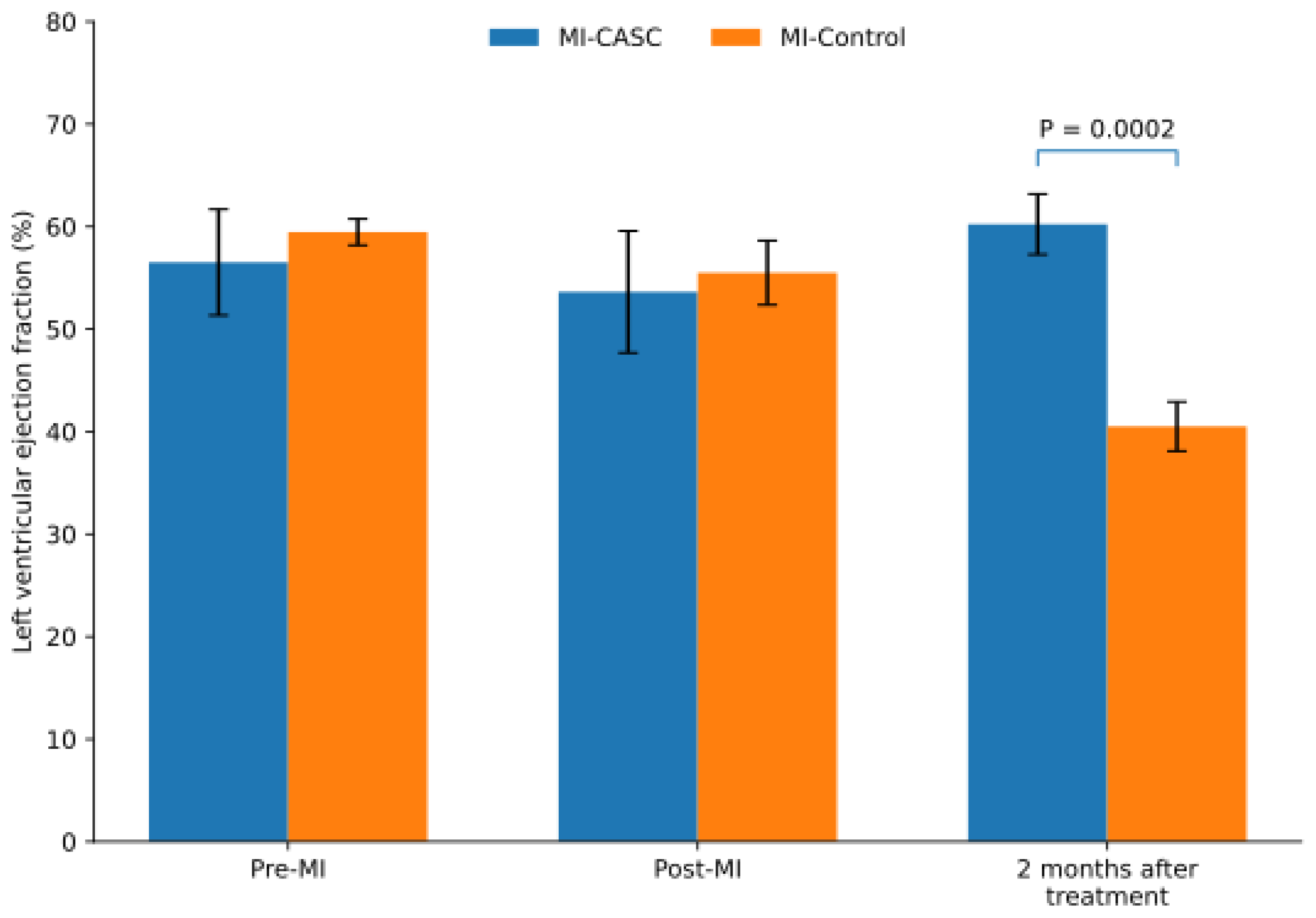
Figure 2.
CASC transplantation preserves left ventricular systolic function in chronic ischemic cardiomyopathy. Left ventricular ejection fraction (LVEF) was assessed by cardiac magnetic resonance imaging before myocardial infarction induction (Pre-MI), after myocardial infarction induction and before treatment (Post-MI), and 2 months after CASC transplantation or control treatment. LVEF remained preserved in the MI-CASC group, whereas a marked reduction was observed in untreated MI-Control animals during the 2-month follow-up period. At final follow-up, LVEF was significantly higher in CASC-treated animals than in MI-Control animals (P = 0.0002). Data are presented as mean ± SD. MI-CASC, n = 6; MI-Control, n = 8. Abbreviations:MI-CASC, myocardial infarction followed by cardiac atrial appendage-derived stem cell transplantation; MI-Control, myocardial infarction without CASC transplantation; LVEF, left ventricular ejection fraction.
Figure 2.
CASC transplantation preserves left ventricular systolic function in chronic ischemic cardiomyopathy. Left ventricular ejection fraction (LVEF) was assessed by cardiac magnetic resonance imaging before myocardial infarction induction (Pre-MI), after myocardial infarction induction and before treatment (Post-MI), and 2 months after CASC transplantation or control treatment. LVEF remained preserved in the MI-CASC group, whereas a marked reduction was observed in untreated MI-Control animals during the 2-month follow-up period. At final follow-up, LVEF was significantly higher in CASC-treated animals than in MI-Control animals (P = 0.0002). Data are presented as mean ± SD. MI-CASC, n = 6; MI-Control, n = 8. Abbreviations:MI-CASC, myocardial infarction followed by cardiac atrial appendage-derived stem cell transplantation; MI-Control, myocardial infarction without CASC transplantation; LVEF, left ventricular ejection fraction.
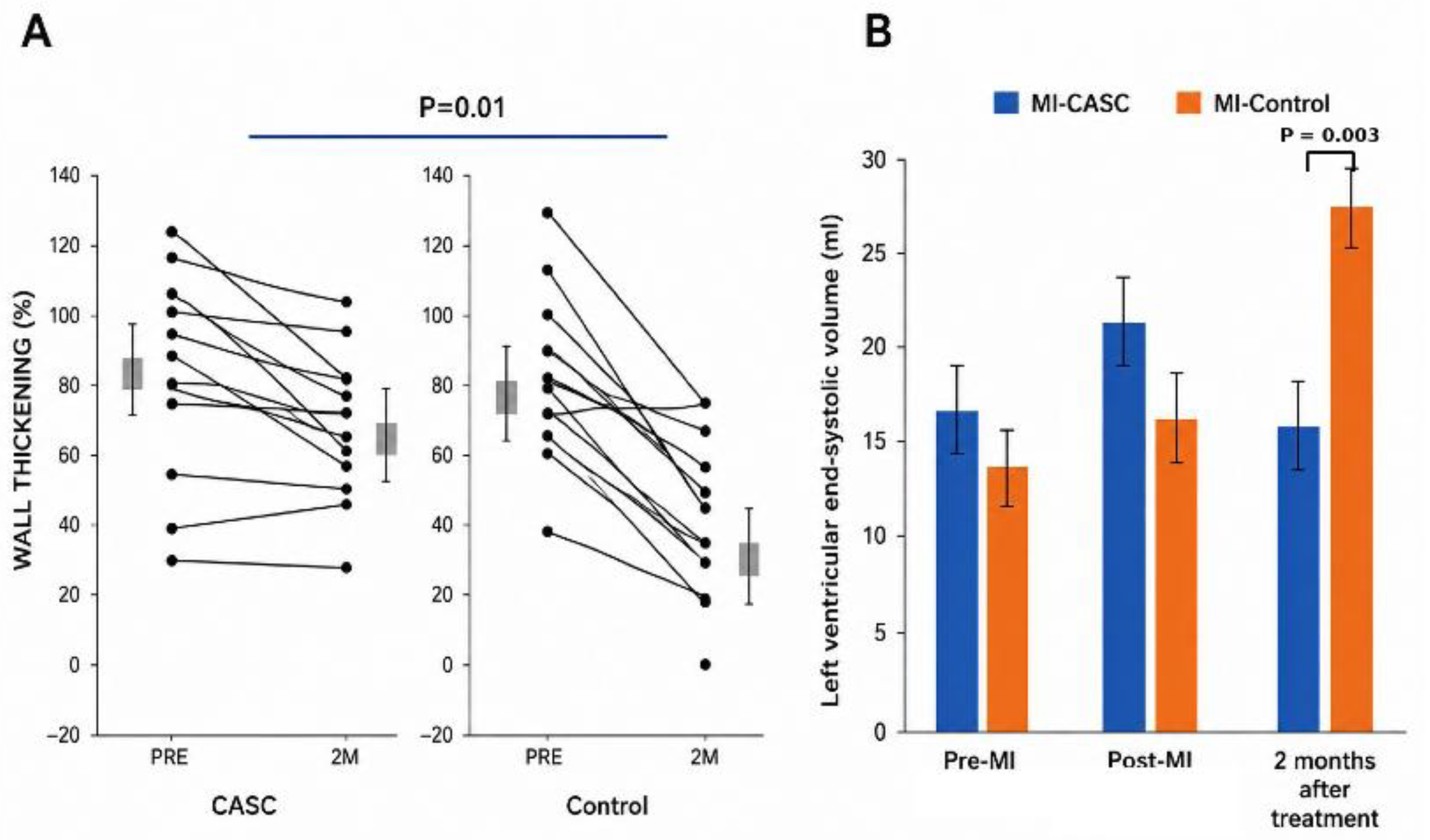
Figure 3.
Electromechanical mapping illustrates improvement in myocardial viability and regional mechanical function following CASC transplantation. Representative three-dimensional electromechanical maps obtained 2 months after myocardial infarction (post-MI) and at 2-month follow-up after treatment. (A) Unipolar voltage map obtained post-MI, demonstrating reduced electrical voltage in the LAD perfusion territory, consistent with chronically injured myocardium. (B) Corresponding local linear shortening (LLS) map obtained post-MI, demonstrating impaired regional mechanical function in the same territory. (C) Unipolar voltage map at 2-month follow-up after CASC transplantation, illustrating improved electrical viability within the previously injured region. (D) Corresponding LLS map at follow-up, illustrating improved regional mechanical function. Abbreviations: CASC, cardiac atrial stem cell; LAD, left anterior descending coronary artery; LLS, local linear shortening; MI, myocardial infarction.
Figure 3.
Electromechanical mapping illustrates improvement in myocardial viability and regional mechanical function following CASC transplantation. Representative three-dimensional electromechanical maps obtained 2 months after myocardial infarction (post-MI) and at 2-month follow-up after treatment. (A) Unipolar voltage map obtained post-MI, demonstrating reduced electrical voltage in the LAD perfusion territory, consistent with chronically injured myocardium. (B) Corresponding local linear shortening (LLS) map obtained post-MI, demonstrating impaired regional mechanical function in the same territory. (C) Unipolar voltage map at 2-month follow-up after CASC transplantation, illustrating improved electrical viability within the previously injured region. (D) Corresponding LLS map at follow-up, illustrating improved regional mechanical function. Abbreviations: CASC, cardiac atrial stem cell; LAD, left anterior descending coronary artery; LLS, local linear shortening; MI, myocardial infarction.
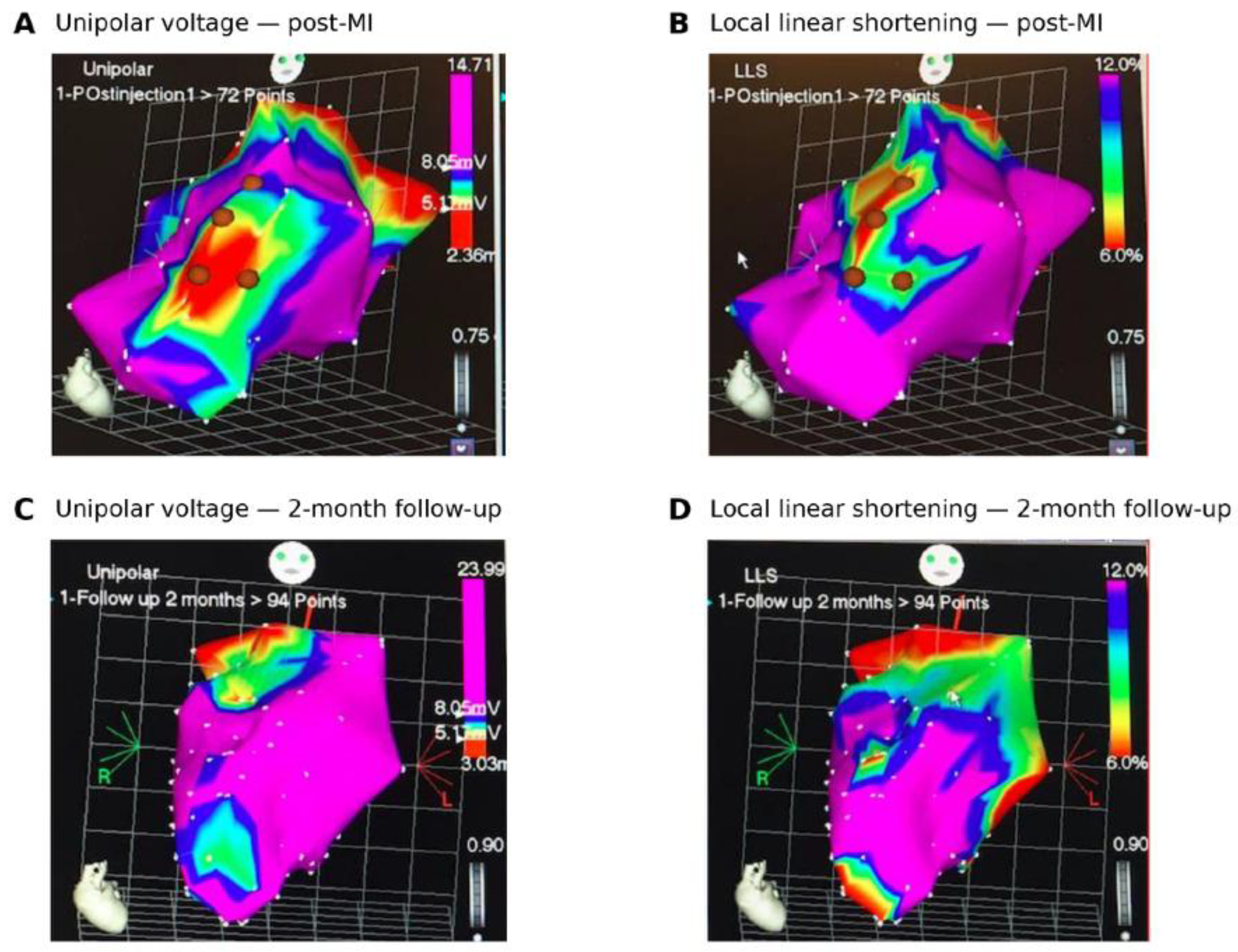
Figure 4.
CASC transplantation is associated with a reduction in LGE-defined infarct mass. (A) Representative late gadolinium enhancement (LGE) cardiac magnetic resonance images obtained after myocardial infarction (MI) and at 2-month follow-up after CASC transplantation/sham (2M post MI). The hyperenhanced area within the left ventricular myocardium, delineated between the endocardial (red dashed line) and epicardial (blue solid line) contours, is reduced at follow-up. (B) Quantitative analysis of LGE-defined infarct mass. Infarct mass decreased in the MI-CASC group from 6.2 ± 2.0 g after MI to 4.0 ± 1.0 g at 2-month follow-up, whereas no reduction was observed in MI-Control animals (5.5 ± 1.1 g to 6.1 ± 1.8 g). At 2-month follow-up, infarct mass was significantly lower in the MI-CASC group than in MI-Control animals (p = 0.04). Bars represent mean ± SEM. MI-CASC, n = 6; MI-Control, n = 8. Abbreviations: CASC, cardiac atrial stem cell; LGE, late gadolinium enhancement; MI, myocardial infarction.
Figure 4.
CASC transplantation is associated with a reduction in LGE-defined infarct mass. (A) Representative late gadolinium enhancement (LGE) cardiac magnetic resonance images obtained after myocardial infarction (MI) and at 2-month follow-up after CASC transplantation/sham (2M post MI). The hyperenhanced area within the left ventricular myocardium, delineated between the endocardial (red dashed line) and epicardial (blue solid line) contours, is reduced at follow-up. (B) Quantitative analysis of LGE-defined infarct mass. Infarct mass decreased in the MI-CASC group from 6.2 ± 2.0 g after MI to 4.0 ± 1.0 g at 2-month follow-up, whereas no reduction was observed in MI-Control animals (5.5 ± 1.1 g to 6.1 ± 1.8 g). At 2-month follow-up, infarct mass was significantly lower in the MI-CASC group than in MI-Control animals (p = 0.04). Bars represent mean ± SEM. MI-CASC, n = 6; MI-Control, n = 8. Abbreviations: CASC, cardiac atrial stem cell; LGE, late gadolinium enhancement; MI, myocardial infarction.
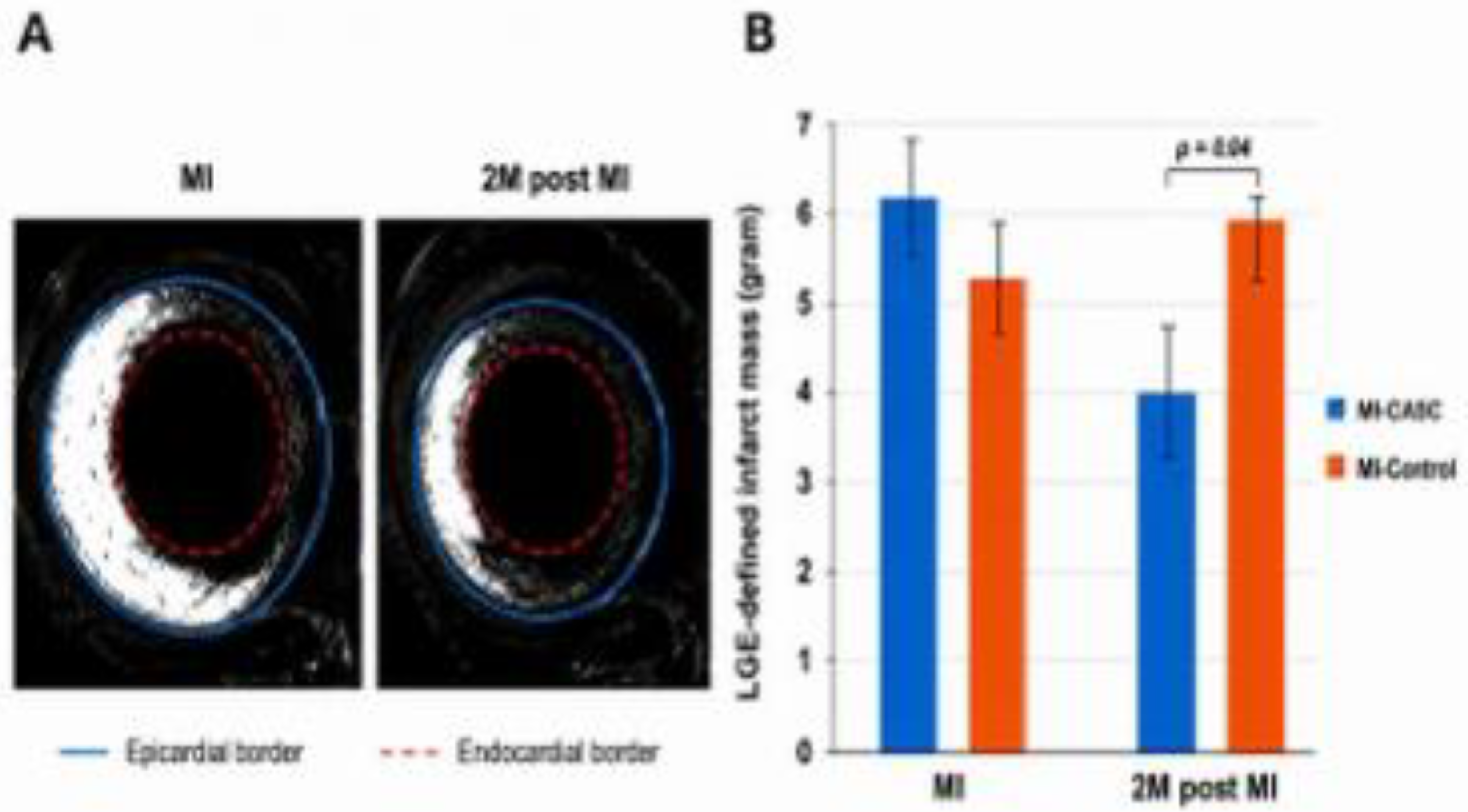
Figure 5.
Transplanted CASCs persist within the chronically injured myocardium and display structural features associated with a cardiomyocyte phenotype. (A) Representative immunofluorescence images of myocardial tissue obtained 2 months after CASC transplantation. GFP-positive transplanted cells are detected within the injured myocardium and co-express cardiac troponin T (cTnT). Individual channels show DAPI-stained nuclei (blue), GFP-positive donor cells (green), and cTnT expression (red), with the merged image demonstrating co-localization of GFP and cTnT. (B) Higher-magnification images illustrating GFP expression, cTnT staining, and the merged image, showing an organized sarcomeric cTnT staining pattern in GFP-positive cells. (C) Representative high-magnification image demonstrating connexin-43 expression (red) at the interface between GFP-positive transplanted cells (green) and adjacent host myocardium. Nuclei are counterstained with DAPI (blue). Abbreviations: CASC, cardiac atrial stem cell; cTnT, cardiac troponin T; DAPI, 4′,6-diamidino-2-phenylindole; GFP, green fluorescent protein.
Figure 5.
Transplanted CASCs persist within the chronically injured myocardium and display structural features associated with a cardiomyocyte phenotype. (A) Representative immunofluorescence images of myocardial tissue obtained 2 months after CASC transplantation. GFP-positive transplanted cells are detected within the injured myocardium and co-express cardiac troponin T (cTnT). Individual channels show DAPI-stained nuclei (blue), GFP-positive donor cells (green), and cTnT expression (red), with the merged image demonstrating co-localization of GFP and cTnT. (B) Higher-magnification images illustrating GFP expression, cTnT staining, and the merged image, showing an organized sarcomeric cTnT staining pattern in GFP-positive cells. (C) Representative high-magnification image demonstrating connexin-43 expression (red) at the interface between GFP-positive transplanted cells (green) and adjacent host myocardium. Nuclei are counterstained with DAPI (blue). Abbreviations: CASC, cardiac atrial stem cell; cTnT, cardiac troponin T; DAPI, 4′,6-diamidino-2-phenylindole; GFP, green fluorescent protein.
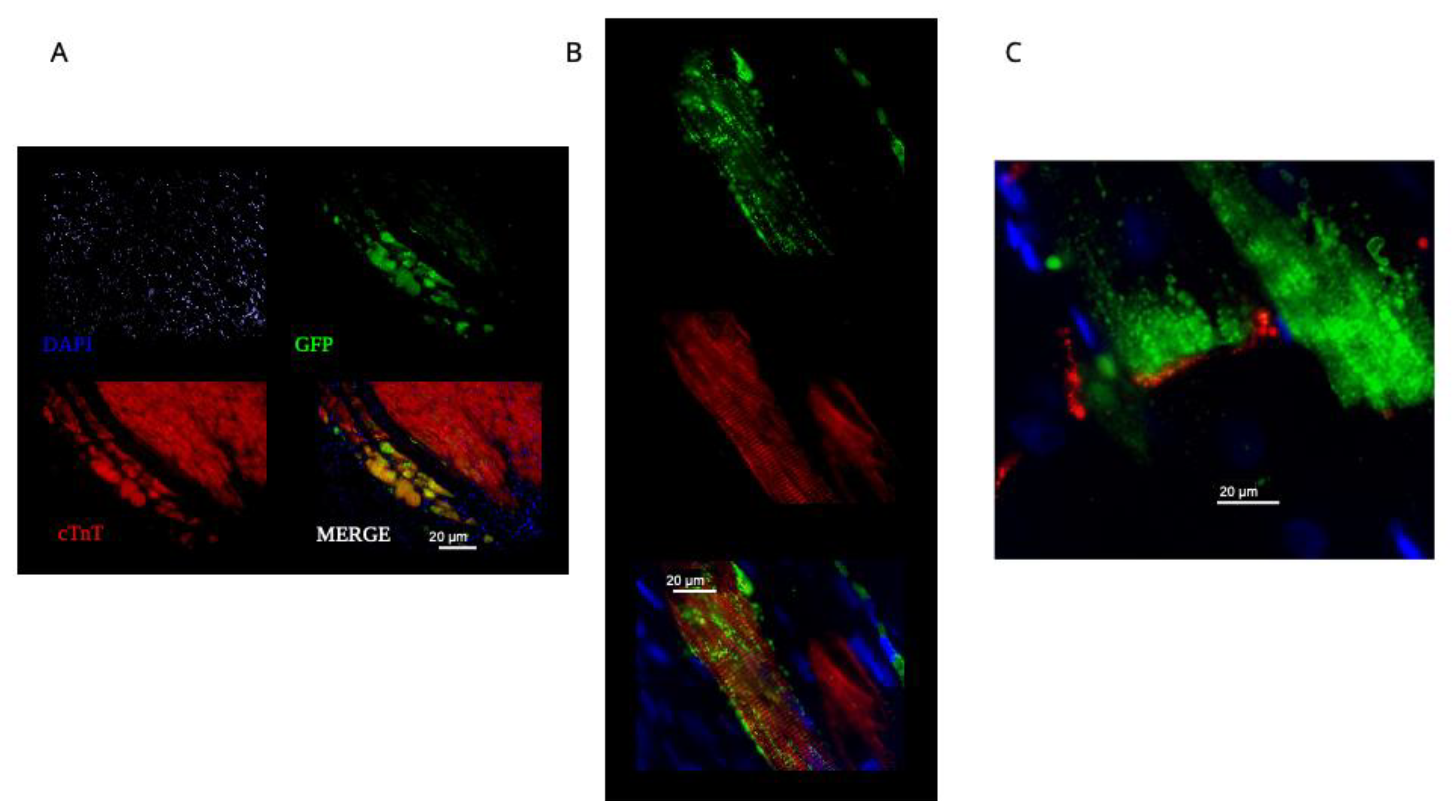
Figure 6.
Study design and experimental timeline. Adult Göttingen minipigs underwent right atrial appendage harvest and induction of myocardial infarction by 2-hour left anterior descending coronary artery occlusion on Day 0. Autologous cardiac atrial stem cells (CASCs) were isolated, expanded and GFP-labelled during the subsequent 2-month recovery period, allowing the development of chronic post-infarction ventricular remodelling. At 2 months post-MI, baseline cardiac magnetic resonance imaging (CMR) and three-dimensional electromechanical mapping (EMM) were performed, followed during the same session by NOGA-guided intramyocardial injection of autologous CASCs or control treatment. Animals were followed for a further 2 months. At 4 months post-MI, CMR and EMM were repeated, followed by euthanasia and tissue collection for histological and immunofluorescence analyses. CMR assessments included left ventricular volumes and ejection fraction, regional wall thickening, and late gadolinium enhancement (LGE)-defined infarct mass. EMM assessed regional myocardial viability using unipolar voltage and regional mechanical function using local linear shortening. Abbreviations: CASC, cardiac atrial stem cell; CMR, cardiac magnetic resonance imaging; EMM, electromechanical mapping; GFP, green fluorescent protein; LAD, left anterior descending coronary artery; LGE, late gadolinium enhancement; MI, myocardial infarction; NOGA, non-fluoroscopic three-dimensional electromechanical mapping system.
Figure 6.
Study design and experimental timeline. Adult Göttingen minipigs underwent right atrial appendage harvest and induction of myocardial infarction by 2-hour left anterior descending coronary artery occlusion on Day 0. Autologous cardiac atrial stem cells (CASCs) were isolated, expanded and GFP-labelled during the subsequent 2-month recovery period, allowing the development of chronic post-infarction ventricular remodelling. At 2 months post-MI, baseline cardiac magnetic resonance imaging (CMR) and three-dimensional electromechanical mapping (EMM) were performed, followed during the same session by NOGA-guided intramyocardial injection of autologous CASCs or control treatment. Animals were followed for a further 2 months. At 4 months post-MI, CMR and EMM were repeated, followed by euthanasia and tissue collection for histological and immunofluorescence analyses. CMR assessments included left ventricular volumes and ejection fraction, regional wall thickening, and late gadolinium enhancement (LGE)-defined infarct mass. EMM assessed regional myocardial viability using unipolar voltage and regional mechanical function using local linear shortening. Abbreviations: CASC, cardiac atrial stem cell; CMR, cardiac magnetic resonance imaging; EMM, electromechanical mapping; GFP, green fluorescent protein; LAD, left anterior descending coronary artery; LGE, late gadolinium enhancement; MI, myocardial infarction; NOGA, non-fluoroscopic three-dimensional electromechanical mapping system.
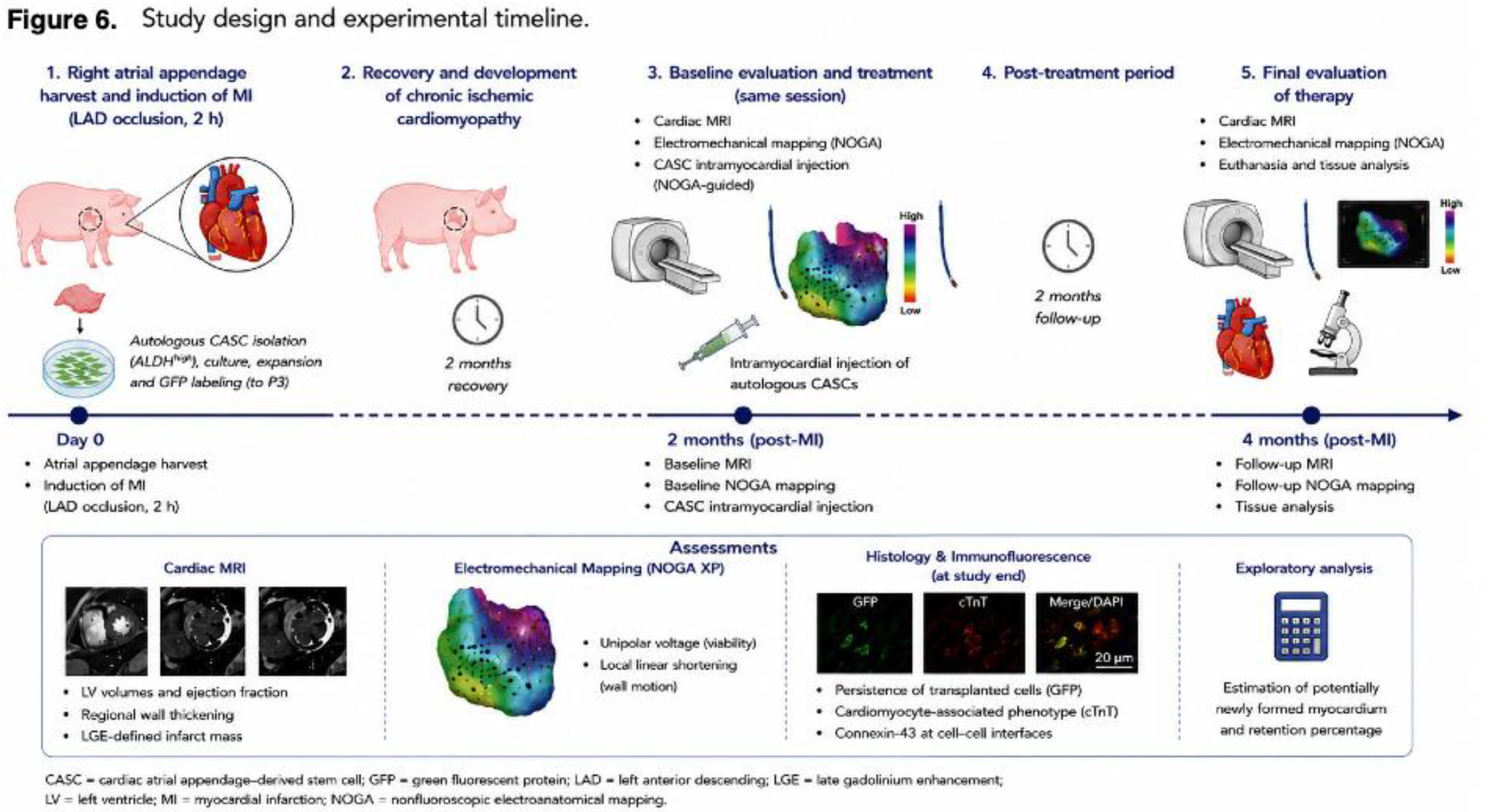

Table 1.
Baseline characteristics at the time of treatment.
| Variable | MI-CASC | MI | P value |
| LVEF, % | 54±15 | 60±4 | NS |
| LVEDV, mL | 48±17 | 36±8 | NS |
| LVESV, mL | 21±4 | 16±3 | NS |
| LGE-defined infarct mass, g | 6.2±2.0 | 5.5±1.1 | NS |
Abbreviations: MI, myocardial infarction; CASC, cardiac atrial appendage-derived stem cell; LVEF, left ventricular ejection fraction; LVEDV, left ventricular end-diastolic volume; LVESV, left ventricular end-systolic volume; LGE, late gadolinium enhancement; NS, not significant. Data are presented as mean ± standard deviation (SD). MI-CASC, n = 6; MI, n = 8.
Disclaimer/Publisher’s Note: The statements, opinions and data contained in all publications are solely those of the individual author(s) and contributor(s) and not of MDPI and/or the editor(s). MDPI and/or the editor(s) disclaim responsibility for any injury to people or property resulting from any ideas, methods, instructions or products referred to in the content. |
© 2026 by the authors. Licensee MDPI, Basel, Switzerland. This article is an open access article distributed under the terms and conditions of the Creative Commons Attribution (CC BY) license (http://creativecommons.org/licenses/by/4.0/).
Copyright: This open access article is published under a Creative Commons CC BY 4.0 license, which permit the free download, distribution, and reuse, provided that the author and preprint are cited in any reuse.