

Submitted:
09 November 2026
Posted:
10 November 2026
You are already at the latest version
Abstract
Background/Objectives: Pseudarthrosis affects 5-35% of patients after lumbar fusion. Anatomical sacral slope (aSS), a recently described spinopelvic parameter measurable without referencing the femoral head, has not been well studied as a pseudarthrosis risk factor. We aimed to identify risk factors for pseudarthrosis after posterior lumbar fusion, with particular focus on aSS and lumbar lordosis (LL).
Methods: We retrospectively analyzed 120 patients who underwent posterior lumbar stabilization for spondylolisthesis or degenerative disease between 2019 and 2024; patients operated on for trauma (n = 9) were excluded to improve cohort homogeneity. Preoperative and postoperative spinopelvic parameters, number of instrumented levels, laminectomy extent, and clinical variables were compared between patients with and without pseudarthrosis, and significant variables were entered into multivariable logistic regression.
Results: Pseudarthrosis occurred in 32.5% (39/120) of patients after a median follow-up of 36 months. Patients with pseudarthrosis were older (p = 0.0012) and had more instrumented levels (p < 0.001); female sex showed a non-significant trend (38.5% vs. 21.4%, p = 0.057), and LL and aSS did not differ between groups. On multivariable analysis, number of instrumented levels (OR 3.44, p < 0.001) was independently associated with pseudarthrosis; female sex showed only a non-significant trend (OR 2.54, p = 0.091) that was further attenuated after covariate adjustment (OR 1.59, p = 0.434). Preoperative aSS was not independently associated with pseudarthrosis. In a sensitivity analysis restricted to radiographic bone non-union alone (28 events), this association was attenuated but remained significant (OR 1.79, p = 0.011).
Conclusions: Number of instrumented levels was the only consistent independent risk factor for pseudarthrosis; female sex showed only a non-significant trend, and preoperative aSS was not independently associated with pseudarthrosis. This association persisted, albeit attenuated, when pseudarthrosis was restricted to bone non-union alone.
Keywords:
lumbar vertebrae
; pseudarthrosis
; spinal fusion
; sacral slope
; risk factors
1. Introduction
Lumbar stabilization surgery is widely used for degenerative, deformity, and traumatic spinal pathologies, and its use has increased alongside advances in technique and instrumentation [1,2]. Pseudarthrosis is among its most common complications, with a reported incidence of 5-35% [3,4], and has been linked to gait disturbance and impaired social and psychological functioning [5]. Established risk factors include age, smoking, osteoporosis, anatomical characteristics, the number of instrumented levels, and dural defects [4,8], but given their complex, multifactorial nature, further investigation may help enable preventive strategies and reduce incidence.
Anatomical sacral slope (aSS), described by Suzuki et al. [6,7], is a recently introduced spinopelvic parameter measurable without referencing the femoral head, which can be difficult to visualize in patients with advanced age, prior hip arthroplasty, or femoral head pathology. We therefore evaluated aSS, together with lumbar lordosis (LL) and preoperative and postoperative clinical characteristics, as potential risk factors for pseudarthrosis after posterior lumbar fusion.
2. Materials and Methods
2.1. Study Design
This retrospective cohort study followed the STROBE reporting guideline for observational studies and was approved by the responsible institutional ethics committee (see Institutional Review Board Statement).
2.2. Patients
We evaluated patients who underwent posterior lumbar stabilization for spondylolisthesis or degenerative spinal disease between 2019 and 2024. Patients were excluded for thoracic extension of instrumentation, interbody cage use, non-degenerative indications (osteoporotic vertebral fracture, tumor, or infection), a bone mineral density T-score below -2.5, inadequate imaging, or incomplete clinical data. Patients operated on for trauma (n = 9) were additionally excluded to reduce population heterogeneity; none of these patients developed pseudarthrosis, so no events were lost. Constructs extending to S1 were retained, since excluding them would have removed 49/120 patients (40.8%) and 28/39 events (71.8%), precluding reliable multivariable analysis. The final cohort comprised 120 patients. Demographics, comorbidities, smoking status, chronic steroid use, body mass index (BMI), bone mineral density T-score (femoral neck dual-energy X-ray absorptiometry), surgical indication, operating surgeon, surgical technique, number of instrumented levels, number of laminectomy levels, graft type and volume, level of pseudarthrosis, LL, aSS, and follow-up duration were recorded. The number of instrumented levels was defined as the number of vertebrae receiving pedicle screw instrumentation (e.g., an L4-L5 construct was classified as 2 instrumented levels).
2.3. Surgical Technique
All procedures used a standardized posterior midline approach with subperiosteal dissection of the paravertebral muscles and fluoroscopically guided transpedicular screw placement; fusion was achieved with autograft, xenograft, or both.
2.4. Radiological Assessment
LL was measured as the angle between the superior endplates of L1 and S1; aSS was measured as the angle between the superior endplate of S1 and a line perpendicular to the anterior pelvic plane, on standing lateral radiographs (Figure 1). Fusion status was assessed on postoperative computed tomography (CT), using the scan closest to each patient’s final follow-up, by an evaluator blinded to clinical and demographic data. Pseudarthrosis was defined as the absence of bridging trabecular bone across the facet joints and posterolateral gutters bilaterally and/or radiographic screw loosening with a peri-screw radiolucent halo. Because this composite definition could in principle be confounded by the number of screws at risk of loosening on imaging (longer constructs carrying more screws), each pseudarthrosis-positive case was additionally classified by its qualifying criterion - bone non-union alone, isolated screw loosening alone, or both - to support a sensitivity analysis restricted to bone non-union (see Section 2.5).
2.5. Statistical Analysis
Categorical variables were compared using the chi-square test or Fisher’s exact test; continuous variables, none of which were normally distributed on Shapiro-Wilk testing, were compared using the Mann-Whitney U test. Age, sex, number of instrumented levels, and preoperative aSS were entered a priori into a multivariable logistic regression model, chosen to maintain an adequate events-per-variable ratio given the 39 observed pseudarthrosis events; a sensitivity model additionally incorporating BMI, bone mineral density T-score, and graft type was also constructed. Linearity of the logit for continuous predictors was assessed using the Box-Tidwell procedure; age showed a significant departure from linearity (p = 0.011) that was not further modeled given its non-significant coefficient in the primary model. A two-sided p value <0.05 was considered statistically significant. To address possible confounding between the composite pseudarthrosis definition and the number of instrumented levels, a sensitivity analysis was performed using a stricter outcome restricted to radiographic bone non-union, excluding the subset of patients whose only qualifying criterion was isolated screw loosening; the primary multivariable model was re-fitted using this stricter outcome definition. All analyses were performed in Python 3 (pandas, statsmodels, scipy, pingouin).
3. Results
A total of 120 patients were included in the study after excluding 9 patients operated on for trauma, none of whom had any comorbidity. Of these, 65.0% (n = 78) were female and 35.0% (n = 42) were male. The median age of the cohort was 51.5 years (IQR 42.0-63.0). Comorbidities were evaluated, and diabetes mellitus (DM) was analyzed separately. A total of 39 patients (32.5%) had at least one comorbidity, while 11 patients (9.2%) had DM.
Regarding surgical indications, 70.8% (n = 85) of patients underwent surgery for lumbar degenerative disease (LDD) and 29.2% (n = 35) for spondylolisthesis. The median follow-up duration was 36 months (IQR 24-36). Pseudarthrosis was identified on postoperative CT in 32.5% of patients (n = 39) (Table 1).
Analysis of preoperative variables revealed no statistically significant differences between patients with and without pseudarthrosis in terms of comorbidities, diabetes mellitus, or surgical indication (Table 2). Female sex showed a non-significant trend toward a higher pseudarthrosis rate than male sex (38.5% [30/78] vs. 21.4% [9/42], p = 0.057). Patients who developed pseudarthrosis had a higher BMI that approached but did not reach statistical significance (28.2 [25.9-30.7] vs. 27.0 [25.2-28.7] kg/m², p = 0.0648) and a significantly lower (more negative) bone mineral density T-score (-0.8 [-1.4 to -0.5] vs. -0.6 [-1.0 to 0.0], p = 0.0128) than those without pseudarthrosis. Graft volume was also significantly greater in patients with pseudarthrosis (25 [20–30] vs. 20 [15–25] mL, p = 0.0002); however, graft volume was strongly correlated with the number of fusion levels (r = 0.87) and is therefore likely to reflect construct length rather than an independent effect of graft amount. Graft type showed a borderline association with pseudarthrosis (p = 0.075), with the highest pseudarthrosis rate observed among patients receiving xenograft alone (50.0%, 10/20) compared with autograft alone (23.7%, 14/59) or combined autograft-xenograft (36.6%, 15/41). Smoking status, chronic steroid use, operating surgeon, and surgical technique were not significantly associated with pseudarthrosis (all p > 0.05).
The median age was 45.0 years (IQR 41.0-61.0) in patients without pseudarthrosis and 59.0 years (IQR 51.0-65.0) in those who developed pseudarthrosis; patients who developed pseudarthrosis were significantly older (p = 0.0012). Additionally, the number of instrumented levels was significantly higher in patients with pseudarthrosis compared to those without pseudarthrosis (p < 0.001) (Table 2).
No statistically significant association was found between pseudarthrosis development and preoperative or postoperative LL or aSS values. When the levels involved by pseudarthrosis were examined, the S1 level was found to be the most frequently involved site. Because a single patient’s construct could span multiple levels, per-level pseudarthrosis rates were calculated relative to the number of constructs including each level and are reported descriptively: L2, 25.0% (6/24); L3, 6.2% (4/65); L4, 1.7% (2/116); L5, 12.3% (14/114); and S1, 55.1% (27/49). The L2 subgroup was small (n = 24 constructs at risk), and these estimates should be interpreted with caution. Although no significant association was observed between the presence of laminectomy and pseudarthrosis development, an increasing number of laminectomy levels was significantly associated with a higher incidence of pseudarthrosis (p < 0.001) (Table 3).
To identify independent risk factors for pseudarthrosis, a multivariable logistic regression model was constructed including age, sex, number of instrumented levels, and preoperative aSS (Table 4). Number of instrumented levels (OR 3.44, 95% CI 1.96-6.03, p < 0.001) was independently associated with pseudarthrosis in this primary model. Female sex showed a non-significant trend toward association (OR 2.54, 95% CI 0.86-7.52, p = 0.091); age (OR 1.02, 95% CI 0.97-1.06, p = 0.454) and preoperative aSS (OR 0.98, 95% CI 0.93-1.03, p = 0.476) were not independently associated with pseudarthrosis. Because the number of laminectomy levels was strongly correlated with the number of instrumented levels (r = 0.67), the two variables were tested separately; when both were entered together, only the number of instrumented levels retained independent significance. As a sensitivity analysis, a second model additionally incorporating BMI, bone mineral density T-score, and graft type (autograft alone vs. any xenograft use) was constructed. In this expanded model, the number of instrumented levels remained the strongest independent predictor of pseudarthrosis, with an even larger effect size (OR 3.90, 95% CI 2.10-7.25, p < 0.001), while the trend toward association with female sex was further attenuated (OR 1.59, 95% CI 0.50-5.02, p = 0.434), and BMI (OR 1.14, 95% CI 0.97-1.35, p = 0.117) and T-score (OR 0.49, 95% CI 0.19-1.25, p = 0.134) showed trends in the expected direction without reaching statistical significance. Given the limited number of events relative to covariates, this expanded model should be regarded as exploratory (Table 4, Panel B).
Because the pseudarthrosis definition combined bone non-union and screw loosening, and because longer constructs inherently carry more screws at risk of showing loosening on imaging, a sensitivity analysis was performed using a stricter, bone-non-union-only definition. Of the 39 pseudarthrosis-positive patients, 7 (17.9%) met only the bone non-union criterion, 11 (28.2%) met only the screw-loosening criterion without bone non-union, and 21 (53.8%) met both. Excluding the 11 patients whose only qualifying criterion was isolated screw loosening (yielding 28 pseudarthrosis events among 120 patients), number of instrumented levels remained independently associated with pseudarthrosis, although the effect size was attenuated relative to the primary model (OR 1.79, 95% CI 1.14-2.79, p = 0.011, versus OR 3.44 in the primary model); age became independently associated with pseudarthrosis in this model (OR 1.05, 95% CI 1.01-1.10, p = 0.024), while female sex (OR 2.03, 95% CI 0.70-5.92, p = 0.194) and preoperative aSS (OR 0.99, 95% CI 0.93-1.04, p = 0.611) remained non-significant (Table 4, Panel C).
4. Discussion
This retrospective cohort study examined risk factors for pseudarthrosis after posterior lumbar fusion, with particular attention to aSS and LL, in addition to conventional demographic and surgical variables. After excluding patients operated on for trauma to improve cohort homogeneity, 120 patients were analyzed, among whom pseudarthrosis was identified in 32.5%, consistent with the 5-35% incidence previously reported for lumbar fusion surgery [3,4].
Previous studies have not consistently identified sex as a significant risk factor for pseudarthrosis [8]. In our cohort, female sex showed a univariate trend toward higher pseudarthrosis risk that did not reach significance (38.5% vs. 21.4%, p = 0.057), and this trend persisted without reaching significance on multivariable analysis (OR 2.54, p = 0.091), attenuating further in a sensitivity model adjusting for BMI, bone mineral density, and graft type (OR 1.59, p = 0.434). This suggests that any association between female sex and pseudarthrosis in our cohort is, at most, a modest trend that may partly reflect confounding by bone quality or body composition rather than a fully independent effect, and warrants confirmation in larger, prospective cohorts.
Age has been more consistently reported as a risk factor for pseudarthrosis, with reduced bone healing capacity proposed as the underlying mechanism; prior studies have proposed age thresholds of 55 and 75 years [9,11]. Patients who developed pseudarthrosis in our cohort were significantly older (median 59.0 vs. 45.0 years, p = 0.0012), but this association did not reach significance on multivariable analysis (OR 1.02, p = 0.454) after adjustment for sex, fusion extent, and aSS. A Box-Tidwell test indicated a significant departure from linearity for age (p = 0.011), raising the possibility of a non-monotonic or threshold-type relationship not captured by a linear term; because age was not independently significant in our model, we did not pursue spline-based modeling, but this warrants further study.
Diabetes mellitus and smoking are established contributors to impaired fusion biology through disrupted bone metabolism and vascularity, respectively [12,13], but we found no significant association for either, likely reflecting our narrow inclusion criteria and limited subgroup sizes. Consistent with reduced bone mineral density and elevated BMI impairing fusion through decreased trabecular bone quality and altered mechanical loading, patients who developed pseudarthrosis had a significantly lower T-score (p = 0.0128) and a non-significantly higher BMI (p = 0.0648), despite osteoporotic patients (T-score <-2.5) having already been excluded. Autograft is generally considered superior to xenograft for fusion, and the pseudarthrosis rate was numerically highest with xenograft alone, though this did not reach significance. We found no association between fusion indication and pseudarthrosis. The number of instrumented levels is a widely accepted risk factor for pseudarthrosis, though the reported threshold varies [4,14]; consistent with this, pseudarthrosis risk increased with both instrumented levels and laminectomy extent in our cohort, and the instrumented-levels association strengthened after excluding trauma patients (OR 3.44, p < 0.001 primary model; OR 3.90, p < 0.001 sensitivity model), representing the most robust and consistent predictor of pseudarthrosis in our cohort. Because our composite pseudarthrosis definition included screw loosening, and because longer constructs carry more screws at risk of showing loosening on imaging, part of this association could in principle reflect ascertainment related to the outcome definition itself rather than a purely biomechanical effect. In a sensitivity analysis restricted to radiographic bone non-union alone, the instrumented-levels association was attenuated but remained statistically significant (OR 1.79, p = 0.011), indicating that this risk factor is not solely an artifact of outcome ascertainment, although the attenuation suggests it may be partly inflated by it.
Sagittal spinopelvic alignment is central to posture and biomechanical loading across fusion segments, and restoring LL relative to pelvic incidence has been linked to improved outcomes after fusion [15,16,17]; aSS, which correlates closely with pelvic incidence but can be measured without referencing the femoral head, was evaluated here as a practical surrogate [6,7]. Prior studies examining spinopelvic parameters and pseudarthrosis have reported inconsistent findings, with some showing no significant association [4,18] and others linking global sagittal imbalance to recurrent pseudarthrosis [10]. In our cohort, neither preoperative nor postoperative LL nor aSS differed significantly between groups, and this null finding for aSS persisted after multivariable adjustment (OR 0.98, p = 0.476), suggesting that, although spinopelvic alignment is important for sagittal balance and functional outcomes, its direct contribution to pseudarthrosis risk may be limited relative to mechanical and surgical factors such as fusion length; given the limited number of events, this should be interpreted as an absence of demonstrable association rather than definitive evidence of no effect.
This retrospective, single-center study has several limitations. To improve homogeneity, patients operated on for trauma were excluded; while this did not reduce the number of pseudarthrosis events, it reduced the overall sample size and likely explains why the sex association seen in our originally analyzed cohort was attenuated to a non-significant trend here. The retrospective design precluded standardized radiological protocols, and the limited number of events (n = 39) constrained the covariates that could be reliably included in the multivariable model, with no a priori power calculation performed. Variability in the interval between surgery and CT-based pseudarthrosis assessment, arising from differing follow-up durations, may have introduced ascertainment variability; reassuringly, follow-up duration itself did not differ significantly between groups (median 30.0 vs. 36.0 months, p = 0.2536), arguing against substantial differential bias. Despite these limitations, our findings are clinically relevant and provide a foundation for larger, prospective studies. Formal inter-observer reliability data for aSS measurement and CT-based fusion assessment were not available for this analysis, as both were performed by a single evaluator; a reliability substudy with independent blinded re-measurement by a second rater in a subsample is in progress.
5. Conclusions
Number of instrumented levels was the only robust, independent risk factor for pseudarthrosis after posterior lumbar fusion in our cohort; preoperative anatomical sacral slope was not independently associated with pseudarthrosis, and female sex showed only a non-significant trend warranting further investigation. This association persisted, albeit attenuated, when pseudarthrosis was restricted to radiographic bone non-union alone, indicating it is not solely an artifact of the composite outcome definition.
Author Contributions
Conceptualization, A.B.; methodology, A.B.; formal analysis, A.B.; data curation, A.B. and N.U.U.; investigation, N.U.U. and G.S.; writing - original draft preparation, A.B.; writing - review and editing, A.B., N.U.U., G.S. and B.E.; supervision, B.E.; validation, B.E. All authors have read and agreed to the published version of the manuscript.
Funding
This research received no external funding.
Institutional Review Board Statement
This study was conducted in accordance with the Declaration of Helsinki and approved by the Harran University Clinical Research Ethics Committee (approval code: HRU/24.11.05; date of approval: 5 August 2024).
Informed Consent Statement
Patient consent was waived due to the retrospective design of this study, which used de-identified data collected during routine clinical care.
Data Availability Statement
The data presented in this study are available on request from the corresponding author due to patient privacy restrictions associated with this retrospective clinical dataset.
Acknowledgments
During the preparation of this manuscript, the authors used Claude (Anthropic) for the purposes of statistical re-analysis and language editing. The authors have reviewed and edited the output and take full responsibility for the content of this publication.
Conflicts of Interest
The authors declare no conflict of interest.
References
- Deyo, R.A.; Mirza, S.K.; Martin, B.I.; Kreuter, W.; Goodman, D.C.; Jarvik, J.G. Trends, major medical complications, and charges associated with surgery for lumbar spinal stenosis in older adults. JAMA 2010, 303, 1259–1265. [Google Scholar] [CrossRef]
- Chun, D.S.; Baker, K.C.; Hsu, W.K. Lumbar pseudarthrosis: a review of current diagnosis and treatment. Neurosurg. Focus 2015, 39, E10. [Google Scholar] [CrossRef]
- Berjano, P.; Langella, F.; Damilano, M.; et al. Fusion rate following extreme lateral lumbar interbody fusion. Eur. Spine J. 2015, 24 (Suppl. 3), 369–371. [Google Scholar] [CrossRef]
- Marques, M.F.; Fiere, V.; Obeid, I.; et al. Pseudarthrosis in adult spine deformity surgery: risk factors and treatment options. Eur. Spine J. 2021, 30, 3225–3232. [Google Scholar] [CrossRef]
- Yamagishi, A.; Sakaura, H.; Ishii, M.; Ohnishi, A.; Ohwada, T. Postoperative loss of lumbar lordosis affects clinical outcomes in patients with pseudoarthrosis after posterior lumbar interbody fusion using cortical bone trajectory screw fixation. Asian Spine J. 2021, 15, 294–300. [Google Scholar] [CrossRef]
- Suzuki, H.; Imai, N.; Nozaki, A.; Hirano, Y.; Endo, N. Anatomical sacral slope, a new pelvic parameter, is associated with lumbar lordosis and pelvic incidence in healthy Japanese women: a retrospective cross-sectional study. J. Orthop. Surg. 2020, 28, 2309499019888809. [Google Scholar] [CrossRef]
- Imai, N.; Suzuki, H.; Sakagami, A.; Hirano, Y.; Endo, N. Correlation of the anatomical sacral slope with pelvic incidence in female patients with developmental hip dysplasia: a retrospective cross-sectional study. J. Orthop. Surg. Res. 2020, 15, 486. [Google Scholar] [CrossRef]
- Boonsirikamchai, W.; Wilartratsami, S.; Ruangchainikom, M.; Korwutthikulrangsri, E.; Tongsai, S.; Luksanapruksa, P. Pseudarthrosis risk factors in lumbar fusion: a systematic review and meta-analysis. BMC Musculoskelet. Disord. 2024, 25, 433. [Google Scholar] [CrossRef]
- Shahzad, H.; Ahmad, M.; Singh, V.K.; et al. Predictive factors of symptomatic lumbar pseudoarthrosis following multilevel primary lumbar fusion. N. Am. Spine Soc. J. 2024, 17, 100302. [Google Scholar] [CrossRef]
- How, N.E.; Street, J.T.; Dvorak, M.F.; et al. Pseudarthrosis in adult and pediatric spinal deformity surgery: a systematic review of the literature and meta-analysis of incidence, characteristics, and risk factors. Neurosurg. Rev. 2019, 42, 319–336. [Google Scholar] [CrossRef]
- Konomi, T.; Yasuda, A.; Fujiyoshi, K.; Yato, Y.; Asazuma, T. Incidences and risk factors for postoperative non-union after posterior lumbar interbody fusion with closed-box titanium spacers. Asian Spine J. 2020, 14, 106–112. [Google Scholar] [CrossRef]
- Wilson, C.; Czernik, P.J.; Elgafy, H.; et al. Diabetes increases risk of lumbar spinal fusion complications: association with altered structure of newly formed bone at the fusion site. JBMR Plus 2024, 8, ziae053. [Google Scholar] [CrossRef]
- Steinmetz, M.P.; O’Toole, J.E.; Harrop, J.S.; et al. Adverse impact of diabetes on spine fusion and patient-reported outcomes: a systematic review and meta-analysis. Spine 2025, 50, E347–E360. [Google Scholar]
- Hofler, R.C.; Swong, K.; Martin, B.; Wemhoff, M.; Jones, G.A. Risk of pseudoarthrosis after spinal fusion: analysis from the Healthcare Cost and Utilization Project. World Neurosurg. 2018, 120, e194–e202. [Google Scholar] [CrossRef]
- Jackson, R.P.; McManus, A.C. Radiographic analysis of sagittal plane alignment and balance in standing volunteers and patients with low back pain matched for age, sex, and size. A prospective controlled clinical study. Spine 1994, 19, 1611–1618. [Google Scholar]
- Pourtaheri, S.; Sharma, A.; Savage, J.; et al. Pelvic retroversion: a compensatory mechanism for lumbar stenosis. J. Neurosurg. Spine 2017, 27, 137–144. [Google Scholar] [CrossRef]
- Legaye, J.; Duval-Beaupère, G.; Hecquet, J.; Marty, C. Pelvic incidence: a fundamental pelvic parameter for three-dimensional regulation of spinal sagittal curves. Eur. Spine J. 1998, 7, 99–103. [Google Scholar] [CrossRef]
- Ko, Y.I.; Ko, M.S.; Bang, C.; et al. Analysis of risk factors affecting lumbosacral pseudoarthrosis in multi-segmental fusion: a CT-based study. Eur. Spine J. 2025, 34, 3978–3987. [Google Scholar] [CrossRef]
Figure 1.
aSS measurement on the patient’s lateral radiograph. PS: Pubic Symphysis; aSS: anatomical sacral slope; L.ASIS: left anterior superior iliac spine; R.ASIS: right anterior superior iliac spine.
Figure 1.
aSS measurement on the patient’s lateral radiograph. PS: Pubic Symphysis; aSS: anatomical sacral slope; L.ASIS: left anterior superior iliac spine; R.ASIS: right anterior superior iliac spine.
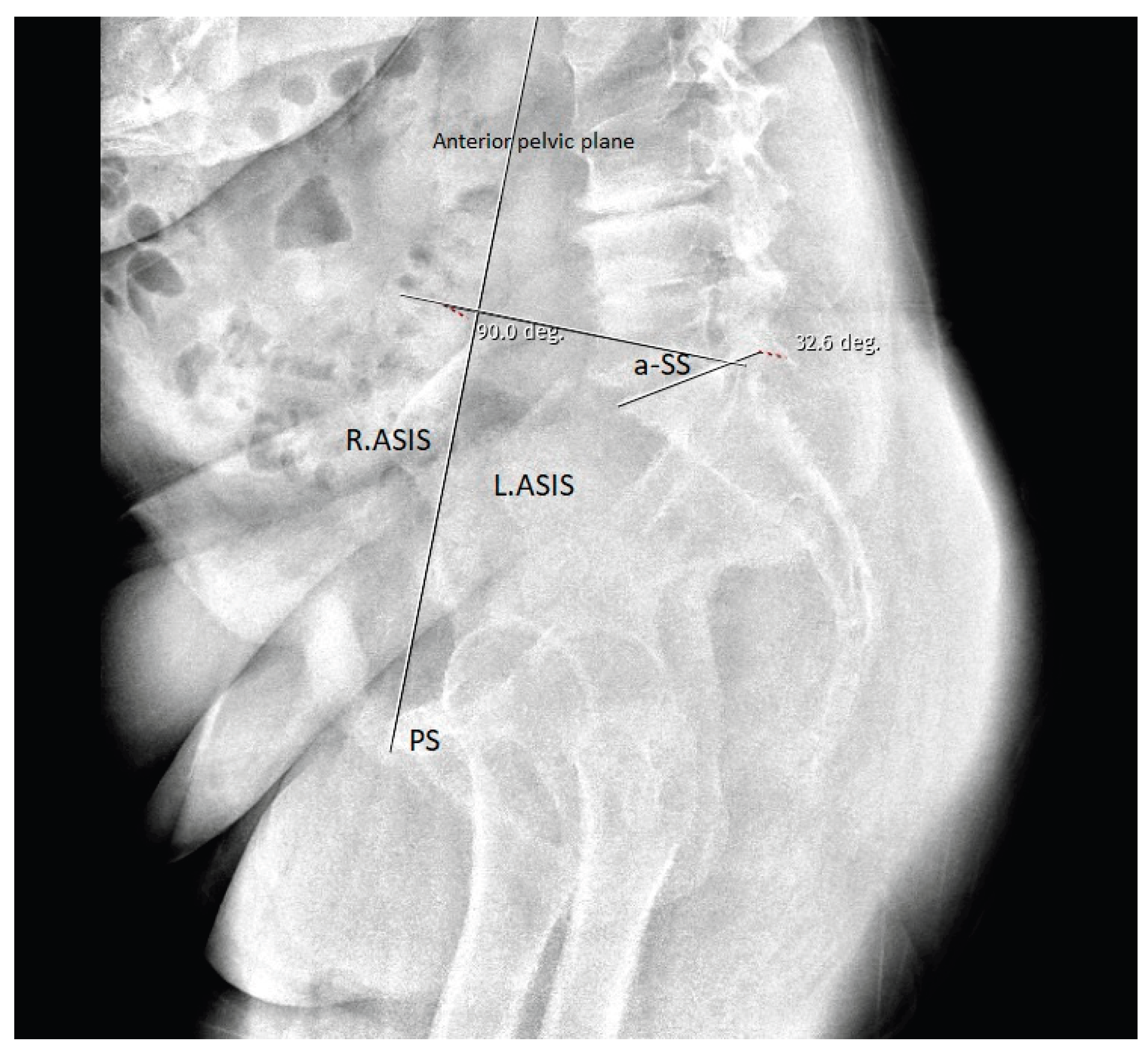

Table 1.
Descriptive Characteristics of the Study Population.
| Variables | n | % | ||
|---|---|---|---|---|
| Sex | Female | 78 | 65.0 | |
| Male | 42 | 35.0 | ||
| Age | Median (25-75 th percentile) | 51.5 (42.0-63.0) | ||
| Comorbidity | Absent Present |
81 39 |
67.5 32.5 |
|
| Diabetes Mellitus (DM) | Absent Present |
109 11 |
90.8 9.2 |
|
| Indication | Lumbar Degenerative Disease (LDD) | 85 | 70.8 | |
| Listhesis | 35 | 29.2 | ||
| Number of instrumented levels | 2 | 41 | 34.2 | |
| 3 | 43 | 35.8 | ||
| >3 | 36 | 30.0 | ||
| Follow-up duration(month) | Median (25-75 p) | 36.0 (24.0-36.0) | ||
| Pseudarthrosis | Absent Present |
81 39 |
67.5 32.5 |
|
|
Number of pseudarthrosis levels| (n=39) |
1 2 3 |
28 8 3 |
71.8 20.5 7.7 |
|
| Pseudarthrosis level | | L2 L3 L4 L5 S1 |
6 4 2 14 27 |
5.0 3.3 1.7 11.7 22.5 |
|
| Laminectomy | Absent Present |
15 105 |
12.5 87.5 |
|
|
Number of laminectomy levels (n=105) |
1 2 3-4 |
55 36 14 |
52.4 34.3 13.3 |
|
| Laminectomy level | L1 L2 L3 L4 L5 |
1 3 39 78 51 |
0.8 2.5 32.5 65.0 42.5 |
|
| BMI (kg/m²) | Median (25-75th percentile) | 27.3 (25.4-29.4) | ||
| Bone Mineral Density T-score | Median (25-75th percentile) | -0.6 (-1.1- -0.2) | ||
| Smoking Status | Never Former Active |
71 (59.2) 26 (21.7) 23 (19.2) |
||
| Chronic Steroid Use | Absent Present |
110 (91.7) 10 (8.3) |
||
| Graft Type | Autograft Autograft + Xenograft Xenograft |
59 (49.2) 41 (34.2) 20 (16.7) |
||
| Graft Volume (mL) | Median (25-75th percentile) | 22.5 (15.0-25.0) | ||
| Operating Surgeon | Surgeon A Surgeon B Surgeon C |
48 (40.0) 43 (35.8) 29 (24.2) |
||
Demographic data, comorbidities, smoking status, chronic steroid use, body mass index (BMI), bone mineral density T-score, surgical indication, operating surgeon, graft type and volume, number of instrumented levels, follow-up duration, pseudarthrosis incidence, pseudarthrosis levels, and laminectomy characteristics of the patients included in the study (n = 120, after excluding 9 patients operated on for trauma, none of whom had any comorbidity). Percentages for number of pseudarthrosis levels per patient (1, 2, or 3) are calculated relative to the number of patients who developed pseudarthrosis (n = 39). Percentages for pseudarthrosis level location (L2-S1) are calculated relative to the total study population (n = 120); because individual patients could have pseudarthrosis at more than one level, these location categories are not mutually exclusive and do not sum to n = 39.
Table 2.
Comparative Analysis of Preoperative Variables in Patients With and Without Pseudarthrosis.
Table 2.
Comparative Analysis of Preoperative Variables in Patients With and Without Pseudarthrosis.
| Pseudarthrosis | Absent n(%) | Present n(%) | p | |
|---|---|---|---|---|
| Sex | Female Male |
48 (59.3) 33 (40.7) |
30 (76.9) 9 (23.1) |
0.057a |
| Age | Median (25th-75th percentile) | 45.0 (41.0-61.0) | 59.0 (51.0-65.0) | 0.0012b |
| Comorbidity | Absent Present |
54 (66.7) 27 (33.3) |
27 (69.2) 12 (30.8) |
0.942a |
| Diabetes Mellitus (DM) | Absent Present |
76 (93.8) 5 (6.2) |
33 (84.6) 6 (15.4) |
0.173c |
| Indication | Lumbar Degenerative Disease (LDD) Listhesis |
57 (70.4) 24 (29.6) |
28 (71.8) 11 (28.2) |
0.872a |
| Number of instrumented levels | 2 3 >3 |
37 (45.7) 31 (38.3) 13 (16.0) |
4 (10.3) 12 (30.8) 23 (59.0) |
<0.001a |
| BMI (kg/m²) | Median (25th-75th percentile) | 27.0 (25.2-28.7) | 28.2 (25.9-30.7) | 0.0648 |
| Bone Mineral Density T-score | Median (25th-75th percentile) | -0.6 (-1.0-0.0) | -0.8 (-1.4- -0.5) | 0.0128 |
| Smoking Status | Never Former Active |
51 (63.0) 18 (22.2) 12 (14.8) |
20 (51.3) 8 (20.5) 11 (28.2) |
0.212 |
| Chronic Steroid Use | Absent Present |
76 (93.8) 5 (6.2) |
34 (87.2) 5 (12.8) |
0.291 |
| Graft Type | Autograft Xenograft Autograft + Xenograft |
45 (55.6) 10 (12.3) 26 (32.1) |
14 (35.9) 10 (25.6) 15 (38.5) |
0.075 |
| Graft Volume (mL) | Median (25th-75th percentile) | 20.0 (15.0-25.0) | 25.0 (20.0-30.0) | 0.0002 |
| Operating Surgeon | Surgeon A Surgeon B Surgeon C |
33 (40.7) 28 (34.6) 20 (24.7) |
15 (38.5) 15 (38.5) 9 (23.1) |
0.917 |
|
a Pearson chi-square test was used. b Mann–Whitney U test was used. c Fisher’s exact test was used. d BMI, body mass index. | ||||
Table 3.
Comparative Analysis of Postoperative Variables in Patients With and Without Pseudarthrosis.
Table 3.
Comparative Analysis of Postoperative Variables in Patients With and Without Pseudarthrosis.
| Pseudarthrosis | Absent n(%) | Present n(%) | P | |
|---|---|---|---|---|
| Preoperative lumbar lordosis angle | Median (25th-75th percentile) | 30.8 (24.3-38.6) | 32.8 (22.1-42.6) | 0.6458a |
| Postoperative lumbar lordosis angle | Median (25th-75th percentile) | 30.0 (25.3-34.0) | 33.2 (22.0-42.0) | 0.7240a |
| Preoperative sacral slope angle | Median (25th-75th percentile) | 35.4 (30.1-40.7) | 33.3 (28.9-41.7) | 0.9019a |
| Postoperative sacral slope angle | Median (25th-75th percentile) | 34.3 (29.7-38.9) | 33.5 (26.0-40.8) | 0.5617a |
| Laminectomy | Absent Present |
12 (14.8) 69 (85.2) |
3 (7.7) 36 (92.3) |
0.381b |
| Number of laminectomy levels | 1 2 3-4 |
44 (63.8) 23 (33.3) 2 (2.9) |
11 (30.6) 13 (36.1) 12 (33.3) |
<0.001c |
| Follow-up duration | Median (25th-75th percentile) | 36.0 (24.0-36.0) | 30.0 (24.0-36.0) | 0.2536a |
|
aMann–Whitney U test was used. bFisher’s exact test was used. cPearson chi-square test was used. | ||||
Table 4.
Multivariable Logistic Regression Analysis of Risk Factors for Pseudarthrosis. Panel A. Primary model (n = 120 patients; 39 pseudarthrosis events). Adjusted odds ratios (OR) with 95% confidence intervals (CI) for pseudarthrosis from a multivariable logistic regression model including age, sex, number of fusion levels, and preoperative anatomical sacral slope (aSS). Panel B. Sensitivity analysis including body mass index, bone mineral density T-score, and graft type (n = 120 patients; 39 pseudarthrosis events). Adjusted odds ratios (OR) with 95% confidence intervals (CI) for pseudarthrosis from an expanded, exploratory multivariable logistic regression model including age, sex, number of instrumented levels, preoperative anatomical sacral slope (aSS), body mass index (BMI), bone mineral density T-score, and graft type. Reference categories: sex, male; graft type, autograft alone (vs. any xenograft use). Panel C. Sensitivity analysis restricted to a stricter, bone-non-union-only pseudarthrosis definition, excluding the 11 patients whose only qualifying criterion was isolated screw loosening without bone non-union (n = 120 patients; 28 pseudarthrosis events). Adjusted odds ratios (OR) with 95% confidence intervals (CI) from a multivariable logistic regression model including the same covariates as Panel A (age, sex, number of fusion levels, and preoperative aSS).
Table 4.
Multivariable Logistic Regression Analysis of Risk Factors for Pseudarthrosis. Panel A. Primary model (n = 120 patients; 39 pseudarthrosis events). Adjusted odds ratios (OR) with 95% confidence intervals (CI) for pseudarthrosis from a multivariable logistic regression model including age, sex, number of fusion levels, and preoperative anatomical sacral slope (aSS). Panel B. Sensitivity analysis including body mass index, bone mineral density T-score, and graft type (n = 120 patients; 39 pseudarthrosis events). Adjusted odds ratios (OR) with 95% confidence intervals (CI) for pseudarthrosis from an expanded, exploratory multivariable logistic regression model including age, sex, number of instrumented levels, preoperative anatomical sacral slope (aSS), body mass index (BMI), bone mineral density T-score, and graft type. Reference categories: sex, male; graft type, autograft alone (vs. any xenograft use). Panel C. Sensitivity analysis restricted to a stricter, bone-non-union-only pseudarthrosis definition, excluding the 11 patients whose only qualifying criterion was isolated screw loosening without bone non-union (n = 120 patients; 28 pseudarthrosis events). Adjusted odds ratios (OR) with 95% confidence intervals (CI) from a multivariable logistic regression model including the same covariates as Panel A (age, sex, number of fusion levels, and preoperative aSS).
| (A) | |||
| Variable | Adjusted OR | 95% CI | p |
| Age (per year) | 1.02 | 0.97–1.06 | 0.454 |
| Sex (female vs. male) | 2.54 | 0.86–7.52 | 0.091 |
| Number of fusion levels | 3.44 | 1.96–6.03 | <0.001 |
| Preoperative aSS (per degree) | 0.98 | 0.93–1.03 | 0.476 |
| Because the number of laminectomy levels was strongly correlated with the number of fusion levels (r = 0.67), these variables were not entered into the model simultaneously; laminectomy extent did not retain independent significance when tested together with fusion levels. | |||
| (B) | |||
| Variable | Adjusted OR | 95% CI | p |
| Age (per year) | 0.98 | 0.93–1.04 | 0.531 |
| Sex (female vs. male) | 1.59 | 0.50–5.02 | 0.434 |
| Number of instrumented levels | 3.90 | 2.10–7.25 | <0.001 |
| Preoperative aSS (per degree) | 0.99 | 0.93–1.04 | 0.616 |
| BMI (per kg/m²) | 1.14 | 0.97–1.35 | 0.117 |
| Bone mineral density T-score | 0.49 | 0.19–1.25 | 0.134 |
| Graft type (any xenograft use vs. autograft alone) | 1.67 | 0.61–4.56 | 0.319 |
| Given the ratio of pseudarthrosis events to covariates in this expanded model (39 events / 7 covariates ≈ 5.6 events per variable), these estimates should be interpreted as exploratory and hypothesis-generating rather than confirmatory; they are not intended to supersede the primary model presented in Panel A. | |||
| (C) | |||
| Variable | Adjusted OR | 95% CI | p |
| Age (per year) | 1.05 | 1.01–1.10 | 0.024 |
| Sex (female vs. male) | 2.03 | 0.70–5.92 | 0.194 |
| Number of fusion levels | 1.79 | 1.14–2.79 | 0.011 |
| Preoperative aSS (per degree) | 0.99 | 0.93–1.04 | 0.611 |
| Because this stricter definition removed the component of the outcome most directly tied to the number of screws in each construct, the attenuation of the instrumented-levels effect (from OR 3.44 in the primary model to OR 1.79 here), while the association remained statistically significant, suggests that part of the primary association may reflect ascertainment related to the composite outcome definition, without fully explaining it. | |||
Disclaimer/Publisher’s Note: The statements, opinions and data contained in all publications are solely those of the individual author(s) and contributor(s) and not of MDPI and/or the editor(s). MDPI and/or the editor(s) disclaim responsibility for any injury to people or property resulting from any ideas, methods, instructions or products referred to in the content. |
© 2026 by the authors. Licensee MDPI, Basel, Switzerland. This article is an open access article distributed under the terms and conditions of the Creative Commons Attribution (CC BY) license (http://creativecommons.org/licenses/by/4.0/).
Copyright: This open access article is published under a Creative Commons CC BY 4.0 license, which permit the free download, distribution, and reuse, provided that the author and preprint are cited in any reuse.