

Submitted:
03 November 2026
Posted:
05 November 2026
You are already at the latest version
Abstract
Background/Objectives: Lateral ankle sprain (LAS) is one of the most common musculoskeletal injuries, and a large proportion of patients go on to develop chronic ankle instability (CAI). Neuromodulation, ranging from transcutaneous to percutaneous techniques, has been proposed as a conservative adjunct for both conditions. This review examined its efficacy in LAS and CAI and compared the available modalities. Methods: We carried out a narrative review with a structured search of PubMed, Scopus, Web of Science and Embase (January–March 2026), reported following PRISMA 2020. Studies published between 2015 and 2026 with levels of evidence 1 to 3 were eligible. Of 511 records identified, 19 studies were included: three systematic reviews, eleven randomized trials, two non-randomized controlled studies, one cohort study and two case series or case-control studies. Results: In LAS (nine studies), results were heterogeneous. Low-frequency stimulation combined with proprioceptive training improved pain, range of motion, strength and joint position sense in one cohort and one randomized trial, and a trial of non-invasive neurostimulation reported less pain and faster return to sport. Two reviews, however, found no added benefit of electrical stimulation in acute sprains. Neuromuscular electrical stimulation (NMES) reduced edema in one pilot trial but not in another study. In CAI (ten studies), six-week NMES programs combined with exercise improved balance, muscle morphology, frontal-plane motor control and patient-reported outcomes, whereas single sessions and two-week programs did not. Percutaneous techniques showed rapid effects in one non-randomized LAS report but only limited acute effects on postural control in CAI. Conclusions: Neuromodulation appears useful as an adjunct to exercise-based rehabilitation rather than as a stand-alone treatment. The evidence is most consistent for NMES combined with exercise in CAI. Standardized protocols and more rigorous trials are required.
Keywords:
lateral ankle sprain
; chronic ankle instability
; neuromodulation
; electrical stimulation
; transcutaneous electrical nerve stimulation
; neuromuscular electrical stimulation
; percutaneous nerve stimulation
; rehabilitation
1. Introduction
Lateral ankle sprains (LAS) are among the most frequent musculoskeletal injuries and a common reason for consultation in podiatry and sports medicine. Most occur during physical activity, and foot inversion is the usual mechanism [1]. The anterior talofibular ligament is the structure most easily damaged, and when the calcaneofibular ligament is also involved the ankle can become considerably unstable [2]. The incidence is estimated at 2–7 cases per 1000 people per year. Although a sprain is often dismissed as a trivial injury that heals on its own, only about one third of patients report full recovery one year later [3].
Initial management has traditionally followed the rest, ice, compression and elevation (RICE) approach. It has more recently been extended into the PEACE & LOVE framework [4]. A second phase of supervised rehabilitation, which begins a few days after the injury depending on severity, aims to restore strength, balance and range of motion (ROM). A final phase is devoted to a gradual return to prior activity and to preventing recurrence [5].
A substantial share of patients does not follow this favorable course. Between 30% and 80% of those with an initial LAS are reported to develop chronic ankle instability (CAI), with repeated episodes of giving way and a persistent sense of dysfunction [6,7]. CAI is not only a mechanical problem. People with CAI show reduced spinal reflex excitability in the affected limb, with fewer alpha motoneurons available for voluntary drive. This has been attributed to a loss of peripheral sensory input after injury, to altered supraspinal sensorimotor control, or to both [7]. Reduced ROM, weakness and impaired neuromuscular control complete the picture [8]. If part of the disability is neural in origin, treatments that act on the nervous system are a reasonable thing to try.
1.1. Anatomy and Mechanism of Injury
The lateral ligament complex joins the fibula to the talus and the calcaneus and provides most of the lateral stability of the ankle. It consists of three ligaments: the anterior talofibular ligament (ATFL), the calcaneofibular ligament (CFL) and the posterior talofibular ligament (PTFL) [1]. The ATFL is a flat, quadrilateral band that runs anteromedially from the anterior border of the lateral malleolus to the body of the talus. The CFL is a stronger, cord-like structure that descends from the malleolus to the lateral surface of the calcaneus and is crossed superficially by the peroneal tendons. The PTFL is the thickest and strongest of the three and is rarely torn in an inversion sprain [2]. On the medial side the deltoid ligament is the main stabilizer, and in chronic instability the anterior and posterior syndesmotic ligaments should not be forgotten either [1].
The most common mechanism of injury is a combination of plantarflexion and inversion, which loads the ATFL first and, when the force continues, the CFL [2]. Severity is graded from I to III. Grade I corresponds to a partial tear with mild tenderness and swelling, little or no functional loss and no mechanical instability. Grade II is an incomplete tear with moderate functional impairment, pain on weight bearing and mild to moderate laxity. Grade III is a complete rupture with loss of ligament integrity, inability to bear weight and mechanical instability [2]. Clinically, the integrity of the ATFL is assessed with the anterior drawer test and that of the CFL with the inversion stress test, comparing the injured side with the healthy one [2]. Imaging, particularly ultrasound, is used to confirm the diagnosis and to guide percutaneous procedures. The peroneus longus and brevis, which lie in the lateral compartment of the leg and are supplied by the superficial peroneal nerve, are the main dynamic restraints against inversion. They are the muscles that most protocols reviewed here chose to stimulate.
1.2. From Sprain to Chronic Instability
CAI is usually defined as a sprain followed by a persistent sensation that the ankle gives way for more than a year after the original injury, with or without pain and swelling. The instability can be mechanical, caused by ligament laxity or rupture that is detectable on examination, or functional, characterized by subjective giving way that may reflect a sensory deficit more than overt laxity. The two forms may coexist or appear independently. CAI is associated with functional impairment, lower activity levels, repeated sprains and, eventually, ankle osteoarthritis [5]. The neuromuscular side of the condition has received growing attention. Reduced strength, impaired neuromuscular control and altered movement patterns are typical [8], and evidence of lowered spinal reflex excitability suggests that part of the deficit lies in how the nervous system handles afferent input from the injured joint [7]. These observations form the rationale for neuromodulation: if the problem is partly one of neural drive, then techniques that change neural excitability may complement the traditional strengthening and balance work.
1.3. Principles of Neuromodulation
According to a widely cited definition, a therapy qualifies as neuromodulation when it is a dynamic, ongoing (or intermittent) intervention rather than a brief single procedure, when it acts on specific neural networks through electrical stimulation or ongoing neuropharmacological action, and when its clinical effect can be adjusted by changing one or more stimulation parameters [9]. Neuromodulation spans invasive and non-invasive techniques. Besides acute changes in neuronal activity, it can produce persistent changes in activity and connectivity. Such neuroplastic changes can be used to reverse maladaptive plasticity, to prevent it, or to reinforce adaptive plasticity [10].
Several mechanisms have been invoked to explain the effects of peripheral electrical stimulation. The gate control theory proposed that activity in large-diameter afferents can close a spinal gate to nociceptive input carried by small fibers. This explains why non-painful electrical stimulation, which recruits large fibers, may reduce pain [11]. Frequency also matters. Experimental work indicates that low-frequency stimulation (around 2 Hz) and high-frequency stimulation (around 100 Hz) release different opioid peptides in the spinal cord, enkephalin and dynorphin respectively, and that alternating both frequencies can produce a stronger analgesic effect than either one alone [12]. At the cellular level, patterns of stimulation can induce long-term potentiation or depression of synaptic transmission, which is the basis of the plasticity-inducing protocols used in percutaneous stimulation [13].
1.4. Peripheral Neuromodulation Modalities
Three modalities appear in the ankle literature. TENS delivers pulsed current through the skin along the trajectory of a nerve. Typical devices allow adjustment of pulse frequency, pulse duration, intensity and pattern. Conventional high-frequency TENS, low-frequency (acupuncture-like) TENS, burst and brief-intense modes can be obtained by combining these parameters. Analgesia arises at local, spinal and supraspinal levels, with low frequencies acting mainly through mu-opioid and other receptors and higher frequencies through delta-opioid receptors [14]. NMES uses stronger, usually biphasic and symmetrical, pulses to depolarize motor nerve branches under the electrodes and elicit a muscle contraction. Its aims are muscle activation, strengthening and maintenance of muscle mass rather than analgesia. PENS places a needle electrode, ideally under ultrasound guidance, next to a target nerve. This allows deeper nerves to be reached with a more precise current distribution. Typical plasticity-inducing protocols use continuous low-frequency stimulation to induce long-term depression and high-frequency bursts to induce long-term potentiation [13]. Data on blood-flow effects and adverse events suggest that percutaneous stimulation is safe in the short term [15], although infection and electrode migration have been reported with peripheral nerve stimulators [16].
1.5. Rationale and Objectives
Despite growing clinical interest, evidence on neuromodulation after ankle sprain is scattered across modalities, populations and outcome measures. Some authors report clear benefits, whereas others recommend against electrical stimulation in the acute phase. This review therefore had three objectives: (1) to evaluate the efficacy of neuromodulation in the management of LAS; (2) to analyze its impact on CAI and on functional recovery; and (3) to compare the different neuromodulation modalities to judge which is most suitable in each condition.
2. Materials and Methods
2.1. Design
We conducted a narrative review with a structured and reproducible literature search. Search and selection were reported following the PRISMA 2020 flow diagram. The included studies differed widely in design, populations, stimulation parameters and outcome instruments, so no meta-analysis was attempted and the findings were synthesized narratively. The review protocol was not registered.
2.2. Search Strategy
Searches were run between January and March 2026 in PubMed (including MeSH terms), Scopus, Web of Science and Embase, through the resources of the Health Sciences Library of the University of Seville. The strategy combined terms for neuromodulation (for example, neuromodulation, electrical stimulation, TENS, NMES, percutaneous nerve stimulation, peripheral nerve stimulation) with terms for the condition (ankle sprain, lateral ankle sprain, lateral ligament injury, ankle instability, chronic ankle instability) using the Boolean operators AND and OR. The strings were adapted to each database; the full versions are given in Appendix A. The first searches returned 511 records: 297 in PubMed, 68 in Scopus, 24 in Web of Science and 122 in Embase.
2.3. Eligibility Criteria
We included studies that (i) were published from 2015 to 2026; (ii) had a level of evidence between 1 and 3, that is, systematic reviews and meta-analyses, randomized controlled trials, cohort studies and case-control studies; (iii) were written in English or Spanish; (iv) had full text available; and (v) addressed neuromodulation in the treatment of LAS or CAI. We excluded studies published before 2015, studies in which neuromodulation was applied to clinical problems unrelated to the foot and lower limb, studies with a level of evidence below 3, and duplicates. Older sources were used only for the theoretical background, and we avoided information that has since been superseded.
2.4. Study Selection
After removing 24 duplicates and 372 records that were not eligible on automated filtering, 115 records were screened against the criteria. Eighty-nine were excluded after reading, and 26 reports were assessed in full. Seven of these were excluded: one had no published results, one was a doctoral thesis and five had a low level of evidence. Nineteen studies were finally included (Figure 1).
2.5. Rationale for the Search Design and Handling of Terminology
Neuromodulation is not a uniformly indexed term in the literature on musculoskeletal rehabilitation. Many authors refer to the same intervention as electrical stimulation, electrotherapy, neurostimulation or peripheral nerve stimulation, and in Spanish-language sources the word itself has only recently become common. For this reason, the four search strings were built around several synonyms and around the specific modalities (TENS, NMES, percutaneous nerve stimulation), and one of them added outcome terms such as pain, range of motion, proprioception and muscle activation. This broad approach increases sensitivity at the cost of precision, which explains why only a small fraction of the 511 initial records reached the eligibility stage. The use of MeSH terms in PubMed helped to filter irrelevant records, and the criteria described above were applied in two steps, first on the basis of titles and abstracts and then on full texts.
Levels of evidence were assigned from the study design. Systematic reviews were classified as Ia, randomized controlled trials as Ib, non-randomized controlled and cohort studies as IIa and IIb, and case series and case-control studies as III. Articles with a level below III were excluded, which is why five reports were removed at the full-text stage. This approach has the advantage of being simple and transparent, but it does not capture the internal quality of each study, a point we return to in the limitations.
2.6. Data Extraction and Synthesis
For each study we recorded authors and year, design, level of evidence, objectives, intervention and main conclusions. Levels of evidence were assigned from the study design (Ia–III). No formal risk-of-bias tool was applied. Studies were split into two groups according to the condition (LAS or CAI), and within each group the findings were organized by outcome and by neuromodulation modality (TENS, NMES and percutaneous techniques).
3. Results
3.1. Characteristics of the Included Studies
The 19 included studies comprised three systematic reviews (one of them a critically appraised paper), eleven randomized controlled trials, two non-randomized controlled studies, one prospective cohort study and two case series or case-control studies. Nine addressed LAS (Table 1) and ten addressed CAI (Table 2). Tables are ordered from the most recent to the oldest publication.
By level of evidence, the LAS group included two Ia sources (the systematic review by Feger et al. and the critically appraised paper by Spooner and Bacon), four Ib randomized trials, one IIb cohort and two level III studies [17,18,19,20,21,22,23,24,25]. The CAI group had one Ia systematic review, six Ib randomized trials and two IIa non-randomized controlled studies [26,27,28,29,30,31,32,33,34,35]. In practice, the strongest sources for LAS were negative on the acute use of electrical stimulation, while the strongest sources for CAI were positive on its combination with exercise. This asymmetry is one of the main reasons why the two conditions are analyzed separately below.
Populations also differed. In LAS, participants were athletes in most trials, with grade I and II sprains being the usual inclusion criterion [17,18,19,21,22]. In CAI, participants were typically young adults, with the exceptions of rock climbers [33] and healthy comparison groups [27,35]. Follow-up was short in nearly every study, ranging from the end of the intervention to a few days, with the 12-month follow-up of Gottlieb et al. being the main exception [31].
3.2. Lateral Ankle Sprain
Pain. Five studies reported benefits, whereas three sources found no advantage over standard care. Zhang et al. [17] and Alahmari et al. [20] reported significant reductions on the visual analogue scale when low-frequency stimulation was combined with proprioceptive training or proprioceptive neuromuscular facilitation. Razzano et al. [22] observed greater pain relief in the treated group, with lower anti-inflammatory use after ten 20-min sessions over two weeks. Shukla and Agrawal [19] and Molsberger and McCaig [23] also described rapid improvements, the latter after as few as three percutaneous sessions. In contrast, the systematic review by Feger et al. [25] found no significant effect of electrical stimulation on pain compared with standard treatment. Wainwright et al. [21] recorded a pain reduction that did not differ from usual care over seven days, and Spooner and Bacon [24] judged the evidence unclear.
Range of motion. Three studies measured ROM, all with a goniometer, and all reported gains. Zhang et al. [17] found additional improvements of 9.2° in dorsiflexion and 6.4° in plantarflexion in martial-arts athletes. Alahmari et al. [20] reported that TENS combined with stretching and facilitation techniques outperformed facilitation alone. Shukla and Agrawal [19] documented better ankle mobility after only five sessions.
Edema. Findings on swelling were the least consistent. Wainwright et al. [21] reported a significant reduction with NMES when swelling was measured by volumetric displacement, but not with tape-measure circumference. Shukla and Agrawal [19] and Molsberger and McCaig [23] also reported reductions, the latter within one to three hours of treatment. Mazloum et al. [18] found that neither NMES nor Kinesio Taping changed ankle volume or perimetry in athletes. Feger et al. [25] and Spooner and Bacon [24] likewise found no benefit, and the latter suggested that late application of the current might explain the null results of the studies they reviewed.
Function and return to sport. Studies used very different functional measures, including walking-disability scales, electromyography, foot and ankle ability scales, and jump, agility and squat tests. Razzano et al. [22] found better walking ability, and Zhang et al. [17], Shukla and Agrawal [19] and Alahmari et al. [20] also reported functional gains. Feger et al. [25], Spooner and Bacon [24] and Mazloum et al. [18] found no functional advantage of adding NMES to conventional care. Return to sport was faster in the trial by Razzano et al. [22], with 71.87% of treated athletes recovered at two months vs. 55.17% of controls. Molsberger and McCaig [23] described unexpectedly fast recoveries, including a ligament rupture in which the athlete returned to sport at three weeks. Zhang et al. [17] argued that improved sensorimotor recovery and movement accuracy may shorten rehabilitation and lower the risk of reinjury. Wainwright et al. [21] and Feger et al. [25], by contrast, found no significant reduction in time to return to activity, although they reported no harm either.
In summary, the evidence for LAS points to possible benefits for pain, ROM and function when stimulation is combined with proprioceptive or facilitation techniques, to inconsistent effects on edema, and to a lack of support for stimulation as a stand-alone adjunct in the acute phase. Return to sport was faster in one randomized trial and one non-randomized report, and was not shortened in the studies that evaluated it with standard care as comparator [21,22,23,25].
3.3. Chronic Ankle Instability
Patient-reported outcomes and function. Studies used scales such as the Cumberland Ankle Instability Tool (CAIT), the Identification of Functional Ankle Instability (IdFAI) and the sport subscale of the Foot and Ankle Ability Measure (FAAM-Sport). Gottlieb et al. [31] showed that 12 sessions of balance exercise combined with electrical stimulation produced lasting improvements, still present at 12 months. Wang et al. [28] reported higher CAIT scores after six weeks. Choi and Jun [29] found better dynamic balance and agility together with muscle changes, and Zheng et al. [30] found safer landing patterns, with a smaller inversion angle and a larger eversion moment. The systematic review by Obeng-Frimpong et al. [26] concluded that electrotherapy added to exercise generally yields better functional results than exercise alone. Short interventions were less convincing. A single 10-min session did not normalize gait in people with CAI [35], and a two-week program of five sessions produced no relevant functional change [32].
Postural control and motor control. Wang et al. [28] used a force platform and found reductions in the area of the center-of-pressure ellipse, in mediolateral displacement and in velocity after six weeks of NMES. Dynamic balance was assessed by Choi and Jun [29] with the Y-Balance Test and the Square Hop Test, with clear improvements, and Saleephon et al. [33] found better hexagon-hop performance after four weeks of neuromuscular training combined with NMES. Needle et al. [32] found no improvement in the Dynamic Postural Stability Index or in jump tests. For percutaneous stimulation, Rodríguez-Rosal et al. [27] reported that a 1.5-min session reduced sway with eyes closed at 24 and 48 h in healthy participants, though the placebo group improved similarly. When the same protocol was applied to people with CAI, only an isolated reduction in anteroposterior amplitude was seen, with no general improvement compared with controls [34].
Muscle morphology and strength. Choi and Jun [29] used ultrasound to show that NMES counteracted the muscle atrophy typical of CAI, with larger cross-sectional area and thickness of the peroneus longus and soleus than in the control group. The authors linked this to the ability of NMES to recruit type II fibers. Saleephon et al. [33] measured isometric strength of the peroneus longus with a wireless handheld dynamometer and found significant increases, although both the exercise-only and the exercise-plus-NMES groups improved.
In summary, the CAI literature favors multi-week NMES programs added to exercise for balance, motor control, muscle morphology and patient-reported outcomes. It does not support very brief interventions, whether transcutaneous or percutaneous, as a way to change function or gait. Two points deserve emphasis. First, in several trials the comparison group also exercised and also improved, so the contribution of stimulation was an increment on top of a robust exercise effect [31,33]. Second, the outcomes were heterogeneous (force-platform variables, hop tests, ultrasound measures, self-reported scales), which makes it hard to say which outcome is most sensitive to stimulation.
3.4. Comparison of Neuromodulation Modalities
Table 3 summarizes how each modality performed in each condition. The dose of stimulation appeared to matter. In LAS, TENS combined with facilitation techniques produced gains within 12 sessions over three weeks [20]. In CAI, protocols of six weeks with NMES were consistently effective [28,29,30], whereas a two-week protocol was not [32]. Percutaneous techniques had the shortest documented times to recovery in LAS, but this rests on a single non-randomized report [23], and their effects in CAI were small and limited to one postural variable [34].
Stimulation dose varied widely across the primary studies, as summarized in Table 4. Interventions that were effective on functional outcomes were generally longer (several weeks and at least 10 to 12 sessions), and the studies with the shortest exposure were the ones with null or marginal findings. This pattern should be read as descriptive, since populations, comparators and outcomes also differ.
3.5. Notes on Individual Studies
Some studies deserve a closer description because they shape the overall picture. The cohort study by Zhang et al. followed athletes with grade I and II sprains for 12 weeks and analyzed the effect of stimulation phase by phase. The gains over proprioceptive training alone were most evident between weeks 2 and 6, which suggests that the early and intermediate stages of rehabilitation may be the window in which stimulation contributes the most [17]. The trial by Alahmari et al. delivered 12 sessions of 30 min over three weeks and measured strength, balance, flexibility and proprioception, so it is one of the broadest assessments of function in LAS [20]. Razzano et al. recruited professional contact-sport athletes and used a sham device, which makes their trial one of the few with a credible blinding procedure in LAS [22].
Among the negative or neutral studies, the pilot trial by Wainwright et al. is informative because it found a significant reduction in edema measured by volumetry but no difference in pain or function at seven days [21]. Mazloum et al. compared NMES with Kinesio Taping and found no significant change in volume or perimetry in either group, so neither technique was shown to be useful for swelling in athletes [18]. The two reviews of the acute phase assessed a similar body of earlier trials and reached comparable conclusions, although with the caveat that the technical variation between studies was large [24,25].
In CAI, the three trials of peroneal NMES by Wang et al., Choi and Jun, and Zheng et al. share a six-week duration and a focus on the peroneus longus or the peroneal group, which makes their consistency more convincing than if they had used unrelated protocols [28,29,30]. The trial by Gottlieb et al. is noteworthy for its long follow-up and for comparing NMES with TENS under the same exercise program [31]. The contrast with the short interventions of Needle et al. and of Gottlieb et al. in 2022 is clear [32,35]. Finally, the two studies by Rodríguez-Rosal et al. tested the acute effect of percutaneous tibial nerve stimulation on postural control, first in healthy participants and then in people with CAI, and found only small and inconsistent effects [27,34].
4. Discussion
4.1. Lateral Ankle Sprain
The literature on LAS is divided. Studies of low-frequency stimulation or neurostimulation added to an active program tended to be favorable [17,19,20,22], whereas the two reviews focused on the acute phase found nothing to recommend [24,25]. Part of this divergence is probably a matter of what was being tested. The negative reviews examined electrical stimulation as an add-on to standard acute care, mostly aimed at edema, and drew on studies with inconsistent technical parameters. The positive trials, in contrast, usually paired stimulation with proprioceptive training or facilitation techniques and targeted pain, balance and strength. The timing of application may also play a role, as Spooner and Bacon suggested for edema [24]. A further difficulty is that the favorable results come largely from small samples, from a case series [19] and from a non-randomized report [23], so they should be read with caution.
Edema deserves a separate comment. Wainwright et al. attributed the reduction with NMES to a pumping effect on venous and lymphatic vessels, but the effect appeared with volumetric displacement and not with tape measurement [21]. This suggests that the measurement method may determine whether an effect is detected at all, and it is one more reason to standardize outcomes in future trials. The absence of significant differences in pain and function at seven days in the same trial indicates that reducing swelling does not automatically translate into a better clinical result in the short term [21].
The most striking claims concern percutaneous stimulation. Molsberger and McCaig described rapid pain relief, reduced inflammation and early return to sport, including after a ligament rupture [23]. These results are attractive, but the design (case-control), the technique (direct current rather than the pulsed low-frequency protocols used in other studies) and the small number of cases do not allow firm conclusions about percutaneous neuromodulation in general.
4.1.1. Outcome-by-Outcome Reading
Looking at each outcome separately helps to explain part of the inconsistency. For pain, the positive studies [17,19,20,22,23] used a variety of stimulation types, but all of them measured pain with a visual analogue scale, which is sensitive to non-specific effects and to the natural course of a sprain. In grade I and II sprains pain subsides quickly with standard care, which leaves little room for an additional effect over a short period, a point that may explain why the trial with the shortest follow-up found no significant between-group difference [21]. For ROM, the three studies that measured it agreed [17,19,20], but all three paired stimulation with another active component, so the ROM gain cannot be attributed to stimulation alone. For function, the diversity of tests, from walking ability scales to hop tests, makes pooling impossible, and the studies with a favorable result and those without it often did not measure the same thing. Return to sport is the outcome most relevant to athletes, yet it was assessed in only a few studies, with very different definitions of recovery.
4.1.2. The Role of the Exercise Component
A recurrent finding is that stimulation seems to work best when it accompanies exercise. The favorable LAS trials combined it with proprioceptive training or facilitation [17,20], and in CAI, the systematic review by Obeng-Frimpong et al. compared electrotherapy plus exercise with exercise alone [26]. Two interpretations are possible. Stimulation might prime the nervous system, increasing excitability or sensory input so that exercise becomes more effective. Alternatively, the exercise effect might dominate and stimulation might add only a small increment. The current evidence cannot separate the two, because few trials include a stimulation-only arm, and when they do, it tends to be weak [32]. Designs with factorial allocation would be the cleanest way to answer this question.
4.2. Chronic Ankle Instability
The picture is more coherent in CAI. Studies using NMES of the peroneal muscles over six weeks reported improvements in static and dynamic balance, patient-reported outcomes and frontal-plane motor control [28,29,30]. This fits the sensorimotor model of CAI. If people with CAI show reduced reflex excitability and fewer motoneurons available [7], repeatedly activating the peroneal motor pathway while the person performs balance or strengthening tasks could plausibly help restore voluntary drive. The ultrasound findings of Choi and Jun, which show reversal of muscle atrophy [29], offer a structural correlate for the functional gains. The 12-month follow-up of Gottlieb et al. is also important, since it suggests that gains in patient-reported outcomes can persist [31].
The studies that found little or no effect share a feature: they were brief. A single gait-training session [35] and a two-week stimulation program [32] did not change function or gait, and the acute percutaneous protocols produced at most minor changes in sway [27,34]. These results are compatible with the idea that plastic changes need repeated stimulation over several weeks. They should not be read as proof that these techniques are ineffective. It is also notable that Saleephon et al. found improvements in both groups [33], and that the placebo group in the healthy-participant trial improved as much as the treated group [27]. Both findings point to the importance of the exercise component and of non-specific effects, which cannot be ignored when interpreting uncontrolled or poorly controlled studies.
4.3. Which Modality and When?
The evidence does not support a single best technique. TENS showed benefit in LAS when combined with facilitation techniques [20] and, in CAI, when paired with exercise [26,31]. NMES showed the most consistent results in CAI and a possible role in edema control in LAS. Percutaneous techniques may offer the fastest effects but rest on the weakest evidence. In the one trial that compared NMES and TENS combined with exercise in CAI, both improved patient-reported outcomes, with a trend favoring NMES [31]. The more useful question is therefore which modality suits the clinical target at a given moment. NMES with exercise appears best for muscle activation and morphology, TENS for pain modulation and possibly reflex excitability, and percutaneous techniques for selected cases in which a deep nerve target is needed, ideally within research protocols for now. Across the studies, the shared message is that neuromodulation works as an adjunct and should be integrated with exercise-based rehabilitation rather than used in isolation.
4.4. Possible Mechanisms
The studies reviewed rarely tested mechanisms directly, so any explanation remains hypothetical. For TENS, analgesia through large-fiber activation and the release of endogenous opioids provides a plausible basis for the pain reductions reported in sprains [11,12,14]. For NMES, the findings on muscle thickness and cross-sectional area [29] fit the known capacity of this modality to recruit motor units that are hard to activate voluntarily, among them type II fibers. In people with CAI, arthrogenic muscle inhibition is thought to limit voluntary activation of the peroneal muscles, and electrical recruitment during functional tasks may bypass part of that limitation. The frontal-plane improvements in landing mechanics [30] are consistent with this idea, since the peroneal muscles control inversion. For percutaneous techniques the proposed mechanism is the induction of synaptic potentiation or depression by specific stimulation patterns [13]. Whether such effects translate into clinical gains in ankle patients remains to be shown, and the acute null findings in CAI [34] suggest that the effect of a single session is small.
Another aspect is that the reduced spinal reflex excitability described in CAI [7] is a measurable neural target. The only study in this review that examined neural excitability found that TENS, and not NMES, might improve it [32], but the intervention was short and clinical outcomes did not change. Future studies could therefore pair clinical outcomes with neurophysiological measures to establish whether modifying excitability is actually the route by which stimulation helps.
4.5. Safety and Tolerability
Adverse events were scarcely reported in the included studies. NMES was described as well tolerated in the pilot trial of Wainwright et al. [21], and the reviews that found no benefit did not identify any harm either [25]. For percutaneous techniques, the available safety data come mainly from studies in other populations. A randomized trial on blood-flow effects found no serious adverse effects and a very low rate of mild ones [15], whereas a review of peripheral nerve stimulators lists infection and electrode migration among the most frequent complications [16]. Contraindications and precautions that apply to invasive techniques, such as bleeding disorders, immunosuppression, pregnancy, cardiac pacemakers and needle phobia, should be checked before any percutaneous application. Given that most patients with a sprain will recover with conservative care, the balance of benefit and risk should be considered particularly carefully for invasive options in acute injuries.
4.6. Populations and Generalizability
The included studies enrolled mostly young and physically active participants, such as athletes, martial-arts practitioners, rock climbers and contact-sport players [17,18,22,33]. Several CAI trials involved young adults [28]. Two studies included healthy participants as controls or as the only population [27,35]. It is not clear that results would be the same in older patients, in sedentary people or in those with more severe structural damage. Likewise, the definitions of CAI and the criteria for grading acute sprains were not identical across studies, which further limits comparison. Clinicians should therefore apply the findings with attention to the profile of their own patients.
4.7. Comparison with Previous Reviews
Our findings agree with earlier work on the acute phase. Feger et al. and Spooner and Bacon concluded that electrical stimulation added to standard care was not supported by the evidence for pain, edema or function after acute LAS [24,25]. The newer primary studies we identified qualify this conclusion, because they tested stimulation within active rehabilitation rather than as a passive add-on and often targeted outcomes other than swelling [17,20,22]. For CAI, our results are in line with the systematic review by Obeng-Frimpong et al., which found that combining electrotherapy with exercise was generally favorable, particularly for balance outcomes [26]. To our knowledge, this review adds a direct comparison across TENS, NMES and percutaneous techniques in both conditions, although the heterogeneity of the evidence limits the strength of that comparison.
4.8. Clinical Implications
For clinicians, the findings suggest several practical points. First, in acute LAS, electrical stimulation should not replace standard care, and expectations about edema reduction should be modest. Second, in patients with CAI, adding peroneal NMES to a balance and strengthening program for around six weeks is supported by randomized evidence and is low risk. Third, because mechanisms such as reduced reflex excitability and muscle atrophy are plausible targets, assessment of neuromuscular function should accompany the usual evaluation of ligament laxity. Fourth, percutaneous techniques should be applied by trained professionals under ultrasound guidance, with attention to contraindications and complications described for invasive nerve stimulation [16].

Table 5.
Tentative guide to the use of neuromodulation derived from the reviewed evidence.
| Clinical Situation | Option with Most Support | Comment |
|---|---|---|
| Acute LAS, grade I–II, swelling as the main problem | Standard care first; NMES as optional adjunct | Edema reduction reported in one pilot trial but not in others; reviews do not recommend routine use [18,21,24,25] |
| Acute or subacute LAS, pain, balance or strength deficits | Low-frequency TENS or electrical stimulation combined with proprioceptive training or PNF | Favorable in a cohort and two trials; short follow-up [17,20,22] |
| Chronic ankle instability with weakness or atrophy of the peroneal muscles | NMES of the peroneal muscles with balance and strengthening exercise for about 6 weeks | Most consistent evidence among all modalities [28,29,30] |
| Chronic ankle instability, persistent patient-reported symptoms | Exercise combined with NMES or TENS | Gains maintained at 12 months in one trial [31] |
| Severe sprain or ligament rupture, athlete seeking fast return | Percutaneous techniques only within controlled or research settings | Promising but from one non-randomized report [23] |
This table is a synthesis by the authors of the reviewed evidence, not a clinical guideline.
For podiatric practice, several points follow from this synthesis. A patient who presents late after a sprain, with persistent swelling, pain and a sense of instability, is at risk of developing CAI, and a plan that includes neuromuscular re-education from the start seems preferable. Stimulation techniques can be incorporated into that plan when the clinician has the necessary training and equipment, but they should not displace the core of the treatment, which is progressive loading, proprioceptive work and education on return to activity [5]. Ultrasound is useful not only for diagnosis but also for guiding percutaneous procedures, and its use is considered essential when needles are placed near a nerve [13]. Podiatrists, physiotherapists and physicians who share patients with ankle instability should agree on dose, frequency and duration of stimulation, because the evidence suggests that these parameters influence the result.
Cost and access are additional practical issues. TENS and NMES devices are inexpensive and portable, and can in principle be used at home under supervision, whereas percutaneous techniques require trained staff, sterile materials and ultrasound equipment. In the absence of cost-effectiveness data in the reviewed studies, we cannot make recommendations on this point, but it should be a priority for future trials.
4.9. Limitations
This review has several limitations. First, the term neuromodulation is not used uniformly. Authors name the same therapy in different ways, so some relevant articles may have been missed despite a broad strategy. Second, protocols were highly heterogeneous in modality, parameters, dose and comparators, which prevented quantitative synthesis. Third, no formal risk-of-bias assessment was performed, and the evidence level was assigned from study design alone. Fourth, many studies had small samples, and several were case series or non-randomized designs. Fifth, evidence on the newest techniques, such as percutaneous neuromodulation, is scarce for LAS. Finally, restricting the search to English and Spanish and to studies from 2015 onwards may have excluded relevant work.
4.10. Methodological Considerations in the Primary Studies
Several design features of the primary studies affect how far their results can be trusted. Blinding is difficult in electrical stimulation, because participants usually feel the current. Only some trials used a sham device or a placebo procedure [22,27], and a few described double-blind designs [28,30]. In the others, expectation effects may have contributed to the subjective outcomes, notably pain and patient-reported function. Sample sizes were generally modest and often not justified by power calculations in the articles we read, which raises the risk of both false positives and false negatives. Outcomes measured immediately after the intervention may not reflect lasting change, and most studies did not follow participants beyond the intervention period [31].
Another issue is the reporting of stimulation parameters. For the results to be reproducible, studies should state pulse frequency, pulse duration, intensity relative to the sensory or motor threshold, electrode placement, session duration and total number of sessions. In several reports, some of these were missing, which is one reason why the technical variation noted by Spooner and Bacon persists [24]. Standard reporting guidance for electrical stimulation trials would help to overcome this problem and to facilitate future meta-analyses. Finally, the comparator matters: when stimulation is compared with no treatment, almost any active intervention appears beneficial, whereas comparison with exercise alone is a much stricter test, and it is the one that is most relevant for clinical decisions [26].
4.11. Future Research
Future trials should use standardized and fully reported stimulation parameters, adequate sham or active comparators, and common outcome sets that include pain, edema measured by volumetry, function, patient-reported outcomes and return-to-sport criteria. For CAI, studies should compare modalities directly, include long follow-up, and clarify the dose needed. For percutaneous techniques, randomized trials in both LAS and CAI are the main priority.
5. Conclusions
Neuromodulation can be a useful adjunct in lateral ankle sprain and chronic ankle instability, but the strength of the evidence varies a great deal between conditions and techniques. In lateral ankle sprain, benefits for pain, ROM and function were reported mainly when low-frequency stimulation was combined with proprioceptive training, while reviews of the acute phase did not support electrical stimulation as an add-on to standard care. In chronic ankle instability, programs of about six weeks of NMES combined with exercise improved balance, muscle morphology, motor control and patient-reported outcomes, whereas very short interventions did not. Percutaneous techniques are promising but supported by very few studies. No modality is superior in all circumstances; the choice should follow the patient’s needs at each stage, and neuromodulation should be integrated with exercise-based rehabilitation. Standardized protocols and more rigorous trials are needed.
From a clinical perspective, the most defensible message is that stimulation, whether transcutaneous or neuromuscular, may be added to an active program, particularly in chronic ankle instability, for periods of several weeks. For acute sprains, a standard approach remains the reference, and stimulation should be considered an optional element whose benefit is still uncertain. Percutaneous neuromodulation is an area worth further study, but at present its effects cannot be generalized beyond the few reports available.
Author Contributions
Conceptualization, M.O.-R. and L.M.G.-F.; methodology, M.O.-R. and I.M.-R.; formal analysis, M.O.-R. and I.M.-R.; investigation, M.O.-R. and I.M.-R.; writing—original draft preparation, M.O.-R.; writing—review and editing, I.M.-R. and L.M.G.-F.; supervision, L.M.G.-F. All authors have read and agreed to the published version of the manuscript.
Funding
This research received no external funding.
Institutional Review Board Statement
Not applicable.
Informed Consent Statement
Not applicable.
Data Availability Statement
No new data were created or analyzed in this study. Data sharing is not applicable to this article.
Acknowledgments
Not applicable.
Use of Artificial Intelligence
Not applicable.
Conflicts of Interest
The authors declare no conflicts of interest.
Abbreviations
The following abbreviations are used in this manuscript:
| CAI | Chronic ankle instability |
| CAIT | Cumberland Ankle Instability Tool |
| FAAM | Foot and Ankle Ability Measure |
| IdFAI | Identification of Functional Ankle Instability |
| LAS | Lateral ankle sprain |
| NMES | Neuromuscular electrical stimulation |
| PENS | Percutaneous electrical nerve stimulation |
| PNF | Proprioceptive neuromuscular facilitation |
| RCT | Randomized controlled trial |
| ROM | Range of motion |
| TENS | Transcutaneous electrical nerve stimulation |
Appendix A. Search Strategies
PubMed: (“neuromodulation” OR “electric stimulation therapy” OR “Transcutaneous Electric Nerve Stimulation”) AND (“ankle sprain” OR “Ankle Injuries” OR “Inversion ankle sprain” OR “Lateral ligament injury”).
Scopus: (“neuromodulation” OR “percutaneous neuromodulation” OR “electrical stimulation” OR “peripheral nerve stimulation”) AND (“lateral ankle sprain” OR “ankle sprain” OR “ankle instability”).
Web of Science: (“ankle sprain” OR “lateral ankle sprain” OR “Chronic ankle instability”) AND (“neuromuscular electrical stimulation” OR NMES OR TENS OR “peripheral nerve stimulation”).
Embase: (“ankle sprain” OR “ankle ligament” OR “lateral ligament” OR “fibular ligament” OR “talofibular ligament” OR “calcaneofibular ligament” OR “chronic ankle instability”) AND (“neuromodulation” OR “PENS” OR “percutaneous nerve stimulation” OR “percutaneous electrical nerve stimulation” OR “neurostimulation” OR “electrical stimulation” OR “electrostimulation”) AND (“efficacy” OR “effectiveness” OR “pain” OR “functional recovery” OR “range of motion” OR “ROM” OR “proprioception” OR “muscle activation”).
References
- Gomes, T.M.; Oliva, X.M.; Sánchez, E.V.; Soares, S.; Diaz, T. Anatomy of the ankle and subtalar joint ligaments. Foot Ankle Clin. 2023, 28, 201–216. [Google Scholar] [CrossRef] [PubMed]
- Wolfe, M.W.; Uhl, T.L.; Mattacola, C.G.; McCluskey, L.C. Management of ankle sprains. Am. Fam. Physician 2001, 63, 93–104. [Google Scholar] [PubMed]
- Bleakley, C.M.; Taylor, J.B.; Dischiavi, S.L.; Doherty, C.; Delahunt, E. Rehabilitation exercises reduce reinjury post ankle sprain, but the content and parameters of an optimal exercise program have yet to be established: A systematic review and meta-analysis. Arch. Phys. Med. Rehabil. 2019, 100, 1367–1375. [Google Scholar] [CrossRef] [PubMed]
- Dubois, B.; Esculier, J.F. Soft-tissue injuries simply need PEACE and LOVE. Br. J. Sports Med. 2020, 54, 72–73. [Google Scholar] [CrossRef] [PubMed]
- Dhillon, M.S.; Patel, S.; Baburaj, V. Ankle sprain and chronic lateral ankle instability. Foot Ankle Clin. 2023, 28, 297–307. [Google Scholar] [CrossRef] [PubMed]
- Lepley, L.K.; McKeon, P.O.; Fitzpatrick, S.G.; Beckemeyer, C.L.; Uhl, T.L.; Butterfield, T.A. Neuromuscular alterations after ankle sprains: An animal model to establish causal links after injury. J. Athl. Train. 2016, 51, 797–805. [Google Scholar] [CrossRef] [PubMed]
- Bowker, S.; Terada, M.; Thomas, A.C.; Pietrosimone, B.G.; Hiller, C.E.; Gribble, P.A. Neural excitability and joint laxity in chronic ankle instability, coper, and control groups. J. Athl. Train. 2016, 51, 336–343. [Google Scholar] [CrossRef] [PubMed]
- Donovan, L.; Hertel, J. A new paradigm for rehabilitation of patients with chronic ankle instability. Phys. Sportsmed. 2012, 40, 41–51. [Google Scholar] [CrossRef] [PubMed]
- Krames, E.S.; Peckham, P.H.; Rezai, A. What is neuromodulation? In Neuromodulation; Krames, E.S., Peckham, P.H., Rezai, A., Eds.; Academic Press: London, UK, 2009; pp. 3–8. [Google Scholar]
- Cramer, S.C.; Sur, M.; Dobkin, B.H.; O’Brien, C.; Sanger, T.D.; Trojanowski, J.Q.; et al. Harnessing neuroplasticity for clinical applications. Brain 2011, 134, 1591–1609. [Google Scholar] [CrossRef] [PubMed]
- Melzack, R.; Wall, P.D. Pain mechanisms: A new theory. Science 1965, 150, 971–979. [Google Scholar] [CrossRef] [PubMed]
- Han, J.S. Acupuncture: Neuropeptide release produced by electrical stimulation of different frequencies. Trends Neurosci. 2003, 26, 17–22. [Google Scholar] [CrossRef] [PubMed]
- Beltrá, P.; Ruiz-del-Portal, I.; Ortega, F.J.; Valdesuso, R.; Delicado-Miralles, M.; Velasco, E. Sensorimotor effects of plasticity-inducing percutaneous peripheral nerve stimulation protocols: A blinded, randomized clinical trial. Eur. J. Pain 2022, 26, 1039–1055. [Google Scholar] [CrossRef] [PubMed]
- Chakravarthy, K.V.; Xing, F.; Bruno, K.; Kent, A.R.; Raza, A.; Hurlemann, R.; et al. A review of spinal and peripheral neuromodulation and neuroinflammation: Lessons learned thus far and future prospects of biotype development. Neuromodulation 2019, 22, 235–243. [Google Scholar] [CrossRef] [PubMed]
- Viudes-Sarrión, N.; Aleixandre-Carrera, F.; Beltrá, P.; Ortega, F.J.; Molina-Payá, F.J.; Velasco, E.; et al. Blood flow effects of percutaneous peripheral nerve stimulation. A blinded, randomized clinical trial. Eur. J. Clin. Investig. 2024, 54, e14091. [Google Scholar] [CrossRef] [PubMed]
- Andrassy, B.; Harris, M.; Mukhdomi, T. Complications associated with use of peripheral nerve stimulators for pain management. Clin. J. Pain 2025. [Google Scholar] [CrossRef] [PubMed]
- Zhang, H.; Yang, W.; Zhao, S.; Cao, Y.; Lee, S. Comprehensive clinical effect evaluation of low-frequency electrical stimulation combined with proprioception training in sports injury rehabilitation. Res. Sports Med. 2025, 1–23. [Google Scholar] [CrossRef] [PubMed]
- Mazloum, V.; Akbari, H.; Gholampour, A. The comparison of the effects of neuromuscular electrical stimulation and Kinesio Taping on ankle swelling in athletes with lateral ankle sprain. J. Exp. Orthop. 2023, 10, 63. [Google Scholar] [CrossRef] [PubMed]
- Shukla, M.; Agrawal, S.S. A novel synergistic device for joint inflammation—Efficacy on ankle sprain cases. J. Med. Eng. Technol. 2022, 46, 265–269. [Google Scholar] [CrossRef] [PubMed]
- Alahmari, K.A.; Silvian, P.; Ahmad, I.; Reddy, R.S.; Tedla, J.S.; Kakaraparthi, V.N.; et al. Effectiveness of low-frequency stimulation in proprioceptive neuromuscular facilitation techniques for post ankle sprain balance and proprioception in adults: A randomized controlled trial. BioMed Res. Int. 2020, 2020, 9012930. [Google Scholar] [CrossRef] [PubMed]
- Wainwright, T.W.; Burgess, L.C.; Middleton, R.G. Does neuromuscular electrical stimulation improve recovery following acute ankle sprain? A pilot randomised controlled trial. Clin. Med. Insights Arthritis Musculoskelet. Disord. 2019, 12, 1179544119849024. [Google Scholar] [CrossRef] [PubMed]
- Razzano, C.; Izzo, R.; Savastano, R.; Colantuoni, C.; Carbone, S. Noninvasive interactive neurostimulation therapy for the treatment of low-grade lateral ankle sprain in the professional contact sport athlete improves the short-term recovery and return to sport: A randomized controlled trial. J. Foot Ankle Surg. 2019, 58, 441–446. [Google Scholar] [CrossRef] [PubMed]
- Molsberger, A.; McCaig, C. Percutaneous direct current stimulation—A new electroceutical solution for severe neurological pain and soft tissue injuries. Med. Devices Evid. Res. 2018, 11, 205–214. [Google Scholar] [CrossRef] [PubMed]
- Spooner, B.M.; Bacon, C.E.W. The current evidence does not support the use of electrical stimulation to decrease pain and edema and improve function following a lateral ankle sprain: A critically appraised paper. Int. J. Athl. Ther. Train. 2017, 22, 1–5. [Google Scholar] [CrossRef]
- Feger, M.A.; Goetschius, J.; Love, H.; Saliba, S.A.; Hertel, J. Electrical stimulation as a treatment intervention to improve function, edema or pain following acute lateral ankle sprains: A systematic review. Phys. Ther. Sport 2015, 16, 361–369. [Google Scholar] [CrossRef] [PubMed]
- Obeng-Frimpong, L.; Winter, S.L.; Fong, D.T.P. Electrotherapy as a rehabilitation modality for chronic ankle instability: A systematic review. J. Athl. Train. 2025, 60, 772–782. [Google Scholar] [CrossRef] [PubMed]
- Rodríguez-Rosal, M.; Sánchez-Sixto, A.; Álvarez-Barbosa, F.; De Hoyo Lora, M. Acute effect of percutaneous tibial nerve stimulation on postural control: A randomized controlled trial. Physiother. Res. Int. 2025, 30, e70099. [Google Scholar] [CrossRef] [PubMed]
- Wang, Y.; Zheng, H.; Wang, J.; Xu, P.; Sun, W. Neuromuscular electrical stimulation of peroneal longus improve balance control ability in young adults with chronic ankle instability: A randomized controlled trial. Am. J. Phys. Med. Rehabil. 2024, 103, 1088–1093. [Google Scholar] [CrossRef] [PubMed]
- Choi, S.; Jun, H.-P. Effects of rehabilitative exercise and neuromuscular electrical stimulation on muscle morphology and dynamic balance in individuals with chronic ankle instability. Medicina 2024, 60, 1187. [Google Scholar] [CrossRef] [PubMed]
- Zheng, H.; Tian, F.; Sun, W.; Zheng, L.; Xiao, W. Neuromuscular electrical stimulation improves frontal ankle motor control in individuals with chronic ankle instability during drop landing. Am. J. Phys. Med. Rehabil. 2024, 103, 890–896. [Google Scholar] [CrossRef] [PubMed]
- Gottlieb, U.; Hayek, R.; Hoffman, J.R.; Springer, S. Exercise combined with electrical stimulation for the treatment of chronic ankle instability—A randomized controlled trial. J. Electromyogr. Kinesiol. 2024, 74, 102856. [Google Scholar] [CrossRef] [PubMed]
- Needle, A.R.; Tinsley, J.E.; Cash, J.J.; Koeval, B.K.; Barton, J.A.; Howard, J.S. The effects of neuromuscular electrical stimulation to the ankle pronators on neural excitability and functional status in patients with chronic ankle instability. Phys. Ther. Sport 2023, 60, 1–8. [Google Scholar] [CrossRef] [PubMed]
- Saleephon, K.; Boonyaroma, O.; Jorrakate, C. Effects of neuromuscular training and electrical stimulation on ankle stability and muscle strength of peroneus longus in rock climbers. Chulalongkorn Med. J. 2020, 64, 205–211. [Google Scholar] [CrossRef]
- Rodríguez-Rosal, M.; Sanchez-Sixto, A.; De Hoyo Lora, M.; Álvarez-Barbosa, F. Acute effects of posterior tibial nerve percutaneous neuromodulation on postural control in individuals with chronic ankle instability. J. Back Musculoskelet. Rehabil. 2026, 1–11. [Google Scholar] [CrossRef]
- Gottlieb, U.; Hoffman, J.R.; Springer, S. The immediate carryover effects of peroneal functional electrical stimulation differ between people with and without chronic ankle instability. Sensors 2022, 22, 1622. [Google Scholar] [CrossRef] [PubMed]
Figure 1.
PRISMA 2020 flow diagram of the study selection process.
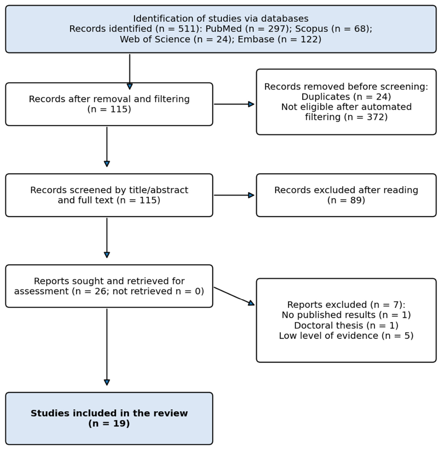
Table 1.
Studies on neuromodulation in lateral ankle sprain.
| Study | Design (Level) | Population and Intervention | Main Findings |
|---|---|---|---|
| Zhang et al., 2025 [17] | Prospective cohort (IIb) | Athletes with grade I–II LAS; low-frequency electrical stimulation plus proprioceptive training vs. proprioceptive training alone (12 weeks, sessions of 20–40 min) | Better pain, pressure tolerance, ROM, muscle strength and joint position accuracy, mainly between weeks 2 and 6 |
| Mazloum et al., 2023 [18] | RCT (Ib) | Athletes with LAS; NMES vs. Kinesio Taping for ankle swelling | No significant change in volumetry or perimetry in either ankle |
| Shukla and Agrawal, 2022 [19] | Prospective case series (III) | Acute grade I–II LAS; device combining the PRICE protocol with electrical stimulation (5 sessions) | Less pain and edema; improved dorsiflexion and plantarflexion ROM |
| Alahmari et al., 2020 [20] | Single-blind RCT (Ib) | Adults after LAS; low-frequency TENS plus proprioceptive neuromuscular facilitation (PNF) vs. PNF alone (12 sessions of 30 min over 3 weeks) | Greater gains in pain, balance, flexibility, proprioception, ROM and strength with TENS plus PNF |
| Wainwright et al., 2019 [21] | Pilot RCT (Ib) | Acute grade I–II LAS; NMES added to standard care for 7 days | Significant edema reduction vs. standard care; no differences in pain or function at 7 days |
| Razzano et al., 2019 [22] | Double-blind RCT (Ib) | Professional contact-sport athletes with grade I–II LAS; non-invasive interactive neurostimulation vs. sham (10 sessions of 20 min over 2 weeks) | Better walking ability, less pain and less anti-inflammatory use; 71.87% vs. 55.17% recovered at 2 months |
| Molsberger and McCaig, 2018 [23] | Case-control (III) | Percutaneous direct current stimulation in soft-tissue injuries, including LAS (up to 3 sessions of 30 min) | Rapid reduction of inflammation and pain; return to sport at 3 weeks in a ligament rupture; similar reductions in 10 further LAS patients |
| Spooner and Bacon, 2017 [24] | Critically appraised paper (Ia) | Electrical stimulation added to standard care in acute LAS | Evidence did not support its use for pain, edema or function; inconsistent parameters |
| Feger et al., 2015 [25] | Systematic review (Ia) | Electrical stimulation plus standard care vs. standard care alone in acute LAS | No significant additional effect on function, edema or pain |
LAS, lateral ankle sprain; RCT, randomized controlled trial; NMES, neuromuscular electrical stimulation; TENS, transcutaneous electrical nerve stimulation; ROM, range of motion; PNF, proprioceptive neuromuscular facilitation.
Table 2.
Studies on neuromodulation in chronic ankle instability.
| Study | Design (Level) | Population and Intervention | Main Findings |
|---|---|---|---|
| Rodríguez-Rosal et al., 2026 [34] | Non-randomized controlled (IIa) | People with CAI vs. healthy controls; ultrasound-guided percutaneous neuromodulation of the posterior tibial nerve | Small change in anteroposterior sway with eyes closed in CAI; no overall postural improvement and no difference vs. controls |
| Obeng-Frimpong et al., 2025 [26] | Systematic review (Ia) | Electrotherapy plus therapeutic exercise vs. exercise alone in CAI | Significant improvements mainly in balance outcomes; most studies favorable |
| Rodríguez-Rosal et al., 2025 [27] | RCT (Ib) | Healthy individuals; one session (1.5 min) of percutaneous tibial nerve stimulation | Better single-leg postural control with eyes closed at 24 and 48 h, but similar in the placebo group |
| Wang et al., 2024 [28] | Double-blind RCT (Ib) | Young adults with CAI; NMES of peroneus longus for 6 weeks (3 sessions of 20 min per week) | Better static balance (smaller center-of-pressure ellipse area, mediolateral displacement and velocity) and higher CAIT scores; clearest in frontal-plane stability |
| Choi and Jun, 2024 [29] | RCT (Ib) | CAI; rehabilitative exercise plus NMES for 6 weeks (3 sessions of 30 min per week) | Improved muscle thickness and cross-sectional area (peroneus longus, soleus) and dynamic balance |
| Zheng et al., 2024 [30] | Double-blind RCT (Ib) | CAI; NMES of peroneus longus for 6 weeks; drop landing | Lower inversion angle and peak inversion angular velocity, higher eversion moment; no difference in frontal-plane ROM |
| Gottlieb et al., 2024 [31] | RCT (Ib) | CAI; balance exercises combined with peroneal NMES or TENS (12 sessions of 20 min over 4–6 weeks), follow-up to 12 months | Both groups improved all patient-reported outcomes at 12 months; consistent trend favoring NMES over TENS |
| Needle et al., 2023 [32] | RCT (Ib) | CAI; 2 weeks of sensory-level NMES or TENS to the ankle pronators (5 sessions of 11 min) | Neither modality alone improved clinical outcomes; TENS may improve reflex excitability |
| Gottlieb et al., 2022 [35] | Non-randomized quasi-experimental (IIa) | CAI vs. healthy controls; one 10-min gait training session with peroneal functional electrical stimulation | Did not normalize gait in CAI; healthy controls increased ankle eversion angle |
| Saleephon et al., 2020 [33] | RCT (Ib) | Rock climbers; neuromuscular training with or without NMES for 4 weeks (3 days per week, 10 min per session) | Both programs significantly increased peroneus longus strength and ankle stability |
CAI, chronic ankle instability; CAIT, Cumberland Ankle Instability Tool; other abbreviations as in Table 1.
Table 3.
Summary of findings by neuromodulation modality.
| Modality | Lateral Ankle Sprain | Chronic Ankle Instability |
|---|---|---|
| TENS | Low-frequency TENS with PNF improved pain, balance, ROM and strength [20]; non-invasive neurostimulation improved walking, pain and return to sport [22] | Useful when combined with exercise [26,31]; limited alone, though it may modulate reflex excitability [32] |
| NMES | Edema reduction in one pilot trial [21]; no effect on swelling vs. Kinesio Taping [18]; reviews found no added benefit in the acute phase [24,25] | Most consistent evidence: six-week programs improved balance, muscle morphology and motor control [28,29,30,33]; trend favoring NMES over TENS with exercise [31] |
| Percutaneous techniques | Rapid reduction of pain and inflammation and early return to sport in one non-randomized report [23] | Small, isolated acute change in postural sway [34]; no difference from placebo in healthy participants [27] |
Table 4.
Stimulation dose in the included primary studies.
| Study | Modality | Dose Reported | Direction of Main Findings |
|---|---|---|---|
| Zhang et al., 2025 [17] | Low-frequency electrical stimulation | 12 weeks; sessions of 20–40 min | Favorable |
| Alahmari et al., 2020 [20] | Low-frequency TENS | 12 sessions of 30 min over 3 weeks | Favorable |
| Razzano et al., 2019 [22] | Non-invasive neurostimulation | 10 sessions of 20 min over 2 weeks | Favorable |
| Shukla and Agrawal, 2022 [19] | Electrical stimulation with PRICE | 5 sessions | Favorable |
| Wainwright et al., 2019 [21] | NMES | 7 consecutive days | Edema only |
| Molsberger and McCaig, 2018 [23] | Percutaneous direct current | Up to 3 sessions of 30 min | Favorable (non-randomized) |
| Wang et al., 2024 [28] | NMES, peroneus longus | 6 weeks; 3 sessions of 20 min per week | Favorable |
| Choi and Jun, 2024 [29] | NMES with exercise | 6 weeks; 3 sessions of 30 min per week | Favorable |
| Zheng et al., 2024 [30] | NMES, peroneus longus | 6 weeks; 3 sessions of 20 min per week | Favorable (frontal-plane control) |
| Gottlieb et al., 2024 [31] | NMES or TENS with exercise | 12 sessions of 20 min over 4–6 weeks | Favorable; sustained to 12 months |
| Saleephon et al., 2020 [33] | NMES with neuromuscular training | 4 weeks; 3 days per week; 10 min | Both groups improved |
| Needle et al., 2023 [32] | NMES or TENS alone | 5 sessions of 11 min over 2 weeks | No clinical benefit |
| Gottlieb et al., 2022 [35] | Peroneal functional electrical stimulation | Single 10-min session | No gait normalization in CAI |
| Rodríguez-Rosal et al., 2025 [27]; 2026 [34] | Percutaneous tibial nerve stimulation | Single session of 1.5 min | Limited or placebo-like acute effects |
Systematic reviews are not listed. CAI, chronic ankle instability; other abbreviations as in Table 1.
Disclaimer/Publisher’s Note: The statements, opinions and data contained in all publications are solely those of the individual author(s) and contributor(s) and not of MDPI and/or the editor(s). MDPI and/or the editor(s) disclaim responsibility for any injury to people or property resulting from any ideas, methods, instructions or products referred to in the content. |
© 2026 by the authors. Licensee MDPI, Basel, Switzerland. This article is an open access article distributed under the terms and conditions of the Creative Commons Attribution (CC BY) license (http://creativecommons.org/licenses/by/4.0/).
Copyright: This open access article is published under a Creative Commons CC BY 4.0 license, which permit the free download, distribution, and reuse, provided that the author and preprint are cited in any reuse.