

Submitted:
04 November 2026
Posted:
05 November 2026
You are already at the latest version
Abstract
This study investigated the prevalence, genotypes, and tissue localization of human papillomavirus (HPV)-DNA in testicular tumor including epididymis and spermatic cord specimens from 116 Japanese patients who underwent radical inguinal orchiectomy. Formalin-fixed, paraffin-embedded tissues were analyzed by nested polymerase chain reaction (PCR), followed by sequencing-based HPV genotyping. HPV-DNA localization was assessed by in situ hybridization (ISH), and E6/E7 mRNA ISH was performed in HPV-positive specimens. HPV-DNA was detected in three patients (3/116, 2.6%), including one testicular tumor specimen (1/116, 0.9%) and two spermatic cord specimens (2/116, 1.7%). All HPV-positive specimens contained HPV16-DNA. HPV-DNA ISH signals were observed in seminoma cells in one testicular specimen and in the vas deferens epithelium in one spermatic cord specimen. On the other hand, ISH signals were not present in epididymis tissue in the HPV-positive testicular tumor tissue, suggesting that HPV-DNA detection rate of epididymis was 0%. E6/E7 mRNA signals were not detected in any HPV-positive specimen. These findings demonstrate that HPV16-DNA was surely present in testicular tumor and the vas deferens epithelium. However we could not provide evidence of transcriptionally active HPV oncogenes, and could not establish a causal role in testicular tumorigenesis.
Keywords:
human papillomavirus
; testicular tumor
; seminoma
; spermatic cord
; vas deferens
1. Introduction
Human papillomavirus (HPV) is a common sexually transmitted virus that affects both men and women [1]. In men, HPV is most frequently detected in the external genitalia, including the glans penis, penile shaft, coronal sulcus, and inner foreskin; however, HPV has also been detected in the urinary tract, urine, and semen [2,3,4,5].
Previous meta-analyses have reported HPV-DNA in semen, including an estimated prevalence of 16% among infertile men, and have suggested associations with impaired sperm concentration, motility, and morphology [4,6]. We previously reported an HPV prevalence of 12.5% in semen samples from Japanese men with infertility [5]. However, HPV detection in semen cannot identify the specific anatomical site of origin of this virus. In other words, it remains unclear whether HPV detected in semen originates from the urethra or from other parts of the genital tract.
The prostate has been investigated as a potential site of HPV detection in the male reproductive tract. A meta-analysis involving 3,122 cases reported HPV detection rates of 25.8% in patients with prostate cancer, 17.4% in patients with benign prostatic hyperplasia (BPH), and 9.2% in healthy men [7]. We also demonstrated that HPV-DNA was detected in 9.6% of the prostate cancer patients who received prostatectomy, whereas HPV-DNA was not detected in seminal vesicle specimens [8]. Although the association between HPV and prostate cancer remains controversial, HPV-DNA has been detected in prostate tissue. On the other hand, the distribution of HPV across the various segments of the seminal tract including spermatic cord, epididymis, and testis, remains unclear. Hence, the present study investigated the prevalence and genotypes of HPV-DNA in seminal tract among 116 Japanese patients with testicular tumors who had received high inguinal orchiectomy.
Persistent infection with oncogenic HPV types is causally associated with several malignancies, including cancers of the cervix, vulva, vagina, penis, anus, and oropharynx [9,10]. Various studies have been conducted on the association between HPV infection and urogenital cancers, reporting diverse associations influenced by geographical differences, histological subtypes, and methodological variations [11]. However, few studies have investigated HPV-DNA in testicular tumors. Since all subjects of our study were included in testicular tumor specimens, we examined HPV-DNA prevalence in tumor tissue and explored its potential relationship with testicular tumorigenesis. Furthermore, in situ hybridization (ISH) analysis was performed to observe HPV-DNA localization in the HPV-positive specimens.
HPV is a non-enveloped virus with an icosahedral capsid and a circular double-stranded DNA genome. In oncogenic HPV infections, dysregulated expression of the viral E6 and E7 oncogenes can contribute to malignant transformation; viral integration may promote this process but is not required for E6/E7 expression. The E6 oncoprotein promotes degradation of the tumor suppressor p53, whereas E7 disrupts retinoblastoma protein (pRb) function; calpain-dependent pRb degradation has also been reported [12]. Detection of E6/E7 transcripts provides evidence of viral oncogene expression but does not, by itself, establish viral integration status. The present study assessed HPV-E6/E7 mRNA expression in HR-HPV-positive samples.
2. Materials and Methods
2.1. Study Population
Japanese patients with testicular tumors who underwent high inguinal orchiectomy at the Department of Urology, Kanazawa University Hospital, or its affiliated institutions between May 2008 and April 2025 were included in this study. Information regarding the study was publicly disclosed, and clinical information, including patient characteristics and tumor features, was retrospectively obtained from medical records. Formalin-fixed paraffin-embedded (FFPE) testicular tumor tissues and spermatic cord tissues were collected from each patient. For the testicular tumor specimens, the sections were collected which contained abundant tumor tissue as well as the epididymis. A total of 116 patients, each contributing testicular tumor and spermatic cord specimens, were included in the analysis.
The study was approved by the Ethics Committee of the Faculty of Medicine, Kanazawa University (approval no. 2024-213).
2.2. HPV-DNA Detection and Genotyping
FFPE blocks of testicular tumor and spermatic cord specimens were sectioned at a thickness of 5 μm and subsequently deparaffinized. All of the testicular tumor specimens included the tumorous lesion and the epididymis. DNA was extracted using the Pinpoint Slide DNA Isolation System (Zymo Research, Orange, CA, USA). The quality of the extracted DNA was assessed by confirming successful PCR amplification of the β-globin gene.
HPV-DNA was detected using nested PCR targeting the L1 region of the HPV genome. MY09/MY11 primers were used for the first-round PCR, followed by GP5+/GP6+ primers for the second-round PCR [13]. To improve reproducibility, PCR was performed at least twice for each sample. Samples were classified as HPV-DNA-positive only when both assays yielded positive results. If the results of the two tests differed, the PCR testing was repeated again. For HPV-DNA-positive samples, PCR products were purified using the NucleoSpin Gel and PCR Clean-up kit (MACHEREY-NAGEL, Germany) and subjected to sequence analysis for HPV genotyping [14]. HPV genotypes were assigned from the resulting nucleotide sequences.
2.3. HPV-DNA In Situ Hybridization
HPV-DNA ISH analysis was performed in all HPV-positive cases to determine the tissue localization of HPV-DNA. Since DNA extraction from testicular tumor specimens including epididymis was conducted as a single sample, it was determined whether the site of HPV infection was the testis or the epididymis based on histological findings of ISH analysis in HPV-positive testicular samples. FFPE blocks from HPV-positive cases were sectioned at a thickness of 5 μm, and hybridization was performed using an HPV-DNA probe (T-1144-400; ZytoVision, Germany) according to the protocol of the ZytoFast™ PLUS CISH Implementation Kit HRP-DAB. This ISH assay detects two low-risk HPV types (HPV6 and HPV11) and 15 high-risk HPV types (HPV16, 18, 31, 33, 35, 39, 45, 51, 52, 56, 58, 59, 66, 68, and 82). Selected HPV-DNA-negative specimens served as negative controls, and an HPV16-positive penile cancer specimen served as a positive control. All experiments were performed with negative and positive controls.
ISH signals were classified into three categories: negative (−), no detectable staining signal; positive (+), signals detectable only at high magnification; and strongly positive (2+), signals clearly detectable even at low magnification.
2.4. HPV-E6/E7 mRNA In Situ Hybridization
HPV-E6/E7 mRNA ISH was performed in high-risk HPV-positive specimens to assess viral oncogene transcription. E6/E7 mRNA expression was evaluated using RNAscope® technology (Advanced Cell Diagnostics, Newark, CA, USA). A specific probe (Code 312591; Advanced Cell Diagnostics) targeting 18 HR-HPV types (HPV16, 18, 26, 31, 33, 35, 39, 45, 51, 52, 53, 56, 58, 59, 66, 68, 73, and 82) was used. Selected HPV-DNA-negative specimens were analyzed as negative controls, and an HPV16-positive penile cancer specimen was used as a positive control. All experiments were performed with negative and positive controls.
Signals were evaluated using the same three-category scoring system as that used for HPV-DNA ISH: negative (−), positive (+), and strongly positive (2+).
2.5. Statistical Analysis
Continuous variables were compared using the Mann–Whitney U test, and categorical variables were compared using the chi-square test. Associations between histopathological tumor categories and HPV-DNA detection were assessed using the chi-square test. All statistical analyses were performed using IBM SPSS Statistics version 29 (IBM Corp., Armonk, NY, USA). A two-sided p value < 0.05 was considered statistically significant.
3. Results
3.1. Patient Characteristics and HPV Prevalence
In all cases, high inguinal orchiectomy was performed within a few days after a diagnosis of testicular tumor. The median age of the 116 patients with testicular tumors was 43 years (range, 14–89 years) (Table 1). Histopathologically, 53 cases (45.7%) were classified as seminoma, 30 (25.9%) as non-seminoma, and 33 (28.4%) as other tumor types. HPV-DNA was detected in three patients (2.6%), and HPV16 was detected in all cases. By anatomical sites, HPV-DNA prevalence was 0.9% (one case) in testicular tumor specimens and 1.7% (2 cases) in spermatic cord ones. All three HPV-positive samples were obtained from patients with seminoma. No statistically significant differences in age, histological tumor classification, or tumor stage were observed between HPV-positive and negative patients.
3.2. HPV-DNA In Situ Hybridization
HPV-DNA ISH signals were observed in seminoma cells in one HPV16-positive testicular tumor specimen (Table 2, Figure 1). However no ISH signals were not observed in the area of epididymis in this case. Among the two HPV16-positive spermatic cord specimens, ISH signals were detected in the vas deferens epithelium in one specimen but not in the other. No ISH signals were detected in the HPV-negative control specimens.
Combining the PCR and ISH results, HPV detection rates were 0.9% (1/116) in testis (testicular tumor), 0% in epididymis, and 1.7% (2/116) in spermatic cord specimens.
3.3. HPV-E6/E7 mRNA Expression
4. Discussion
In our cohort, nested PCR detected HPV-DNA in 2.6% (3/116) of patients with testicular tumors. By anatomical sites, HPV-DNA was detected in 0.9% (one case) of testicular tumor (testis), in 0% of epididymis, and in 1.7% (2 cases) of spermatic cord ones. HPV-DNA ISH localized viral DNA to seminoma cells in one specimen and to the vas deferens epithelium in another. No E6/E7 mRNA signals were detected in any of the three HPV-positive cases. Currently, there have been no previous reports investigating HPV-DNA detection in the seminal tract including testicular tumor and spermatic cord in a single individual. The paired analysis of testicular tumor and spermatic cord specimens, together with tissue-level localization in the vas deferens, adds to the limited evidence regarding HPV distribution in the male seminal tract.
HPV-DNA was detected in one (0.9%) of 116 testicular tumor specimens. A few studies have investigated HPV-DNA in testicular tumors [11]. A previous study of semen samples reported an HPV prevalence of 9.7% in patients with testicular germ cell tumors and 2.4% in healthy controls [15], which was much higher than the prevalence observed in our tissue specimens. However, HPV-DNA detected in semen may originate from multiple anatomical sites, including the prostate, urethra, and external genitalia, making direct comparisons with tissue-based estimates difficult. Other studies did not detect HPV-DNA in testicular tumor specimens or control tissues [16,17]. A meta-analysis did not show an association between HPV infection and testicular tumor (OR 2.79, 95% CI 0.84–9.29, p = 0.09) [18]. The low detection rate in our cohort, together with the absence of detectable E6/E7 transcripts, does not support an etiological association in these specimens. Nevertheless, the small number of HPV-positive cases and lack of a non-neoplastic control group preclude definitive conclusions.
We found that HPV16 was detected in all HPV-positive cases. HPV-DNA ISH signals localized to tumor cells in one HPV16-positive seminoma specimen. HPV16 is a major oncogenic genotype in several HPV-associated cancers. Some previous studies demonstrated that HPV16 was also the most frequent genotype detected from bladder cancer [19] and prostate cancer [7,8]. Although the number of cases was extremely small, HPV16 was detected in one seminoma tissue, and ISH analysis demonstrated that HPV-DNA was surely present in testicular tumor tissue in this case.
In HPV-associated malignancies, persistent high-risk HPV infection and dysregulated expression of the viral E6 and E7 oncogenes can contribute to carcinogenesis [20,21,22]. Accordingly, E6/E7 mRNA or protein expression in tumor tissue is often investigated when evaluating the potential oncogenic activity of HPV [20,21,23]. In the present study, however, E6/E7 mRNA expression was not detected in any of the HPV-positive cases. Thus, despite detection of HPV-DNA in a seminoma specimen, we found no evidence of detectable E6/E7 transcription in this specimen. However, the present study included an extremely limited number of HPV-positive cases, further studies are required to clarify the potential role of HPV-E6/E7 expression in the development of testicular tumors.
An additional strength of this study was the evaluation of paired testicular tumor, epididymis, and spermatic cord tissues from each patient. HPV-DNA detection rates were 0.9% in testis (testicular tumor), 0% in epididymis, and 1.7% in spermatic cord specimens. HPV infection in the seminal tract was extremely low in our cohort. The spermatic cord contains vas deferens, blood vessels, lymphatic vessels, and interstitial tissue. The presence of HPV-DNA signals in vas deferens epithelial cells in one specimen by ISH analysis suggests that this epithelium may harbor viral DNA. There have been no previous studies to demonstrate a clear observation of HPV-DNA in epithelial cells of vas deferens, which is likely to be valuable.
Several studies focusing on testicular tumors or testicular biopsies have investigated HPV-DNA prevalence in the testis [11,15,16,17,18], whereas there have been limited studies examining HPV-DNA detection rate in the epididymis or vas deferens. A previous study described that HPV-DNA prevalence in vas deferens was 7% (3 cases) among 43 healthy men undergoing vasectomy [24]. In addition, Svec et al. reported that HPV-DNA was detected in the epididymis of 6 (27.2%) out of 22 men diagnosed with non-tuberculous epididymitis [25]. Furthermore, HPV-DNA was found to be present in the tissue of the vas deferens in one case (4.5%) [25]. Previously reported detection rates in vas deferens and epididymis tissue were higher than those observed in our cohort; differences in study populations, sampling methods, and laboratory procedures may contribute to this variation. Taken together, these findings suggest that HPV-DNA may be detectable in vas deferens tissue, although its clinical significance remains uncertain.
This study has several limitations. First, only three patients were HPV-DNA-positive, limiting the precision of prevalence estimates and the interpretation of E6/E7 mRNA ISH findings. In particular, HPV-DNA was detected in only one case in testicular tumor specimens (seminoma). The absence of detectable E6/E7 transcripts does not support HPV-driven tumorigenesis in the HPV-DNA-positive seminoma, but the small sample size and assay limitations do not exclude an association in other settings. Second, all specimens were obtained from patients with testicular tumors, without a non-neoplastic control group; therefore, these findings cannot be generalized to healthy men. Larger studies incorporating non-neoplastic testicular and reproductive tract tissues are needed. A strength of this study is the use of complementary molecular methods—PCR, HPV-DNA ISH, and HPV-E6/E7 mRNA ISH—to evaluate HPV detection at multiple anatomical sites of seminal tract, and the demonstration of HPV-DNA presence within seminoma and vas deferens epithelium.
5. Conclusions
HPV16-DNA was detected in one of 116 testicular tumor specimens (0.9%) and two of 116 spermatic cord specimens (1.7%), suggesting that HPV infection in the seminal tract among the patients with testicular tumor was very rare. HPV-DNA ISH localized viral DNA to seminoma cells in one case and to the vas deferens epithelium in another. No E6/E7 mRNA signals were detected in HPV-DNA-positive specimens. These results demonstrate the presence of HPV-DNA in these anatomical sites, but do not support an etiological role in testicular tumorigenesis. Larger controlled studies are needed to clarify the biological significance of these findings.
Author Contributions
Conceptualization, K.S., S.K., and R.S.; methodology, S.T., K.S., S.K. and R.S.; formal analysis, S.T; investigation, S.T., K.F., R.S., H.N., and N.S.; resources, S.T., K.S., and S.K.; data curation, S.T., K.S., K.F., S.K., Y.K., and T.N.; writing—original draft preparation, S.T.; writing—review and editing, K.S.; supervision, K.S., K.I., and A.M. All authors have read and agreed to the published version of the manuscript.
Funding
This research received no external funding.
Institutional Review Board Statement
The study was conducted in accordance with the Declaration of Helsinki and approved by the Ethics Committee of the Graduate School of Medical Sciences, Kanazawa University (protocol code 2024-213; date of approval: 18 December 2024).
Informed Consent Statement
Patient consent was waived owing to the retrospective design of the study; information about the study was made publicly available and patients were given the opportunity to opt out.
Data Availability Statement
The data presented in this study are available on reasonable request from the corresponding author. The data are not publicly available due to patient privacy restrictions.
Conflicts of Interest
The authors declare no conflicts of interest.
References
- Dunne, E.F.; Nielson, C.M.; Stone, K.M.; Markowitz, L.E.; Giuliano, A.R. Prevalence of HPV infection among men: A systematic review of the literature. J. Infect. Dis. 2006, 194, 1044–1057. [CrossRef]
- Giuliano, A.R.; Nielson, C.M.; Flores, R.; Dunne, E.F.; Abrahamsen, M.; Papenfuss, M.R.; Markowitz, L.E.; Smith, D.; Harris, R.B. The optimal anatomic sites for sampling heterosexual men for human papillomavirus (HPV) detection: The HPV detection in men study. J. Infect. Dis. 2007, 196, 1146–1152. [CrossRef]
- Shigehara, K.; Sasagawa, T.; Namiki, M. Human papillomavirus infection and pathogenesis in urothelial cells: A mini-review. J. Infect. Chemother. 2014, 20, 741–747. [CrossRef]
- Laprise, C.; Trottier, H.; Monnier, P.; Coutlée, F.; Mayrand, M.H. Prevalence of human papillomaviruses in semen: A systematic review and meta-analysis. Hum. Reprod. 2014, 29, 640–651. [CrossRef]
- Kato, Y.; Shigehara, K.; Nakagawa, T.; Nakata, H.; Iijima, M.; Nakashima, K.; Kawaguchi, S.; Izumi, K.; Kadono, Y.; Mizokami, A. Human papillomavirus detected in sperm of Japanese infertile males affects reproductive parameters. Int. J. Infect. Dis. 2021, 112, 294–299. [CrossRef]
- Weinberg, M.; Sar-Shalom Nahshon, C.; Feferkorn, I.; Bornstein, J. Evaluation of human papilloma virus in semen as a risk factor for low sperm quality and poor in vitro fertilization outcomes: A systematic review and meta-analysis. Fertil. Steril. 2020, 113, 955–969.e4. [CrossRef]
- Tsydenova, I.A.; Ibragimova, M.K.; Tsyganov, M.M.; Litviakov, N.V. Human papillomavirus and prostate cancer: Systematic review and meta-analysis. Sci. Rep. 2023, 13, 16597. [CrossRef]
- Shinzawa, R.; Shigehara, K.; Nakata, H.; Takada, S.; Fukukawa, K.; Kato, Y.; Nakagawa, T.; Kawaguchi, S.; Izumi, K.; Mizokami, A. Human papillomavirus prevalence in the prostate and seminal vesicles: Does this virus have an etiological role in the development of prostate cancer? Viruses 2025, 17, 1304. [CrossRef]
- Smola, S. Human papillomaviruses and skin cancer. Adv. Exp. Med. Biol. 2014, 810, 192–207. [CrossRef]
- Haedicke, J.; Iftner, T. Human papillomaviruses and cancer. Radiother. Oncol. 2013, 108, 397–402 . [CrossRef]
- Karaoğlan, B.B.; Ürün, Y. Unveiling the role of human papillomavirus in urogenital carcinogenesis: A comprehensive review. Viruses 2024, 16, 667. [CrossRef]
- Dornell, G.A.; Schroder, W.A.; Antalis, T.M.; Lambley, E.; Major, L.; Gardner, J.; Birrell, G.; Cid-Arregui, A.; Suhrbier, A. Human papillomavirus E7 requires the protease calpain to degrade the retinoblastoma protein. J. Biol. Chem. 2007, 282, 37492–37500. [CrossRef]
- de Roda Husman, A.M.; Walboomers, J.M.; van den Brule, A.J.; Meijer, C.J.; Snijders, P.J. The use of general primers GP5 and GP6 elongated at their 3′ ends with adjacent highly conserved sequences improves human papillomavirus detection by PCR. J. Gen. Virol. 1995, 76, 1057–1062. [CrossRef]
- Tanzi, E.; Bianchi, S.; Fappani, C.; Gori, M.; Colzani, D.; Passera, I.; Tincati, C.; Canuti, M.; Raviglione, M.; Amendola, A. Detection of human papillomavirus in fresh and dried urine through an automated system for cervical cancer screening in low- and middle-income countries. J. Med. Virol. 2023, 95, e28802. [CrossRef]
- Garolla, A.; Pizzol, D.; Bertoldo, A.; Ghezzi, M.; Carraro, U.; Ferlin, A.; Foresta, C. Testicular cancer and HPV semen infection. Front. Endocrinol. 2012, 3, 172. [CrossRef]
- Bertazzoni, G.; Sgambato, A.; Migaldi, M.; Grottola, A.; Sabbatini, A.M.T.; Nanni, N.; Farinetti, A.; Iachetta, F.; Giacobazzi, E.; Pecorari, M.; Reggiani Bonetti, L. Lack of evidence for an association between seminoma and human papillomavirus infection using GP5+/GP6+ consensus primers. J. Med. Virol. 2013, 85, 105–109. [CrossRef]
- Meyts, E.R.; Hørding, U.; Nielsen, H.W.; Skakkebæk, N.E. Human papillomavirus and Epstein–Barr virus in the etiology of testicular germ cell tumours. APMIS 1994, 102, 38–42. [CrossRef]
- Garolla, A.; Vitagliano, A.; Muscianisi, F.; Valente, U.; Ghezzi, M.; Andrisani, A.; Ambrosini, G.; Foresta, C. Role of viral infections in testicular cancer etiology: Evidence from a systematic review and meta-analysis. Front. Endocrinol. 2019, 10, 355. [CrossRef]
- Sun, J.X.; Xu, J.Z.; Liu, C.Q.; An, Y.; Xu, M.Y.; Zhong, X.Y.; Zeng, N.; Ma, S.Y.; He, H.D.; Hu, J.; Liu, Z.; Wang, S.G.; Xia, Q.D. The association between human papillomavirus and bladder cancer: Evidence from meta-analysis and two-sample Mendelian randomization. J. Med. Virol. 2023, 95, e28208. [CrossRef]
- Sharma, P.K.; Panaiyadiyan, S.; Kurra, S.; Kumar, R.; Nayak, B.; Kaushal, S.; Sharma, A.; Kumar, R.; Seth, A.; Singh, P. Association of human papillomavirus in penile cancer: A single-center analysis. Indian J. Urol. 2022, 38, 210–215. [CrossRef]
- Yin, J.; Li, Y.; Song, C.; Liu, Y.; Zhao, J.; Zhao, D.; Zhang, S.; Zhang, X.; Zhao, F.; Qiao, Y. Evaluation of an E6/E7 PCR-capillary electrophoresis fragment analysis in the genotyping of human papillomavirus in archival FFPE samples of oropharyngeal cancer. J. Med. Virol. 2024, 96, e29716. [CrossRef]
- Phanuphak, N.; Teeratakulpisarn, N.; Keelawat, S.; Pankam, T.; Barisri, J.; Triratanachat, S.; Deesua, A.; Rodbamrung, P.; Wongsabut, J.; Tantbirojn, P.; Numto, S.; Ruangvejvorachai, P.; Phanuphak, P.; Palefsky, J.M.; Ananworanich, J.; Kerr, S.J. Use of human papillomavirus DNA, E6/E7 mRNA, and p16 immunocytochemistry to detect and predict anal high-grade squamous intraepithelial lesions in HIV-positive and HIV-negative men who have sex with men. PLoS ONE 2013, 8, e78291. [CrossRef]
- Abumsimir, B.; Mrabti, M.; Laraqui, A.; Ameur, A.; Koraishi, S.I.; Mzibri, M.; Lamsisi, M.; Ennaji, Y.; Almahasneh, I.; Ennaji, M.M. Molecular characterization of human papillomavirus and mouse mammary tumor virus-like infections in prostate cancer tissue and relevance with tumor characteristics. Mol. Clin. Oncol. 2022, 16, 97. [CrossRef]
- Fedder, J.; Ørnskov, D.; Engvad, B.; Kristensen, T.K.; Lomholt, M.; Marcussen, N.; Waldström, M. Seminal human papillomavirus originates from the body surface and is not a frequent aetiological factor in azoospermia. Andrologia 2019, 51, e13202. [CrossRef]
- Svec, A.; Mikysková, I.; Hes, O.; Tachezy, R. Human papillomavirus infection of the epididymis and ductus deferens: An evaluation by nested polymerase chain reaction. Arch. Pathol. Lab. Med. 2003, 127, 1471–1474. [CrossRef]
Figure 1.
HPV-DNA in situ hybridization (ISH). ISH signals were observed in seminoma cells (a) (×200; Case 2). ISH signals were also detected in the vas deferens epithelium in one specimen(b) (×200; Case 3). No ISH signals were detected in the HPV-DNA-negative control specimens(c) (×200). ISH signals were detected in the HPV16-positive control specimens(d) (×200).
Figure 1.
HPV-DNA in situ hybridization (ISH). ISH signals were observed in seminoma cells (a) (×200; Case 2). ISH signals were also detected in the vas deferens epithelium in one specimen(b) (×200; Case 3). No ISH signals were detected in the HPV-DNA-negative control specimens(c) (×200). ISH signals were detected in the HPV16-positive control specimens(d) (×200).
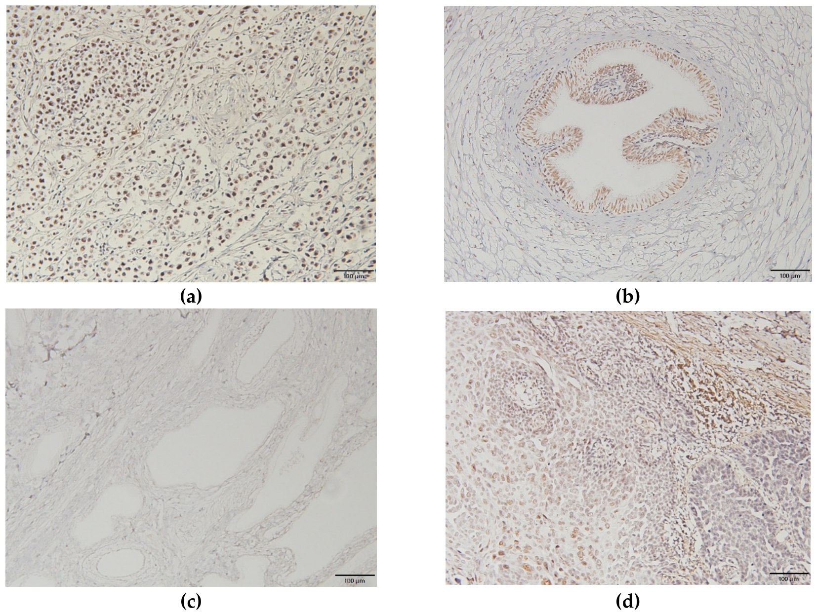
Figure 2.
HPV-E6/E7 mRNA in situ hybridization (ISH). No HPV-E6/E7 mRNA signals were observed in seminoma cells (a) (×200; Case 2). No ISH signals were also detected in the vas deferens epithelium in one specimen(b) (×200; Case 3). No ISH signals were detected in the HPV-negative control specimens(c) (×200). ISH signals were detected in the HPV16-positive control specimens(d) (×200).
Figure 2.
HPV-E6/E7 mRNA in situ hybridization (ISH). No HPV-E6/E7 mRNA signals were observed in seminoma cells (a) (×200; Case 2). No ISH signals were also detected in the vas deferens epithelium in one specimen(b) (×200; Case 3). No ISH signals were detected in the HPV-negative control specimens(c) (×200). ISH signals were detected in the HPV16-positive control specimens(d) (×200).
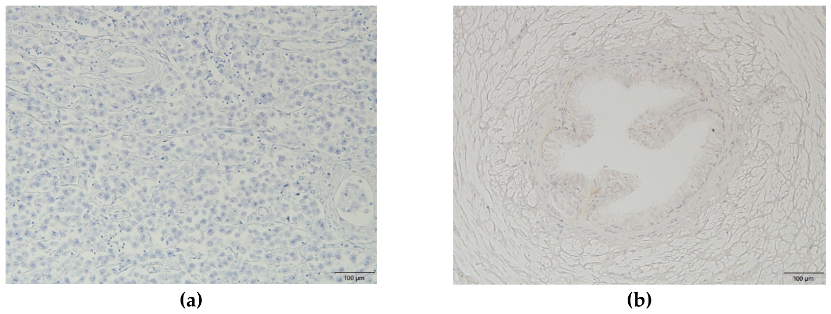
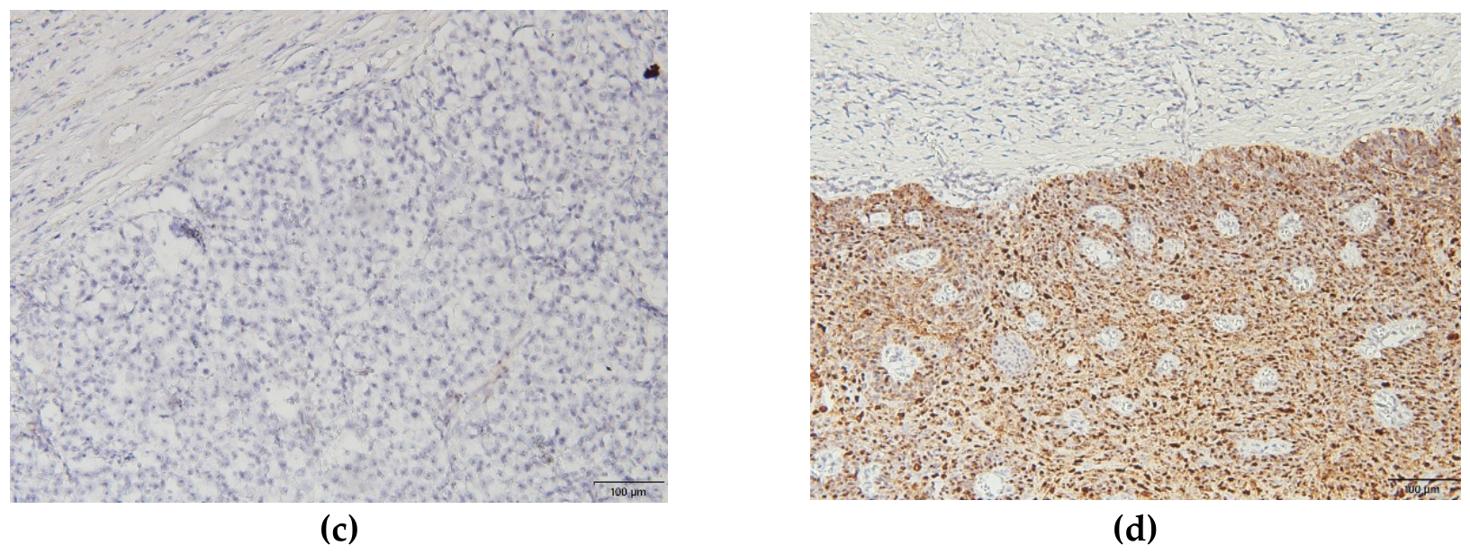

Table 1.
Patient backgrounds and tumor characteristics.
| Characteristics | Overall n=116 |
HPV(+) n=3 |
HPV(-) n=113 |
p value |
| Age(median, range) | 43(14-89) | 37(37-39) | 44(14-89) | 0.506 |
| Tumor type | 0.16 | |||
| Seminoma | 53 | 3 | 50 | |
| Non-Seminoma | 30 | 0 | 30 | |
| Others | 33 | 0 | 33 | |
| Tumor stage (n=83*) | 0.092 | |||
| T1 | 29 | 0 | 0 | |
| T2 | 48 | 3 | 3 | |
| T3 | 6 | 0 | 0 | |
| HPV, human papillomavirus *Cases of germ cell carcinoma | ||||
Table 2.
Summary of the HPV genotypes, HPV-DNA in situ hybridization and HPV-E6/E7 expression in HPV-positive specimens.
Table 2.
Summary of the HPV genotypes, HPV-DNA in situ hybridization and HPV-E6/E7 expression in HPV-positive specimens.
| No | Age | Diagnosis | Specimens | Genotype | HPV-DNA ISH | E6/E7 mRNA ISH | ||
| signals | lesions | signals | lesions | |||||
| 1 | 37 | Seminoma | Tumor | 16 | 2+ | tumor | - | - |
| 2 | 37 | Seminoma | Spermatic cord | 16 | 2+ | vas deferens | - | - |
| 3 | 39 | Seminoma | Spermatic cord | 16 | - | - | - | - |
| HPV, human papillomavirus; ISH, in situ hybridization. Staining score; negative (−; no staining), positive (+; visible only at high magnification), and strongly positive (2+; visible clearly at low magnification). | ||||||||
Disclaimer/Publisher’s Note: The statements, opinions and data contained in all publications are solely those of the individual author(s) and contributor(s) and not of MDPI and/or the editor(s). MDPI and/or the editor(s) disclaim responsibility for any injury to people or property resulting from any ideas, methods, instructions or products referred to in the content. |
© 2026 by the authors. Licensee MDPI, Basel, Switzerland. This article is an open access article distributed under the terms and conditions of the Creative Commons Attribution (CC BY) license (http://creativecommons.org/licenses/by/4.0/).
Copyright: This open access article is published under a Creative Commons CC BY 4.0 license, which permit the free download, distribution, and reuse, provided that the author and preprint are cited in any reuse.