

Submitted:
03 November 2026
Posted:
05 November 2026
You are already at the latest version
Abstract
Methicillin-resistant Staphylococcus aureus (MRSA) is a major antimicrobial-resistant pathogen worldwide and a high-priority target for new therapeutic strategies. Methicillin resistance is primarily mediated by the mecA gene, which encodes penicillin-binding protein 2a (PBP2a), a transpeptidase with reduced affinity for β-lactam antibiotics. Because PBP2a plays a key role in bacterial survival under β-lactam pressure, highly specific to Methicillin-resistant Staphylococci, and immunologically recognizable, it represents an attractive target for immunotherapeutic approaches. This review summarizes current knowledge of PBP2a structure, biological function, and immunological relevance, focusing on vaccine and antibody-based strategies. Studies employing DNA vaccines, recombinant proteins, multi-epitope constructs, and passive immunization have demonstrated protective immune responses and reduced bacterial burden in animal models of MRSA infection. Monoclonal antibodies targeting PBP2a have also shown prophylactic and therapeutic efficacy, including activity independent of Fc-mediated effector functions, suggesting direct interference with PBP2a function. Their potential use in rapid immunodiagnostic assays for methicillin resistance is also discussed. Emerging strategies, including antibody engineering, glycoengineering, antibody-drug conjugates, mucosal vaccination, and combination therapies with antibiotics, may further enhance clinical applicability. Although evidence remains preclinical, PBP2a represents a promising target for active and passive immunization against Methicillin-resistant staphylococci.
Keywords:
methicillin-resistant Staphylococcus aureus
; MRSA
; PBP2a
; mecA
; monoclonal antibodies
; immunotherapy
; vaccine
; passive immunization
; antimicrobial resistance
; immunodiagnosis
1. Introduction
The discovery of penicillin by Alexander Fleming and the subsequent development and clinical use of β-lactam antibiotics can be considered one of the greatest advances in modern medicine, leading to a reduction in deaths caused by bacterial infections and an increase in life expectancy in the last century [1]. These drugs follow the principle of selective toxicity, established by Paul Ehrlich [2], where the target - penicillin-binding proteins (PBPs) are found exclusively in bacteria [3].
However, the widespread and indiscriminate use of antibiotics has contributed to the selection and dissemination of antimicrobial resistance to β -lactams and other classes of antimicrobials [4]. The mechanism of action of β-lactams involves the inhibition of cell-wall synthesis through binding to PBPs, thereby interfering with the transpeptidation reaction and blocking peptidoglycan biosynthesis [5]. Among the resistance mechanisms developed by staphylococci, the first to be described was the production of β-lactamases, enzymes capable of hydrolyze the β-lactam ring of penicillins and their derivatives [6]. The pharmaceutical industry responded with group M penicillins (methicillin and oxacillin), which are resistant to the action of anti-staphylococcal β-lactamases [7]. However, in 1961, Patricia Jevons reported for the first time a case of an infection caused by a Staphyococcus aureus resistant to Celbenin (methicillin) [8]. The era of MRSA (methicilin-resistant Staphylococcus aureus) infections began.
In subsequent years, MRSA outbreaks were reported in hospitals across several countries [9]. The treatment of MRSA infections, relying primarily on glycopeptides, was further challenged by the emergence of vancomycin-intermediate S. aureus (VISA) [10] and, subsequently, of vancomycin-resistant S. aureus (VRSA), resulting from the acquisition of resistance genes shared with vancomycin-resistant enterococci (VRE) [11]. Since its initial inclusion on the list in 2017 through the most recent update in 2024, the World Health Organization (WHO) has classified MRSA as a high-priority pathogen for the research and development (R&D) of new antimicrobials, among those pathogens that pose a threat to human health [12].
MRSA is a major bacterial pathogen responsible for a broad spectrum of infections, ranging from skin and soft-tissue infections to pneumonia, bacteremia, endocarditis, osteomyelitis, and device-associated infections. According to global analyses of the burden of antimicrobial resistance conducted by the Global Research on Antimicrobial Resistance (GRAM) Project and published in The Lancet, (MRSA) stands out as the single antibiotic-resistant bacterial pathogen responsible for the highest absolute number of deaths worldwide. In 2019, MRSA was responsible for more than 100,000 deaths directly attributable to antimicrobial resistance and was associated with over 120,000 additional deaths worldwide. The overall 30-day mortality rate associated with MRSA bacteremia ranges from 20% to 30%, substantially exceeding the mortality observed in infections caused by methicillin-susceptible S. aureus (MSSA) [13].
The global dissemination of multidrug-resistant lineages has substantially complicated antimicrobial management and has reinforced the need for alternative or complementary approaches to conventional antimicrobial therapy [14].
The principal molecular basis of methicillin resistance is the acquisition of the mecA gene, which encodes penicillin-binding protein 2a (PBP2a). Unlike the native penicillin-binding proteins of S. aureus, PBP2a has markedly reduced affinity for most β-lactam antibiotics. Consequently, peptidoglycan synthesis can continue despite the pharmacological inhibition of susceptible PBPs, allowing MRSA to survive exposure to β-lactams concentrations that would otherwise inhibit cell wall synthesis [15,16,17].
PBP2a beyond MRSA: PBP2a-mediated β-lactam resistance is not restricted to S. aureus. In recent years, there have been several reports of an increase in the prevalence of coagulase-negative Staphylococcus carrying the mecA/PBP2a determinant (methi-R CoNS), causing major concern and expanding the resistance spectrum to β-lactams in Staphylococci [18]. Although CoNS are considered less virulent than S. aureus [19], once they show higher resistance to antimicrobials, the issues seen in MRSA infections are likely to repeat in methi-R CoNS, such as treatment difficulties with longer hospital stays, prolonged antibiotic therapy, and probably higher morbidity [20]. Therefore, it is important to consider that the entire discussion in this review can apply, besides MRSA, to methi-R CoNS.
2. Pbps and Pbp2a
PBPs exhibit characteristics that make them attractive targets for antimicrobial development, as they are enzymes found exclusively in bacteria. They participate in the biosynthesis of peptidoglycan, a macromolecule that coats the bacterial membrane, ensuring the cell’s structural integrity and osmotic stability [5]. Like PBPs, peptidoglycan is found exclusively in bacteria, except for Mycoplasma spp. [21].
PBPs are complex molecules, and according to Goffin and Ghuysen, they are classified into low- and high-molecular-weight PBPs. All PBPs possess transpeptidase activity, while some high-molecular-weight PBPs can be bifunctional, containing one domain responsible for transpeptidase activity and another for transglycosylase activity [3]. The transpeptidase domain contains three conserved motifs essential for enzymatic activity: SXXK, which includes the active-site serine, and two additional motifs, SXN and KXG, where X represents any amino acid. The three-dimensional arrangement of these motifs corresponds to the catalytic center of the penicilloyl-serine transferases, enabling the transpeptidase activity of PBPs for peptidoglycan biosynthesis [5].
PBP2a is a class B multimodular PBP with only transpeptidase activity. It was described by Reynolds and Fuller (1986) [22]. The crystal structure of PBP2a was determined by Lim and Strynadka in 2002. These authors described that the groove of the active site of PBP2a is significantly narrower and less accessible than that of PBPs susceptible to β-lactams, forming a "closed" cavity that makes it hard for β-lactam antibiotics to enter and fit. This shape leads to low affinity for β-lactams and is the structural basis for MRSA resistance (Figure 1B) [17].
3. Why Pbp2a May Be an Attractive Immunotherapeutic Target
Several characteristics support the investigation of PBP2a as a potential vaccine antigen. Among them, we can mention: (i) Direct association with MRSA resistance: PBP2a is encoded by mecA, a major determinant of methicillin resistance [9]; (ii) Functional importance: PBP2a is required for efficient cell wall synthesis under β-lactam pressure [17]; (iii) Potential antibody accessibility: Experimental studies indicate that PBP2a can be recognized by antibodies associated with the bacterial surface [23]; (iv) Functional immune responses: PBP2a vaccination induces opsonophagocytic antibodies in animal models [24]; (v) Potential therapeutic synergy: PBP2a-directed immunity could complement β-lactam therapy; and (vi) Specificity: PBP2a is a unique feature of Methicillin-resistant Staphylococci (Methi-R Staph), and this target is not present in Methicillin-sensitive Staphylococci (Methi-S Staph) belonging to the normal microbiota, which can be preserved.
4. Biological Rationale for Targeting Pbp2a
Beyond its role in β-lactam resistance, the structural organization of PBP2a provides a basis for investigating whether antibody binding could interfere with its function. Structural studies have identified an allosteric site in PBP2a that is spatially distinct from its catalytic site [25]. The binding of specific ligands to this region can induce conformational changes that facilitate the opening of the active-site region, providing an important basis for the development of β-lactam potentiators [15,16].
From an immunological perspective, this structural organization raises the possibility that antibodies could be designed to recognize functionally important or surface-accessible regions of PBP2a. In addition to its central role in β-lactam resistance, PBP2a contains structurally and functionally defined domains that may be accessible to antibodies. These characteristics of PBP2a, including its specificity and its potential exposure on the outer surface of MRSA, a Gram-positive bacterium, make it a particularly attractive target for therapeutic intervention via immunological approaches.
Most of the vaccines discussed here used a similar internal region of PBP2a, comprising the active center, including Ser403 (Figure 2). Comparing these approaches may help clarify whether antibody recognition of such regions contributes to protective immunity.
5. Anti-Pbp2a Vaccines
5.1. Dna Vaccines
The first experimental studies for generating an anti-PBP2a immune response used a strategy based on a DNA vaccine approach.
DNA vaccines deliver a gene encoding the protein of interest into host cells, which then produce the encoded antigen. This approach can elicit both arms of the immune response: humoral, by antibody production, and cellular immune responses [26]. The main advantages of DNA vaccines are safety, stability, rapid development and relatively low-cost manufacturing [26].
Ohwada et al. developed a DNA vaccine containing the full-length mecA gene, which encodes PBP2a protein, which is the main factor behind methicillin resistance in MRSA. Vaccination of mice with a plasmid containing the mecA gene induced the production of anti-PBP2a antibodies, boosted MRSA phagocytosis, and significantly lowered kidney bacterial load (approximately 50% bacteria recovered than the no-immunized mice control) after experimental infection [27].
Senna et al. demonstrated that a DNA vaccine encoding a fragment of the MRSA PBP2a elicited a specific humoral immune response and significantly reduced bacterial burden in challenged mice, approximately 1,000-fold. In this study, the authors used an internal fragment with 88 amino acids comprising the active enzymatic center (STQK) of PBP2a [23].
Both studies are important because they showed that (i) DNA vaccines encoding PBP2a can elicit specific antibody responses; (ii) these antibodies can recognize and bind to PBP2a on the bacterial surface; and (iii) the binding of these antibodies to their epitopes on PBP2a can provide protection in an animal model.
5.2. Recombinant Pbp2a Vaccines
Recombinant protein-based vaccines constitute a well-established approach for inducing the production of specific antibodies against a selected protein target. Immunizations are usually performed using recombinant protein formulated with an adjuvant to boost the immune response [28].
Haghighat et al., using a recombinant protein corresponding to an internal region of PBP2a, formulated with Freund's adjuvant, demonstrated the generation of anti-PBP2a antibodies in a mouse model. Vaccination induced high levels of PBP2a-specifc IgG antibodies. Different IgG isotypes were also detected, indicating the activation of immune pathways associated with both Th1 and Th2 profiles. The bacterial load in the kidneys from challenged mice was approximately 1,000-fold lower than that in the negative controls (non-immunized mice), and the opsonophagocytic activity of sera from immunized mice was significantly increased. Finally, the survival of immunized mice after bacterial challenge was extended compared to that of control mice [24].
Similar levels of protection and improved survival were obtained using the same recombinant protein from a previous study [24], but in different formulations containing various adjuvants or delivery systems: (i) combined with recombinant S. aureus autolysin [29]; (ii) formulated with Montanide and aluminum [30]; (iii) combined with autolysin and PLGA (poly lactic-co-glycolic acid) [31]; and (iv) formulated with Biogenic Selenium Nanoparticles and MF59 [32].
5.3. Multiepitope and Immunoinformatics-Based Vaccines
The increasing availability of S. aureus genomic and structural information has enabled the application of reverse vaccinology and immunoinformatics to PBP2a vaccine design. Computational studies have identified predicted B-cell- and T-cell epitopes within PBP2a, and proposed multiepitope vaccine constructs designed to stimulate both humoral and cellular immunity [33].
More recent approaches have incorporated PBP2a-derived epitopes and antigens associated with virulence and iron acquisition, including IsdB and hemolysin-associated components [34].
This approach is conceptually attractive because S. aureus pathogenesis is multifactorial [35]. A vaccine that simultaneously targets resistance, virulence, and bacterial survival mechanisms may be more effective than a monovalent antigen. Nevertheless, computational prediction remains hypothesis-generating. Predicted antigenicity, MHC binding, structural stability, or immunogenicity cannot substitute for experimental validation in appropriate animal models and, ultimately, clinical trials. However, this type of approach, including antigens present in Methi-S Staph strains, loses the specificity feature for Methi-R strains and can also affect commensal Staphylococci.
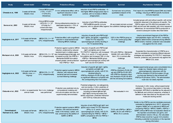
Table 1, divided into two parts, summarizes the main characteristics of the anti-PBP2a vaccines discussed in this review.

Table 1.
The main characteristics of the anti-PBP2a vaccines (part I).
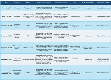 |
Table 1.
The main characteristics of the anti-PBP2a vaccines (part II).
 |
5.4. Is It Worth Using a Vaccine to Prevent Mrsa Infections?
In general, MRSA infections can occur in both community and hospital settings, usually caused by strains with specific characteristics classified as community-associated (CA-MRSA) or hospital-associated (HA-MRSA) [36]. Hospital-acquired infections caused by MDR bacteria are often linked to infectious opportunism, associated with prolonged hospital stays and previous antibiotic therapy. In most cases, patients who contract these infections from MDR bacteria usually present immunodepression [37].
In these situations, vaccination strategies might not be the best option, because (i) it is difficult to predict in the general population who will be hospitalized and infected with MRSA; (ii) even if immunized, in cases of immunosuppression, the production of antibodies would be compromised; (iii) vaccinating a patient in a hospital bed would not make sense, since the time needed to generate antibodies would not help fight a recent infection. However, in contexts where we have populations at high risk of contracting an MRSA infection, a vaccination strategy is justified. Immunizations prior to surgeries with a high risk of MRSA infections (e.g., prosthesis implantation) could use vaccines to prevent MRSA infections.
A promising alternative for preventing the spread of MRSA would be the development of a vaccine aimed at preventing MRSA colonization or promoting nasal decolonization in asymptomatic carriers, potentially reducing bacterial transmission and the risk of subsequent infection.
6. Mrsa Nasal Carriage and Potential Vaccine for Nasal Decolonization
S. aureus naturally colonizes approximately 20%–30% of the population, with rates potentially reaching even higher levels in certain groups [38]. Although several body sites can be colonized in humans, the anterior nares are the most frequent site of S. aureus carriage [39]. According to CDC, approximately 2% of the population carries MRSA, although prevalence may be higher in hospitals, long-term care facilities, and intensive care units [40]. Gupta et al. demonstrated that patients colonized with MRSA have significantly higher risks of MRSA infection and death compared to non-colonized patients, even after adjusting for healthcare exposure and comorbidities [41].
The most used strategy for nasal decolonization involves topical mupirocin, while antiseptic baths with agents such as chlorhexidine and povidone-iodine are used to reduce skin colonization [42]. However, healthcare workers can become colonized by patients and pose a risk of transmission [43]. A study by Santos Goes et al. revealed a high rate (74.6%) of S. aureus nasal carriage among healthcare workers included in the dynamic of units of Primary Health Care, while the prevalence of MRSA was 53.9%. The reported prevalence of S. aureus carriers was higher than that reported in most studies conducted among professionals in hospital settings worldwide. Several studies have reported that the rate of MSSA and MRSA nasal carriage among health workers ranged from 5.5–34% and 6.1–25.5%, respectively [44]. Despite the benefits of decolonization, its effects may not be permanent, since recolonization can occur weeks or months after treatment, and mupirocin resistance has been reported [45].
Although MRSA nasal colonization is considered an important risk factor for infection and hospital-based transmission [44], there is currently no approved vaccine for prevention or decolonization. A vaccine for nasal decolonization of MRSA would be useful for healthcare workers as well as for patients undergoing procedures with a high risk of MRSA infection. However, the potential of anti-PBP2a vaccines to prevent or eliminate nasal colonization remains to be established. In particular, whether these vaccines can induce protective mucosal immunity sufficient to reduce nasal carriage requires experimental investigation, as this would be crucial for vaccine effectiveness.
7. Passive Immunotherapy Against Pbp2a
Passive immunotherapy differs fundamentally from vaccination because protective antibodies are administered directly to the patient. This approach may be particularly attractive for severe MRSA infections because patients with acute infections cannot wait several weeks for vaccine-induced adaptive immunity to develop. Therefore, PBP2a-specific antibodies (monoclonal antibodies or serum therapy) could potentially be administered prophylactically to high-risk individuals or therapeutically during established infection. In the context of hospital-acquired infections, direct administration of antibodies would be the best way to overcome the limitations of vaccine strategies, as previously discussed.
7.1. Passive Serotherapy
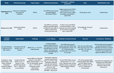
Naghshbandi et al. described passive immunization using polyclonal antibodies produced in rabbits against the recombinant PBP2a (rPBP2a) from MRSA (previously been used in study [31]). An experimental MRSA sepsis in murine model was used, and the results showed that passive immunization with anti-rPBP2a significantly improved the survival rate, similar to vancomycin treatment, when compared with control groups. Furthermore, anti-PBP2a IgG reduced the bacterial load in internal organs. These findings suggest that passive immunization could be a promising therapeutic alternative for MRSA infections [46].
7.2. Monoclonal Antibodies
Monoclonal antibodies (mAbs) offer several advantages over conventional serum therapy (hyperimmune serum or polyclonal immunoglobulins), especially in the treatment of infectious diseases, toxins, and some immune disorders [47]. Among these, the following can be highlighted: (i) the higher specificity when compared to serology (polyclonal antibodies); (ii) the lower risk of adverse reactions: Therapeutic serums are often produced in animals, such as horses, and can trigger serum sickness, immediate hypersensitivity, and other immune reactions. Humanized or fully human mAbs have significantly lower immunogenicity, making their use safer; (iii) reproducibility and standardization: The composition of mAbs is uniform and can be reproduced indefinitely in cell culture; (iv) lower risk of pathogen transmission: mAbs have a lower risk of biological contamination than products derived from plasma or animals used in serotherapy because they are produced in controlled cell culture systems.; (v) Improvement by molecular engineering: mAbs can be modified using genetic engineering techniques to increase affinity, half-life, effector activity, or even carry drugs, radioisotopes, and toxins. This capability does not exist in conventional serum therapy.
7.2.1. Mab Anti-Pbp2a
Experimental murine mAbs targeting PBP2a were generated and characterized by Senna et al. These mAbs were selected against a recombinant protein corresponding to an internal PBP2a fragment comprising the active center of this enzyme. They demonstrated binding to the bacterial surface, as assessed by flow cytometry, and high target affinity interactions, as determined by surface plasmon resonance [48]. Saraiva et al. conducted prophylactic studies using these mAbs in a murine model, demonstrating a significant reduction in kidney bacterial load in groups treated with either intact IgG (greater than 3 logs) or F(ab’)2 fragments (approximately 2 logs) when compared with the control groups (untreated). Following challenge with a lethal dose of MRSA, treated mice also showed higher survival rates. In a therapeutic assay, treatment with the mAb reduced the bacterial load in the kidneys similar to that of mice treated with vancomycin. Combination therapy with the mAb and vancomycin was also effective [49]
In addition to the importance of demonstrating the protective activity of antibodies, the protection observed in animals treated with an antibody lacking the Fc region (F[ab’]2 fragment) suggests that the mAb can act independently of the Fc-mediated effector functions. As several MRSA-infected patients can be immunosuppressed, neutralizing activity could be considered an important feature of therapeutic approaches for these infections [49].
These findings demonstrate that anti-PBP2a mAb may be a promising therapeutic agent for treating MRSA infections and provide experimental evidence supporting PBP2a as an immunotherapeutic target. Table 2 summarizes the main characteristics of the anti-PBP2a passive immunotherapy by serum and mAbs presented here.
7.2.2. Cross-Reactivity with Low Affinity E. faecium Pbp5
Recent findings have identified an unexpected possibility: antibodies originally developed against PBP2a may also bind related penicillin-binding proteins in other Gram-positive bacteria. A recent study demonstrated cross-reactivity between an anti-PBP2a mAb and PBP5 of Enterococcus faecium. Both MRSA PBP2a and E.faecium PBP5 are classified as multimodular PBPs of subclass B1, characterized by their low affinity for b-lactam antibiotics [3]. Nano-isothermal titration calorimetry (Nano-ITC) revealed a high-affinity binding between mAb and PBP5, with a nanomolar-scale affinity. Immunofluorescence assays confirmed mAb recognition of both MRSA and E. faecium by the mAb. Futhermore, the mAb provided protection in a murine model infected with vancomycin-resistant E. faecium, demonstrating the efficacy of the prophylactic treatment. [50]. A comparative sequence analysis of these two PBPs showed amino acid similarity in the region surrounding the active center of these enzymes.
7.3. Why Is the Mab Anti-Pbp2a Effective? Studies of Molecular Interactions Between Mab and Pbp2a
The anti-PBP2a mAb was selected using a recombinant protein corresponding to the amino acid sequence used in previous vaccine studies [23,24]. This selection process involved immunization in a mouse model followed by hybridoma selection. The potential antigenicity of the internal PBP2a region was investigated using different computational tools, including ANTHEPROT [http://pbil.ibcp.fr/NPSA]) [51] and the algorithm developed by Kolaskar and Tongaonkar to find antigenic determinants (http://imed.med.ucm.es/Tools/antigenic.pl) [52]. Both methods indicated favorable antigenic characteristics for this protein sequence. The Kolaskar/Tongaonkar analysis identified two potential antigenic determinants: peptides EPLLNKF and TQKILTAMI.
Teixeira et al. [50] employed in silico analyses, such as molecular docking, to evaluate the interactions between the antibody and PBP2a. They identified 18 PBP2a residues involved in interactions with 27 residues of the antibody. This interaction included two salt bridges, 13 hydrogen bonds, and additional non-bonded contacts. Furthermore, the constant of dissociation (Kd) value at 37°C was predicted to be 7,2 ×10⁻¹⁰ M, suggesting strong binding between the mAb and its target. Notably, 12 of the 18 identified residues were located in the region of the recombinant protein used to select the mAb, while eight residues were located near the active center (STQK). These findings suggest that the binding of the mAb inhibits the transpeptidation enzymatic activity normally exerted by PBP2a (Figure 2).
8. Could Pbps Transglycosylases Be Targets for Immunological Approaches?
As previously discussed, PBPs are not exclusively transpeptidase enzymes. According to the classification of PBPs by Goffin and Ghuysen [3], multimodular class B PBPs are composed of two modules: transpeptidase and transglycosylase. Despite the widespread use of antibiotics targeting transpeptidase activity (β-lactams and glycopeptides), the transglycosylase domain has been poorly explored as an antibacterial target. The only transglycosylation inhibitor described to date is Moenomycin, which is isolated from Streptomyces. However, besides being toxic, its low absorption property makes it ineffective in the treatment of human infections [53]. PBP2 is the only bifunctional enzyme of S. aureus, including MRSA strains. Its structure presents a bilobular folding with the glycosyl transferase domain (GT) in the N-terminal region and the transpeptidase domain (TP) in the C-terminal region, separated by a binding region that exhibits some flexibility [54].
Argondizzo et al. generated specific antibodies in a murine model using a recombinant protein encoding the internal PBP2 transglycosylase domain. Immunized mice were challenged with a sublethal dose of MRSA, and the bacterial loads in kidneys and spleen indicate a reduction of 2 logs in organs from immunized mice compared with the negative controls [55]. Although these findings support the potential of targeting PBP transglycosylase domains through immunological approaches, the presence of alternative transglycosylases may represent a limitation to this strategy. For example, S. aureus possesses monofunctional transglycosylases, such as MGT/SgtB, that can support peptidoglycan biosynthesis in the absence of PBP2 transglycosylase activity [56].
9. Challenges and Future Perspectives
Despite the promising results presented in this review, anti-PBP2a immunotherapies still need to overcome some obstacles to be a real alternative for therapeutic and prophylactic use in infections caused by methi-R Staph. The following points are important to consider.
i. Clinical Development
There is currently no evidence that anti-PBP2a vaccines or immunotherapies have advanced to the point of demonstrating clinical effectiveness in humans. This is a crucial point: all positive results available to date are preclinical, in mouse models, showing proof of concept and cannot be interpreted as evidence of therapeutic efficacy in humans.
ii. Vaccine Prophylaxis, Applicability
One of the main challenges in developing a PBP2a vaccine is defining the target population to be vaccinated, which is linked to the characteristics of infections caused by methi-R Staph. Nasal vaccines to prevent nasal colonization in patients at high risk of MRSA infections and healthcare workers (HCW) seem to be the most suitable populations for anti-PBP2a vaccination strategies to reduce or prevent cross transmission between HCW and patients.
iii. Therapy/Prophylaxis Through Immunotherapy (Mostly mAbs): Final Costs
Anti-PBP2a mAbs would need to undergo humanization process before clinical use in humans. This process involves modifying the antibody molecule as well as demonstrating that its activity is maintained. Additionally, upstream and downstream processes would need to be developed and optimized for conducting clinical trials, and, ultimately, large-scale production. Besides, mAbs usually have a high added value, which is reflected in a high final cost, and this can limit a wider use of these molecules in the treatment and prevention of infections caused by methi-R Staph, when compared with costs of conventional antibiotic therapies.
iv. Combination with Traditional Antibiotics
One of the most promising applications of anti-PBP2a mAbs will likely not be to replace antibiotics but to enhance them, working in combination with conventional antimicrobials. The combination with glycopeptides is particularly interesting and should be explored because it is a class of antimicrobials used in the treatment of methi-R Staph infections. These antibiotics inhibit bacterial cell-wall synthesis, primarily by binding to the D-Ala-D-Ala terminus of peptidoglycan precursors, thereby interfering with peptidoglycan cross-linking and cell-wall assembly [57]. In the study by Saraiva et al, animals treated with a combination of the mAb anti-PBP2a and vancomycin showed better results than animals treated with either the antibody or vancomycin alone [49].
The rationale could be also applied in the case of β-lactams. PBP2a enables MRSA to maintain peptidoglycan synthesis when conventional PBPs are inhibited by β-lactams. Thus, combining anti-PBP2a mAbs with β-lactams could inhibit the transpeptidase activity of susceptible Staphylococci PBPs (1-4), while the antibodies would target PBP2a, for which β-lactams are ineffective.
iv.i. Antibody-Drug Conjugate (ADC)
Antibody-drug conjugates (ADCs) are a class of biotherapeutics that combine the high specificity of a mAb with the activity of a drug payload, attached to the antibody via a chemical linker. The goal of this strategy is to target the drug selectively to target cells, potentially increase therapeutic efficacy and reduce systemic toxicity [58]. ADCs consist of three main components: (i) mAb: recognizes specific target antigen expressed on intended site of action; (ii) Linker: connects the antibody to the payload and is designed to maintain the stability of the conjugate in circulation, while enabling drug release at the desired site; and (iii) Payload: the active molecule responsible for the intended therapeutic effect after the ADC binds to or is internalized by the target. In antibacterial context, the payload could be an antibiotic.
Currently, the main use of ADCs is in oncology. Several ADCs have already been approved for tumors, including breast cancer, lymphomas, leukemias, and lung cancer. Beyond oncology, recent research has explored their use in infectious, inflammatory diseases, and other conditions where it is possible to identify specific cellular targets [57]. In the context of Methi-R Staph, PBPs could represent potential targets.
Among the main advantages of using ADCs in antibacterial strategies, the following can be highlighted: (i) greater therapeutic selectivity, where the antibiotic is specifically directed to the site of action. This would lead to a reduction in toxicity due to less exposure to host tissues; (ii) the possibility of reducing doses, as the delivery would also take place exactly at the site of action; and (iii) the possibility of longer intervals between doses, since the antibiotic conjugated to IgG isotype antibodies has a relatively long plasma half-life of about 21 days [59]. It is important to highlight once again that the classes of antimicrobials most suitable for ADC strategies against methi-R Staph would be glycopeptides and β-lactams.
v. Antibody Bioengineering—Modulating Pharmacokinetics, Tissue Penetration, and Immune Response Recruitment
Special attention should be given to the bioengineering of mAbs. These molecules can be subjected to structural modifications to alter their characteristics. Reducing their size through the generation of antibody fragments can improve tissue penetration [60,61]. Modifications like glycosylation, particularly sialylation in the Fc region, can also influence pharmacokinetic properties of therapeutic antibodies [62]. Additionally, modifications of glycosylation can affect the interaction of the Fc region with Fc gamma receptors (FcγRs) and complement system proteins. In this way, changes in glycosylation profiles can increase or decrease effector mechanisms, such as (i) antibody-dependent cellular cytotoxicity (ADCC), (ii) antibody-dependent cellular phagocytosis (ADCP), and (iii) activation of the complement system [63].
10. Other Applications—Immunodiagnostic of Methicillin-Resistance Using Anti-PBP2a mAbs
Immunological detection of Methicillin resistance is based on the use of anti-PBP2a mAbs, which can specifically recognize this protein. Unlike conventional phenotypic methods (antibiograms), which rely on bacterial growth and can take 24 to 48 hours, PBP2a detection can be performed from an isolated bacterial colony, providing results in just a few minutes and helping make faster and more accurate treatment decisions [64].
One of the first approaches developed was the latex agglutination test, in which latex particles coated with anti-PBP2a mAbs aggregate in the presence of the target protein. The MRSA-Screen assay showed high sensitivity and specificity when compared with PCR for detecting the mecA gene, which is considered the gold standard for molecular diagnosis. In a classic study, sensitivity reached about 97% and specificity 100% for MRSA isolates [64].
More recently, lateral flow immunochromatographic assays have been developed based on use the same principles of recognition by mAbs, but in a simpler and faster format. These tests enable direct detection of PBP2a in bacterial colonies or in subcultures from positive blood cultures, showing high agreement with molecular detection of mecA and making it easier to use in routine clinical laboratories [65]. Among the main advantages of immunodiagnostic methods based on anti-PBP2a mAbs are rapid results, relatively low cost, operational simplicity, and the ability to directly detect the functional product responsible for resistance. Moreover, these assays do not require sophisticated molecular infrastructure, making them an attractive option for labs with limited resources. However, some limitations should be considered. The expression of PBP2a can be heterogeneous among different isolates, affecting the sensitivity of the test. In some cases, prior to induction of mecA expression by exposure to β-lactams may be necessary to increase protein detection. Additionally, recent variants of resistance genes, such as mecC, may not be recognized by all available immunoassays, potentially resulting in false-negative results [64]. Borderline oxacillin-resistant S. aureus (BORSA) strains can also be a problem. Unlike conventional Methi-R Staph strains carrying mecA or mecC, BORSA strains lack these resistance determinants and do not produce PBP2a. Their reduced susceptibility to oxacillin is generally associated with overproduction of β-lactamases and/or changes in PBPs 2 or 4, which makes it hard to diagnose using anti-PBP2a immunological methods and conventional molecular tests [66].
11. Conclusions
MRSA remains one of the most important antimicrobial-resistant pathogens worldwide, and the emergence of methi-R CoNS further broadens the clinical relevance of resistance mechanisms mediated by PBP2a. Because PBP2a is the central determinant of Methicillin resistance and appears accessible to antibody recognition, it represents a particularly attractive target for immunological intervention.
The studies reviewed herein provide preclinical proof-of-concept evidence that PBP2a can induce protective immune responses. DNA vaccines, recombinant protein formulations, multiepitope constructs, passive polyclonal immunization, and mAb-based approaches have all demonstrated varying degrees of protection in experimental animal models. Importantly, anti-PBP2a mAbs have shown both prophylactic and therapeutic efficacy, reducing bacterial burden, improving survival, and maintaining activity even when administered as F(ab')₂ fragments, suggesting that direct interference with PBP2a function may contribute to protection independently of Fc-mediated immune mechanisms.
Beyond their therapeutic potential, anti-PBP2a antibodies have demonstrated practical clinical utility in the diagnosis of Methicillin resistance through rapid immunological assays. This successful diagnostic application further validates PBP2a as a robust and accessible biological target and supports the feasibility of expanding antibody-based technologies toward therapeutic applications.
Nevertheless, significant challenges remain. All available evidence supporting anti-PBP2a immunotherapies derives from preclinical studies, and no vaccine or mAb directed against PBP2a has yet demonstrated efficacy in human clinical trials. Questions concerning the optimal target populations (particularly for vaccines), cost-effectiveness, antibody humanization, manufacturing scalability, tissue penetration, and long-term safety must still be addressed. Furthermore, the complexity of S. aureus pathogenesis suggests that PBP2a-directed strategies may be most effective when integrated with conventional antimicrobial therapy.
Future developments in antibody engineering, Fc glycoengineering, long-acting formulations, antibody-drug conjugates, and mucosal immunization strategies may significantly enhance the clinical applicability of PBP2a-targeted interventions in MRSA infections. Particularly promising is the concept of combining anti-PBP2a mAbs with β-lactams or glycopeptides, exploiting complementary mechanisms that simultaneously interfere with multiple steps of peptidoglycan synthesis.
In conclusion, PBP2a represents one of the most compelling examples of a bacterial resistance determinant that can be exploited as both a diagnostic marker and an immunotherapeutic target. Although substantial translational work remains necessary, the accumulated evidence indicates that anti-PBP2a vaccines and antibody-based therapies constitute a promising and innovative approach for combating infections caused by Methi-R Staph and may contribute to expanding the therapeutic arsenal available against antimicrobial resistance in the coming years.
Author Contributions
JPS – conceptualization, writing, original draft, writing review. JPCB – writing review and editing (table, figures), data curation. Authors have read and agreed to the submitted version of the manuscript.
Funding
No funds were used for the preparation of this manuscript.
Data Availability Statement
The data are available upon request to the corresponding author.
Acknowledgments
We would like to thank Dr. Haroldo Cid da Silva Júnior for the excellent and helpful review of the manuscript. José Procopio Senna thanks Dr. Diógenes Santiago Santos for encouraging work on this important line of research (in memoriam).
Conflicts of Interest
The authors declare no conflicts of interest.
IA, software platform use
Paperpal was used to improve English writing. Graphical abstract was created using BioRender (BioRender.com).
References
- Hutchings, M.I.; Truman, A.W.; Wilkinson, B. Antibiotics: Past, present and future. Curr. Opin. Microbiol. 2019, 51, 72–80. [Google Scholar] [CrossRef] [PubMed]
- Van Miert, A.S.J.P.A.M. The sulfonamide-diaminopyrimidine story. J. Vet. Pharmacol. Ther. 1994, 17, 309–316. [Google Scholar] [CrossRef] [PubMed]
- Goffin, C.; Ghuysen, J.-M. Multimodular penicillin-binding proteins: An enigmatic family of orthologs and paralogs. In Microbiol. Mol. Biol. Rev.; PubMed Central (PMC), 1998; Volume 62, pp. 1079–1093. [Google Scholar] [CrossRef] [PubMed]
- De Angelis, G.; Del Giacomo, P.; Posteraro, B.; Sanguinetti, M.; Tumbarello, M. Molecular mechanisms, epidemiology, and clinical importance of β-lactam resistance in Enterobacteriaceae. Int. J. Mol. Sci. 2020, 21, 5090. [Google Scholar] [CrossRef] [PubMed]
- Sauvage, E.; Kerff, F.; Terrak, M.; Ayala, J.A.; Charlier, P. The penicillin-binding proteins: Structure and role in peptidoglycan biosynthesis. In FEMS Microbiol. Rev.; Wiley Online Library, 2008; Volume 32, pp. 234–258. [Google Scholar] [CrossRef]
- Chambers, H.F. The changing epidemiology of Staphylococcus aureus? Emerg. Infect. Dis. 2001, 7, 178–182. [Google Scholar] [CrossRef] [PubMed]
- Gajdács, M. The continuing threat of methicillin-resistant Staphylococcus aureus. Antibiotics 2019, 8, 52. [Google Scholar] [CrossRef] [PubMed]
- Jevons, M.P. “Celbenin”-resistant staphylococci. Br. Med. J. 1961, 1, 124–125. [Google Scholar] [CrossRef]
- Boyce, J.M.; Cookson, B.; Christiansen, K.; Hori, S.; Vuopio-Varkila, J.; Kocagöz, S.; et al. Methicillin-resistant Staphylococcus aureus. Lancet Infect. Dis. 2005, 5, 653–663. [Google Scholar] [CrossRef] [PubMed]
- Hiramatsu, K.; Hanaki, H.; Ino, T.; Yabuta, K.; Oguri, T.; Tenover, F.C. Methicillin-resistant Staphylococcus aureus clinical strain with reduced vancomycin susceptibility. J. Antimicrob. Chemother. 1997, 40, 135–136. [Google Scholar] [CrossRef] [PubMed]
- Périchon, B.; Courvalin, P. VanA-type vancomycin-resistant Staphylococcus aureus. In Antimicrob. Agents Chemother.; PubMed Central (PMC), 2009; Volume 53, pp. 4580–4587. [Google Scholar] [CrossRef] [PubMed]
- World Health Organization. WHO Bacterial Priority Pathogens List, 2024: Bacterial Pathogens of Public Health Importance to Guide Research, Development and Strategies to Prevent and Control Antimicrobial Resistance; World Health Organization: Geneva, Switzerland, 2024. [Google Scholar]
- GBD 2019 Antimicrobial Resistance Collaborators. Global mortality associated with 33 bacterial pathogens in 2019: A systematic analysis for the Global Burden of Disease Study 2019. Lancet 2022, 400, 2221–2248. [Google Scholar] [CrossRef] [PubMed]
- Turner, N.A.; Sharma-Kuinkel, B.K.; Maskarinec, S.A.; Eichenberger, E.M.; Shah, P.P.; Carugati, M.; Holland, T.L.; Fowler, V.G., Jr. Methicillin-resistant Staphylococcus aureus: An overview of basic and clinical research. Nat. Rev. Microbiol. 2019, 17, 203–218. [Google Scholar] [CrossRef] [PubMed]
- Fishovitz, J.; Hermoso, J.A.; Chang, M.; Mobashery, S. Penicillin-binding protein 2a of methicillin-resistant Staphylococcus aureus. IUBMB Life 2014, 66, 572–577. [Google Scholar] [CrossRef] [PubMed]
- Rosado, P.C.; Marques, M.M.; Justino, G.C. Targeting MRSA penicillin-binding protein 2a: Structural insights, allosteric mechanisms, and the potential of adjuvant inhibitors. Biochem. Pharmacol. 2025, 239, 117048. [Google Scholar] [CrossRef] [PubMed]
- Lim, D.; Strynadka, N.C.J. Structural basis for the β-lactam resistance of PBP2a from methicillin-resistant Staphylococcus aureus. Nat. Struct. Biol. 2002, 9, 870–876. [Google Scholar] [CrossRef] [PubMed]
- Katkowska, M.; Kosecka-Strojek, M.; Wolska-Gębarzewska, M.; Kwapisz, E.; Wierzbowska, M.; Międzobrodzki, J.; et al. Emerging challenges in methicillin resistance of coagulase-negative staphylococci. Antibiotics 2025, 14, 37. [Google Scholar] [CrossRef] [PubMed]
- Becker, K.; Heilmann, C.; Peters, G. Coagulase-negative staphylococci. Clin. Microbiol. Rev. 2014, 27, 870–926. [Google Scholar] [CrossRef] [PubMed]
- Michels, R.; Last, K.; Becker, S.L.; Papan, C. Update on coagulase-negative staphylococci: What the clinician should know. Microorganisms 2021, 9, 830. [Google Scholar] [CrossRef] [PubMed]
- Razin, S.; Yogev, D.; Naot, Y. Molecular biology and pathogenicity of mycoplasmas. Microbiol. Mol. Biol. Rev. 1998, 62, 1094–1156. [Google Scholar] [CrossRef] [PubMed]
- Reynolds, P.E.; Fuller, C. Methicillin-resistant strains of Staphylococcus aureus: Presence of a novel penicillin-binding protein. FEMS Microbiol. Lett. 1987, 41, 305–308. [Google Scholar] [CrossRef]
- Senna, J.P.M.; Roth, D.M.; Oliveira, J.S.; Machado, D.C.; Santos, D.S. Protective immune response against methicillin-resistant Staphylococcus aureus in a murine model using a DNA vaccine approach. Vaccine 2003, 21, 2661–2666. [Google Scholar] [CrossRef]
- Haghighat, S.; Siadat, S.D.; Sorkhabadi, S.M.R.; Sepahi, A.A.; Sadat, S.M.; Yazdi, M.H.; Mahdavi, M. Recombinant PBP2a as a vaccine candidate against methicillin-resistant Staphylococcus aureus: Immunogenicity and protectivity. Microb. Pathog. 2017, 108, 32–39. [Google Scholar] [CrossRef] [PubMed]
- Otero, L.H.; Rojas-Altuve, A.; Llarrull, L.I.; Carrasco-López, C.; Kumarasiri, M.; Lastochkin, E.; Fisher, J.F.; Chang, M.; Mobashery, S.; Hermoso, J.A. How allosteric control of Staphylococcus aureus penicillin-binding protein 2a enables methicillin resistance and physiological function. Proc. Natl. Acad. Sci. USA 2013, 110, 16808–16813. [Google Scholar] [CrossRef]
- Kozak, M.; Hu, J. DNA vaccines: Their formulations, engineering and delivery. Vaccines 2024, 12, 71. [Google Scholar] [CrossRef] [PubMed]
- Ohwada, A.; Sekiya, M.; Hanaki, H.; Kuwahara-Arai, K.; Nagaoka, I.; Hori, S.; et al. DNA vaccination by mecAsequence evokes an antibacterial immune response against methicillin-resistant Staphylococcus aureus. J. Antimicrob. Chemother. 1999, 44, 767–774. [Google Scholar] [CrossRef] [PubMed]
- Soler, E.; Houdebine, L.M. Preparation of recombinant vaccines. Biotechnol. Annu. Rev. 2007, 13, 65–94. [Google Scholar] [CrossRef] [PubMed]
- Haghighat, S.; Siadat, S.D.; Sorkhabadi, S.M.R.; Sepahi, A.A.; Mahdavi, M. A novel recombinant vaccine candidate comprising PBP2a and autolysin against methicillin-resistant Staphylococcus aureus confers protection in experimental mice. In Mol. Immunol.; PubMed, 2017; Volume 91, pp. 1–7. [Google Scholar] [CrossRef] [PubMed]
- Mortazavi, S.S.; Haghighat, S.; Mahdavi, M. Recombinant PBP2a of methicillin-resistant S. aureus formulation in Alum and Montanide ISA266 adjuvants induced cellular and humoral immune responses with protection in BALB/c mice. Microb. Pathog. 2020, 140, 103945. [Google Scholar] [CrossRef] [PubMed]
- Haghighat, S.; Siadat, S.D.; Akhavan Sepahi, A.; Mahdavi, M. Recombinant PBP2a/autolysin conjugate as PLGA-based nanovaccine induced humoral responses with opsonophagocytosis activity, and protection versus methicillin-resistant Staphylococcus aureus infection. Iran. J. Basic Med. Sci. 2022, 25, 442–450. [Google Scholar] [CrossRef] [PubMed]
- Daneshpajooh Rahmani, M.; Yazdi, M.H.; Haghighat, S.; Zarrinpanah, T. Synergistic immunoenhancement of recombinant PBP2a vaccine candidate by biogenic selenium nanoparticles and MF59 against methicillin-resistant S. aureus. Curr. Microbiol. 2026, 83, 211. [Google Scholar] [CrossRef] [PubMed]
- Olatunde, S.K.; Kolawole, O.E.; Owolabi, J.F. Designing a novel in silico multi-epitope vaccine against penicillin-binding protein 2A in Staphylococcus aureus. Inform. Med. Unlocked 2022, 33, 101080. [Google Scholar] [CrossRef]
- Das Mitra, S.; Kumar, B.; Rajegowda, S.; Bandopadhyay, S.; Karunakar, P.; Pais, R. Reverse vaccinology & immunoinformatics approach to design a multiepitope vaccine (CV3Ag-antiMRSA) against methicillin-resistant Staphylococcus aureus (MRSA)—A pathogen affecting both human and animal health. J. Biomol. Struct. Dyn. 2024, 42, 11792–11811. [Google Scholar] [CrossRef] [PubMed]
- Foster, T.J.; Geoghegan, J.A.; Ganesh, V.K.; Höök, M. Adhesion, invasion and evasion: The many functions of the surface proteins of Staphylococcus aureus. Nat. Rev. Microbiol. 2014, 12, 49–62. [Google Scholar] [CrossRef] [PubMed]
- David, M.Z.; Daum, R.S. Community-associated methicillin-resistant Staphylococcus aureus: Epidemiology and clinical consequences of an emerging epidemic. Clin. Microbiol. Rev. 2010, 23, 616–687. [Google Scholar] [CrossRef] [PubMed]
- Ye, Q.; Chen, X.; Zhang, J.; Lin, J. Meta-analysis of risk factors for infection by multi-drug-resistant organisms in intensive care unit patients. J. Hosp. Infect. 2025, 158, 1–10. [Google Scholar] [CrossRef]
- Sakr, A.; Brégeon, F.; Mège, J.-L.; Rolain, J.-M.; Blin, O. Staphylococcus aureus nasal colonization: An update on mechanisms, epidemiology, risk factors, and subsequent infections. Front. Microbiol. 2018, 9, 2419. [Google Scholar] [CrossRef]
- Wertheim, H.F.L.; Vos, M.C.; Ott, A.; van Belkum, A.; Voss, A.; Kluytmans, J.A.J.W.; van Keulen, P.H.J.; Vandenbroucke-Grauls, C.M.J.E.; Meester, M.H.M.; Verbrugh, H.A. Risk and outcome of nosocomial Staphylococcus aureus bacteraemia in nasal carriers versus non-carriers. Lancet 2004, 364, 703–705. [Google Scholar] [CrossRef] [PubMed]
- Centers for Disease Control and Prevention. Clinical overview of methicillin-resistant Staphylococcus aureus (. MRSA) in healthcare settings. 2025. Available online: https://www.cdc.gov/mrsa/hcp/clinical-overview/index.html (accessed on 5 September 2026).
- Gupta, K.; Martinello, R.A.; Young, M.; Strymish, J.; Cho, K.; Lawler, E. MRSA nasal carriage patterns and the subsequent risk of conversion between patterns, infection, and death. PLoS ONE 2013, 8, e53674. [Google Scholar] [CrossRef] [PubMed]
- Smith, M.; Herwaldt, L. Nasal decolonization: What antimicrobials and antiseptics are most effective before surgery and in the ICU. Am. J. Infect. Control 2023, 51, A64–A71. [Google Scholar] [CrossRef] [PubMed]
- Kinnevey, P.M.; Kearney, A.; Shore, A.C.; Earls, M.R.; Brennan, G.; Poovelikunnel, T.T.; Humphreys, H.; Coleman, D.C. Methicillin-resistant Staphylococcus aureus transmission among healthcare workers, patients and the environment in a large acute hospital under non-outbreak conditions investigated using whole-genome sequencing. J. Hosp. Infect. 2021, 118, 99–107. [Google Scholar] [CrossRef] [PubMed]
- Goes, I.C.R.D.S.; Romero, L.C.; Turra, A.J.; Gotardi, M.A.; Rodrigues, T.F.S.O.; Santos, L.O.; Dores, J.C.D.; Nascimento, M.U.D.; Cavalleri, A.C.; Pinheiro-Hubinger, L.; Eller, L.K.W.; Pereira, V.C. Prevalence of nasal carriers of methicillin-resistant Staphylococcus aureus in primary health care units in Brazil. Rev. Inst. Med. Trop. São Paulo 2021, 63, e14. [Google Scholar] [CrossRef] [PubMed]
- Dadashi, M.; Hajikhani, B.; Darban-Sarokhalil, D.; van Belkum, A.; Goudarzi, M. Mupirocin resistance in Staphylococcus aureus: A systematic review and meta-analysis. J. Glob. Antimicrob. Resist. 2020, 20, 238–247. [Google Scholar] [CrossRef] [PubMed]
- Naghshbandi, R.Z.; Haghighat, S.; Mahdavi, M. Passive immunization against methicillin-resistant Staphylococcus aureus recombinant PBP2a in sepsis model of mice: Comparable results with antibiotic therapy. Int. Immunopharmacol. 2018, 56, 186–192. [Google Scholar] [CrossRef] [PubMed]
- Lipman, N.S.; Jackson, L.R.; Trudel, L.J.; Weis-Garcia, F. Monoclonal versus polyclonal antibodies: Distinguishing characteristics, applications, and information resources. ILAR J. 2005, 46, 258–268. [Google Scholar] [CrossRef] [PubMed]
- Senna, J.P.M.; Teixeira, M.G.M.; Santiago, M.A.; Batoréu, N.; Valadares, N.; Galler, R. Generation and characterization of murine monoclonal antibodies anti-PBP2a of methicillin-resistant Staphylococcus aureus. Monoclon. Antib. Immunodiagn. Immunother. 2015, 34, 257–262. [Google Scholar] [CrossRef] [PubMed]
- Saraiva, F.B.; de Araújo, A.C.C.; de Araújo, A.E.V.; Senna, J.P.M. Monoclonal antibody anti-PBP2a protects mice against MRSA (methicillin-resistant Staphylococcus aureus) infections. PLoS ONE 2019, 14, e0225752. [Google Scholar] [CrossRef] [PubMed]
- Teixeira, J.H.L.; de Souza, N.P.; Boechat, J.P.C.; Argondizzo, A.P.; da Silva Junior, H.C.; Gonçalves, V.L.; Bastos, R.C.; de Souza Junior, J.P.; Souza Oliveira, D.A.; Lara, F.A.; Rodrigues-da-Silva, R.N.; Teixeira, L.M.; Senna, J.P.M. When cross-reaction is welcome: Monoclonal antibody anti-PBP2a from methicillin-resistant Staphylococcus aureus (MRSA) binds PBP5 from Enterococcus faecium and confers protection in a murine model. Microb. Pathog. 2026, 210, 108160. [Google Scholar] [CrossRef] [PubMed]
- Deléage, G.; Combet, C.; Blanchet, C.; Geourjon, C. ANTHEPROT: An integrated protein sequence analysis software with client/server capabilities. Comput. Biol. Med. 2001, 31, 259–267. [Google Scholar] [CrossRef] [PubMed]
- Kolaskar, A.S.; Tongaonkar, P.C. A semi-empirical method for prediction of antigenic determinants on protein antigens. FEBS Lett. 1990, 276, 172–174. [Google Scholar] [CrossRef] [PubMed]
- Goldman, R.C.; Gange, D. Inhibition of Transglycosylation Involved in Bacterial Peptidoglycan Synthesis. Curr. Med. Chem. 2000, 7, 801–820. [Google Scholar] [CrossRef] [PubMed]
- Lovering, A.L.; de Castro, L.H.; Lim, D.; Strynadka, N.C.J. Structural Insight into the Transglycosylation Step of Bacterial Cell-Wall Biosynthesis. Science 2007, 315, 1402–1405. [Google Scholar] [CrossRef]
- Argondizzo, A.P.C.; Peres, A.M.N.; Saraiva, F.B.; Almeida, M.; Senna, J.P.M. Evaluation of the PBP2 Transglycosylase Region of Staphylococcus aureus as a Target for Immunotherapeutic Approaches. Microb. Pathog. 2021, 157, 105000. [Google Scholar] [CrossRef] [PubMed]
- Reed, P.; Veiga, H.; Jorge, A.M.; Terrak, M.; Pinho, M.G. Monofunctional Transglycosylases Are Not Essential for Staphylococcus aureus Cell Wall Synthesis. J. Bacteriol. 2011, 193, 2549–2556. [Google Scholar] [CrossRef] [PubMed]
- Müller, A.; et al. Approved glycopeptide antibacterial drugs: mechanism of action and resistance. Cold Spring Harb. Perspect. Med. 2016, 6(12), a026989. [Google Scholar] [CrossRef] [PubMed]
- Wu, G.; Yuan, Z.; Chen, M.; Tang, X.; Wang, F.; Zhang, D. Antibody-Drug Conjugates (ADCs): A Review of Structural Design, Technological Evolution, and Future Perspectives. Molecules 2026, 31, 1180. [Google Scholar] [CrossRef] [PubMed]
- Bas, M.; Terrier, A.; Jacque, E.; Dehenne, A.; Pochet-Béghin, V.; Beghin, C.; Dezetter, A.-S.; Dupont, G.; Engrand, A.; Beaufils, B.; et al. Fc Sialylation Prolongs Serum Half-Life of Therapeutic Antibodies. J. Immunol. 2019, 202, 1582–1594. [Google Scholar] [CrossRef] [PubMed]
- Boechat, J.P.C.; Ramos, F.R.S.; Saraiva, F.B.; Gonçalves, V.L.; Silva, F.S.; Alves, C.R.; Senna, J.P.M.; Silva Júnior, H.C. Generation and Characterization of a Monoclonal Antibody Fab Fragment Targeting PBP2a in Methicillin-Resistant Staphylococcus aureus. J. Immunol. Methods 2024, 534, 113774. [Google Scholar] [CrossRef] [PubMed]
- Araújo, A.E.V.; Souza, N.P.; Sousa, A.P.B.; Lara, F.A.; Senna, J.P.M. Production and Characterization of F(ab')2 Fragments Obtained by Enzymatic Digestion from Murine Anti-MRSA PBP2a Monoclonal Antibodies. Appl. Biochem. Biotechnol. 2018, 185, 72–80. [Google Scholar] [CrossRef] [PubMed]
- Bussel, J.B.; Cines, D.B.; Blumberg, R.S. Neonatal Fc Receptor: Biology and Therapeutics. N. Engl. J. Med. 2025, 392, 1621–1635. [Google Scholar] [CrossRef] [PubMed]
- Jennewein, M.F.; Alter, G. The Immunoregulatory Roles of Antibody Glycosylation. Trends Immunol. 2017, 38, 358–372. [Google Scholar] [CrossRef] [PubMed]
- Van Leeuwen, W.B.; Van Pelt, C.; Luijendijk, A.; Verbrugh, H.A.; Goessens, W.H.F. Rapid Detection of Methicillin Resistance in Staphylococcus aureus Isolates by the MRSA-Screen Latex Agglutination Test. J. Clin. Microbiol. 1999, 37, 3029–3030. [Google Scholar] [CrossRef] [PubMed]
- Kolesnik-Goldmann, N.; Bodendoerfer, E.; Röthlin, K.; Herren, S.; Imkamp, F.; Marchesi, M.; Mancini, S. Rapid Detection of PBP2a in Staphylococci from Shortly Incubated Subcultures of Positive Blood Cultures by an Immunochromatographic Assay. Microbiol. Spectr. 2021, 9, e00462-21. [Google Scholar] [CrossRef] [PubMed]
- Sawhney, S.S.; Ransom, E.M.; Wallace, M.A.; Reich, P.J.; Dantas, G.; Burnham, C.A.D. Comparative Genomics of Borderline Oxacillin-Resistant Staphylococcus aureus Detected during a Pseudo-outbreak of Methicillin-Resistant S. aureus in a Neonatal Intensive Care Unit. mBio 2022, 13, e03196-21. [Google Scholar] [CrossRef] [PubMed]
Figure 1.
Molecular surface structure of PBP2a (PDB 1MWR). A. The N-terminal extension represented in green; the non-penicillin binding domain is colored orange. The transpeptidase domain is blue. The groove of the catalytic site of PBP2a is represented in light blue. B. Expanded transpeptidase domain highlighting the groove of the catalytic site. In red, amino acids GSTQK, indicating the position of the active center in the groove. Red arrow indicates the active center amino acids.
Figure 1.
Molecular surface structure of PBP2a (PDB 1MWR). A. The N-terminal extension represented in green; the non-penicillin binding domain is colored orange. The transpeptidase domain is blue. The groove of the catalytic site of PBP2a is represented in light blue. B. Expanded transpeptidase domain highlighting the groove of the catalytic site. In red, amino acids GSTQK, indicating the position of the active center in the groove. Red arrow indicates the active center amino acids.
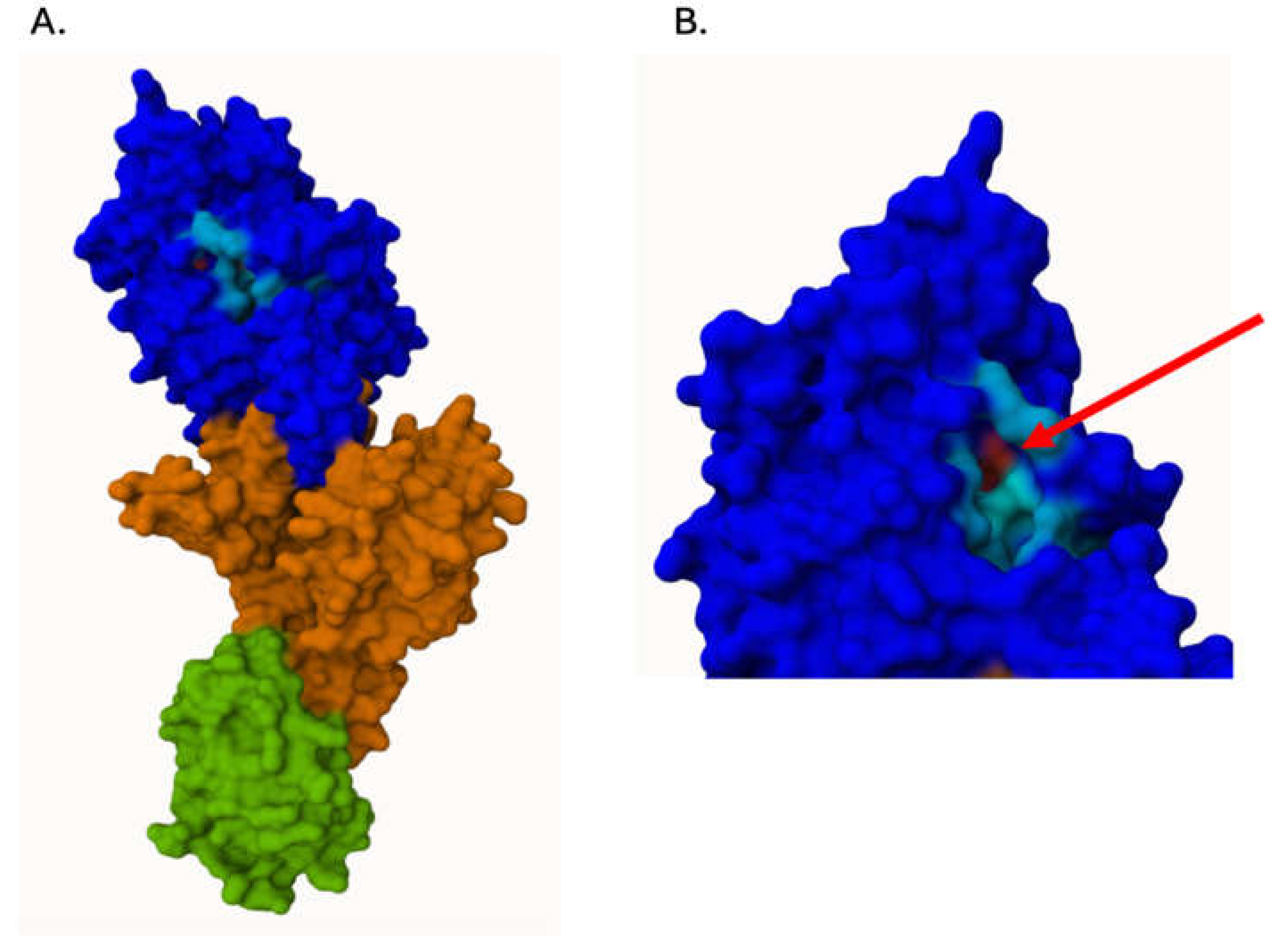
Figure 2.
PBP2a in molecular surface representation. Left: two different views of PBP2a. Green: N-terminal extension. Orange: non-penicilin-binding domain. Blue: transpeptidase domain. Light blue: groove of the catalytic PBP2a site. Magenta: PBP2a residues (amino acids) involved in interactions with antibody (mAb) residues [49]. Right: PBP2a amino acid sequence indicating the regions of PBP2a structure represented in the left. The three conserved domains essential for enzymatic activity – STQK, SDN and KSG are represented in bold. The underlined region corresponds to the amino acid sequence employed in the vaccine and immunotherapy to generate antibodies anti-PBP2a [23,24,29,30,31,32,46,48].
Figure 2.
PBP2a in molecular surface representation. Left: two different views of PBP2a. Green: N-terminal extension. Orange: non-penicilin-binding domain. Blue: transpeptidase domain. Light blue: groove of the catalytic PBP2a site. Magenta: PBP2a residues (amino acids) involved in interactions with antibody (mAb) residues [49]. Right: PBP2a amino acid sequence indicating the regions of PBP2a structure represented in the left. The three conserved domains essential for enzymatic activity – STQK, SDN and KSG are represented in bold. The underlined region corresponds to the amino acid sequence employed in the vaccine and immunotherapy to generate antibodies anti-PBP2a [23,24,29,30,31,32,46,48].
Table 2.
The main characteristics of anti-PBP2a passive immunotherapy.
 |
Disclaimer/Publisher’s Note: The statements, opinions and data contained in all publications are solely those of the individual author(s) and contributor(s) and not of MDPI and/or the editor(s). MDPI and/or the editor(s) disclaim responsibility for any injury to people or property resulting from any ideas, methods, instructions or products referred to in the content. |
© 2026 by the authors. Licensee MDPI, Basel, Switzerland. This article is an open access article distributed under the terms and conditions of the Creative Commons Attribution (CC BY) license (http://creativecommons.org/licenses/by/4.0/).
Copyright: This open access article is published under a Creative Commons CC BY 4.0 license, which permit the free download, distribution, and reuse, provided that the author and preprint are cited in any reuse.