

Submitted:
03 November 2026
Posted:
05 November 2026
You are already at the latest version
Abstract
Background/Objectives: Life-threatening haemoptysis primarily fatal due to airway obstruction, and the culprit vessel must be identified before it can be occluded. Bronchial artery embolisation (BAE) is the established haemostatic treatment, yet its success is largely determined upstream, by the accuracy of the diagnostic chain that precedes it: computed tomography angiography (CTA), bronchoscopy and selective angiography. These modalities are conventionally deployed sequentially and by separate teams. Case Presentation: An 84-year-old male patient presented with abrupt life-threatening haemoptysis. CTA demonstrated an ectatic left bronchial artery with arterial-phase venous opacification, diagnostic of a bronchial arteriovenous fistula (BAVF). Because this phenotype determines both the embolic agent and the definition of technical success, bronchoscopy and angiography were performed concurrently. Endoscopy localised pulsatile bleeding to the left upper lobe bronchus and secured the airway; superselective angiography confirmed the fistula, which was occluded with detachable platinum microcoils under continuous endoscopic observation. Conclusions: Outcome in life-threatening haemoptysis depends less on the embolic device than on whether imaging, endoscopy and angiography operate within a single decision-making environment. We propose a phenotype-driven, dual-endpoint workflow, supported by a structured CTA reading checklist and an embolic-agent selection framework, as a reproducible model for the multidisciplinary management of pulmonary vascular emergencies.
Keywords:
haemoptysis
; bronchial artery embolisation
; bronchial arteriovenous fistula
; bronchoscopy
; computed tomography angiography
; interventional radiology
; airway management
; case report
1. Introduction
Haemoptysis occupies a singular position in respiratory medicine: a symptom that is usually trivial and occasionally lethal within minutes. Between 5% and 14% of patients who present with haemoptysis bleed at a life-threatening rate, and mortality in this subgroup ranges from 9% to 38%, driven less by the volume expectorated than by aetiology, comorbidity and the speed with which the bleeding vessel is identified [1,2,3]. The clinical task is therefore diagnostic before it is haemostatic, and it must be completed under time pressure.
Volume-based definitions have not survived scrutiny. Proposed thresholds span 100 to 1000 mL per 24 h, patients and clinicians estimate blood loss unreliably, and an identical volume carries entirely different consequences in a patient with normal lungs and in one with advanced fibrosis. Severity is now defined functionally, by the capacity of the bleeding to obstruct the airway, impair gas exchange or destabilise the circulation [1,2]. Fartoukh and colleagues translated this concept into a validated bedside score combining bleeding mechanism, underlying disease and respiratory status, which predicts in-hospital mortality more reliably than any volumetric threshold [4], and the multicentre Italian cohort reported by Mondoni et al. confirmed in the CT era that aetiology and comorbidity, rather than the amount of blood, determine outcome [5].
The anatomical rationale for endovascular treatment is precise. Approximately 90% of severe bleeding arises from the bronchial systemic circulation, which operates at aortic pressure, rather than from the low-pressure pulmonary bed [6,7]. Chronic inflammation—bronchiectasis, tuberculosis sequelae, chronic pulmonary aspergillosis, malignancy—induces bronchial arterial hypertrophy, peribronchial neovascularisation and systemic-to-pulmonary anastomoses, and these fragile vessels rupture into the airway [8]. Less frequently, the same remodelling produces a discrete lesion: a bronchial artery aneurysm or a bronchial arteriovenous fistula (BAVF), in which a high-flow communication bypasses the capillary bed altogether [9,10,11]. The distinction is not taxonomic. A diffuse hypervascular territory and a focal high-flow shunt appear different on CTA, require different catheters and different embolic agents, and are cured by different endpoints.
Because death follows airway flooding rather than blood loss, the first therapeutic act is airway protection and isolation of the healthy lung [1,12]. This gives bronchoscopy an irreducible role: rigid instruments provide suction capacity, clot extraction, tamponade and device deployment, whereas the flexible scope reaches the distal airways, identifies the bleeding segment and detects endobronchial lesions [12,13]. CTA, meanwhile, has become the diagnostic reference standard, identifying aetiology more often than endoscopy, mapping bronchial and non-bronchial systemic arteries and serving as the procedural roadmap that shortens catheterisation and fluoroscopy [6,14,15].
These two pillars are habitually framed as competitors, and the framing is unproductive. CTA maps the probable vascular territory and potential culprit vessels; bronchoscopy answers which airway is bleeding now and keeps the patient alive while the first question is resolved. Comparative work has begun to show that adding interventional bronchoscopy to BAE improves immediate haemostasis, reduces post-procedural respiratory complications and lowers one-year recurrence [16,17].
We report such a case—a rare high-flow BAVF occluded with detachable platinum microcoils under continuous endoscopic surveillance—and use it as the clinical anchor for a critical synthesis of the diagnostic workflow in life-threatening haemoptysis. The objective of this paper is not to add another rare lesion to the literature, but to propose an integrated endoscopic–endovascular workflow, with explicit indications, contraindications and endpoints, that is reproducible in any centre with a conventional angiography suite and two available interventionalists.
2. Case Presentation
An 84-year-old man with a history of permanent atrial fibrillation on direct oral anticoagulant therapy, hypertensive heart disease, depressive disorder, and dilation of the ascending thoracic aorta, as well as recurrent episodes of haemoptysis, presented to the emergency department with acute worsening of bleeding over the preceding hours. The exact volume of haemoptysis could not be reliably quantified; however, the recurrence pattern and associated respiratory impairment were consistent with a clinically significant episode.
On arrival, the patient was haemodynamically stable, with no signs of circulatory compromise. Arterial blood gas analysis demonstrated hypoxemic respiratory failure (pH 7.37, PaO₂ 54 mmHg, PaCO₂ 42.3 mmHg), while laboratory testing showed haemoglobin 12.8 g/dL and platelet count 152 ×10⁹/L, with no evidence of coagulopathy or significant systemic inflammation. Based on functional criteria, the episode was classified as life-threatening haemoptysis [4].
Chest radiography demonstrated parenchymal opacities predominantly involving the left lung, associated with ipsilateral pleural effusion. Urgent contrast-enhanced multidetector computed tomography angiography (CTA) of the thorax revealed complex parenchymal abnormalities, including ground-glass opacities, fibroatelectasis, cylindrical bronchiectasis, and multiple calcified nodules, predominantly involving the left upper lobe. In addition, a markedly tortuous and hypertrophic left bronchial artery arising from the descending thoracic aorta was identified, with early opacification of an adjacent vascular structure during the arterial phase, consistent with a high-flow bronchial arteriovenous fistula draining into the pulmonary venous circulation. No non-bronchial systemic arterial supply or radiculomedullary branches were detected on the pre-procedural study.
The identification of a focal high-flow shunt, rather than diffuse bronchial hypervascularity, directly influenced the therapeutic strategy. A multidisciplinary decision was made to proceed with combined endoscopic and endovascular management performed concurrently in the angiography suite.
Under conscious sedation, flexible bronchoscopy was performed as the initial step. The examination revealed blood and clots occupying the left main bronchus, while the right bronchial tree was free of blood. A free-floating clot was visualised in the distal portion of the left main bronchus, firmly adherent to a small crater in the mucosa of its anteromedial wall, with an apparent transparietal implantation and mild pulsatile bleeding at that level. The patient was positioned with the bleeding side dependent, and blood was aspirated to ensure airway patency while carefully avoiding dislodgement of the adherent clot. Flushing with cold saline achieved temporary hemostasis, creating a stable time window for subsequent angiographic intervention.
Selective catheterisation was then performed via the right common femoral artery. Thoracic aortography followed by superselective bronchial angiography confirmed a hypertrophied and tortuous left bronchial artery supplying a high-flow arteriovenous fistula with early venous return, anatomically corresponding to the pulmonary segment and bronchial lesion identified at bronchoscopy.
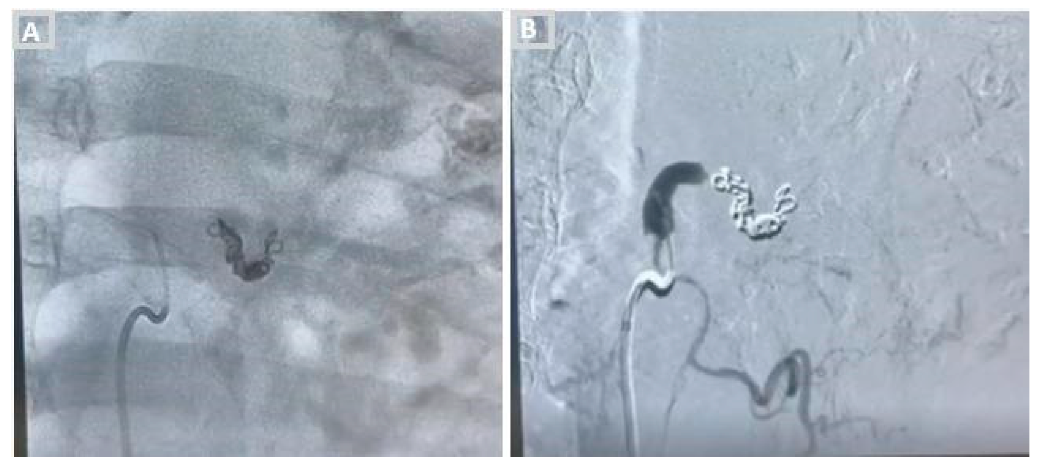
Given the focal and high-flow nature of the lesion, embolisation was performed using detachable platinum microcoils. Two coils (4 × 15 mm and 3 × 5 mm) were deployed across the fistulous communication, with careful positioning and stability assessment prior to release to minimise the risk of distal migration. Completion angiography demonstrated complete occlusion of the fistula, with cessation of pathological shunting and preservation of flow within the proximal bronchial artery and adjacent vascular territories.
Throughout the embolization procedure, the airways were monitored endoscopically. Bronchoscopy aided in the identification of the bronchial lesion and the transparietal clot and documented the immediate cessation of pulsatile bleeding after coil insertion, allowing direct, real-time confirmation of successful hemostasis. After the procedure, the bronchial mucosa appeared viable, with no evidence of ischemic damage or uncontrolled embolization.
The post-procedural course was uneventful. No recurrence of haemoptysis was observed during hospitalisation, and haemoglobin levels remained stable. No neurological complications, chest pain, or dysphagia occurred. Follow-up bronchoscopy confirmed the absence of active bleeding and preserved bronchial mucosal integrity, with persistence of the mucosal crater in the medial wall of the distal left main bronchus and traces of the adherent clot, but without signs of new bleeding. The patient was discharged in stable clinical condition with indication for clinical and radiological follow-up.
Figure 1.
An 84-year-old man presenting with haemoptysis. Contrast-enhanced computed tomography of the chest shows marked volume loss and fibro-atelectatic changes of the left lung with bronchiectatic changes. The coronal image at the level of the distal left main bronchus demonstrates a small vascular abnormality adjacent to its medial wall, corresponding topographically to the site of the transparietal clot identified at bronchoscopy.
Figure 1.
An 84-year-old man presenting with haemoptysis. Contrast-enhanced computed tomography of the chest shows marked volume loss and fibro-atelectatic changes of the left lung with bronchiectatic changes. The coronal image at the level of the distal left main bronchus demonstrates a small vascular abnormality adjacent to its medial wall, corresponding topographically to the site of the transparietal clot identified at bronchoscopy.
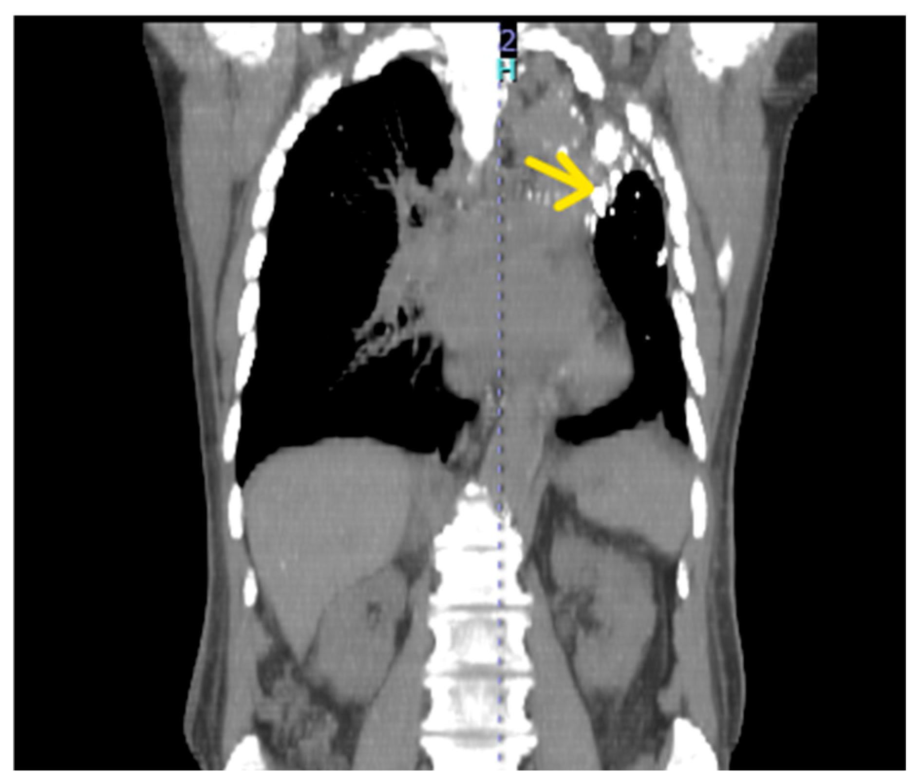
Figure 2.
Bronchoscopy revealed a dark floating blood clot anchored to a small crater in the mucosa of the anteromedial wall of the distal left main bronchus, with an apparent trans-parietal implantation. The clot was deliberately left in situ because of the risk of reinitiating active bleeding. The finding corresponded topographically to the vascular abnormality identified on CT angiography.
Figure 2.
Bronchoscopy revealed a dark floating blood clot anchored to a small crater in the mucosa of the anteromedial wall of the distal left main bronchus, with an apparent trans-parietal implantation. The clot was deliberately left in situ because of the risk of reinitiating active bleeding. The finding corresponded topographically to the vascular abnormality identified on CT angiography.
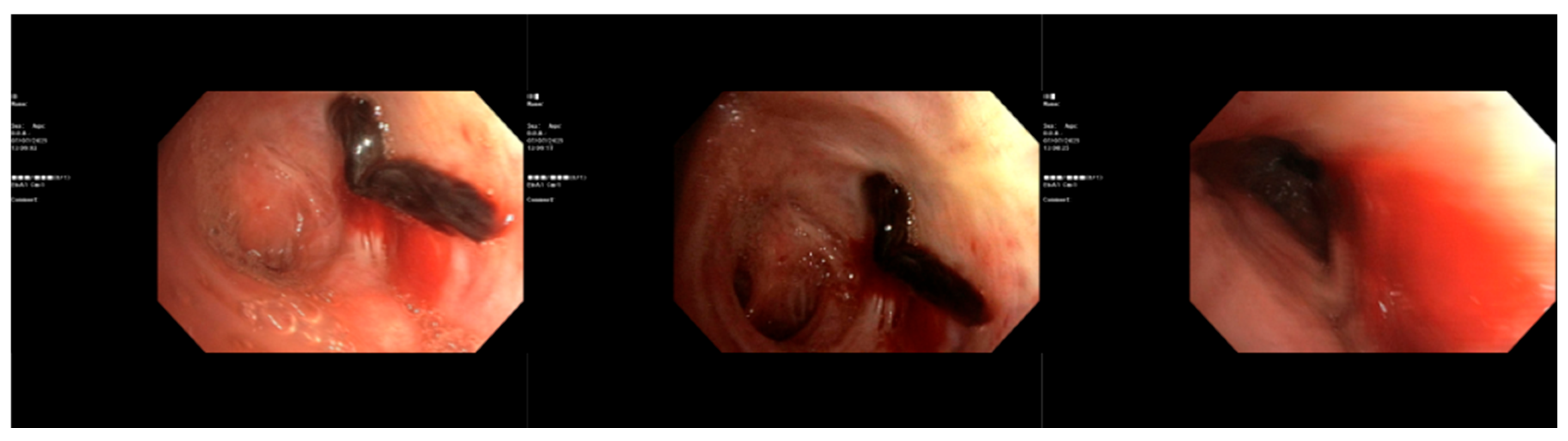
Figure 3.
Selective left bronchial artery angiography, demonstrating opacification of the hypertrophic and tortuous left bronchial artery and the high-flow arteriovenous fistula.
Figure 3.
Selective left bronchial artery angiography, demonstrating opacification of the hypertrophic and tortuous left bronchial artery and the high-flow arteriovenous fistula.
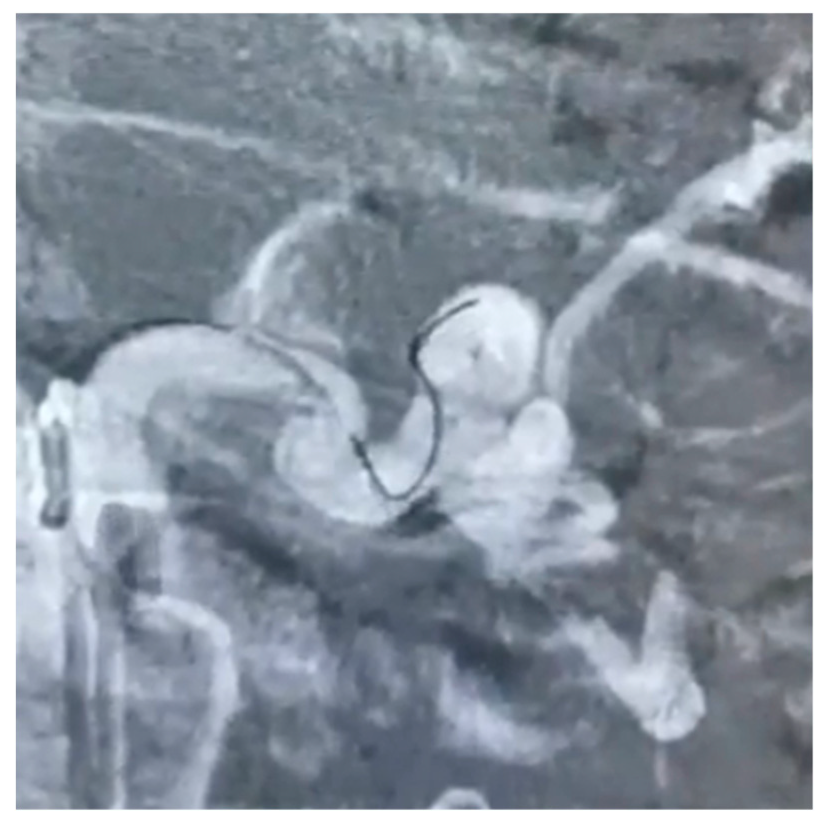
Figure 4.
Embolization of the left bronchial artery with two controlled-release platinum coils (Ruby Soft 4 × 15 mm and 3 × 5 mm). Completion angiography before (A) and after (B) injection of contrast medium demonstrates complete occlusion of the fistulous communication with no residual arteriovenous shunting and no residual opacification of the arteriovenous fistula.
Figure 4.
Embolization of the left bronchial artery with two controlled-release platinum coils (Ruby Soft 4 × 15 mm and 3 × 5 mm). Completion angiography before (A) and after (B) injection of contrast medium demonstrates complete occlusion of the fistulous communication with no residual arteriovenous shunting and no residual opacification of the arteriovenous fistula.
3. Discussion
The lesion described in this case is uncommon; however, its clinical relevance lies less in its rarity than in the diagnostic and therapeutic approach it illustrates. This report uses a discrete high-flow bronchial arteriovenous fistula as a model to examine how lesion phenotype can guide the diagnostic strategy, influence procedural planning, and determine the definition of technical success. The following sections analyse the respective roles and limitations of computed tomography angiography (CTA) and bronchoscopy, the rationale for their simultaneous use, the selection of embolic agents, and the diagnostic measures that prevent complications. Each subsection concludes with a clinically applicable message.
3.1. Lesion Phenotype as the Organising Principle of Diagnosis
The bronchial arteries supply the airways, the vasa vasorum, mediastinal structures and the visceral pleura, and in disease they give rise to the overwhelming majority of severe haemoptysis [6,7]. Sustained inflammation triggers angiogenic remodelling: trunks dilate beyond the accepted 2 mm threshold, become tortuous and generate a peribronchial neovascular network with systemic-to-pulmonary anastomoses [8]. In most patients this yields a diffuse hypervascular territory in which the culprit vessel is inferred from the distribution of parenchymal disease, since active extravasation is visible angiographically in only a minority of cases.
BAVF is the antithesis of that picture: a discrete, anatomically defined, high-flow communication. The published experience is dominated by bronchial artery aneurysms fistulising to the pulmonary artery, in which coil occlusion of the fistulous neck is the technique of choice [9,10]. Lin et al. [11] showed that true aneurysms with a wide neck or a fistulous communication require coils, whereas pseudoaneurysms are adequately treated with particulate or liquid agents. The distinction is diagnostic before it is therapeutic, because it redefines what counts as success: in diffuse hypervascularity, success is devascularisation of a territory; in a fistula, success is complete occlusion of a single conduit with preservation of the parent artery. Applying the first definition to the second lesion yields either an incomplete result or an untreatable vessel.
3.2. Computed Tomography Angiography: What to Acquire, What to Look for, What Is Usually Missed
CTA has displaced both chest radiography and bronchoscopy as the first-line test in haemoptysis of undetermined origin. It identifies aetiology more frequently than endoscopy, characterises the parenchymal substrate and maps arterial anatomy before catheterisation [6]. Le et al. showed prospectively that pre-procedural CTA improves culprit-vessel identification and predicts early recurrence after BAE [14], and a two-centre retrospective study documented shorter procedure and fluoroscopy times with higher technical success when CTA preceded embolisation [15]. The evidence here is consistent and unopposed: CTA should be strongly considered before BAE in haemodynamically stable patients when it can be obtained without delaying haemostatic treatment.
Four reading tasks determine whether that advantage is realised. Localisation identifies the bleeding territory from ground-glass opacity, alveolar filling or endobronchial clot, even when no extravasation is seen. Arterial mapping records the number, origin, calibre and course of the bronchial arteries and of any bronchointercostal or anomalous trunk. The search for non-bronchial systemic arteries—intercostal, internal mammary, inferior phrenic, subclavian, axillary—recruited across a thickened pleura is the task most often omitted and the classical reason for failure or early recurrence after an apparently successful embolisation [7]. The fourth task, specific to aneurysmal and fistulous disease, is the detection of early venous or pulmonary arterial opacification during the arterial phase, which establishes shunt physiology and, as in our patient, changes the embolic plan before the patient reaches the table.
3.3. Bronchoscopy: Timing Matters More Than Yield
The diagnostic contribution of bronchoscopy is time-dependent. Mondoni et al. showed that bronchoscopy performed while bleeding is active or recent substantially increases localisation of the anatomical source, particularly in moderate-to-severe episodes, even though the overall aetiological yield changes little [18]. That distinction is precisely what the interventional radiologist requires: not the cause, but the side and the segment. In the massive scenario the role of endoscopy is therefore topographic and therapeutic rather than aetiological. Sakr and Dutau remains the reference synthesis, positioning rigid bronchoscopy as the instrument of airway control in torrential bleeding, given its suction capacity and its accommodation of tamponade devices, with the flexible scope passed through the rigid barrel for peripheral localisation [12].
The counter-evidence deserves to be taken seriously rather than neutralised. Nishihara et al. questioned the value of routine pre-BAE bronchoscopy in non-intubated patients with bronchiectasis or chronic pulmonary infection, in whom CT already identifies the bleeding side [19]. We regard this finding as compatible with our position rather than contradictory to it. Where disease is diffuse, chronic and unambiguously unilateral on CT, endoscopy adds little and delays treatment. Where the airway is threatened, where laterality is uncertain, or where a discrete high-flow lesion demands confirmation that the correct segment has been devascularised, endoscopy cannot be substituted by imaging. The productive question is not whether bronchoscopy should be performed, but in whom and at what moment.
When bleeding must be controlled before angiography, cold saline lavage, topical vasoconstrictors, balloon blockade and oxidised regenerated cellulose tamponade [13], topical tranexamic acid [20] and, in refractory or recurrent cases, unidirectional endobronchial valves [21] are available. None of these treats the vascular lesion; each buys the diagnostic time needed to find it.
3.4. The Limits and Methodological Value of Simultaneous Bleeding Localization
In extensive neovascularization across multiple systemic arterial territories, precisely localizing the bleeding source is challenging. Bronchoscopy and CT angiography (CTA) are complementary tools that narrow the probable location rather than reliably identifying the exact ruptured vessel. Bronchoscopy excels during active hemorrhage to clear clots, lateralize bleeding, and maintain airway patency, but cannot pinpoint peripheral sources. CTA maps bronchial and non-bronchial systemic arteries, vascular enlargement, shunts, and parenchymal hemorrhage. While CTA localizes the affected lobe in 63–100% of cases, widespread hypertrophied vessels complicate distinguishing the culprit vessel from the abnormal network; thus, these abnormalities are strong anatomical correlates rather than definitive proof [22].
This distinction applies directly here. Bronchoscopy revealed an adherent transparietal clot on the medial wall of the distal left main bronchus, corresponding topographically to a CTA-identified vascular abnormality adjacent to the bronchial wall. This rare anatomical bridge represents a fortunate convergence rather than a standard expectation. Digital subtraction angiography (DSA) is essential for embolization, but direct contrast extravasation into the airway occurs in only 2–10.7% of cases. Angiography more often shows indirect signs like hypervascularity, tortuous arteries, or shunts. Because multiple abnormal vessels may appear to supply the bleeding territory without a visible rupture point, the interventional strategy must integrate angiographic patterns with pre-procedural CTA and bronchoscopic findings.
This case demonstrates the methodological value of performing these complementary assessments simultaneously. Rather than following a sequential model, concurrent bronchoscopy and angiography create a dynamic real-time loop where each modality refines the other. Endoscopic identification of the bleeding site narrows the angiographic search, while angiographic characterization explains endoscopic findings that might otherwise be attributed to common causes of hemoptysis. Here, simultaneous visualization successfully identified the target vessel within an extensive neovascular network.
Continuous endoscopic observation also establishes a direct clinical endpoint: cessation of airway bleeding, whereas angiographic occlusion is merely a surrogate marker of technical success. In our patient, these endpoints coincided within seconds of coil deployment—an observation impossible in a sequential model. Endoscopy also allows immediate assessment for complications like bronchial mucosal ischemia or coil migration, and enables residual clot removal to prevent post-procedural atelectasis and obstructive pneumonia [17]. We therefore propose defining technical success in discrete high-flow lesions by a dual endpoint: angiographic flow arrest combined with an endoscopically verified dry airway and preserved mucosal integrity.
However, this approach carries logistical costs, requiring an angiography suite that accommodates anesthesia and endoscopy, two simultaneous interventional specialists, and a radiation-protection protocol for the endoscopist. Without a hybrid room, intraprocedural cone-beam CT can partly compensate by improving culprit-vessel detection and identifying hazardous radiculomedullary arteries [23,24].
3.5. Phenotype-Directed Selection of the Embolic Agent
BAE achieves immediate haemostasis in the large majority of patients, and meta-analytical data confirm a significant reduction in re-bleeding compared with conservative management [25,26,27]. Recurrence nevertheless remains substantial during long-term follow-up, driven by incomplete devascularisation, recruitment of non-bronchial collaterals and progression of the underlying disease [28]. Agent selection is where phenotype re-enters the argument. Calibrated polyvinyl alcohol particles and microspheres remain the workhorse for diffuse hypervascular territories, occluding at the pre-capillary level and preserving the option of repeat embolisation. N-butyl-2-cyanoacrylate produces faster and more durable occlusion, with lower long-term recurrence than particles in comparative cohorts, at the cost of a steeper learning curve and a risk of catheter adhesion [29,30,31]. Coils occlude proximally and, if used indiscriminately in diffuse disease, may render the parent vessel uncatheterisable—a legitimate objection that must be met directly rather than ignored.
The fistulous phenotype inverts that logic. In a high-flow arteriovenous communication, particles and liquid agents may transit the shunt into the pulmonary or systemic venous circulation, producing non-target embolisation. Detachable platinum microcoils permit deployment, angiographic assessment and repositioning before irreversible release, achieving controlled flow arrest at the fistulous neck while sparing the parent trunk [9,10]. Reported cases of BAVF and bronchial artery aneurysm converge on this strategy [11,32,33]. The choice of coils in our patient was therefore not operator preference but the direct consequence of a diagnostic characterisation made before arterial access was obtained.
3.6. Synthesis of the Evidence on Combined Endoscopic–Endovascular Management
Two comparative studies underpin combined management. Lin et al. found that endobronchial tamponade added to BAE improved oxygenation, accelerated haemostasis and significantly reduced one-year recurrence relative to embolisation alone, without an excess of major adverse events [16]. Li et al. reported that interventional bronchoscopy combined with BAE produced a higher immediate response rate and fewer post-procedural respiratory complications—obstructive pneumonia, consolidation and atelectasis—with favourable mid- and long-term follow-up [17]. Read together, they converge on a coherent mechanism: clearing the airway and controlling flow address two different causes of failure, and neither modality compensates for the other. Read critically, both are retrospective and single-centre, both compare a combined intervention against a historical or contemporaneous single-modality group, and neither uses a common definition of haemostatic success. Allocation bias cannot be excluded, and the magnitude of benefit is therefore uncertain even where its direction is not.
This is the state of the evidence into which our case fits, and it differs from those series in two respects. First, the procedures were simultaneous rather than sequential, permitting bidirectional exchange of information during embolisation rather than before or after it. Second, the target was a discrete high-flow fistula rather than diffuse inflammatory hypervascularity, a setting in which the precision gained from endoscopic confirmation is greatest and the tolerance for error is smallest. The principles established in bronchiectasis and tuberculosis cohorts extend to this setting, but they extend with a narrower margin.
Three questions remain genuinely unanswered. Does simultaneity add anything beyond combination, or is the benefit attributable to airway clearance alone, whenever it is performed? Does endoscopic verification of a dry airway predict one-year recurrence better than angiographic flow arrest? And what is the incremental radiation and anaesthetic cost of the integrated model in unselected patients? None of these questions can be resolved by further case reports.
3.7. Complications Are Prevented Diagnostically, Not Technically
The most feared complication of BAE is spinal cord infarction caused by inadvertent embolisation of a radiculomedullary branch arising from a bronchial or intercostobronchial trunk. A Japanese nationwide observational study established an incidence of approximately 1.4% and identified the anatomical risk factors [34]; recent case-based reviews reinforce the need for meticulous pre-embolisation angiographic scrutiny [35]. Bronchial and oesophageal wall ischaemia, transient chest pain and dysphagia are more common and usually self-limiting. Non-target embolisation, which may be systemic in fistulous lesions, is device-dependent and largely preventable.
Each of these complications has a diagnostic countermeasure rather than a technical rescue: high-resolution pre-procedural CTA to define arterial origins; superselective microcatheter positioning distal to any radiculomedullary branch; intraprocedural cone-beam CT where the origin remains ambiguous [23,24]; retrievable, controlled-release devices in high-flow lesions; and, as illustrated here, direct endoscopic inspection of the bronchial mucosa before the procedure is declared complete.
3.8. The Integrated Endoscopic–Endovascular Workflow
The workflow set out below is the principal deliverable of this paper. It is written to be executable in a conventional angiography suite and specifies, at each step, both the action and the decision that it supports.
- Step 1. Assess severity functionally at first contact—airway compromise, gas exchange, haemodynamics—supported by a validated score rather than by estimated volume [4].
- Step 2. Protect the airway and position the patient with the bleeding side dependent.
- Step 4. Triage by lesion phenotype: diffuse hypervascularity versus discrete aneurysmal or fistulous lesion.
- Step 5. Choose the procedural model: simultaneous bronchoscopy and angiography for unstable patients, ambiguous laterality or a discrete high-flow lesion; direct BAE, with bronchoscopy reserved for failure or recurrence, for stable patients with unequivocal CT localisation of diffuse disease [19].
- Step 6. Select the embolic agent from the phenotype.
- Step 7. Verify both endpoints before terminating the procedure: angiographic flow arrest and endoscopically confirmed cessation of bleeding with intact mucosa.
- Step 8. Follow up in a structured fashion, with clinical review, CTA and bronchoscopy where mucosal integrity is in doubt.
Two boundaries deserve emphasis, since a workflow without contraindications is a slogan. The integrated model should not be used when it would delay embolisation in a patient with unambiguous diffuse disease, and it should not be attempted where anaesthetic support, radiation protection for the endoscopist or a second interventionalist cannot be guaranteed. In those settings the sequential model, with cone-beam CT compensating for the missing endoscopic information, remains appropriate.

Table 1.
Schematic workflow and procedural model for endovascular management of life-threatening haemoptysis.
Table 1.
Schematic workflow and procedural model for endovascular management of life-threatening haemoptysis.
| Phase | Step | Actions and key points |
| Initial assessment and management | 1. Assess severity functionally at first contact | Assess airway compromise, gas exchange and haemodynamics; classification supported by a validated score rather than by estimated expectorated volume. |
| 2. Airway protection and patient positioning | Protect the airway; position the patient with the bleeding side dependent. | |
| Diagnostic evaluation | 3. CT angiography (CTA) | Perform contrast-enhanced CTA in every stable patient and read it against a structured checklist (localisation, arterial mapping, non-bronchial systemic supply, shunt physiology). |
| Procedural triage and action | 4. Triage by lesion phenotype | Distinguish diffuse hypervascularity from a discrete aneurysmal or fistulous lesion. |
| 5. Choose the procedural model | Unstable patient, ambiguous laterality or discrete high-flow lesion: simultaneous bronchoscopy and angiography. Stable patient with unequivocal CT localisation of diffuse disease: direct BAE, with bronchoscopy reserved for failure or recurrence. | |
| 6. Select the embolic agent | Derive the agent from the phenotype (particle size, coils or liquid agents). | |
| Post-procedural management | 7. Verify procedural endpoints | Angiographic flow arrest together with endoscopically confirmed cessation of bleeding and intact mucosa. |
| 8. Structured follow-up | Clinical review, CTA and bronchoscopy where mucosal integrity is in doubt. |
3.9. Rescue Strategies in Refractory Bleeding
When bleeding persists despite technically successful embolisation, the differential diagnosis comprises recruitment of non-bronchial systemic collaterals, a pulmonary arterial source such as a Rasmussen aneurysm, or a coagulopathic contribution. Each demands a different diagnostic response, not a repetition of the same procedure. Veno-venous extracorporeal membrane oxygenation, including anticoagulation-free protocols, has been used successfully as a bridge to definitive embolisation or resection when airway flooding precludes conventional ventilation [36,37]. Emergency surgical resection is now reserved for refractory bleeding or for lesions that are inherently surgical [2].
3.10. Limitations
This is a single case, and no causal inference about the superiority of simultaneous over sequential management can be drawn from it. The comparative literature supporting combined endoscopic–endovascular treatment is retrospective, single-centre and heterogeneous in its definitions of haemostatic success [16,17]. BAVF is rare, and the published experience consists of case reports and small series subject to publication bias favouring success [9,10,11]. The simultaneous model also presupposes resources—an appropriately equipped suite, anaesthetic support and two immediately available interventionalists—that are not universally accessible, which limits external validity. Prospective multicentre registries with predefined endpoints, including one-year recurrence, radiation dose and a standardised definition of endoscopic success, are required before this workflow can be recommended as a standard of care.
4. Conclusions
Life-threatening haemoptysis has long been treated as a haemostatic problem with a diagnostic prelude. This case argues for the reverse order. The phenotype identified on CTA determined the embolic device; the segment identified endoscopically determined where to look angiographically; and continuous airway visualisation converted a surrogate angiographic endpoint into a verified clinical one, while allowing immediate assessment of bronchial mucosal integrity and other endobronchial complications.
What follows is a change of model rather than of technique. Bronchoscopy and bronchial artery embolisation are still organised as consecutive procedures owned by different specialties and separated by transport, handover and time that the patient may not have. Performed together, they behave as a single diagnostic–therapeutic act with a dual endpoint, and the information each generates becomes immediately actionable by the other. We have set out the components required to reproduce that act: a phenotype-driven algorithm, a structured CTA reading checklist, an embolic-agent selection framework, and explicit criteria for when the integrated pathway should and should not be used.
Whether simultaneity improves one-year recurrence, and at what radiation and organisational cost, remains to be established in prospective multicentre registries with standardised endpoints. Until those data exist, the argument for integration rests on mechanism and on consistency of direction across the available evidence—a reasonable basis for adopting a workflow, and an insufficient one for declaring a standard. Centres treating these emergencies should nonetheless begin to measure their outcomes against both endpoints, angiographic and endoscopic. Pulmonary vascular emergencies are unlikely to be solved by better devices alone; they will be solved by better-integrated teams using the devices they already have.
Author Contributions
Conceptualization: C.C., D.L. and F.M.; Formal Analysis: G.P. and G.T.; Data Curation: G.P. and M.N.; Writing—Original Draft Preparation: G.P. and G.T.; Writing—Review & Editing: G.P. and C.C. All authors have read and agreed to the published version of the manuscript.
Funding
This research received no external funding.
Institutional Review Board Statement
Ethics committee review and approval were not required for this case report, as clinical management followed guidelines and routine clinical practice.
Informed Consent Statement
Written informed consent was obtained from patient’s next-of-kin for publication of the details of medical case and accompanying images.
Data Availability Statement
Anonymized data showed are available to editors, reviewers, and readers.
Conflicts of Interest
The authors declare no conflict of interest.
Abbreviations
BAE, bronchial artery embolisation; BAVF, bronchial arteriovenous fistula; CTA, computed tomography angiography; NBCA, N-butyl-2-cyanoacrylate; PVA, polyvinyl alcohol.
References
- Kathuria, H.; Hollingsworth, H.M.; Vilvendhan, R.; Reardon, C. Management of life-threatening hemoptysis. J. Intensive Care 2020, 8, 23. [Google Scholar] [CrossRef] [PubMed]
- Radchenko, C.; Alraiyes, A.H.; Shojaee, S. A systematic approach to the management of massive hemoptysis. J. Thorac. Dis. 2017, 9 (Suppl. 10), S1069–S1086. [Google Scholar] [CrossRef] [PubMed]
- Charya, A.V.; Karim, N.A.; Wang, C.; Choi, J.; Grage, R. Management of life-threatening hemoptysis in the ICU. J. Thorac. Dis. 2021, 13, 5139–5158. [Google Scholar] [CrossRef] [PubMed]
- Fartoukh, M.; Khoshnood, B.; Parrot, A.; Khalil, A.; Carette, M.-F.; Stoclin, A.; Mayaud, C.; Cadranel, J.; Ancel, P.Y. Early prediction of in-hospital mortality of patients with hemoptysis: an approach to defining severe hemoptysis. Respiration 2012, 83, 106–114. [Google Scholar] [CrossRef] [PubMed]
- Mondoni, M.; Carlucci, P.; Job, S.; Parazzini, E.M.; Cipolla, G.; Pagani, M.; Tursi, F.; Negri, L.; Fois, A.; Canu, S.; et al. Observational, multicentre study on the epidemiology of haemoptysis. Eur. Respir. J. 2018, 51, 1701813. [Google Scholar] [CrossRef] [PubMed]
- Ketai, L.H.; Mohammed, T.L.; Kirsch, J.; Kanne, J.P.; Chung, J.H.; Donnelly, E.F.; Ginsburg, M.E.; Heitkamp, D.E.; Henry, T.S.; Kazerooni, E.A.; et al. ACR Appropriateness Criteria Hemoptysis. J. Am. Coll. Radiol. 2020, 17, S148–S159. [Google Scholar] [CrossRef] [PubMed]
- Li, H.; Ding, X.; Zhai, S.; Gao, K. A retrospective study on the management of massive hemoptysis by bronchial artery embolization: risk factors associated with recurrence of hemoptysis. BMC Pulm. Med. 2023, 23, 87. [Google Scholar] [CrossRef] [PubMed]
- Almeida, J.; Leal, C.; Figueiredo, L. Evaluation of the bronchial arteries: normal findings, hypertrophy and embolization in patients with hemoptysis. Insights Imaging 2020, 11, 70. [Google Scholar] [CrossRef] [PubMed]
- Li, Y.; Gu, G.C.; Liu, B.; Shao, J.; Chen, Y.; Zheng, Y.H. Endovascular treatment of multiple bronchial artery aneurysms with prominent fistula to pulmonary artery in a patient with interstitial lung disease: a case report and literature review. Vasc. Endovasc. Surg. 2019, 53, 499–503. [Google Scholar] [CrossRef] [PubMed]
- Hsieh, C.G.; Le, T.; Fogelfeld, K.; Kamangar, N. Bronchial artery aneurysm with associated bronchial artery to pulmonary artery fistula: treatment by embolization. J. Clin. Imaging Sci. 2017, 7, 2. [Google Scholar] [CrossRef] [PubMed]
- Lin, J.-L.; Ji, Y.-Y.; Zhang, M.-Z.; Tang, Y.; Wang, R.-L.; Ruan, D.-D.; Zhou, Y.-F.; Wu, S.-J.; Cai, S.-L.; Zhang, J.-H.; et al. Rare cases of bronchial aneurysm and comparison of interventional embolization in the treatment of true bronchial aneurysm and pseudobronchial aneurysm. Front. Cardiovasc. Med. 2022, 9, 856684. [Google Scholar] [CrossRef] [PubMed]
- Sakr, L.; Dutau, H. Massive hemoptysis: an update on the role of bronchoscopy in diagnosis and management. Respiration 2010, 80, 38–58. [Google Scholar] [CrossRef] [PubMed]
- Valipour, A.; Kreuzer, A.; Koessler, W.; Vonbank, K.; Burghuber, O.C. Bronchoscopy-guided topical hemostatic tamponade therapy for the management of life-threatening hemoptysis. Chest 2005, 127, 2113–2118. [Google Scholar] [CrossRef] [PubMed]
- Le, H.Y.; Le, V.N.; Pham, N.H.; Phung, A.T.; Nguyen, T.T.; Do, Q. Value of multidetector computed tomography angiography before bronchial artery embolization in hemoptysis management and early recurrence prediction: a prospective study. BMC Pulm. Med. 2020, 20, 231. [Google Scholar] [CrossRef] [PubMed]
- Ravetta, P.; Vouche, M. Added value of computed tomography angiography prior to bronchial artery embolization for hemoptysis: a retrospective two-center study. J. Belg. Soc. Radiol. 2024, 108, 1. [Google Scholar] [CrossRef] [PubMed]
- Lin, C.; Chen, Y.; Cai, D.; Chen, Z.; Peng, Z.; Lai, H.; Liu, D. The efficacy and safety of combined therapy with endobronchial tamponade and bronchial artery embolization for massive hemoptysis. BMC Pulm. Med. 2024, 24, 314. [Google Scholar] [CrossRef] [PubMed]
- Li, K.; Lian, T.; Wang, Z.; Zhang, Z.; Wang, W.; Qian, J. Short-term prognosis, mid- and long-term clinical follow-up of bronchoscopic intervention therapy combined with bronchial artery embolization on massive hemoptysis. Front. Surg. 2025, 12, 1642449. [Google Scholar] [CrossRef] [PubMed]
- Mondoni, M.; Carlucci, P.; Cipolla, G.; Fois, A.; Gasparini, S.; Marani, S.; Centanni, S.; Sotgiu, G. Bronchoscopy to assess patients with hemoptysis: which is the optimal timing? BMC Pulm. Med. 2019, 19, 36. [Google Scholar] [CrossRef] [PubMed]
- Nishihara, T.; Ishikawa, H.; Tsuyuguchi, K.; Fukuda, S.; Sumikawa, H. Reevaluating the role of bronchoscopy prior to bronchial artery embolization in nonintubated patients with hemoptysis due to bronchiectasis and chronic pulmonary infection. CHEST Pulm. 2025, 3, 100128. [Google Scholar] [CrossRef] [PubMed]
- Korraa, E.; Madkour, A.; Galal, I.; El-Saidy, I.M.I. Bronchoscopic instillation of tranexamic acid to control bronchopulmonary bleeding. Egypt. J. Bronchol. 2017, 11, 185–190. [Google Scholar] [CrossRef] [PubMed]
- Patel, B.; Abi-Fadel, D.; Rosenheck, J.; Bartter, T.; Boujaoude, Z.; Abouzgheib, W. Endobronchial valves for treatment of hemoptysis. J. Bronchol. Interv. Pulmonol. 2019, 26, e22–e24. [Google Scholar] [CrossRef] [PubMed]
- Meena, P.; Bhalla, A.S.; Goyal, A.; Naranje, P.; Kumar, K.P. Imaging findings as predictors of the site of bleeding in patients with hemoptysis: Comparison between split-bolus dual-energy CT angiography and digital subtraction angiography. Diagn. Interv. Radiol. 2022, 28, 344–351. [Google Scholar] [CrossRef] [PubMed]
- Grosse, U.; Grözinger, G.; Syha, R.; Ketelsen, D.; Partovi, S.; Nikolaou, K.; Hoffmann, R. Intra-procedural bronchial artery embolization planning: the usefulness of cone-beam CT. Acta Radiol. 2019, 60, 1438–1444. [Google Scholar] [CrossRef] [PubMed]
- Zhang, Q.; Li, J.; He, G.; Tang, J.; Zhang, G. Utility of intra-procedural cone-beam computed tomography imaging for the determination of the artery of Adamkiewicz suspected by angiography during transarterial embolization for hemoptysis. Diagn. Interv. Radiol. 2023, 29, 713–718. [Google Scholar] [CrossRef] [PubMed]
- Panda, A.; Bhalla, A.S.; Goyal, A. Bronchial artery embolization in hemoptysis: a systematic review. Diagn. Interv. Radiol. 2017, 23, 307–317. [Google Scholar] [CrossRef] [PubMed]
- Fan, S.; Cheng, X.; Wang, X.; Liu, Y.; He, W.; Chen, H. Bronchial artery embolization versus conservative treatment for hemoptysis: a systematic review and meta-analysis. BMC Pulm. Med. 2024, 24, 428. [Google Scholar] [CrossRef] [PubMed]
- Ravetta, P.; Bruyneel, M.; Vouche, M. New insights on bronchial artery embolization (BAE) for hemoptysis: a systematic review. BMC Pulm. Med. 2025, 25, 541. [Google Scholar] [CrossRef] [PubMed]
- Frood, R.; Karthik, S.; Mirsadraee, S.; Clifton, I.; Flood, K.; McPherson, S.J. Bronchial artery embolisation for massive haemoptysis: immediate and long-term outcomes—a retrospective study. Pulm. Ther. 2020, 6, 107–114. [Google Scholar] [CrossRef] [PubMed]
- Taninokuchi Tomassoni, M.; Perini, D.; Porta, F.; Braccischi, L.; Zanella, S.; Basile, A.; Modestino, F.; Mosconi, C. Bronchial artery embolization for the treatment of hemoptysis: permanent versus temporary embolic materials, a single center study. CVIR Endovasc. 2023, 6, 32. [Google Scholar] [CrossRef]
- Lee, J.H.; Yoon, C.J.; Jung, Y.S.; Choi, W.S.; Lee, C.-H.; Lee, G.M. Comparison of n-butyl-2-cyanoacrylate and polyvinyl alcohol particles for bronchial artery embolisation in primary lung cancer: a retrospective cohort study. Respir. Res. 2022, 23, 257. [Google Scholar] [CrossRef] [PubMed]
- Mazıcan, M.; Karluka, I.; Fındıkcıoglu, A.; Andıc, C. Bronchial artery embolization in patients with life-threatening massive hemoptysis: comparison of the efficacy and safety of particulate embolizing agents and n-2-butyl-cyanoacrylate. Eur. Rev. Med. Pharmacol. Sci. 2024, 28, 310–318. [Google Scholar] [CrossRef] [PubMed]
- Li, X.; Zhang, H.; Ma, W.; Lv, F.; Zhou, W. Case report: combined transcatheter arterial embolization and aortic stent-graft have better efficacy for bronchial artery aneurysms. Front. Cardiovasc. Med. 2024, 11, 1328674. [Google Scholar] [CrossRef] [PubMed]
- Suzuki, K.; Kai, R.; Hirano, T.; Yagi, M.; Dohi, K. Bronchial arterial embolization for an unruptured bronchial artery aneurysm in patient with hemoptysis. Intern. Emerg. Med. 2024, 19, 2079–2080. [Google Scholar] [CrossRef] [PubMed]
- Ishikawa, H.; Ohbe, H.; Omachi, N.; Morita, K.; Yasunaga, H. Spinal cord infarction after bronchial artery embolization for hemoptysis: a nationwide observational study in Japan. Radiology 2021, 298, 673–679. [Google Scholar] [CrossRef] [PubMed]
- Ghouri, R.G.; Ali, M.M.; Naeem, H.; Raza, M.S.; Arshad, W. Anterior spinal artery syndrome after bronchial embolization for hemoptysis: a case report and literature review. BMJ Neurol. Open 2024, 6, e000684. [Google Scholar] [CrossRef] [PubMed]
- Kimura, T.; Kobayashi, T.; Kobori, F.; Goto, M.; Takemitsu, M.; Tanaka, A.; Sato, J.; Isomine, S. Massive hemoptysis managed by prolonged anticoagulation-free veno-venous extracorporeal membrane oxygenation with which thoracic surgeries were accompanied: a case report. JA Clin. Rep. 2022, 8, 11. [Google Scholar] [CrossRef] [PubMed]
- Ryan, D.G.; Miller, K.A.; Capaldi, C.; Pasquarello, C.; Yang, Q.; et al. Massive hemoptysis bridged with VV-ECMO: a case report. Front. Cardiovasc. Med. 2022, 9, 997990. [Google Scholar] [CrossRef] [PubMed]
Disclaimer/Publisher’s Note: The statements, opinions and data contained in all publications are solely those of the individual author(s) and contributor(s) and not of MDPI and/or the editor(s). MDPI and/or the editor(s) disclaim responsibility for any injury to people or property resulting from any ideas, methods, instructions or products referred to in the content. |
© 2026 by the authors. Licensee MDPI, Basel, Switzerland. This article is an open access article distributed under the terms and conditions of the Creative Commons Attribution (CC BY) license (http://creativecommons.org/licenses/by/4.0/).
Copyright: This open access article is published under a Creative Commons CC BY 4.0 license, which permit the free download, distribution, and reuse, provided that the author and preprint are cited in any reuse.