

Submitted:
02 November 2026
Posted:
05 November 2026
You are already at the latest version
Abstract
Background: Perinatal asphyxia is a critical condition resulting from various complications around birth. Despite advances in neonatal care reliable prediction of long-term neurodevelopmental outcomes remains challenging. Therefore, standardized clinical follow-up assessments are recommended to detect impairments early and provide optimal support. This study aimed to characterize five-year neurodevelopmental outcomes in children with history of perinatal asphyxia treated at a tertiary neonatal care center in Austria.
Methods: This retrospective analysis included infants with perinatal asphyxia, defined as umbilical artery pH < 7.0, admitted to the neonatal intensive care unit in Graz, Austria, between January 2004 and September 2020. The primary outcome was neurodevelopmental status at five years of age. This was assessed by trained clinicians during routine follow-up examinations and classified as age-appropriate development or mild, moderate, or severe developmental delay. Where feasible, the intelligence quotient was additionally quantified by certified psychologists using the standardized Wechsler Preschool and Primary Scale of Intelligence (WPPSI).
Demographic characteristics, causes of asphyxia, neonatal clinical course and short-term outcomes were also assessed. Data were extracted from the local electronic patient documentation system and analyzed descriptively.
Results: Of 139 eligible neonates, 25 (18%) died. Five-year follow-up data were available for 35 (31%) of the 114 survivors, their median gestational age was 36.6 (IQR 31.3–39.3) weeks; 16 (46%) were term infants and 19 (54%) preterm infants. Hypoxic-ischemic encephalopathy was diagnosed in 19 (54%) infants. 10 (29%) infants received therapeutic hypothermia, and 30 (86%) required invasive respiratory support.
At five years, five (31%) term infants achieved age-appropriate development, while five (31%) had mild, three (19%) moderate, and three (19%) severe developmental delay. Among preterm infants, four (21%) showed age-appropriate development, whereas 10 (53%) had mild, three (16%) moderate, and two (11%) severe developmental delay.
Conclusion: Developmental delay was common among both term and preterm infants assessed five years after perinatal asphyxia. However, the small cohort and incomplete follow-up limit the generalizability of these findings. Improved strategies for risk stratification, neuroprotective interventions and long-term developmental surveillance are warranted.
Keywords:
neonate
; preterm infants
; term infants
; perinatal asphyxia
; neonatal resuscitation
; long-term neurodevelopmental outcome
; neonatal intensive care
Introduction
Perinatal asphyxia occurs in 1 to 6 per 1000 live births and accounts for approximately 23% of neonatal deaths, making it the third leading cause of neonatal mortality worldwide, according to the World Health Organization (WHO) [1,2]. Asphyxia results from various events in utero, during labor and immediately after delivery, including placental abruption, umbilical cord complications, meconium aspiration, transplacental anesthetic transmission affecting the respiratory drive and traumatic birth [3]. These events can impair oxygen delivery and tissue perfusion, leading to hypoxemia, acidemia, and multiorgan dysfunction, including hypoxic-ischemic encephalopathy (HIE) [4,5]. The diagnosis of perinatal asphyxia is based on a combination of clinical findings, including the Apgar score, umbilical cord blood gas analysis and a detailed neurological assessment, including scores like the Sarnat score for grading HIE [5]. HIE is one of the primary causes of severe neurological impairment in childhood and occurs in approximately 28% of infants affected by perinatal asphyxia [2,4]. Hypoxia-induced brain injury may result in acute complications such as seizures and impaired consciousness. In the long term, it is associated with an increased risk of permanent neurological impairments, including cerebral palsy, epilepsy and cognitive deficits [6].
Management of infants affected by perinatal asphyxia begins immediately after birth, with particular attention to appropriate monitoring, respiratory and cardiocirculatory support as well as adequate thermoregulation [7]. Therapeutic hypothermia is the standard of care for infants born at ≥ 36 weeks of gestation with moderate-to-severe HIE who meet established eligibility criteria [8]. When initiated within six hours after birth to a target core temperature of 33.5 °C, therapeutic hypothermia reduces brain injury and improves neurodevelopmental outcomes by reducing cerebral inflammation and free radical formation [9].
Outcome of infants diagnosed with perinatal asphyxia vary widely, ranging from age-appropriate development and mild developmental delay to severe neurological impairments, cerebral palsy, seizures or death, especially in infants with severe HIE [10,11]. Developmental follow-up assessments are essential in severely affected infants to monitor the progression and extent of impairment over time and to ensure optimal guidance for the child and parents [12]. In infants with milder involvement, cognitive deficits may only emerge after toddlerhood as cognitive demands increase [13]. Therefore, continued observation is important to assess the developmental status and to provide appropriate educational and therapeutic support for the child in later life [14]. The most commonly used neurodevelopmental assessment tools performed at two years of age are the Griffiths Scales of Child Development and the Bayley Scales of Infant and Toddler Development [11]. At five to six years of age, children are evaluated by standardized intelligence tests. The Wechsler Preschool and Primary Scale of Intelligence (WPPSI) evaluates verbal comprehension, visual-spatial ability, fluid reasoning, working memory, processing speed and provides a full-scale IQ value. Diagnostic testing is particularly important to assess a child’s cognitive and behavioral abilities and provides decision support on whether a child is ready for school entry [15,16].
Despite broad early follow-up studies, evidence on the neurodevelopmental outcomes of children at five years of age after perinatal asphyxia across different gestational age groups remain limited. Assessments at five years enable a more reliable evaluation of cognitive function and school readiness than assessments during infancy. Therefore, this study aimed to characterize long-term developmental outcomes at five years of age in infants affected by perinatal asphyxia (umbilical artery pH < 7.0), integrating cognitive performance, cerebral abnormalities and developmental impairment. Outcomes were stratified by gestational age to evaluate potential differences between term and preterm infants.
Methods
Study Design
This retrospective analysis included infants diagnosed with perinatal asphyxia who were admitted to the tertiary neonatal intensive care unit (NICU) at the Medical University of Graz. The study was conducted in accordance with the Declaration of Helsinki and approved by the Ethics Committee of the Medical University of Graz (ethics committee number: 36-130-ex 23/24).
Participants and Data Collection
Infants admitted to the NICU at the Medical University of Graz between January 2004 and September 2020 were initially identified using the International Classification of Diseases 10th Revision (ICD-10) codes for severe (P21.0), mild and moderate (P21.1), and unspecified perinatal asphyxia (P21.9). Their medical records were subsequently reviewed to verify eligibility based on a documented umbilical artery pH < 7.0, which served as a surrogate for perinatal asphyxia. Ten infants were excluded a priori because of incomplete clinical data. The reasons for exclusion were an unavailable umbilical artery pH (n = 8) and initial postnatal management at an external hospital (n = 2). Surviving children with available neurodevelopmental assessments at five years of age were included in the long-term follow-up analysis.
Data were extracted from electronic medical records in the openMEDOCS documentation system (Steiermaerkische Krankenanstaltengesellschaft, Graz, Austria) and anonymously stored in Microsoft Excel (Version 16, Microsoft Corporation, Redmond, WA, USA).
Outcome Parameter
The primary outcome was the neurodevelopmental status at five years of age in children with a history of perinatal asphyxia, assessed by trained clinicians during routine follow-up examinations. Outcomes were categorized as age-appropriate development or mild, moderate or severe developmental delay. This categorization reflected the clinical impression as documented in the medical records and did not follow a predefined, standardized assessment scale. The presence of cerebral palsy, epilepsy or microcephaly was also documented. Where feasible, the intelligence quotient (IQ) was evaluated by certified psychologists using the standardized WPPSI in 3rd and 4th edition. IQ testing was performed independently of the clinically assigned neurodevelopmental outcome category.
Data were stratified by gestational age into preterm (delivered < 37+0) and term infants. Preterm infants were further classified as moderate-to-late preterm infants (MLPI), born ≥ 32+0 and < 37+0 weeks of gestation, very preterm infants (VPI) born ≥ 28+0 and < 32+0 weeks of gestation and extremely preterm infants (EPI) < 28+0 weeks of gestation.
Demographic and perinatal characteristics included gestational age, sex, birthweight, birth length, head circumference, Apgar score, umbilical artery pH, and mode of delivery. Moreover, causes of asphyxia and the neonatal clinical course were also assessed, including therapeutic hypothermia, respiratory support, catecholamine requirement, seizures, hypoglycemia, renal, hepatic or cardiovascular dysfunction and HIE. The severity of HIE was graded using the Sarnat classification. Length of hospital stay and short-term outcomes at hospital discharge, including in-hospital mortality, were also analyzed. Mortality was assessed in the entire eligible cohort (n=139).
Statistics
Statistical analyses were performed using Microsoft Excel (Version 16, Microsoft Corporation, Redmond, WA, USA) together with IBM SPSS Statistics (Version 29, IBM Corp., Armonk, NY, USA). Primary outcome data were analyzed using descriptive statistics, including absolute and relative frequencies. Developmental outcomes were compared between term and preterm infants using the Fisher’s exact test. The presence of cerebral palsy, epilepsy and microcephaly was compared between groups accordingly. All tests were two-sided, and a p-value < 0.05 was considered statistically significant. Additionally, bar charts were used to visualize the results. Demographic data and secondary outcomes were summarized using median and interquartile range (IQR), as well as absolute and relative frequencies.
Results
In total, 13,130 infants were admitted to the NICU between January 2004 and September 2020. Of these, 149 infants had a documented diagnosis of perinatal asphyxia. After exclusion of 10 infants with incomplete medical records, a total of 139 patients with an umbilical artery pH < 7.0 were eligible for the study. (Figure 1).
Of 139 infants eligible, 25 (18%) died during initial hospitalization after birth. Five-year neurodevelopmental follow-up data were unavailable for 79 (69%) of the 114 survivors. The remaining 35 children (31% of survivors) were included in the primary outcome analysis. Demographic and perinatal characteristics are presented in Table 1.
Five-Year Outcome Analysis
Thirty-five infants with a median gestational age of 36.6 weeks (IQR 31.3–39.3) were included in the five-year outcome analysis. Sixteen (46%) infants were born at term. Among preterm infants, 9 (47%) were classified as MLPI, 7 (37%) as VPI and 3 (16%) as EPI. Five-year neurodevelopmental outcomes are presented in Table 2 and Figure 2.
Among the children included in the primary outcome analysis, neurodevelopmental assessment using the WPPSI was performed in 10 children (two born at term and eight born preterm) and revealed a median IQ of 95 (IQR 85–105), with individual scores ranging from 76 to 114. None of the developmental outcome measures differed significantly between term and preterm infants.
Causes and Associated Risk Factors of Perinatal Asphyxia
In the study cohort (n=35), the most frequent cause of perinatal asphyxia was placental abruption, observed in 11 (31%) infants. Prolonged labor occurred in three (9%) and meconium aspiration in four (11%) of all infants. Documented umbilical cord complications included nuchal cord in two cases (6%) and velamentous cord insertion in one (3%). Delivery-related complications, such as shoulder dystocia was identified in one (3%) infant. Regarding antenatal risk factors, preeclampsia was present in the mothers of two (6%) infants, whereas placental insufficiency was documented in one (3%) and chorioamnionitis in one (3%) pregnancy. Additionally, maternal uterine rupture occurred in one case (3%). Other causes of perinatal asphyxia were reported in eight (23%) infants. When comparing term and preterm infants, term infants more frequently experienced meconium aspiration and prolonged labor whereas placental abruption and preeclampsia were more common among preterm infants. Other causes of asphyxia were similarly distributed between the two groups.
Clinical Course During NICU Hospitalization
The frequencies of therapeutic interventions, mechanical ventilation, medication use, organ involvement, seizures, and hypoglycemia are shown in Table 3. The incidence of renal dysfunction increased with decreasing gestational age. Likewise, the duration of hospitalization increased with decreasing gestational age, with preterm infants requiring longer hospital stays than term infants. Hypoglycemia also occurred more frequently in preterm than in term infants, with the highest rate observed in the VPI group.
Among the 10 infants (median gestational age 39.1 (IQR 36.9 – 41.3)) who received therapeutic hypothermia during the neonatal period, four (40%) infants demonstrated age-appropriate development at five years, four (40%) had mild developmental delay, and two (20%) had severe developmental delay. In comparison, among the 25 infants (median gestational age 33.0 (IQR 24.6 – 41.3)) who did not receive therapeutic hypothermia, five (20%) showed age-appropriate development, 11 (44%) had mild, six (24%) moderate and three (12%) severe developmental delay.
Short-Term Outcome at Discharge
HIE was diagnosed in 19 (54%) of the 35 infants. Table 4 presents the distribution of HIE severity according to the Sarnat classification across gestational age groups.
Cranial magnetic resonance imaging (cMRI) was performed in 7 (20%) infants from the follow-up cohort at a median age of 8 days (range 5–16 days). Findings were unremarkable in one (14%) infant. Among the remaining six (86%) infants, extra-axial intracranial hemorrhage was identified in three (50%) cases, and one (17%) infant showed residual hemorrhage in the form of hemosiderin deposits. Hypoxic-ischemic brain lesions were detected in one (17%) infant, while another exhibited cerebral edema.
Mortality
Among the 139 eligible infants, 25 (18%) died during NICU hospitalization. The median age at death was 2 days (range 1–7 days). The mortality was 14% (12 of 86) among term infants and 25% (13 of 53) among preterm infants (n = 53), reaching 40% (4 of 10) in the extremely preterm subgroup. The most frequent cause of death was multi-organ failure, occurring in 12 (48%) infants, frequently in combination with intraventricular hemorrhage. Six (24%) infants sustained severe hypoxic-ischemic brain injury, after which care was redirected to palliative measures. Additional causes of death included isolated cardiovascular or pulmonary failure and therapy-refractory status epilepticus.
Discussion
This study describes five-year neurodevelopmental outcomes in children born at term or preterm who were treated for perinatal asphyxia in a tertiary neonatal care center in Austria. In this retrospective cohort, 25 of 139 eligible infants (18%) died during neonatal hospitalization. Five-year neurodevelopmental follow-up data were available for 35 of the 114 survivors (31%). Developmental delay was identified in 26 of the 35 children assessed (74%), with mild delay being the most common finding. No statistically significant differences in neurodevelopmental outcomes were observed between children born at term and those born preterm, although the small sample size and incomplete follow-up limit interpretation.
Previous studies have reported similar findings regarding severe developmental delay following perinatal asphyxia. Blommaert et al. documented long-term neurodevelopmental outcomes in 63 children with history of HIE who were treated with therapeutic hypothermia, reporting severe developmental delay in 14% and cerebral palsy in 14% of these children at five years of age [17]. These proportions were numerically similar to those observed in the present study, whereas age-appropriate development was reported in 52% of their cohort and 26% in the present study. These descriptive comparisons should be interpreted cautiously because of differences in outcome definitions and cohort characteristics. Blommaert et al. included only infants born at ≥ 35 weeks of gestation. Furthermore, all infants in their cohort received therapeutic hypothermia, compared with only 29% in the present cohort [17]. Similarly, Cainelli et al. reported a median full-scale IQ of 100 (IQR 87–110) in 40 children with history of HIE treated with therapeutic hypothermia and found no significant differences compared with healthy controls [18]. Compared with the present study, the IQ values reported by Cainelli et al. were slightly higher. However, their cohort consisted exclusively of infants born at > 36 weeks of gestation and excluded children with major neurological impairments such as cerebral palsy or epilepsy [18]. In contrast, Murray et al. compared 46 infants with HIE to a healthy control cohort at five years of age and demonstrated significantly lower full-scale IQ scores in the HIE group. The median WPPSI IQ in the HIE group was 99 (IQR 94–124), which was higher than that observed in the present study [19]. Additionally, Dzikiene et al. reported a mean IQ of 87 (SD 16.9) in 32 children with a history of perinatal asphyxia assessed at 8 to 9 years of age, which was significantly lower than that of healthy controls and also lower than the median IQ observed in the present study; notably, none of the children in their cohort had received therapeutic hypothermia during the neonatal period [20]. Overall, present findings are broadly consistent with those reported in previous cohorts although differences in gestational age, treatment strategies, inclusion criteria, and outcome assessments limit direct comparisons. This underscores the need for standardized diagnostic criteria, uniform treatment protocols and structured long-term follow-up assessments to improve the comparability across future studies and ultimately optimize care for affected infants.
A particular strength of the present study is the inclusion of preterm infants, a population that has been underrepresented in previous studies on long-term outcomes following perinatal asphyxia. Accordingly, Robertsson Gossmann et al. investigated long-term outcomes following HIE treated with therapeutic hypothermia, including late preterm infants. Their results demonstrated that all included infants born < 36 weeks of gestation experienced adverse outcomes, suggesting that prematurity may aggravate the consequences of perinatal asphyxia [13]. Although the neurodevelopmental outcomes did not differ significantly between term and preterm infants in the present study, adverse outcomes and the need for intensive therapeutic interventions tended to be more frequent among preterm infants. For example, none of the EPI and only 14% of VPI achieved age-appropriate development, compared with 31% of term infants. Likewise, the need for mechanical ventilation, administration of catecholamines and the duration of hospitalization appeared to increase with prematurity. These findings are consistent with those of Logitharajah et al., who reported more severe outcomes in preterm infants (≤ 36+0 weeks of gestation) with perinatal asphyxia than term infants. Their documented mortality rate (30%) and incidence of cerebral palsy (26%) among preterm infants were higher than those observed in term infants of the present study [21]. In contrast, Lademann et al. reported comparable long-term neurodevelopmental outcomes between preterm and term infants at the age of two years using the Bayley Scales of Infant Development; however, their analysis was restricted to late preterm infants (LPI) [22]. Although age-appropriate development was numerically less frequent among children born preterm, the small subgroups and absence of a comparison group without perinatal asphyxia preclude conclusions regarding the independent contribution of asphyxia to outcomes associated with prematurity. Further prospective studies focusing on preterm infants with perinatal asphyxia and HIE are warranted to improve our understanding of gestational age-specific risks and to optimize management strategies in this particularly vulnerable population.
The beneficial effects of therapeutic hypothermia in term infants with moderate-to-severe HIE, are well established [23]. In recent years, however, the use of therapeutic hypothermia has increasingly expanded to infants with mild HIE. Van Oldenmark et al. demonstrated a marked increase in the use of therapeutic hypothermia for mild HIE over the past 15 years, but found no clear reduction in adverse outcomes or mortality [24]. In an observational study, Montaldo et al. reported reduced MRI-defined brain injury in infants with mild HIE treated with therapeutic hypothermia, suggesting a potential neuroprotective effect [25]. A subsequent pilot randomized controlled trial by the same group found no improvement in cerebral magnetic resonance biomarkers with therapeutic hypothermia in mild HIE; however, infants in the hypothermia group were more severely affected at study entry [26]. These conflicting results highlight the need for further research on both short- and long-term neurodevelopmental outcomes in infants with mild HIE. Notably, Reiss et al. observed that a substantial proportion of infants with mild HIE, who did not receive therapeutic hypothermia developed neurological abnormalities, raising the question of whether selected infants with mild HIE could benefit from therapeutic hypothermia [27]. Nevertheless, the present descriptive findings should be interpreted with caution, as our study was neither designed nor powered to evaluate the efficacy of therapeutic hypothermia, and treatment allocation was not randomized. Consequently, the presented data do not allow conclusions regarding treatment efficacy.
The mortality rate observed in the present analysis (18%) was similar to that reported in previous studies. For example, Blommaert et al. reported a mortality rate of 15% [17]. Although mortality rates in high-resource settings have improved considerably over recent decades, perinatal asphyxia remains associated with substantial mortality. In contrast, markedly higher mortality rates continue to be reported from low-income countries. In a Ugandan cohort, Namusoke et al. reported a mortality rate of 26% among infants with HIE during the first week of life, with approximately two-thirds classified as Sarnat stage III HIE [28]. Similarly, a cohort from Ethiopia reported a mortality rate of 33% among infants with perinatal asphyxia over a median follow-up of 15 days [29]. Current evidence suggests that therapeutic hypothermia alone does not substantially improve outcomes in low-income countries. Instead, strengthening hospital infrastructure and ensuring adequate supportive care may improve survival and long-term neurodevelopmental outcomes in these regions [30].These findings highlight the persistent burden of perinatal asphyxia and emphasize the need for further studies to improve mortality rates and long-term outcomes across different healthcare settings.
Strengths and Limitations
A strength of this study is the inclusion of 139 eligible infants with perinatal asphyxia over a 16-year observation period, at a tertiary single-center cohort. The inclusion of both term and preterm infants, including very and extremely preterm infants, together with the stratification of therapeutic interventions and outcomes across gestational age groups, provides insights into the clinical course and long-term developmental outcomes following perinatal asphyxia. Neurodevelopmental assessment at five years of age allows the identification of cognitive and functional difficulties that may not be apparent during infancy or toddlerhood.
Several limitations should also be acknowledged. First, five-year neurodevelopmental follow-up data were available for only 31% of survivors introducing potential risk of selection bias. Consequently, the reported long-term outcomes may not fully reflect those of the entire study population. Second, developmental categories were based on clinical assessments documented during routine follow-up rather than a uniform standardized assessment protocol, potentially limiting the consistency of classification and comparability with other studies. Formal cognitive assessment using the WPPSI included only ten children, two born at term and eight born preterm. This imbalance may have introduced bias, as additional morbidities associated with prematurity could have influenced the full-scale IQ scores. The selective availability of testing and the small, heterogenous subgroup limit the interpretation and generalizability of the IQ findings. Case identification relied on ICD-10 coding followed by confirmation of an umbilical artery pH < 7.0, potentially excluding infants with perinatal asphyxia who were not appropriately coded or lacked a documented pH value. In addition, clinical management of infants with perinatal asphyxia evolved over the 16-year study period, particularly with the introduction of therapeutic hypothermia. Comparisons between children who received therapeutic hypothermia and those who did not are therefore subject to confounding by indication, gestational age and treatment era and cannot establish treatment efficacy. Finally, the small follow-up cohort limits statistical power, particularly for comparison between gestational age subgroups, while the retrospective single-center study design limits the generalizability of the findings.
Conclusion
Perinatal asphyxia remains associated with substantial long-term neurodevelopmental morbidity. Although developmental outcomes varied considerably, mild developmental delay was the most common finding at five years of age. However, the small cohort and incomplete follow-up limit the generalizability of these results. Structured long-term developmental surveillance and larger prospective multicenter studies are warranted.
Funding
This research received no external funding.
Institutional Review Board Statement
The study was conducted in accordance with the Declaration of Helsinki and approved by the Ethics Committee of the Medical University of Graz (ethics committee number: 36-130-ex 23/24).
Abbreviations
| cMRI | cranial magnetic resonance imaging |
| ElCD | elective cesarean delivery |
| EmCD | emergency cesarean delivery |
| EEG | electroencephalogram |
| EPI | extremely preterm infants |
| HIE | hypoxic-ischemic encephalopathy |
| ICD-10 | International Classification of Diseases, 10th Revision |
| IQ | intelligence quotient |
| IQR | interquartile range |
| LPI | late preterm infants |
| MLPI | moderate-to-late preterm infants |
| NICU | neonatal intensive care unit |
| SVD | spontaneous vaginal delivery |
| VED | vacuum extraction delivery |
| VPI | very preterm infants |
| WHO | World Health Organization |
| WPPSI | Wechsler Preschool and Primary Scale of Intelligence |
References
- Iribarren, I.; Hilario, E.; Álvarez, A.; Alonso-Alconada, D. Neonatal multiple organ failure after perinatal asphyxia. An. Pediatría (Engl Ed) 2022, 97(4), 280.e1–280.e8. [Google Scholar] [CrossRef]
- Walas, W.; Wilińska, M.; Bekiesińska-Figatowska, M.; Halaba, Z.; Śmigiel, R. Methods for assessing the severity of perinatal asphyxia and early prognostic tools in neonates with hypoxic–ischemic encephalopathy treated with therapeutic hypothermia. Adv. Clin. Exp. Med. 2020, 29(8), 1011–1016. [Google Scholar] [CrossRef]
- McGuire, W. Perinatal asphyxia. BMJ Clin. Evid. 2007, 2007, 0320. [Google Scholar]
- Fattuoni, C.; Palmas, F.; Noto, A.; Fanos, V.; Barberini, L. Perinatal Asphyxia: A Review from a Metabolomics Perspective. Molecules 2015, 20(4), 7000. [Google Scholar] [CrossRef]
- Bower, A.; Lorain, P.; Kayem, G.; Dommergues, M.; Foix-L’Hélias, L.; Guellec, I. Perinatal outcome and need of care for term asphyxiated newborns without moderate or severe hypoxic-ischemic encephalopathy. Acta Paediatr. 2022, 111(3), 576–83. [Google Scholar] [CrossRef]
- Greco, P.; Nencini, G.; Piva, I.; Scioscia, M.; Volta, C.A.; Spadaro, S. Pathophysiology of hypoxic–ischemic encephalopathy: a review of the past and a view on the future. Acta Neurol. Belg. 2020, 120(2), 277–88. [Google Scholar] [CrossRef]
- Bruckner, M.; Lista, G.; Saugstad, O.D.; Schmölzer, G.M. Delivery Room Management of Asphyxiated Term and Near-Term Infants. Neonatology 2021, 118(4), 487–99. [Google Scholar] [CrossRef]
- Zanelli, S.A.; Wusthoff, C.J.; Lucke, A.M.; Kaufman, D.A. Committee on Fetus and Newborn, Section on Neurology. Therapeutic Hypothermia for Neonatal Hypoxic-Ischemic Encephalopathy: Clinical Report. Pediatrics 2026, 157(2), e2025073627. [Google Scholar] [CrossRef]
- Silveira, R.C.; Procianoy, R.S. Hypothermia therapy for newborns with hypoxic ischemic encephalopathy. J. Pediatr. (Rio J) 2015, 91(6), S78–83. [Google Scholar] [CrossRef]
- Adhikari, S.; Rao, K.S. Neurodevelopmental outcome of term infants with perinatal asphyxia with hypoxic ischemic encephalopathy stage II. Brain Dev. 2017, 39(2), 107–11. [Google Scholar] [CrossRef]
- Tuiskula, A.; Stjerna, S.; Saure, E.; Metsäranta, M.; Haataja, L. Perinatal asphyxia with no or mild hypoxic-ischaemic encephalopathy: Two-year neurodevelopmental outcome. Early Hum. Dev. 2025, 205, 106266. [Google Scholar] [CrossRef]
- Robertson, C.M.; Perlman, M. Follow-up of the term infant after hypoxic-ischemic encephalopathy. Paediatr. Child Health 2006, 11(5), 278–82. [Google Scholar] [CrossRef]
- Robertsson Grossmann, K.; Eriksson Westblad, M.; Blennow, M.; Lindström, K. Outcome at early school age and adolescence after hypothermia-treated hypoxic–ischaemic encephalopathy: an observational, population-based study. Arch. Dis. Child Fetal Neonatal Ed. 2023, 108(3), 295–301. [Google Scholar] [CrossRef]
- Schreglmann, M.; Ground, A.; Vollmer, B.; Johnson, M.J. Systematic review: long-term cognitive and behavioural outcomes of neonatal hypoxic–ischaemic encephalopathy in children without cerebral palsy. Acta Paediatr. 2020, 109(1), 20–30. [Google Scholar] [CrossRef]
- Edmonds, C.J.; Cianfaglione, R.; Cornforth, C.; Vollmer, B. Children with neonatal Hypoxic Ischaemic Encephalopathy (HIE) treated with therapeutic hypothermia are not as school ready as their peers. Acta Paediatr. 2021, 110(10), 2756–65. [Google Scholar] [CrossRef]
- Kenyhercz, F.; Kósa, K.; Nagy, B.E. Perinatal, neonatal, developmental and demographic predictors of intelligence at 4 years of age among low birth weight children: a panel study with a 2-year follow-up. BMC Pediatr. 2022, 22, 88. [Google Scholar] [CrossRef]
- Blommaert, D.; Bos, A.F.; den Heijer, A.E.; ter Horst, H.J. Five-year outcomes after cooling for hypoxic-ischemic encephalopathy and predictive value of aEEG. Early Hum. Dev. 2026, 213, 106455. [Google Scholar] [CrossRef]
- Cainelli, E.; Vedovelli, L.; Mastretta, E.; Gregori, D.; Suppiej, A.; Bisiacchi, P.S. Long-Term Outcomes after Neonatal Hypoxic-Ischemic Encephalopathy in the Era of Therapeutic Hypothermia: A Longitudinal, Prospective, Multicenter Case-Control Study in Children without Overt Brain Damage. Children 2021, 8(11), 1076. [Google Scholar] [CrossRef]
- Murray, D.M.; O’Connor, C.M.; Ryan, C.A.; Korotchikova, I. Early EEG Grade and Outcome at 5 Years After Mild Neonatal Hypoxic Ischemic Encephalopathy. Pediatrics 2016, 138(4), e20160659. [Google Scholar] [CrossRef]
- Dzikienė, R.; Lukoševičius, S.; Laurynaitienė, J.; Marmienė, V.; Nedzelskienė, I.; Tamelienė, R. Long-Term Outcomes of Perinatal Hypoxia and Asphyxia at an Early School Age. Medicina 2021, 57(9), 988. [Google Scholar] [CrossRef]
- Logitharajah, P.; Rutherford, M.A.; Cowan, F.M. Hypoxic-Ischemic Encephalopathy in Preterm Infants: Antecedent Factors, Brain Imaging, and Outcome. Pediatr. Res. 2009, 66(2), 222–9. [Google Scholar] [CrossRef]
- Lademann, H.; Abshagen, K.; Janning, A.; Däbritz, J.; Olbertz, D. Long-Term Outcome after Asphyxia and Therapeutic Hypothermia in Late Preterm Infants: A Pilot Study. Healthcare 2021, 9(8), 994. [Google Scholar] [CrossRef]
- Bedetti, L.; Lugli, L.; Guidotti, I.; Roversi, M.F.; Muttini, E.D.C.; Pugliese, M. Real-World Therapeutic Hypothermia for Neonatal HIE: Neurodevelopmental Outcomes and Predictors. Acta Paediatr. 2025, 114(11), 2874–84. [Google Scholar] [CrossRef]
- van Oldenmark, B.O.; van Steenis, A.; de Vries, L.S.; Groenendaal, F.; Steggerda, S.J. Fifteen Years of Neonatal Therapeutic Hypothermia: Clinical Trends Show Unchanged Post-Rewarming Outcomes despite Reduction in Hypoxic-Ischemic Encephalopathy Severity. Neonatology 2024, 122(2), 191–201. [Google Scholar] [CrossRef]
- Montaldo, P.; Lally, P.J.; Oliveira, V.; Swamy, R.; Mendoza, J.; Atreja, G. Therapeutic hypothermia initiated within 6 hours of birth is associated with reduced brain injury on MR biomarkers in mild hypoxic-ischaemic encephalopathy: a non-randomised cohort study. Arch. Dis. Child Fetal Neonatal Ed. 2019, 104(5), F515–20. [Google Scholar] [CrossRef]
- Montaldo, P.; Cirillo, M.; Burgod, C.; Caredda, E.; Ascione, S.; Carpentieri, M. Whole-Body Hypothermia vs Targeted Normothermia for Neonates with Mild Encephalopathy: A Multicenter Pilot Randomized Clinical Trial. JAMA Netw. Open 2024, 7(5), e249119. [Google Scholar] [CrossRef]
- Reiss, J.; Sinha, M.; Gold, J.; Bykowski, J.; Lawrence, S.M. Outcomes of Infants with Mild Hypoxic Ischemic Encephalopathy Who Did Not Receive Therapeutic Hypothermia. BioMed Hub. 2019, 4(3), 1–9. [Google Scholar] [CrossRef]
- Namusoke, H.; Nannyonga, M.M.; Ssebunya, R.; Nakibuuka, V.K.; Mworozi, E. Incidence and short term outcomes of neonates with hypoxic ischemic encephalopathy in a Peri Urban teaching hospital, Uganda: a prospective cohort study. Matern Health Neonatol. Perinatol. 2018, 4, 6. [Google Scholar] [CrossRef]
- Daka, D.T.; Wubneh, C.A.; Alemu, T.G.; Terefe, B. Incidence and predictors of mortality among neonates admitted with perinatal asphyxia at west Oromia tertiary hospitals, Ethiopia, 2022. BMC Pediatr. 2023, 23(1), 475. [Google Scholar] [CrossRef]
- Bellos, I.; Devi, U.; Pandita, A. Therapeutic Hypothermia for Neonatal Encephalopathy in Low- and Middle-Income Countries: A Meta-Analysis. Neonatology 2022, 119(3), 300–10. [Google Scholar] [CrossRef]
Figure 1.
Flowchart.
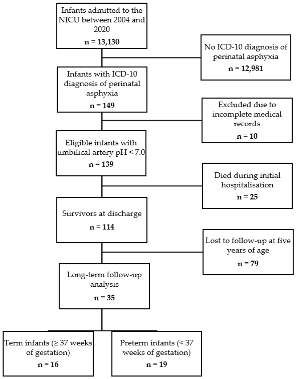
Figure 2.
Five-year neurodevelopmental outcomes of children born preterm or at term with a history of perinatal asphyxia.
Figure 2.
Five-year neurodevelopmental outcomes of children born preterm or at term with a history of perinatal asphyxia.
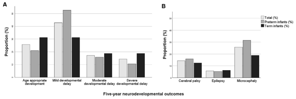

Table 1.
Demographic characteristics, place of birth and mode of delivery of included infants.
| Total n = 35 |
Term n = 16 |
Preterm n = 19 |
EPI n = 3 |
VPI n = 7 |
MLPI n = 9 |
|
|---|---|---|---|---|---|---|
| Gestational age, weeks | 36.6 (31.3–39.3) | 39.4 (38.6–40.0) | 31.9 (29.7–34.1) | 26.3 (24.6–27.3) | 30.1 (29.7–31.3) | 34.1 (33.0–36.6) |
| Female sex | 17 (49) | 7 (44) | 10 (53) | 3 (100) | 4 (57) | 3 (33) |
| Birthweight, g | 2605 (1560–3365) | 3408 (2899–3850) | 1560 (1260–2335) | 595 (460–750) | 1500 (1260–1900) | 2335 (1560–2920) |
| Birth length, cm | 49 (42-51) | 51 (49–53) | 43 (39–48) | 32 (28–34) | 40 (39–43) | 48 (43–51) |
| Head circumference, cm | 32 (28–34) | 34 (33–36) | 29 (27–31) | 21 (20–21) | 28 (27–30) | 31 (31–33) |
| Apgar score at 1 min | 2 (1–4) | 3 (1–5) | 2 (1–3) | 2 (0–2) | 3 (1–4) | 2 (1–3) |
| Apgar score at 5 min | 6 (4–7) | 5 (4–7) | 6 (4–7) | 6 (4–6) | 6 (4–7) | 4 (4–6) |
| Apgar score at 10 min | 7 (6–8) | 7 (6–8) | 7 (6–8) | 6 (4–7) | 7 (7–9) | 7 (5–8) |
| Umbilical artery pH | 6.90 (6.78–6.95) | 6.89 (6.79–6.95) | 6.90 (6.77–6.94) | 6.89 (6.84–6.94) | 6.92 (6.88–6.98) | 6.82 (6.71–6.92) |
| Mode and place of delivery | ||||||
| SVD | 5 (14) | 3 (19) | 2 (11) | 1 (33) | 0 (0) | 1 (11) |
| VED | 1 (3) | 1 (6) | 0 (0) | 0 (0) | 0 (0) | 0 (0) |
| ElCD | 2 (6) | 2 (13) | 0 (0) | 0 (0) | 0 (0) | 0 (0) |
| EmCD | 27 (77) | 10 (63) | 17 (90) | 2 (67) | 7 (100) | 8 (89) |
| Hospital with NICU | 24 (69) | 8 (50) | 16 (84) | 2 (67) | 5 (71) | 9 (100) |
| Hospital without NICU | 10 (29) | 8 (50) | 2 (11) | 0 (0) | 2 (29) | 0 (0) |
| Out-of-hospital birth | 1 (3) | 0 (0) | 1 (5) | 1 (33) | 0 (0) | 0 (0) |
Abbreviations: ElCD, elective cesarean delivery; EmCD, emergency cesarean delivery; EPI, extremely preterm infants (< 28 weeks of gestation); MLPI, moderate-to-late preterm infants (≥ 32+0 and < 37+0 weeks of gestation); NICU, neonatal intensive care unit; SVD, spontaneous vaginal delivery; VED, vacuum extraction delivery; VPI, very preterm infants (≥ 28+0 and < 32+0 weeks of gestation). Data are presented as median (interquartile range) or n (%).
Table 2.
Five-year neurodevelopmental outcomes of children with a history of perinatal asphyxia according to gestational age at birth.
Table 2.
Five-year neurodevelopmental outcomes of children with a history of perinatal asphyxia according to gestational age at birth.
| Total n = 35 |
Term n = 16 |
Preterm n = 19 |
EPI n = 3 |
VPI n = 7 |
MLPI n = 9 |
|
|---|---|---|---|---|---|---|
| Age-appropriate development | 9 (26) | 5 (31) | 4 (21) | 0 (0) | 1 (14) | 3 (33) |
| Mild developmental delay | 15 (43) | 5 (31) | 10 (53) | 2 (67) | 3 (43) | 5 (56) |
| Moderate developmental delay | 6 (17) | 3 (19) | 3 (16) | 0 (0) | 3 (43) | 0 (0) |
| Severe developmental delay | 5 (14) | 3 (19) | 2 (11) | 1 (33) | 0 (0) | 1 (11) |
| Cerebral palsy | 5 (14) | 2 (13) | 3 (16) | 1 (33) | 1 (14) | 1 (11) |
| Epilepsy | 2 (6) | 1 (6) | 1 (5) | 0 (0) | 0 (0) | 1 (11) |
| Microcephaly | 9 (26) | 3 (19) | 6 (32) | 2 (67) | 1 (14) | 3 (33) |
Abbreviations: EPI, extremely preterm infants (< 28 weeks of gestation); MLPI, moderate-to-late preterm infants (≥ 32+0 and < 37+0 weeks of gestation); VPI, very preterm infants (≥ 28+0 and < 32+0 weeks of gestation). Developmental categories are mutually exclusive. Cerebral palsy, epilepsy, and microcephaly may coexist with any developmental category. Data are presented as n (%).
Table 3.
Clinical course during hospital stay in infants with perinatal asphyxia.
| Total n = 35 |
Term n = 16 |
Preterm n = 19 |
EPI n = 3 |
VPI n = 7 |
MLPI n = 9 |
|
|---|---|---|---|---|---|---|
| Therapeutic hypothermia | 10 (29) | 9 (56) | 1 (5) | 0 (0) | 0 (0) | 1 (11) |
| Mechanical ventilation | 30 (86) | 13 (81) | 17 (89) | 3 (100) | 6 (86) | 8 (89) |
| Duration of mechanical ventilation, days | 6 (4–8) | 6 (3–8) | 6 (4–8) | 6 (4–14) | 7 (4–8) | 6 (2–7) |
| Inhaled nitric oxide | 1 (3) | 1 (6) | 0 (0) | 0 (0) | 0 (0) | 0 (0) |
| Catecholamine use | 21 (60) | 7 (44) | 14 (74) | 1 (33) | 6 (86) | 7 (78) |
| Seizures | 10 (29) | 5 (31) | 5 (26) | 0 (0) | 3 (43) | 2 (22) |
| Hypoglycemia | 8 (23) | 3 (19) | 5 (26) | 1 (33) | 3 (43) | 1 (11) |
| Renal dysfunction | 17 (49) | 6 (38) | 11 (58) | 2 (67) | 4 (57) | 5 (56) |
| Elevated liver enzymes | 25 (71) | 11 (69) | 14 (74) | 2 (67) | 6 (86) | 6 (67) |
| Hospital stay, days | 26 (13–44) | 15 (11–24) | 37 (24–65) | 120 (105–232) | 49 (36–65) | 27 (22–34) |
Abbreviations: EPI, extremely preterm infants (< 28 weeks of gestation); MLPI, moderate-to-late preterm infants (≥ 32+0 and < 37+0 weeks of gestation); VPI, very preterm infants (≥ 28+0 and < 32+0 weeks of gestation). Data are presented as median (interquartile range) or n (%).
Table 4.
Severity of hypoxic-ischemic encephalopathy (HIE) classified by the Sarnat score and stratified by gestational age.
Table 4.
Severity of hypoxic-ischemic encephalopathy (HIE) classified by the Sarnat score and stratified by gestational age.
| Total n = 35 |
Term n = 16 |
Preterm n = 19 |
EPI n = 3 |
VPI n = 7 |
MLPI n = 9 |
|
|---|---|---|---|---|---|---|
| HIE | 19 (54) | 11 (69) | 8 (42) | 1 (33) | 3 (43) | 4 (44) |
| HIE stage I (including I/II) | 13 (68) | 7 (64) | 6 (75) | 1 (100) | 2 (67) | 3 (75) |
| HIE stage II (including II/III) | 5 (26) | 3 (27) | 2 (25) | 0 (0) | 1 (33) | 1 (25) |
| HIE stage III | 1 (5) | 1 (9) | 0 (0) | 0 (0) | 0 (0) | 0 (0) |
Abbreviations: EPI, extremely preterm infants (< 28 weeks of gestation); MLPI, moderate-to-late preterm infants (≥ 32+0 and < 37+0 weeks of gestation); VPI, very preterm infants (≥ 28+0 and < 32+0 weeks of gestation). Intermediate stages (I/II and II/III) were grouped with the respective lower grade for analysis. Data are presented as n (%). Percentages for HIE refer to all infants in the respective gestational age group, whereas percentages for individual HIE stages refer only to infants diagnosed with HIE within that group. Percentages may not sum to 100% due to rounding.
Disclaimer/Publisher’s Note: The statements, opinions and data contained in all publications are solely those of the individual author(s) and contributor(s) and not of MDPI and/or the editor(s). MDPI and/or the editor(s) disclaim responsibility for any injury to people or property resulting from any ideas, methods, instructions or products referred to in the content. |
© 2026 by the authors. Licensee MDPI, Basel, Switzerland. This article is an open access article distributed under the terms and conditions of the Creative Commons Attribution (CC BY) license (http://creativecommons.org/licenses/by/4.0/).
Copyright: This open access article is published under a Creative Commons CC BY 4.0 license, which permit the free download, distribution, and reuse, provided that the author and preprint are cited in any reuse.