

Submitted:
31 October 2026
Posted:
04 November 2026
You are already at the latest version
Abstract
Image-guided thermal ablation has matured into a recognized local therapy for medically inoperable early-stage non-small cell lung cancer and selected pulmonary metastases, and transbronchial approaches delivered through navigational bronchoscopy are now extending ablation to patients who cannot tolerate pleural puncture. Yet ablation planning remains strikingly empirical: target coverage is estimated from manufacturer-provided geometric charts derived from ex vivo or non-pulmonary tissue, while the two features that most distinguish the lung from other ablated organs, respiratory motion and the low thermal and electrical conductivity of aerated parenchyma, are handled by operator experience rather than by patient-specific modeling. Digital twin (DT) technology, defined here as a patient-specific computational replica coupled to its physical counterpart through bidirectional data flow, offers a framework to close this gap. In this narrative review we (i) summarize the clinical maturity of percutaneous and transbronchial lung ablation that makes planning the rate-limiting step; (ii) propose a three-tier terminological hierarchy (static model, digital shadow, and digital twin) to discipline a literature in which the term is increasingly diluted; (iii) map a four-layer technical stack for a lung ablation DT, spanning four-dimensional anatomical modeling, respiratory-motion compensation, energy–tissue coupling simulation, and intraoperative closed-loop updating, and show that every layer already has validated components in adjacent fields, including a biophysical DT of liver microwave ablation, histology-validated dielectric characterization of human lung carcinoma, and retrospective computational validation of lung microwave ablation models against post-treatment imaging; and (iv) outline a staged validation pathway and a five-item research agenda, arguing that the first integrated, closed-loop DT for pulmonary ablation, percutaneous or transbronchial, remains to be built, and that the specialty which builds it will define the next standard of ablation planning.
Keywords:
digital twin
; lung neoplasms
; microwave ablation
; bronchoscopy
; electromagnetic navigation
; treatment planning
; respiratory motion
; computational modeling
1. Introduction: From Empirical Ablation to Model-Based Ablation
The detection of small pulmonary nodules has risen sharply with the global adoption of low-dose computed tomography (CT) screening, and with it the population of patients (elderly, comorbid, or bearing multifocal ground-glass lesions) for whom surgical resection is either unsafe or anatomically wasteful [1]. Image-guided thermal ablation, encompassing radiofrequency ablation (RFA), microwave ablation (MWA), and cryoablation, has progressively filled this space alongside stereotactic ablative radiotherapy [2]: for medically inoperable stage I non-small cell lung cancer (NSCLC), contemporary series report local control and survival outcomes that have justified inclusion of ablation among accepted local therapies alongside stereotactic body radiation therapy (SBRT) [3,4]. Long-term follow-up of percutaneous ablation in stage IA disease now extends beyond five years [5,6], and the evidence base extends to frail and comorbid populations, including multicenter retrospective evaluation in octogenarians [7] and cohorts with comorbid idiopathic pulmonary fibrosis [8].
Against this clinical maturity, the planning of an individual ablation remains conspicuously pre-modern. The operator selects power, duration, and applicator position to achieve an ablation zone that encompasses the nodule plus a surrounding margin of normal tissue [9], using manufacturer charts that relate energy settings to expected ablation-zone dimensions. Those charts are generated in ex vivo tissue or in healthy porcine liver and lung, under conditions that ignore the patient’s tumor size and geometry, surrounding emphysema, adjacent vessels and their convective cooling, prior ipsilateral treatments and, uniquely among solid organs, the respiratory displacement of the target [9,10]. The consequences are measurable: local progression after lung ablation is driven substantially by treatment-technical factors: in a 91-tumor risk-factor analysis, tumor shape and energy delivered per unit tumor volume emerged as independent predictors of local progression [11]; and a comparison of achieved versus intended ablation zones in lung tumors has documented systematic discrepancies between plan and result [12]. In short, the field possesses mature energy delivery systems but no patient-specific engine for predicting what those systems will do in a given lung.
Digital twin (DT) technology is the leading candidate for such an engine. Originating in manufacturing and now rapidly diffusing into medicine [13,14], a DT is a computational replica of a specific patient that is synchronized with its physical counterpart through ongoing data exchange and is used to simulate, predict, and optimize interventions before or while they are delivered [15,16]. DT frameworks have achieved their greatest medical maturity in cardiology, where patient-specific virtual hearts have been used to test antiarrhythmic drug response and to plan ablation strategies before the catheter is ever inserted [17,18], and a biophysical DT that predicts microwave ablation zones in the liver under varying steatosis grades has been reported as a computational proof of concept [19]. In oncology more broadly, DT concepts are being explored from predictive modeling to personalized treatment strategies [20], including radiotherapy planning and image-guidance applications [21].
The lung ablation field, however, sits at an instructive position: its enabling components exist but remain unassembled. Patient-specific statistical prediction of lung MWA zone size from clinical covariates has been demonstrated [22]; computational MWA models have been validated retrospectively against post-treatment imaging in patients with lung tumors [9]; the dielectric properties of human lung carcinoma have been characterized and validated against histology [23]; and four-dimensional (4D) respiratory-motion modeling is routine in thoracic radiation oncology [24,25]. What does not yet exist, for either the percutaneous or the transbronchial route, is an integrated, closed-loop DT that couples these components into a planning and intraoperative guidance system.
Evidence acquisition. This is a narrative review with a translational roadmap; references were selected for relevance to each layer of the proposed framework. The novelty claim was verified reproducibly: a PubMed search on 16 October 2026 for “digital twin” AND lung AND ablation returned 0 records, and a search for “digital twin” AND (bronchoscopic OR transbronchial OR “cone beam”) AND ablation likewise returned 0 records; an independent Europe PMC title/abstract query (“digital twin” AND “lung” AND “ablation”) on the same date returned 0 hits. A broad full-text Europe PMC query returned 178 records, whose screening confirmed the nearest lung digital-twin framework to be a light-dosimetry model for photodynamic and photobiomodulation therapies [26], accounted for in Section 3, rather than any thermal-ablation application.
This review therefore pursues four aims: (i) to define why planning, rather than energy delivery, is now the rate-limiting step in pulmonary ablation; (ii) to impose terminological discipline on “digital twin” claims in procedural lung medicine; (iii) to lay out a four-layer technical stack for a lung ablation DT, mapping each layer to validated components in adjacent fields and to the gaps that remain; and (iv) to propose a staged validation pathway and an explicit, testable research agenda. Our scope encompasses both CT-guided percutaneous ablation and transbronchial ablation, because the two routes share the same predictive core while facing distinct spatial constraints that a DT framework is uniquely positioned to formalize.
2. Clinical Maturity of Pulmonary Ablation: The Application Soil for a Digital Twin
2.1. Percutaneous CT-Guided Ablation: Efficacy Established, Planning Empirical
CT-guided percutaneous ablation is the most extensively validated thermal approach in the lung. RFA established proof of durability more than a decade ago, with long-term survival reported in stage I disease [5]. MWA has since become the dominant modality, favored for higher intratumoral temperatures, shorter ablation times, larger and more spherical zones, and relative insensitivity to convective heat loss [27,28], with early high-powered series establishing feasibility in medically inoperable stage I disease [29]. Safety and efficacy of CT-guided percutaneous MWA in stage I NSCLC have been confirmed in comorbid populations, including patients with idiopathic pulmonary fibrosis [8]; long-term outcomes of MWA in early-stage NSCLC have been reported [6]; and comparative analyses against surgical resection in stage I disease, while favoring surgery in fit patients, quantify the outcomes that ablation must match to be competitive [30]. Cryoablation occupies a complementary niche, with reported series in ground-glass-predominant nodules and in the very elderly [31,32]. Durable outcomes have likewise been reported for RFA of colorectal lung metastases [33]; comparative effectiveness and cost-effectiveness analyses against sublobar resection and SBRT frame the competitive context in which ablation must justify its planning deficits [34,35].
Across modalities, however, local progression remains the Achilles’ heel. Risk-factor analyses identify tumor shape and energy deployed per unit tumor volume as independent determinants of local progression [11], and quantitative work on ablation-zone involution has introduced energy-normalized metrics (ablation work and ablation resistance scores) in an explicit attempt to rationalize dosing across patients [36]. Machine-learning models combining clinical variables with CT radiomics to predict local recurrence after lung MWA further reinforce that recurrence risk is substantially a planning-and-coverage problem that better modeling could anticipate [37]. The gap between intended and achieved treatment is not hypothetical: direct comparison of actual versus predicted ablation zones after CT-guided lung tumor ablation has demonstrated clinically relevant discrepancies [12].
2.2. Transbronchial Ablation: A Second Route Reaching Clinical Maturity
Transbronchial ablation eliminates pleural puncture and with it the pleura-related complications, particularly pneumothorax, that dominate percutaneous morbidity [38]. Following preclinical feasibility work, including bronchoscopic thermal vapor and laser approaches in porcine models [39] and device safety assessment of bronchoscopic MWA in swine peripheral lung [40], clinical translation has been rapid. Chan and colleagues reported the initial 30-case experience of electromagnetic navigation bronchoscopy (ENB)-guided transbronchial MWA [41], followed by the multicenter NAVABLATE study [42], a prospective single-arm multicenter study of image-guided flexible-probe transbronchial MWA for stage 1 lung cancer using cone-beam CT (CBCT) with augmented fluoroscopy [38], and reports of safety and feasibility in subpleural nodules and of concomitant ablation of multiple nodules in a single session [43,44]. Editorials now openly debate whether transbronchial ablation is “ready for prime time” [45,46], and dedicated reviews frame bronchoscopic treatment of early-stage peripheral lung cancer as an emerging frontier of interventional pulmonology [47,48,49]. A multicenter prospective protocol now couples bronchoscopic diagnosis, transbronchial ablation, and surgical resection with histopathological assessment of treatment effect (a treat-and-resect design; NCT05281237) [50], which will provide an unprecedented imaging–pathology validation substrate.
The transbronchial route intensifies rather than relaxes the planning problem. The flexible applicator approaches the target through the airway tree, so achievable antenna orientation is constrained by bronchial anatomy; the target may be displaced between navigation CT, registration, and energy delivery; and CBCT confirmation captures only a single respiratory state [51,52]. The very features that make the route attractive (no transthoracic trajectory, multiple targets in one session) make patient-specific prediction of energy deposition both harder and more valuable.
2.3. The Shared Unmet Need
Whether the applicator arrives through the chest wall or through the bronchoscope, the operator’s core question is identical: where, at what settings, and for how long will energy deposition cover this tumor plus margin in this lung, at this respiratory phase, given these vessels? Today that question is answered by experience, vendor charts, and post hoc imaging. The remainder of this review argues that DT methodology is the systematic way to answer it prospectively.
3. What Counts as a Digital Twin: A Three-Tier Hierarchy for Procedural Lung Medicine
The term “digital twin” is at risk of dilution in proportion to its popularity. We therefore propose a pragmatic three-tier hierarchy, adapted from frameworks in manufacturing and in medical DT reviews [13,15,16], including musculoskeletal and orthopedic applications [53,54], classified by the direction and continuity of data flow between the physical patient and the virtual replica (Table 1).
A Tier 1 static patient-specific model is built once from pre-procedural data and used for simulation without subsequent updating: most computational ablation models, however sophisticated, live here, including image-based 3D modeling validated against tissue-mimicking phantoms [55] and the liver MWA biophysical DT proof of concept, which, despite its title, performs virtual experiments on a static replica rather than maintaining a live link to a patient [19]. A Tier 2 digital shadow adds one-directional flow from physical to virtual: the model is updated with intraoperative or longitudinal data, informing the operator but not automatically steering therapy. Augmented-reality (AR) navigation for pulmonary nodule localization, in which a preoperative 3D thoracic model is registered to the operative field, progressing from canine models [56] to human pilot studies [57] to a randomized noninferiority trial against CT-guided localization [58], exemplifies this tier in procedural lung medicine. A Tier 3 digital twin closes the loop bidirectionally: the replica is continuously or repeatedly synchronized with the patient and its predictions feed back to modulate the intervention itself. In cardiology, virtual-heart systems that test drug or ablation strategies patient-specifically before or during treatment approach this tier [17,18]; in musculoskeletal medicine, DT systems combining finite-element analysis with wearable-sensor data streams are being assembled toward it [59].
Applied to lung ablation, this taxonomy yields a crisp verdict: the field currently possesses Tier 1 components (biophysical and statistical models [9,22]) and Tier 2 shadows (AR and CBCT guidance), but no Tier 3 system. The taxonomy is not pedantry: it converts “we need digital twins” into an engineering specification: which data, flowing how often, feeding which decision. The nearest digital-twin framework in pulmonary medicine, a Monte Carlo light-dosimetry twin built from CT-derived thoracic anatomy to personalize photodynamic and photobiomodulation lung therapies [26], targets light rather than thermal energy, involves no ablation, and likewise qualifies only as Tier 1 under our criteria; it is accounted for here rather than ignored, and it reinforces the verdict that no closed-loop twin exists for any form of lung ablation. The taxonomy thus also clarifies the review’s central claim: the first Tier 3 digital twin for pulmonary ablation remains to be constructed, and its construction is an assembly problem more than an invention problem.
4. A Four-Layer Technical Stack for the Lung Ablation Digital Twin
We organize the assembly problem into four layers (Figure 1). For each, we identify the validated components that already exist, the lung-specific barriers, and the concrete gap. Table 2 summarizes the mapping, and Figure 2 contrasts the route-specific intervention points of the percutaneous and transbronchial pathways.
4.1. Layer 1: Patient-Specific Anatomical Modeling
The geometric substrate is a segmentation of tumor, airways, pulmonary and bronchial vasculature, fissures, and chest wall from thin-section CT, assembled into a mesh suitable for simulation. This layer is the most mature: it is standard practice in AR-guided localization and virtual bronchoscopic navigation [57], and image-based 3D modeling pipelines for ablation simulation have been validated in other organs [55]; orthopedic surgery offers the most mature precedent for patient-specific, image-derived modeling in procedural medicine [60]. Two lung-specific caveats remain. First, part-solid and pure ground-glass targets, the fastest-growing ablation population, have indistinct margins whose segmentation uncertainty propagates directly into margin planning. Second, the ablation applicator itself must be localized in the same coordinate frame; peri-procedural CT with the applicator in situ, as assembled in retrospective modeling datasets [9], provides the template. For the transbronchial route, the airway tree segmentation additionally constrains feasible applicator orientation, a geometric input that no current planning system exploits.
4.2. Layer 2: Respiratory-Motion Modeling and Compensation
Respiratory displacement reaches its greatest amplitude in the lung among commonly ablated organs and is compounded there by respiration-driven changes in aeration and tissue properties; static manufacturer geometric charts account for neither [9]. The enabling mathematics is, however, mature in thoracic radiation oncology: 4D-CT acquisition, deformable image registration (DIR) for motion estimation with quantified nonlinearity and hysteresis [24], modern landmark-driven deep-learning registration of lung 4D-CT [25], and multi-institutional evaluation of DIR accuracy in the thorax establish both feasibility and known error bounds [61,62]. DIR between average-intensity-projection and phase-binned 4D reconstructions has been validated for geometric and dosimetric accuracy [62], and its influence on 4D dose simulation has been quantified for extracranial SBRT [63]. Within interventional radiology, fast CT/CT-fluoroscopy registration with respiratory-motion compensation has been demonstrated for percutaneous procedures [64].
The ablation-specific gap is integration: motion models must be coupled to the thermal simulation so that predicted energy deposition is accumulated over the true trajectory of tissue through the treatment field, and intraoperative CBCT, acquired in an arbitrary respiratory state, must be deformably registered back to the planning 4D model. Respiration has not been entirely ignored in the modeling literature: finite-element analyses have flagged respiratory movement as a key obstacle to accurate lung MWA prediction [65], and a 4D-CT-based study of 100 radiotherapy patients quantified how respiration-driven changes in lung physical properties alter the microwave ablation zone [66]. What has not been reported, including in the two most advanced lung ablation modeling studies [9,22], is integration of phase-resolved tissue motion into thermal-dose accumulation for planning. This is simultaneously the field’s largest error source and its clearest opportunity for measurable improvement. In the swine lung, single-lung ventilation illustrates that the problem is tractable: inducing atelectasis significantly enlarged ablation volume (10.74 cm³ versus 7.35 cm³ in ventilated controls) [67], a pragmatic bridge toward motion and aeration management while motion-coupled models are built.
4.3. Layer 3: Energy–Tissue Coupling Simulation
The biophysical core couples electromagnetic energy deposition with heat transfer and thermal damage kinetics. Three classes of inputs now have published foundations. First, tissue properties: the dielectric properties of human lung carcinoma and non-neoplastic lung have been measured across ablation-relevant frequencies and validated against histology [23]; temperature dependence of thermal, dielectric, and perfusion properties relevant to thermal ablation has been systematically reviewed [68]; and global sensitivity analysis has shown that MWA model outputs are materially sensitive to biophysical-property uncertainty, the first step toward probabilistic treatment planning [69]. Aerated lung, with its low electrical and thermal conductivity, produces ablation zones that are smaller, less predictable, and more sensitive to local composition than in solid organs; emphysema, fibrosis, and tumor infiltration alter these properties regionally in ways a patient-specific model can encode but a vendor chart cannot. Second, convective cooling: the heat-sink effect of adjacent vasculature has been quantified experimentally in ex vivo hepatic MWA [70] and modeled numerically under high-flow conditions [71], and the lung’s dual perfusion (pulmonary and bronchial arterial) adds an organ-specific wrinkle [27]. Third, damage integration: Arrhenius-type thermal-dose models, as embedded in the liver DT framework [19], translate spatiotemporal temperature fields into predicted zones of coagulation, with surface-distance-based agreement metrics available for validation against imaging [9].
Two works document the clinical need most directly and themselves delimit what remains undone. A statistical model predicting immediate-phase lung MWA zone dimensions from patient- and tumor-specific covariates in 164 lesions demonstrated that clinical variables carry predictive signal, but it is correlative, two-dimensional, and cannot generalize across devices or unconventional geometries [22]. A computational-modeling study compared biophysical predictions against manufacturer geometric models using retrospective CT data from 50 patients treated with lung MWA, directly demonstrating the inadequacy of geometric charts and the promise of model-based prediction, but in a static, off-line, percutaneous-only implementation without motion or intraoperative updating [9]. Tier 3 function requires that this layer run fast enough to be re-solved intraoperatively, which motivates reduced-order or machine-learning-accelerated surrogates; planning software reviews have catalogued the corresponding techniques and their trade-offs [72].
4.4. Layer 4: Intraoperative Closed-Loop Updating
Loop closure is what promotes a model to a twin. Three signal sources are already in clinical use and await integration: (i) CBCT, which provides intraoperative 3D imaging of applicator position and the evolving ground-glass ablation zone in both percutaneous suites and hybrid operating rooms used for transbronchial ablation [51,52]; (ii) applicator-embedded telemetry (power, delivered energy, and impedance or permittivity feedback in some contemporary MWA systems), which constrains the real-time thermal state; and (iii) navigation-system tracking, which localizes the applicator within the registered airway or CT coordinate frame. A Tier 3 system would assimilate these streams to update the thermal simulation during energy delivery, compare predicted versus observed zone evolution, and issue concrete corrective recommendations (extend duration, reposition, add a second applicator) rather than leaving the operator to interpolate vendor charts at the console. The analogy to cardiac electrophysiology, where patient-specific virtual models already stratify therapeutic response before intervention [17], indicates the direction; the engineering task in the lung is harder because the organ moves and because the ablation zone is directly imageable, which, conveniently, also makes the lung the ideal organ in which to validate a closed-loop ablation twin.
5. A Staged Validation Pathway: From Phantom to Prospective Trial
We propose a five-rung validation ladder (Table 3), ordered by evidentiary weight and modeled on how adjacent fields earned clinical trust.
Rung 1: Phantom and ex vivo validation. Tissue-mimicking phantoms and ex vivo perfused lung, in which image-based ablation modeling has precedents in other organs [55], establish ground-truth agreement between simulated and measured zones under controlled properties, using surface-distance and volumetric overlap metrics [9].
Rung 2: In vivo large-animal validation. The healthy porcine lung is the established preclinical substrate for bronchoscopic ablation safety [40], including long-term survival evaluation of endobronchial RFA [73], and enables synchronized 4D imaging, telemetry, and immediate explant histopathology, the first setting in which a motion-coupled, closed-loop prototype can be tested end-to-end. Multimodality imaging–pathology correlation of ablation lesions has been demonstrated in rabbit lung tumor models [74,75], establishing the comparative-imaging methodology that the human rung will inherit.
Rung 3: Imaging–pathology correlation in humans. The treat-and-resect trial design now entering the transbronchial field [50] provides the gold standard: the actual treatment-effect boundary on histopathology against the DT-predicted boundary. This rung is where a DT earns or loses the right to guide margins.
Rung 4: Retrospective clinical concordance. The existing template is the 50-patient retrospective assessment of computational versus manufacturer predictions [9]; extending such concordance testing to transbronchial cohorts and to prospective-assembled registries with uniform CBCT protocols, and reporting quantitative zone-agreement endpoints of the kind introduced for pulmonary MWA [36], establishes generalizability across devices, operators, and nodule phenotypes.
Rung 5: Prospective utility trials. The AR-localization literature supplies the design template: a randomized noninferiority comparison of AR-guided versus CT-guided localization [58] maps directly onto a trial of DT-planned versus conventional ablation, with minimal-margin error as primary endpoint and local progression-free survival as the key secondary endpoint; prospective single-arm multicenter templates for transbronchial ablation likewise exist [38,42].
Two cross-cutting considerations deserve emphasis. First, regulatory positioning: a lung ablation DT is software as a medical device whose risk class depends on whether it informs or executes; medical-DT reviews consistently flag data integration, privacy, validation, and regulatory uncertainty as the dominant translational bottlenecks [14,16]. Second, reporting standards: the field should adopt quantitative zone-agreement metrics and pre-registered prediction-versus-observation analyses from the outset, so that the DT literature does not recapitulate the heterogeneity that has limited pooling in the lung ablation evidence base [3,4].
6. A Five-Item Research Agenda
We close the loop from review to action with five testable propositions, ordered roughly by dependency.
P1: In-vivo calibration of aerated-lung properties. Ex vivo dielectric characterization of lung carcinoma [23] must be extended to in-vivo, temperature-dependent, regionally resolved properties of aerated, emphysematous, and fibrotic lung. Without this, every downstream simulation rests on ex-vivo assumptions whose in-vivo validity is unknown.
P2: Motion-coupled prediction reduces margin error. A DT incorporating 4D respiratory motion should reduce the discrepancy between predicted and achieved ablation boundaries relative to static models. This is directly measurable with existing retrospective datasets augmented by 4D-CT, and success criteria can be pre-specified against the discrepancies already documented between actual and predicted zones [9,12].
P3: Prospective utility of DT-guided planning. A randomized comparison of DT-planned versus conventional ablation, using the noninferiority-then-superiority logic established by AR localization trials [58], with minimal-margin error and local progression as endpoints. Statistical-prediction [22] and radiomics-based progression models [37] should be benchmarked as comparators, not ignored: a DT that cannot beat a regression model does not deserve the name.
P4: Transbronchial-specific modeling. The airway-constrained applicator orientation, bronchus-sign dependency, and multi-nodule single-session workflows of transbronchial ablation [41,44] constitute a formally different planning problem that no modeling study has addressed; its multi-target nature makes it arguably the highest-value DT use case, because the combinatorial planning space most exceeds unaided human optimization.
P5: Post-ablation twin for surveillance. Extending the twin beyond the procedure (predicting zone involution [36] and integrating surveillance imaging to distinguish expected evolution from progression) would convert a planning tool into a longitudinal management platform, aligning pulmonary ablation with the broader vision of medical DTs as companions across the care continuum [16,20].
7. Conclusions
Pulmonary thermal ablation has outgrown its planning tools. Its energy platforms are mature, its clinical indications are consolidating across percutaneous and transbronchial routes, and its failure modes are dominated by coverage and margin problems that are computational in nature. Every component required for a lung ablation digital twin (anatomical modeling, respiratory-motion mathematics, tissue-property data, validated energy–tissue simulation, and intraoperative imaging and telemetry) exists and has been validated in isolation or in adjacent organs. What is missing is the integrated, closed-loop assembly, together with the in-vivo property calibration and prospective utility evidence that clinical adoption will demand. The ablate-then-resect trial design offers a ready-made histopathological validation substrate, and the cardiac electrophysiology experience offers a regulatory and translational precedent. The contribution we intend is deliberately falsifiable: each layer of the stack carries a measurable success criterion (Table 2), each proposition a pre-specifiable endpoint (Section 6), and the framework as a whole stands or falls on whether model-based planning can beat both vendor charts and simple regression [22] on quantitative zone agreement. The first group to assemble these components into a working Tier 3 system will not merely improve ablation planning; it will define the standard against which the next generation of pulmonary ablation is judged.
Supplementary Materials
The following supporting information can be downloaded at the website of this paper posted on Preprints.org.
Author Contributions
Y.Z. conceived the review, defined the clinical framework and research agenda, and drafted the manuscript. C.M. curated the imaging literature, contributed the radiological perspective on image-guided ablation workflows, and critically revised the manuscript. Both authors approved the final version.
Funding
None.
Ethical approval and consent to participate
Not applicable (narrative review).
Consent for Publication
Not applicable.
Availability of Data and Materials
Not applicable; all cited works are publicly available.
Competing interests
The authors declare no competing interests. Commercial systems are mentioned solely to describe the technological state of the art, without endorsement.
References
- David S Buchberger, Rishabh Khurana, Michael Bolen, Gregory M M Videtic. The Treatment of Patients with Early-Stage Non-Small Cell Lung Cancer Who Are Not Candidates or Decline Surgical Resection: The Role of Radiation and Image-Guided Thermal Ablation. Journal of clinical medicine. 2024. [CrossRef] [PubMed]
- Megan E Daly, Patrick Beagen, Mohammad H Madani. Nonsurgical Therapy for Early-Stage Lung Cancer. Hematology/oncology clinics of North America. 2023. [CrossRef] [PubMed]
- Paul Laeseke, Calvin Ng, Nicole Ferko, Andrada Naghi, George W J Wright, Yuxin Zhang, et al. Stereotactic body radiation therapy and thermal ablation for treatment of NSCLC: A systematic literature review and meta-analysis. Lung cancer (Amsterdam, Netherlands). 2023. [CrossRef] [PubMed]
- Miao Lin, Pat Eiken, Shanda Blackmon. Image guided thermal ablation in lung cancer treatment. Journal of thoracic disease. 2020. [CrossRef] [PubMed]
- Chaitan K Narsule, Praveen Sridhar, Divya Nair, Avneesh Gupta, Roy G Oommen, Michael I Ebright, et al. Percutaneous thermal ablation for stage IA non-small cell lung cancer: long-term follow-up. Journal of thoracic disease. 2017. [CrossRef] [PubMed]
- Henry Zhao, Karin Steinke. Long-term outcome following microwave ablation of early-stage non-small cell lung cancer. Journal of medical imaging and radiation oncology. 2020. [CrossRef] [PubMed]
- Xiaoying Han, Xia Yang, Guanghui Huang, Chunhai Li, Licheng Zhang, Yuanxun Qiao, et al. Safety and clinical outcomes of computed tomography-guided percutaneous microwave ablation in patients aged 80 years and older with early-stage non-small cell lung cancer: A multicenter retrospective study. Thoracic cancer. 2019. [CrossRef] [PubMed]
- JinZhao Peng, ZhiXin Bie, YuanMing Li, RunQi Guo, XiaoGuang Li. Safety and efficacy of CT-guided percutaneous microwave ablation for stage I non-small cell lung cancer in patients with comorbid idiopathic pulmonary fibrosis. European radiology. 2023. [CrossRef] [PubMed]
- Jan Sebek, Pinyo Taeprasartsit, Chanok Pathomparai, Damian E Dupuy, Henky Wibowo, Punit Prakash. Computational modeling of microwave ablation of lung tumors: Assessment of model-predictions against post-treatment imaging. Medical physics. 2025. [CrossRef] [PubMed]
- V Lopresto, R Pinto, L Farina, M Cavagnaro. Treatment planning in microwave thermal ablation: clinical gaps and recent research advances. International journal of hyperthermia: the official journal of European Society for Hyperthermic Oncology, North American Hyperthermia Group. 2016. [CrossRef] [PubMed]
- Thomas J Vogl, Thomas S Worst, Nagy N N Naguib, Hanns Ackermann, Tatjana Gruber-Rouh, Nour-Eldin A Nour-Eldin. Factors influencing local tumor control in patients with neoplastic pulmonary nodules treated with microwave ablation: a risk-factor analysis. AJR. American journal of roentgenology. 2013. [CrossRef] [PubMed]
- Xiaoying Han, Tiehong Zhang, Min Meng, Jiao Wang, Guanghui Huang, Jianjian Dai, et al. Comparison of the difference between the actual ablation zone in lung tumors after computed tomography-guided percutaneous microwave ablation and the predicted value in the planning system: A retrospective study. Journal of cancer research and therapeutics. 2026. [CrossRef] [PubMed]
- Yunqi Zhang, Kuangyu Shi, Biao Li. A Brief History of Digital Twin Technology. PET clinics. 2025. [CrossRef] [PubMed]
- R Laubenbacher, B Mehrad, I Shmulevich, N Trayanova. Digital twins in medicine. Nature computational science. 2024. [CrossRef] [PubMed]
- Kang Zhang, Hong-Yu Zhou, Daniel T Baptista-Hon, Yuanxu Gao, Xiaohong Liu, Eric Oermann, et al. Concepts and applications of digital twins in healthcare and medicine. Patterns (New York, N.Y.). 2024. [CrossRef] [PubMed]
- Christoph Sadée, Stefano Testa, Thomas Barba, Katherine Hartmann, Maximilian Schuessler, Alexander Thieme, et al. Medical digital twins: enabling precision medicine and medical artificial intelligence. The Lancet. Digital health. 2025. [CrossRef] [PubMed]
- Taehyun Hwang, Byounghyun Lim, Oh-Seok Kwon, Moon-Hyun Kim, Daehoon Kim, Je-Wook Park, et al. Clinical usefulness of digital twin guided virtual amiodarone test in patients with atrial fibrillation ablation. NPJ digital medicine. 2024. [CrossRef] [PubMed]
- Paschalis Karakasis, Antonios P Antoniadis, Panagiotis Theofilis, Panayotis K Vlachakis, Nikias Milaras, Dimitrios Patoulias, et al. Digital Twin Models in Atrial Fibrillation: Charting the Future of Precision Therapy? Journal of personalized medicine. 2025. [CrossRef] [PubMed]
- Frankangel Servin, Jarrod A Collins, Jon S Heiselman, Katherine C Frederick-Dyer, Virginia B Planz, Sunil K Geevarghese, et al. Simulation of Image-Guided Microwave Ablation Therapy Using a Digital Twin Computational Model. IEEE open journal of engineering in medicine and biology. 2023. [CrossRef] [PubMed]
- David B Olawade, Emmanuel O Oisakede, Oluwakemi Jumoke Bello, Claret Chinenyenwa Analikwu, Eghosasere Egbon, Adeyinka Ojo. Digital twins in oncology: From predictive modelling to personalised treatment strategies. Critical reviews in oncology/hematology. 2026. [CrossRef] [PubMed]
- David B Olawade, Oluwatosin Akinro, Emmanuel O Oisakede, Oluwakemi Jumoke Bello, Claret Chinenyenwa Analikwu, Eghosasere Egbon. Digital twin applications in radiology and radiotherapy: Applications, challenges, and future perspectives. European journal of radiology. 2026. [CrossRef] [PubMed]
- Daniel H Kim, Jason Chiang, Lucia Chen, Ashley Lamba, Fereidoun Abtin, Scott Genshaft, et al. Patient-Specific Prediction of Immediate Phase Lung Microwave Ablation Zone Size. Journal of vascular and interventional radiology: JVIR. 2024. [CrossRef] [PubMed]
- Laura Farina, Giuseppe Ruvio, Ramadan Shatwan, Aliaa Shalaby, Martin O’Halloran, Alexandra White, et al. Histology-Validated Dielectric Characterisation of Lung Carcinoma Tissue for Microwave Thermal Ablation Applications. Cancers. 2023. [CrossRef] [PubMed]
- Vlad Boldea, Gregory C Sharp, Steve B Jiang, David Sarrut. 4D-CT lung motion estimation with deformable registration: quantification of motion nonlinearity and hysteresis. Medical physics. 2008. [CrossRef] [PubMed]
- Luke Matkovic, Yang Lei, Yabo Fu, Tonghe Wang, Aparna H Kesarwala, Marian Axente, et al. Deformable lung 4DCT image registration via landmark-driven cycle network. Medical physics. 2023. [CrossRef] [PubMed]
- Johan Sebastián Diaz Tovar, Flavio Pola Dos Reis, Lilian Tan Moriyama, Paulo Manuel Pêgo-Fernandes, Cesar Higa Nomura, Vladislav Yakovlev, et al. Anatomy-resolved digital twin framework for personalized light dosimetry in lung therapies. Proceedings of the National Academy of Sciences of the United States of America. 2026. [CrossRef] [PubMed]
- Clara Prud’homme, Frederic Deschamps, Benjamin Moulin, Antoine Hakime, Marc Al-Ahmar, Salma Moalla, et al. Image-guided lung metastasis ablation: a literature review. International journal of hyperthermia: the official journal of European Society for Hyperthermic Oncology, North American Hyperthermia Group. 2019. [CrossRef] [PubMed]
- Peyton Cramer, Bradley B Pua. The Latest on Lung Ablation. Seminars in interventional radiology. 2022. [CrossRef] [PubMed]
- Howard Liu, Karin Steinke. High-powered percutaneous microwave ablation of stage I medically inoperable non-small cell lung cancer: a preliminary study. Journal of medical imaging and radiation oncology. 2013. [CrossRef] [PubMed]
- Michael Vinchill Chan, Ya Ruth Huo, Christopher Cao, Lloyd Ridley. Survival outcomes for surgical resection versus CT-guided percutaneous ablation for stage I non-small cell lung cancer (NSCLC): a systematic review and meta-analysis. European radiology. 2021. [CrossRef] [PubMed]
- Shupeng Liu, Bing Liang, Yanhong Li, Jiongyuan Xu, Wei Qian, Mao Lin, et al. CT-Guided Percutaneous Cryoablation in Patients with Lung Nodules Mainly Composed of Ground-Glass Opacities. Journal of vascular and interventional radiology: JVIR. 2022. [CrossRef] [PubMed]
- Tarub S Mabud, David Swilling, Phillip Guichet, Yuli Zhu, Sophia Manduca, Bhavin Patel, et al. Pulmonary Cryoablation Outcomes in Octogenarians and Nonagenarians with Primary Lung Cancer. Journal of vascular and interventional radiology: JVIR. 2023. [CrossRef] [PubMed]
- Yusuke Matsui, Takao Hiraki, Hideo Gobara, Toshihiro Iguchi, Hiroyasu Fujiwara, Takeshi Nagasaka, et al. Long-term survival following percutaneous radiofrequency ablation of colorectal lung metastases. Journal of vascular and interventional radiology: JVIR. 2015. [CrossRef] [PubMed]
- Jianwei Liu, Caixia Wang, Weiwei Yi, Hang Zheng, Aimin Zheng. A retrospective comparative study of microwave ablation and sublobectomy in the treatment of early subpleural nonsmall cell lung cancer. International journal of hyperthermia: the official journal of European Society for Hyperthermic Oncology, North American Hyperthermia Group. 2022. [CrossRef] [PubMed]
- David J Sher, Jon O Wee, Rinaa S Punglia. Cost-effectiveness analysis of stereotactic body radiotherapy and radiofrequency ablation for medically inoperable, early-stage non-small cell lung cancer. International journal of radiation oncology, biology, physics. 2011. [CrossRef] [PubMed]
- Ramsey A Al-Hakim, Fereidoun G Abtin, Scott J Genshaft, Erin Kutay, Robert D Suh. Defining New Metrics in Microwave Ablation of Pulmonary Tumors: Ablation Work and Ablation Resistance Score. Journal of vascular and interventional radiology: JVIR. 2016. [CrossRef] [PubMed]
- Shengmei Ma, Jingshuo Li, Yuxian Chen, Ziqi Zhang, Li Hu, Chunhai Li, et al. Machine Learning Based on Clinical Information and Integrated CT Radiomics to Predict Local Recurrence of Stage Ia Lung Adenocarcinoma after Microwave Ablation. Journal of vascular and interventional radiology: JVIR. 2024. [CrossRef] [PubMed]
- Michael A Pritchett, Janani S Reisenauer, Ryan Kern, David S Wilson, Erin E Meyers, Philippe O Szapary, et al. Novel Image-Guided Flexible-Probe Transbronchial Microwave Ablation for Stage 1 Lung Cancer. Respiration; international review of thoracic diseases. 2023. [CrossRef] [PubMed]
- Daniel P Steinfort, Michael Christie, Phillip Antippa, Kanishka Rangamuwa, Robert Padera, Michael Rolf Müller, et al. Bronchoscopic Thermal Vapour Ablation for Localized Cancer Lesions of the Lung: A Clinical Feasibility Treat-and-Resect Study. Respiration; international review of thoracic diseases. 2021. [CrossRef] [PubMed]
- Hector De Leon, Kevin Royalty, Louie Mingione, David Jaekel, Sarvesh Periyasamy, David Wilson, et al. Device safety assessment of bronchoscopic microwave ablation of normal swine peripheral lung using robotic-assisted bronchoscopy. International journal of hyperthermia: the official journal of European Society for Hyperthermic Oncology, North American Hyperthermia Group. 2023. [CrossRef] [PubMed]
- Joyce W Y Chan, Rainbow W H Lau, Jenny C L Ngai, Carita Tsoi, Cheuk Man Chu, Tony S K Mok, et al. Transbronchial microwave ablation of lung nodules with electromagnetic navigation bronchoscopy guidance-a novel technique and initial experience with 30 cases. Translational lung cancer research. 2021. [CrossRef] [PubMed]
- Kelvin K W Lau, Rainbow W H Lau, Ralitsa Baranowski, Julie Krzykowski, Calvin S H Ng. Transbronchial Microwave Ablation of Peripheral Lung Tumors: The NAVABLATE Study. Journal of bronchology & interventional pulmonology. 2024. [CrossRef] [PubMed]
- Aliss Tsz Ching Chang, Joyce Wing Yan Chan, Ivan Chi Hin Siu, Rainbow Wing Hung Lau, Calvin Sze Hang Ng. Safety and feasibility of transbronchial microwave ablation for subpleural lung nodules. Asian cardiovascular & thoracic annals. 2024. [CrossRef] [PubMed]
- Joyce W Y Chan, Rainbow W H Lau, Aliss T C Chang, Ivan C H Siu, Cheuk Man Chu, Tony S K Mok, et al. Concomitant electromagnetic navigation transbronchial microwave ablation of multiple lung nodules is safe, time-saving, and cost-effective. JTCVS techniques. 2023. [CrossRef] [PubMed]
- Daniel P Steinfort, Felix J F Herth. Bronchoscopic treatments for early-stage peripheral lung cancer: Are we ready for prime time? Respirology (Carlton, Vic.). 2020. [CrossRef] [PubMed]
- Joyce W Y Chan, Calvin S H Ng. Transbronchial ablation for lung cancers: Ready for prime time? Respirology (Carlton, Vic.). 2024. [CrossRef] [PubMed]
- Kassem Harris, Jonathan Puchalski, Daniel Sterman. Recent Advances in Bronchoscopic Treatment of Peripheral Lung Cancers. Chest. 2016. [CrossRef] [PubMed]
- Udit Chaddha, D Kyle Hogarth, Septimiu Murgu. Bronchoscopic Ablative Therapies for Malignant Central Airway Obstruction and Peripheral Lung Tumors. Annals of the American Thoracic Society. 2019. [CrossRef] [PubMed]
- Aristides J Armas Villalba, David E Ost. Bronchoscopic treatment of early-stage peripheral lung cancer. Current opinion in pulmonary medicine. 2024. [CrossRef] [PubMed]
- Jason Beattie, Ray Parrish, Laith Ayasa, Paula Aranguren, Francisco L Uribe-Buritica, Mariana Nino Lopez, et al. Safety and feasibility of bronchoscopic microwave ablation technology for peripheral lung cancer: a multi-center, prospective, single-arm study protocol. BMC surgery. 2025. [CrossRef] [PubMed]
- Joyce W Y Chan, Aliss T C Chang, Ivan C H Siu, Rainbow W H Lau, Calvin S H Ng. Electromagnetic navigation bronchoscopy transbronchial lung nodule ablation with IllumisiteTM platform corrects CT-to-body divergence with tomosynthesis and improves ablation workflow: a case report. AME case reports. 2023. [CrossRef] [PubMed]
- Calvin S H Ng, William S Krimsky, Kazuhiro Yasufuku. Editorial: The Hybrid Operating Room in Modern Thoracic Surgery. Frontiers in surgery. 2021. [CrossRef] [PubMed]
- Tianze Sun, Xiwang He, Xueguan Song, Liming Shu, Zhonghai Li. The Digital Twin in Medicine: A Key to the Future of Healthcare? Frontiers in medicine. 2022. [CrossRef] [PubMed]
- Tianze Sun, Jinzuo Wang, Moran Suo, Xin Liu, Huagui Huang, Jing Zhang, et al. The Digital Twin: A Potential Solution for the Personalized Diagnosis and Treatment of Musculoskeletal Diseases. Bioengineering (Basel, Switzerland). 2023. [CrossRef] [PubMed]
- Zhigang Wang, Isshaa Aarya, Mariana Gueorguieva, Dun Liu, Hongyan Luo, Luigi Manfredi, et al. Image-based 3D modeling and validation of radiofrequency interstitial tumor ablation using a tissue-mimicking breast phantom. International journal of computer assisted radiology and surgery. 2012. [CrossRef] [PubMed]
- Chengqiang Li, Yuyan Zheng, Ye Yuan, Hecheng Li. Augmented reality navigation-guided pulmonary nodule localization in a canine model. Translational lung cancer research. 2021. [CrossRef] [PubMed]
- Chengqiang Li, Anqi Ji, Zheng Jian, Yuyan Zheng, Xijia Feng, Wei Guo, et al. Augmented reality navigation-guided intraoperative pulmonary nodule localization: a pilot study. Translational lung cancer research. 2023. [CrossRef] [PubMed]
- Zuodong Song, Yin Li, Qing Tian, Chao Sun, Hongfeng Liu, Kongyong Chong, et al. Augmented reality guided versus computed tomography guided percutaneous lung nodule localization: a noninferiority randomized clinical trial. International journal of surgery (London, England). 2025. [CrossRef] [PubMed]
- Pedro Diniz, Bernd Grimm, Frederic Garcia, Jennifer Fayad, Christophe Ley, Caroline Mouton, et al. Digital twin systems for musculoskeletal applications: A current concepts review. Knee surgery, sports traumatology, arthroscopy: official journal of the ESSKA. 2025. [CrossRef] [PubMed]
- Zhonghai Li. Digital Orthopedics: The Future Developments of Orthopedic Surgery. Journal of personalized medicine. 2023. [CrossRef] [PubMed]
- Sebastian Sarudis, Anna Karlsson, Dan Bibac, Jan Nyman, Anna Bäck. Evaluation of deformable image registration accuracy for CT images of the thorax region. Physica medica: PM: an international journal devoted to the applications of physics to medicine and biology: official journal of the Italian Association of Biomedical Physics (AIFB). 2019. [CrossRef] [PubMed]
- Yulun He, Guillaume Cazoulat, Carol Wu, Christine Peterson, Molly McCulloch, Brian Anderson, et al. Geometric and dosimetric accuracy of deformable image registration between average-intensity images for 4DCT-based adaptive radiotherapy for non-small cell lung cancer. Journal of applied clinical medical physics. 2021. [CrossRef] [PubMed]
- Nik Mogadas, Thilo Sothmann, Tobias Knopp, Tobias Gauer, Cordula Petersen, René Werner. Influence of deformable image registration on 4D dose simulation for extracranial SBRT: A multi-registration framework study. Radiotherapy and oncology: journal of the European Society for Therapeutic Radiology and Oncology. 2018. [CrossRef] [PubMed]
- Po Su, Jianhua Yang, Kongkuo Lu, Nam Yu, Stephen T Wong, Zhong Xue. A fast CT and CT-fluoroscopy registration algorithm with respiratory motion compensation for image-guided lung intervention. IEEE transactions on bio-medical engineering. 2013. [CrossRef] [PubMed]
- Marija Radmilović-Radjenović, Martin Sabo, Marta Prnova, Lukaš Šoltes, Branislav Radjenović. Finite Element Analysis of the Microwave Ablation Method for Enhanced Lung Cancer Treatment. Cancers. 2021. [CrossRef] [PubMed]
- Defu Yang, Miao Cao. Effect of changes in lung physical properties on microwave ablation zone during respiration. Biomedical engineering letters. 2020. [CrossRef] [PubMed]
- Ricardo S Santos, Jianmin Gan, Carl J Ohara, Benedict Daly, Michael I Ebright, Michael Desimone, et al. Microwave ablation of lung tissue: impact of single-lung ventilation on ablation size. The Annals of thoracic surgery. 2010. [CrossRef] [PubMed]
- Christian Rossmann, Dieter Haemmerich. Review of temperature dependence of thermal properties, dielectric properties, and perfusion of biological tissues at hyperthermic and ablation temperatures. Critical reviews in biomedical engineering. 2014. [CrossRef] [PubMed]
- Jan Sebek, Nathan Albin, Radoslav Bortel, Bala Natarajan, Punit Prakash. Sensitivity of microwave ablation models to tissue biophysical properties: A first step toward probabilistic modeling and treatment planning. Medical physics. 2016. [CrossRef] [PubMed]
- Kristina I Ringe, Carolin Lutat, Christian Rieder, Andrea Schenk, Frank Wacker, Hans-Juergen Raatschen. Experimental Evaluation of the Heat Sink Effect in Hepatic Microwave Ablation. PloS one. 2015. [CrossRef] [PubMed]
- Gangadhara Boregowda, Panchatcharam Mariappan. Effect of high blood flow on heat distribution and ablation zone during microwave ablation-numerical approach. International journal for numerical methods in biomedical engineering. 2024. [CrossRef] [PubMed]
- Gray R Lyons, Bradley B Pua. Ablation Planning Software for Optimizing Treatment: Challenges, Techniques, and Applications. Techniques in vascular and interventional radiology. 2018. [CrossRef] [PubMed]
- K Y Yoneda, F Herth, T Spangler, S Raina, D Panescu. Long-term Survival Results following Endobronchial RF Ablation in a Healthy-Porcine Model. Annual International Conference of the IEEE Engineering in Medicine and Biology Society. IEEE Engineering in Medicine and Biology Society. Annual International Conference. 2020. [CrossRef] [PubMed]
- Jian Chen, Xiao-Nan Lin, Xian-Hua Miao, Jin Chen, Rui-Xiang Lin, Huai-Ying Su, et al. Evaluation of the correlation between infrared thermal imaging-magnetic resonance imaging-pathology of microwave ablation of lesions in rabbit lung tumors. Journal of cancer research and therapeutics. 2020. [CrossRef] [PubMed]
- Jin Chen, Yuan Yan, QingFeng Lin, Jian Chen, Jie Chen, ZhengYu Lin. The correlation between multimodal radiomics and pathology about thermal ablation lesion of rabbit lung VX2 tumor. Frontiers in oncology. 2022. [CrossRef] [PubMed]
Figure 1.
The four-layer technical stack of a lung ablation digital twin. Layer 1, patient-specific anatomical modeling; Layer 2, respiratory-motion modeling and compensation; Layer 3, energy–tissue coupling simulation; Layer 4, intraoperative closed-loop updating. Arrows indicate bidirectional data flow between the physical patient (left) and the virtual replica (right) required for Tier 3 function.
Figure 1.
The four-layer technical stack of a lung ablation digital twin. Layer 1, patient-specific anatomical modeling; Layer 2, respiratory-motion modeling and compensation; Layer 3, energy–tissue coupling simulation; Layer 4, intraoperative closed-loop updating. Arrows indicate bidirectional data flow between the physical patient (left) and the virtual replica (right) required for Tier 3 function.
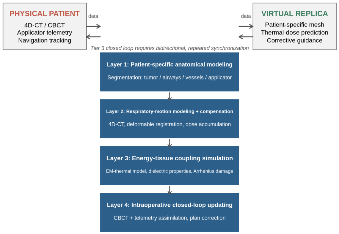
Figure 2.
Digital-twin intervention points along the two ablation routes. (A) CT-guided percutaneous route: applicator trajectory planning, respiratory gating/compensation, margin prediction under heat-sink constraints. (B) Transbronchial route: airway-tree-constrained applicator orientation, bronchus-sign-dependent targeting, multi-nodule session sequencing. Shared core: motion-coupled energy–tissue simulation with CBCT-based closed-loop updating.
Figure 2.
Digital-twin intervention points along the two ablation routes. (A) CT-guided percutaneous route: applicator trajectory planning, respiratory gating/compensation, margin prediction under heat-sink constraints. (B) Transbronchial route: airway-tree-constrained applicator orientation, bronchus-sign-dependent targeting, multi-nodule session sequencing. Shared core: motion-coupled energy–tissue simulation with CBCT-based closed-loop updating.
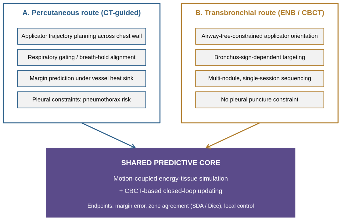
Figure 3.
Five-item research agenda on an indicative timeline (P1, in-vivo property calibration; P2, motion-coupled prediction; P3, prospective utility trial; P4, transbronchial-specific modeling; P5, post-ablation surveillance twin), mapped onto the five-rung validation ladder of Table 3.
Figure 3.
Five-item research agenda on an indicative timeline (P1, in-vivo property calibration; P2, motion-coupled prediction; P3, prospective utility trial; P4, transbronchial-specific modeling; P5, post-ablation surveillance twin), mapped onto the five-rung validation ladder of Table 3.
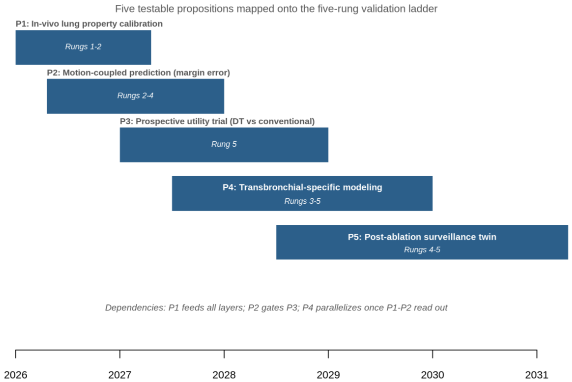
Figure 4.
End-to-end proof-of-concept workflow: 4D-CT acquisition → segmentation and meshing → motion-coupled thermal simulation → applicator placement under navigation/CBCT → telemetry- and CBCT-assimilated intraoperative updating → quantitative predicted-versus-observed zone comparison feeding model refinement. Abbreviations in figures: 4D-CT = four-dimensional computed tomography; CBCT = cone-beam computed tomography; DT = digital twin.
Figure 4.
End-to-end proof-of-concept workflow: 4D-CT acquisition → segmentation and meshing → motion-coupled thermal simulation → applicator placement under navigation/CBCT → telemetry- and CBCT-assimilated intraoperative updating → quantitative predicted-versus-observed zone comparison feeding model refinement. Abbreviations in figures: 4D-CT = four-dimensional computed tomography; CBCT = cone-beam computed tomography; DT = digital twin.
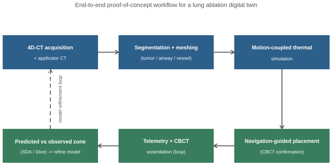

Table 1.
A three-tier terminological hierarchy for digital-twin claims in procedural lung medicine.
| Tier | Designation | Data flow | Defining property | Lung-field exemplars |
|---|---|---|---|---|
| 1 | Static patient-specific model | None after construction | Built once from pre-procedural imaging; used for offline simulation | Biophysical MWA models validated retrospectively [9]; statistical zone prediction [22]; liver MWA DT proof of concept [19] |
| 2 | Digital shadow | Physical → virtual, unidirectional | Intraoperative data update the replica; human remains the actuator | AR-guided nodule localization [57,58]; CBCT-confirmed transbronchial ablation [51] |
| 3 | Digital twin | Bidirectional, closed loop | Replica continuously synchronized; predictions modulate therapy | Not yet reported for pulmonary ablation; nearest analogues in cardiac electrophysiology [17,18] |
AR = augmented reality; CBCT = cone-beam computed tomography; MWA = microwave ablation; DT = digital twin.
Table 2.
The four-layer lung ablation digital-twin stack: validated components and remaining gaps.
| Layer | Function | Validated components (representative) | Lung-specific barrier | Gap to Tier 3 |
|---|---|---|---|---|
| 1. Anatomical modeling | Patient-specific geometry of tumor, airways, vessels, applicator | AR/navigation segmentation pipelines [57]; image-based ablation modeling [55] | Ground-glass margin uncertainty; applicator co-registration | Uncertainty-quantified segmentation feeding simulation |
| 2. Respiratory motion | 4D motion model; phase-resolved dose accumulation | 4D-CT DIR with error quantification [24,25,62]; CT-fluoro motion compensation [64] | Motion absent from all current ablation planning | Motion-coupled thermal simulation; CBCT-to-4D registration |
| 3. Energy–tissue coupling | EM-thermal-damage simulation | Lung carcinoma dielectric data [23]; property sensitivity analysis [69]; heat-sink modeling [70,71]; validated lung MWA models [9]; liver DT template [19] | Aerated-tissue property variability; dual perfusion | In-vivo pulmonary property calibration; fast surrogate solvers [72] |
| 4. Closed-loop updating | Intraoperative assimilation and corrective guidance | CBCT hybrid-OR imaging [51,52]; applicator telemetry | None conceptual; integration is the barrier | Real-time data assimilation; prospective closed-loop trials |
4D = four-dimensional; DIR = deformable image registration; CBCT = cone-beam computed tomography; MWA = microwave ablation; EM = electromagnetic.
Table 3.
Staged validation pathway for a lung ablation digital twin.
| Rung | Substrate | Key validation question | Existing template |
|---|---|---|---|
| 1 | Phantom / ex vivo lung | Does the model reproduce zones under controlled properties? | Phantom-validated image-based modeling [55] |
| 2 | In vivo porcine/rabbit lung | Does the closed loop function end-to-end with motion and telemetry? | Bronchoscopic ablation swine safety and survival studies [40,73]; imaging–pathology correlation in rabbit models [74,75] |
| 3 | Human ablate-then-resect | Does the predicted boundary match histopathological treatment effect? | Treat-and-resect protocol (NCT05281237) [50] |
| 4 | Retrospective clinical cohorts | Does prediction beat vendor charts across centers and devices? | 50-patient computational validation [9]; zone-metric frameworks [36] |
| 5 | Prospective utility trial | Does DT planning improve margins and local control? | AR localization noninferiority RCT [58]; prospective transbronchial ablation designs [38,42] |
RCT = randomized controlled trial; DT = digital twin.
Disclaimer/Publisher’s Note: The statements, opinions and data contained in all publications are solely those of the individual author(s) and contributor(s) and not of MDPI and/or the editor(s). MDPI and/or the editor(s) disclaim responsibility for any injury to people or property resulting from any ideas, methods, instructions or products referred to in the content. |
© 2026 by the authors. Licensee MDPI, Basel, Switzerland. This article is an open access article distributed under the terms and conditions of the Creative Commons Attribution (CC BY) license.
Copyright: This open access article is published under a Creative Commons CC BY 4.0 license, which permit the free download, distribution, and reuse, provided that the author and preprint are cited in any reuse.