

Submitted:
28 October 2026
Posted:
29 October 2026
You are already at the latest version
Abstract
Background: This proof-of-concept clinical study evaluated the feasibility, applicability, tolerability, and preliminary response of a customised 3D-printed, thermoformed intraoral device for gingival topical treatment. Immune-mediated conditions, including oral lichen planus, mucous membrane pemphigoid, and pemphigus vulgaris often cause desquamative gingivitis. Topical corticosteroids are often ineffective in the oral environment, and frequent reapplication may increase systemic absorption. Methods: Two adults with immune-mediated gingival disease—oral lichen planus and mucous membrane pemphigoid—were unresponsive to topical corticosteroids. Following intraoral scanning, digital design, 3D printing, and thermoforming, each received a custom device with reservoirs over the lesions. They applied the corticosteroid gel and wore the device for 30 minutes, three times daily, in four cycles of five treatment days and two rest days. Results: Both cases completed the digital workflow successfully, with high compliance and no device-related mucosal irritation or pressure lesions. Mean pain scores fell from 8.5 at T0 to 4 at T1 (day 20) and 1.5 at T2 (day 40). Erythema and erosive areas decreased markedly, while symptoms nearly resolved. Conclusions: Device-mediated clobetasol was feasible, well tolerated, and effective in two topical treatment–resistant patients, but is currently limited to keratinized, dentition-supported sites (gingiva and palate). Randomized controlled trials must confirm efficacy versus conventional application.
Keywords:
lichen planus
; oral
; pemphigoid
; benign mucous membrane
; gingivitis
; clobetasol
; drug delivery systems
; printing
; three-dimensional
; computer-aided design
1. Introduction
Immune-mediated lesions with gingival localization frequently converge on a shared presentation, desquamative gingivitis (DG), characterized by erythema, desquamation, erosions, and pain of the free and attached gingiva [1]. DG is not a diagnosis in itself but a clinical sign shared by a heterogeneous group of chronic, relapsing mucocutaneous diseases, of which oral lichen planus (OLP), mucous membrane pemphigoid (MMP), and pemphigus vulgaris (PV) account for the large majority of cases [1,2].
Although individually uncommon or rare, these conditions carry a disproportionate clinical burden. They are chronic and recurrent, have no definitive cure, and cause pain, impaired mastication and oral hygiene, and a marked decline in oral-health-related quality of life [2]. Because the gingiva is often the site of onset, dentists have a key role, and a corresponding responsibility, in recognizing, referring, and managing these patients [3].
Oral lichen planus is also a critical oral potentially malignant disorder (OPMD), with a variable risk of progression to oral squamous cell carcinoma (OSCC), as are conditions such as leukoplakia, erythroplakia, and oral submucous fibrosis. Early recognition of these lesions is essential to facilitate timely intervention. A detailed medical history, including the patients’ habits such as smoking, alcohol use, and substance misuse, should always be obtained [4].
However, distinguishing oral lichen planus and other OPMDs from benign reactive lesions or immune-mediated conditions remains challenging in routine dental practice [5]. Delays in clinical assessment may postpone essential specialist referrals, biopsies, and definitive care. Once a diagnosis is established, topical corticosteroids are the first-line treatment for localized gingival diseases. Systemic immunosuppression is reserved for severe, extensive, or systemically threatening disease [6,7].
The topical route delivers the drug directly to the affected site while minimizing systemic exposure. In practice, however, its efficacy is substantially limited by the oral environment [8,9]. Salivary washout and the mechanical stresses of mastication, speech, and swallowing reduce drug–tissue contact time. Conventional formulations, including gels, pastes, ointments, and mouth rinses, also exhibit poor adherence to the gingival mucosa. Consequently, local bioavailability is reduced [10,11]. This often results in incomplete efficacy, the need for repeated daily applications, dispersion of the drug throughout the oral cavity with subsequent systemic absorption, and a burdensome application regimen that frequently leads to poor treatment adherence [12,13].
Systemic therapy circumvents the challenges associated with local delivery; however, it carries a toxicity burden that appears disproportionate when the disease is clinically confined to the gingiva [7,14]. A therapeutic need therefore remains for a delivery modality that retains the drug at a concentrated level and in prolonged contact with the target gingiva, while minimizing dispersion, systemic absorption, and patient burden. Advances in digital dentistry now enable highly precise, reproducible fabrication of devices tailored to individual anatomy. This advancement facilitates the development of intraoral appliances specifically designed to deliver medication to the site of disease.
This study reports the design, fabrication, and preliminary clinical application of a custom intraoral device designed to serve as a reservoir and delivery vehicle for a topical medication. The device is intended to maintain prolonged, direct contact between the medication and gingival lesions, protect it from salivary washout, and localize its action to the treated site. Specifically, the study aimed to demonstrate the technical feasibility of the complete digital workflow, from arch acquisition through reservoir design and device fabrication; evaluate the device’s clinical applicability for delivering a topical drug to gingival lesions and document the preliminary clinical response, with particular attention to patient-reported pain and lesion appearance; assess device tolerability and patient compliance; and explore, at a theoretical level, whether device-mediated delivery can overcome the recognized limitations of conventional topical application.
1.1. Corticosteroid Effect
Topical corticosteroids are the first-line treatment for immune-mediated gingival lesions. Clobetasol propionate 0.05%, a class I (super-potent) topical corticosteroid, is among the most widely used and best-supported agents for oral lichen planus and its desquamative forms [6,7,15]. Its therapeutic rationale is based on broad anti-inflammatory and immunomodulatory effects that directly counteract the pathogenic mechanisms underlying these diseases [16].
Corticosteroids are lipophilic molecules that cross the keratinocyte membrane and bind to the cytoplasmic glucocorticoid receptor. The activated complex translocates to the nucleus and modulates gene transcription. Corticosteroids activate anti-inflammatory genes and, most importantly, suppress pro-inflammatory pathways such as nuclear factor-κB and activator protein-1. This results in reduced cytokine production, decreased T-lymphocyte activation and proliferation, and a less dense inflammatory infiltrate. Consequently, the local immune-mediated response is suppressed, and the affected epithelium is stabilized [15,17].
This mechanism is directly relevant to the target diseases. Oral lichen planus is a T-cell-mediated disorder in which cytotoxic lymphocytes drive apoptosis of basal keratinocytes. Here, corticosteroids attenuate the cytotoxic infiltrate and the resulting damage to the basal layer [18,19]. In mucous membrane pemphigoid and pemphigus vulgaris, tissue damage is antibody-mediated, and corticosteroids suppress the associated inflammatory response [20,21]. Clinically, this leads to reduced erythema, progressive re-epithelialization of erosive-atrophic areas, and pain relief.
Clobetasol and other commonly used molecules (fluocinonide, betamethasone, triamcinolone acetonide, and dexamethasone) are conventionally administered as adhesive pastes, gels, ointments, or mouth rinses [9,10]. This route has several drawbacks. Local adverse effects include mucosal atrophy and, most commonly, oral candidiasis, warranting concomitant antifungal prophylaxis. Potent agents applied to extensive, eroded surfaces may also result in clinically significant systemic absorption [15,22]. The well-established adverse effects associated with prolonged systemic corticosteroid exposure point up the interest in steroid-sparing strategies [22]. Accordingly, the primary objective in improving topical corticosteroid therapy is to enhance the efficiency and localization of drug delivery rather than the potency of the molecule.
1.2. State of the Art
Topical calcineurin inhibitors, primarily tacrolimus and pimecrolimus, have been proposed as steroid-sparing agents for recalcitrant oral lichen planus and desquamative gingivitis. Their short-term efficacy is comparable to that of corticosteroids [23,24]. However, their use is limited by measurable systemic absorption through eroded mucosa and by regulatory warnings regarding a potential neoplastic risk [25,26]. Regardless of the agent used, clinical benefit at the gingiva depends on maintaining contact between the drug and the target mucosa for a sufficient duration.
Mucoadhesive polymers (Carbopol, sodium alginate, gelatin, and chitosan) have been incorporated into films, tablets, gels, and dissolving lozenges to prolong drug residence time. [27,28]. However, they do not fully overcome salivary wash out and mechanical clearance on the gingiva. A complementary physical strategy involves using intraoral devices as drug reservoirs. Custom trays that deliver corticosteroids or tacrolimus under occlusion have been associated with improved outcomes in case reports, retrospective series, and open trials. [12,13,29,30].
Nevertheless, the only randomized trial of tray-applied clobetasol—a three-week crossover study involving 22 patients with desquamative gingivitis—reported no significant difference compared with placebo [31]. Several sustained-release intraoral devices have also been developed. These include a 3D-printed drug-eluting mouthguard evaluated in a first-in-human study [32] and a mucoadhesive clobetasol patch evaluated in a phase 2 randomized trial [33].
However, none has yet achieved established clinical use [34]. Accordingly, a fully digital workflow that maps the reservoirs to the precise lesional sites remains largely unexplored. The present study investigates this approach.
2. Materials and Methods
The treatment consisted of administering therapy using a custom intraoral drug-delivery device in accordance with a cyclical protocol. The overall observation period was 40 days, with clinical and photographic assessments executed at baseline (T0), after 20 days (T1), and at the final follow-up visit (T2, 40 days). Before enrollment, both patients were informed of their conditions, the proposed treatment, and the objectives of the study. Both provided written informed consent to receive treatment, participate in the study, and permit the anonymous publication of their clinical data and intraoral images.
The study was conducted in accordance with the ethical principles of the Declaration of Helsinki, as revised in 2013. It was approved by the Council of the Department of Oral and Maxillofacial Sciences, Sapienza University of Rome, on 26 February 2026 (meeting minutes, item 17.2; Extract No. 15).
2.1. Inclusion Criteria
The inclusion criteria considered in the study design were: adult age; an immune-mediated oral mucosal lesion (oral lichen planus or mucous membrane pemphigoid) with clinical and histopathological confirmation; a clinical presentation with prevalent gingival localization, in the form of desquamative gingivitis, amenable to topical corticosteroid treatment; an insufficient or incomplete response to conventional, manually applied topical therapy; dento-anatomical conditions appropriate for the fabrication and retention of a custom intraoral device; and willingness to provide informed consent and to adhere to the treatment protocol and follow-up visits.
All consecutive eligible patients were included in the study. The final sample comprised two patients. Both patients were informed of the research objectives and treatment methods before providing written informed consent to participate.
2.2. Exclusion Criteria
The exclusion criteria included lesions confined to non-keratinized, mobile mucosal sites not amenable to sealing by a dentition-retained device (buccal mucosa, tongue, floor of the mouth, lips, or soft palate); insufficient dental support or edentulism that precluded adequate device retention; and inability or unwillingness to adhere to the application protocol.
2.3. Clinical Study Group
Two patients were enrolled on a voluntary basis after providing informed consent. Both presented with immune-mediated oral mucosal disease predominantly affecting the gingiva: oral lichen planus in Case 1 and mucous membrane pemphigoid in Case 2. Histological examination of samples obtained through scalpel-incisional biopsy confirmed the diagnoses.
Case 1 was a 67-year-old woman affected by atrophic-erosive oral lichen planus localized on the attached gingiva. She had a medical history of arterial hypertension and was currently receiving treatment with olmesartan medoxomil and hydrochlorothiazide. She also had microcytosis. The patient denies any allergic hypersensitivities and reports being a non-smoker.
Case 2 was a 59-year-old man affected by mucous membrane pemphigoid limited to the attached gingiva. He had a history of benign prostatic hypertrophy and was currently assuming terazosin hydrochloride. The patient also reported no known allergic hypersensitivities to medications or other substances and reported being a non-smoker.
Both patients had received a first-line treatment with topical clobetasol propionate 0.05% gel three times daily for 40 days, with an unsatisfactory response. Afterwards, we administered systemic corticosteroid therapy with methylprednisolone at a daily dose of 16 mg for 40 days, resulting in partial improvement in Case 1 and no response in Case 2. On the other hand, both patients showed intense acute adverse effects of systemic corticosteroid therapy such as insomnia, gastritis, rapid mood changes and fluid retention.
Baseline pain, self-rated using a 0–10 Numeric Rating Scale (NRS), was 9/10 in Case 1 and 8/10 in Case 2.
2.4. Digital Workflow for Device Fabrication
We acquired scans of the maxillary and mandibular arches with an intraoral scanner (i700; Medit Corp., Seoul, Republic of Korea).. Digital scanning was preferred over an alginate impression because a loaded impression tray presses directly on eroded, painful gingival tissues. Scanning avoided this uncomfortable pressure and improved patient compliance and cooperation. The scans were imported into a dental computer-aided design (CAD) software (Blender for Dental, a dental CAD add-on suite based on the open-source Blender platform), and a separate digital model was generated for each arch. The overall workflow is summarized in Fig. 1.
Each model was then subjected to a block-out procedure. In digital appliance design, block-out refers to the elimination of undercuts—surface regions below the height of contour that would otherwise mechanically lock the appliance onto the model—by uniformly building up the model surface. In the present case, the surface volume was increased by approximately 120 µm. This step smooths the residual undercuts, enabling the thermoformed device to be seated and removed without interference while creating the controlled offset space required to accommodate the drug between the device and the mucosa.
At this stage, the model was processed in PLY (Polygon File Format) rather than the more commonly used STL (Standard Tessellation Language). Both formats represent the surface as a polygon mesh. However, STL stores geometry only and supports a single color (monochrome), whereas PLY also retains per-vertex color information. Since the intraoral scan’s color rendering was essential for identifying the erythematous lesion and accurately mapping its extent on the model—and, consequently, for extending the device to the desired anatomical region—the monochrome STL format was excluded at this stage in favor of the color-capable PLY format. In order to correspond with the clinically identified lesions, 2-mm-thick reservoir boxes were created and fused to the base model using a Boolean union.
The completed digital model was exported in STL format and imported into the dedicated slicing software of the 3D printer (Inkspire LCD printer; Zortrax S.A., Olsztyn, Poland). It was printed using KeyModel Ultra™ resin (Keystone Industries, Gibbstown, NJ, USA), with a layer thickness of 50 µm and a 405 nm light source. The model was oriented at 30° on the build platform without supports to minimize deformation.
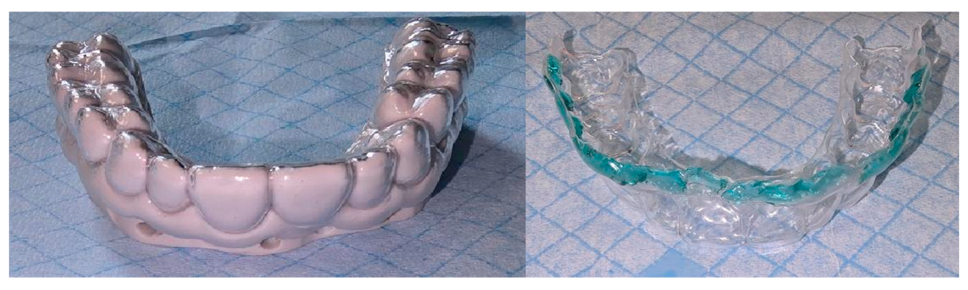
Post-processing consisted of automated washing in 90% isopropyl alcohol for 6 minutes, followed by overnight air-drying. The specimens were then post-cured under 30 multidirectional UV LEDs for 5 minutes per side, in accordance with the resin manufacturer's instructions. A 0.5-mm thermoforming resin sheet was then thermoformed onto the printed model to obtain the final device (Figure 2). This intentionally thin, lightweight profile was selected to enhance comfort, as a thicker appliance could cause pressure lesions. In the stratified 2-mm reservoir areas, the device reproduces cavities designed to contain the drug.
The device was trimmed just above the lesional layer to ensure a seal against healthy perilesional tissue, and its margin was polished to remove any roughness before delivery to the patient. To accommodate this sealing margin, the initial intraoral scan was deliberately extended beyond the lesional border, allowing the device to be trimmed and finished on sound tissue immediately above the lesion.
Figure 1.
Digital workflow for the fabrication of the custom delivery tray, from intraoral scanning to delivery of the drug-loaded device.
Figure 1.
Digital workflow for the fabrication of the custom delivery tray, from intraoral scanning to delivery of the drug-loaded device.
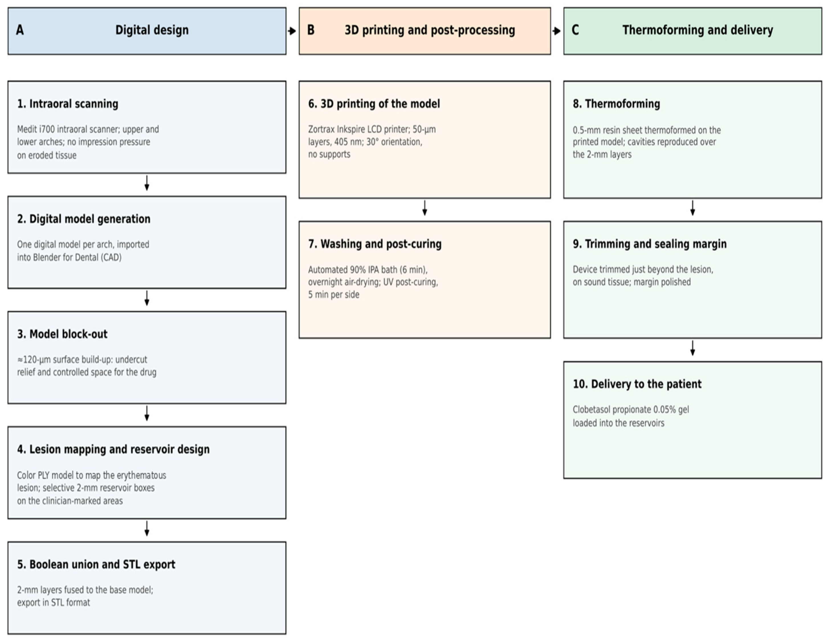
Figure 2.
Intraoral device for topical drug delivery.
2.5. Therapeutic Protocol
The active treatment consisted of a galenic (pharmacy-compounded) formulation containing clobetasol propionate 0.05%, prepared as an alcohol-free oral gel. This formulation was selected to avoid the irritant and burning effects of alcohol on eroded mucosa and to provide a consistency suitable for loading into the device reservoirs.
Nystatin oral suspension was co-prescribed as antifungal prophylaxis against oral candidiasis, a recognized complication of prolonged topical corticosteroid contact. Nystatin rinses were scheduled at least 30 minutes apart from clobetasol propionate 0.05% application to prevent the corticosteroid from being washed away. Nystatin was selected over azole antifungals because it is a polyene with negligible mucosal and systemic absorption and no inhibitory activity on cytochrome P450 3A4. In contrast, azoles inhibit CYP3A4 and could increase systemic exposure to any absorbed fraction of the corticosteroid.
The device, loaded with clobetasol propionate 0.05% gel in its reservoirs (handled with a glove to minimize contamination), was worn three times daily for 30 minutes per application following oral hygiene. Treatment was administered according to a cyclical schedule consisting of five consecutive treatment days (three applications per day), followed by a two-day rest period. This schedule was repeated for a total of four cycles. Patients were instructed to abstain from eating and drinking for 30 minutes after each application. Throughout treatment, patients also performed nystatin rinses twice daily using 4 mL of undiluted suspension, held in the mouth for approximately one minute without swallowing.
Each patient received structured instruction in oral hygiene preparation; appropriate gel dosing and loading; device insertion and removal; adherence to application and post-application intervals; device cleaning and storage; and recognition of adverse effects, including burning, white patches, taste changes suggestive of candidiasis, and pressure-related mucosal injury.
2.6. Outcome Measures
Two principal parameters were assessed at baseline (T0), 20 days (T1), and 40 days (T2): (1) patient-reported pain, measured using the NRS (0–10); and (2) clinical lesion appearance, documented through standardized photography and, where applicable, evaluated with reference to validated grading instruments, including the Thongprasom sign score [36] and the Oral Disease Severity Score (ODSS), also known as the Escudier score (Escudier et al.) [37,39].
Secondary parameters included device tolerability (discomfort, mucosal irritation, and decubitus lesions) and patient adherence to the protocol. Following the active treatment phase, patients entered a follow-up program that included clinical assessments every 3–6 months.
For the case of the patient with oral lichen planus, periodic follow-up also serves an oncological surveillance function, as oral lichen planus is a potentially malignant disorder with a pooled malignant transformation rate of approximately 1.1%, which is higher in atrophic-erosive forms [35].
3. Results
The digital workflow was successfully completed for both patients. Intraoral scanning was well tolerated and eliminated the need for a pressure-loaded impression on painful, eroded tissue. Both patients tolerated the device well. Its 0.5-mm thickness and polished margins contributed to patient comfort. No appliance-related discomfort, pressure lesions, or mucosal irritation were reported. Nystatin prophylaxis was well tolerated, and no clinical signs of oral candidiasis were observed during treatment. Both patients adhered closely to the cyclical protocol and reported a favorable overall experience with both the device and the atraumatic digital impression.
3.1. Case 1 (Oral Lichen Planus).
At baseline, the patient presented with diffuse erythema of the attached gingiva, with atrophic-erosive areas (Figure 3 and Figure 4). She reported persistent burning pain (NRS 9/10), exacerbated by contact with certain foods. Prior treatment with topical clobetasol propionate 0.05% gel yielded no meaningful benefit, whereas systemic methylprednisolone resulted in partial improvement. The patient received in-person instruction on the proper application of the gel into the custom device and its placement in the oral cavity for the initial application, which was demonstrated by the clinician (Figure 5).
Following two cycles of device-mediated clobetasol propionate 0.05% therapy, erythema and inflammation had visibly improved by T1 (Figure 6 and Figure 7) and improved further by T2 (Figure 8 and Figure 9). The gingival mucosa appeared largely normalized, and the burning symptoms were reported to have nearly resolved completely, occurring only after the consumption of acidic, hot, or spicy foods. At the conclusion of treatment, the patient reported a final NRS score of 1/10.
3.2. Case 2 (Mucous Membrane Pemphigoid).
At baseline, the patient presented with erythematous, desquamating gingiva and reported pain rated 8/10 on the NRS. The lesions were first observed two years earlier and had previously been treated with antifungal agents alone (Figure 10). Neither the previous topical clobetasol propionate 0.05% gel treatment nor the subsequent systemic methylprednisolone treatment achieved the desired outcome. In this case, the patient was also instructed on the proper application of the gel to the custom device and its placement in the oral cavity during the initial application (Figure 11 and Figure 12).
Following device-mediated therapy, a marked regression in erythema and erosions was observed at T1 (Figure 13), with further improvement at T2 (Figure 14 and Figure 15). The patient reported that the burning sensation had almost completely resolved, with a final NRS score of 2/10.
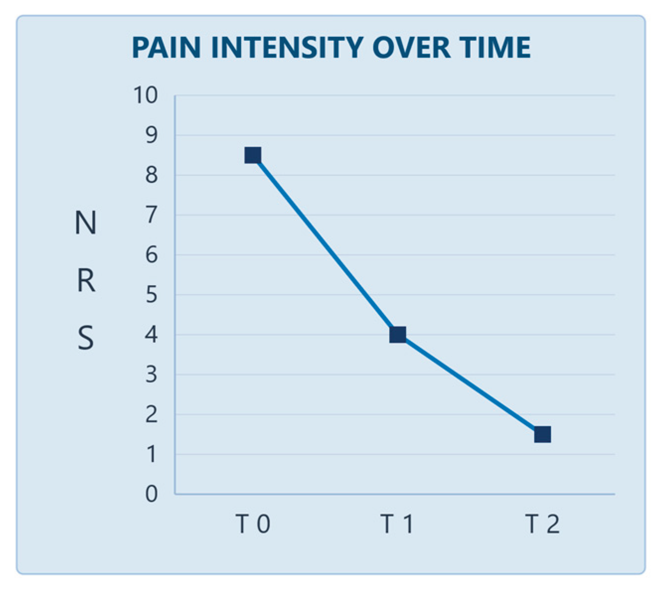
Across both patients, the mean NRS pain score steadily decreased from approximately 8.5 at T0 to 4 at T1 and 1.5 at T2, paralleling the clinical improvement observed in lesion appearance for both cases (Figure 16).
The validated severity scores demonstrated a similar trend. In Case 1, the Thongprasom sign score decreased from 5 at T0 to 4 at T1 and 2 at T2, while the ODSS decreased from 36 to 19 and 8, respectively. In Case 2, the ODSS decreased from 32 at T0 to 21 at T1 and 10 at T2. Thus, between T0 and T2, the ODSS decreased by approximately 78% in Case 1 and 69% in Case 2.
4. Discussion
This study demonstrates that a fully digital workflow can be employed to fabricate a patient-specific intraoral device capable of delivering a topical drug directly to immune-mediated gingival lesions. In both treated cases, clinical improvement was observed despite previous unsatisfactory responses to the same medication administered through conventional manual application. Since the active compound (clobetasol propionate 0.05%) remained unchanged, the observed benefit is more plausibly attributable to the modified delivery modality—specifically, confinement of the drug within a sealed reservoir maintained in prolonged contact with the target mucosa—than to any difference in pharmacological potency.
The magnitude of the observed change also appears clinically meaningful: the mean reduction in NRS was approximately 7 points, substantially exceeding the 2-point minimal important change established for the NRS in oral lichen planus. [36]. This interpretation is consistent with prior reports indicating that gingival corticosteroid therapy yields improved outcomes when occlusive or retentive custom trays are used to minimize salivary washout [12,13,29,30]. It is also consistent with the broader concept of sustained-release intraoral devices [32,33,34]. At the same time, the only randomized trial of tray-applied clobetasol in desquamative gingivitis found no benefit over placebo after three weeks of treatment [31]. Differences in tray design, lesion-mapped reservoir geometry, treatment duration, and patient selection may explain this discrepancy and draw attention to the need for controlled confirmation of these findings.
Mechanistically, the device targets the two parameters believed to govern local topical bioavailability—contact time and local drug concentration—through a physical rather than chemical strategy, complementing mucoadhesive formulation approaches. By sealing the corticosteroid against the lesional gingiva and its immediate perilesional margin, the device is expected to reduce three undesirable phenomena associated with manual application: drug dispersion onto adjacent healthy tissue, partial swallowing, and consequent secondary systemic absorption. This is particularly relevant for patients, including both cases reported here, who had previously required systemic corticosteroid exposure. Accordingly, the device may represent a potential systemic steroid-sparing strategy, an attribute of interest in chronic immune-mediated diseases that often affect older, comorbid, or polymedicated patients [14,22].
Tolerability and compliance findings were similarly favorable. The device profile appeared to avoid the mechanical irritation that a bulkier appliance might cause in already fragile mucosa. In addition, the atraumatic nature of digital impression-taking, compared with a pressure-loaded conventional impression, likely contributed to a positive experience from the outset of care. Standardization of the application procedure may also reduce operator- and patient-dependent variability inherent in manual topical application. This factor may be relevant to the good adherence observed in both cases, given the chronic, relapsing nature of these diseases.
Despite these insights, several limitations must be acknowledged. The primary and most inherent limitation is anatomical. As a thermoformed device retained by the dental arch and sealed against adjacent tissue, it can deliver the drug effectively only to sites that are stable and adjacent to its retentive base. Lesions of the lining mucosa (buccal mucosa, tongue, floor of the mouth, lips, soft palate), which are mobile and elastic, cannot be sealed or reached by an arch-retained appliance and therefore fall outside the scope of this approach.
Additional limitations pertain to the study design, including the extremely small sample size (n = 2), the absence of a control group, randomization, or blinding, and a sample size insufficient to support statistical inference. The 40-day observation period captures only short-term responses and does not allow for the assessment of durability, recurrence rates, or the utility of repeated treatment cycles—all critical considerations in chronic relapsing disease. Finally, practical and logistical constraints may limit reproducibility and scalability in less-equipped settings. Adequate dental support is also required to ensure device retention, thereby limiting applicability in edentulous or periodontally compromised patients.
Confirmation of these preliminary findings will require larger, preferably randomized controlled trials that directly compare device-mediated delivery with conventional manual application. Such trials should use standardized outcome measures (NRS [36], Thongprasom score [37], Oral Disease Severity Score [38,39,40]), incorporate oral-health-related quality-of-life assessments, and include extended follow-up to characterize recurrence and evaluate the value of repeated treatment cycles.
Further avenues for development include optimizing device materials and reservoir geometry, particularly for patients with reduced dental support; combining the device with sustained- or controlled-release drug formulations or alternative agents (e.g., topical calcineurin inhibitors); conducting pharmacokinetic studies to objectively quantify any reduction in systemic absorption; adapting the design to extend its applicability to non-keratinized mucosal sites; and integrating the mapping step with AI-assisted lesion segmentation based on color intraoral scans to streamline and standardize device design.
5. Conclusion
In this preliminary experience, a fully digital workflow enabled the fabrication of a patient-specific intraoral device that successfully delivered a topical corticosteroid to immune-mediated gingival lesions in two patients who had previously not responded to conventional manual topical application. The treatment was associated with a favorable clinical response, high tolerability, and good patient compliance. These findings support the technical feasibility and clinical plausibility of this approach and provide a rationale for larger, controlled studies to establish its comparative efficacy, optimal design, and role in the management of chronic immune-mediated gingival disease.
Author Contributions
Conceptualization, G.P., A.Po and D.P.; Methodology, N.P., U.R.; Software, N.P., F.M.G.; Validation, G.P., A.Pu. and U.R.; Formal Analysis, A.Pu.; Investigation, A.Pu. and G.P.; Resources, S.H. and F.M.G.; Data Curation, S.H. and D.P.; Writing – Original Draft Preparation, S.H. and A.Pu.; Writing – Review & Editing, A.Pu. and N.P.; Visualization, D.P. and F.M.G.; Supervision, U.R. and A.Po; Project Administration, G.P.
Funding
This research received no external funding.
Institutional Review Board Statement
The study was conducted in accordance with the ethical principles of the Declaration of Helsinki, as revised in 2013. The study protocol, entitled “Digital Workflow-Guided Topical Drug Delivery in Oral Mucosal Diseases: A Clinical Study on a Customized Intraoral Device,” was approved by the Council of the Department of Oral and Maxillofacial Sciences, Sapienza University of Rome, on 26 February 2026 (meeting minutes, item 17.2; Extract No. 15).
Informed Consent Statement
Written informed consent was obtained from both patients for treatment, for participation in the study, and for the publication of this report and the accompanying intraoral images. The images were anonymized and contain no identifying features.
Data Availability Statement
The datasets used and/or analyzed during the current study are available from the corresponding author on reasonable request.
Conflicts of Interest
The authors declare no conflict of interest.
References
- Maderal, A.D.; Lee Salisbury, P., 3rd; Jorizzo, J.L. Desquamative gingivitis: Clinical findings and diseases. J. Am. Acad. Dermatol. 2018, 78(5), 839–848. [Google Scholar] [CrossRef]
- Lo Russo, L.; Fedele, S.; Guiglia, R.; Ciavarella, D.; Lo Muzio, L.; Gallo, P.; Di Liberto, C.; Campisi, G. Diagnostic pathways and clinical significance of desquamative gingivitis. J. Periodontol. 2008, 79(1), 4–24. [Google Scholar] [CrossRef]
- Leao, J.C.; Ingafou, M.; Khan, A.; Scully, C.; Porter, S. Desquamative gingivitis: retrospective analysis of disease associations of a large cohort. Oral Dis. 2008, 14(6), 556–60. [Google Scholar] [CrossRef]
- Palaia, G.; Mohsen, M.; Pergolini, D.; Bartone, V.; Purrazzella, A.; Romeo, U.; Polimeni, A. E-Cigarette: A Safe Tool or a Risk Factor for Oral Cancer? A Systematic Review. J. Clin. Exp. Dent. 2025, 17, e219–e228. [Google Scholar] [CrossRef]
- Mohsen, M.; Pergolini, D.; Purrazzella, A.; Pierini, E.; Pranno, N.; Romeo, U.; Polimeni, A.; Palaia, G. Diagnostic Performance of a Point-of-Care Salivary Biomarker-Based Test for Oral Potentially Malignant Disorders: A Case–Control Clinical Study. Appl. Sci. 2026, 16, 9379. [Google Scholar] [CrossRef]
- Lodi, G.; Carrozzo, M.; Furness, S.; Thongprasom, K. Interventions for treating oral lichen planus: a systematic review. Br. J. Dermatol. 2012, 166(5), 938–47. [Google Scholar] [CrossRef]
- Buonavoglia, A.; Leone, P.; Dammacco, R.; Di Lernia, G.; Petruzzi, M.; Bonamonte, D.; Vacca, A.; Racanelli, V.; Dammacco, F. Pemphigus and mucous membrane pemphigoid: An update from diagnosis to therapy. Autoimmun. Rev. 2019, 18(4), 349–358. [Google Scholar] [CrossRef]
- Lodi, G.; Manfredi, M.; Mercadante, V.; Murphy, R.; Carrozzo, M. Interventions for treating oral lichen planus: corticosteroid therapies. Cochrane Database Syst. Rev. 2020, 2(2), CD001168. [Google Scholar] [CrossRef]
- Carbone, M.; Conrotto, D.; Carrozzo, M.; Broccoletti, R.; Gandolfo, S.; Scully, C. Topical corticosteroids in association with miconazole and chlorhexidine in the long-term management of atrophic-erosive oral lichen planus: a placebo-controlled and comparative study between clobetasol and fluocinonide. Oral Dis. 1999, 5(1), 44–9. [Google Scholar] [CrossRef]
- González-Moles, M.A.; Scully, C. Vesiculo-erosive oral mucosal disease--management with topical corticosteroids: (1) Fundamental principles and specific agents available. J. Dent. Res. 2005, 84(4), 294–301. [Google Scholar] [CrossRef]
- Hearnden, V.; Sankar, V.; Hull, K.; Juras, D.V.; Greenberg, M.; Kerr, A.R.; Lockhart, P.B.; Patton, L.L.; Porter, S.; Thornhill, M.H. New developments and opportunities in oral mucosal drug delivery for local and systemic disease. Adv. Drug Deliv. Rev. 2012, 64(1), 16–28. [Google Scholar] [CrossRef]
- Torrezani, A.; Gallo, C.B.; Motta, A.C.F.; Siqueira, C.S.; Rabelo, G.D.; Júnior, C.A.L. Desquamative gingivitis treatment with topical tacrolimus applied to a custom tray: an open trial regarding its efficacy on patients' symptoms. Oral Surg. Oral Med. Oral Pathol. Oral Radiol. 2024, 137(1), 30–36. [Google Scholar] [CrossRef]
- Kavlakova, L.; Bachurska, S. Topical Occlusive Corticosteroid Therapy for the Treatment of Gingival Manifestation of Mucous Membrane Pemphigoid - a Case Report. Folia Med. 2020, 62(4), 866–870. [Google Scholar] [CrossRef]
- Kridin, K. Emerging treatment options for the management of pemphigus vulgaris. Ther. Clin. Risk Manag 2018, 14, 757–778. [Google Scholar] [CrossRef]
- Zheng, T.; Liu, C.; Wang, Y.; Zhou, R.; Wu, D.; Tan, J.; Zhu, K. Efficacy and safety of topical clobetasol propionate in comparison with alternative treatments in oral lichen planus: an updated systematic review and meta-analysis. Front Med. 2024, 11, 1391754. [Google Scholar] [CrossRef]
- Pergolini, D.; Purrazzella, A.; Mohsen, M.; Di Gioia, C.R.T.; Polimeni, A.; Palaia, G. Traumatic Ulcerative Granuloma with Stromal Eosinophilia Treated with Intralesional Injections of Triamcinolone Acetonide: A Case Report. Reports 2025, 8, 254. [Google Scholar] [CrossRef]
- Roopashree, M.R.; Gondhalekar, R.V.; Shashikanth, M.C.; George, J.; Thippeswamy, S.H.; Shukla, A. Pathogenesis of oral lichen planus--a review. J. Oral Pathol. Med. 2010, 39(10), 729–34. [Google Scholar] [CrossRef]
- Sugerman, P.B.; Savage, N.W.; Walsh, L.J.; Zhao, Z.Z.; Zhou, X.J.; Khan, A.; Seymour, G.J.; Bigby, M. The pathogenesis of oral lichen planus. Crit. Rev. Oral Biol. Med. 2002, 13(4), 350–65. [Google Scholar] [CrossRef]
- Schmidt, E.; Kasperkiewicz, M.; Joly, P. Pemphigus. Lancet 2019, 394(10201), 882–894. [Google Scholar] [CrossRef]
- Kasperkiewicz, M.; Ellebrecht, C.T.; Takahashi, H.; Yamagami, J.; Zillikens, D.; Payne, A.S.; Amagai, M. Pemphigus. Nat. Rev. Dis. Primers 2017, 3, 17026. [Google Scholar] [CrossRef]
- Schmidt, E.; Zillikens, D. Pemphigoid diseases. Lancet 2013, 381(9863), 320–32. [Google Scholar] [CrossRef]
- Decani, S.; Federighi, V.; Baruzzi, E.; Sardella, A.; Lodi, G. Iatrogenic Cushing's syndrome and topical steroid therapy: case series and review of the literature. J. Dermatol. Treat. 2014, 25(6), 495–500. [Google Scholar] [CrossRef]
- Sun, S.L.; Liu, J.J.; Zhong, B.; Wang, J.K.; Jin, X.; Xu, H.; Yin, F.Y.; Liu, T.N.; Chen, Q.M.; Zeng, X. Topical calcineurin inhibitors in the treatment of oral lichen planus: a systematic review and meta-analysis. Br. J. Dermatol. 2019, 181(6), 1166–1176. [Google Scholar] [CrossRef]
- Samycia, M.; Lin, A.N. Efficacy of topical calcineurin inhibitors in lichen planus. J. Cutan. Med. Surg. 2012, 16(4), 221–9. [Google Scholar] [CrossRef]
- Vohra, S.; Singal, A.; Sharma, S.B. Clinical and serological efficacy of topical calcineurin inhibitors in oral lichen planus: a prospective randomized controlled trial. Int. J. Dermatol. 2016, 55(1), 101–5. [Google Scholar] [CrossRef]
- Chancellor, M.B. Rationale for the Use of Topical Calcineurin Inhibitors in the Management of Oral Lichen Planus and Mucosal Inflammatory Diseases. Cureus 2024, 16(11), e74570. [Google Scholar] [CrossRef]
- Hombach, J.; Bernkop-Schnürch, A. Mucoadhesive drug delivery systems. Handb. Exp. Pharmacol. 2010, (197), 251–66. [Google Scholar] [CrossRef]
- Dhal, S.; Pal, A.; Gramza-Michalowska, A.; Kim, D.; Mohanty, B.; Sagiri, S.S.; Pal, K. Formulation and Characterization of Emulgel-Based Jelly Candy: A Preliminary Study on Nutraceutical Delivery. Gels 2023, 9(6), 466. [Google Scholar] [CrossRef]
- Gonzalez-Moles, M.A.; Ruiz-Avila, I.; Rodriguez-Archilla, A.; Morales-Garcia, P.; Mesa-Aguado, F.; Bascones-Martinez, A.; Bravo, M. Treatment of severe erosive gingival lesions by topical application of clobetasol propionate in custom trays. Oral Surg. Oral Med. Oral Pathol. Oral Radiol. Endod. 2003, 95(6), 688–92. [Google Scholar] [CrossRef]
- Porat Ben Amy, D.; Kassem, R. Custom-made trays for targeted treatment of oral vesiculobullous diseases: A proposed protocol. Dermatol. Ther. 2020, 33(6), e14446. [Google Scholar] [CrossRef]
- Motta, A.C.F.; Domaneschi, C.; Komesu, M.C.; Souza, C.S.; Aoki, V.; Migliari, D.A. Double-blind, crossover, placebo-controlled clinical trial with clobetasol propionate in desquamative gingivitis. Braz. Dent. J. 2009, 20(3), 231–6. [Google Scholar] [CrossRef]
- Liang, K.; Carmone, S.; Brambilla, D.; Leroux, J.C. 3D printing of a wearable personalized oral delivery device: A first-in-human study. Sci. Adv. 2018, 4(5), eaat2544. [Google Scholar] [CrossRef]
- Brennan, M.T.; Madsen, L.S.; Saunders, D.P.; Napenas, J.J.; McCreary, C.; Ni Riordain, R.; et al. Efficacy and safety of a novel mucoadhesive clobetasol patch for treatment of erosive oral lichen planus: A phase 2 randomized clinical trial. J. Oral Pathol. Med. 2022, 51(1), 86–97. [Google Scholar] [CrossRef]
- Alghanem, S.; Dziurkowska, E.; Ordyniec-Kwaśnica, I.; Sznitowska, M. Intraoral medical devices for sustained drug delivery. Clin. Oral Investig. 2023, 27(12), 7157–7169. [Google Scholar] [CrossRef]
- González-Moles, M.Á.; Ruiz-Ávila, I.; González-Ruiz, L.; Ayén, Á.; Gil-Montoya, J.A.; Ramos-García, P. Malignant transformation risk of oral lichen planus: A systematic review and comprehensive meta-analysis. Oral Oncol. 2019, 96, 121–130. [Google Scholar] [CrossRef]
- Wiriyakijja, P.; Porter, S.; Fedele, S.; Hodgson, T.; McMillan, R.; Shephard, M.; Ni Riordain, R. Meaningful improvement thresholds in measures of pain and quality of life in oral lichen planus. Oral Dis. 2020, 26(7), 1464–1473. [Google Scholar] [CrossRef]
- Thongprasom, K.; Luangjarmekorn, L.; Sererat, T.; Taweesap, W. Relative efficacy of fluocinolone acetonide compared with triamcinolone acetonide in treatment of oral lichen planus. J. Oral Pathol. Med. 1992, 21(10), 456–8. [Google Scholar] [CrossRef]
- Escudier, M.; Ahmed, N.; Shirlaw, P.; Setterfield, J.; Tappuni, A.; Black, M.M.; et al. A scoring system for mucosal disease severity with special reference to oral lichen planus. Br. J. Dermatol. 2007, 157(4), 765–70. [Google Scholar] [CrossRef]
- Ormond, M.; McParland, H.; Donaldson, A.N.A.; Andiappan, M.; Cook, R.J.; Escudier, M.; et al. An Oral Disease Severity Score validated for use in oral pemphigus vulgaris. Br. J. Dermatol. 2018, 179(4), 872–881. [Google Scholar] [CrossRef]
- Ormond, M.; McParland, H.; Thakrar, P.; Donaldson, A.N.A.; Andiappan, M.; Cook, R.J.; et al. Validation of an Oral Disease Severity Score (ODSS) tool for use in oral mucous membrane pemphigoid. Br. J. Dermatol. 2020, 183(1), 78–85. [Google Scholar] [CrossRef]
Figure 3.
Clinical presentation at the start of treatment (T0): marked erythema of the attached gingiva with a desquamative appearance, frontal view.
Figure 3.
Clinical presentation at the start of treatment (T0): marked erythema of the attached gingiva with a desquamative appearance, frontal view.
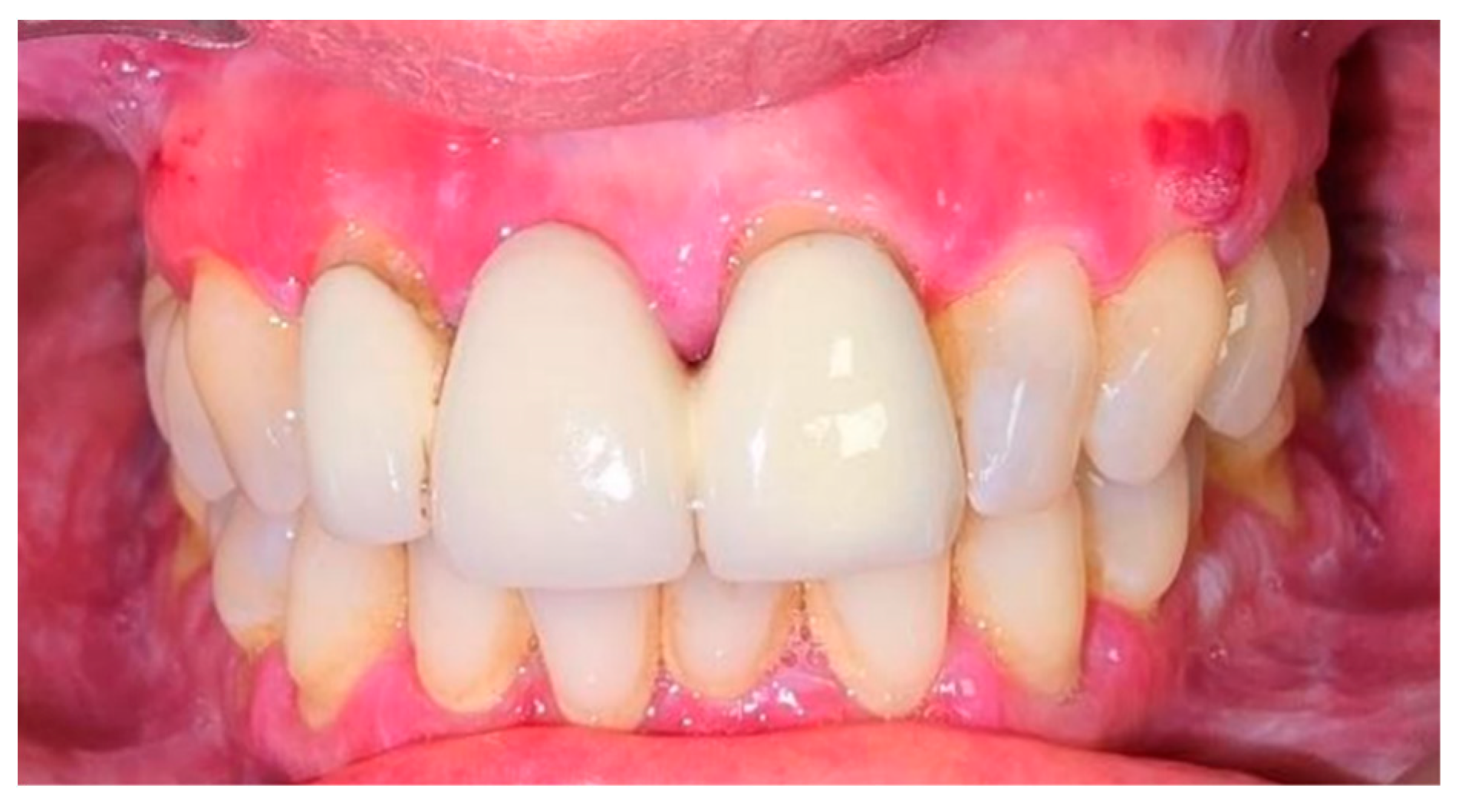
Figure 4.
Lateral views at T0.
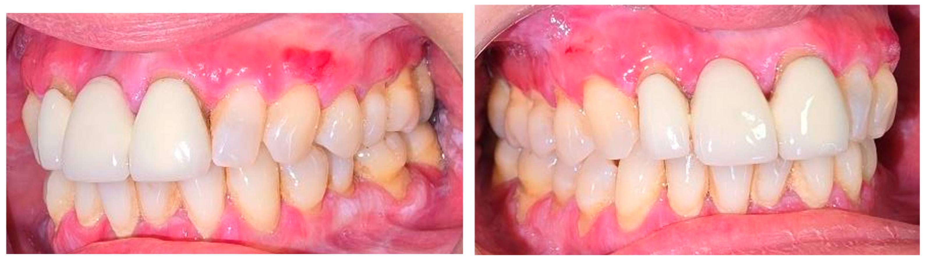
Figure 5.
Device positioned, frontal and lateral views.
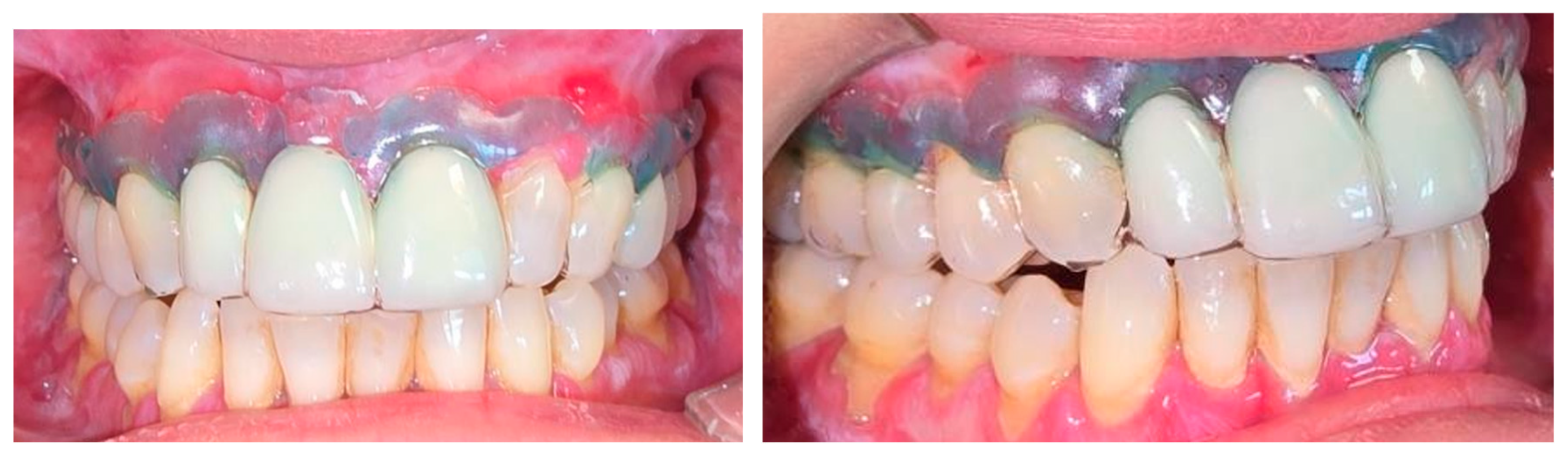
Figure 6.
20-day follow-up (T1), frontal view.
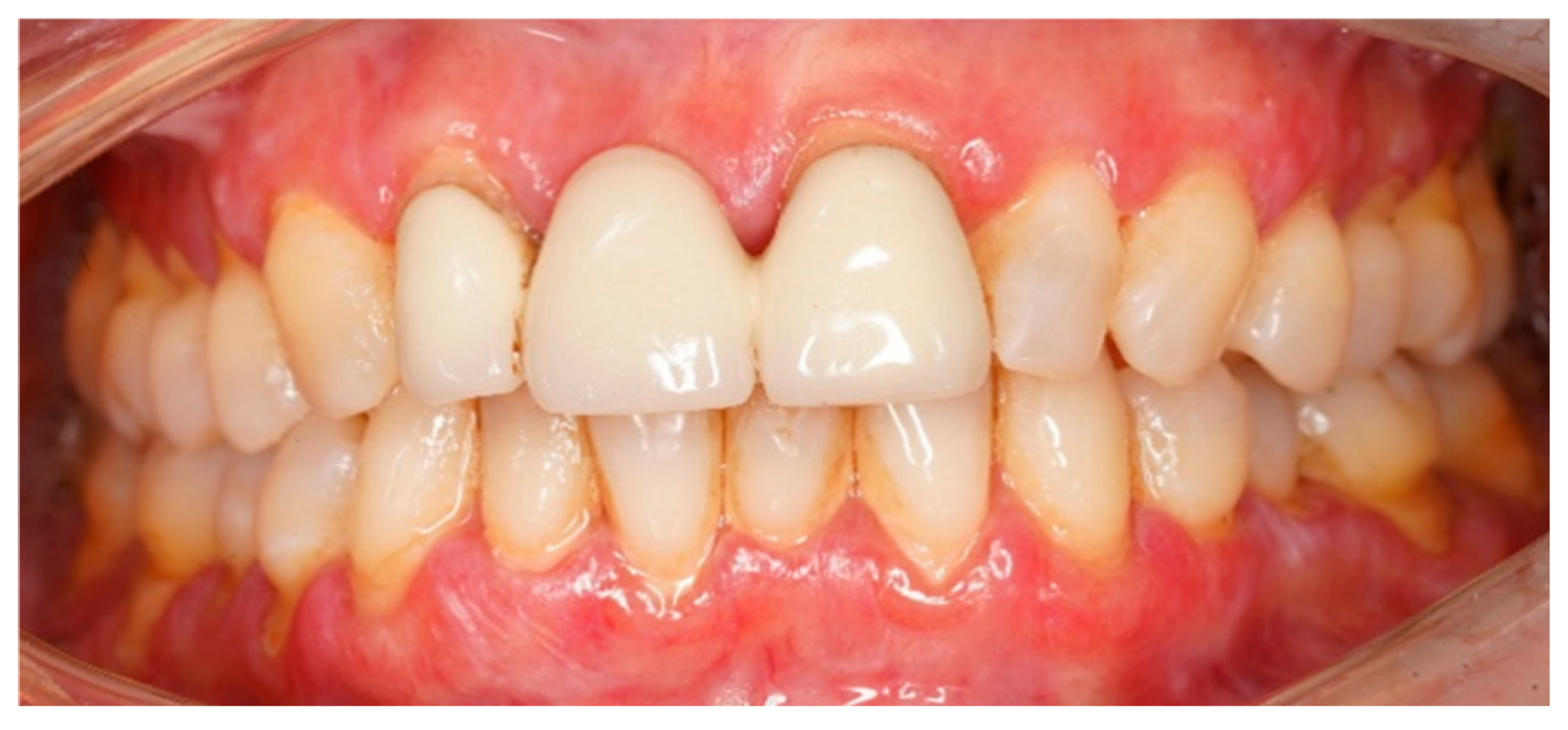
Figure 7.
20-day follow-up (T1), lateral views.
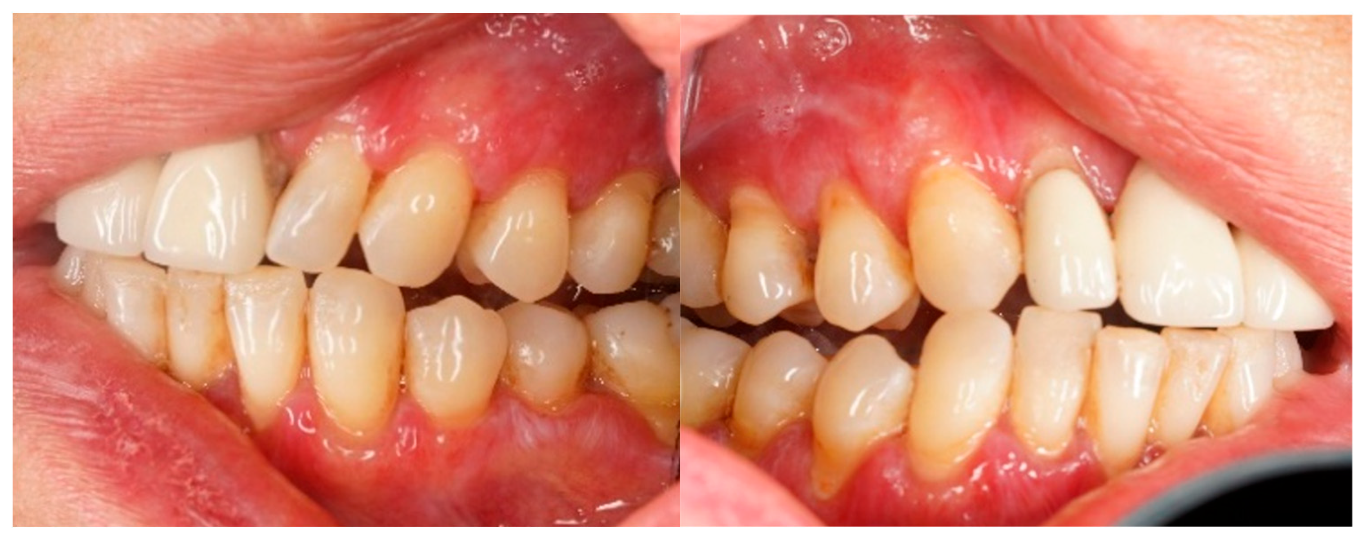
Figure 8.
40-day follow-up (T2), frontal view.
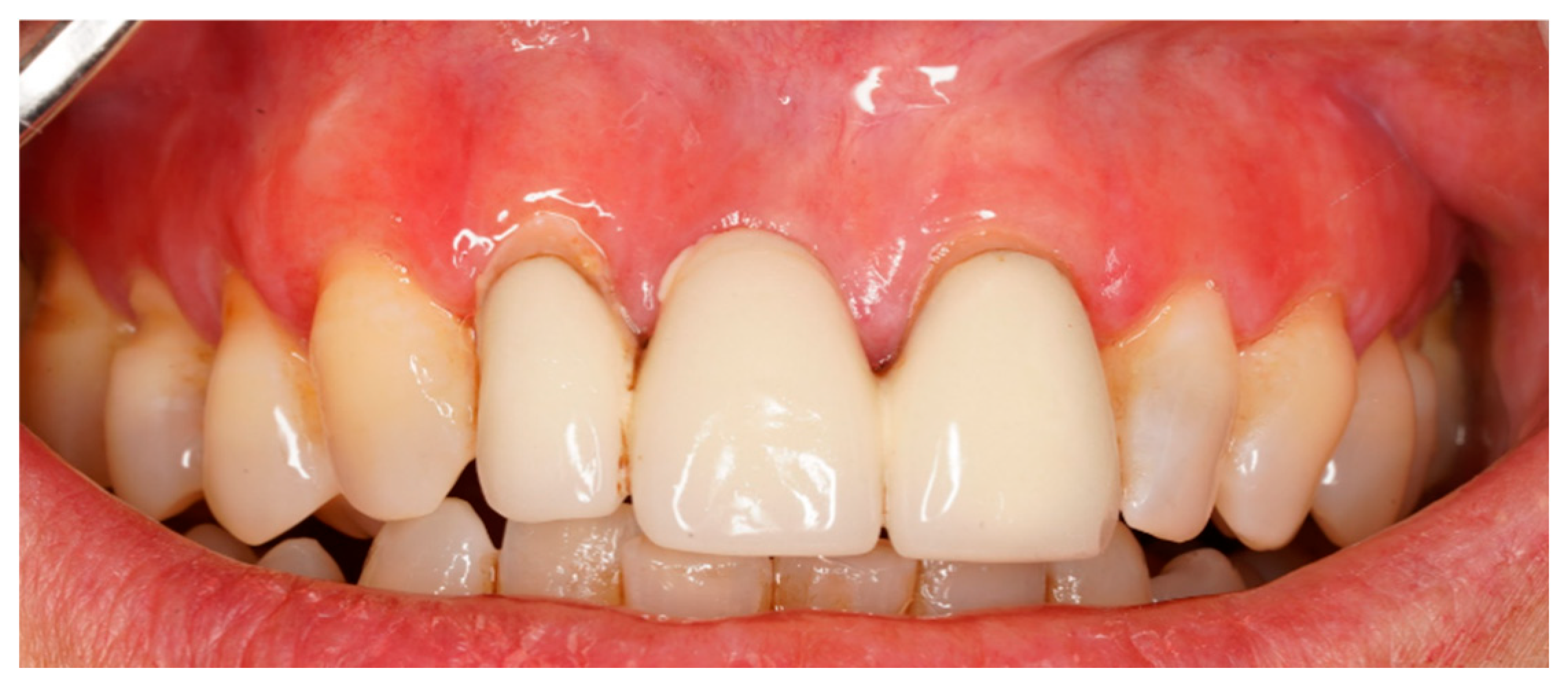
Figure 9.
40-day follow-up (T2), lateral views.
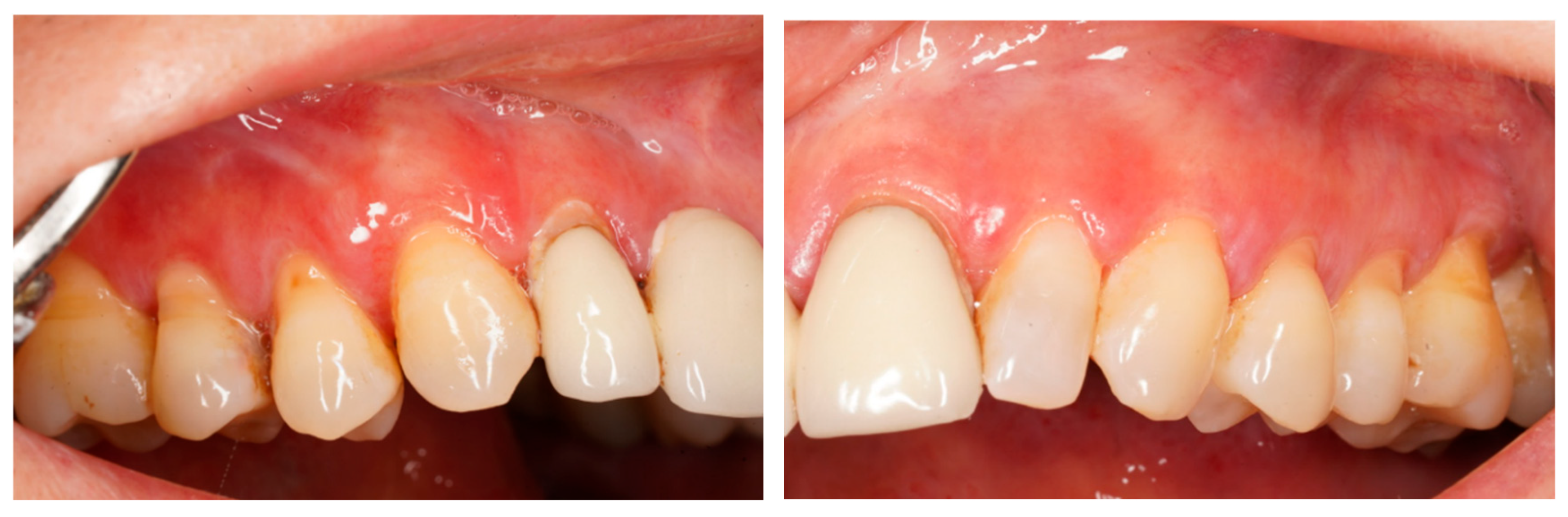
Figure 10.
Baseline clinical picture (T0): diffuse erythematous and desquamating attached gingiva, frontal view.
Figure 10.
Baseline clinical picture (T0): diffuse erythematous and desquamating attached gingiva, frontal view.
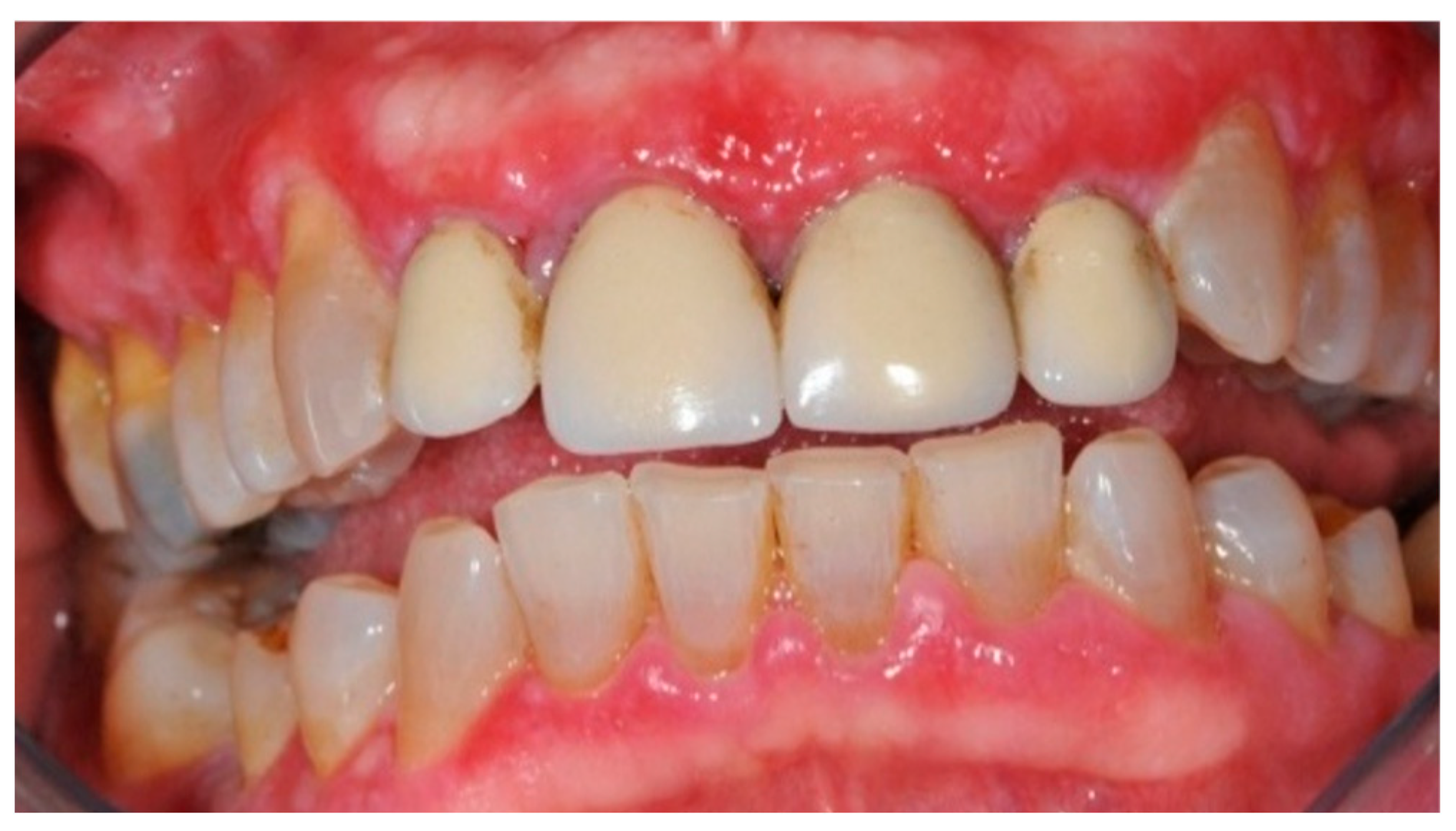
Figure 11.
Customized intraoral device.
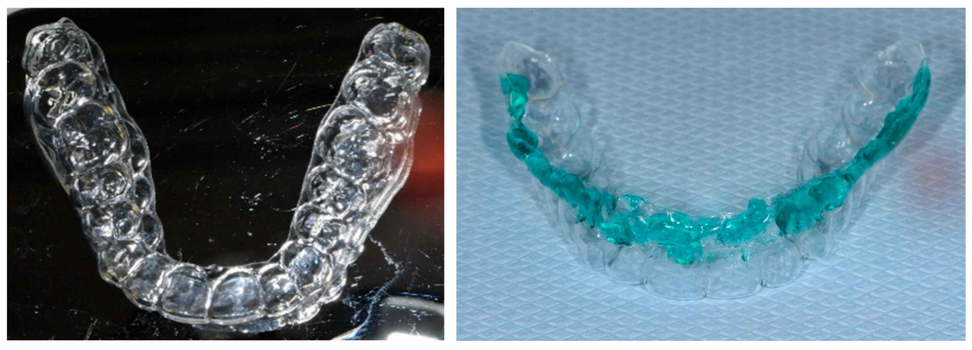
Figure 12.
Device positioned, frontal and lateral views.
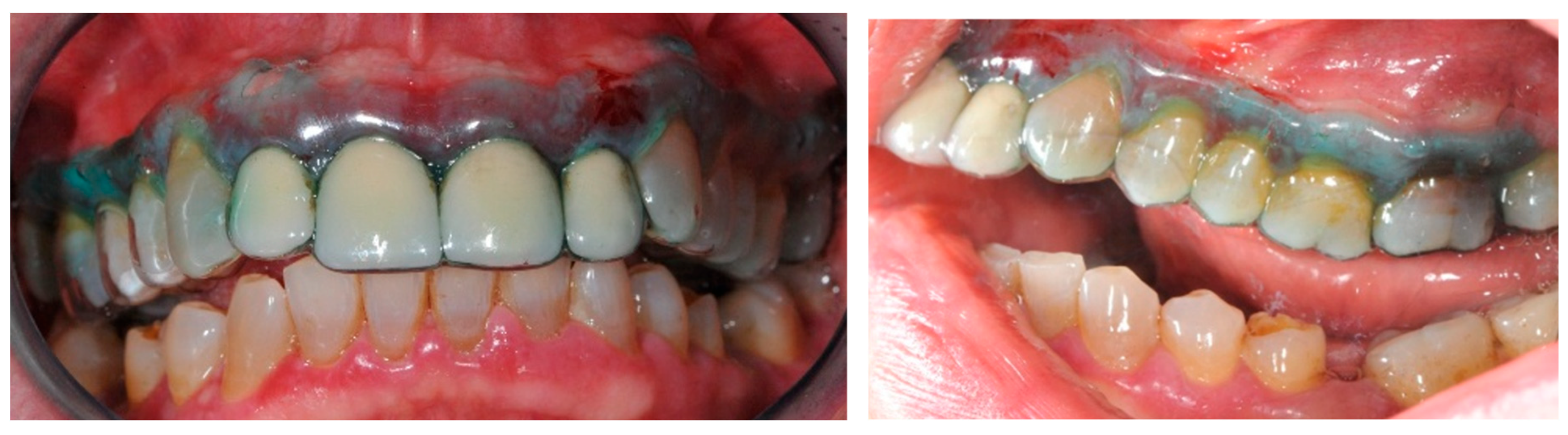
Figure 13.
20-day follow-up (T1), lateral views.
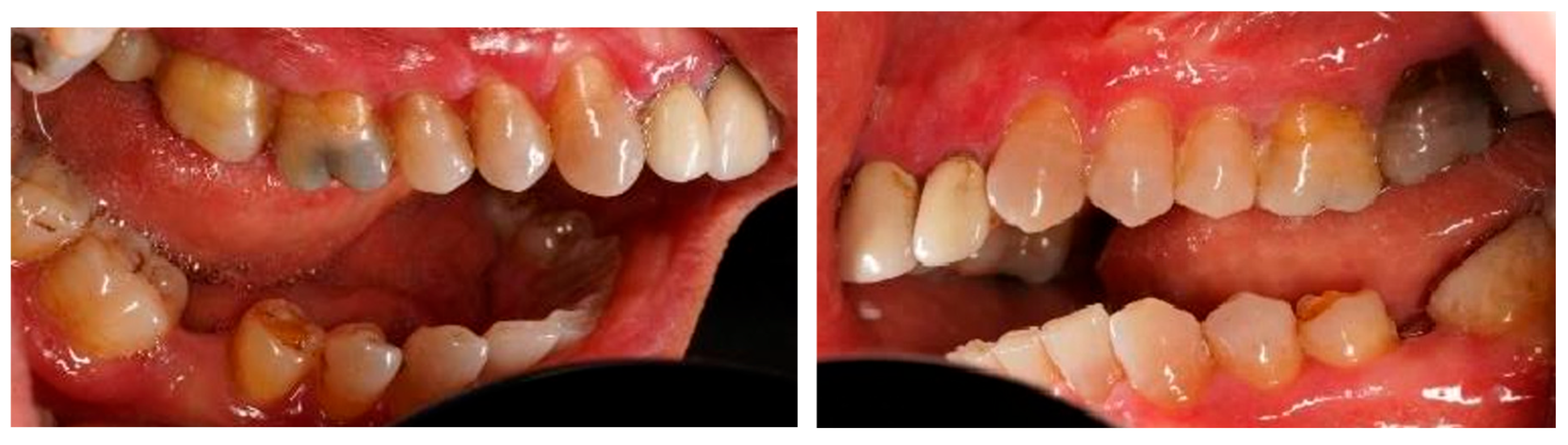
Figure 14.
40-day follow-up (T2), frontal view.
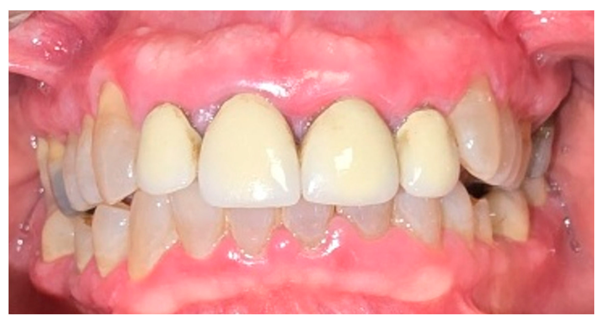
Figure 15.
40-day follow-up (T2), lateral views.
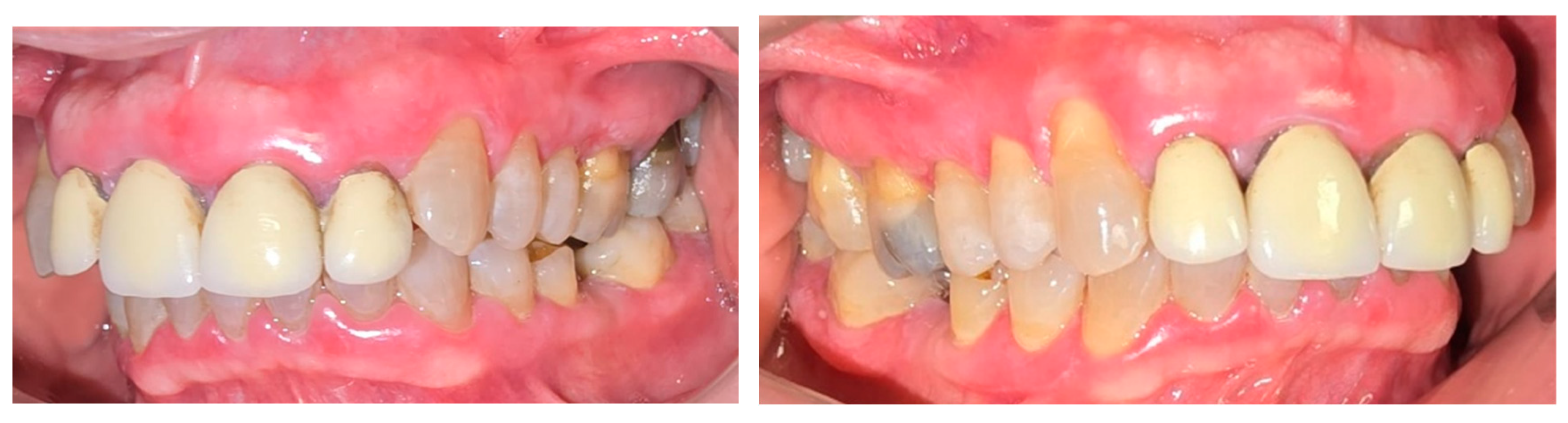
Figure 16.
Pain intensity over time (mean NRS score of the two patients at T0, T1, and T2.
Disclaimer/Publisher’s Note: The statements, opinions and data contained in all publications are solely those of the individual author(s) and contributor(s) and not of MDPI and/or the editor(s). MDPI and/or the editor(s) disclaim responsibility for any injury to people or property resulting from any ideas, methods, instructions or products referred to in the content. |
© 2026 by the authors. Licensee MDPI, Basel, Switzerland. This article is an open access article distributed under the terms and conditions of the Creative Commons Attribution (CC BY) license (http://creativecommons.org/licenses/by/4.0/).
Copyright: This open access article is published under a Creative Commons CC BY 4.0 license, which permit the free download, distribution, and reuse, provided that the author and preprint are cited in any reuse.