

Submitted:
21 October 2026
Posted:
23 October 2026
You are already at the latest version
Abstract
Airways’ patency and protection, and the feasibility of ventilation, are necessary pre-requisites to have for oxygenation during the induction of a general anesthesia or before intubation in other contexts. The text presents a bulleted and holistic approach to the interwoven dynamics of the two fundamental variables for a safe airway management i.e. oxygenation and ventilation, which is intuitive, easy to grasp and rapid to apply on the scene. The ‘cannot ventilate, cannot intubate’ (CVCI) situation is analysed in its crucial variables and patterns. The concept of anticipated and unanticipated difficult airway is an useful theoretical leverage for the right mind gear. The recently emphasized term of ‘physiologically difficult airway’ is instead a misnomer as it rather refers to special patients at risk of desaturation, hypotension or cardiac arrest on preoxygenation and induction. Emphasis is in an approach on the airways, which is independent on the specialistic background. Control of the airways outdoors follows the same patterns of approach and decision making of indoors, but in difficult or unfriendly contexts.
Keywords:
difficult airway
; general anesthesia
; induction
; oxygenation
; ventilation
; endotracheal intubation
1. Introduction
Airway control is needed in general anaesthesia (GA) induction and in emergencies (ED, ICU or on the street outdoors).
Airways’ patency and protection with feasibility of ventilation, and oxygenation are safely and optimally ensured by endotracheal intubation (ETI). Other devices such as supralaryngeal/supraglottic airways (SLA/SGA) and facial masks (FM) are suitably used for ventilation and oxygenation, and nasal devices for oxygenation.
By Difficult Airway (DA) is generally meant “a clinical situation in which a conventionally trained anaesthesiologist experiences difficulty with face mask or laryngeal mask ventilation, and difficulty with tracheal intubation” [1]. DA is essentially an anatomical problem. It can also be more specifically defined as any situation of difficult to impossible mask ventilation and difficult or failed tracheal intubation after two-three attempts by experienced operators with 50-100 among emergency and elective endotracheal intubations, determined essentially by an anatomical problem.
Physiologically Difficult Airway is a misnomer introduced in the last two decades to emphasize the problem of inducing GA in patients with deranged physiology or already hypoxemic, namely patients with severe metabolic acidosis, respiratory failure, pump failure, hypotension [2]. In fact, there is no such a thing as ‘physiologically difficult airway’. There is only a spectrum of situations where control of the airways is done on patients at risk of deterioration and cardiac arrest on induction or within 10 minutes.
Critical Airway is instead “a life-threatening situation of impossible or inadequate ventilation in general anesthesia or in a resuscitation context, or any other specific emergency/urgency scenario of impending/actual obstruction rising ex abrupto, requiring an emergency surgical airway (ESA)” [3].
The rule and general principles of airway management are that at any stage of inadequate manual ventilation (MV) with facemask (FM) or a supra-glottic airway (SGA) or impossible endotracheal intubation (ETI), a stab crico-thyroidotomy followed by manual ventilation via a 5-6 Fr ETT should be immediately done.
In the situation of critical airway the patient will have at any moment a respiratory-cardiac arrest by hypoxia, depending on the individual FRC, if oxygen is not swiftly introduced into the lungs with surgical methods. Temporary oxygenation can be achieved with percutaneous transtracheal jet ventilation but it will not last long (20-40 min) in supporting also ventilation, without damaging the lower airways.
Incidence of Difficult and Critical airway. In an operating room the rate of failed intubations and difficult airway is commonly deemed to be around 3%; outside e.g. emergency department, on the road and in ICU, failed intubation in critically ill patients occurs in around 6-10% with peaks up to 30% [4,5].
The need for an ESA has been reported in different studies as <2% in elective and emergency ENT GA [6].
The incidence of DA in routine anesthesia with inability to ventilate/intubate is 1:2000-5000, 1:300 in obstetrics and 1:100 in ED, ICU and outdoors. Emergency surgical airway is required in 1:50000 routine GA and 1:200 in ED. An inadequately or mismanaged difficult airway (DA) has been identified as a significant factor in up to 25% of anesthesia-related deaths [7].
The rate of successful first attempt intubation depends on several factors, whether indoor or outdoor, whether elective or emergency, the experience and expertise of the attending healthcare professional, and the presence of risk factors in the patient [8].
Obesity has an incidence up to 15% for difficult airway (ventilation and intubation).
In obese patients, the incidence of difficult intubation was twice more frequent in ICU than in the OT, with severe life-threatening complications occurring 20-fold more often in ICU [9].
2. The Endotracheal Intubation Drill
Before a standard general anesthesia, after checking the equipment, setting head and neck in the optimal anatomical position and emptying a full stomach when needed, the airway drill is carried out in a predefined fashion and order of timely steps in consecutio et continuum, namely, i) positioning, ii) peri-oxygenation, ii) ventilation, and iv) induction/intubation sequence [10,11,12,13].
A premedication with a benzodiazepine or an opioid is usually done in elective GA done to curb the psychological and biological stress of the patient. It is, though, contraindicated or risky in emergency anesthesia/surgery for the potential effects on ventilation and pressures.
2.1. Position and Pre-Oxygenation
The standard sniff position, with flexed neck, head extended at the atlanto-occipital joint and patient lying on a cushion 3-7 7 cm thick, is the optimal position for routine endotracheal intubation (ETI) in normal adults.
Pregnant should have the trunk tilted to the left.
Children must be ventilated and intubated with a padding below trunk and arts, counteracting the natural tendency to anterior flexion of the neck and the oropharynx soft tissues impairing visualization of the larynx.
In obese patients, the 25-30° back-up head-elevated ‘ramp’ position (Fowler position), with blankets or other devices placed under the patient's head and shoulders and removed once intubation is accomplished, is the standard position. It can also be achieved by reconfiguring the normally flat operating room (OR) table by flexing the table at the trunk-thigh hinge and raising the back (trunk) portion of the table (OR table ramp), obviating manipulations of the patient [14]
The Fowler position also has shown to be superior with significantly less complications to supine position in emergency intubations on patients outside theatre, eg in ICU or in the wards for not cardiac arrest patients [15].
In morbidly obese patients, the back-up position at 30° above the horizontal axis of a breaking table with legs at 15° Trendelenburg, known as Reverse Trendelenburg position, provided the longest safest apnea period, the lower SaO2 drop and the shortest time to SaO2 recover when compared to the 30 degrees Back Up Fowler and Horizontal-Supine positions [16].
Pre-oxygenation is a standard step before elective and emergency intubation, except for critical airways scenarios where rapid oxygenation/ventilation is undeferrably lifesaving.
Maximizing oxygen stores, and subsequently the time to desaturation, is essential to all intubations, especially of the critically ill.
By ensuring a higher reserve of oxygen than FRC at rest (denitrogenation), it delays the onset of hypoxemia during the apnea periods of the drill.
Suction by Yankauer sucker is required in some cases before oxygenation to remove oropharyngeal secretions, saliva, blood, vomit.
In healthy adults, the duration of apnoea without desaturation is limited to 1–2 min while breathing room air, whereas can safely undergo apnea for 8 min if preoxygenated at around 100% SaO2 [17,18,19].
Preoxygenation is carried out on the awake and cooperative patient spontaneously breathing through a non-rebreather mask and on the unconscious patient via manual ventilation with a bag-valve mask.
In healthy awake adults, it is carried out for 3 min with non-rebreather mask at 15 L/min with open valve deliver 90-100% O2 on tidal volume breath or with 8 deep breaths in 1 min (or 4 deep breaths in 30 sec); in an anesthetized or unconscious patient with an oropharyngeal cannula in situ, it is carried out with the practitioner holding tightly the mask with one hand, or with two hands and another operator squeezing the bag.
Haemoglobin desaturation during apnoea was more rapid following pre-oxygenation using the four deep breath technique than following pre-oxygenation either by the traditional tidal volume breathing for 3 min or by the eight deep breath technique for 60 sec. The four breaths in 30 sec technique should be reserved to young and healthy patients.
The safe apnea period, conventionally until the SaO2 reached 90%, can be extended in normal healthy subjects up to 10 min with a CPAP mask or PEEP at 5 cm H2O [20/44].
Special patients.
Obese patientsare more prone to desaturation and adverse events are more frequent.
The ramped position with the head positioned 25° up is the optimal position for preoxygenation in obese: the laryngeal view is superior and results in 20-25% higher PaO₂ and SaO₂ [21,22].
A study of preoxygenation on obese with eight deep breaths within 60 s and an oxygen flow of 10 L/min in obese in sitting vs supine position, extended tolerance to apnea with desaturation at 90% before RSII was achieved in 3½ min in the sitting group vs 2½ min in the supine group [23].
Adding CPAP mask with additional positive end-expiratory pressure (PEEP) of 7.5-10 cm H₂O for 5 min offers higher safety margins from hypoxemia in apnea. In a study with pre-oxygenation with PEEP vs preoxygenation without PEEP, the nonhypoxic apnea duration was longer in the PEEP group compared with the control group (188 +/- 46 versus 127 +/- 43 sec) ie apneic hypoxia onset was delayed and PaO2 was higher before apnea in the PEEP group. After induction of anesthesia, patients were mechanically ventilated with the pressure-controlled mode at 14 cm H2O, PEEP 10 cm H2O, respiratory rate 8 x min for another 5 min until tracheal intubation. In conclusion, application of CPAP (10 cm H2O) for 5 minutes in conscious morbidly obese patients followed by 5 minutes of mechanical ventilation with PEEP (10 cm H2O) during anesthesia induction is safe, simple, and well tolerated [24].
In spontaneously breathing patients with trismus e.g tetanus or epileptics pre-oxygenation is done via nasopharyngeal cannula or nasal mask.
In patients obliged to stay sitting such as severe maxillofacial injuries, preoxygenation is done with a full-blown gas facemask placed close to the mouth, without though much gain in pre-oxygenation.
Asthmatic and Dyspnoic must also be pre-oxygenated in sitting position.
In maxillofacial injuries pre-oxygenation with high-flow nasal cannulae (HFNC) is a good choice if the naso-ethmoidal complex and maxillae are integral and the stomach is not full, provided blood and debris has been pre-emptively suctioned with Yankauer sucker in the nasal-oropharynx.
In presence of not stoppable intraoral dribbling blood, the patient must be managed without preoxygenation with straight RSII preceded by suction, or SGA if ETI appears difficult or is unsuccessful.
Patients with intestinal obstructionmust have stomach contents drained beforehand, and soon afterwards intubated with RSII, skipping preoxygenation.
Patients with strangulated colon volvulus with toxaemia and full blown compartment syndrome must have a preliminary decompression by percutaneous or semiopen cecostomy in LA before induction to prevent the high risk of death on the operating table at induction (pers obs).
Vomiting patients must be put on lateral decubitus and intubated in that position if the vomit cannot be stopped and drained before induction or have preoxygenation done with HFNC if drainage successful.
Psychotic adults refusing induction or childrenmust be sedated or anesthetized before preoxygenation. Drugs such as ketamine im and DSII are especially useful.
Cervical Spine Injury or Non-Exclusion. Cervical spine injury (CSI) or not-exclusion per se is not an issue, as almost always endotracheal intubation (ETI) can be done with careful, little or no head and neck manipulation. Moreover, manual in-line stabilization does not effectively immobilize the cervical spine or decreases the likelihood of difficult and failed tracheal intubation [25].
The problem in the end remains whether the patient can be ventilated or not.
Apneic preoxygenation is pre-oxygenation without ventilation with insufflation of oxygen via nasal route. It was originally developed to see if could substitute facemask ventilation in situations of crowded airway.
In fact, at a flow of 5 L/min via a nasopharyngeal catheter it can be the only modality in sedation or iv GA induction for bronchoscopies and fiberoptic intubation.
In the first studies on nasal oxygenation, patients were pre-oxygenated at the onset of apnoea following induction and myorelaxation. The nasal airway sustained 10 min of adequate oxygenation with SaO2 at ≥ 97% for 3 min with tidal volume breathing, followed by pharyngeal O2 insufflation via 36 Fr nasal cannula with oxygen insufflated with a 8 Fr catheter placed within the first 2 cm of the nasal airway, In contrast, in the absence of naso-pharyngeal O2 insufflation, the SaO2 fell to 91% within a mean apnoea time of 6-7 min [26].
In a landmark study, facemask pre-oxygenation with the 30 sec four deep breath technique was followed by insufflation of oxygen at a flow of 5 L/min via a nasopharyngeal catheter, commenced after removing the facemask at the onset of apnoea following induction and myorelaxation. The method maintained a SaO2 > 95% for 5.5-6 min (5,5 min in obese) whereas it dropped below 95% within 3.5 min in the control groups with only standard facemask preoxygenation [27].
In a second landmark study on obese patients, preoxygenated breathing 100% oxygen via a standard tight-fitting facemask for 3 minutes and then with controlled manual or mechanical ventimation delivering positive-pressure ventilation with peak inspiratory pressure of < 20 cm H2O after induction and myorelaxation before laryngoscopy, the group who had additional nasal O2 at 5L/min administered with nasal prongs kept during prolonged laryngoscopy attempts a SaO2 ≥ 95% around 2 min longer than those who did not receive additional nasal oxygen [28].
In a further landmark study on patients, including obese patients, with anticipated difficult airways undergoing complex upper airway surgery, OptiFlow nasal cannula at 70L/min O2, kept for 10 min with patients head up 40°, generating a CPAP-like effect and positive end-expiratory pressure (PEEP) estimated roughly at around 1 cmH₂O of PEEP for every 10 L/min of flow, markedly lengthened apnea tolerance and postponed desaturation with a median apnea time of 14 min. Nasal cannula/catheter and nasal prongs were humidified with warm humidified O2 before use. After induction and myorelaxation, jaw-thrust at the same time keeping open mouth, using double hands and fingers, was performed immediately when the patient became unconscious and was maintained throughout the apnoeic period, to ensure upper airway patency until the end of the operation. Facemask or SGA for positive pressure ventilation were kept handy during the procedure if needed and used, likewise TTJV or ETI, at the end of the operation if the patient had not recovered from GA with sufficient spontaneous breathing [29].
Severe pathological hypercapnia during apnoea preoxygenation is managed with controlled hyperventilation. An elevation of PCO₂, likewise hypoxemia, raises the blood pressure.
2.2. The “ABCD Vertical Train” Approach to the ETI drill
Strategies. The ETI drill is usually effectuated in three steps [induction, myo-relaxation, intubation], which can be done different phases: Rapid Sequence Induction/Intubation (RSII), Stepped Sequence Induction/Intubation (SSII) with peri-ventilation/oxygenation in between induction and intubation, or Delayed Sequence Induction/Intubation with delay between induction and subsequent intubation (DSII) [30,31]. [Fig 1]
Figure 1.
ETI=endotracheal intubation; NMB=neuromuscular blockade; SGA=supraglottic airway; HS=hemorrhagic shock; TBI=traumatic brain injury; DA=difficult airway.
Figure 1.
ETI=endotracheal intubation; NMB=neuromuscular blockade; SGA=supraglottic airway; HS=hemorrhagic shock; TBI=traumatic brain injury; DA=difficult airway.
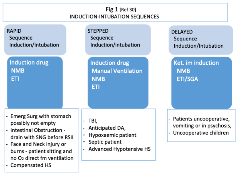
The difference between RSII and SSII bases on the possibility of finding difficult mask ventilation after induction drug, a scenario that requires immediate ETI.
RSII can be carried out with a predetermined dose of the induction drug or with titration to loss of consciousness. The neuromuscular blocking drug (NMBD) should immediately follow the induction or be given after establishing loss of consciousness [32].
In SSII, before manually ventilating the lungs after induction via a bag-valve-mask, it serves to assess the feasibility of rescuing a paralyzed patient in the event of anticipated or unanticipated intubation scenario. Crucially, it excludes the possibility of over-relaxing of the upper airways smooth muscle tone caused by an excess of induction drug, or the possibility of a reflex spasm that can occur when there is not enough relaxation due to an inadequate dosage of the induction drug [33].
SSII may be particularly relevant in those cases involving patients with poor respiratory reserves, sepsis, or high metabolic requirements, who are likely to decompensate during the attempts of a difficult intubation.
In DSII, im ketamine allows for induction within a 5–10-minute time frame in patients who are uncooperative, agitated, uncooperative or children requiring urgent surgical intervention [30,31].
The airway operator should know in advance which SII – see [Fig 2] - apply to the patient.
Suction of saliva/secretions/ blood/vomit by a handy Yankauer sucker is used before induction in presence of blood or vomit in the oropharynx.
Anticholinergic agents such as atropine and glycopyrronium are used for premedication to reduce oral and respiratory secretions respectively before the secretions-inducer Ketamine or in children, with glycopyrrolate preferred to atropine, due to the lesser odds of a dysrhythmias, lesser oropharyngeal secretions and the fact that does not pass the blood-brain barrier [34,35].
A simple, clear, bulleted, ‘revised’ [3] ‘ABCD vertical train of thought” for airway control during anticipated or unanticipated difficult airway is illustrated in [Fig 2].
Figure 2.
ETI=endotracheal intubation; RSI/ETT=rapid sequence induction/endotracheal tube, SGA=supraglottic airway; GA=general anesthesia; VC=vocal cords; ESA=emergency surgical airway; CTY=cricothyroidotomy; OST/SOT=open standard tracheostomy/semi open tracheostomy, PTJV=percutaneous transtracheal jet ventilation, prn=pro re nata.
Figure 2.
ETI=endotracheal intubation; RSI/ETT=rapid sequence induction/endotracheal tube, SGA=supraglottic airway; GA=general anesthesia; VC=vocal cords; ESA=emergency surgical airway; CTY=cricothyroidotomy; OST/SOT=open standard tracheostomy/semi open tracheostomy, PTJV=percutaneous transtracheal jet ventilation, prn=pro re nata.
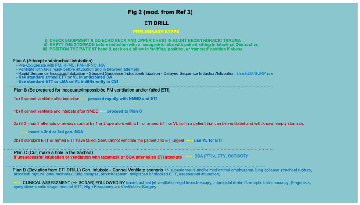
Intubation attempts should not last longer than 90 seconds, after which an increased incidence of aspiration and desaturation is found [36].
If surgery is elective and not life-threatening, and intubation attempts fail in an unanticipated DA but the patient can be safely oxygenated and ventilated with facemask or SGA eg i-gel, a safe option is VideoLaryngoscopy (VL) or to wake the patient and reschedule for an Awake Fiber-Optic Intubation under sedation and spontaneous breathing.
In anticipated DA to start with VL is sensible. VL is superior to ILMA for emergency intubation, offering higher first-pass success, faster intubation, and fewer complications. VL should be prioritized in critical care setting [37] [Fig 3].
Induction. The objective of induction is unconsciousness + analgesia during intubation and starting GA.
Induction can be achieved with anesthetic gases or drugs.
The anesthetist, intensivist or the emergency operator will use the most suitable, effective and safe tactic or combinations of tactics for each patient.
Inhaled anesthetic agents (nitrous oxide, halothane, isoflurane, desflurane, sevoflurane) are used in crowded airway, endoscopy for foreign body in the airway or esophagus [38] and for induction and maintenance of GA in elective neurosurgery [39].
The major variables of inhalational anesthetics are the blood-gas (B-G) solubility coefficient and the minimum alveolar concentration (MAC). The B-G solubility coefficient measures the uptake of an agent by blood. In general, less soluble agents (lower B-G solubility coefficients), such as nitrous oxide and desflurane, more rapid are induction and emergence from anesthesia, whereas induction and emergence with agents having high solubility in blood such as halothane are slower. MAC measures the efficacy of the gas and is conventionally defined as the concentration of an agent required to prevent movement in response to skin incision in 50% of patients. The higher is MAC the less potent is the volatile anesthetic. Halothane is the most potent anesthetic gas, followed by desflurane and then isoflurane which is the most irritant of the three.
It is not advised to use gas anesthesia for induction in life-threatening or emergency GA in general, except on pre-cardiac arrest hemorrhagic shock (HS) - Stage IV of the ‘Revised Physiological’ classification -, where the life is on the balance of an equilibrium that if broken will bring to rapid exitus on the table [30,40].
Gas induction, despite being slower to act, would offer in this situation a safe, more inert, less interfering alternative to iv induction.
Intravenous induction is standard preferred method of administration. Barbiturates, Etomidate, Ketamine, Propofol, and less frequently synthetic opioids, are commonly used.
Each agent has its own specific properties, onset, duration, side effects, indications and contraindications. Effects and peaking time of the most common intravenous induction drugs must be known [41,42,43,44,45]. [Tab 1a&b]
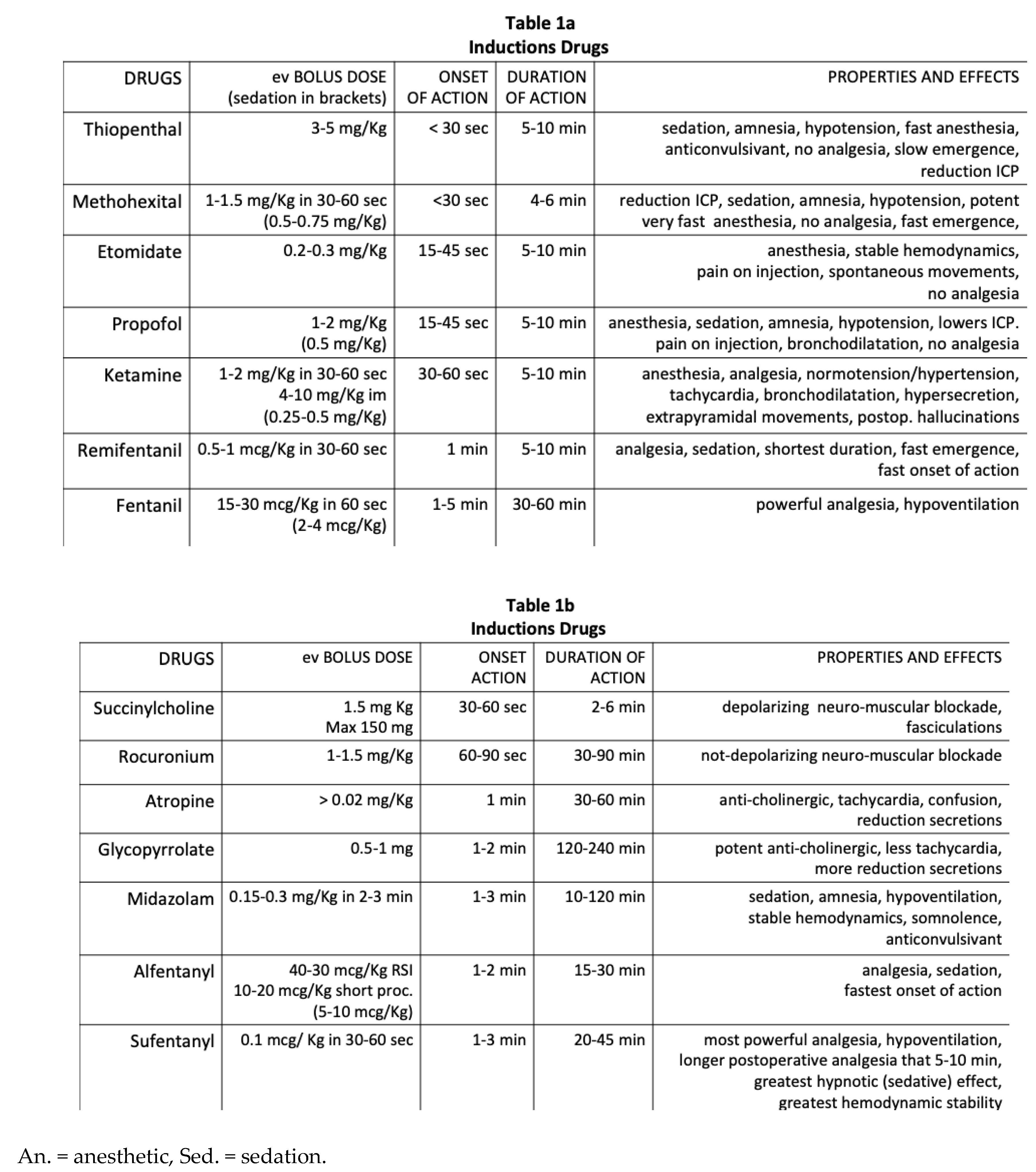
Synthetic opioids (SO) are commonly used in routine anesthesia for perioperative analgesia and often as adjuncts to patients receiving regional or local anesthesia. Opioids produce profound analgesia and minimal cardiac depression. Their disadvantages include ventilatory depression, delayed awakening, chest wall rigudity, hyperalgesia and inconsistent hypnosis and amnesia, which must usually be provided by other agents during GA. Added to standard induction drug, SO facilitate ETI even in absence of a NMBD [46].
Induction with solely synthetic opioid is done in cardiac surgery or cases involving profoundly unstable cardiovascular systems eg in the advanced Stage III & IV of hemorrhagic shock [30,40]
In critically ill, mortality is not different between etomidate and ketamine, but ketamine, despite probably causing more peri-intubation hemodynamic instability in terms of possible temporary arterial pressure rise, remains more dose adjustable at various levels of hemodynamic instability if subsequently used as maintenance as well. Propofol, a widely used induction agent, is not advised in hypotensive patients in virtue of its vasodilatory effect.
In severe maxillofacial injuries, once approximative preoxygenation is accomplished and iv induction drug rapidly administered, the patient is carefully put in supine position for rapid sequence induction-intubation (RSII).
Intramuscular induction is done with ketamine and only in DSII on difficult uncooperative patients. Once the patient is anesthetized under vigil control and assistance, he/she is put supine for rapid ETI.
Ventilation and Peri-Oxygenation. If an unconscious patient, especially after induction or the NMBD, cannot be ventilated due to the obstruction of the tongue, an oropharyngeal cannula (Guedell or Choi) is inserted before ventilation.
Oxygen is administered in continuity with pre-induction oxygenation between assisted ventilation and endotracheal intubation (ETI) attempts that should not last more than 10 seconds. Several attempts at tracheal intubation must be avoided when there is difficulty, as is failed oxygenation and pulmonary aspiration, and not failed tracheal intubation, that harm patients [47,48,49].
Most of the times facemask ventilation can be done by a single operator with one hand on the facemask sealing the mouth and one hand on the bag. Some operators cannot make it to gain maximum seal and optimal oxygen administration with one hand and need two hands just for the facemask facial seal. Two operators’ cooperation becomes hence necessary.
Manual ventilation after NMB before tracheal intubation with moderate pressures of ≤ 10 cm H2O PEEP or manually < 20 cm H2O without PEEP is acceptable to safely "test" the ability to mask ventilate without stomach overinflation or ventilate in the peri ventilation period [24,32,50,51].
In the event of the ineffectiveness or impossibility of MV with a SGVD following induction, the rapid neuro-muscular blockade and ETI serve to prevent death by asphyxia.
A rare but life-threatening event, difficult or failed mask ventilation after the administration of induction drugs and a neuromuscular blocking agent (NMBA) can occur, as consequence 2f an excessive or inadequate dose of the induction agent [30,33,52]. In the event of an unknown silent lesion in the airways, both scenarios have the potential to transform a potential partial stenosis into a complete or significant occlusion [30,33,52].
If the patient cannot be ventilated with a facemask or a SGA, cut-off the pre-oxygenation phase and proceed soon after induction for rapid RSI with or without myo-relaxation.
Patients with severe maxillofacial injuries can be ventilated only after rapid ketamine iv in sitting position and rapidly laid down after induction in standing position, by being intubated in RSII. Bag valve mask manual ventilation is contraindicated in face and neck trauma where the mask cannot be laid upon or there are blood or tissues that can be pushed downwards [3,53,54,55,56]. Often preoxygenation is not feasible through nose or facemask, and the laryngeal mask cannot be inserted if the patient is awake. A swift rapid ETI is the best choice left. In rare cases an emergency surgical airway is required at once if ETI and laryngeal mask cannot be done [53].
When both mask and SGA ventilation fail and ETI cannot be accomplished – cannot cevtilate, canot intubate (CVCI) situation - a stab CTY followed by a 5-6 Fr ETT is lifesaving. If post traumatic swelling by edema or hematoma obscure landmarks, then an open tracheostomy is what saves life [53].
Myorelaxation. Suxamethonium alias Succinylcholine is a fast and short-acting neuro-muscular blocking agent, inducing myorelaxation within 1 min and a spontaneous reversal within 5-10 minutes. Another advantage is the attenuation of thoracic rigidity induced by opioids, when used for induction. It has two drawbacks in the induction of fasciculations with increase of oxygen consumption and in producing a fast onset of oxygen desaturation during the subsequent apnoea.
In comparison, the other myorelaxant rocuronium [57,58] has a slower desaturation mean time to a SaO2 < 95% (5,5 +/- 0,5 min R vs 4,5 +/- 0,5 min S) in apnea, an advantage that may become relevant in the event of pre-existent partial airway obstruction
In overweight patients, Succinylcholine has a longer recovery time of normalizing oxygen saturation to > 95% on restarting effective ventilation compared to Rocuronium [58].
Suxamethonium bolus effect lasts usually 3-6 min whereas Rocuronium bolus duration of action is 30-90 min according to the dosage used, making it not an optimal option for RSII, and requires reversal [59,60].
Rocuronium-induced neuro-muscular blockade has though a unique advantage, when compared to Succinylcholine, in that it can be reversed with sugammadex at 16 mg/kg allowing recovery of spontaneous ventilation (usually 2,5-3 min) faster than spontaneous suxamethonium recovery (mean 8-9 min) [59,60].
Duration and effects of both the neuromuscular blocking drugs (NMBD) are influenced by dosage. Whereas Suxamethonium is effective at 1-1,5 mg/Kg according if an opioid or lidocaine or neither is given before or RSII is the choice, Rocuronium appropriate dosages vary from 0.9 to 12 to 1.5 mg/Kg, respectively according to whether if used for RSII in alternative to Suxamethonium or for SSII or to make sure laryngopharynx paralysis is obtained in contexts where contexts where minimizing airway reactivity is paramount [61,62].
Suxamethonium is standard in RSII. Rocuronium finds the main indication and advantage in an unanticipated difficult airway scenario during an SSII and in TBI. Another advantage is the presence of a pre-existent unknown silent stenosis that after myo-relaxation under RSII would make ventilation and intubation difficult to impossible [30,33,52].
Rocuronium requires supportive manual ventilation in between injection and ETI, in order to avoid desaturation.
Manouvres and Adjuncts. Three manouvres can be used by anesthetists to facilitate ETI under different rationale: cricoid pressure (CP), back-upwards-right pressure (BURP) or external laryngeal manouvring (ELM) and an armoured ETT.
Cricoid Pressure, used often in the past with the rationale of preventing gastric insufflation and regurgitation while simultaneously facilitating ETI, remains unproven and a controversial topic.
BURP/ELM is instead used by many anesthetists when it is wanted to visualize the laryngeal inlet.
The armed ETT solution is advisable in any foreseen or actual DA and whether to use a stylet or a bougie as adjuvant in ETI is left to the operator preference.
To awaken the patient during induction of anesthesia is an option only in elective deferable cases where other preoxygenation and induction options are practicable and safe. It is done by manually assisting ventilation until satisfactory spontaneou uptaking of regular breathing by the patient after clearing hypoventilating drugs affect, sometimes accordingly fastened by using drugs such as Naloxone, Anexate and Sugammadex.
2.3. Risks, Failures of the ETI Drill
EndoTracheal Tube position check. While the tube entry cannot be missed with clear vision of epiglottis and a free margin of tissues around, problem may arise when only the epiglottis is seen, and the tube covers the laryngeal inlet vision. In this instance, BURP/ELM manoeuvre can be helpful. A stylet or lightwand armoured ETT is another useful asset in the first attempt of a foreseeable difficult airway.
Wave capnography (WC), despite limitations as for example it cannot pick up unwitting bronchial intubation, has a sensitivity and specificity of 100% in confirming ETT position, compared to clinical appraisal or to sonar. Another limitation is that is slower and less effective than clinical auscultation in picking up ETT misplacement in cardiac arrest scenario where remains, though, the most reliable tool to signal success in resuscitation [63].
Capnography is routinely used in ICU on patients in coma to gauge EtCO2, or during GA to troubleshoot ETT displacement [64,65].
Esophageal intubation remains the most threatening scenario: besides failing direct oxygenation, ventilating into the stomach can trigger regurgitation of its contents into the airways.
Sonar is successfully utilized to detect the position of the endotracheal tube [66,67]. A recent study has shown advantages of using the sonar during rapid-sequence-induction intubation drill, particularly in detecting esophageal intubation earlier than capnography and five-point auscultation [66].
Right main bronchus intubation occurs if the ETT length passes the tracheal bifurcation. It is prevented by keeping at least 2 cm of ETT above dental arcade and picking up at auscultation the absence of beath sounds on the left side with full hearing on right side. The ETT is then repositioned above tracheal bifurcation.
Aspiration into the airways, directly from intraoral introduction or from aspiraton of stomach contents is normally treated with suction with flexible fiberoptic or rigid bronchoscopes, and different methods of neutralization of the alien substance or stopping the bleeding in case of blood.
The incidence of a full stomach at the time of anesthetic induction has been reported up to 56% of patients undergoing emergency surgery, and pulmonary aspiration of gastric contents in up to 5% of emergency department intubations. Over 50% of airway-related deaths in anaesthesia are caused by peri-operative aspiration [67].
The stomach status can be beforehand assessed by ultrasound. Tecnical limits, especially in emergencies without ileus or obstruction, do not always allow such tool to help in critical scenarios. The risk of regurgitation and aspiration of stomach contents into the airways increases with insufflation pressures ≥ 20 cm H2O during the manual ventilation or ETI attempts [24,32,50,51].
Patients need to have immediately the contents sucked and to protect the lower airways from further soiling with LMA or ETI (direct or fiberoptic-assisted or video-laryngoscope mediated or with double lumen) [68,69].
Rapid sequence intubation (RSI) is the standard for definitive airway management in these patients. Even with RSI, aspiration of gastric contents is not avoided when vomiting or regurgitation occurs at an interval before tracheal intubation.
The ‘suction assisted laryngoscopy and airway decontamination’ (SALAD) is a method suggested on the patient positioned at 20-25° with stomach or blood contents or pus into the airways, needing GA induction for emergency surgery, using simultaneously a glydescope or a DL, or better a McIntosh VL and a large bore rigid suction catheter slightly curved and with a straight end. The catheter is advanced ahead of the laryngoscope, clearing all contents through oropharynx, laryngopharynx (hypopharynx), laryngeal inlet, contiguous upper trachea, and till the upper oesophagus too. A last suction is done in the upper trachea before ETI [70,71]. The stomach is aspirated too through a large bore tube with suction catheter inside.
Patients who are currently vomiting stomach contents or blood but not aspirated yet in the lungs, can be intubated with SGA or ETT in lateral position to prevent contents getting into the lungs [72,73].
CVCI situation. If the patient cannot be ventilated with a facemask or a SGA, cut-off the pre-oxygenation phase and proceed soon after induction for rapid RSI with or without myo-relaxation. When both mask and SGA ventilation fail and ETI cannot be accomplished, a stab crichothyroidotomy (CTY) followed by a 5-6 Fr ETT is lifesaving. CTY is also the best ESA in CA. If post traumatic swelling by edema or hematoma obscure landmarks, then an open tracheostomy is what saves life [53]. Manual ventilation with CTY can be safely effected up max 48-72 hours, after which cartilage damage by erosion and stricture can be expected [74,75].
In contexts different than GA induction, other ESA are used for oxygenation and temporary ventilation.
Percutaneous trans-tracheal jet ventilation (PTJV) set up manually (a 12-14 gauge cannula after withdrawing the needle is connected to intravenous or nasogastric tube attached to a 40-50 psi source delivering oxygen at 15 l/minute, and intermittent insufflation of 1 second on and 4 second off is done from controlling a tube near the end hole9 or automated and transportable, can provide satisfactory jet insufflation of oxygen. Trans-tracheal jet ventilation (TTJV) ie insufflation of oxygen at high pressure into the trachea directly from the oropharynx is used in the ‘crowded airway during foreign body removal in the lung under bronchoscopy.
It is also used in incomplete foreign body upper airways obstruction where it can dislodge the obstruction upwards and can be done by medics or paramedics on the scene but has a temporary short ventilation capacity and cannot be used for longer than 20-40 min to prevent significant hypercapnia by hypoventilation
PTJV is the preferred ESA in children < 10 where a CTY would be traumatic.
The author does not advice a PCT as it takes precious minutes and has complications augmented by the emergency. Instead, a semi open tracheostomy (SOT) is better.
OST by a skilled operator is also rarely required if anterior neck landmarks - hyoid bone (HB), thyroid cartilage (TC), cricoid cartilage (CC) -, are not palpable, eg in trauma to the neck with hematoma or oedema.
Semiopen or standard open tracheostomy are the preferred ESA in DA/CA in adults, as for the speed of execution and the relative safety.
In maxilla-facial surgery and in trauma to the airways with loss of cervical landmarks, tracheostomy remains the preferred and most used ESA [77,78], despite remaining a rare indication in the order of <4% of all ESA requirements [79].
A tracheostomy is required if long term ventilation is expected.
Complications and Mortality of DA
Three mechanisms of injury accounted for three-fourths of the adverse respiratory events in the US: inadequate ventilation (38%), esophageal intubation (18%) and difficult tracheal intubation (17%). The incidence of death or permanent brain damage associated with airway management during general anesthesia was found to be much higher than the incidence associated with cardiovascular management [80].
The NAP4 study has shown that the commonest factor associated with serious airway complications was difficulty with tracheal intubation due to airway obstruction, whereas pulmonary aspiration was the commonest cause of serious airway complications not only during induction of anesthesia but also after surgery [81].
In a multicentre study, up 50% of patients with Difficult Airways in ED experience serious life-threatening complications and there is still a 15-20% complication rate on first attempt [82].
Mortality post-ETI is around 3%, 37% of which due to hypotension by advanced III/IV stage HS, cardiac failure/ischemia or hypotension from other causes [83].
Peri-intubation cardiac arrest has an incidence up to 4% and is most of the times due to peri-intubation oxygen desaturation and hypotension [84].
In two studies on critically ill patients in ICU, Ed and wards [85] and only ED [86], major events occur in 32-45% of cases, with hypotension in 20-43%, severe hypoxemia in 9-12% and cardiac arrest in 3-3.5% of cases, and 33% mortality [85,86].
In particular, patients with septic shock given a fixed amount of 500 mL of fluids did not significantly decrease the incidence or often prevent cardiovascular collapse compared on induction with no fluid bolus group, except for the subgroup already on PPV [85].
3.1. Diffficult Airways
Anticipated Difficult Airway. Clinical appraisal can point out before hand to the likelihood of difficult ventilation and intubation for the presence of risk factors that mentally make aware and gear to action requiring variations from standard steps.
Body mass index (BMI) greater than 25 kg/m2, age greater than 55 years, history of macroglossia/snoring/apnea, the presence of beard, edentulousness/limited jaw protrusion, a Mallampati grade III or IV, and a thyro-mental distance of less than 6 cm are considered independent risk factors for inadequate, unstable, difficult-to-impossible mask ventilation (MV) [87,88,89,90,91].
The use of a gauze between the mask and the face may facilitate face-mask ventilation, in case of bearded patient and an assistant’s hands can help in maintaining an open airway in cases where the anesthesiologist has small hands and cannot hold the chin and the mask with one hand.
In the event a patient is unable to be adequately ventilated with a facemask, a supra-glottic airway device may be employed as an alternative means of ventilation.
Supra-glottic airways (SGA) prevent the tongue from obstructing the oropharynx and, when correctly positioned, can effectively ventilate the lungs in manner that is more efficient than that achieved by a face mask. SGA can be inserted blindly, but not in an awake patient, where it would trigger local reflexes (gagging, coughing, vomiting) and a systemic sympatho-adrenal autonomic response (an increase in blood pressure, heart rate and respiratory rate).
Among the SGA, the Intubating-Laryngeal-Mask-Airway [92] and the Intubating-Gel [93,94] allow passage of a nasogastric tube and fiber-optic-bronchoscope (FOB), and the AIR-Q its suited as the only ventilation conduit during general anesthesia [95,96].
Patients with a mouth opening of less than 2.5 cm (the average for an adult), a glottis that is off midline, a history of neck radiotherapy, and post-fibrotic distorted anatomy, as well as lesions to the oropharynx or epiglottis, supra-glottic stenosis, can impair ventilation with SGA [97].
With advances such as sonar as preliminary assessment tool and the advent of the video-laryngoscope, the preventive knowledge of risk factors for a DA has become less crucial.
A pre-induction sonar can identify specific causes of anticipated DA such as epiglottitis or an unknown supra-laryngeal tumour [98].
The use of video-laryngoscope in emergency, likewise SGA with ETT, requires basic and specific pre-training practice in election.
SGA such as the PLMA, the Supreme or the I-gel [99,100,101], which reduce the risk of aspiration and decubitus pressure, and permit esophageal drain tube and ETT introduction, are optimal devices of airway control in CSI, maxilla-facial injury, and cardiac arrest. Furthermore, SGAs are optimal invaluable as temporary rescue oxygenation devices, if the facemask is contraindicated or fails. SGAs are contraindicated in the awake patient where can trigger regurgitation and vomit, and in anterior neck injuries, when an ETI or an ESA are required. Like in difficult intubation, no more than three attempts should be done with SGA introduction.
Video-laryngoscope, likewise SGAs, is particularly useful in suspected or proven CSI and is invaluable in presence of an obvious CSI.
There are three types of VL: the non-angulated one (eg Mcintosh curved and Miller straight blade), the hyperangulated one (eg glydescope) and the channelled ones (eg AirTraq). The non angulated allow easier ETI, the hyperangulated ones a better thorough view and the channelled the addition of stylets and bougies or straight cannulae for extra oxygen.
VL requires a slightly longer execution time, is associated with higher first-pass intubation rate and reduced esophageal intubation rate than DL in ED, trauma, ICU and in patients with anatomically predicted difficult airways [102-107
In a study on Helicopter EMS staff, the first pass success rate was 88% (85% in CPR, 86% in trauma patients and 93% in non-trauma patients and a first pass rate in difficult airways of 87-89%), 99% overall intubation rate, with indoors and ambulance rates superior to outdoors rates, using the C-MAC video laryngoscope with Macintosh (not hyperangulated) blade 0-4 [105].
In particular, the Macintosh-style and channelled VLs reduce rates of hypoxaemic events and the hyperangulated VLs reduces rates of oesophageal intubation, in respect of standard laryngoscopes.
When both tracheal intubation and manual/SGA ventilation are impossible, an ESA is required (CTY, or PCTy/SOT) and the patient can be meanwhile oxygenated via PTJV.
Unanticipated difficult airway is a scenario ‘all emergency operators’, i.e. non only anesthetists, should be aware, trained, and prepared to face and manage.
Some authors have questioned the practical value of definitions such as "anticipated or unanticipated difficult airway" in the context of the mandatory awareness and vigilance for risks that operators must maintain regardless of whether a difficult airway is expected or not. Indeed, the predictive criteria for difficult airways may demonstrate high sensitivity but have low specificity and positive predictive value [108,109].
In a prospective study, out of 3391 difficult intubations, 3154 (93%) were unanticipated with only 25% of positively predicted DAs; difficult mask ventilation was unanticipated in 94% of cases with only 22% of positively predicted difficult mask ventilation [110].
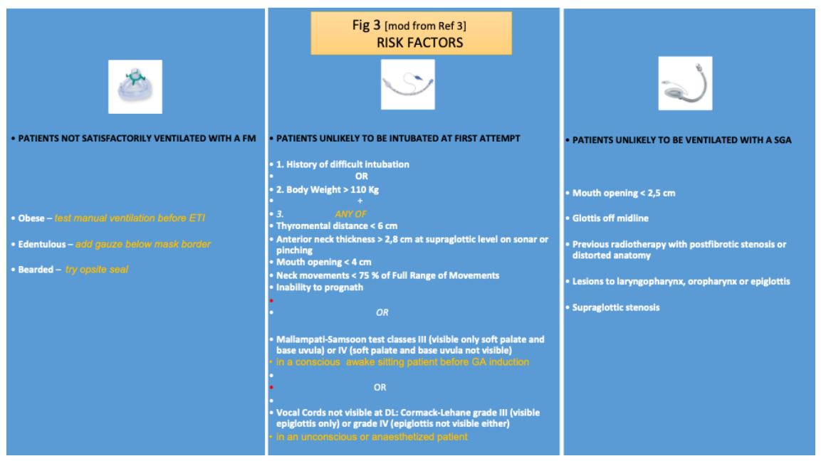
This author believes that “anticipated/unanticipated difficult airway” remains a useful concept. It is beneficial to know beforehand who is unlikely or impossible to be ventilate or intubate [87,88,89,90,91]. [Fig 3] It enhances inner tension to action and would prompt not as much to have at hand the armamentarium possibly needed for obtaining oxygenation and a patent airway, namely [stylets, bougies, ESA sets, video-laryngoscope (VL), SGA devices], but crucially to ‘oil’ the mental gear, which fundamental in scenarios of crisis.
Severe life-threatening complications (adverse events). Severe complications occurred in 36% of patients who had a non-difficult intubation, compared to 51% in patients with unanticipated difficult intubations) [111].
Figure 3.
FM=facemask; ETI=endotracheal intubation; SGA=supraglottic airway.
3.2. Difficult Patients
Lateral Position. Some patients have to be intubated with SGA or ETT in the lateral position: i) the ones who cannot stay supine and must stay in a lateral position, ii) who are currently vomiting stomach contents or blood but not aspirated yet in the lungs, or iii) have unilateral lung soiling of pus or blood, should be intubated in lateral position. Gravity from Trendelenburg position helps in the case of airways soiling. The ETI can be standard, fiberoptic-assisted or video-laryngoscope mediated or with double lumen [72,73,112].
The combination of sniffing position and left lateral decubitus at 90° or extended semi-lateral position where the trunk is inclined at 75°-90° to the horizontal plane, effectively reduces the risk of reflux and aspiration [113], due to a much-reduced upper airway collapsibility compared to the supine position, particularly at the level of the soft palate and epiglottis [112]. The lateral position prevents collapse of the oropharyngeal space and decreases the odds of gastric content reflux, especially on left decubitus [112].
In children undergoing surgery for adenotonsillar hypertrophy, lateral positioning combined with chin lift and jaw thrust provided better airway patency than supine positioning [114].
The enlarged oropharyngeal space situation occurring in the lateral position does not affect, though, the overall pressures, oxygenation, perfusion and ventilation. This occurs because of the [118–123increased perfusion of the dependent lung (by gravity, by abdominal organs pressure and by hypoxic pulmonary vasocostriction of the not-dependent lung), coupled with the inverted pattern of ventilation, which is decreased and with increased inhomogeneity in the dependent lung [115,116].
The paradoxical improvement of Obstructive Sleep Apnea episodes, more frequent and severe in supine position and improved in lateral position, can be explained by the fact it is more heavily dependent on the patient's airway collapse rather than the overall change in pressures [117].
Specific indications and scenarios where tracheal intubation in lateral position (TILP) is required in emergency situations are in [Tab 2] [118,119,120,121,122,123].
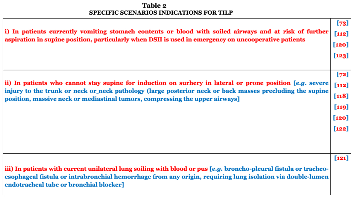
TILP=tracheal intubation in lateral position.
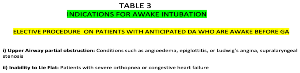
Awake Intubation. Awake fiber-optic Intubation (AFOI) or awake tracheal intubation can be used in an awake spontaneously breathing patient lightly sedated and then sprayed thorough enough topical LA blunting gag and cough reflexes, in situations of elective GA/surgery and predicted DA for supra-laryngeal obstruction or stenosis. The operator stands in front of the patient. The method allows also suctioning via its working channel and insufflation of oxygen through the scope if needed [124,125]. [Tab 3]
Complication rates like severe hypoxemia or aspiration during the procedure are very low in elective settings.
The time to intubate is important: AFOI cannot be used in acute emergent “crash” airways where RSII is preferred.
A randomized trial compared awake fiberoptic intubation with awake video laryngoscope intubation under remifentanil infusion at sedative dosages in anticipated difficult airways, and found both had similarly high success rates, despite a slower AWOI execution time, [126]. AFI with a flexible fiberoptic bronchoscope (FOB) has now been replaced with AFOI by VL with the operator behind the patient.
Patients with severe acute respiratory distress syndrome (ARDS) and reduced FRC to a third of the healthy patients, if induced with anesthetic and ETI can be affected precipitously immediately upon induction by the pendelluft phenomenon i.e. gas transfer from the non-dependent to dependent portions of the lung, causing a sudden worsening of the V/Q mismatch and rapid desaturation, a phenomenon avoided with the patient spontaneously breathing in AFOI [127].
FOB remains electively the gold standard in tumors, abscesses and lesions from mouth to above larynx, which are partially obstructing but can abut in complete obstruction with any sedation or anestesia or spontaneoously.
3.3. Special Patients at Risk on Induction
Patients with metabolic acidosis, hypoxemia, hypotension and ischemic or failing heart, are at risk of brain damage or cardiac arrest on induction or within 10 min from the ETI drill and require skilled inductions [2,128,129].
Patients in severe Metabolic Acidosis.
Patients who have deranged physiology and have metabolic acidosis (MA) from severe infection, diabetes, acute kidney failure and postcardiac arrest with a PH < 7.2-7.1, if not well preoxygenated and stabilized hemodynamically and metabolically are at risk of dysrhythmias and collapse on induction [130].
In severe metabolic acidosis the demand on alveolar ventilation requirement is compensated by the patient hyperventilation often inadequate in advanced MA. So even a brief apneic period can lead to a precipitous drop in oxygenation and pH given the loss of the already inadequate respiratory compensation [2]. Moreover, the reliance on circulating catecholamines for adequate inotropy and vascular tone in metabolic acidosis can be obtunded during induction of anaesthesia with high risk of hemodynamic deterioration after intubation [131,132].
Induction with Etomidate or Ketamine rapid-acting potent opioids e.g., Alfentanil or Remifentanil and rapid-acting muscle relaxants such as Suxamethonium in lieu of Rocuronium with its rapid onset of action and rapid offset, are safest induction drugs in these patients.
Vasodilating hypotensive agents such as barbiturates, propofol and midazolam are better be avoided, and judicious fluid management is managed by appropriate vasopressor management in non-responders [2].
Preoxygenation with initial NIV and attempt to buffer the acidotic trend as much as possible [130], depending on the criticity of the surgical timing, prevents the hypotensive trend of positive pressure ventilation and hypoxaemic deterioration.
Mix ICU patients with respiratory and metabolic acidosis.
In a study in ICU on critically ill patients in shock, coma or respiratory failure mix, the group of patients who are already hypoxemic before the ETI drill, high fraction-inspired oxygen facial mask (HFFM) could not avoid desaturation going down to below 80-85% in less than half-a-minute in a quarter of patients [133].
In a study in ICU on a mix of patients, apneic preoxygenation at 15 L/min 100% O2 with high flow nasal cannula (HFNC) left in place during intubation attempts did not significantly increase the lowest oxygen saturations compared to no apneic oxygenation [134].
In another study on a mix of patients in ICU, the ones preoxygenated with bag-mask ventilation at 15 L/min of 100% oxygen, PEEP at 5-10 cm, and tidal volume ventilation breaths ≥ 5 min/L, had better SaO2 and a lesser incidence of severe hypoxemia with SaO2 < 80 % than those receiving no ventilation without increasing the risk of aspiration [51].
In a landmark study and secondary analysis of the above studies [51,134], bag-mask ventilation was found associated with higher oxygen saturation during intubation compared to apneic oxygenation with nasal cannulae with 15 L/min 100% O2 [135].
Standard oxygen masks are simple to set up and can deliver a fraction of inspired oxygen (Fio2) as high as 100% under ideal conditions eg non rebreather mask with reservoir, but can actually release an effective 50% when ambient air filters through inside a not-tight fitting mask. Only a tight fitting mask capable to deliver PEEP and PS can guarantee optimal oxygenation in those patients by enhancing alveolar recruitment, preserving FRC, and optimizing oxygen storage.
In another ICU mix patients’ study, comparing NIV and facemask preoxygenation, the incidence of hypoxemia was lower by 9.4 % among those who received preoxygenation with noninvasive ventilation than among those who received preoxygenation with an oxygen mask. Moreover, preoxygenation with noninvasive ventilation did not appear to increase the incidence of aspiration. [In the NIV group, a tight-fitting mask was connected for 3 min to either a conventional mechanical ventilator in ICU or a dedicated noninvasive ventilator in ED set up on BiPAP at FiO2 of 100%, expiratory pressure of at least 5 cm of water, inspiratory pressure of at least 10 cm of water, and respiratory rate of at least 10 breaths per minute]. For the patients in the oxygen-mask group, supplemental oxygen was administered for 3 min either with a nonrebreather mask or bag-mask device without manual ventilation at the highest flow rate of oxygen available (≥15 L/min)] [136].
Preoxygenation with high flow nasal cannula (HFNC) at 20-70 L/min with head elevated position has also been studied in the last decade or so. In contexts with non-severe upper facial trauma or suspected skull base fracture, it improves oxygenation by increasing gas flow and pressure that can be maintained in situ during ETI, so allowing continuous oxygenation during the drill, whereas a FM would have to be removed for the intubation; moreover, it can be used below the facemask. The problem with HFNO in those cases was technical in that interferes with the fitting of CPAP mask often needed in several hypoxemic scenarios, diminishing its seal and the overall efficiency. It was noted that CPAP improves oxygenation in 10 min; HFNC and NIPPV are superior to facemask oxygenation in reducing the incidence of peri-intubation hypoxemia, and NIPPV reduces the risk of hypoxemia compared to HFNC.
Patients already in Respiratory Failure.
In a study only on hypoxemic patients in ICU, comparing HFNC with HFFM preoxygenation for 4 min, where HFNC was maintened throughout the procedure and HFFM was removed at the end of general anaesthesia induction, apneic preoxygenation with HFNC at O2 at 60L/min did not reduce the lowest level of desaturation compared to high flow facemask (HFFM) at 15 L/min oxygen flow [137].
A ICU randomized trial of 3-5 min preoxygenation on patients with hypoxemic ARF in semi-recumbent position at 30° compared HFNC at 60L/min O2 vs noninvasive ventilation NIPPV with tidal volume between 6 and 8 mL/kg, a positive end-expiratory pressure (PEEP) level of 5 cm H2O and a FiO2 of 1.0. Both methods did not significantly change the overall risk of severe hypoxemia (oxygen saturation below 80%) in adults with acute hypoxaemic respiratory failure. This risk seemed though to be decreased with non-invasive ventilation for patients with moderate-to-severe hypoxemia (PaO2/FiO2 ≤200 mm Hg): in these patients severe hypoxaemia occurred less frequently after preoxygenation with non-invasive ventilation than with high-flow oxygen (24% vs 35%. The study proved HFNC as an effective strategy for mild hypoxemia, NIPPV to be beneficial for moderate to severe hypoxemia, whereas a combination of multiple modalities is necessary for the most severe patients [138].
In low resource settings where NIPPV is unavailable, preoxygenation in patients with no or mild AHRF, can be successfully done combining an oxygen mask at a flush rate (fully opening a standard oxygen flowmeter beyond 15 L/min) with a nasal cannula at 15 L/min after mask removal, and head up 20-30°. In critically ill patients with moderate-to-severe AHRF or with risk factors for reduced oxygen reserves (e.g., obesity, pleural effusion, and intra-abdominal hypertension), BMV with a PEEP valve, or a Mapleson C circuit, connected to a tight-fitting mask after induction and prior to laryngoscopy, prevent severe hypoxemia and can be used for preoxygenation [139].
In a study on hypoxemic patients with full stomach, preoxygenation was performed before a rapid sequence intubation for a 3-min period using a nonrebreather bag-valve mask (control group) or pressure support ventilation delivered by an ICU ventilator through a face mask (NIV group). BiPAP with PS delivered by a ventilator was superior to BVM in reducing desaturation before ETI. [The bilevel positive airway pressure (BiPAP) for 3 min delivered by ICU ventilator ensured better SaO2 and PaO2, values than the usual pre-oxygenation method with a non-rebreather mask at 15 L/min and occasional pump ventilation. The ventilator was set at pressure limited module ≤ 20 cm H2O PSV and normal tidal volumes, PEEP at 5 cm H2O with FiO2 of 1, and applied for 3-5 min before ETI]. SaO2 values were still better 5 minutes after intubation in the NIV group than in the control group. The improvement of oxygenation with 3 min of pre-oxygenation with NIV persists up to 30 min [140].
An analogue study by the same group above showed no difference in mortality between the two methods and reassert the need to continue uninterrupted NIV until intubation to avoid worsening the situation by thwarting the benefits of NIV [141].
In another study on hypoxemic patients from variable etiology in ICU, receiving 4 min preoxygenation at 30° of head-up inclination, the group pre-oxygenated with HFNC [flow > 60 L/min of humidified oxygen at FiO2 of 1 + a bilevel positive airway pressure (BiPAP) with PS of 10 cm H2O or continuous positive airway pressure (CPAP) and PEEP at 5 cm H2O], was superior in reducing the severity of desaturation to the group pre-oxygenated only with NIV at the same parameters of the interventional group. It was also recommended to never exceed a total insufflation airway pressure (PS + PEEP) of 15 cmH2O, which is below the threshold for gastric air insufflation of 20 cm H2O. The study, however, failed to show a significant difference in complications and mortality outcomes in ICU [142].
In the above study [142], it was noted that supernormal values of oxygen were used for a period of preoxygenation and apneic oxygenation overall not >15 min and the maximum duration of severe hypoxemia with SaO2 < 80% did not last over 6 min [143].
HFNC predictably finds an absolute indication in patients at risk of aspiration.
Patients at high risk of hypoxemia from aspiration of vomit, unresolved intestinal obstruction, hematemesis or hemoptysis, should be pre-oxygenated with 60-70L/min 100% heated humidified O2 via HFNC or with supplemental oxygen via standard nasal cannula or standard facemask at insufflation pressures < 20 cm H2O [144].
Cardiopathic patients with IHD and pump failure. In patients with right ventricular failure, the vasodilation by induction agents that reduces preload (upon which the failing RV relies to maintain cardiac output); the apnoea that can result in hypoxemia, hypercarbia, and acidosis, which increase pulmonary vascular resistance (PVR), and the positive pressure ventilation resulting in additional reduction of venous return/RV preload and RV afterload, are variables that must be prevented and managed before induction [145].
The preliminary adjustment of rhythm, rate, and fluid balance, the judicious use of inotropic and vasotropic drugs, a low PEEP, the FiO2 manipulation of O2, the ventilation rate control of CO2, and the use of induction agents such as synthetic opioids, ketamine, etomidate and Propofol are normally used to target the key variables of GA induction in right pump failure, aiming to maintain mean arterial pressure (MAP) > 60 mmHg and mean pulmonary arterial pressure (mPAP) < 20 mm Hg. CVP and arterial lines are needed and impromptu ECHOcardio is useful [146,147].
When the ratio cannot be maintained because LV failure is the prima causa of the systemic cardiovascular failure, a more aggressive approach with VA extracorporeal circuits or left ventricular assist devices is required [146].
Patients with left ventricular failure and acute or acute on chronic IHD undergoing no-cardiac surgery need cardiothoracic anaesthesia expertise, logistics and organization [148].
Hypotensive patients. Hypotension may be present before GA due to relative hypovolaemia in medical patients or to trauma and/or blood loss in surgical patients. Contingent pre-GA hypotension is managed by fluid replacement and vasopressors, which are essential primary tools to restore temporary hypotension in absence of major physio-pathological changes.
Hypotension occurring within 10 min from induction is an emergency. Even in the apparent normotensive patient, perioperative hypotension below 20% of baseline systolic blood pressure (SBP) for >60 minutes, has been associated with adverse cardiovascular, renal and neurological outcomes [149]. This is particularly true in patients who are hypertensive and used to high SBP, in whom a drop of 20-30 torr from usual level, deemed as wihin the nornal range in normotensive individuals, in these patients equals to hypotension.
One method for picking up post-induction hypotension is the use of the Shock Index (SI). The SI is the ratio of the heart rate to systolic blood pressure (SBP) and is an inverse marker of cardiac work, such that a raised SI indicates deteriorating cardiac performance. The normal SI is 0.5 to 0.7 and values >0.8 predict compensated shock [150].
In a study on trauma patients, post-intubation hypotension (PIH) occurred in 36.3% of cases and the ones who developed PIH had an in-hospital mortality of 29.8% of patients, compared to 15.9% of patients in the non-PIH group [151].
Fluids, drugs and optimal ventilation patterns are the tools available to manage hypotension at induction.
Post induction/intubation hypotension is caused by blood loss, induction agents, muscle relaxants, and positive pressure ventilation as effect of vasodilatation, reduced venous return and decreased cardiac output [152]. Hypotension caused by some induction drugs [153] but also by the acceleration of bleeding by the hypertensive laryngeal peak reflex triggered by a not sufficiently dampened reflex before ETT is prevented by using optimal induction drugs combinations [154].
Etomidate and ketamine are considered safe in hemodynamically unstable patients. E causes transient adrenal suppression and ketamine increases heart arte and work with risk of CA in patients with depleted cathecolamines store [155]. Small does of Propofol judiciously used can curb the hypertensive response to laryngeal stimulaton, that can potentialy accelerate blood loss and put strain to the pump [156].
In otherwise normal individuals, 500 ml bolus of crystalloid reduces or normalizes haemodynamic instability. More prudent is to use small boluses (200-100 mL) monitoring the haemodynamic response, in titrated hypotension resuscitation style. Adrenergic agonists form the mainstay of management of hypotension not treated by fluids in both the perioperative patient and the critically ill. Giving norepinephrine continuously during induction of general anaesthesia, compared with giving it as repeated manual boluses, improved blood pressure stability in higher-risk individuals undergoing noncardiac surgery [157].
NIV devices based on facemask or HFNO as preoxygenation and mechanical ventilation or low PEEP and PS not interfering with heart and hemodynamics are generally preferred oxygenation and ventilation modes in structured hypotension scenarios.
Advanced HS is safely induced with Ketamine or Etomidate. Although debated by some authors [158,159], the most advanced is the shock [160] the best and safer is to do a rapid ETI in OT before surgery, also avoiding high oxygen flows [161], than to do it in ED or on the scene unless the patient is in cardiac arrest by exsanguination. Moreover, ETI done in ED interferes with GA and puts in difficulty the receiving anesthetist if the patient is transferred to another centre [54,162,163,164].
Peri-intubation cardiac arrest occurrences during emergent endotracheal intubation reached up to 3%, showing a similar rate both within and outside the ED. ETI in hypotensive patients should be done only after the hemodynamics are optimized [165].
4. Special Contexts - Control of the Airways Outdoors
Outdoors, e.g. at the scene of a cardiac arrest or a severe life-threatening trauma, airway assessment and management follows the same practical guidelines and technical steps of ordinary settings such ED, theatre or ICU. The difference and liability reside instead in the context, in which option the best and safest in the context [Fig 4].
Figure 4.
FM=facemask; MV=manual ventilation SGA= supraglottic airway; TBI=traumatic brain injury; CA=cardiac arrest; CPR=cardiopulmonary resuscitation; CTY=cricothyroidotomy; ECLS=extracorporeal life support; ETI=endotracheal intubation; ETT=endotracheal tube; GA=general anesthesia; OT=operating theatre NMBD=neuromuscular blocking drug; ESA=emergency surgical airway.
Figure 4.
FM=facemask; MV=manual ventilation SGA= supraglottic airway; TBI=traumatic brain injury; CA=cardiac arrest; CPR=cardiopulmonary resuscitation; CTY=cricothyroidotomy; ECLS=extracorporeal life support; ETI=endotracheal intubation; ETT=endotracheal tube; GA=general anesthesia; OT=operating theatre NMBD=neuromuscular blocking drug; ESA=emergency surgical airway.
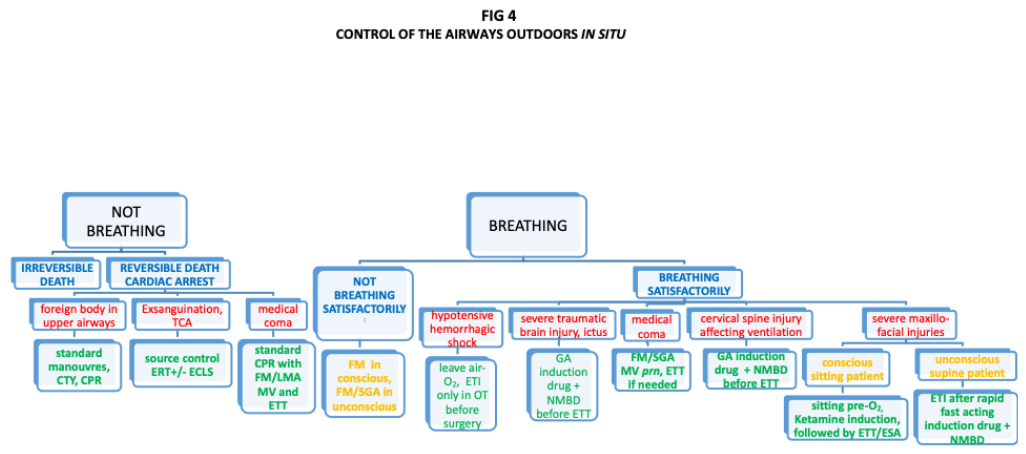
The decision is taken on the scene within seconds considering all factors and variables in a holistic comprehensive picture, with knowledge, previous experience and skills playing a role in the decision.
Clinical acumen (inspection, palpation), anamnesis by bystanders witnesses, the context, the availability of equipment and tools for oxygenation, ventilation or a simple pulse oximeter, and, most importantly the right operator mind-frame, personality, emotive empathy and capacity to read the situation from the anatomical and physiological aspect, is what makes difference between saving or not saving a rescuable patient needing airway control.
The initial variables to compute for the decision-making at impact on the scene outdoors are if the patient is breathing and or is unconscious or conscious.
In presence of an unconscious patient requiring per se control of airways in principle, first we need to assess whether is “breathing or not”.
At impact with a “non-breathing patient”, not moving and with impalpable central pulses, it can be a scenario of ‘irreversible death’ or ‘reversible-death/cardiac-arrest’.
Rigor mortis, livor mortis, lesions incompatible with life, and the time factor of witnessed immobility and no ventilation/ breathlessness are four certain criteria of irreversible death to rely upon in the decision whether to resuscitate or not as indoors as outdoors.
On a dead patient nothing needs to be done, and efforts should be concentrated on other patients e.g. in a scenario of multiple casualties.
In a cardiac arrest scenario, next question is to exclude or have confirmed three special scenarios, which are: i) foreign body in the upper airways that caused reflex laryngeal spasm; exsanguination or traumatic cardiac arrest (ECA/TCA) and iii) medical CA.
Patients in fact may have immediate cardiac arrest by vagal mediated laryngeal spasm or very shortly by respiratory-cardiac arrest by hypoxia depending on individual FRC if not complete obstruction.
It is almost impossible to identify stand-by complete foreign body obstruction of the upper airways unless witnessed and CPR can damage the trachea and lower airways.
To manually ventilate a patient with a foreign body in the upper airways would only cause the obstruction to become more complete, definitive.
If the obstruction cannot be expelled by postural manoeuvres, oxygen must rapidly be introduced into the lung and ventilation rapidly effected through any surgical airway, varying from the simple needle, catheter or improvised device to a PTJV to CTY and tracheostomy.
If the postural manouvres for a supine patient as recommended in the ALS/PALS manuals are unsuccessful, a stab CTY is first choice. Usually, these patients recover shortly when flooded of oxygen. If CTY is not possible or not done for some reasons a PTJV with automated machine if available is next choice.
Good tactics is to use PTJV alternatively directed down to lower airways for oxygenation and up towards the upper airways with the hope to dislodge the foreign body.
In complete obstruction PTJV cannot ventilate longer than 20-40 min without causing damage to vocal cords and alveoli before taking the patient to nearest medical facility or do a CTY on the way.
Any improvised airway device can save life and predictably has more limitations of PTJV.
In exsanguination or traumatic cardiac arrest (ECA/TCA), CPR is deleterious and will accelerate exitus. Heroic surgical steps such as emergency resuscitative thoracotomy (ERT) or ECLS via sternotomy and simultaneous damage control surgery will have to be undertaken.
Hypertensive pneumothorax and cardiac tamponade are excluded clinically or with portable echo.
Any TCA or asystolic CA requires stand-by ETI.
Guidelines for airway control in cardiac arrest/ictus have been standardized by the ERC and the AHA [166,167].
Standard CA from main medical cause (eg dysrrhytmias, acute myocardial infarction, ictus) is initially treated with Def/CPR and MV/LMA ventilation followed by ETI after 5 min in shockable rhythms, and ETI stat in asystole. Before ETI in shockable rhytms and before transfer or before standby synchronous damage control surgery (DCS) in not shockable rhytms, a short-acting brief duration synthetic opioid such as Sufentanil is advised by this author.
In OHCA, despite a first pass rate of 70% and the negative effects of cumulative chest interruption and the insertion technique of SGA easiness, ETI is associated with higher odds of a return of spontaneous circulation (ROSC), 24-hour survival and hospital discharge with favorable neurologic status than SGA [168].
In the PAR Trial compared initial airway management with the LT and initial airway management with ETI, both preceded by BVM for 10-12 min. The strategy of initial LT resulted in higher 72-hour-to-discharge survival than a strategy of initial ETI. Any failed attempt (52%) was rescued by LT with a 91% success rate in the ETI arm [169]. The low first pass rate with ETI was probably biased by the subconscious reassurance of the SGA availability as alternative rescue method.
The AIRWAYS-2 Trial comparing i-gel with ETI preceded by BVM for 7-8 min registered no difference in survival, regurgitation and aspiration beetween the two methods, despite the faster initial ventilation in the i-gel group [170].
In the CAAM Trial, comparing the effects of initial ETI or BVM ventilation followed by ETI after ROSC, practically no difference in neurological outcomes was found between the two tactics, but regurgitation of gastric contents, aspiration rate and difficult ventilation were found higher in the BVM group [171].
In a previous study [172], BVM had been found to have slightly better neurological outcome probably biased from the patients different conditions preCA.
A psychological bias of sense of security with ETI in respect of BVM was universally found among all rescuers.
A group showed a remarkable OHCA 91% first pass rate with ETI [173].
In a study on OHCA managed by trained paramedics no difference was found in neurocognitive outcomes, survival to discharge or 90 days, and quality of life between 3 groups (i-gel, LMA Supreme, conventional management with BVM +/- ETI ≤3 attempts). The first pass success rate was 85% with ETI, 79% with i-gel and 75% with of LMAS [174].
A group with almost totally paramedic (90% + 10% medics) as OHCA rescuers, had a i-gel first pass rate of 90% [175].
In a training manikin study, LT has been reported with a first pass rate of 92% in two attempts (71% at first attempt) [176].
In an experimental model SGA were found to reduce carotid blood flow [177]. Admittedly, pigs must have a tighter oropharyngeal space than humans.
Specific management of cardiac arrest from other medical causes (eg potassium severe derangement, severe diabetes, drug overdose, pulmonary embolism, drowning and hypothermia, anaphylactic shock) is well described in ALS guidelines [166].
In a “breathing patient”, next question is to see if he/she is “breathing satisfactorily or not” and if is “conscious or unconscious”, particularly in post-TBI or ictus, advanced hemorrhagic shock stage III & IV, CSI affecting breathing or ventilation, maxillofacial trauma, convulsions, or other medical coma causes.
“If the patient does not breathe satisfactorily”(clinical assessment, cyanosis, low SaO2 and scarce chest excursion), MV is first thing to do on a ‘conscious patient’, making sure to apply an insufflation pressure < 20 cm H2O [24,50]. SGA is a good alternative to MV in an ‘unconscious patient’.
If ventilation does not ensure satisfactory SaO2, colour and consciousness, or the patient is cyanotic, a DSII under Ketamine or a synthetic opioid followed by suxamethonium is a correct choice.
Ketamine has the advantage of not affecting the scarce breathing of the patient but takes 1 min for action, whereas other drugs are quicker to act and better suited for RSII.
Airway management in trauma and outdoors has been well appraised in its criticalities [178].
Hypoxia and airway mismanagement contribute up to a third of pre-hospital trauma deaths [179].
In trauma with patient breathing normally and requiring mask oxygenation or ventilation, head tilt (not in patients with cervical trauma or not excluded possible cervical trauma), and chin lift/jaw thrust or the use of basic adjuncts such as oropharyngeal airway in unresponsive patients without gag reflex and/or nasopharyngeal airway in patients with more active reflexes but without evidence of fracture of base of skull, are the initial steps to do.
ETI is recommended in presence of post severe TBI apnoea; need for airway protection from aspiration (vomitus, bleeding); Glasgow Coma Scale <8; severe maxillo-facial fractures; impending or potential airway compromise eg inhalation injury; inability to maintain SpO2> 90% by facemask or LMA oxygenation [180].
Surgical airway should be resorted to when there is severe glottis oedema or oropharyngeal haemorrhage, fracture/hemathom/edema of the larynx with no palpable anterior neck (HB, CC, TC) landmarks and when endotracheal tube fails to be passed through the vocal cords.
In problematic intubations a LMA or LT suffice for ventilation in unconscious patients.
On patients in evident HS oxygen, likewise blood and flids, should be calibrated to avoid worsening of the hemorrhage [30,160,161].
To intubate a patient before theatre unless is in ECA is a big error as it risks increasing the hemorrhage speed and exsanguination from the hypertensive peak at laryngoscopy, with the consequent need of deep GA not allowing the correct physiological reading; it risks a further post induction hypotension that can be fatal; finally, it messes up appraisal for the destination anesthetist in case of transport to a trauma center [30].
Categorization of the degree of HS and its continuous clinical monitoring of the hemodynamic dynamics is essential [40].
In Traumatic Brain Injury, apnea requires immediate inubation. There are 3 mechanisms by which apnea may occur in TBI: severe or catastrophic brain injury, impact brain apnea, loss of consciousness with resultant functional airway obstruction.
Several drugs are suitable for induction before ETI.
Etomidate is safe in neurosurgery and TBI but cannot be given im as ketamine and can cause hypotension on volume-depleted patients. It also causes myoclonus and pain on injection and postoperative nausea and vomit.
Ketamine can be given im, is useful in sedating combactive patients, and increases sympathetic tone and catecholamines release. It is the preferred choice in cardiac tamponade and appears to be antidepressive and have neuroprotective property indipendent from the prevention of hypotension, but may cause hypotension in cathecolamine depleted patients..
Propofol is a sedative hypnotic agent with rapid onset of action, short duration and a profound amnesia effect. It blunts the sympathetic hypertensive stimulation on laryngoscopy and decreases intracranial pressure. It is a cardiovascular depressant and is relatively contraindicated in hemodynamic instability; however coupled with a peripheral vasocostrictor such as phenylephrime or with ketamine can be given also in mild cardiovascular instability [181].
Synthetic opioids are not advised on their own in TBI but as support the above drugs as stabilizers to lessen other drugs in balanced anaesthesia fashion [182].
In epilepsy, propofol is the ideal induction/therapy to block convulsions and reduce intracranial pressure in absence of hypotension. Ketamine is the alternative in hypotensive patients. Midazolam at high doses is the alternative but is slower to uptake. ETI is followed by NMBD and supported by BZD before and after ETI.
Same considerations for patients with ictus. The hypertensive peak due to a non-curbed laryngeal reflex is particularly relevant in ictus, where internal bleeding and endocranial pressure are increased. Induction pre-ETI with a not hypotensive anesthetic drug lowering ICP, such as etomidate and ketamine, followed by a NMBD, is best suited for hypotensive ictus whereas propofol is mandatory in hypertensive ictus [153].
Obvious or highly suspected CSIaffecting ventilation requires induction and NMBD in situ, on a supine or laterally placed patient, followed by ETT.
Severe injuries to the maxillofacial complexes on a conscious sitting patient require preoxygenation and induction in sitting position with ketamine and then be put carefully and rapidly in supine position for ETI. If impossible, an ESA follows.
If medical coma, the need to use an ETT immediately occurs only if the laryngeal and gag reflexes are obtunded, not a frequent situation. These patients can be meanwhile managed well with MV or LGA ventilation before ETI.
In Pulmonary Oedema/AcuteHF, Etomidate is the best induction agent followed by BiPAP or BiPAP and Rocuronium before ETI.
In Asthma or reacutization of COPD: BiPAP, Ketamine and Rocuronium are given before ETI.
Agitated uncooperative patients or children or psychotic adults who are cyanotic and dyspnoic are induced by a DSII using Ketamine im and follow with preoxygnation or direct ETI with a NMBD soon after the drug has been uptaken and the patient placed supine [30,31]. On patients not needing emergency ETI, haloperidol im is best choice and the drill can be done after 30-60 min.
5. Conclusions and Directions
The vertical train flow for the intubation drill is universal, independent from the different specialist backgrounds, and patient age [183].
Since the introduction of SGAs and VL, the difficult airway has become more manageable scenario but still accounts for a mortality of GA in the induction phase together with the hypotensive effect of the induction drugs, particularly in patients needing GA for HS.
The thre sequences of induction/intubation [Fig 2] are synthetized in consensus. [Table 4]
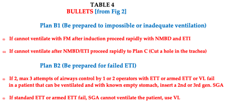
Nasal mask is increasingly used for preoxygenation [184. High-flow nasal oxygen therapy and NIV are useful option for patients with mild hypoxemia, while PEEP added to facemask is advised for obese patients.
The long-term future of difficult airway is in a heat-laser-magnet guided blind ETI insertion with the ETT cuff stopping above tracheal bifurcation as directed by a percutaneous landmark or identified with an external sonar on the anterior aspect of the neck at a level above the carina.
“Styletubation” is the most recent advancement in airway management combining videolaryngoscopy and intubation in one device [185,186]. The technique is fast, safe, effective, 100% succesful at first attempt, with 0 complications.
The potential use of the method, well studied in some centers with experience by now of the order of 100.000 patients, is enormous and the device is destined to become universal as for its applicability in many scenario and patients with normal or difficult airway [187].
A new parameter, the ‘oxygen reserve index’ can be calculated with the multiwave pulse oxymeter (multiple sensors to fingers controlled by a software), more sensitive than the standard oxymeter in detecting early desaturation in obese. It detects a fall of PaO2 at SaO2 levels of 98% whereas classic oxymeter does it when PaO2 reaches 70%. Another drawback is the risk of giving excess oxygen when trying to compensate SaO2 falls, due to an over-estimation up to 4% of common pulse oxymeter [188].
Whether an emergency physician, an anesthetist, or a trauma surgeon, a one-and-only linearly consequential, “vertical, fast-tracked”, holistic orderly decision-making must be followed, when approaching and dealing with an airway before general anesthesia or in other settings.
Trials between i-gel and ETI, and between SGA and BVM seem contextually lacking.
6. DECLARATIONS
I AM THE ONLY AUTHOR
THE WORK HAS NO FUNDING
THERE IS NO NEED OF APPROVAL ON HUMAN OR ANIMAL STUDIES
THERE IS NO NEED FOR AN INFORMED CONSENT
THERE ARE NO DATA
THERE IS NO CONFLICT OF INTEREST
Abbreviations
FM=facemask; ETT=endotracheal tube; SGA=supraglottic airway; ETI=endotracheal intubation; NMB=neuromuscular blockade; SGA=supraglottic airway; HS=hemorrhagic shock; TBI=traumatic brain injury; DA=difficult airway; CA=critical airway; RSI/ETT=rapid sequence induction/intubation; SGA=supraglottic airway; GA=general anesthesia, VC=vocal cords PTJV=percutaneous transtracheal jet ventilation; prn=pro re data; EtCO2=endotracheal CO2; SpO2/SaO2=peripheral oxygen saturation; ED=emergency department.
References
- Apfelbaum, J.L.; Hagberg, C.A.; Caplan, R.A. Practice guidelines for management of the difficult airway: an updated report by the American Society of Anesthesiologists Task Force on Management of the Difficult Airway. Anesthesiology 2013, 118, 251–270. [Google Scholar] [PubMed]
- Mosier, J.M.; Joshi, R.; Hypes, C.; Pacheco, G.; Valenzuela, T.; Sakles, J.C. The Physiologically Difficult Airway. West J. Emerg. Med. 2015, 16(7), 1109–17. [Google Scholar] [PubMed]
- Bonanno, F.G. The critical airway in adults - The facts. J. Emerg. Trauma Shock 2012, 5(2), 153–159. [Google Scholar] [PubMed]
- Walls, R.M.; Brown, C.A.; Bair, A.E. NEAR II Investigators. Emergency Airway Management: A Multi-Center Report of 8937 Emergency Department Intubations. J. Emerg. Med. 2011, 41, 347–354. [Google Scholar] [PubMed]
- Martin, L.D.; Mhyre, J.M.; Shanks, A.M. 3,423 emergency tracheal intubations at a university hospital: airway outcomes and complications. Anesthesiology 2011, 114, 42–48. [Google Scholar] [PubMed]
- Rosenstock, C.V.; Nørskov, A.K.; Wetterslev, J. Emergency surgical airway management in Denmark: a cohort study of 452 461 patients registered in the Danish Anaesthesia Database. Br. J. Anaesth. 2016, 117 Suppl 1, i75–i82. [Google Scholar] [CrossRef] [PubMed]
- Cook, T.M.; MacDougall-Davis, S.R. Complications and failure of airway management. Br. J. Anaesth. 2012, 109, i68–85. [Google Scholar] [PubMed]
- Kheterpal, S.; Han, R.; Tremper, K.K.; Shanks, A.; Tait, A.R.; O’Reilly, M.; et al. Incidence and predictors of difficult and impossible mask ventilation. Anesthesiology 2006, 105(5), 885–91. [Google Scholar] [PubMed]
- De Jong, A.; Molinari, N.; Pouzeratte, Y. Difficult intubation in obese patients: incidence, risk factors, and complications in the operating theatre and in intensive care. [PubMed]
- Goto, T.; Goto, Y.; Hagiwara, Y.; Okamoto, H.; Watase, H.; Hasegawa, K. Advancing emergency airway management practice and research. Acute Med. Surg. 2019, 6, 336–351. [Google Scholar] [PubMed]
- Japanese Society of Anesthesiologists. JSA airway management guideline 2014: to improve the safety of induction of anesthesia. J. Anesth. 2014, 28, 482–493. [Google Scholar] [PubMed]
- Frerk, C.; Mitchell, V.S.; McNarry, A.F. Difficult Airway Society 2015 guidelines for management of unanticipated difficult intubation in adults. Br. J. Anaesth. 2015, 115, 827–48. [Google Scholar] [PubMed]
- Law, J.A.; Duggan, V.A.; Asselin, M.; Bakr, P.; Crosby, E.; Downey, A.; et al. Canadian Airway Focus Group updated consensus-based recommendations for management of the difficult airway: part 1. Difficult airway management encountered in an unconscious patient. Can. J. Anaesth. 2021, 68(9), 1373–1404. [Google Scholar] [PubMed]
- Rao, S.; Kunselman, A.R.; Schuler, H.G.; DesHarnais. S. Laryngoscopy and tracheal intubation in the head-elevated position in obese patients: a randomized, controlled, equivalence trial. Anesth. Analg. 2008, 107(6), 1912–8. [Google Scholar] [CrossRef] [PubMed]
- Khandelwal, N.; Khorsand, S.; Mitchell, S.H.; Joffe, A.M. Head-Elevated Patient Positioning Decreases Complications of Emergent Tracheal Intubation in the Ward and Intensive Care Unit. Anesth. Analg. 2016, 122(4), 1101–7. [Google Scholar] [PubMed]
- Boyce, J.R.; Ness, T.; Castroman, P.; Gleysteen, J.J. A preliminary study of the optimal anesthesia positioning for the morbidly obese patient. Obes. Surg. 2003, 13(1), 4–9. [Google Scholar] [CrossRef] [PubMed]
- Weingart, S.D.; Levitan, R.M. Preoxygenation and prevention of desaturation during emergency airway management. Ann. Emerg. Med. 2012, 59, 165–175. [Google Scholar] [CrossRef] [PubMed]
- Heier, T.; Feiner, J.R.; Lin, J. Hemoglobin desaturation after succinylcholine-induced apnea: a study of the recovery of spontaneous ventilation in healthy volunteers. Anesthesiology 2001, 94, 754–759. [Google Scholar] [PubMed]
- Tanoubi, I.; Drolet, P.; Donati, F. Optimizing preoxygenation in adults. Can. J. Anaesth. 2009, 56, 449–466. [Google Scholar] [PubMed]
- Herriger, A.; Frascarolo, P.; Spahn, D.R.; Magnusson, L. The effect of positive airway pressure during pre-oxygenation and induction of anaesthesia upon duration of non-hypoxic apnoea. Anaesthesia 2004, 59, 243–7. [Google Scholar] [PubMed]
- Collins, J.S.; Lemmens, H.J.; Brodsky, J.B. Laryngoscopy and morbid obesity: a comparison of the “sniff” and “ramped” positions. Obes. Surg. 2004, 14, 1171–1175. [Google Scholar] [PubMed]
- Dixon, B.J.; Dixon, J.B.; Carden, J.R. Preoxygenation is more effective in the 25 degrees head-up position than in the supine position in severely obese patients: a randomized controlled study. Anesthesiology 2005, 102, 1110–1115. [Google Scholar] [PubMed]
- Altermatt, F.R.; Muñoz, H.R.; Delfino, A.E.; Cortínez, L.I. Preoxygenation in the obese patient: effects of position on tolerance to apnoea. Br. J. Anaesth. 2005, 95(5), 706–9. [Google Scholar] [CrossRef] [PubMed]
- Gander, S.; Frascarolo, P.; Suter, M. Positive end-expiratory pressure during induction of general anesthesia increases duration of non-hypoxic apnea in morbidly obese patients. Anesth. Analg. 2005, 100, 580–584. [Google Scholar] [PubMed]
- Wiles, M.D. Airway management in patients with suspected or confirmed traumatic spinal cord injury: a narrative review of current evidence. Anaesthesia 2022, 77, 1120–1128. [Google Scholar] [CrossRef] [PubMed]
- Teller, L.; Alexander, C.; Frumin, J.; Gross, J. Pharyngeal insufflation of oxygen prevents arterial desaturation during apnea. Anesthesiology 1988, 69, 980–2. [Google Scholar] [PubMed]
- Taha, S.K.; Siddik-Sayyid, S.M.; El-Khatib, M.F.; Dagher, C.M.; Hakki, M.A.; Baraka, A.S. Nasopharyngeal oxygen insufflation following pre-oxygenation using the four deep breath technique. Anaesthesia 2006, 61(5), 427–30. [Google Scholar] [PubMed]
- Ramachandran, S.K.; Cosnowski, A.; Shanks, A.; Turner, C.R. Apneic oxygenation during prolonged laryngoscopy in obese patients: a randomized, controlled trial of nasal oxygen administration. J. Clin. Anesth. 2010, 22(3), 164–8. [Google Scholar] [CrossRef] [PubMed]
- Patel, A.; Nouraei, S.A.R. Transnasal Humidified Rapid-Insufflation Ventilatory Exchange (THRIVE): A Physiological Method of Increasing Apnoea Time in Patients with Difficult Airways. Anaesthesia 2015, 70, 323–329. [Google Scholar] [PubMed]
- Bonanno, F.G. General Anaesthesia in the Context of Haemorrhagic Shock and Trauma, in Anesthesiology - New Insights, Ed MU Gerbershagen; IntechOpen: Rijeka, 2025. [Google Scholar] [CrossRef]
- Weingart, S.D.; Trueger, N.S.; Wong, N.; Scofi, J.; Singh, N.; Rudolph, S.S. Delayed Sequence Intubation: A Prospective Observational Study. Ann. Emerg. Med. 2015, 65(4), 349–55. [Google Scholar] [CrossRef] [PubMed]
- Neilipovitz, D.T.; Crosby, E.T. No evidence for decreased incidence of aspiration after rapid sequence induction. Can. J. Anaesth. 2007, 54(9), 748–64. [Google Scholar] [PubMed]
- El-Orbany, M.; Connolly, L.A. Rapid sequence induction and intubation: current controversy. Anesth. Analg. 2010, 110, 1318–1325. [Google Scholar] [PubMed]
- Bonanno, F.G. Ketamine in war/tropical surgery (a final tribute to the racemic mixture). Injury 2002, 33(4), 323–7. [Google Scholar] [CrossRef] [PubMed]
- Tao, Y.-B.; Tang, Z.-L.; Lin, Z.-L.; Lei, W.-P.; Lu, X.-L.; Sun, J.-L. Effects of glycopyrrolate and atropine for oral secretions and perioperative hemodynamics in children undergoing tonsillectomy and adenoidectomy: a prospective, single-center, randomized, double-blind, controlled trial. Front. Pharmacol. 2024, 15–2024. [Google Scholar] [CrossRef] [PubMed]
- El Orbany, M. Opioids and rapid-sequence induction. BJA 2007, 99(4), 598. [Google Scholar] [PubMed]
- Aggarwal, A.; Mustahsin, M.; Shishir, P. Comparison of intubating laryngeal mask airway and video laryngoscope during emergency intubation in patients who are critically ill. World J. Crit. Care Med. 2026, 15(1), 112345. [Google Scholar] [CrossRef] [PubMed]
- Panda, C.K.; Karim, H.M.R.; Neema, P.K. Bronchoscopy examination of a patient with tracheobronchial injury having intractable cough: Role of sevoflurane sedation and paraoxygenation. Ann. Card. Anaesth. 2021, 24(1), 121–123. [Google Scholar] [PubMed]
- Baloi, D.; Rawson, C.; Montgomery, D.; Karsy, M.; Pahlevani, M. Perioperative Anesthetic Strategies in Emergent Neurosurgery During Severe Traumatic Brain Injury. Trauma Care 2026, 6, 5. [Google Scholar] [CrossRef]
- Bonanno, F. Time to Change ATLS Classifications of Hemorrhagic Shock. J. Emerg. Trauma Shock Epub;Researchgate Update December 2025. 2024, 17(4), 252–4. [Google Scholar] [CrossRef] [PubMed]
- Smischney, N.J.; Nicholson, W.T.; Brown, D.R.; Gallo De Moraes, A.; Hoskote, S.S.; Pickering, B.; et al. Ketamine/propofol admixture vs etomidate for intubation in the critically ill: KEEP PACE randomized clinical trial. J. Trauma Acute Care Surg. 2019, 87(4), 883–91. [Google Scholar] [PubMed]
- Russotto, V.; Tassistro, E.; Myatra, S.N.; Parotto, M.; Antolini, L.; Bauer, P. for the INTUBE Study Investigators. Peri-intubation cardiovascular collapse in patients who are critically ill: insights from the INTUBE study. Am. J. Respir. Crit. Care Med. 2022, 206(4), 449–458. [Google Scholar] [PubMed]
- Schmidt, R.C.; Zampieri, F.G.; Da Silva Ramos, F.J.; Santos Cavatoni Serra, F.; Maccagnan Pinheiro Besen, B.A.; Fonseca Nunes, N.; et al. Propofol versus ketamine for rapid sequence intubation of critically ill patients (PROMINE): a randomized clinical trial. Intensive Care Med. 2026. [Google Scholar] [CrossRef] [PubMed]
- Casey, J.D.; Seitz, K.P.; Driver, B.E.; Gibbs, K.W.; Ginde, A.A.; Trent, S.A.; et al. RSI Investigators and the Pragmatic Critical Care Research Group. Ketamine or Etomidate for Tracheal Intubation of Critically Ill Adults. N Engl. J. Med. 2026, 394(16), 1608–1620. [Google Scholar] [CrossRef] [PubMed]
- Zampieri, F.G.; Schmidt, R.C.; Besen, B.A.M.P.; Ramos, F.J.D.S.; Lamontagne, F.; Adhikari, N.K.J.; et al. Induction agents for emergency tracheal intubation in critically ill adults: a systematic review and network meta-analysis. Crit. Care 2026, 30(1), 296. [Google Scholar] [CrossRef] [PubMed]
- Klemola, U.M.; Mennander, S.; Saarnivaara, L. Tracheal intubation without the use of muscle relaxants: remifentanil or alfentanil in combination with propofol. Acta Anaesthesiol. Scand. 2000, 44(4), 465–9. [Google Scholar] [CrossRef] [PubMed]
- Isono, S.; Ishikawa, T. Oxygenation, not intubation, does matter. Anesthesiology 2011, 114, 7–9. [Google Scholar] [CrossRef] [PubMed]
- Asai, T. Strategies for difficult airway management—the current state is not ideal. J. Anesth. 2013, 27, 157–60. [Google Scholar] [PubMed]
- Liu, E.H.; Asai, T. Cannot intubate cannot ventilate—focus on the ‘ventilate’. J. Anesth. 2015, 29, 323–325DOI. [Google Scholar] [CrossRef] [PubMed]
- Bouvet, L.; Albert, M.L.; Augris, C. Real-time detection of gastric insufflation related to facemask pressure-controlled ventilation using ultrasonography of the antrum and epigastric auscultation in non-paralyzed patients: a prospective, randomized, double-blind study. Anesthesiology 2014, 120, 326–334. [Google Scholar] [PubMed]
- Casey, J.D.; Janz, D.R.; Russell, D.W. Bag-mask ventilation during tracheal intubation of critically ill adults. N Engl. J. Med. 2019, 380, 811–21. [Google Scholar] [PubMed]
- Mendonca, C. Sugammadex to rescue a ‘can’t ventilate’ scenario in an anticipated difficult intubation: is it the answer? Anaesthesia 2013, 68, 795–799. [Google Scholar] [PubMed]
- Bonanno, F.G. An appraisal on emergency tracheostomy. Inj. Extra 2007, 38, 346–349. [Google Scholar]
- Glapa, M.; Kourie, J.F.; Doll, D.; Degiannis, E. Early management of gunshot injuries to the face in civilian practice. World J. Surg. 2007, 31, 2104–2110. [Google Scholar] [CrossRef] [PubMed]
- Jose, A.; Nagori, S.A.; Agarwal, B. Management of maxilla-facial trauma in emergency. An update of challenges and controversies. J. Emerg. Trauma Shock 2016, 9, 73–80. [Google Scholar] [PubMed]
- Rupprecht, H.; Gaab, K. Prehospital tracheotomy in a case of avulsion of the larynx with a comminuted fracture of the jawbone. Clin. Exp. Emerg. Med. 2019, 6(2), 173–176. [Google Scholar] [CrossRef] [PubMed]
- Taha, S.K.; El-Khatib, M.F.; Baraka, A.S. Effect of suxamethonium vs rocuronium on onset of oxygen desaturation during apnoea following rapid sequence induction. Anaesthesia 2010, 65, 358–361. [Google Scholar] [PubMed]
- Tang, L.; Li, S.; Huang, S. Desaturation following rapid sequence induction using succinylcholine vs rocuronium in overweight patients. Acta Anaesthesiol. Scand. 2011, 55, 203–208. [Google Scholar] [PubMed]
- Lee, C.; Jahr, J.S.; Candiotti, K.A.; Warriner, B.; Zornow, M.H.; Naguib, M. Reversal of profound neuromuscular block by sugammadex administered three minutes after rocuronium: a comparison with spontaneous recovery from succinylcholine. Anesthesiology 2009, 110(5), 1020–5. [Google Scholar] [PubMed]
- Sørensen, M.K.; Bretlau, C.; Gätke, M.R.; Sørensen, A.M.; Rasmussen, L.S. Rapid sequence induction and intubation with rocuronium-sugammadex compared with succinylcholine: a randomized trial. Br. J. Anaesth. 2012, 108(4), 682–9. [Google Scholar] [PubMed]
- Wright, P.M.; Caldwell, J.E.; Miller, R.D. Onset and duration of rocuronium and succinylcholine at the adductor pollicis and laryngeal adductor muscles in anesthetized humans. Anesthesiology 1994, 81(5), 1110–5. [Google Scholar] [CrossRef] [PubMed]
- Hayes-Bradley, C.; Tarrant, M. Rocuronium ≤1.5 mg/kg. Emerg. Med. Australas. 2022, 34(6), 892–897. [Google Scholar] [CrossRef] [PubMed]
- Takeda, T.; Tanigawa, K.; Tanaka, H.; Hayashi, Y.; Goto, E.; Tanaka, K. The assessment of three methods to identify tracheal tube placement in the emergency setting. Resuscitation 2003, 56, 153–7. [Google Scholar] [PubMed]
- Kerslake, I.; Kelly, F. Uses of capnography in the critical care unit. BJA Educ. 2017, 17, 178–183. [Google Scholar]
- Cook, T.M. The winds of change--progress in the implementation of universal capnography. Anaesthesia 2016, 71, 363–368. [Google Scholar] [CrossRef] [PubMed]
- Mishra, P.R.; Bhoi, S.; Sinha, T.P. Integration of point-of-care ultrasound during rapid sequence induction in trauma resuscitation. J. Emerg. Trauma Shock 2018, 11, 92–97. [Google Scholar] [PubMed]
- Godschalx, V.; Vanhoof, M.; Soetens, F.; Van de Putte, P.; Hadzic, A.; Van de Velde, M.; et al. The role of gastric ultrasound in anaesthesia for emergency surgery: a review and clinical guidance. Eur. J. Anaesthesiol. Intensive Care 2023, 2(4), e0027. [Google Scholar] [CrossRef] [PubMed]
- Charya, A.V.; Holden, V.K.; Pickering, E.M. Management of life-threatening hemoptysis in the ICU. J. Thorac. Dis. 2021, 13(8), 5139–58. [Google Scholar] [PubMed]
- Håkanson, E.; Konstantinov, I.E.; Fransson, S.G.; Svedjeholm, R. Management of life- threatening haemoptysis. Br. J. Anaesth. 2002, 88(2), 291–5. [Google Scholar] [PubMed]
- Root, C.W.; Mitchell, O.J.L.; Brown, R.; Evers, C.B.; Boyle, J.; Griffin, C. Suction Assisted Laryngoscopy and Airway Decontamination (SALAD): A technique for improved emergency airway management. Resusc. Plus 2020, 1-2, 100005. [Google Scholar] [CrossRef] [PubMed]
- Turkstra, T.P.; Regan, W.D. Provider/patient conflict: is it time to reconsider the contraindication for videolaryngoscope use in bleeding/soiled airway? Can. J. Anaesth. 2022, 69(1), 177–178. [Google Scholar] [CrossRef] [PubMed]
- Komatsu, R.; Nagata, O.; Sessler, D.I.; Ozaki, M. The intubating laryngeal mask airway facilitates tracheal intubation in the lateral position. Anesth. Analg. 2004, 98(3), 858–61. [Google Scholar] [CrossRef] [PubMed]
- Takenaka, I.; Aoyama, K. Prevention of aspiration of gastric contents during attempt in tracheal intubation in the semi-lateral and lateral positions. World J. Emerg. Med. 2016, 7(4), 285–9. [Google Scholar] [CrossRef] [PubMed]
- Paw, H.G.; Sharma, S. Cricothyroidotomy: a short-term measure for elective ventilation in a patient with challenging neck anatomy. Anaesth. Intensive Care 2006, 34(3), 384–7. [Google Scholar] [CrossRef] [PubMed]
- DeVore, E.K.; Redmann, A.; Howell, R.; Khosla, S. Best practices for emergency surgical airway: A systematic review. Laryngoscope Investig. Otolaryngol. 2019, 4(6), 602–608. [Google Scholar] [CrossRef] [PubMed]
- Bonanno, F.G. Issues of critical airway management (Which anesthesia; Which surgical airway?). J. Emerg. Trauma Shock. 2012, 5(4), 279–84. [Google Scholar] [CrossRef] [PubMed]
- Gilyoma, J.M.; Balumuka, D.D.; Chalya, P.L. Ten-year experiences with Tracheostomy at a University teaching hospital in Northwestern Tanzania: A retrospective review of 214 cases. World J. Emerg. Surg. 2011, 6, 38. [Google Scholar] [PubMed]
- Shaikh, A.A.; Rafique, M.; Farrukh, M.S. Emergency tracheostomy - an analysis of its indications. Med. Channel 2014, 20(1), 65–69. [Google Scholar]
- Ono, Y.; Yokoyama, H.; Matsumoto, A.; Kumada, Y.; Shinohara, K.; Tase, C. Surgical airways for trauma patients in an emergency surgical setting: 11 years' experience at a teaching hospital in Japan. J. Anesth. 2013, 27, 832–827. [Google Scholar] [CrossRef] [PubMed]
- Caplan, R.A.; Posner, K.L.; Ward, R.J.; Cheney, F.W. Adverse respiratory events in anesthesia: a closed claim analysis. Anesthesiology 1990, 72, 828–33. [Google Scholar] [PubMed]
- Cook, T.M.; Woodall, N.; Frerk, C. Major complications of airway management in the UK: results of the Fourth National Audit Project of the Royal College of Anaesthetists and the Difficult Airway Society. Part 1: anaesthesia. Br. J. Anaesth. 2011, 106, 617–31. [Google Scholar] [PubMed]
- Brown, C.A.; Bair, A.E.; Pallin, D.J.; Walls, R.M. NEAR III Investigators. Techniques, success, and adverse events of emergency department adult intubations. Ann. Emerg. Med. 2015, 65(4), 363–70. e1. [Google Scholar] [PubMed]
- Südfeld, S.; Brechnitz, S.; Wagner, J.Y.; Reese, P.C.; Pinnschmidt, H.O.; Reuter, D.A.; et al. Post-induction hypotension and early intraoperative hypotension associated with general anaesthesia; British Journal of Anaesthesia: BJA, 2017; Volume 119, 1, pp. 57–64. [Google Scholar]
- Ali, N.; Kapadia, N.N.; Shomar, S.M.; Raheem, A.; Habibullah, M.; Habib, Z.; et al. Risk Factors of Peri-Intubation Cardiac Arrest in Critically Ill Patients Presenting to the Emergency Department of a Low-Income Country – A Case-Control. J. Emerg. Med. 2025, 76, 26–32. [Google Scholar] [CrossRef] [PubMed]
- Russotto, V.; Myatra, S.N.; Laffey, J.G.; Tassistro, E.; Antolini, L.; Bauer, P.; et al. Intubation Practices and Adverse Peri-intubation Events in Critically Ill Patients From 29 Countries. JAMA 2021, 325(12), 1164–72. [Google Scholar] [PubMed]
- Maia, I.W.A.; Besen, B.A.M.P.; Silva, L.O.J.E.; von Hellmann, R.; Abrahao Hajjar, L.; Sandefur, B.J.; et al. Peri-intubation adverse events and clinical outcomes in emergency department patients: the BARCO study. Crit. Care 2025, 29, 155. [Google Scholar] [CrossRef] [PubMed]
- El-Ganzhouri, A.R.; McCarthy, R.J.; Tuman, K.J. Preoperative airway assessment: Predictive value of a multivariate risk index. Anesth. Analg. 1996, 82, 1197–1204. [Google Scholar]
- Langeron, O.; Masso, E.; Huraux, C. Prediction of difficult mask ventilation. Anesthesiology 2000, 92, 1229–1236. [Google Scholar] [PubMed]
- Levitan, R.M.; Everett, W.W.; Ochroch, E.A. Limitations of difficult airway prediction in patients intubated in the emergency department. Ann. Emerg. Med. 2004, 44, 307–313. [Google Scholar] [PubMed]
- Shiga, T.; Wajima, Z.; Inoue, T. Predicting difficult intubation in apparently normal patients: a meta-analysis of bedside screening test performance. Anesthesiology 2005, 103, 429–437. [Google Scholar] [PubMed]
- Kheterpal, S.; Martin, L.; Shanks, A.M. Prediction and Outcomes of Impossible Mask Ventilation. Anesthesiology 2009, 110, 891–897. [Google Scholar] [PubMed]
- Gerstein, N.S.; Braude, D.A.; Hung, O.; Sanders, J.C.; Murphy, M.F. The Fast-Trach intubating laryngeal mask airway: an overview and update. Can. J. Anaesth. 2010, 57, 588–601. [Google Scholar] [CrossRef] [PubMed]
- Singh, A.; Bhalotra, A.R.; Anand, R. A comparative evaluation of ProSeal laryngeal mask airway, I-gel and supreme laryngeal mask airway in adult patients undergoing elective surgery: a randomised trial. Indian J. Anaesth. 2018, 62, 858–864. [Google Scholar] [CrossRef] [PubMed]
- Kim, M.H.; Lee, J.H.; Choi, Y.S.; Park, S.; Shin, S. Comparison of the laryngeal mask airway supreme and the i-gel in paralysed elderly patients: A randomised controlled trial. Eur. J. Anaesthesiol. 2018, 35(8), 598–604. [Google Scholar] [PubMed]
- Attarde, V.B.; Kotekar, N.; Shetty, S.M. Air-Q intubating laryngeal airway: a study of the second generation supraglottic airway device. Indian J. Anaesth. 2016, 60, 343–348. [Google Scholar] [PubMed]
- Windpassinger, M.; Plattner, O.; Gemeiner, J. Pharyngeal oxygen insufflation during Air Traq laryngoscopy slows arterial desaturation in infants and small children. Anesth. Analg. 2016, 122, 1153–1157. [Google Scholar] [PubMed]
- Atlas, G.M. A comparison of fiberoptic compatible oral airways. J. Clin. Anest. 2004, 16, 66–73. [Google Scholar] [CrossRef] [PubMed]
- Cergan, R.; Dumitru, M.; Vrinceanu, D.; Neagos, A.; Jeican, I.I.; Ciuluvica, R.C. Ultrasonography of the larynx: Novel use during the SARS-CoV-2 pandemic (Review). Exp. Ther. Med. 2021, 21(3), 273. [Google Scholar] [CrossRef] [PubMed]
- Taneja, S.; Agarwalt, M.; Dali, J.S.; Agrawal, G. Ease of Proseal Laryngeal Mask Airway insertion and its fibreoptic view after placement using Gum Elastic Bougie: a comparison with conventional techniques. Anaesth. Intensive Care 2009, 37, 435–440. [Google Scholar] [CrossRef] [PubMed]
- Li, S.Y.; Yao, W.Y.; Yuan, Y.J. Supreme™ laryngeal mask airway uses in general Anesthesia for category 2 and 3 Cesarean deliveries: a prospective cohort study. BMC Anesthesiol. 2017, 17, 169. [Google Scholar] [PubMed]
- Kim, M.; Lee, J.H.; Choi, Y.S.; Park, S.; Shin, S. Comparison of the laryngeal mask airway supreme and the i-gel in paralysed elderly patients: a randomised controlled trial. Eur. J. Anaesthesiol. 2018, 35, 598–604. [Google Scholar] [PubMed]
- Aziz, M.F.; Dillman, D.; Fu, R.; Brambrink, A.M. Comparative effectiveness of the C-MAC video laryngoscope versus direct laryngoscopy in the setting of the predicted difficult airway. Anesthesiology 2012, 116, 629–636. [Google Scholar] [CrossRef] [PubMed]
- Ruderman, B.T.; Mali, M.; Kaji, A.; Kilgo, R.; Watts, S.; Wells, R.; et al. Direct vs Video Laryngoscopy for Difficult Airway Patients in the Emergency Department: A National Emergency Airway Registry Study. West. J. Emerg. Med. Integr. Emerg. Care With Popul. Health 2022, 23(5), 706–715. [Google Scholar] [CrossRef] [PubMed]
- Prekker, M.E.; Driver, B.E.; Trent, S.A.; Resnick-Ault, D.; Seitz, K.P.; Russell, D.W.; et al. Videolaryngoscopy versus Direct Laryngoscopy for tracheal intubation of critical ill adults. NEJM August 3, 2023. N Engl. J. Med. 2023, 389, 418–429. [Google Scholar] [PubMed]
- Knapp, J.; Eberle, B.; Bernhard, M.; Theiler, L.; Pietsch, U.; Albrecht, R. Analysis of tracheal intubation in out-of-hospital helicopter emergency medicine recorded by video laryngoscopy. Scand. J. Trauma Resusc. Emerg. Med. 2021, 29(1), 49. [Google Scholar] [PubMed]
- Russotto, V.; Lascarrou, J.B.; Tassistro, E.; Parotto, M.; Antolini, L.; Bauer, P.; et al. Efficacy and adverse events profile of videolaryngoscopy in critically ill patients: subanalysis of the INTUBE study. Br. J. Anaesth. 2023, 131(3), 607–616. [Google Scholar] [PubMed]
- Araújo, B.; Rivera, A.; Martins, S.; et al. Video versus direct laryngoscopy in critically ill patients: an updated systematic review and meta-analysis of randomized controlled trials. Crit. Care 2024, 28, 1. [Google Scholar] [CrossRef] [PubMed]
- Yentis, S.M. Predicting difficult intubation – worthwhile exercise or pointless ritual? Anaesthesia 2002, 57(2), 105–9. [Google Scholar] [PubMed]
- Huitink, J.M.; Bouwman, R.A. The myth of the difficult airway: airway management revisited. Anaesthesia 2015, 70(3), 244–9. [Google Scholar] [CrossRef] [PubMed]
- Nørskov, A.K.; Rosenstock, C.V.; Wetterslev, J.; Astrup, G.; Afshari, A.; Lundstrøm, L.H. Diagnostic accuracy of anaesthesiologists’ prediction of difficult airway management in daily clinical practice: a cohort study of 188064 patients registered in the Danish Anaesthesia Database. Anaesthesia 2015, 70, 272–281. [Google Scholar] [PubMed]
- De Jong, A.; Molinari, N.; Terzi, N.; Mongardon, N.; Arnal, J.M.; Guitton, C. AzuRéa Network for the Frida-Réa Study Group. Early identification of patients at risk for difficult intubation in the intensive care unit: development and validation of the MACOCHA score in multicenter cohort study. Am. J. Respir. Crit. Care Med. 2013, 187(8), 832–9. [Google Scholar] [CrossRef] [PubMed]
- Cui, P.; Wen, T.; Wang, B.; Wu, S.; Chen, S.; Fang, X.; TILP consortium. Tracheal intubation in the lateral position in emergency medicine: a narrative review and clinical protocol. World J. Emerg. Med. 2025, 16(2), 103–112. [Google Scholar] [CrossRef] [PubMed]
- Schuitenmaker, J.M.; Kuipers, T.; Oude Nijhuis, R.A.B.; Schijven, M.P.; Smout, A.J.P.M.; Fockens, P.; et al. Sleep positional therapy for nocturnal gastroesophageal reflux: a double-blind, randomized, sham-controlled trial. Clin. Gastroenterol. Hepatol. 2022, 20, 2753–62. [Google Scholar] [PubMed]
- Arai, Y.P.; Fukunaga, K.; Ueda, W.; Hamada, M.; Ikenaga, H.; Fukushima, K. The endoscopically measured effects of airway maneuvers and the lateral position on airway patency in anesthetized children with adenotonsillar hypertrophy. Anesth. Analg. 2005, 100, 949–952. [Google Scholar] [CrossRef] [PubMed]
- Wang, Y.; Xu, H.S.; Li, H.; Cheng, B.L.; Fang, X.M. Lung aeration and ventilation after general anesthesia in left lateral position: a prospective observational study using electrical impedance tomography. Ann. Palliat. Med. 2021, 10(2), 1285–95. [Google Scholar] [PubMed]
- Licker, M.; Hagerman, A.; Jeleff, A.; Schorer, R.; Ellenberger, C. The hypoxic pulmonary vasoconstriction: from physiology to clinical application in thoracic surgery. Saudi J. Anaesth. 2021, 15(3), 250–63. [Google Scholar] [CrossRef] [PubMed]
- Walsh, J.H.; Leigh, M.S.; Paduch, A.; Maddison, K.J.; Armstrong, J.J.; Sampson, D.D. Effect of body posture on pharyngeal shape and size in adults with and without obstructive sleep apnea. Sleep 2008, 31(11), 1543–9. [Google Scholar] [PubMed]
- Prasad, M.K.; Sinha, A.K.; Bhadani, U.K.; Chabra, B.; Rani, K.; Srivastava, B. Management of difficult airway in penetrating cervical spine injury. Indian J. Anaesth. 2010, 54(1), 59–61. [Google Scholar] [CrossRef] [PubMed]
- Nakao, K.T.; Komasawa, N.; Kuzukawa, Y.; Fujitate, Y.; Minami, T. Successful double-lumen tube intubation with the Pentax-AWS Airwayscope and a tracheal tube introducer in the lateral position for a patient with a giant mediastinal tumor. Masui 2014, 63(6), 658–61. [Google Scholar] [PubMed]
- Kumar, N.; Bindra, A.; Mahajan, C.; Yadav, N. Airway management in a patient of ankylosing spondylitis with traumatic cervical spine injury. Saudi J. Anaesth. 2015, 9(3), 327–9. [Google Scholar] [CrossRef] [PubMed]
- Almeida, C.; Freitas, M.J.; Brandão, D.; Assunção, J.P. Use of bronchial blocker in emergent thoracotomy in presence of upper airway hemorrhage, and cervical spine fracture: a difficult decision. Braz. J. Anesthesiol. (Engl Ed) 2018, 68(4), 408–11. [Google Scholar]
- Lim, J.A.; Jeong, M.Y.; Kim, J.H. Airway management using laryngeal mask airway (LMA) in a patient in a lateral decubitus position: a case report. Medicine 2019, 98(51), e18287. [Google Scholar] [PubMed]
- Wen, T.T.; Liu, Z.L.; Zeng, M.; Zhang, Y.; Cheng, B.L.; Fang, X.M. Lateral position intubation followed by endoscopic ultrasound-guided angiotherapy in acute esophageal variceal rupture: a case report. World J. Clin. Cases 2021, 9, 372–8. [Google Scholar] [PubMed]
- Galway, U.; Khatib, R.; Zura, A.; Khanna, S.; Wang, M.; Thida, F.; et al. Awake Fiberoptic Intubation: A narrative clinical review based on the Cleveland Clinic Experience. Trends Anaesth. Crit. Care 2021, 41(12), 50–60. [Google Scholar]
- Vora, J.; Leslie, D.; Stacey, M. Awake tracheal intubation. BJA Educ. 2022, 22(8), 298–305. [Google Scholar] [CrossRef] [PubMed]
- Rosenstock, C.V.; Thøgersen, B.; Afshari, A.; Christensen, A.-L.; Eriksen, C.; Gätke, M.R. Awake Fiberoptic or Awake Video Laryngoscopic Tracheal Intubation in Patients with Anticipated Difficult Airway Management: A Randomized Clinical Trial. Anesthesiology 2012, 116, 1210–1216. [Google Scholar] [PubMed]
- Mosier, J. The Physiologically Difficult Airway and Management Considerations. Curr. Anesthesiol. Rep. 2024, 14, 446–457. [Google Scholar] [CrossRef]
- Jabaley, C.S. Managing the Physiologically Difficult Airway in Critically Ill Adults. Crit. Care 2023, 27(1), 91. [Google Scholar] [CrossRef] [PubMed]
- Lacy, A.J.; Long, B.; Gottlieb, M.; Mosier, J.M. Emergency medicine updates: The physiologically difficult airway. Am. J. Emerg. Med. 2026, 108, 180–191. [Google Scholar] [CrossRef] [PubMed]
- Kraut, J.A.; Kurtz, I. Use of base in the treatment of severe acidemic states. Am. J. Kidney Dis. 2001, 38, 703–727. [Google Scholar] [CrossRef] [PubMed]
- Schotola, H.; Toischer, K.; Popov, A.F.; Renner, A.; Schmitto, J.D.; Gummert, J.; et al. Mild metabolic acidosis impairs the β-adrenergic response in isolated human failing myocardium. Crit. Care 2012, 16(4), R153. [Google Scholar] [CrossRef] [PubMed]
- Gonçalves, A.; Gonçalves Pereira, F.; Fernandes, S.; Gonçalves Pereira, J. Insights into the Pathophysiology of Catecholamine-Refractory Shock: A Narrative Review. Cureus 2025, 17(6), e86224. [Google Scholar] [CrossRef] [PubMed]
- Jaber, S.; Amroui, J.; Lefrant, F.J.; Arich, C.; Cohendy, R.; Landreau, L.; et al. Clinical practice and risk factors for immediate complications of endotracheal intubation in the intensive care unit: a prospective multi-center study. Crit. Care Med. 2026, 34, 2355–61. [Google Scholar]
- Semler, M.W.; Janz, D.R.; Lentz, R.J.; Matthews, D.T.; Norman, B.C.; Assad, T.R.; et al. Randomized Trial of Apneic Oxygenation during Endotracheal Intubation of the Critically Ill. Am. J. Respir. Crit. Care Med. 2016, 193, 273–280. [Google Scholar] [CrossRef] [PubMed]
- Vaughan, E.M.; Seitz, K.P.; Janz, D.R.; Russell, D.W.; Dargin, J.; Vonderhaar, D.J.; et al. Bag-Mask Ventilation Versus Apneic Oxygenation During Tracheal Intubation in Critically Ill Adults: A Secondary Analysis of 2 Randomized Trials. J. Intensive Care Med. 2022, 37(7), 899–907. [Google Scholar] [CrossRef] [PubMed]
- Gibbs, K.W.; Semler, M.W.; Driver, B.E.; Seitz, K.P.; Stempek, S.B.; Taylor, C. PREOXI Investigators and the Pragmatic Critical Care Research Group. Noninvasive Ventilation for Preoxygenation during Emergency Intubation. N Engl. J. Med. 2024, 390(23), 2165–2177. [Google Scholar] [CrossRef] [PubMed]
- Vourc’h, M.; Asfar, P.; Volteau, C.; Bachoumas, K.; Clavieras, N.; Egreteau, P.Y.; et al. High-flow nasal cannula oxygen during endotracheal intubation in hypoxemic patients: a randomized controlled clinical trial. Intensive Care Med. 2015, 41, 1538–1548. [Google Scholar] [CrossRef] [PubMed]
- Frat, J.P.; Ricard, J.D.; Quenot, J.P.; Pichon, N.; Demoule, A.; Forel, J.M.; et al. Non-invasive ventilation versus high-flow nasal cannula oxygen therapy with apnoeic oxygenation for preoxygenation before intubation of patients with acute hypoxaemic respiratory failure: a randomised, multicentre, open-label trial. Lancet Respir. Med. 2019, 7(4), 303–312. [Google Scholar] [CrossRef] [PubMed]
- Huynh, D.Q.; Trieu, N.H.K.; Pham, T.T.N. Preoxygenation strategies for endotracheal intubation in resource limited settings: reframing the basics. Crit. Care 2025, 29, 259. [Google Scholar] [CrossRef] [PubMed]
- Baillard, C.; Fosse, J.P.; Sebbane, M. Noninvasive ventilation improves preoxygenation before intubation of hypoxic patients. Am. J. Respir. Crit. Care Med. 2006, 174, 171–7. [Google Scholar] [PubMed]
- Baillard, C.; Prat, G.; Jung, B.; Futier, E.; Lefrant, J.Y.; Vincent, F.; et al. Effect of preoxygenation using non-invasive ventilation before intubation on subsequent organ failures in hypoxaemic patients: a randomized clinical trial. Br. J. Anaesth. 2018, 120(2), 361–7. [Google Scholar] [PubMed]
- Jaber, S.; Monnin, M.; Girard, M. Apnoeic oxygenation by high-flow nasal cannula oxygen combined with non-invasive ventilation preoxygenation for intubation of hypoxaemic patients in the intensive care unit: the single centre, blinded, randomised controlled OPTINIV trial. Intensive Care Med. 2016, 42, 1877–87.54. [Google Scholar] [PubMed]
- Bauchmuller, K.B.; Glossop, A.J.; De Jong, A.; Jaber, S. Combining high-flow nasal cannula oxygen and non-invasive ventilation for pre-oxygenation in the critically ill: is a double-pronged approach warranted? Intensive Care Med. 2017, 43(2), 288–290. [Google Scholar] [CrossRef] [PubMed]
- Casey, J.D.; Semler, M.W.; High; K. Self, W.H. How I manage a difficult intubation. Crit. Care 2019, 23, 177. [Google Scholar] [CrossRef] [PubMed]
- Arrigo, M.; Huber, L.C.; Winnik, S.; et al. Right ventricular failure: pathophysiology, diagnosis and treatment. Card. Fail Rev. 2019, 5, 140–146. [Google Scholar] [CrossRef] [PubMed]
- Murphy, E.; Shelley, B. Clinical presentation and management of right ventricular dysfunction. BJA Educ. 2019, 19(6), 183–90. [Google Scholar] [PubMed]
- Price, L.C.; Martinez, G.; Brame, A.; Pickworth, T.; Samaranayake, C.; Alexander, D.; et al. Perioperative management of patients with pulmonary hypertension undergoing non-cardiothoracic, non-obstetric surgery: a systematic review and expert consensus statement. Br. J. Anaesth. 2021, 126(4), 774–90. [Google Scholar] [PubMed]
- Lees, H.; Charlesworth, M. Anaesthesia for patients with cardiac disease undergoing non-cardiac surgery. Anaesth. Intensive Care Med. 2024, 25, 256–259. [Google Scholar]
- Biccard, B.M.; Rodseth, R.N. What evidence is there for intraoperative predictors of perioperative cardiac outcomes? A systematic review. Perioper. Med. 2013, 2(1), 14. [Google Scholar]
- Heffner, A.C.; Swords, D.S.; Nussbaum, M.L.; Kline, J.A.; Jones, A.E. Predictors of the complication of postintubation hypotension during emergency airway management. J. Crit. Care 2012, 27(6), 587–93. [Google Scholar] [PubMed]
- Green, R.S.; Butler, M.B.; Erdogan, M. Increased mortality in trauma patients who develop post-intubation hypotension. J. Trauma 2017, 83, 569–574. [Google Scholar]
- Saunders, H.; Khadka, S.; Shrestha, R.; Baig, H.Z.; Helgeson, S.A. A systematic review and meta-analysis of prophylactic vasopressors for the prevention of peri-intubation hypotension. Diseases 2024, 13. [Google Scholar] [CrossRef] [PubMed]
- Smischney, N.J.; Williams, G.; Jabaley, C.S.; Khanna, A.K.; Bouldin, B.; Petrilli, A.R.; et al. Outcomes of Sedative Hypnotic Agents Used for Endotracheal Intubation in Critically Ill Adults: A Systematic Review with Exploratory Meta-Analysis. J. Intensive Care Med. 2026, 41(8), 706–723. [Google Scholar] [CrossRef] [PubMed]
- Gan, Y.; Yang, X.; Wang, W.; Zhang, H.; Luo, X.; Wang, M.; et al. Induction strategies for preventing hemodynamic changes after intubation in non-cardiac surgery patients: a network meta-analysis of randomized controlled trials. Front. Med. 2026, 12, 1694700. [Google Scholar] [CrossRef] [PubMed]
- April, M.D.; Arana, A.; Schauer, S.G.; Davis, W.T. Ketamine Versus Etomidate and Peri-intubation Hypotension: A National Emergency Airway Registry Study. Acad. Emerg. Med. 2020, 27(11), 1106–14. [Google Scholar] [PubMed]
- Kotani, Y.; Pruna, A.; Turi, S.; Borghi, G.; Lee, T.C.; Zangrillo, A.; et al. Propofol and survival: an updated meta-analysis of randomized clinical trials. BMC 2023, 27, 139. [Google Scholar]
- Vokuhl, C.; Kouz, K.; Flick, M.; Krause, L.; Kröker, A.; Moll-Khosrawi, P.; et al. Continuous versus bolus norepinephrine administration and arterial blood pressure stability during induction of general anaesthesia in high-risk noncardiac surgery patients: a randomised trial. Br. J. Anaesth. 2025. [Google Scholar] [CrossRef] [PubMed]
- Corcostegui, S.P.; Galant, J.; Cazes, N. Intubation of bleeding patients in the emergency department or the operating room: A medical decision to be justified. J. Trauma Acute Care Surg. 2023, 95(4), e36. [Google Scholar] [CrossRef] [PubMed]
- Clavier, T.; Macre, Q.; Bourgeois, A.; Compagnon, B.; Delhaye, N.; Fremery, A. for Trauma base Group. Association of prehospital vs. in-hospital intubation with mortality in hemorrhagic shock after severe trauma: a propensity-matched study. Eur. J. Emerg. Med. 2025, 32(6), 421–429. [Google Scholar] [CrossRef] [PubMed]
- Bonanno, F.G. Management of Hemorrhagic Shock: Physiology Approach, Timing and Strategies. J. Clin. Med. 2022, 12(1), 260. [Google Scholar] [CrossRef] [PubMed]
- Bonanno, F.G. Pathways to Reduce Late Mortality in Hemorrhagic Shock. In Contemporary Approach to Trauma and Emergency Surgery; IntechOpen; Karcioglu, O., Akman, C., Eds.; Rijeka, Croatia, 22 April 2024. [Google Scholar] [CrossRef]
- Price, J.; Lachowycz, K.; Major, R.; McLachlan, S.; Keeliher, C.; Finbow, B.; et al. Prehospital Postintubation Hypotension and Survival in Severe Traumatic Brain Injury. JAMA Netw. Open 2025, 8(11), e2544057. [Google Scholar] [CrossRef] [PubMed]
- Subramanian, M.; Jopling, J.; Byrne, J.P. Intubation of bleeding patients in the emergency department or the operating room: A medical decision to be justified - reply. J. Trauma Acute Care Surg. 2023, 95(4), e36–e38. [Google Scholar] [CrossRef] [PubMed]
- Dunton, Z.; Seamon, M.J.; Subramanian, M.; Jopling, J.; Manukyan, M.; Kent, A.; et al. Emergency department versus operating room intubation of patients undergoing. [PubMed]
- immediate hemorrhage control surgery. J. Trauma Acute Care Surg. 2023. [PubMed]
- 95(1), 69–77. [CrossRef] [PubMed]
- 165; Meelarp, N.; Wongtanasarasin, W. Incidence and predisposing factors associated with. [CrossRef]
- peri-intubation cardiac arrest: a systematic review and meta-analysis. Turk. J. Emerg.
- Med. 2025, 25, 130–138. [CrossRef] [PubMed]
- Soar, J.; Böttiger, B.W.; Carli, P.; Jiménez, F.C.; Cimpoesu, D.; Cole, G. European Resuscitation Council Guidelines 2025 Adult Advanced Life Support. Resuscitation 2025, 166 215 Suppl 1, 110769. [Google Scholar] [CrossRef] [PubMed]
- 167.Wigginton; Agarwal, J.G.; Bartos, S.; Coute, J.A.; Drennan, R.A.; Haamid, I.R.A.; et al. Adult Advanced Life Support: 2025 American Heart Association Guidelines for Cardiopulmonary Resuscitation and Emergency Cardiovascular Care. J. Circ. 2025, 152((16) S2). [Google Scholar] [CrossRef] [PubMed]
- 172. 168; Wang, H.E.; Benger, J.R. Endotracheal intubation during out-of-hospital cardiac arrest: New insights from recent clinical trials. J. Am. Coll. Emerg. Physicians Open 2019, 1(1), 24. [Google Scholar] [CrossRef] [PubMed]
- Wang, H.E.; Schmicker, R.H.; Daya, M.R.; Stephens, S.W.; Idris, A.H.; Carlson, J.N.; et al. Effect of a strategy of initial laryngeal tube insertion vs endotracheal intubation on 72-hour survival in adults with out-of-hospital cardiac arrest: a randomized clinical trial. JAMA 2018, 320, 769–778. [Google Scholar] [CrossRef] [PubMed]
- 170; Benger, J.R.; Kirby, K.; Black, S.; Brett, S.J.; Clout, M.; Lazaroo, M.J.; et al. Effect of a strategy of a supraglottic airway device vs tracheal intubation during out-of-hospital cardiac arrest on functional outcome: the AIRWAYS-2 randomized clinical trial. JAMA 2018, 320, 779–791. [Google Scholar] [PubMed]
- Jabre, P.; Penaloza, A.; Pinero, D.; Duchateau, F.X.; Borron, S.W.; Javaudin, F.; et al. Effect of bag-mask ventilation vs endotracheal intubation during cardiopulmonary resuscitation on neurological outcome after out-of-hospital cardiorespiratory arrest: a randomized clinical trial. JAMA 2018, 319, 779–787. [Google Scholar] [CrossRef] [PubMed]
- Hasegawa, K.; Hiraide, A.; Chang, Y.; Brown, D.F. Association of prehospital advanced airway management with neurologic outcome and survival in patients with out-of-hospital cardiac arrest. JAMA 2013, 172 309, 257–266. [Google Scholar]
- Lyon, R.M.; Ferris, J.D.; Young, D.M.; McKeown, D.W.; Oglesby, A.J.; Robertson, C. Field intubation of cardiac arrest patients: a dying art? Emerg. Med. J. 2010, 27(4), 321–3. [Google Scholar] [CrossRef] [PubMed]
- Benger, J.; Coates, D.; Davies, S.; Greenwood, R.; Nolan, J.; Rhys, M.; et al. Randomised comparison of the effectiveness of the laryngeal mask airway supreme, i-gel and current practice in the initial airway management of out of hospital cardiac arrest: a feasibility study. Br. J. Anaesth. 2016, 116, 262–268. [Google Scholar] [PubMed]
- Häske, D.; Schempf, B.; Gaier, G.; Niederberger, C. Performance of the i-gelTM during pre-hospital cardiopulmonary resuscitation. Resuscitation 2013, 564, 72–77. [Google Scholar]
- Länkimäki, S.; Alahuhta, S.; Kurola, J. Feasibility of a laryngeal tube for airway management during cardiac arrest by first responders. Resuscitation 2013, 176 84(4), 446–9. [Google Scholar] [CrossRef] [PubMed]
- 177; Segal, N.; Yannopoulos, D.; Mahoney, B.D.; Frascone, R.J.; Matsuura, T.; Cowles, C.G.; et al. Impairment of carotid artery blood flow by supraglottic airway use in a swine model of cardiac arrest. Resuscitation 2012, 83, 1025–1030. [Google Scholar] [CrossRef] [PubMed]
- Kovacs, G.; Sowers, N. Airway Management in Trauma. Emerg. Med. Clin. North Am. 2018, 178 36(1), 61–84. [Google Scholar] [CrossRef] [PubMed]
- Hussain, L.; Redmond, A. Are pre-hospital deaths from accidental injury preventable? BMJ 1994, 179 308, 1077–80. [Google Scholar] [CrossRef] [PubMed]
- 180.American College of Surgeons; ATLS-advanced trauma life support student course manual, 11th ed.; Chicago, IL, 2025.
- Sikorski, R.A.; Koerner, A.K.; Fouche-Weber, G.S.M., Jr. Choice of general anesthetics for trauma patients. Curr. Anesthesiol. Rep. 2014, 4, 225–232. [Google Scholar] [CrossRef]
- Kannabiran, N.; Bidkar, P.U. Total intravenous anesthesia in neurosurgery. J. Neuroanaesth. Crit. Care 2018, 5(3), 141–149. [Google Scholar] [CrossRef]
- 183; Doctor, J.R.; Shah, A.P.; Kundra, P.; Ramkumar, V.; Malde, A.D.; Yaddanapudi, S.; et al. India Difficult Airway Association 2025 guidelines for the management of unanticipated difficult airway in paediatrics under general anaesthesia. Indian J. Anaesth. 2025, 69(11), 1167–1186. [Google Scholar] [CrossRef] [PubMed]
- Pate, A.; El-Boghdadly, K. Facemask or high-flow nasal oxygenation: time to switch? 184.
- Anaesthesia 2022, 77(1), 7–11. [PubMed]
- Luk, H.-N.; Luk, H.-N.; Qu, J.Z.; Shikani, A. A Paradigm Shift of Airway Management: The Role of Video-Assisted Intubating Stylet Technique IN Advances in Tracheal Intubation; Lovich-Sapola, Jessica A., Lebak, Kelly, Bassett, Michael D., Eds.; p. 185.
- Luk, H.-N.; Qu, J.Z. Styletubation versus Laryngoscopy: A New Paradigm for Routine. 186. [PubMed]
- Tracheal Intubation. Surgeries 2024, 5, 135–161. [CrossRef]
- Lee, H.C.; Wu, B.G.; Chen, B.C.; Luk, H.N.; Qu, J.Z. Structured Routine Use of Styletubation for Oro-Tracheal Intubation in Obese Patients Undergoing Bariatric Surgeries-A Case Series Report. Healthcare 2024, 12(14), 1404. [Google Scholar] [CrossRef] [PubMed]
- Saraçoğlu, K.T.; Arslan, G.; Saraçoğlu, A.; Sezen, Ö.; Ratajczyk, P.; Gaszynski, T. Oxygen reserve index vs. peripheral oxygen saturation for the prediction of hypoxemia in morbidly obese patients: a prospective observational study. BMC Anesthesiol. 2024, 24(1), 367. [Google Scholar] [CrossRef] [PubMed]
- Hung, K.C.; Chang, Y.J.; Chen, I.W.; Lin, C.M.; Liao, S.W.; Chin, J.C.; et al. Comparison of video-stylet and video-laryngoscope for endotracheal intubation in adults with cervical neck immobilisation: A meta-analysis of randomised controlled trials. Anaesth. Crit. Care Pain Med. 2021, 40(6), 100965. [Google Scholar] [CrossRef] [PubMed]
- Zhang, T.; Zhao, K.Y.; Zhang, P.; Li, R.H. Comparison of video laryngoscope, video stylet, and flexible videoscope for transoral endotracheal intubation in patients with difficult airways: a randomized, parallel-group study. Trials 2023, 24(1), 599. [Google Scholar] [CrossRef] [PubMed]
Disclaimer/Publisher’s Note: The statements, opinions and data contained in all publications are solely those of the individual author(s) and contributor(s) and not of MDPI and/or the editor(s). MDPI and/or the editor(s) disclaim responsibility for any injury to people or property resulting from any ideas, methods, instructions or products referred to in the content. |
© 2026 by the authors. Licensee MDPI, Basel, Switzerland. This article is an open access article distributed under the terms and conditions of the Creative Commons Attribution (CC BY) license (http://creativecommons.org/licenses/by/4.0/).
Copyright: This open access article is published under a Creative Commons CC BY 4.0 license, which permit the free download, distribution, and reuse, provided that the author and preprint are cited in any reuse.