

Submitted:
19 October 2026
Posted:
21 October 2026
You are already at the latest version
Abstract
Background and Objectives: Inflammatory bowel disease (IBD) is associated with chronic inflammation and an increased risk of impaired bone metabolism. Wnt-pathway regula-tors -including sclerostin (sclerostin), dickkopf-1 (DKK-1), and periostin (periostin)- are key mediators of osteoblast and osteocyte function, yet data on their role in IBD-related bone alterations remain limited. This study aimed to evaluate serum levels of DKK-1, scle-rostin, and periostin in IBD patients. Materials and Methods: In this observational case–control study, 89 adults with IBD (58 CD, 31 UC) and 73 age- and sex-matched healthy controls were evaluated. Fasting serum levels of sclerostin, DKK-1, periostin, calcium, phosphorus, magnesium, and 25(OH)D were measured. BMD at the lumbar spine and femoral regions was assessed by DXA. Results: Patients with IBD showed elevated in-flammation, reflected by higher CRP and ESR compared with controls. Sclerostin levels were significantly lower in IBD and independently associated with disease status, age, corticosteroid use, vitamin D supplementation, and disease activity. DKK-1 levels were significantly increased in IBD and were independently associated with ESR, with addi-tional correlations to inflammatory markers and disease activity in subtype analyses. Per-iostin levels were reduced in IBD and independently associated by IBD status, sex, and BMI, with gender remaining a consistent determinant across analyses. Conclusions: IBD patients show distinct alterations in Wnt-pathway biomarkers, indicating increased bone turnover and disrupted osteoblast/osteocyte activity despite preserved DXA measure-ments. These findings suggest that biochemical markers may detect early bone impairment before measurable declines in BMD and highlight the utility of Wnt-related proteins as early indicators of skeletal risk in IBD.
Keywords:
inflammatory bowel disease
; bone loss
; dickkopf factor-1
; periostin
; sclerostin
1. Introduction
Inflammatory bowel disease (IBD) is a persistent inflammatory condition affecting the gastrointestinal tract, clinically including Crohn’s disease (CD) and ulcerative colitis (UC) [1]. The prevalence of IBD increases over time because of its early onset, chronic nature, and relatively low mortality. Consequently, the IBD population is progressively aging. Recent Canadian data reports an IBD prevalence of 725.0 per 100.000, steadily increasing at an Average Annual Percentage Change (AAPC) of +2.86%. Similarly, IBD prevalence is on the rise in Europe, with CD ranging from 1.5 to 213.0 per 100.000 and UC from 2.4 to 294.0 per 100.000. Overall, an estimated 0.3% of the European population, totalling 2.5–3 million individuals, is diagnosed with IBD [2].
This rising prevalence of IBD has significant systemic consequences, including its impact on bone health. Chronic inflammation, a hallmark of IBD, plays a critical role in reducing bone mineral density (BMD), increasing the risk of osteopenia and osteoporosis. As the number of IBD patients continues to grow globally, addressing these complications becomes increasingly important for long-term disease management [3]. Osteopenia and osteoporosis are observed in 22-55% and 3-6% of CD patients, respectively, while in UC patients the rates are 32-67% and 4-50%, respectively [4]. Factors that contribute to the decline in BMD among individuals with IBD include age over 65 [5,6], menopause, smoking, poor dietary habits, insufficient physical activity, glucocorticoid usage, and vitamin D deficiency [7].
Bone metabolism is tightly regulated by multiple signaling pathways, among which the RANK (Receptor Activator of Nuclear Factor κB), RANKL (Receptor Activator of Nuclear Factor κB Ligand), and OPG (Osteoprotegerin) axis is the principal regulator of osteoclast differentiation and bone resorption [8]. Inflammatory cytokines such as TNF-α (Tumor Necrosis Factor Alpha), IL-1 (Interleukin 1), and IL-6 (Interleukin 6) disrupt this balance in IBD, promoting bone loss [9]. The Wnt signaling pathway (Wingless-Type Signaling Pathway) also contributes to skeletal homeostasis by modulating OPG expression and osteoblast activity, linking inflammatory and bone-regulating mechanisms [10]. Thus, Wnt pathway inhibitors may play a critical role in IBD-associated bone metabolism.
The role of Wnt signaling pathway in maintaining bone density was identified more than 25 years ago [11]. Several factors participate in Wnt signaling including the Dickkopf factor 1 (DKK-1), sclerostin and periostin [12]. DKK-1 functions as an endogenous inhibitor of the Wnt/β-catenin pathway. Studies have demonstrated that DKK-1 enhances the expression of genes such as vascular endothelial growth factor (VEGF), the transcription factor RUNX-2 (Runt-Related Transcription Factor 2), and other genes associated with increased cell growth and osteogenesis. Furthermore, DKK-1 has been implicated in the etiology and progression of various autoimmune diseases, including osteoarthritis, ankylosing spondylitis, and rheumatoid arthritis [13]. Sclerostin binds to lipoprotein receptors and inhibits the Wnt/β-catenin signaling pathway [14,15], which normally activates TNF-α, forming a positive feedback mechanism [16]. Through this mechanism, sclerostin regulates OPG in a dose-dependent manner, increasing the nuclear factor-κB receptor/OPG mRNA ratio. This exerts a catabolic effect by promoting osteoclast formation and osteocyte activity [17]. Studies in mice with induced colitis have shown that treatment with a sclerostin antibody prevents bone loss [18]. Bone strength significantly improved in treated mice, suggesting that sclerostin antibodies could be a potential treatment for preventing bone loss in patients with IBD. Periostin promotes allergic inflammation by interacting with α-integrin, as it activates NF-κB (Nuclear Factor Kappa B) signaling in keratinocytes [19]. Studies have also revealed that periostin expression is elevated in the lamina propria of patients with UC [20]. In mice, periostin has been shown to regulate intestinal inflammation through NF-κB activation [21].
Clinical data regarding the signaling pathways that influence bone metabolism in patients with IBD are scarce [22]. Therefore, we aimed to assess serum levels of Wnt pathway regulators (DKK-1, sclerostin, and periostin) in patients with IBD.
2. Materials and Methods
2.1. Patients and Control Subjects
The study was designed as an observational, cross-sectional, case - control study. We evaluated a cohort of 89 patients diagnosed with IBD (64% male; 65% with CD and 35% with UC), with a median age of 39 years (interquartile range [IQR]: 28–54). All patients were treated at the Gastroenterology Clinic of “Agios Panteleimon” General Hospital of Nikaia-Piraeus during the period from 2020 to 2023. In addition, 73 healthy controls [median age: 42 years (IQR: 29–54)], matched for age (p=0.672) and sex (p=0.195), were recruited from patients’ relatives and close friends.
Inclusion criteria included:
- Adults aged 18–80 years;
- Patients with confirmed CD and UC who should have complete visits and examinations data more than once;
- Controls should be healthy individuals without any chronic illness or subjects with only well-controlled thyroid disease for over two years;
- Patients and controls should have given a written consent.
Exclusion criteria included:
- Patients with incomplete data.
- Adults younger than 18 years or older than 80 years. Below 18, skeletal growth and puberty profoundly alter bone turnover and DXA/biomarker reference ranges, making comparisons with adults inappropriate. Above 80, advanced age-related changes in bone metabolism, increased frailty, and a higher burden of comorbidities (e.g., chronic kidney disease), which may confound the assessment of IBD-specific effects on bone turnover markers.
- Patients and controls with comorbidities other than osteoporosis that are known to adversely affect bone metabolism, such as parathyroid disease, uncontrolled thyroid disease, Cushing’s syndrome, and anorexia nervosa.
- Patients with diseases requiring medications affecting bone metabolism, including bronchial asthma or rheumatic diseases requiring chronic corticosteroid use, were excluded; however, patients receiving short-term corticosteroid therapy for active inflammatory bowel disease, including in the pre-operative setting, were included.
- Patients with diseases requiring chronic use of antipsychotics or neuroleptic drugs.
- Patients with undifferentiated IBD disease and irritable bowel syndrome.
- Patients who could not understand questionnaire completion.
2.2. Biochemical Profile and Laboratory Assays
Samples of peripheral venous blood were obtained in the morning, after an overnight fast. A complete blood count and biochemical parameters including calcium [corrected calcium (mg/dL) was calculated using the following formula: (0.8 × (Normal albumin − patient’s albumin)) + measured serum calcium], magnesium, phosphorus and 25-hydroxycholecalciferol (25-hydroxyvitamin D3) was performed. Levels of sclerostin (Human sclerostin/Sclerostin, Cat. No. E-EL-H1544; Intra Assay Variation CV% 4.06% and Inter Assay Variation CV% 5.47%), DKK-1 (Human DKK-1/Dickkopf related protein 1, Cat. No. E-EL-H0057; Intra Assay Variation CV% 4.42% and Inter Assay Variation CV% 5.34%), and periostin (Human periostin/OSF-2-Periostin, Cat. No. E-EL-H6160; Intra Assay Variation CV% 3.09% and Inter Assay Variation CV% 3.94%) were also investigated using ELISA assay kits.
2.3. Bone Mineral Density Evaluation
Bone mineral density (BMD) measurements were performed for patients and controls and evaluated sites were the femoral total and neck and the lumbar L1 to L4 vertebrae, using the Hologic Explorer Dexa Scanner. BMD values were expressed as actual estimations, T-scores and Ζ-scores that were automatically calculated from the DXA software in comparison with the reference embedded Mediterranean population.
Z-scores were used to interpret DXA scans, as they are more appropriate for individuals under 50 years of age. They allow comparison of an individual’s bone density with that of age-, sex-, and ethnicity-matched peers. This approach is also recommended in cases of secondary osteoporosis, particularly when assessing hip measurements [23].
2.4. Ethical Considerations
The research protocol received approval from the Research Ethics Committee of the University of Athens (Approval No. 356/21.01.2021) and the “Agios Panteleimon” General Hospital of Nikaia-Piraeus Scientific Committee (No. 24/20.01.2021) and complied with the Declaration of Helsinki and good clinical practice guidelines. All participants provided written informed consent.
2.5. Statistical Analysis
Data were analyzed using SPSS v27 (IBM SPSS Statistics, Chicago, IL, United States). Variables were expressed as frequencies, means ± SD, or medians with interquartile ranges, as appropriate. Continuous variables were compared using Student’s t-test (for normal distributions, assessed by skewness and kurtosis since our sample is large enough) or the Mann-Whitney U test (for non-normal distributions). Categorical variables were compared with χ2 or Fisher’s exact tests. Associations between variables were assessed with Spearman’s correlation coefficient. Multivariate linear and regression models identified independent predictors. Only variables with P < 0.10 in univariate analysis were entered into multivariate models. The assumptions of linear regression were verified prior to analysis. Specifically, multicollinearity was assessed using the Variance Inflation Factor (VIF) and Tolerance values, with a VIF threshold of < 5 indicating no significant overlap between predictors. To determine the unique contribution of each independent variable, standardized (β) and unstandardized (B) coefficients were reported. Statistical significance was defined as p<0.05, with 95% confidence intervals calculated for all B coefficients. The significance of the overall model was assessed using the F-test, and the proportion of variance explained was determined by the coefficient of determination (R2). Individual predictors were then evaluated for significance using the t-test and 95% Confidence Intervals. Two-sided P values < 0.05 were considered statistically significant. Since this was a post hoc analysis of de-identified, previously collected data, ethical approval was deemed not required.
3. Results
3.1. Participant Characteristics
In our study 36 men and 22 women suffered from CD, while 21 men and 10 women suffered from UC. The IBD cohort included newly diagnosed (treatment-naïve) patients, patients in clinical remission receiving maintenance therapy who had previously been treated with corticosteroids but had not received corticosteroids within the preceding year, and post-surgical patients who had received short-term corticosteroid therapy. There were no significant differences in age (p=0.672) or sex (p=0.195) between the IBD group and the controls. Patients with IBD were more frequently treated with vitamin D (p<0.05) and systemic corticosteroids at the time of assessment (p<0.001). This difference remained significant after excluding patients treated with budesonide (p<0.001). In addition, markers of systemic inflammation were significantly elevated in patients compared to controls, as reflected by higher CRP (p<0.001) and ESR (p<0.001) (Table 1). The demographic and clinical characteristics of the participants are shown in Table 2.
3.2. Sclerostin
Patients with IBD exhibited significantly lower sclerostin levels compared to controls (p<0.01) (Figure 1). A multivariable analysis to predict sclerostin levels based on age and patient status was performed. Only the presence of IBD independently predicted lower sclerostin levels (B=−381.54, β=−0.25, p<0.01). There was no overlap between the independent variables with the model reaching statistical significance (p<0.01) (Table 3).
Focusing on the IBD cohort, higher sclerostin levels were observed among patients receiving systemic corticosteroids (excluding budesonide) (824.3±824.2 vs 467.6±309.5, p<0.01) and those receiving vitamin D supplementation (891.9±986.4 vs 485.4±339.0, p<0.05). Furthermore, sclerostin levels were positively correlated with ESR (p<0.01). These findings remained significant in the multivariable analysis, where both corticosteroid treatment (B = 354.69, β = 0.289, p<0.01) and vitamin D supplementation (B = 409.8, β = 0.264, p < 0.05) were independently correlated to sclerostin levels. The overall model was significant (p<0.01) (Table 3).
In disease-specific analyses, sclerostin levels were significantly lower in patients with CD compared to controls (p<0.01) (Figure 1). Within this subgroup, higher sclerostin levels were associated with both vitamin D supplementation (p<0.01) and systemic corticosteroid treatment (p<0.001), while positive correlations were also observed with disease activity (HBI, p<0.05) and age (p<0.01). Multivariable analysis confirmed that corticosteroid use (besides budesonide) (p<0.01) and vitamin D supplementation (p<0.01) were independent predictors, with the model demonstrating strong explanatory power (p<0.001) (Table 3).
3.3. DKK-1
DKK-1 levels were significantly higher in patients with IBD compared to controls (p<0.001) (Figure 2). Within the IBD cohort, DKK-1 levels were positively correlated with BMI (p<0.05), CRP (p<0.05), and ESR (p<0.01). However, when multivariable analysis was performed, only ESR remained an independent predictor of DKK-1 levels (B=7.638, β=0.384, p<0.01), with the overall model being significant (p<0.05) (Table 4).
3.4. Periostin
Periostin levels were significantly higher in females (266.9±144.9 vs 184.1±106.5, p<0.001) and negatively correlated with BMI (r=−0.185, p<0.05) in the total cohort. There were also markedly lower in patients with IBD compared to controls (p<0.001) (Figure 3). These relationships remained robust in multivariable analysis, where IBD status (p<0.001), gender (p<0.001), and BMI (p<0.001) independently predicted periostin levels. No overlap between the independent variables was observed and the overall model was significant (p<0.001) (Table 5).
Within the IBD group, periostin levels continued to be higher in females (p<0.001) and demonstrated a modest negative correlation with CRP (p<0.05). However, in multivariable analysis, only gender retained statistical significance (B= 99.971 β=0.423, p<0.001) with the overall model being significant (p<0.001) (Table 5).
Subgroup analyses showed that periostin levels were reduced in both CD (p<0.01) and UC (p<0.01) compared to controls (Figure 3), while remaining consistently higher in females (both p<0.05). Notably, in UC, vitamin D supplementation was associated with increased periostin levels (p<0.01), and both gender (p<0.001) and vitamin D supplementation (p<0.01) emerged as independent predictors in multivariable analysis (p<0.001) (Table 5).
4. Discussion
We demonstrated that the levels of DKK-1, sclerostin, and periostin are significantly altered in patients with IBD overall, as well as within the CD and UC subgroups. Our results provide valuable information on the otherwise well-documented risk of impaired bone metabolism in IBD patients, regarding the functional role of these three bone-metabolism-related proteins.
Regarding the main outcomes of our study, the higher DKK-1 levels (in total IBD cohort, CD and UC patients) together with lower periostin levels indicated suppressed osteoblast activity, while lower sclerostin levels (in total IBD cohort and CD patients) point to reduced osteocyte activity. These results confirm that IBD patients exhibited increased bone turnover, characterized by a predominance of bone resorption over bone formation, reduced osteocyte function, and diminished osteoblastic activity. Although the difference in periostin levels between controls and UC patients did not reach statistical significance, a clear overall trend was observed. It is therefore likely that a larger sample size would have yielded more robust evidence for the combined effect of the markers examined.
Sclerostin, secreted by osteocytes, is a key inhibitor of bone formation through the Wnt/β-catenin pathway. We establish that age increases sclerostin levels in healthy subjects and in IBD patients, but IBD patients have lower levels compared to controls. In IBD patients, this can be attributed to the lower levels seen, especially in CD patients. Nonetheless, analysis showed that IBD remains an independent predictor of lower sclerostin levels. Reduced sclerostin levels may represent a compensatory skeletal response aimed at maintaining osteoblast activity under chronic inflammatory stress. Sclerostin levels in UC patients were lower than those of controls, although not reaching statistical significance (p=0.073); however, the same pattern was identified, suggesting a similar osteocyte regulation to CD.
Chronic inflammation, mediated by cytokines, increases DKK-1 expression [24], inhibits osteoblast maturation [25] and promotes osteoclast overactivation [26]. Our finding that ESR independently predicts DKK-1 levels suggests that bone alterations in IBD are primarily inflammation-driven rather than purely nutritional or hormonal. Kim et al. (2019) also reported a significant correlation between DKK-1 levels and inflammatory markers, including CRP and erythrocyte sedimentation rate (ESR), in patients with CD [27]. Reducing DKK-1 levels might have therapeutic potential for bone loss by enhancing Wnt signaling, and this hypothesis was recently verified by Du et al. in a mouse model of IBD treated with the Wnt pathway activator WAY-262611. Treatment with the Wnt pathway activator also improved colon tissue health and reduced intestinal inflammation, as the histological analysis revealed [28].
Regarding periostin levels in IBD, Coelho et al. (2021) reported elevated periostin concentrations in pediatric CD patients, particularly within areas of active inflammation, supporting its involvement in IBD-related tissue remodelling and fibrosis [29]. Similarly, Keskin and Topkaya (2022) identified periostin as a promising diagnostic biomarker for CD, reporting a sensitivity of 72.5% and a specificity of 77.5%, highlighting its potential clinical utility [30]. Takedomi et al. also observed significantly higher periostin levels in UC patients compared with healthy controls [20]. More recently, in a study involving 49 IBD patients and 30 healthy individuals, treatment with conventional anti-inflammatory agents significantly reduced periostin levels in CD patients (p<0.001). In the same study, periostin also emerged as a promising biomarker capable of distinguishing both UC and CD patients from healthy individuals [31]. In contrast to these findings, our observation of decreased periostin levels in both CD and UC patients suggests a potentially age-dependent, drug-dependent, or time-dependent modulation of periostin. We also identified that gender, BMI and IBD status were independent predictors of periostin, with females IBD, CD and UC patients characterized by higher periostin levels than men.
Both sclerostin and DKK-1 are inhibitors of the Wnt signaling pathway. However, given the suppression of bone formation in IBD, one might expect both inhibitors to be elevated. In contrast, we observed an increase only in DDK-1 levels, while sclerostin levels were reduced. This could be an indication that DKK-1 acts as the dominant Wnt inhibitor under inflammatory conditions, potentially predominating over the compensatory downregulation of sclerostin. This could be a plausible effect, given the differential cellular origin of these two biomarkers, with sclerostin deriving from osteocytes and DKK-1 produced from osteoblasts and inflammatory cells which could be the reason why the latter is being more directly influenced by systemic inflammation.
Corticosteroid treatment was also correlated with an increase in sclerostin levels. This effect was observed in post-surgical patients who received short-term corticosteroid therapy. This aligns with known effects of glucocorticoids, such as the increase in osteocyte apoptosis [32], the increase in sclerostin expression [33] - although human and mice studies provide controversial data- and the reduction in bone formation [34]. Our findings suggest that corticosteroids may override the compensatory suppression of sclerostin (sclerostin deficiency has a protective effect against bone loss), worsening bone metabolism. This highlights corticosteroid exposure as a key modifiable factor contributing to altered bone signaling in IBD.
Vitamin D supplementation association with higher sclerostin levels in IBD and CD patients and higher periostin levels in UC patients should be interpreted with caution. Patients receiving vitamin D supplementation are more likely to have underlying vitamin D deficiency, increased fracture risk or suspected bone metabolism impairment. The lack of data on baseline vitamin D status, dosage, and treatment duration further limits causal interpretation. Therefore, the direct biological effect of vitamin D supplementation is questionable.
Although CD and UC are characterized by distinct inflammatory pathways (predominantly Th1 and Th2 responses, respectively), similar biomarker profiles were observed. CD is associated with transmural inflammation and increased TNF-α activity, factors known to stimulate DKK-1 expression [35] and disrupt osteoblast function (Pinzone et al., 2009), whereas UC involves superficial mucosal inflammation and a different cytokine profile [37]. A feasible explanation could be that these biomarkers do not express disease-specific mechanisms altered in CD or UC but rather reflect systemic inflammation.
Our study had several strengths, including the simultaneous evaluation of three Wnt-related biomarkers, the inclusion of both CD and UC populations, and the use of multivariable regression models to identify independent predictors. In contrast, the cross-sectional design of our study limited the ability to establish causality. Additionally, heterogeneity in disease activity and prior treatment exposure may have influenced biomarker levels while the mismatch between corticosteroid use and the administration of vitamin D and calcium supplementation may have affected bone metabolism. Although the study included 58 patients, which limits the generalizability of findings to larger populations, the sample size allowed for in-depth qualitative analysis that revealed significant information of the biomarker’s expression in patients with IBD.
5. Conclusions
Our findings support the role of Wnt-pathway biomarkers as early indicators of skeletal impairment, potentially preceding measurable changes in BMD. However, longitudinal and interventional studies are needed to clarify their predictive value. In conclusion, modulation of the Wnt signaling pathway may represent a promising strategy for improving bone health and long-term outcomes in IBD patients.
Author Contributions
Conceptualization, M.E.P., M.T., C.T. and G.K. methodology, M.E.P., T.V., I.P., A.V. and G.K.; validation, M.E.P., A.M., I.P., A.V. and G.K.; formal analysis, M.E.P., T.V., D.G. and G.K.; investigation, M.E.P., A.M. and C.T.; resources, M.T. D.G. and G.K; data curation, A.M. and T.V.; writing—original draft preparation, M.E.P., A.M., C.T., D.G., T.V. and G.K.; writing—review and editing, M.E.P., M.T., T.V., I.P., A.V. and G.K.; visualization, M.E.P., I.P., and A.V.; supervision, M.T., C.T., and D.G.; project administration, G.K.; funding acquisition, M.T. and G.K. All authors have read and agreed to the published version of the manuscript.
Funding
This research was funded by a research grant (2020EOMIFNEp6) from the Hellenic Group for the Study of Inflammatory Bowel Disease (EOMIFNE).
Institutional Review Board Statement
The study was conducted in accordance with the Declaration of Helsinki, and approved by t Research Ethics Committee of the University of Athens (Approval No. 356/21.01.2021) and the “Agios Panteleimon” General Hospital of Nikaia-Piraeus Scientific Committee (No. 24/20.01.2021).” for studies involving humans.
Informed Consent Statement
Informed consent was obtained from all subjects involved in the study.
Data Availability Statement
Data available on request due to restrictions.
Conflicts of Interest
The authors declare no conflicts of interest.
Abbreviations
The following abbreviations are used in this manuscript:
| AAPC | Average Annual Percentage Change |
| 5-ASA | 5-Aminosalicylic Acid |
| BMD | Bone Mineral Density |
| BMI | Body Mass Index |
| CD | Crohn’s Disease |
| CRP | C-Reactive Protein |
| CV | Coefficient of Variation |
| DKK-1 | Dickkopf-1 |
| DXA | Dual-Energy X-ray Absorptiometry |
| EIMs | Extraintestinal Manifestations |
| ELISA | Enzyme-Linked Immunosorbent Assay |
| ESR | Erythrocyte Sedimentation Rate |
| HBI | Harvey–Bradshaw Index |
| IBD | Inflammatory Bowel Disease |
| IL-1 | Interleukin-1 |
| IL-6 | Interleukin-6 |
| IL-12/23 | Interleukin-12/23 |
| IQR | Interquartile Range |
| IRAs | Integrin Receptor Antagonists |
| JAK | Janus Kinase |
| NF-κB | Nuclear Factor Kappa B |
| OPG | Osteoprotegerin |
| PSC | Primary Sclerosing Cholangitis |
| RANK | Receptor Activator of Nuclear Factor κB |
| RANKL | Receptor Activator of Nuclear Factor κB Ligand |
| RUNX-2 | Runt-Related Transcription Factor 2 |
| TNF-α | Tumor Necrosis Factor Alpha |
| UC | Ulcerative Colitis |
| VEGF | Vascular Endothelial Growth Factor |
| VIF | Variance Inflation Factor |
| 25(OH)D | 25-Hydroxyvitamin D |
References
- Higashiyama, M.; Hokaria, R. New and Emerging Treatments for Inflammatory Bowel Disease. Digestion 2023, 104. [Google Scholar] [CrossRef] [PubMed]
- Mak, W.Y.; Zhao, M.; Ng, S.C.; Burisch, J. The Epidemiology of Inflammatory Bowel Disease: East Meets West. J. Gastroenterol. Hepatol. 2020, 35. [Google Scholar] [CrossRef] [PubMed]
- Lima, C.A. Risk Factors for Osteoporosis in Inflammatory Bowel Disease Patients. World J. Gastrointest. Pathophysiol. 2015, 6, 210. [Google Scholar] [CrossRef] [PubMed]
- Katz, S.; Weinerman, S. Osteoporosis and Gastrointestinal Disease. Gastroenterol. Hepatol. 2010, 6, 506. [Google Scholar] [PubMed]
- Canalis, E.; Mazziotti, G.; Giustina, A.; Bilezikian, J.P. Glucocorticoid-Induced Osteoporosis: Pathophysiology and Therapy. Osteoporos. Int. 2007, 18, 1319–1328. [Google Scholar] [CrossRef] [PubMed]
- Mitra, R. Adverse Effects of Corticosteroids on Bone Metabolism: A Review. PM&R 2011, 3, 466–471. [Google Scholar] [CrossRef] [PubMed]
- Bernstein, C.N.; Leslie, W.D. The Pathophysiology of Bone Disease in Gastrointestinal Disease. Eur. J. Gastroenterol. Hepatol. 2003, 15, 857–864. [Google Scholar] [CrossRef] [PubMed]
- Boyce, B.F.; Xing, L. The RANKL/RANK/OPG Pathway. Curr. Osteoporos. Rep. 2007, 5. [Google Scholar] [CrossRef] [PubMed]
- Moschen, A.R.; Kaser, A.; Enrich, B.; Ludwiczek, O.; Gabriel, M.; Obrist, P.; Wolf, A.M.; Tilg, H. The RANKL/OPG System Is Activated in Inflammatory Bowel Diseases and Relates to the State or Bone Loss. Gut 2005, 54, 479–487. [Google Scholar] [CrossRef] [PubMed]
- Kobayashi, Y.; Maeda, K.; Takahashi, N. Roles of Wnt Signaling in Bone Formation and Resorption. Jpn. Dent. Sci. Rev. 2008, 44. [Google Scholar] [CrossRef]
- Ducy, P.; Zhang, R.; Geoffroy, V.; Ridall, A.L.; Karsenty, G. Osf2/Cbfa1: A Transcriptional Activator of Osteoblast Differentiation. Cell 1997, 89, 747–754. [Google Scholar] [CrossRef] [PubMed]
- Kawano, Y.; Kypta, R. Secreted Antagonists of the Wnt Signalling Pathway. J. Cell Sci. 2003, 116, 2627–2634. [Google Scholar] [CrossRef] [PubMed]
- Ma, Y.; Zhang, X.; Wang, M.; Xia, Q.; Yang, J.; Wu, M.; Han, R.; Chen, M.; Hu, X.; Yuan, Y.; et al. The Serum Level of Dickkopf-1 in Patients with Rheumatoid Arthritis: A Systematic Review and Meta-Analysis. Int. Immunopharmacol. 2018, 59, 227–232. [Google Scholar] [CrossRef] [PubMed]
- Li, X.; Zhang, Y.; Kang, H.; Liu, W.; Liu, P.; Zhang, J.; Harris, S.E.; Wu, D. Sclerostin Binds to LRP5/6 and Antagonizes Canonical Wnt Signaling. J. Biol. Chem. 2005, 280, 19883–19887. [Google Scholar] [CrossRef] [PubMed]
- Clevers, H. Wnt/Beta-Catenin Signaling in Development and Disease. Cell 2006, 127, 469–480. [Google Scholar] [CrossRef] [PubMed]
- Hiyama, A.; Yokoyama, K.; Nukaga, T.; Sakai, D.; Mochida, J. A Complex Interaction between Wnt Signaling and TNF-α in Nucleus Pulposus Cells. Arthritis Res. Ther. 2013, 15, 1–11. [Google Scholar] [CrossRef]
- Appel, H.; Ruiz-Heiland, G.; Listing, J.; Zwerina, J.; Herrmann, M.; Mueller, R.; Haibel, H.; Baraliakos, X.; Hempfing, A.; Rudwaleit, M.; et al. Altered Skeletal Expression of Sclerostin and Its Link to Radiographic Progression in Ankylosing Spondylitis. Arthritis Rheum. 2009, 60, 3257–3262. [Google Scholar] [CrossRef] [PubMed]
- Eddleston, A.; Marenzana, M.; Moore, A.R.; Stephens, P.; Muzylak, M.; Marshall, D.; Robinson, M.K. A Short Treatment with an Antibody to Sclerostin Can Inhibit Bone Loss in an Ongoing Model of Colitis. J. Bone Mineral Res. 2009, 24, 1662–1671. [Google Scholar] [CrossRef] [PubMed]
- Masuoka, M.; Shiraishi, H.; Ohta, S.; Suzuki, S.; Arima, K.; Aoki, S.; Toda, S.; Inagaki, N.; Kurihara, Y.; Hayashida, S.; et al. Periostin Promotes Chronic Allergic Inflammation in Response to Th2 Cytokines. J. Clin. Invest. 2012, 122, 2590–2600. [Google Scholar] [CrossRef] [PubMed]
- Takedomi, H.; Nunomura, S.; Nanri, Y.; Honda, Y.; Yokomizo, K.; Akutagawa, T.; Tsuruoka, N.; Sakata, Y.; Conway, S.; Kawaguchi, A.; et al. Serum Periostin Is Able to Stratify Type 2-Dominant Ulcerative Colitis. Inflamm. Bowel Dis. 2025, 31. [Google Scholar] [CrossRef] [PubMed]
- Koh, S.J.; Choi, Y.; Kim, B.G.; Lee, K.L.; Kim, D.W.; Kim, J.H.; Kim, J.W.; Kim, J.S. Matricellular Protein Periostin Mediates Intestinal Inflammation through the Activation of Nuclear Factor Κb Signaling. PLoS ONE 2016, 11, 1–17. [Google Scholar] [CrossRef] [PubMed]
- Palatianou, M.E.; Karamanolis, G.; Tsentidis, C.; Gourgiotis, D.; Papaconstantinou, I.; Vezakis, A.; Tzouvala, M. Signaling Pathways Associated with Bone Loss in Inflammatory Bowel Disease. Ann. Gastroenterol. 2023, 36. [Google Scholar] [CrossRef] [PubMed]
- Sheu, A.; Diamond, T. Bone Mineral Density: Testing for Osteoporosis. Aust. Prescr. 2016, 39. [Google Scholar] [CrossRef] [PubMed]
- Jaschke, N.P.; Pählig, S.; Sinha, A.; Adolph, T.E.; Colunga, M.L.; Hofmann, M.; Wang, A.; Thiele, S.; Schwärzler, J.; Kleymann, A.; et al. Dickkopf1 Fuels Inflammatory Cytokine Responses. Commun. Biol. 2022, 5. [Google Scholar] [CrossRef] [PubMed]
- Baum, R.; Gravallese, E.M. Impact of Inflammation on the Osteoblast in Rheumatic Diseases. Curr. Osteoporos. Rep. 2014, 12. [Google Scholar] [CrossRef] [PubMed]
- Zhao, W.; Li, J.; Su, T.; Wang, C.; Fu, Y.; Li, C.; Hua, P.; Liang, X.; Zhu, Y.; Cui, H. Osteoclast Activation and Inflammatory Bone Diseases: Focusing on Receptors in Osteoclasts. J. Inflamm. Res. 2025, 18. [Google Scholar] [CrossRef] [PubMed]
- Kim, M.J.; Choe, Y.H. Correlation of Dickkopf-1 with Inflammation in Crohn Disease. Indian Pediatr. 2019, 56, 929–932. [Google Scholar] [CrossRef]
- Du, B.; Luo, S.; Zhu, X.; Hu, M.; Feng, X.; Yu, Q.; Bai, B.; Xu, J.; Wang, J. WAY-262611 Ameliorates the Inflammatory Bowel Disease by Activating Wnt/β-Catenin Pathway. In Vitro Cell. Dev. Biol. Anim. 2024, 60. [Google Scholar] [CrossRef] [PubMed]
- Coelho, T.; Sonnenberg-Riethmacher, E.; Gao, Y.; Mossotto, E.; Khojanazarov, A.; Griffin, A.; Mukanova, S.; Ashimkhanova, A.; Haggarty, R.; Borissenko, A.; et al. Expression Profile of the Matricellular Protein Periostin in Paediatric Inflammatory Bowel Disease. Sci. Rep. 2021, 11, 1–13. [Google Scholar] [CrossRef] [PubMed]
- Keskin, M.; Topkaç, A. The Predictive Value of Periostin to Diagnose Crohn’s Disease. Turk. J. Gastroenterol. 2022, 33. [Google Scholar] [CrossRef] [PubMed]
- Górecka, A.; Jura-Półtorak, A.; Szeremeta, A.; Komosinska-Vassev, K. Diagnostic Potential of Periostin, Galectin-3 and Tenascin C Serum Measurements in Inflammatory Bowel Disease: Pilot Study. Int. J. Mol. Sci. 2025, 26. [Google Scholar] [CrossRef] [PubMed]
- Gado, M.; Baschant, U.; Hofbauer, L.C.; Henneicke, H. Bad to the Bone: The Effects of Therapeutic Glucocorticoids on Osteoblasts and Osteocytes. Front. Endocrinol. . 2022, 13. [Google Scholar] [CrossRef] [PubMed]
- Vasiliadis, E.S.; Evangelopoulos, D.S.; Kaspiris, A.; Benetos, I.S.; Vlachos, C.; Pneumaticos, S.G. The Role of Sclerostin in Bone Diseases. J. Clin. Med. 2022, 11. [Google Scholar] [CrossRef] [PubMed]
- Canalis, E.; Delany, A.M. Mechanisms of Glucocorticoid Action in Bone. In Proceedings of the Annals of the New York Academy of Sciences; 2002; Vol. 966. [Google Scholar]
- Li, S.; Yin, Y.; Yao, L.; Lin, Z.; Sun, S.; Zhang, J.; Li, X. TNF-α Treatment Increases DKK1 Protein Levels in Primary Osteoblasts via Upregulation of DKK1 MRNA Levels and Downregulation of MiR-335-5p. Mol. Med. Rep. 2020, 22. [Google Scholar] [CrossRef] [PubMed]
- Pinzone, J.J.; Hall, B.M.; Thudi, N.K.; Vonau, M.; Qiang, Y.W.; Rosol, T.J.; Shaughnessy, J.D. The Role of Dickkopf-1 in Bone Development, Homeostasis, and Disease. Blood 2009, 113, 517. [Google Scholar] [CrossRef] [PubMed]
- Gologan, S.; Iacob, R.; Iancu, D.; Iacob, S.; Cotruta, B.; Vadan, R.; Catuneanu, A.M.; Constantinescu, I.; Barbarii, L.; Gheorghe, C.; et al. Inflammatory Gene Expression Profiles in Crohn’s Disease and Ulcerative Colitis: A Comparative Analysis Using a Reverse Transcriptase Multiplex Ligation-Dependent Probe Amplification Protocol. J. Crohns Colitis 2013, 7. [Google Scholar] [CrossRef] [PubMed]
Figure 1.
Sclerostin levels in controls and patients with inflammatory bowel disease (IBD), including Crohn’s disease (CD) and ulcerative colitis (UC). **, p<0.01.
Figure 1.
Sclerostin levels in controls and patients with inflammatory bowel disease (IBD), including Crohn’s disease (CD) and ulcerative colitis (UC). **, p<0.01.
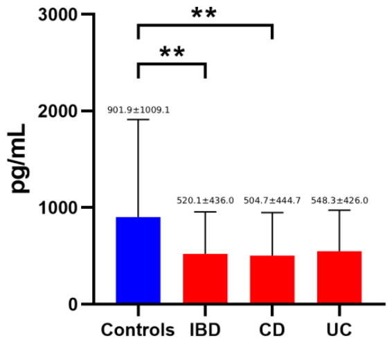
Figure 2.
DKK-1 levels in controls and patients with inflammatory bowel disease (IBD), including Crohn’s disease (CD) and ulcerative colitis (UC). ***, p<0.001.
Figure 2.
DKK-1 levels in controls and patients with inflammatory bowel disease (IBD), including Crohn’s disease (CD) and ulcerative colitis (UC). ***, p<0.001.
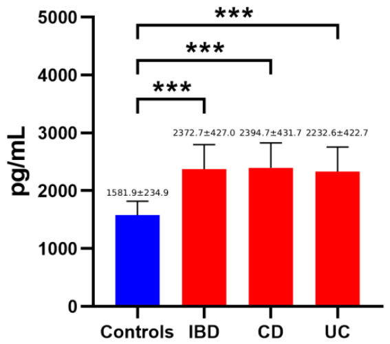
Figure 3.
Periostin levels in controls and patients with inflammatory bowel disease (IBD), including Crohn’s disease (CD) and ulcerative colitis (UC). *, p<0.05; **, p<0.01; ***, p<0.001.
Figure 3.
Periostin levels in controls and patients with inflammatory bowel disease (IBD), including Crohn’s disease (CD) and ulcerative colitis (UC). *, p<0.05; **, p<0.01; ***, p<0.001.
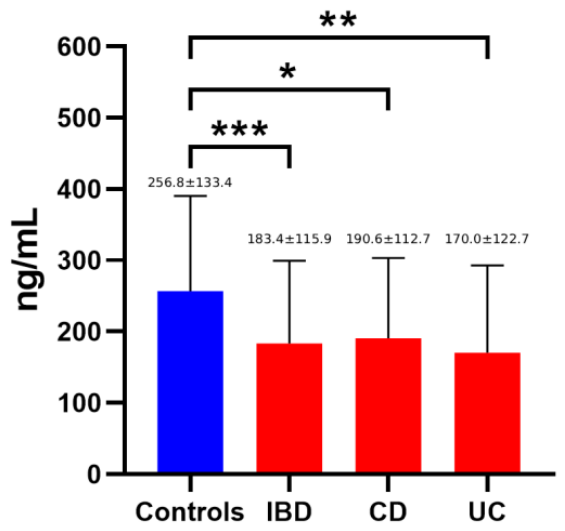

Table 1.
Baseline characteristics and clinical parameters of controls and patients with IBD.
| Parameter | Controls (n=73) |
IBD Patients (n=89) |
p-value |
|---|---|---|---|
| Age (years) | 42 [29-54] | 39 [28-54] | 0.672 |
| Sex (male/female) | 41 / 32 | 57 / 32 | 0.195 |
| Vitamin D supplementation (%) | 0 | 7.9 | p<0.05 |
| Systemic corticosteroids | 0 | 20.2 | p<0.001 |
| Systemic corticosteroids (excluding budesonide) | 0 | 15.7 | p<0.001 |
| CRP | 2.06±3.04 | 19.07±31.70 | p<0.001 |
| ESR | 6.84±7.49 | 25.49±21.54 | p<0.001 |
Table 2.
Demographic data of the study population.
| Parameter | Controls (n=73) |
CD Patients (n=58) |
UC Patients (n=31) |
||
|---|---|---|---|---|---|
| Age (years) | 41.3 ± 15.0 | 39.7 ± 16.4 | 44.2 ± 15.3 | ||
| Sex (male/female) | 41 / 32 | 36 / 22 | 21 / 10 | ||
| Disease duration, median (IQR) (years) | - | 9.5 [15.3 – 0.8] | 3 [13.0 – 0.3] | ||
| Smokers (%) | 30 (41%) | 26 (45%) | 1 (3%) | ||
| Treatment (%) | - | 5-ASA | 30 (51%) | 5-ASA | 29 (94%) |
| Corticosteroids: | 39 (67%) | Corticosteroids: | 23 (74%) | ||
| Immunomodulators: | Immunomodulators: | ||||
| Azathioprine | 27 (47%) | Azathioprine | 6 (19%) | ||
| Mercaptopurine | 11 (19%) | Mercaptopurine | 0 (0%) | ||
| Anti-TNF | Anti-TNF | ||||
| Infliximab | 26 (45%) | Infliximab | 11 (35%) | ||
| Adalimumab | 17 (29%) | Adalimumab | 4 (13%) | ||
| Golinumab | 0 (0%) | Golinumab | 1 (3%) | ||
| IRAs | 5 (9%) | IRAs | 7 (23%) | ||
| IL-12/23 antagonists | 14 (24%) | IL-12/23 antagonists | 1 (3%) | ||
| JAK inhibitor | 2 (4%) | JAK inhibitor | 1 (3%) | ||
| Localization (%) | - | L1 L2 L3 |
24 (42.1%) 2 (3.5%) 32 (54%) |
E1 E2 E3 |
1 (3%) 6 (19%) 24 (78%) |
| EIMs (%) | - | None Joint pain Skin PSC |
25 (43%) 25 (43%) 7 (12%) 1 (2%) |
None Joint pain Skin PSC |
18 (58%) 9 (29%) 3 (10%) 1 (3%) |
Table 3.
Sclerostin levels in controls, patients with IBD, CD and UC patients and their correlation with factors possibly affecting its levels.
Table 3.
Sclerostin levels in controls, patients with IBD, CD and UC patients and their correlation with factors possibly affecting its levels.
| Controls | IBD | CD | UC | ||||||
|---|---|---|---|---|---|---|---|---|---|
| R | p-value | R | p-value | R | p-value | R | p-value | ||
| Age | 0.435 | <0.001 | 0.301 | <0.010 | 0.396 | 0.003 | 0.074 | 0.705 | |
| BMI | 0.053 | 0.667 | 0.040 | 0.727 | 0.226 | 0.108 | -0.250 | 0.218 | |
| ESR | -0.066 | 0.594 | 0.333 | 0.003 | 0.299 | 0.138 | 0.282 | 0.154 | |
| CRP | 0.197 | 0.108 | 0.211 | 0.058 | 0.77 | 0.696 | 0.345 | 0.078 | |
| HBI | 0.275 | 0.048 | |||||||
| Partial Mayo Score | 0.353 | 0.055 | |||||||
| Levels | p-value | Levels | p-value | Levels | p-value | Levels | p-value | ||
|
Sex |
Male Female |
911.5 ± 1265.3 887.5 ± 400.2 |
0.924 |
515.1 ± 453.1 528.0 ±414.8 |
0.897 |
525.4 ± 531.1 475.5 ± 291.9 |
0.692 |
498.3 ± 297.3 643.4 ±610.3 |
0.393 |
|
Vit D supplementation |
Yes No |
891.9 ± 986.4 485.4 ± 339.0 |
0.017 |
1003.1 ± 1184.3 452.8 ± 265.1 |
0.007 |
614.2 ± 106.3 543.5 ± 441.2 |
0.826 |
||
|
Systemic corticosteroid treatment |
Yes No |
824.3 ± 824.2 467.6 ± 309.5 |
0.008 |
986.4 ± 1007.2 431.4 ± 227.3 |
0.001 |
602.2 ± 488.6 537.1 ± 422.6 |
0.762 |
||
|
Smoking |
Active Never |
1007.6 ± 1334.7 823.3 ± 683.7 |
0.460 |
566.4 ± 574.1 510.0 ± 373.6 |
0.607 |
592.3 ± 621.5 463.5 ± 280.6 |
0.328 |
430.1 ± 179.9 568.7 ± 465.7 |
0.567 |
A multivariable linear regression analysis was performed: a) to evaluate the independent effects of age and IBD status on sclerostin levels. The overall model was significant (F = 4.791, p < .01, R2 = .061). b) to identify independent predictors of sclerostin levels in patients with IBD. The overall model was significant (F = 3.629, p < .01, R2 = .162). In the Crohn’s disease subgroup to identify independent predictors of sclerostin levels. The overall model was significant (F = 5.918, p < .001, R2 = .335).
Table 4.
DKK-1 levels in controls, patients with IBD, CD and UC patients and their correlation with factors possibly affecting its levels.
Table 4.
DKK-1 levels in controls, patients with IBD, CD and UC patients and their correlation with factors possibly affecting its levels.
| Controls | IBD | CD | UC | ||||||
|---|---|---|---|---|---|---|---|---|---|
| R | p-value | R | p-value | R | p-value | R | p-value | ||
| Age | 0.035 | 0.775 | 0.118 | 0.290 | 0.240 | 0.084 | -0.058 | 0.765 | |
| BMI | 0.364 | 0.002 | 0.228 | 0.044 | 0.269 | 0.054 | 0.215 | 0.291 | |
| ESR | 0.285 | 0.010 | 0.123 | 0.391 | 0.467 | 0.011 | |||
| CRP | 0.251 | 0,023 | 0.193 | 0.166 | 0.401 | 0.031 | |||
| HBI | 0.298 | 0.032 | |||||||
| Partial Mayo Score | 0.169 | 0.381 | |||||||
| Levels | p-value | Levels | p-value | Levels | p-value | Levels | p-value | ||
|
Sex |
Male Female |
1617.3 ± 252.8 1528.3 ± 197.6 |
0.128 |
2357.7 ± 396.9 2396.2 ± 475.9 |
0.693 |
2325.9 ± 347.3 2491.6 ± 522.0, |
0.171 |
2186.3 ± 270.2 2409.5 ± 472.6 |
0.181 |
|
Vit D supplementation |
Yes No |
2437.0 ± 485.3 2366.7 ± 424.3 |
0.680 |
2563.7 ± 340.7 2377.2 ± 486.5 |
0.363 |
2120.5 ± 817 2348.3 ± 403.7 |
0.472 |
||
|
Systemic corticosteroid treatment |
Yes No |
2497.5 ± 200.7 2351.3 ± 452.2 |
0.274 |
2498.8 ± 232.3 2378.9 ± 454.2 |
0.499 |
2495.7 ± 172.6 2298.6 ± 453.2 |
0.352 |
||
|
Smoking |
Active Never |
1563.9 ± 270.2 1595.5 ± 207.7 |
0.587 |
2462.1 ± 356.8 2333.6 ± 451.7 |
0.212 |
2507.4 ± 335.4 2329.6 ± 495.9 |
0.162 |
2224.1 ± 422.8 2338.1 ± 437.6 |
0.633 |
A multivariable linear regression analysis was performed to identify independent predictors of DKK-1 levels in patients with IBD. The overall model was significant (F = 4.350, p < .05, R2 = .102).
Table 5.
Periostin levels in controls, patients with IBD, CD and UC patients and their correlation with factors possibly affecting its levels.
Table 5.
Periostin levels in controls, patients with IBD, CD and UC patients and their correlation with factors possibly affecting its levels.
| Controls | IBD | CD | UC | ||||||
|---|---|---|---|---|---|---|---|---|---|
| R | p-value | R | p-value | R | p-value | R | p-value | ||
| Age | 0.104 | 0.399 | 0.154 | 0.168 | 0.186 | 0.181 | 0.186 | 0.181 | |
| BMI | -0.275 | 0.023 | -0.121 | 0.291 | -0.064 | 0.652 | -0.064 | 0.652 | |
| ESR | -0.130 | 0.251 | -0.128 | 0.370 | -0.202 | 0.292 | |||
| CRP | -0.219 | 0.048 | -0.162 | 0.247 | -0.106 | 0.585 | |||
| HBI | 0.158 | 0.262 | |||||||
| Partial Mayo Score | -0.227 | 0.146 | |||||||
| Levels | p-value | Levels | p-value | Levels | p-value | Levels | p-value | ||
|
Sex |
Male Female |
231.7 ± 18.6 295.0 ± 28.3 |
0.055 |
145.1 ± 76.1 243.1 ± 141.2 |
<0.001 |
158.9 ± 88.1 235.4 ± 131.4 |
0.014 | 122.5 ± 50.5 260.3 ± 166.9 |
0.014 |
|
Vit D supplementation |
Yes No |
199.4 ± 166.3 181.8 ± 111.6 |
0.705 |
120.0 ± 28.9 197.9 ± 115.7 |
0.143 |
397.6 ± 229.1 153.1 ± 99.5 |
0.004 |
||
| Systemic corticosteroid treatment |
Yes No |
159.8 ± 88.6 187.4 ± 120.1 |
0.451 |
176.5 ± 112.4 192.8 ± 113.8 |
0.726 |
136.5 ± 38.5 176.9 ± 133.3 |
0.512 |
||
|
Smoking |
Active Never |
281.7 ± 118.6 238.3 ± 142.2 |
0.186 |
173.7 ± 121.7 188.5 ± 115.6 |
0.608 |
180.6 ± 123.6 197.3 ± 104.9 |
0.609 |
137.6 ± 120.9 177.3 ± 129.2 |
0.563 |
A multivariable linear regression analysis was performed: a) to identify independent predictors of periostin levels. IBD status, sex, and BMI were independent predictors. The model was significant (F = 14.033, p < .001, R2 = .229). b) within the IBD cohort to identify independent predictors of periostin levels. The overall model was significant (F = 8.306, p < .001, R2 = .174). c) in the UC subgroup to identify independent predictors of periostin levels. The overall model was significant (F = 13.757, p < .001, R2 = .514).
Disclaimer/Publisher’s Note: The statements, opinions and data contained in all publications are solely those of the individual author(s) and contributor(s) and not of MDPI and/or the editor(s). MDPI and/or the editor(s) disclaim responsibility for any injury to people or property resulting from any ideas, methods, instructions or products referred to in the content. |
© 2026 by the authors. Licensee MDPI, Basel, Switzerland. This article is an open access article distributed under the terms and conditions of the Creative Commons Attribution (CC BY) license (http://creativecommons.org/licenses/by/4.0/).
Copyright: This open access article is published under a Creative Commons CC BY 4.0 license, which permit the free download, distribution, and reuse, provided that the author and preprint are cited in any reuse.