

Submitted:
16 October 2026
Posted:
20 October 2026
You are already at the latest version
Abstract
Objective: To characterize the HBB mutation spectrum in a contemporary Bulgarian pediatric cohort with transfusion-dependent β-thalassemia (TDT) and to explore genotype–phenotype relationships, including organ-specific iron loading by magnetic resonance imaging (MRI) T2*. Materials and Methods: We retrospectively analyzed 37 children and adolescents with TDT receiving regular red-cell transfusions and iron chelation. HBB variants identified by Sanger sequencing and multiplex ligation-dependent probe amplification were classified as β0, β+, or β++. Patients with two identified alleles were grouped by predicted genotype severity. Pretransfusion hemoglobin, serum ferritin, and cardiac, hepatic, and pancreatic MRI T2* were evaluated. All comparisons were exploratory. Results: Molecular characterization was available in 28 of 37 patients: 26 had a complete two-allele genotype and 2 had a single identified allele. Among these 28 genetically informative patients, 54 alleles were identified. IVS-I-110 predominated (18/54; 33.3%), followed by IVS-I-6 (13.0%), IVS-II-745 (13.0%), and FS5 (11.1%). Pretransfusion hemoglobin and serum ferritin did not differ significantly among genotype-severity groups (p=0.208 and p=0.289). Hepatic T2* was <6.3 ms in 9/14 patients and cardiac T2* was <20 ms in 2/19. Pancreatic T2* was ≤2.4 ms in all 7 patients examined, a small and possibly selected subgroup. Conclusion: The mutation spectrum closely reflects the established Bulgarian profile. Under regular transfusion therapy, broad genotype-severity categories were not reflected by pretransfusion hemoglobin or serum ferritin, whereas MRI demonstrated organ-specific heterogeneity. The pancreatic findings are preliminary and require prospective confirmation.
Keywords:
β-thalassemia
; HBB
; genotype–phenotype correlation
; transfusion-dependent thalassemia
; iron overload
; MRI T2*
; pediatric
1. Introduction
Transfusion-dependent β-thalassemia (TDT) is among the most intensively managed inherited anemias. Pathogenic variants in the HBB gene reduce or abolish β-globin synthesis, resulting in ineffective erythropoiesis, chronic hemolytic anemia, and, in the most severe phenotype, lifelong dependence on red-cell transfusion. Contemporary transfusion protocols, iron chelation, and systematic surveillance for organ complications have substantially changed the natural history of TDT and shifted the clinical focus toward prevention of treatment-related morbidity and preservation of long-term quality of life [1,2,3].
Bulgaria lies at the northern margin of the traditional Mediterranean thalassemia region and has a well-characterized β-thalassemia population. The estimated carrier frequency is approximately 3%, while around 270 patients with TDT are registered nationally [4,5]. Molecular studies extending over more than three decades have demonstrated a characteristic and remarkably stable mutation spectrum dominated by IVS-I-110, followed by codon 39, IVS-I-6, IVS-II-745, IVS-I-1, and FS5 [4,6,7]. Specialized multidisciplinary care, access to the major iron chelators, and centralized MRI-based assessment of tissue iron are established components of contemporary TDT management in the country [5].
The primary HBB genotype remains an important determinant of intrinsic disease severity because the amount of residual β-globin production varies substantially among β0, β+, and milder β++ alleles [8]. However, genotype does not operate in isolation. Studies from Azerbaijan, Thailand, Syria, Malaysia, India, China, and more recently Iraq demonstrate substantial population-specific differences in mutation spectra and considerable phenotypic overlap among molecular classes [9,10,11,12,13,14,15,16,17,18,19]. Consequently, genotype–phenotype interpretation is most informative when anchored in the genetic background of the population being studied.
At the same time, molecular diagnosis itself is evolving. Standardized interpretation of hemoglobinopathy variants, improved in silico assessment, next-generation sequencing, and third-generation sequencing increasingly allow characterization of variants that may be missed by restricted mutation panels [20,21,22,23].
In TDT, iron overload represents a second, increasingly treatment-dependent phenotype. Transfusional iron is distributed unevenly among organs, and serum ferritin cannot reliably describe tissue-specific iron burden [24,25,26,27]. MRI has therefore become central to non-invasive assessment of iron loading. Clinically relevant tissue iron may appear early in chronically transfused children, while myocardial iron accumulation and cardiac morbidity represent later and particularly important complications [28,29,30]. The liver remains a major target of transfusional iron toxicity and an important site for longitudinal assessment of chelation efficacy [25,31].
Contemporary pediatric data integrating HBB genotype with multi-organ MRI assessment remain scarce in Central and Southeastern Europe. We therefore characterized the mutation spectrum in a Bulgarian pediatric TDT cohort and, in an exploratory analysis, examined whether predicted genotype severity was associated with pretransfusion hemoglobin, serum ferritin, or organ-specific iron loading.
2. Materials and Methods
2.1. Study Design and Patients
This was a single-center, retrospective, exploratory cohort study of children and adolescents with TDT followed at the Specialized Hematology Hospital, Sofia, Bulgaria, between 2022 and 2025. All patients aged <19 years with molecularly and/or hematologically confirmed β-thalassemia who were receiving regular transfusions and were under active follow-up during the study period were eligible. Patients with non-transfusion-dependent thalassemia and those without an available clinical record were excluded.
The study was conducted in accordance with the Declaration of Helsinki and was approved by the Ethics Committee for Scientific Research of the Specialized Hospital for Active Treatment of Hematological Diseases, Sofia, Bulgaria (Decision No. 19, 21 August 2026). The study was retrospective and non-interventional and used pseudonymized routinely collected clinical data. Written informed consent for genetic testing was obtained from the patients and/or their legal guardians as part of routine clinical care; where applicable, the standard consent forms included authorization for the secondary use of clinical and genetic data in future research and registry-based studies.
2.2. Clinical Data, Transfusion, and Chelation
Demographic, clinical, laboratory, genetic, and imaging data were extracted from the institutional clinical registry and verified against individual medical records. Pretransfusion hemoglobin was calculated as the arithmetic mean of the available pretransfusion measurements during the study period, and serum ferritin analogously from available measurements. The most recent available cardiac, hepatic, and pancreatic MRI T2* measurements were used for the imaging analysis.
All patients received regular red-cell transfusions, generally every 2–4 weeks, with a target pretransfusion hemoglobin of approximately 90–100 g/L. Iron chelation included deferasirox, deferiprone, and deferoxamine, administered alone or in combination according to iron burden and tolerance. Chelation efficacy was monitored by serial serum ferritin and tissue-iron MRI. Cumulative transfusion volume and individual adherence were not systematically available and were not analyzed.
2.3. Molecular Analysis and Genotype Classification
Molecular results were retrieved from the available institutional records. HBB analysis was performed by direct Sanger sequencing, complemented by multiplex ligation-dependent probe amplification (MLPA) for large deletions or duplications. Next-generation and third-generation sequencing were not routinely used. Some patients therefore had incomplete molecular characterization because earlier testing had been more limited.
Identified variants were reported using HGVS nomenclature together with their traditional designation and classified according to their expected effect on β-globin synthesis: β0 (IVS-I-1, IVS-II-1, codon 39, codons 82/83, FS5, FS6, FS8); β+ (IVS-I-110, IVS-II-745); and β++ (IVS-I-6, Hb Knossos). For the exploratory genotype–phenotype analysis, patients with two identified alleles were categorized as severe (β0/β0), intermediate (β0/β+ or β0/β++), or mild (β+/β+, β+/β++, or β++/β++). Patients with only one identified allele or incomplete molecular characterization were excluded from between-group comparisons.
2.4. Mri T2* Assessment
Cardiac, hepatic, and pancreatic iron loading was assessed by MRI T2* at the national reference center, using a multi-echo gradient-echo sequence Cardiac T2* <20 ms was considered indicative of myocardial iron loading. Hepatic T2* <6.3 ms was considered evidence of significant hepatic iron accumulation according to the reference protocol used by the reporting center. No formal pancreatic T2* severity threshold was applied, and pancreatic values are reported descriptively.
2.5. Statistical Analysis
Categorical variables are presented as number and percentage. Continuous variables are reported as median and range and, where appropriate, as mean ± standard deviation. Differences among genotype-severity groups were evaluated with the Kruskal–Wallis test because of the small subgroup sizes and non-normal data distribution. A two-sided p-value <0.05 was considered statistically significant. Analyses were performed using SPSS v14. Given the limited number of complete genotypes and the smaller, non-identical MRI subsets, all genotype–phenotype and organ-iron comparisons were defined as exploratory and hypothesis-generating.
3. Results
3.1. Cohort Characteristics and Genetic Characterization
The cohort included 37 patients: 21 boys (56.8%) and 16 girls (43.2%). Median age was 10.5 years (range, 0.6–18.3 years), and mean age was 11.2 ± 4.3 years. The principal demographic, laboratory, and imaging characteristics are summarized in Table 1.
Molecular characterization was available in 28 of 37 patients: 26 had a complete two-allele genotype and 2 had a single identified allele. Of the remaining 9 patients, sequencing detected no HBB variant in 1, while molecular data were unavailable or testing had not been performed in 8. The 28 patients with at least one identified allele formed the genetically informative group for mutation-spectrum analysis; genotype-severity comparisons included only the 26 patients with two identified alleles. The 2 patients with a single identified allele had not undergone MLPA, so a second, possibly deletional, allele could not be excluded. MRI T2* data were available for different subsets: cardiac, n=19; hepatic, n=14; and pancreatic, n=7.
3.2. Mutation Spectrum
Among the 28 genetically informative patients, 54 HBB alleles were identified. IVS-I-110 was the dominant variant, accounting for 18/54 alleles (33.3%), followed by IVS-I-6 (7/54; 13.0%), IVS-II-745 (7/54; 13.0%), FS5 (6/54; 11.1%), and IVS-I-1 (5/54; 9.3%). IVS-II-1, codons 82/83, Hb Knossos, and codon 39 each accounted for 2 alleles (3.7%), while codon 29, FS8, and FS6 were each represented by a single allele (1.9%). The complete distribution is presented in Table 2 and Figure 1.
3.3. Genotype Distribution
A complete two-allele genotype was defined in 26 patients. The distribution of genotypes and of the categories with incomplete molecular data is shown in Table 3, and the complete genotypes are illustrated by predicted severity class in Figure 2. The most frequent complete genotype was β0/β+ (n=9), followed by β+/β+ (n=7).
3.4. Exploratory Genotype–phenotype Relationships
Among the 26 patients with a complete genotype, neither pretransfusion hemoglobin nor serum ferritin differed significantly among the predicted genotype-severity groups (p=0.208 and p=0.289, respectively). Age also did not differ significantly (p=0.144), although the β0/β0 group had the highest median age. Cardiac and hepatic T2* comparisons were based on very small subgroups, and pancreatic data were insufficient for between-group analysis. Detailed values are presented in Table 4 and Figure 3.
3.5. Organ-Specific Iron Loading
Serum ferritin exceeded 1000 ng/mL in 12/37 patients (32.4%). Hepatic MRI T2* was available in 14 patients: the median hepatic T2* was 4.2 ms (range, 0.2–28.3 ms), and 9/14 patients (64.3%) had a hepatic T2* <6.3 ms. Cardiac MRI T2* was available in 19 patients, of whom 2 (10.5%) had a value <20 ms; both were in the mild range (16.6 and 17.0 ms). Pancreatic MRI T2* was available in only 7 patients, in all of whom the value was ≤2.4 ms. Individual cardiac, hepatic, and pancreatic measurements are shown in Figure 4.
4. Discussion
This exploratory single-center study confirms that the HBB mutation spectrum in our pediatric TDT cohort closely reflects the established Bulgarian profile. Under regular transfusion therapy, genotype severity was not reflected by pretransfusion hemoglobin or serum ferritin. MRI showed marked tissue-specific differences in iron loading. However, the genotype–phenotype and organ-iron analyses were limited by small and unequal subgroups and should be regarded as hypothesis-generating.
IVS-I-110 accounted for one third of identified alleles, almost identical to the approximately 31% frequency reported in the broader Bulgarian molecular dataset [4,5]. IVS-I-6, IVS-II-745, IVS-I-1, and FS5 were likewise prominent, consistent with historical and contemporary Bulgarian studies [4,6,7]. This concordance is relevant because the present cohort was selected for a severe, transfusion-dependent pediatric phenotype, whereas previous national molecular data include a broader spectrum of affected individuals and carriers.
The main divergence was codon 39. Despite being the second most frequent variant in the wider Bulgarian carrier population, it accounted for only two alleles in our cohort [4,5]. Given the cohort size and its restriction to pediatric TDT, this finding should be considered cohort-specific rather than evidence of a changing national mutation spectrum.
Comparison with other populations demonstrates why local molecular data remain clinically relevant. The predominant mutations reported in Azerbaijan, Thailand, Syria, Malaysia, India, China, and Iraq differ substantially from the Bulgarian spectrum [9,10,11,12,13,14,15,16,17,18,19]. These studies also show considerable phenotypic variability among patients carrying apparently comparable molecular classes. HBB genotype is therefore a major biological determinant, but not a complete clinical prediction model.
The absence of significant differences in pretransfusion hemoglobin among genotype groups should not be interpreted as absence of a biological genotype effect. In established TDT, pretransfusion hemoglobin is deliberately treatment-controlled. Regular transfusion suppresses the hematologic consequences of ineffective erythropoiesis and narrows the phenotypic range that would otherwise separate severe from milder genotypes [1,3,8,10].
Serum ferritin is influenced by cumulative transfusion exposure, age, chelation efficacy and adherence, inflammation, and individual iron handling rather than by HBB genotype alone. Its overlap among genotype groups is therefore expected and reinforces its limited value as a stand-alone measure of tissue iron [24,26,27]. The present exploratory comparisons cannot exclude clinically relevant genotype effects, which would require a larger, modifier-aware, longitudinal study.
Primary HBB genotype represents only one level of phenotypic regulation. Co-inherited globin defects and modifiers of fetal hemoglobin production may substantially alter clinical expression [8,10]. The milder alleles represented in our cohort are particularly relevant in this context. Hb Knossos, for example, has been reported in compound states producing a thalassemia-intermedia phenotype [32]. Interpretation of mild or borderline β-globin abnormalities may be further complicated by erythroid regulators such as KLF1 and by variants with subtle hematologic expression [33,34].
These observations are also relevant to the molecular strategy. Targeted mutation panels remain efficient in populations with a limited number of predominant variants, but restricted testing may leave one allele unidentified. Contemporary sequencing can improve the characterization of uncommon point variants and structural abnormalities [20,21,22,23], while combined molecular approaches using gap-PCR and targeted sequencing can increase carrier detection and diagnostic yield [35,36]. This remains important in our cohort, in which 11 patients had incomplete, absent, or inconclusive molecular characterization.
The organ-iron findings show substantial tissue-specific discordance. Significant hepatic iron loading was present in almost two thirds of imaged patients, whereas myocardial iron was detected in only 2 of 19 and was mild in both.
Previous studies have shown that clinically relevant iron accumulation can occur from an early age in chronically transfused children and that serum ferritin cannot reliably predict individual organ burden [24,26,27,28,29]. Cardiac T2* remains crucial because myocardial iron is directly related to subsequent cardiac morbidity [28,29,30]. Our findings indicate that a reassuring cardiac T2* does not necessarily imply a low extra-cardiac iron burden.
The pancreatic findings require particular caution. Only seven patients underwent pancreatic MRI, and this subgroup may have been selected according to perceived clinical risk. The data therefore neither establish a pancreatic T2* threshold nor estimate the prevalence of pancreatic iron loading in the cohort, and they cannot demonstrate a temporal sequence of organ iron deposition. The observation is preliminary and hypothesis-generating, but it supports prospective assessment of pancreatic iron together with glucose metabolism in a larger pediatric cohort.
The Bulgarian care model provides an appropriate setting for this approach. Specialized follow-up, availability of modern chelation, and centralized MRI assessment provide the infrastructure for moving from conventional systemic markers toward complementary organ-specific monitoring [5,25,31].
Beyond individual patient management, a well-defined national mutation spectrum has direct implications for genetic counseling, carrier identification, and prevention. Accurate interpretation of routine hematologic findings remains a critical first step [37], particularly in individuals with borderline HbA2 values [38]. Reproductive carrier screening must be adapted to the population in which it is implemented [39], while modern molecular techniques increasingly allow accurate prenatal diagnosis even in families carrying uncommon variants [40].
The present study has several limitations. It is retrospective and single-center. Although updated molecular data provided complete two-allele genotypes for 26 of 37 patients, 11 still had incomplete, absent, or inconclusive molecular characterization. The two patients with a single identified allele had not undergone MLPA, so a second, possibly deletional, allele could remain undetected. The cohort also included one sibling pair, and the allele and genotype observations were therefore not fully independent. MRI data were available in smaller and non-identical subsets, particularly for pancreatic T2* (n=7), and imaging may have been performed according to clinical risk. The analysis did not account for cumulative transfusion volume, individual chelation regimen and adherence, splenectomy status, or known genetic modifiers, and MRI parameters were analyzed cross-sectionally. The genotype and organ-iron comparisons are consequently exploratory and underpowered.
The cohort nevertheless has two strengths. It represents a uniformly managed pediatric TDT population and integrates molecular data with cardiac, hepatic, and pancreatic MRI. This allows genotype and treated phenotype to be considered separately rather than assuming that one directly predicts the other.
5. Conclusions
The HBB mutation spectrum in Bulgarian children with TDT is dominated by IVS-I-110 and closely reflects the established national profile. Under regular transfusion therapy, broad genotype-severity categories did not discriminate pretransfusion hemoglobin or serum ferritin.
Multi-organ MRI showed clinically important heterogeneity, with frequent hepatic iron loading and pancreatic T2* values ≤2.4 ms in all seven patients examined, despite relative myocardial sparing. The pancreatic observation is preliminary and requires prospective confirmation. These findings support a model in which genotype defines intrinsic disease biology, while the phenotype observed during contemporary treatment is shaped by transfusion exposure, chelation, genetic modifiers, and organ-specific iron handling.
Larger multicenter longitudinal studies should determine whether pancreatic T2* adds clinically useful information to established ferritin, hepatic, and cardiac monitoring and whether it can contribute to earlier identification of patients at risk of iron-related metabolic morbidity.
Author Contributions
A.B.: conceptualization, methodology, and writing—original draft. B.M. and S.A.: data curation. I.M. and G.T.: formal analysis. A.B., I.M., and B.M.: writing—review and editing, supervision, and project administration. All authors have read and approved the final manuscript.
Funding
This research received no external funding.
Institutional Review Board Statement
The study was conducted in accordance with the Declaration of Helsinki and was approved by the Ethics Committee for Scientific Research of the Specialized Hospital for Active Treatment of Hematological Diseases, Sofia, Bulgaria (Decision No. 19, 21 August 2026).
Informed Consent Statement
Written informed consent was obtained for all genetic investigations as part of routine clinical care. Where applicable, the standard consent forms included authorization for the secondary use of clinical and genetic data in future research and registry-based studies.
Data Availability Statement
De-identified data supporting the findings of this study are available from the corresponding author upon reasonable request, subject to applicable institutional and legal requirements.
Conflicts of Interest
The authors declare no conflicts of interest.
Abbreviations
HBB, beta-globin gene; MRI, magnetic resonance imaging; T2*, effective transverse relaxation time; TDT, transfusion-dependent β-thalassemia; MLPA, multiplex ligation-dependent probe amplification.
References
- Taher, A.T.; Weatherall, D.J.; Cappellini, M.D. Thalassaemia. Lancet 2018, 391(10116), 155–167. [Google Scholar] [CrossRef] [PubMed]
- Harteveld, C.L.; Achour, A.; Arkesteijn, S.J.; et al. The hemoglobinopathies, molecular disease mechanisms and diagnostics. Int. J. Lab Hematol. 2022, 44 (Suppl 1), 28–36. [Google Scholar] [CrossRef] [PubMed]
- Cappellini, M.D.; Farmakis, D.; Porter, J.; Taher, A. (Eds.) Guidelines for the Management of Transfusion Dependent Thalassaemia (TDT), 4th ed.; Thalassaemia International Federation: Nicosia, 2023. [Google Scholar]
- Andonova, S.; Bradinova, I.; Savov, A. Genetic diagnosis of beta-thalassemia in Bulgaria - what we learned from our patients. Rare Dis. Orphan Drugs 2023, 14(1), 32–38. [Google Scholar]
- Banchev, A.; Stoyanova, D.; Sapunarova, K. Thalassemia and hemoglobinopathies in Bulgaria. Pediatriya 2025, 64(3), 7–10. [Google Scholar]
- Kalaydjieva, L.; Eigel, A.; Horst, J. The molecular basis of beta-thalassaemia in Bulgaria. J. Med. Genet. 1989, 26(10), 614–618. [Google Scholar] [CrossRef] [PubMed]
- Petkov, G.H.; Efremov, G.D. Molecular basis of beta-thalassemia and other hemoglobinopathies in Bulgaria: an update. Hemoglobin 2007, 31(2), 225–232. [Google Scholar] [CrossRef] [PubMed]
- Danjou, F.; Anni, F.; Galanello, R. Beta-thalassemia: from genotype to phenotype. Haematologica 2011, 96(11), 1573–1575. [Google Scholar] [CrossRef] [PubMed]
- Asadov, C.; Abdulalimov, E.; Mammadova, T.; Gafarova, S.; Guliyeva, Y.; Aliyeva, G. Genotype-phenotype correlations of β-thalassemia mutations in an Azerbaijani population. Turk. J. Hematol. 2017, 34, 258–263. [Google Scholar] [CrossRef] [PubMed]
- Traivaree, C.; Monsereenusorn, C.; Rujkijyanont, P.; Prasertsin, W.; Boonyawat, B. Genotype-phenotype correlation among beta-thalassemia and beta-thalassemia/HbE disease in Thai children: predictable clinical spectrum using genotypic analysis. J. Blood Med. 2018, 9, 35–41. [Google Scholar] [CrossRef] [PubMed]
- Shoujaa, A.; Moasses, F.; Mukhalalaty, Y.; Murad, H.; Al-Quobaili, F. Genotype/phenotype correlation of β-thalassemia in Syrian patients: a cross-sectional study. Hemoglobin 2020, 44(1), 42–46. [Google Scholar] [CrossRef] [PubMed]
- Yasin, N.M.; Abdul Hamid, F.S.; Hassan, S.; Sudin, A.; Yassim, H.; Mohd Sahid, E.N.; Mat Yusoff, Y.; Esa, E.; Saleem, M. Molecular and hematological studies in a cohort of beta zero South East Asia deletion (β0-thal SEA) from Malaysian perspective. Front Pediatr. 2022, 10, 974496. [Google Scholar] [CrossRef] [PubMed]
- Samanta, A.; Bhattacharyya, N. Genotype-phenotype correlation of β-thalassemia in the Indian college student population: insights and challenges in screening and molecular diagnosis. Biosci. Biotechnol. Res. Asia 2024, 21(3), 1043–1053. [Google Scholar] [CrossRef]
- Huang, N.; Wang, Y.; Huang, H.; Chen, Z.; Zhang, Z. Genotype-phenotype correlation analysis of patients with thalassemia in Quanzhou City, southeast of China. Heliyon 2024, 10, e40144. [Google Scholar] [CrossRef] [PubMed]
- Singha, K.; Pansuwan, A.; Fucharoen, G.; Fucharoen, S. Genotype-phenotype correlation of seven known and novel β-globin gene variants. Int. J. Mol. Sci. 2025, 26, 8872. [Google Scholar] [CrossRef] [PubMed]
- Jasim, H.A.; Mohammed, H.A.; Al-Omairi, R. Genotype-phenotype correlation of β-globin mutations and hematological parameters in Iraqi β-thalassemia patients. Cell Physiol. Biochem. 2026, 60, 408–416. [Google Scholar] [CrossRef] [PubMed]
- Khan, A.M.; Al-Sulaiti, A.M.; Younes, S.; Yassin, M.; Zayed, H. The spectrum of beta-thalassemia mutations in the 22 Arab countries: a systematic review. Expert Rev. Hematol. 2021, 14(1), 109–122. [Google Scholar] [CrossRef] [PubMed]
- Suali, L.; Mohammad Salih, F.A.; Ibrahim, M.Y.; et al. Genotype-phenotype study of β-thalassemia patients in Sabah. Hemoglobin 2022, 46(6), 317–324. [Google Scholar] [CrossRef] [PubMed]
- Abdullah, U.Y.; Ibrahim, H.M.; Mahmud, N.B.; et al. Genotype-phenotype correlation of β-thalassemia in Malaysian population: toward effective genetic counseling. Hemoglobin 2020, 44(3), 184–189. [Google Scholar] [CrossRef] [PubMed]
- Kountouris, P.; Stephanou, C.; Lederer, C.W.; Traeger-Synodinos, J.; Bento, C.; Harteveld, C.L.; et al. Adapting the ACMG/AMP variant classification framework: a perspective from the ClinGen Hemoglobinopathy Variant Curation Expert Panel. Hum. Mutat. 2022, 43, 1089–1096. [Google Scholar] [CrossRef] [PubMed]
- Tamana, S.; Xenophontos, M.; Minaidou, A.; Stephanou, C.; Harteveld, C.L.; Bento, C.; et al. Evaluation of in silico predictors on short nucleotide variants in HBA1, HBA2, and HBB associated with haemoglobinopathies. eLife 2022, 11. [Google Scholar] [CrossRef] [PubMed]
- Mahabhol, C.; Yothindamrongkul, L.; Nuntanajaroenkul, N.; Nawasod, P.; Tepakhan, W.; Karnpean, R.; et al. The validation of whole β-globin gene sequencing for detecting β-thalassemia mutations found in Thailand using next-generation sequencing. Ann. Hematol. 2025, 104, 2653–2661. [Google Scholar] [PubMed]
- Liu, Q.; Chen, Q.; Zhang, Z.; Peng, S.; Liu, J.; Pang, J.; et al. Identification of rare thalassemia variants using third-generation sequencing. Front Genet. 2023, 13, 1076035. [Google Scholar] [CrossRef] [PubMed]
- Hsu, C.C.; Senussi, N.H.; Fertrin, K.Y.; et al. Iron overload disorders. Hepatol. Commun. 2022, 6, 1842–1854. [Google Scholar] [CrossRef] [PubMed]
- Georgiev, P.G.; Sapunarova, K.G.; Goranova-Marinova, V.S.; et al. Reduction of liver iron load in adult patients with β-thalassemia major treated with modern chelation modalities. Folia Med. 2020, 62(2), 265–270. [Google Scholar] [CrossRef] [PubMed]
- Porter, J.B.; Elalfy, M.; Taher, A.; et al. Limitations of serum ferritin to predict liver iron concentration responses to deferasirox therapy in patients with transfusion-dependent thalassaemia. Eur. J. Haematol. 2017, 98(3), 280–288. [Google Scholar] [CrossRef] [PubMed]
- Jarisch, A.; Salzmann-Manrique, E.; Cario, H.; et al. Serum ferritin is not a reliable predictor to determine iron overload in thalassemia major patients post-hematopoietic stem cell transplantation. Eur. J. Haematol. 2018, 101(6), 791–797. [Google Scholar] [CrossRef] [PubMed]
- Yang, G.; Liu, R.; Peng, P.; et al. How early can myocardial iron overload occur in beta thalassemia major? PLoS ONE 2014, 9. [Google Scholar] [CrossRef] [PubMed]
- Berdoukas, V.; Nord, A.; Carson, S.; et al. Tissue iron evaluation in chronically transfused children shows significant levels of iron loading at a very young age. Am. J. Hematol. 2013, 88. [Google Scholar] [CrossRef] [PubMed]
- Marsella, M.; Borgna-Pignatti, C.; Meloni, A.; et al. Cardiac iron and cardiac disease in males and females with transfusion-dependent thalassemia major: a T2* magnetic resonance imaging study. Haematologica 2011, 96, 515–520. [Google Scholar] [CrossRef] [PubMed]
- Kofinova, D. Liver complications in β-thalassemia. Rare Dis. Orphan Drugs 2023, 14(1), 5–8. [Google Scholar]
- Moassas, F.; Nweder, M.S.; Murad, H. Hb Knossos (HBB:c.82G>T), β-globin CD5 (-CT) (HBB:c.17_18delCT) and δ-globin CD59 (-A) (HBD:c.179delA) mutations in a Syrian patient with β-thalassemia intermedia. BMC Pediatr. 2019, 19, 61. [Google Scholar] [PubMed]
- Srivorakun, H.; Thawinan, W.; Fucharoen, G.; Sanchaisuriya, K.; Fucharoen, S. Thalassemia and erythroid transcription factor KLF1 mutations associated with borderline hemoglobin A2 in the Thai population. Arch. Med. Sci. 2022, 18, 112–120. [Google Scholar] [PubMed]
- Long, J.; Liu, E. Identification of the β-thalassemia allele β-50 and analysis of hematology data of carriers in a southern Chinese population. Ann. Hum. Genet. 2022, 86, 63–70. [Google Scholar] [CrossRef] [PubMed]
- Lam, T.T.; Nguyen, D.T.; Le, Q.T.; et al. Combined gap-polymerase chain reaction and targeted next-generation sequencing improve α- and β-thalassemia carrier screening in pregnant women in Vietnam. Hemoglobin 2022, 46(4), 233–239. [Google Scholar] [CrossRef] [PubMed]
- Wichian, P.; Yamsri, S.; Sanchaisuriya, K.; Fucharoen, S. Direct amplification of whole blood and amniotic fluid specimens for prenatal and postnatal diagnosis of Hb E-β0-thalassemia diseases. Lab Med. 2021, 52(5), 460–468. [Google Scholar] [CrossRef] [PubMed]
- Rangelova, M.; Petkova, Z. The interpretation of laboratory results - a key factor in the prevention of severe hemoglobinopathies. Rare Dis. Orphan Drugs 2023, 14(1), 25–31. [Google Scholar]
- Colaco, S.; Nadkarni, A. Borderline HbA2 levels: dilemma in diagnosis of beta-thalassemia carriers. Mutat. Res. Rev. Mutat. Res. 2021, 788, 108387. [Google Scholar] [CrossRef] [PubMed]
- Delatycki, M.B.; Alkuraya, F.; Archibald, A.; et al. International perspectives on the implementation of reproductive carrier screening. Prenat. Diagn. 2020, 40(3), 301–310. [Google Scholar] [CrossRef] [PubMed]
- Jiang, F.; Zhou, J.; Zuo, L.; et al. Utilization of multiple genetic methods for prenatal diagnosis of rare thalassemia variants. Front Genet. 2023, 14, 1208102. [Google Scholar] [CrossRef] [PubMed]
Figure 1.
Allele frequencies of the identified HBB variants in 28 genetically informative patients (54 alleles), classified according to predicted functional effect as β0, β+, or β++.
Figure 1.
Allele frequencies of the identified HBB variants in 28 genetically informative patients (54 alleles), classified according to predicted functional effect as β0, β+, or β++.
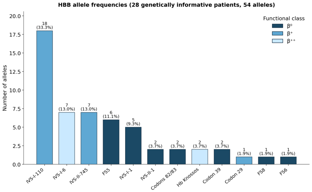
Figure 2.
Distribution of the 26 complete HBB genotypes according to predicted genotype-severity group. Patients with a single identified allele or incomplete molecular characterization are not included.
Figure 2.
Distribution of the 26 complete HBB genotypes according to predicted genotype-severity group. Patients with a single identified allele or incomplete molecular characterization are not included.
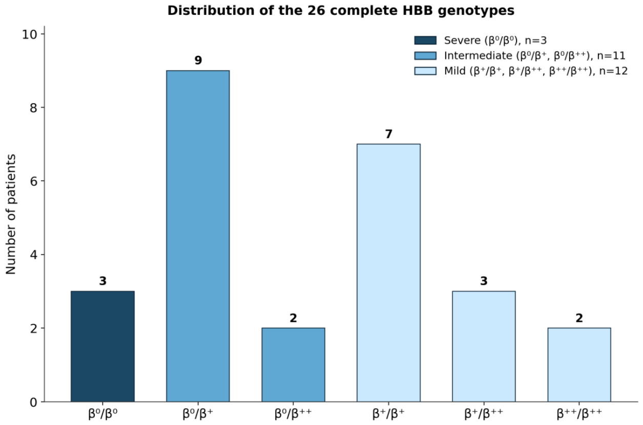
Figure 3.
Age, pretransfusion hemoglobin, and serum ferritin according to predicted genotype-severity group. Each point represents an individual patient; horizontal bars indicate medians. Kruskal–Wallis p-values are shown.
Figure 3.
Age, pretransfusion hemoglobin, and serum ferritin according to predicted genotype-severity group. Each point represents an individual patient; horizontal bars indicate medians. Kruskal–Wallis p-values are shown.
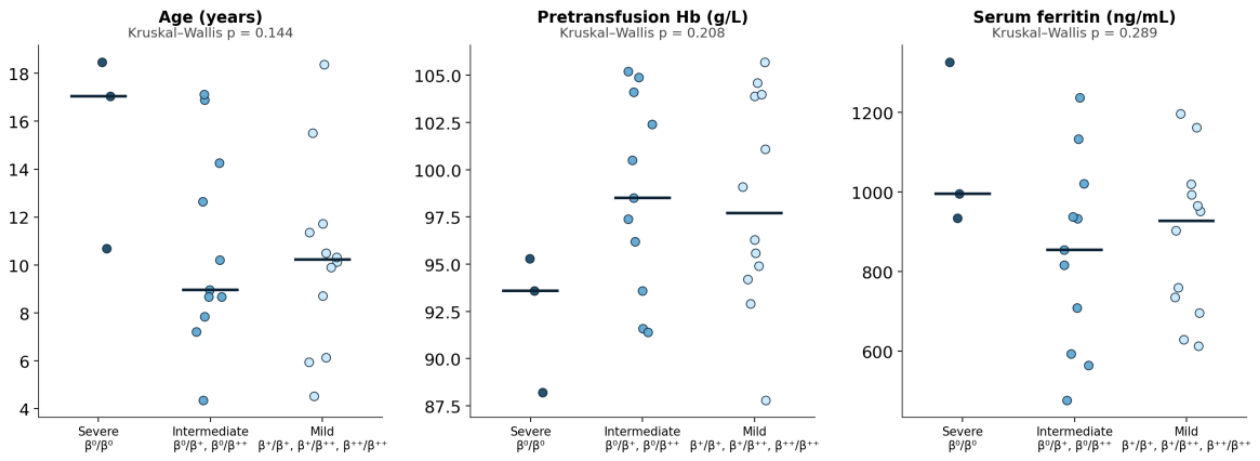
Figure 4.
Organ-specific iron loading assessed by MRI T2*. Individual cardiac, hepatic, and pancreatic T2* measurements are shown on a logarithmic scale. The dashed 20 ms threshold applies to cardiac T2* only.
Figure 4.
Organ-specific iron loading assessed by MRI T2*. Individual cardiac, hepatic, and pancreatic T2* measurements are shown on a logarithmic scale. The dashed 20 ms threshold applies to cardiac T2* only.
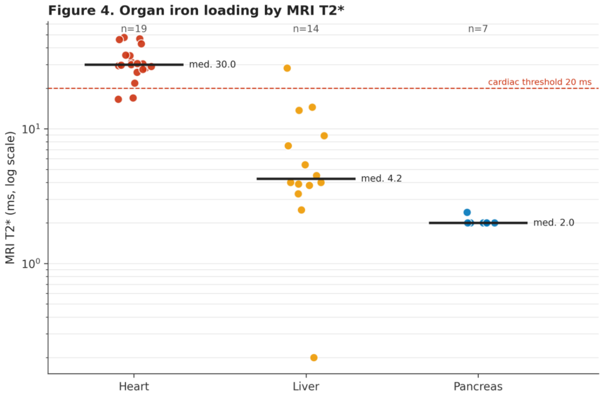

Table 1.
Demographic, laboratory, and imaging characteristics of the cohort.
| Parameter | Value |
| Number of patients | 37 |
| Sex, male/female | 21 (56.8%) / 16 (43.2%) |
| Age, years, median (range) | 10.5 (0.6–18.3) |
| Age, years, mean ± SD | 11.2 ± 4.3 |
| Pretransfusion hemoglobin, g/L, median (range) | 96.2 (84–108) |
| Serum ferritin, ng/mL, median (range) | 934 (42–1326) |
| Ferritin >1000 ng/mL, n (%) | 12 (32.4%) |
| Complete two-allele genotype, n | 26 |
| Genetically informative (≥1 allele), n | 28 |
| MRI T2* performed, heart/liver/pancreas | 19 / 14 / 7 |
| Cardiac iron, T2* <20 ms, n (%) | 2/19 (10.5%) |
| Hepatic T2*, ms, median (range) | 4.2 (0.2–28.3) |
| Hepatic T2* <6.3 ms, n (%) | 9/14 (64.3%) |
| Pancreatic T2*, ms | ≤2.4 ms in 7/7 examined |
Table 2.
Distribution and functional classification of identified HBB alleles.
| Mutation | HGVS nomenclature | Functional class | Alleles, n | % |
| IVS-I-110 | HBB:c.93-21G>A | β+ | 18 | 33.3 |
| IVS-I-6 | HBB:c.92+6T>C | β++ | 7 | 13.0 |
| IVS-II-745 | HBB:c.316-106C>G | β+ | 7 | 13.0 |
| FS5 | HBB:c.17_18delCT | β0 | 6 | 11.1 |
| IVS-I-1 | HBB:c.92+1G>A | β0 | 5 | 9.3 |
| IVS-II-1 | HBB:c.315+1G>A | β0 | 2 | 3.7 |
| Codons 82/83 | HBB:c.251delG | β0 | 2 | 3.7 |
| Hb Knossos | HBB:c.82G>T | β++ | 2 | 3.7 |
| Codon 39 | HBB:c.118C>T | β0 | 2 | 3.7 |
| Codon 29 | HBB:c.90C>T | β+ | 1 | 1.9 |
| FS8 | HBB:c.25_26delAA | β0 | 1 | 1.9 |
| FS6 | HBB:c.20delA | β0 | 1 | 1.9 |
Table 3.
Distribution of HBB genotypes in the study cohort.
| Genotype/category | n |
| β0/β+ | 9 |
| β+/β+ | 7 |
| β0/β0 | 3 |
| β+/β++ | 3 |
| β0/β++ | 2 |
| β++/β++ | 2 |
| Complete genotype, subtotal | 26 |
| Single identified allele | 2 |
| No mutation detected by available sequencing | 1 |
| No molecular genetic data available | 8 |
Table 4.
Phenotypic parameters according to predicted genotype-severity group.
| Parameter | Severe β0/β0 | Intermediate β0/β+ or β0/β++ | Mild β+/β+, β+/β++, or β++/β++ | p* |
| Patients, n | 3 | 11 | 12 | — |
| Age, years | 17.0 (10.7–18.5) | 9.0 (4.4–17.1) | 10.2 (4.5–18.4) | 0.144 |
| Pretransfusion Hb, g/L | 93.6 (88–95) | 98.5 (91–105) | 97.7 (88–106) | 0.208 |
| Ferritin, ng/mL | 995.7 (934–1326) | 854.6 (477–1236) | 926.8 (613–1196) | 0.289 |
| Cardiac T2*, ms | 21.8 (17.0–30.0); n=3 | 33.2 (16.6–46.1); n=4 | 30.0 (26.3–47.7); n=8 | 0.279 |
| Hepatic T2*, ms | 6.0 (4.5–7.5); n=2 | 3.8 (3.3–4.0); n=3 | 8.8 (2.5–28.3); n=6 | 0.296 |
| Pancreatic T2*, ms | 2.2 (2.0–2.4); n=2 | — | 2.0 (2.0–2.0); n=3 | — |
* Kruskal–Wallis test; computed only where subgroup size was sufficient.
Disclaimer/Publisher’s Note: The statements, opinions and data contained in all publications are solely those of the individual author(s) and contributor(s) and not of MDPI and/or the editor(s). MDPI and/or the editor(s) disclaim responsibility for any injury to people or property resulting from any ideas, methods, instructions or products referred to in the content. |
© 2026 by the authors. Licensee MDPI, Basel, Switzerland. This article is an open access article distributed under the terms and conditions of the Creative Commons Attribution (CC BY) license (http://creativecommons.org/licenses/by/4.0/).
Copyright: This open access article is published under a Creative Commons CC BY 4.0 license, which permit the free download, distribution, and reuse, provided that the author and preprint are cited in any reuse.