

Submitted:
08 October 2026
Posted:
09 October 2026
You are already at the latest version
Abstract
Background: The aim of this study was to find out a combination between the endodontic treatment and the restorative procedure to be performed in one step. For that, the validity of a combined warm vertical condensation technique, an immediate washout of the bi-oceramic sealer (Bioceramic Immediate Wash Out, BIWO), and an immediate build-up (IBU) of the endodontically treated tooth was evaluated. Methods: One hundred ten single-root human teeth were selected. Then, the sample roots were divided into 3 Groups according to the used the endodontic sealer and the wash out technique: Group 1. CeraSeal root canal sealer with the continuous wave of condensation technique (CWC), BIWO and IBU; Group 2. Ceraseal with a week delayed BDWO (Bioceramic Delayed Wash Out, BDWO) and a delayed build-up (DBI); Group 3. Pulp Canal Sealer EWT (PCS) with the BIWO and IBU; Restorations were made with a fiber post or using a fiber re-inforced flowable resin composite. The samples were tested by push out and SEM ob-servation to evaluate the cleanliness of root canal walls. Also, apical leakage test was performed to evaluate if BIWO might negatively affect the apical seal. Results: Group 1 post scored the highest values (15.23 ± 4.2 MPa), followed closely by Group 1 composite (14.51 ± 3.1 MPa), and then Group 2 post (12.83 ± 3.1 MPa), and Group 2 composite (12.52 ± 2.1 MPa). The lowest values were recorded for Group 3 post (9.55±1.32) and Group 3 composite (9.12±1.67). Statistically significant differences were detected among these Groups (p > 0.05). Regarding the influence of sealer type, the groups obturated with BES (Group 1 and 2) demonstrated significantly higher bond strength values than those obturated with Pulp Canal Sealer (p < 0.05). From the SEM observation was evident that the BIWO technique obtained a better cleaned dentin surface from the smear layer. The apical leakage test did not show any differences among BIWO and BDWO, and BES and PCS. Conclusions: From the findings of this in vitro study, the following conclusions can be drawn: 1. The BIWO is more effective than BDWO. 2. The BIWO achieved clean dentinal wall surfaces, available for bonding. 3. The BIWO dos not affect the apical seal of the endodontic obturation.
Keywords:
endodontic treated teeth
; bioceramic endodontic sealers
; build up
; bioceramic immediate wash out
; immediate build up
; push out
; SEM evaluation
; rotary endodontic instrumentation
; leakage test
1. Introduction
The aim of endodontic treatment is to clean the root canal space from the pulp and to restore the tooth to normal function [1,2].
In the last 30 years, adhesive luted post restorations have become very popular, but in case of retreatment, post removal is difficult and uncertain [3,4]. For that, the use of direct resin composite material was advocated [5,6].
The effective bonding to root dentinal walls is difficult because of some factors such as: complete cleaning of root canal walls from the smear layer [7,8], unfavorable C factor inside the root canal [9], incomplete polymerization of bonding-luting material related to the type of post composition and its light transmission [10,11].
The complete cleaning of the root canal walls has been a focus of many authors, and various techniques have been proposed [12,13,14]. However, the bond strength achieved with root dentin [13,15] was always lower than that which can be achieved with coronal dentin [15].
Recently, bioceramic endodontic sealers (BES), also called hydraulic endodontic sealers, were launched into the dental market and were very well received by the scientific and clinical dental community [16,17].
The BES have many good properties, like to be osseoinductive, biocompatible, speeding up the healing process of periapical lesions, being used for apical plug, sealing of stripping and root perforations, pulp cupping, and, of course, as an endodontic sealer [18,19,20].
From the beginning, the BES were proposed in combination with the single cone obturation technique [21,22,23], but more recently also in combination with the warm vertical condensation technique [24].
After the endodontic obturation of the root canals, the so-called prosthodontic space must be prepared to receive a post or a direct adhesive restoration. The preparation of the prosthodontic space of the root may weaken the root, and for that a very conservative, non-aggressive preparation is desired. However, the post or the direct restorative material used for the build-up must be bonded to the root canal walls, and to achieve this, a very clean surface must be obtained [25].
Also, the tubules’ orifices must be clean and open to be infiltrated by the adhesive material [4]. The cleanliness of the root canal walls can be related to the type of endodontic sealer used [7] and how the prosthodontic space is prepared and cleaned from the smear layer [8].
In the laboratory studies, the cleanliness of root canal walls is evaluated with SEM and its bond strength established by a push-out test [4,7,8].
When a traditional endodontic sealer is used, the buildup of an endodontically treated tooth is made at least 1-2 days later, while between 1 and 4 weeks when a BES was used [23,24,25].
The possibility of building up the endodontically treated tooth immediately after the endodontic obturation might be extremely desirable to immediately seal the coronal margin and to save working time at the chair. Unfortunately, based on the scientific knowledge and common clinical experience, there is no clear proposal of a build-up technique yet that can be done in the same appointment immediately after the obturation was performed. The possible efficacy and success of similar clinical procedure might be related to different endodontic instrumentations, type of endodontic sealer, timing of cleaning the root canal walls, type of restoration were used.
This study aimed to find out a combination between the endodontic treatment and the restorative procedure to be performed in one step. For that, the validity of a combined warm vertical condensation technique, an immediate washout of the bioceramic sealer (Bioceramic Immediate Wash Out, BIWO), and an immediate build-up (IBU) of the endodontically treated tooth was evaluated by microscopic observations, apical leakage test and bond strength test under laboratory conditions.
The following null hypotheses were tested: 1. There were no differences in bond strength between BIWO/IBU and BDWO/DBU when BES was used; 2. There were no differences in bond strength between BIWO/IBU when BES vs traditional endodontic sealer were used; 3. When BIWO/IBU combined procedures were used, there were no differences about the quantity of remaining smear layer and the number of open tubules along the dentinal walls between the two types of endodontic sealers; 4. There were no differences in apical leakage among all tested Groups.
2. Materials and Methods
2.1. Specimen Selection
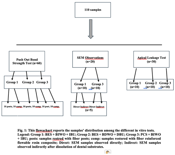
One hundred ten single-root human teeth devoid of fractures, previous root canal treatments, and root defects were selected for this study. Figure 1 reports the samples distribution among the different groups and type of experimental tests. Teeth were chosen between incisors, canines, and mandibular and maxillary premolars, extracted for orthodontic, prosthetic, or periodontal causes. All extracted teeth had a length to permit to have 5 mm restorative space and to leave at least 5 mm of gutta-percha apically. Pre-operative intraoral radiographs were taken to examine the endodontic anatomy of each sample. After extraction, teeth were maintained in a saline solution at room temperature to avoid dehydration and to guarantee a good preservation of the samples.
Among the 110 sample teeth 23 were incisors, 22 were canines, 32 maxillary premolars and 33 mandibular premolars. The selected teeth were randomly divided into three Groups, keeping the types of teeth equally represented, based on the used endodontic sealer. The use of extracted teeth was approved by the ethical Committee of the University of Siena.
2.2. Root Canal Treatment (Shaping Technique and Endodontic Sealers)
The samples were prepared for the following shaping and cleaning procedures to remove residues of plaque, calculus, and human tissue using ultrasonic instruments by one operator (DIKP) to reduce operator bias.
Using diamond burs mounted on a high-speed handpiece equipped with a water-cooling system, a standardized cavity access was made for each tooth. A long shaft round diamond bur was used to access the pulp chamber and, subsequently, a non-end cutting diamond bur that was oriented perpendicularly to the occlusal plane was used to make the finishing phase.
Once the access cavity was completed, a pre-curved stainless steel manual K-file #10 (Sweden & Martina; Due Carrare, Italy) was inserted into each canal with a watch winding movement up to the apical level, scouting the canal and defining the working length. After access cavity preparation, a #10 K-file (Dentsply Sirona, Ballaigues, Switzerland) was introduced into the root canal until its tip was just visible at the major apical foramen under ×10 magnification using a dental operating microscope. The working length (WL) was established by subtracting 0.5 mm from this measurement. The apical diameter was determined by inserting K-files of progressively increasing size until the file snugly fitted the apical foramen, as confirmed under magnification. The corresponding ISO size was recorded as the initial apical diameter before canal instrumentation. Teeth in wich a size #15 or #20 K-file first bound at working lenght were included in this study to ensure comparable canal anatomy. To standardize canal preparation, all root canals were instrumented to the same final apical preparation size (ISO 25, .06 taper). The Mtwo rotary system (Sweden & Martina; Due Carrare, Italy) was used for shaping root canals accordingly with manufacturer’ recommended sequences to achieve the final preparation.
All the chemo-mechanical preparation procedures of the samples were performed by one operator to reduce operator bias (DIKP).
Instruments were used with continuous rotation movement through an endodontic motor (Tri Auto ZX, Kuraray) at 250 rpm for the MTwo group and at 350rpm for the B-4U group.
Following the sequence indicated by the manufacturer, the shaping was carried out in the same way for all sample roots. 2.5 ml of 5,25% sodium hypochlorite (Niclor 5 dental, Ogna Lab srl, MB, Italy) for 30 seconds was used between each instrument by a side venting syringe-needle irrigation. The sodium hypochlorite used for all shaping and cleasing phases was preheated to 50 °C by immersing the syringes containing it in 60° C water. The apical patency was maintained by using a # 10 K-type file, brought 0.5mm beyond the apex, after each larger file. The final irrigation phase was carried out with 2 ml of 17% EDTA (Ogna Lab srl) for 3 minutes, followed by the last washing of 5 ml of 5% NaOCl for 5 minutes, to optimize the removal of inorganic and organic components.
Once the root canal instrumentation was completed, the apical gauging was performed. All the canals were dried with endodontic paper cones.
Then, the sample roots were divided into 3 Groups as follows:
Group 1. CeraSeal root canal sealer (Meta Biomed; Osongeup, Republic of Korea) with the continuous wave of condensation technique, BIWO and IBU(n=20);
Group 2. Ceraseal (Meta Biomed) with the continuous wave of condensation technique, BDWO and DBU(n=20);
Group 3. Pulp Canal Sealer (Kerr Dental, Brea, California, USA) with the continuous wave of condensation technique, BIWO and IBU(n=20).
The composition of CeraSeal is 20-30%tricalcium disilicate, 1-10% dicalcium silicate, 1-10% Tricalcium aluminate, and 45-50% zirconium oxide, accordingly with manufacturer’s information. In all Groups the continuous wave of condensation technique was ended after the apical third of the root canal (approximately 1/3 or half of the root canal, related to the length of the root). More specifically the obturation protocol was the following: Canal preparation: The canal is cleaned, shaped, and dried. A thin, uniform layer of endodontic sealer is applied to the canal walls. A gutta-percha cone matching the final apical preparation is selected. The cone should exhibit tug-back at the working length, indicating a snug apical fit. A heated plugger, EQ-V Pack, (EQ-V Meta Biomed, South Korea) is selected so that it can reach 4–5mm short of the working length without binding (in short canals can be half of the canal, in longer ones can be one third of the canal). The heated plugger is activated and driven apically through the gutta-percha in one continuous movement (“continuous wave”) until the predetermined depth is reached. Heat is discontinued while maintaining apical pressure for approximately 10 seconds to compensate for gutta-percha shrinkage during cooling. The plugger is briefly reheated to separate it from the compacted gutta-percha and removed, leaving a dense apical plug approximately 4–5 mm long.
2.3. BIWO Technique
In Groups 1, and 3 when the endodontic therapy was completed and before doing the build up with the adhesive restorative materials, the middle and coronal thirds of the root canals were washed with tap water carried into the canal with an endodontic syringe followed by the motion of ultrasonic tip for 30 seconds to remove the unset sealer yet from the root canal walls and permit an Immediate Build Up (IBU) in which the adhesive procedure can be performed properly. More specifically: The washout procedure consisted of applying the high-pressure air-water spray from the dental unit syringe for 30 seconds. The resulting shear stress may enhance the removal of residual root canal sealer from the canal walls. The residual sealer was removed using 5ml of saline delivered through a 27-gauge side-vented irrigation needle into the root canal and activated with a P5 Newtron ultrasonic device (Satelec Acteon, Mèrignac, France) equipped with an Irrisafe ultrasonic tip (Satelec Acteon, Mèrignac, France) ISO size 25, taper .00, at power setting 4, for 30 seconds. Ultrasonic activation enhanced irrigant flow and generated shear stress along the canal walls, thereby promoting the removal of residual sealer.
In Group 2 the wash out of the endodontic sealer was delayed one week, and for that was called Bioceramic Delayed Wash Out (Bioceramic Delayed Wash Out).
2.4. IBU
The build-up of Groups 1 and 3 was performed in 10 samples luting a fiber post (GC Europe N.V.) (Group Posts) and in another 10 samples using a fiber-reinforced flowable resin composite (everXFlow, GC Europe, N.V., Leuven, Belgium (Group Flow). In Group 1 Posts, a fiber post was calibrated in the coronal third of each root sample according to the shape and size of the root canal. The post was cut at the proper length with a diamond bur under water spray, dried, and then silanized (Multiprimer, GC Europe N.V.) and left for at least 1 minute on the tray. The root canal walls were treated with experimental adhesive system (AFB-00, GC Co., Tokyo, Japan) and experimental cement (ASAC-02, GC Co, Tokyo, Japan) strictly following the manufaturer’s instructions. When the samples of Group 1 Resin were built up, the adhesive procedure to the root canal walls was performed with the experimental adhesive system and then followed by the insertion of several layers, around 2 mm in thickness, of fiber-reinforced flow resin composite everXFlow, GC Europe N.V.). Each layer was light cured for 20 seconds. All materials used for the build-up were used strictly following the manufacturer's instructions. Finally, all samples were kept in a saline solution at 37° C in an incubator for 3 weeks.
In Group 2 the build-up was made with the same sequence described for Groups 1 and 2 but one week after the endodontic treatment (Delayed Build Up).
2.5. Preparation of the Samples for the Push-Out Bond Strength Test
After three weeks, 3 or 4 2-mm-thick slices were obtained by sectioning the coronal and medium thirds of the root perpendicular to the long axis, under water-cooling (Isomet, Buehler, Lake Bluff, IL, USA). The machine worked at a constant rotation speed of 150rpm. Between 36 and 40 samples were thus obtained for each group (n = 36-40 / group). Each specimen was marked on its coronal surface with a marker, and the precise thickness of each slice was measured using a digital caliper with 0,02mm accuracy (Mitutoyo, Tokyo, Japan).
2.6. Push-Out Test
The dislodgement resistance of the materials was measured using a universal testing machine (TRIAX 50 Controls, Milano, Italy). The root slices were mounted on an acrylic block of 2 × 2 cm in dimension. In order to provide the most extended coverage over the filling material, the material was loaded with cylindrical stainless-steel plungers of different diameters: 0,8mm for the middle third, and 1mm for the coronal third. The loading was performed at the speed of 0,5mm/min in an apical-coronal direction to avoid any interference because of the root canal taper.
The maximum load applied to the filling material before debonding was recorded in Newtons. To express the bond strength in MegaPascals, the load recorded in Newtons was divided by the area of the bonded interface (mm2).
The area (SL) was calculated as the lateral surface area of a truncated cone with the formula:
SL = π(R+r)[(h2 + (R-r)2]0.5, where “π” is the constant 3.14, R represents the coronal post radius, r the apical post radius and h the thickness of the slice in millimeters.
Roots were considered as statistical units as the bond strength data were normally distributed (Kolmogorov-Smirnov test), with homogeneous group variances (Levene test), and that the root of origin of the slices did not significantly affect bond strength (regression analysis). A two-way ANOVA was performed to assess the influence of post system and timing of post space preparation on retention strength. Post-hoc comparisons were performed using the Tukey test. The level of significance was set at p<0.05 for all analyses (SPSS 11.0 software; SPSS Inc., Chicago, IL).
After the push-out test was performed, the slices were mounted on stubs and examined under Stereomicroscope (Nikon H550L, Tokyo, Japan) and also observed under a SEM (Jeol Co., Jsm-6060LV Scanning Electron Microscope, Tokyo, Japan) to detect the type of failure after the push-out test.
2.7. SEM Observation
Other 20 roots were divided into two Groups of ten samples each, and endodontically treated following the procedure described for Group 1 (BIWO) and Group 2 (BDWO). The same BES was used in both Groups. After the endodontic treatment was completed as described above, 50% of the samples in each group were washed with BIWO while the other 50% were not washed immediately but after 1 week (BDWO). Then, 5 samples of each group were directly observed under the SEM (SubGroup Direct) to evaluate how the root walls and tubules’ orefices were cleaned and free from endodontic sealer, while the other 5 samples received build-up procedures with a post (SubGroup Indirect).
The last 5 samples, after being restored, stayed in saline solution for 24 hours and then were kept in a 37% hydrochloric acid solution for 24 hours and then in a 5% sodium hypochlorite solution for 24 hours to dissolve all the mineralized and organic components of the teeth and to leave only the bonding restorations to detect the penetration of resin tags into the dentinal tubules.
The samples were mounted on matrices and gold-sputtered using a sputtering device (EMITECH K550); after that, the samples were observed using a scanning electron microscope (Jeol Co.) at different magnifications: 500x, 1000x and 7000x. The different magnifications were useful to observe the amount of smear layer (lower magnification) and the tubules’orefices (higher magnification). Because the surface of the samples was clearly uniform regarding the presence of smear layer and the number of open tubules, pictures were made only in the middle third of the root canal space preparation, always the same area for all samples. Two observations were made of each sample: the first observation was made approximately in the center of the medium third root wall and the second 2 mm more apically. In this way, 10 scores were collected for each SubGroup.
Using a grading system ranging from 1 to 5, the amount of smear layers and open dentinal tubules on the root surface by examining images captured under a microscope at various magnification levels were assessed. All microscopic observations were made at the medium third of the canal preparation. At the same level, pictures were made directly on the post surface. For each area of observation, 3 pictures at progressive magnifications were made. For each sample, one single score was made reporting the amount of the smear layer and obstructed tubules. The scores were done by two independent operators (MF, EFC) and in case of different scores, the samples were reevaluated by the two observers and an agreement was found.
The amount of residual smear layer and of obstructed dentinal tubules by observational investigations using a scale from 1 to 5 were reported as below: 1. more than or equal to 50% of residua smear layer and more than or equal to 90% of obstructed tubules; 2. more than or equal to 40% of residua smear layer and more than or equal to 70% of obstructed tubules 3. more than or equal to 30% of residua smear layer and more than or equal to 50% of obstructed tubules 4. more than or equal to 20% of residua smear layer and more than or equal to 20% of obstructed tubules 5. more than or equal to 10% of residua smear layer and more than or equal to 10% of obstructed tubules.
A statistical test (Wilcoxon p-value test) was used to assess the collected data, with a p-value set at p<0.01 [7].
Also, the 5 samples of Group 1 that were directly observed were also used to evaluate the interfacial adaptation of the gutta-percha and the bioceramic sealer to the root canal walls, to investigate the possible presence of gaps and voids and note if the BIWO affected the seal of the endodontic obturation.
2.8. Apical Leakage Test
Thirty recently extracted intact human single-root maxillary premolars were included in the study. After cleaning of remaining tissues, a ×4.5 stereomicroscope (Nikon SMZ645; Nikon, Tokyo, Japan) was employed to rule out any external radicular cracks. Samples were stored in 0.9% saline solution (SALF SPA, Cenate Sotto, Italy) at a temperature of 37°C to prevent dehydration. The teeth, randomly divided into three Groups, and endodontically treated and built up as described above for Groups A1-A3, and submitted to BIWO (A1, A3) and BDWO (A2). After the root canal obturation, all tooth surfaces were covered with nail varnish. The 2 mm around the area of the apex of each tooth were left exposed. A diluted ammoniacal silver nitrate solution (1:4 ratio of ammoniacal silver nitrate to distilled water) was prepared, and the diluted solution was filtered with a Millipore filter (0.22-nm filter, Carrigtwohill, County Cork, Ireland) mounted on a syringe. Under laboratory light, each tooth was placed in a test tube with diluted ammoniacal silver nitrate solution. After 24 h, specimens were thrice rinsed in water for 10 min. Nail varnish around the tooth was removed with acetone, and each tooth was placed in a test tube with the diluted photo-developer solution (Kodak, Rochester, NY, USA; 1:10 ratio of photo-developer solution to distilled water). After 8 h, teeth were thrice rinsed in water for 10 min. Each tooth was embedded in transparent self-curing acrylic resin. The teeth were then sliced with a low-speed diamond saw underwater cooling to prevent frictional heat (Isomet; Buehler, Lake Bluff, NY, USA) into two slices along their long axis and perpendicularly to the root. Samples were examined with a digital microscope. Two observers (DP, MF) independently scored the amount of tracer along with the interface. The apical leakage was evaluated following the procedure described by Limkangwalmongkol et al [26]. In case of a score discrepancy, the sample was evaluated again by the two operators (MF, EFC), and a common decision was taken. The Bonferroni and Anova tests were used to assess differences among groups, and to separately determine whether leakage significantly differed among groups. The significance level was set at P < 0.05, and the analyses were performed with the software package SPSS IBM Statistics version 21 for Mac (SPSS Inc., Chicago, IL, USA).
3. Results
3.1. Push Out Test
Table 1 presents the mean bond strength values (MPa) of tested groups according the endodontic sealer used and BIWO and BDWO. The Table 1 revealed a highly statistically significant difference among the studied groups (p ≤ 0.05).
Among the post groups, the highest mean bond strength was observed in Group 1 post (15.23 ± 4.2 MPa), followed closely by Group 1 composite (14.51 ± 3.1 MPa), Group 2 post (12.83 ± 3.1 MPa), and Group 2 composite (12.52 ± 2.1 MPa). The lowest values were recorded for Group 3 post (9.55±1.32) and Group 3 composite (9.12±1.67). Statistically significant differences were detected among the three Groups (p > 0.05) while no differences were present between samples restored with post or composite within the same Group.
Regarding the influence of sealer type, groups obturated with BES demonstrated significantly higher bond strength values than those obturated with Pulp Canal Sealer. The Pulp Canal Sealer Group 1 exhibited significantly lower bond strength compared with BES Groups independently if BIWO or BDWO was done (p < 0.05).
After the push-out test, the slides were also observed under microscope to detect the type of failure that occurred: the results were reported in Table 3. Examples of types of failure are reported in Figure 2. Cohesive failure of the resin composite was noted when the fiber-reinforced flowable resin composite was used, while when the post was used, the Cohesive failure was mainly within the cement (Groups 1-3). In Groups 3 post and composite, a wider number of Adhesive and Mixed failures were recorded.
3.2. SEM Observation
The scanning electron microscope observations were made to evaluate two groups of samples that, after being shaped and obturated with the same procedure and the same endodontic material (MTwo rotating system and Ceraseal endodontic sealer), were restored differently: Group 1 with BIWO and IBU and Group 2 in which samples were treated with BDWO and DBU. SEM observations were made in half of the samples, directly observing the root canal walls of specimens (Figure 3 and Figure 4), and in the other half, after being restored as described above, dissolving with strong acid the dental structure and consequently looking at formed resin tags made by the penetration into the tubules of the adhesive material (Figure 5). Table 4 report the scores related to the presence of smear layer and obstructed dentinal tubules.
Legend. Direct: Direct observation of the root walls; Indirect: Observation of the adhesive restoration after dissolving the tooth structure; The scores: 1. more than or equal to 50% of residual smear layer and more than or equal to 90% obstructed tubules; 2. less than or equal to 40% of residual smear layer and less than or equal to 70% of obstructed tubules; 3. less than or equal to 30% of residual smear layer and less than or equal to 50%; 4. less than or equal to 20% of residual smear layer and less than or equal to 20% of obstructed tubules; 5. less than or equal to 10% of residual smear layer and less than or equal to 10% obstructed tubules.
The SEM observations showed that when BIWO was done (Group 1), the surface of root canal walls was better cleaned than those samples in which BIWO was not used (Group 3) (Figure 3a,b, Figure 4).
At higher magnifications was possible to note that the bioceramic sealer was partially or totally closing the tubule orifices when the BDWO was used (Group 3)(Figs 3a and 3b), while when the BIWO was done (Group 1), tubules were completely open and clean (Figure 4).
Looking at formed resin tags made by the penetration into the tubules of the adhesive material was evident that long resin tags’ formation was possible after BIWO and IBU ((Figure 5) while when BDWO and DBU were used, very short resin tags were noted and also smear layer still present along the root canal walls (Figure 6a-c). That was due to the fact that tubules were still covered totally by smear layer or at least partially filled by the endodontic sealer.
The differences between the scores of the two groups were statistically significant (p-value Wilcoxon test < 0.01).
From the SEM observation was evident that the BIWO technique obtained a better cleaned dentin surface from the smear layer and consequently that root dentinal tubules were infiltrated, while when the bioceramic sealer had the time to set completely also into the tubules, although the delayed Wash Out technique was used (BDWO), an important amount of smear layer remained and some tubules were filled by the endodontic cement itself.
3.3. Apical Leakage Test
The apical leakage scores are reported below (Table 5). The apical leakage of the Groups with vertical condensation, BES cements and BIWO was the lowest recorded in this study (Group 1). However, no statistically significant differences were found among the 3 Groups (Table 5). Also, no statistically significant differences were found between the sealing ability of the two endodontic types of cement and between BIWO and BDWO samples.
4. Discussion
The BIWO is a new clinical procedure that is based on the possibility to wash out the BES before is set using the water spray or/and a syringe of water and an ultrasonic tip. It is possible to immediately remove the bioceramic sealer because this material is water soluble before starting to set. The main purpose is to completely clean the root canal walls from the endodontic cement and have a clean bonding surface available for adhesion. In this in vitro study, all the performed investigations showed that the BIWO is effective, and it can be speculated that, if translated it to clinical reality, several advantages might be achieved, such as reduced number of appointments, obtaining a good coronal seal in continuity with the root canal obturation step with a reduced risk of reinfection, and performing the build-up under rubber dam. The IBU was for many years a desire of practitioners and scientists, but before the launch of BES into the market was not possible because the zinc phosphate endodontic sealer creates a sticky smear layer that is difficult to be removed also with the rotary instruments [27].
The BES were originally proposed to be used with the single cone obturation technique, but in this case the BIWO should be not applied because it can be expected that the washout might involve a wide part of the bioceramic cement when the BES is no set yet, with the risk to lose the tridimensional seal of the root canal. At the end, the BIWO can be used only as described in the materials and methods along this paper because the vertical compaction of the warm gutta-percha, ended approximately between the medium and apical third of the root, might be effective to seal and prevent the washout of the BES from the apical portion of the root (Figure 7 and Figure 8).
In this study, three Groups of samples were tested, comparing one bioceramic sealer, and one traditional endodontic cement, the BIWO versus BDWO, and two different restorative procedures. The push-out bond strength test showed statistically significant differences among the three tested Groups, and more in particular, when the BIWO was used it showed the highest value, whilst when the traditional endodontic sealer was used in combination with BIWO the lowest value. Regarding the type of reconstruction, in all Groups the samples restored with a post showed a higher bond strength but not statistically significant. For that, the type of material and technique to restore the endodontic treated teeth is determined by other clinical parameters, such as presence of ferrule, the amount of coronal residual structure, the complexity of the treatment planning, the position of the tooth, presence of sign of bruxism, etc [25].
In this study, some null hypotheses were tested; The bond strength to root dentin under three different experimental conditions was evaluated by push-out. The statistical analysis showed that when BIWO was used in combination with the IBU, the bond strength was superior and statistically significantly different than when BDWO was used in combination DBU; For that, the first null hypothesis was rejected. Also, Group 1 showed statistical significantly higher bond strength than Group 3: for that, the second null hypothesis was rejected. This can be due to the fact that the BES is a soluble in water before starting to set and the BIWO can effectively remove the endodontic sealer from the tubules and the surface of root dentinal walls; after a week, when BDWO was made, the BES was already set and it was not possible to remove it completely.
In all tested Groups, those restored with fiber post showed the highest scores while the Groups restored with fiber reinforced resin composite showed lower bond strength but not significantly. This can be due to the different stiffness of the restorative materials and their different behavior under the push-out test, in which a punch is pushing the restoration in the center of the slide.
The results of the push-out bond strength test obtained in this study are similar to that already published in the last 30 years [29]. The push-out test was mainly born to evaluate the bond strength to root dentin and is now widely accepted. The range of bond strength that can be found in the literature is mainly from 6 to 15 MPa, in the range of those found in this study [30].
The third null hypothesis tested if when BIWO/IBU combined procedures were used, there were no differences about the quantity of remaining smear layer and the number of open tubules along the dentinal walls between the two types of endodontic sealers; accordingly with the results of this study after the instrumentation of the prosthodontic space, there were statistical significant differences between the samples in which BIWO was used and those in which BDWO was applied; In Group 1 the root canal walls appeared clean and tubules were open in a wide number and available for an effective bonding procedure while in those where BDWO was applied (Group 2), a smear layer of endodontic sealer was found to cover the dentinal walls and/or to close completely or at least partially the tubules; For that, the third null hypothesis was rejected. Also, the failure types were reported (Table 2) showing that there was a wide frequence of cohesive failures into the material (cement and restorative component) when the bond strength showed the highest scores, while adhesive failures were more often noted when the scores were mainly lower. This was in accordance with the existing literature [28].
In the past, many Authors investigated bonding to root dentin that is considered pivotal to achieve a durable restoration and to seal the root canal margin coronally [7,8,9]. The SEM observation was largely used to investigate the cleanliness of the root canal walls, and different irrigant solutions in combination or not with an ultrasonic device were proposed [7,8]. All these investigations were performed on root samples that were obturated and then, some days later, reprepared, bonded and restored. This traditional procedure needs to prepare the root canal space with drills removing some root dentinal walls and creating a smear layer that is difficult to be completely removed; Also, when a BES is used, the cement can deeply penetrate the dentinal tubules and set inside [31]: the set bioceramic cement is then very difficult to be removed and continue to obstacle the adhesive material to penetrate the tubules and consequently reducing the bond strength of the restoration. It must also be considered that when BIWO is used, there is no need to reprepare the root canal to create a ‘prosthodontic space,’ while when the restoration is postponed, and the BIWO is not applied, the root canal must be reprepared with the possibility to take out some amount of root canal dentin and making the root weaker.
Regarding the apical leakage test, it was observed that in the three Groups, they were not statistically significant differences and for that the fourth null hypothesis was accepted. That can be due to the good seal obtained when warm gutta-percha vertical condensation technique was used, independently from the type of endodontic sealer that was used and BIWO or BDWO. It can be speculated that the apical seal of root canals treated with BIWO was no different from those can be achieved with traditional endodontic sealer and obturation techniques. For that the null hypothesis was accepted. The leakage data that were found in this study are in agreement with other similar in vitro tests [31,32,33,34,35,36].
However, some limitations of this study such as the in vitro design, the use of extracted teeth, the possible anatomical variability between samples, must be point out. For sure the outcomes of this in vitro study can’t be extrapolated to clinical conditions. The results of this study encourage to make clinical trial to evaluate the BIWO in combination with IBU. Future researches are in progress, such as in vitro tests to verify the standardized model, and randomized clinical studies to validate BIWO and IBU under clinical conditions.
5. Conclusions
From the findings of this in vitro study, the following conclusions can be drawn: 1. The BIWO under in vitro conditions is a procedure that permit to immediate clean the root canal walls and to make an immediate bonding procedure. 2. Using BIWO a higher bond strength than BDWO can be achieved. 3. The BIWO dos not affect negatively the apical seal of the endodontic treated tooth. 4. The IBU is a useful and effective clinical procedure when used in combination with the BIWO.
Supplementary Materials
The datasets used and/or analyzed during the current study are available from the corresponding author on reasonable request.
Author Contributions
Conceptualization, M.F., D.I.K.P.; Methodology, M.F., D.I.K.P.; Software, W.S.H., G.V., D.M; Validation, M.F., E.F.C., D.M.; Formal Analysis, M.F., E.F.C..; Investigation, D.I.K.P., M.F.; Resources, M.F..; Data Curation, D.I.K.P., E.F.C.; Writing – Original Draft Preparation, M.F., D.I.K.P., Writing – Review & Editing, M.F., E.F.C., D.M.; Visualization, W.S.H., G.V.; Project Administration, M.F.; Funding Acquisition, M.F. All authors have read and agreed to the published version of the manuscript.
Funding
This research received no external funding.
Data Availability Statement
The original contributions presented in this study are included in the article. Further inquiries can be directed to the corresponding author.
Acknowledgments
The authors would like to thank the following industries for providing the needed materials: Sweden & Martina; GC Co and the dental laboratory of MDT Giovanni Bonadeo for the technical support. The authors want also to thank Dr. Doaa Al Emam, University of Mansoura, Egypt for the statistical analysis.
Conflicts of Interest
The authors declare no conflict of interest. The funders had no role in the design of the study; in the collection, analyses, or interpretation of data; in the writing of the manuscript, or in the decision to publish the results.
References
- Ng, Y.L.; Mann, V.; Gulabivala, K. Tooth survival following non-surgical root canal treatment: a systematic review of the literature. Int. Endod. J. 2010, 43, 171–89. [Google Scholar] [CrossRef] [PubMed]
- Consensus report of the European Society of Endodontology on quality guidelines for endodontic treatment. Int. Endod. J. 1994, 115–24. [CrossRef] [PubMed]
- Ferrari, M.; Ferrari Cagidiaco, E.; Pontoriero, D.I.K.; Ercoli, C.; Chochlidakis, K. Survival rates of endodontically treated posterior teeth restored with all-ceramic partial-coverage crowns: when systematic review fails. Int. J. Environ. Res. Publ. Health 2022, 19, 1971. [Google Scholar] [CrossRef] [PubMed]
- Haragushiku, G.A.; Sousa-Neto, M.D.; Silva-Souss, Y.T.C.; Alfredo, E.; Silva, S.C.; Silva, R.G. Adhesion of endodontic sealer to human root dentine submitted to different surface treatments. Photomed. Laser Surg. 2010, 28, 405–10. [Google Scholar] [CrossRef] [PubMed]
- Garoushi, S.; Säilynoja, E.; Vallittu, V.K.; Lassila, L. Physical properties and depth of cure of a new short fiber reinforced composite. Dent. Mater. 2017, 29(8), 835–841. [Google Scholar] [CrossRef] [PubMed]
- Garoushi, Vallitu S; Gargoum, A.; Vallittu, P.K.; Lassila, L. Short fiber-reinforced composite restorations: a review of the current literature. J. Investig. Clin. Dent. 2016, 9, e12330. [Google Scholar]
- Marchese, M.; Pontoriero, D.I.K.; Ferrari Cagidiaco, E.; Iandolo, A.; Grandini, S.; Ferrari, M. Endodontic Irrigants and Their Activation Efficacy on Cleansing Post-Space Root Canal Walls. Prosthesis 2021, 3, 406–414. [Google Scholar] [PubMed]
- Haichal, W.S.; Pontorero, D.I.K.; Ferrari Cagidiaco, E.; Verniani, G.; Ferrari, M. Evaluation of the efficacy of different irrigation materials to remove the bioceramic sealer in endodontic retreatment. J. Osseoint. 2025. [Google Scholar] [CrossRef]
- Franklin R Tay, F.R.; Loushine, R.J.; Lambrechts, P.; Weller, R.N.; Pashley, D.H. Geometric factors affecting dentin bonding in root canals: a theoretical modeling approach. J. Endod. 2005, 31, 584–9. [Google Scholar] [CrossRef] [PubMed]
- Goracci, C.; Ferrari, M. Current perspectives on post systems: a literature review. Aust. Dent. J. 2011, 56 Suppl 1, 77–83. [Google Scholar] [CrossRef] [PubMed]
- Goracci, C.; Corciolani, G.; Ferrari, M. Light-transmitting Ability of Marketed Fiber Posts. J.Dent.Res. 2008. [Google Scholar] [CrossRef] [PubMed]
- Gandolfi, M.G.; Taddei, P.; Pondrelli, A.; Zamparini, F.; Prati, C.; Spagnuolo, G. Demineralization, Collagen Modification and Remineralization Degree of Human Dentin after EDTA and Citric Acid Treatments. Materials 2018, 21;12(1), 25. [Google Scholar]
- Pontoriero, D.I.K.; Verniani, G.; Manfredini, D.; Ferrari, M.; Ferrari Cagidiaco, E. Efficacy on cleansing post space root canal walls after Hydraulic Endodontic Sealers were used. J. Osseoint. 2024. [Google Scholar]
- Pontoriero, D.I.K.; Villani, R.; Bianchi, G.; Ferrari Cagidiaco, E. Push-Out Bond Strenght of four different bioceramic sealers tested with three different obturating systems: an In Vitro study. J. Osseoint. 2023, 15, 210–6. [Google Scholar]
- Belsare, L.; Patil, S.; Bhede, R.; Gade, J.; Gade, V. Evaluation of push-out bond strength of endosequence BC sealer with lateral condensation and thermoplasticized technique: An in vitro study. J. Conserv. Dent. 2015, 18, 124. [Google Scholar] [CrossRef] [PubMed]
- Prati, C.; Gandolfi, M.G. Calcium silicate bioactive cements: Biological spectives and clinical applications. Dent. Mater. 2015, 31, 351–70. [Google Scholar] [CrossRef] [PubMed]
- Niua, L.; Jiaob, K.; Wang, T.; Zhang, W.; Camilleri, J.; Bergeron, B.J.; Feng, H.; Mao, J.; Chen, J.; Pashley, H.D.; Tay, F.R. A review of the bioactivity of hydraulic calcium silicate cements. J. Dent. 2014, 42, 517–533. [Google Scholar] [CrossRef] [PubMed]
- Cardinali, F.; Camilleri, J. A. critical review of the material properties guiding the clinician’s choice of root canal sealers. Clin. Oral Invest. 2023. [Google Scholar] [CrossRef] [PubMed]
- Jeong, J.W.; DeGraft-Johnson, A.; Dorn, S.O.; Di Fiore, P.M. Dentinal Tubule penetration of a calcium silicate–based root canal sealer with different obturation. J. Endod. 2017, 5, 1–5. [Google Scholar] [CrossRef] [PubMed]
- Lee, B.N.; Hong, J.U.; Kim, S.M. Anti-inflammatory and osteogenic effects of calcium silicate-based root canal sealers. J. Endod. 2019, 45, 73–78. [Google Scholar] [CrossRef] [PubMed]
- Garrib, M.; Camilleri, J. Retreatment efficacy of hydraulic calcium silicate sealers used in single cone obturation. J. Dent. 2020, 98, 103370. [Google Scholar] [CrossRef] [PubMed]
- Chybowski, E.A.; Glickman, G.N.; Patel, Y. Clinical outcome of non-surgical root canal treatment using a single-cone technique with Endosequence bioceramic sealer: a retrospective analysis. J. Endod. 2018, 44, 941–945. [Google Scholar] [CrossRef] [PubMed]
- Bardini, G.; Casula, L.; Ambu, E.; Musu, D.; Mercadè, M.; Cotti, E. A 12-month follow-up of primary and secondary root canal treatment in teeth obturated with a hydraulic sealer. Clin. Oral Investig. 2021, 15, 2757–64. [Google Scholar] [CrossRef] [PubMed]
- Pontoriero, D.I.K.; Ferrari Cagidiaco, E.; Maccagnola, V.; Manfredini, D.; Ferrari, M. Outcomes of Endodontic treated teeth obturated with calcium silicate-based root canal sealers in combination with warm gutta-percha obturation techniques: a prospective clinical study. J. Clin. Med. 2023, 12. [Google Scholar] [PubMed]
- Ferrari, M.; Ferrari Cagidiaco, E.; Goracci, C.; Sorrentino, R.; Zarone, F.; Grandini, S.; Joda, T. Posterior partial crowns out of lithium disilicate (LS2) with or without posts: A randomized controlled prospective clinical trial with a 3-year follow up. J. Dent. 2019, 83, 12–17. [Google Scholar] [CrossRef] [PubMed]
- Limkangwalmongkol, S.; Burtscher, P.; Abbott, P.V.; Sandler, A.B.; Bishop, B.M. A comparative study of the apical leakage of four root canal sealers and laterally condensed gutta-percha. J. Endodon 1991, 10, 495–499. [Google Scholar] [CrossRef] [PubMed]
- Saikaew, P.; Sattabanasuk, V.; Harnirattisai, C.; Chowdhury, A.F.M.A.; Carvalho, R.; Sano, H. Role of the smear layer in adhesive dentistry and the clinical applications to improve bonding performance. Jpn. Dent. Sci. Rev. 2022, 29, 59–66. [Google Scholar] [CrossRef] [PubMed]
- Brichko, J.; Burrow, M.F.; Parashos, P. Design Variability of the Push-out Bond Test in Endodontic Research: A Systematic Review. J. Endod. 2018, 44, 1237–1245. [Google Scholar] [CrossRef] [PubMed]
- Goracci, C.; Tavares, A.U.; Fabianelli, A.; Raffaelli, O.; Cardoso, P.C.; Ferrari, M. The adhesion between fiber posts and root canal walls: comparison between microtensile and push-out bond strength measurements. Eur. J. Oral Sci. 2004, 112, 353–61. [Google Scholar] [CrossRef] [PubMed]
- Habib, S.R.; Ansari, A.S.; Khan, A.S.; Alamro, N.M.; Alzaaqi, M.A.; Alkhunefer, Y.A. Push-out bond strength of endodontic posts cemented to extracted teeth: an in-vitro evaluation. Materials 2022, 15, 6792. [Google Scholar] [CrossRef] [PubMed]
- Ashkar, I.; Sanz, J.L.; Forner, L.; Llena, C.; Melo, M. Comparative Dentinal Tubule Penetration of Three Calcium Silicate-Based Sealers Under Conventional and Sonic-Activated Final Irrigation Protocols: An In Vitro Confocal Microscopy Study. Dent. J. 2026, 14, 593. [Google Scholar] [CrossRef] [PubMed]
- Viapiana, R.; Guerreiro-Tanomaru, J.M.; Tanomaru-Filho, M.; Camilleri, J. Investigation of the effect of sealer used on the heat generated at the external root surface during root canal obturation using warm vertical compaction technique with System B heat source. J. Endod. 2014, 40, 555–61. [Google Scholar] [CrossRef] [PubMed]
- Amanda, B.; Suprastiwi, E.; Usman, M. Comparison of Apical Leakage in Root Canal Obturation Using Bioceramic and Polydimethylsiloxane Sealer (In Vitro). Open J. Stomatol. 2018, 8, 21–31. [Google Scholar]
- Moccia, E.; Carpegna, G.; Dell’Acqua, A.; Alovisi, M.; Comba, A.; Pasqualini, D.; Berutti, E. Evaluation of the root canal tridimensional filling with warm vertical condensation, carrier-based technique and single cone with bioceramic sealer: a micro-CT study. G. Ital. Di Endod. 2020, 34, 10–7. [Google Scholar] [CrossRef]
- Dzankovic, A.; Hadziabdic, N.; Korac, S.; Tahmiscija, I.; Konjhodzic, A.; Hasic-Brankovic, L. Sealing Ability of Mineral Trioxide Aggregate, Biodentine and Glass Ionomer as Root-End Materials: A Question of Choice. Acta Med. Acad. 2020, 49, 232–239. [Google Scholar] [CrossRef] [PubMed]
- Pontoriero, D.I.K.; Madaro, G.; Vanagolli, V.; Benedicenti, S.; Verniani, G.; Ferrari Cagidiaco, E.; Grandini, S.; Ferrari, M. Sealing ability of a bioceramic sealer used in combination with cold and warm obturation techniques. J. Osseoint 2021, 13, 248–255. [Google Scholar]
Figure 2.
Example of Adhesive failure (A), Cohesive Failure (C) and Mixed Failure (M).
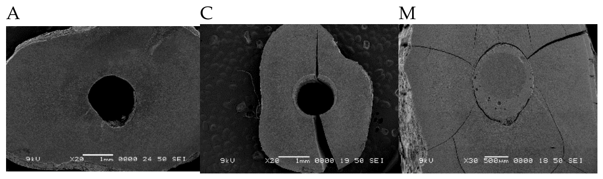
Figure 3.
a,b. The dentinal wall surface shows some area with smear layer and smear plugs to close the tubules’ orifices when BDWO was used.
Figure 3.
a,b. The dentinal wall surface shows some area with smear layer and smear plugs to close the tubules’ orifices when BDWO was used.
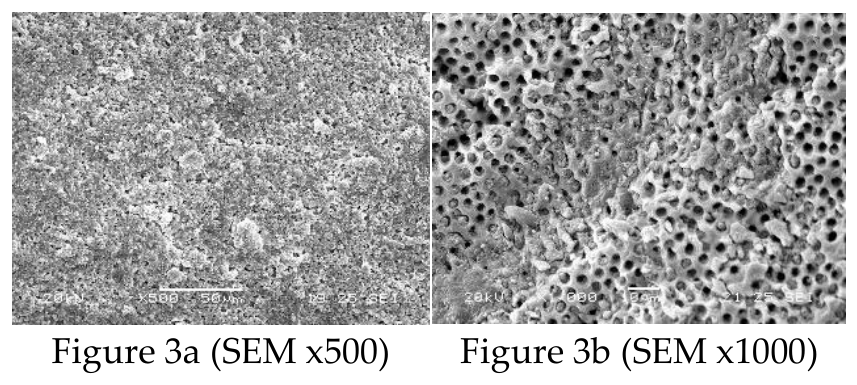
Figure 4.
The tubules are completely open and the dentinal wall cleaned from smear layer after BIWO was used. (SEM x1000).
Figure 4.
The tubules are completely open and the dentinal wall cleaned from smear layer after BIWO was used. (SEM x1000).
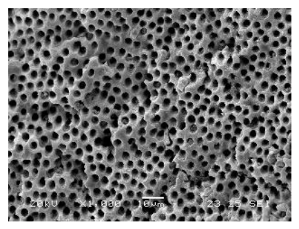
Figure 5.
Long resin tags are filling all tubules (SEM x2000).
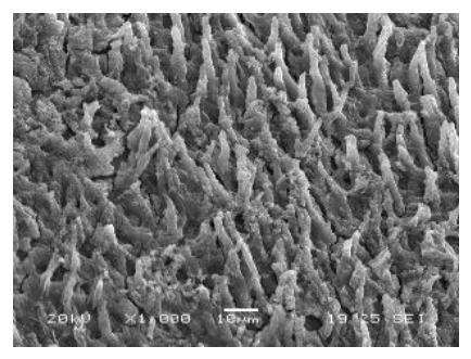
Figure 6.
Legend: in Figure 6a (SEM x300) it is possible to detect an area in which short resin tags are formed (lower part) and another area in which smear layer is covering the dentinal walls (upper part); in Figure 6b are visible short resin tags at higher magnification (x7000) while in Figure 6c tubules are mainly partially or totally filled by BES (x7000). The presence of completely set BES into the tubules can’t permit to the adhesive material to penetrate into the tubules and form resin tags. Also, some BES is covering the intertubular dentin as well.
Figure 6.
Legend: in Figure 6a (SEM x300) it is possible to detect an area in which short resin tags are formed (lower part) and another area in which smear layer is covering the dentinal walls (upper part); in Figure 6b are visible short resin tags at higher magnification (x7000) while in Figure 6c tubules are mainly partially or totally filled by BES (x7000). The presence of completely set BES into the tubules can’t permit to the adhesive material to penetrate into the tubules and form resin tags. Also, some BES is covering the intertubular dentin as well.
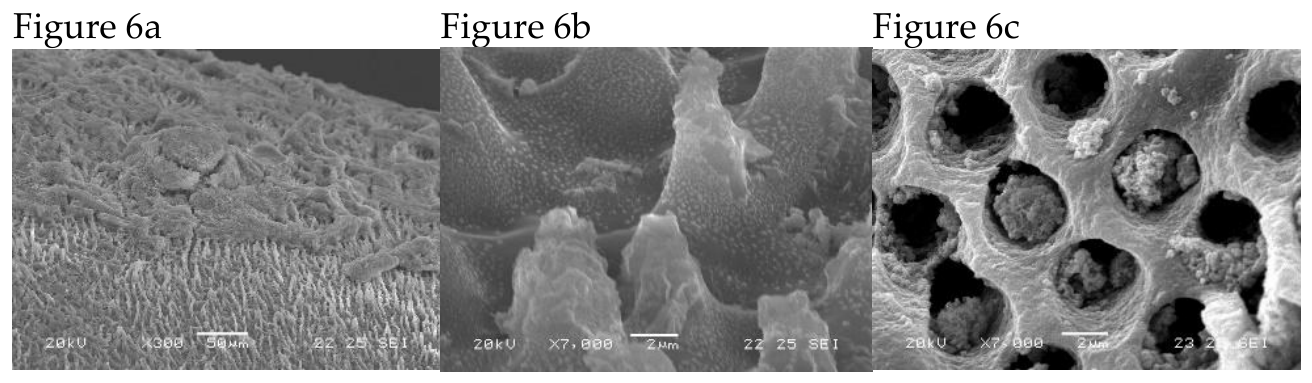
Figure 7.
Two examples of composite pictures of the same root canal after being obturated (SubGroup A1). There is no evident gap or void visible between the obturation and the cavity walls (SEM x17 and x50).
Figure 7.
Two examples of composite pictures of the same root canal after being obturated (SubGroup A1). There is no evident gap or void visible between the obturation and the cavity walls (SEM x17 and x50).
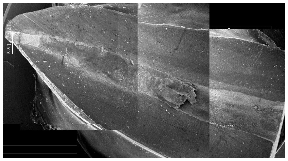
Figure 8.
Two examples of composite pictures of the same root canal after being obturated (SubGroup A1). There is no evident gap or void visible between the obturation and the cavity walls (SEM x17 and x50).
Figure 8.
Two examples of composite pictures of the same root canal after being obturated (SubGroup A1). There is no evident gap or void visible between the obturation and the cavity walls (SEM x17 and x50).
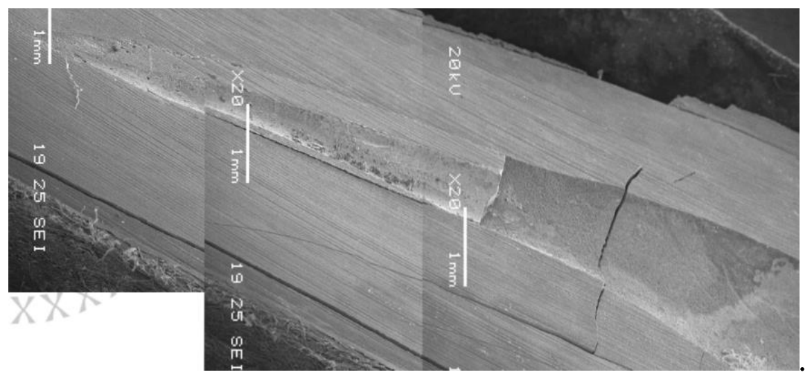

Table 1.
Bond strength among the studied Groups. Post= Group in which post was bonded; Comp: Group in which fiber reinforced flowable resin composite was used; n: number of root’s slides were tested. F: ANOVA test, *significant p≤0.05, p1: vs. 1 post, p2: vs. Group 1 composite, p3: vs. Group 2 post, p4: vs. Group 2 composite, p5: vs. Group 3 post, p6: vs. Group 3 composite.
Table 1.
Bond strength among the studied Groups. Post= Group in which post was bonded; Comp: Group in which fiber reinforced flowable resin composite was used; n: number of root’s slides were tested. F: ANOVA test, *significant p≤0.05, p1: vs. 1 post, p2: vs. Group 1 composite, p3: vs. Group 2 post, p4: vs. Group 2 composite, p5: vs. Group 3 post, p6: vs. Group 3 composite.
| Groups | Mean ± SD | |||||||
| P1 | P2 | P3 | P4 | P5 | P6 | |||
| Group 1 post (n=49) | 15.23±4.2 | - | - | - | - | - | - | |
| Group 1 comp (n=48) | 14.51 ±3.1 | 0.0 | - | - | - | - | - | |
| Group 2 post (n=50) | 12.83±3.0 | 0.0 | .88 | - | - | - | - | |
| Group 2 comp (n=48) | 12.52±2.1 | 0.0 | .03 | .015 | - | - | - | |
| Group 3 post (n=49) | 9.55±1.32 | 0.0 | 0.0 | 0.0 | 0.0 | .346 | - | |
| Group 3 comp (n=48) | 9.12±1.67 | 0.0 | 0.0 | 0.0 | 0.0 | .218 | .55 | |
Table 2.
It compares the mean bond strength values between the post and composite specimens within each experimental Group. In all evaluated groups the post specimens did not show significantly higher bond strength values than the corresponding composite specimens (p < 0.05).
Table 2.
It compares the mean bond strength values between the post and composite specimens within each experimental Group. In all evaluated groups the post specimens did not show significantly higher bond strength values than the corresponding composite specimens (p < 0.05).
| Post | Composite | Test of significance | P value | |
| Group 1 | 15.23±4.2 | 14.51 ±3.1 | t=2.54 | 0.001* |
| Group 2 | 12.83±3.0 | 12.52±2.1 | t=2.78 | 0.007* |
| Group 3 | 9.55±1.32 | 9.12±1.67 | t=3.52 | 0.004* |
Table 3.
Type of failure of samples. Legend: Adhesive: Failure at the bonding interface between dentin and adhesive material; Cc: Cohesive failure of the cement; Cm: Cohesive failure of the mate-rial; Mix: Adhesive and Cohesive failure; Comp: fiber reinforced flowable resin composite; Post: fiber post.
Table 3.
Type of failure of samples. Legend: Adhesive: Failure at the bonding interface between dentin and adhesive material; Cc: Cohesive failure of the cement; Cm: Cohesive failure of the mate-rial; Mix: Adhesive and Cohesive failure; Comp: fiber reinforced flowable resin composite; Post: fiber post.
| Group | Adhesive | Mixed | Cohesive |
| Group 1 post(n=36) | 6 | 6 | 24Cc |
| Group 1 comp(n=35) | 5 | 5 | 25Cm, 1Cc |
| Group 2 post(n=38) | 10 | 4 | 24Cc |
| Group 2 comp(n=38) | 11 | 6 | 21Cm |
| Group 3 post(n=37) | 18 | 12 | 7Cc |
| Group 3 comp(n=35) | 17 | 13 | 7Cc |
Table 4.
Amount of residual smear layer along the root walls and of obstructed dentinal tubules.
| Groups | |||||
| Score 1 | Score 2 | Score 3 | Score 4 | Score 5 | |
| Group 1 Direct | 2 | 8 | |||
| Group 1 Indirect | 3 | 7 | |||
| Group 2 Direct | 6 | 2 | 2 | ||
| Group 2 Indirect | 6 | 3 | 1 | ||
Table 5.
Apical leakage of 3 groups. The Groups with the same letter did not show statistically significant differences.
Table 5.
Apical leakage of 3 groups. The Groups with the same letter did not show statistically significant differences.
| Groups | Mean of apical leakage (mm) | Standard Deviation |
| Group 1 (n=10) | 0,430 a | 0,199 |
| Group 2 (n=10) | 0,561 a | 0,184 |
| Group 3 (n=10) | 0,463 a | 0,210 |
Disclaimer/Publisher’s Note: The statements, opinions and data contained in all publications are solely those of the individual author(s) and contributor(s) and not of MDPI and/or the editor(s). MDPI and/or the editor(s) disclaim responsibility for any injury to people or property resulting from any ideas, methods, instructions or products referred to in the content. |
© 2026 by the authors. Licensee MDPI, Basel, Switzerland. This article is an open access article distributed under the terms and conditions of the Creative Commons Attribution (CC BY) license (http://creativecommons.org/licenses/by/4.0/).
Copyright: This open access article is published under a Creative Commons CC BY 4.0 license, which permit the free download, distribution, and reuse, provided that the author and preprint are cited in any reuse.