

Submitted:
06 October 2026
Posted:
08 October 2026
You are already at the latest version
Preprints on COVID-19 and SARS-CoV-2
Abstract
Feline infectious peritonitis (FIP) can be overlooked in older cats with competing causes of effusion. An older female domestic shorthair cat with hyperthyroidism and a hypertrophic cardiomyopathy phenotype developed abdominal effusion in December 2023. Initial effusion total protein was 5.46 g/dL, the albumin-to-globulin ratio was 0.48 and delta total nucleated cell count ratio (ΔTNC) was 60.58, yielding an FIP Effusion Index of 126.2. Coronavirus RT-PCR detected FCoV RNA, with M1058L mutation positivity, and cell tube block immunohistochemistry (CTB-IHC) demonstrated viral antigen within macrophages. A second effusion sample collected 9 days after the first had ΔTNC of 2.15 and a FIP Effusion Index of 7.17, despite higher total protein and a lower albumin-to-globulin ratio. GS-441524 treatment began on 20 December 2023 and ended on 22 March 2024. Abdominal fluid was no longer detected at the assessment on 25 January. No confirmed FIP relapse was documented before death during cardiopulmonary decompensation in January 2026. This case illustrates complementary contributions of the FIP Effusion Index and CTB-IHC, with an exploratory observation of serial FIP Effusion Index change. Threshold comparison is retrospective, and the serial measurements do not establish a validated marker of treatment response.
Keywords:
abdominal effusion
; case report
; cell tube block
; feline coronavirus
; feline infectious peritonitis
; Sysmex XN-1000V
1. Introduction
Feline infectious peritonitis (FIP) is a systemic inflammatory disease associated with feline coronavirus (FCoV) infection, characterised by monocyte/macrophage involvement, vascular injury and variably distributed pyogranulomatous inflammation [1]. In-creased vascular permeability can produce protein-rich abdominal, pleural or pericardial effusions [2,3]. Although FIP is strongly associated with young age, disease in an older cat remains possible [4]. Age therefore modifies clinical suspicion but cannot determine whether further investigation is warranted [3,4,5].
An effusion provides a relatively accessible specimen for assessing both the inflammatory response and the presence of FCoV. Protein concentrations, the albumin-to-globulin (A/G) ratio, cytomorphology and the Rivalta test contribute to the diagnostic assessment, whereas molecular assays and immunostaining provide evidence of the agent [1,3,6,7]. None of these findings should be interpreted without considering the sample type, clinical context and analytical limitations. This distinction is particularly relevant when signalment is atypical and alternative causes of abdominal effusion remain plausible.
Automated haematology analysers can provide an additional diagnostic clue through discordant nucleated-cell counts in different channels. Delta total nucleated cells (ΔTNC), initially investigated on the Sysmex XT-2000iV [8], has subsequently been assessed on the Sysmex XN-1000V [9,10,11]. Despite its name, ΔTNC is a dimensionless ratio. The FIP Effusion Index combines ΔTNC with the effusion A/G ratio [9]. These parameters support the selection and interpretation of further diagnostic tests, including molecular assays and cell tube block immunohistochemistry (CTB-IHC) [1,6,12,13,14,15,16,17,18,19,20,21].
This case report examines the complementary contributions of the FIP Effusion Index and CTB-IHC in an older cat with pre-existing cardiac and thyroid disease. Two effusion analyses performed 9 days apart provide an exploratory longitudinal observation. The clinical course, including GS-441524 treatment and prolonged follow-up, supplies context for interpreting the diagnostic findings.
2. Case Presentation
2.1. Clinical History and Initial Assessment
A 14-year-old female domestic shorthair cat with established hyperthyroidism and an echocardiographic hypertrophic cardiomyopathy phenotype was investigated for abdominal effusion in December 2023. Maintenance treatment included thiamazole, benazepril, furosemide and clopidogrel. Previous episodes of dyspnoea and pleural effusion in October 2023 had been managed in the context of cardiac disease. The pleural fluid was described as a non-septic exudate. At reassessment on 2 December, apparent weight gain was associated with free abdominal fluid. Abdominal ultrasonography identified no obvious mass, and thoracic radiographs showed a bronchointerstitial pattern. The abdominal effusion was classified as a non-septic exudate, prompting FCoV testing.
Subsequent molecular and serological findings, interpreted together with the compatible effusion phenotype and clinical presentation, supported a diagnosis of effusive FIP, which was recorded in the clinical record on 15 December. Progressive fluid accumulation required therapeutic abdominocentesis on 17 December, when approximately 250 mL was removed to improve comfort. On 19 December, the cat was hospitalised for marked lethargy and inadequate spontaneous food intake. Haematology showed anaemia (haematocrit, 23.7%; reference interval, 26–47%) and lymphopenia (0,27 × 10⁹/L; reference interval, 0,73–7,86 × 10⁹/L). The clinical record also noted Heinz bodies (3+) and toxic neutrophil changes. Serum albumin (2.6 g/dL), creatinine (0.8 mg/dL), alanine aminotransferase (14 U/L) and total bilirubin (0.2 mg/dL) were within the respective laboratory reference intervals. The essential clinical sequence is summarised in Table 1.
2.2. Effusion Biochemistry and Cytology
Abdominocentesis yielded clear-to-yellow fluid, submitted in ethylenediaminetetraacetic acid (EDTA) and additive-free tubes. Initial total serum protein was 5.46 g/dL, albumin was 1.78 g/dL and calculated globulins were 3.68 g/dL, giving an A/G ratio of 0.48. Protein measurements were performed on a Beckman Coulter AU680e analyser (Beckman Coulter, Cassina de’ Pecchi, Italy) and globulins were calculated as total protein minus albumin [9]. The Rivalta test, performed using the published acetic-acid precipitation method [9], was positive. Direct smears and cytocentrifuge preparations were stained with a Romanowsky-type May–Grünwald–Giemsa stain using a RAL Stainer (CellaVision RAL Diagnostics, Martillac, France). Activated macrophages predominated, with admixed non-degenerate neutrophils and occasional small, mature lymphocytes in a proteinaceous background (Figure 1). No microorganisms or overtly neoplastic cell population were identified on cytological examination.
The protein-rich inflammatory effusion, low A/G ratio and positive Rivalta reaction supported investigation for FIP. Cytology alone did not establish the aetiology or exclude a poorly exfoliative neoplasm.
2.3. Automated Cell Counts and the FIP Effusion Index
The abdominal effusion collected in EDTA was analysed on a Sysmex XN-1000V (Sysmex Corporation, Kobe, Japan) in whole-blood mode. The initial total nucleated cell count (TNCC) obtained in the white blood cell differential (WDF) channel (TNCC-WDF) was 7,267 × 103/µL, whereas the TNCC obtained in the white cell nucleated (WNR) channel (TNCC-WNR) was 0,120 × 103/µL with a ΔTNC of 60.58 (Figure 2). In accordance with the published method, ΔTNC was defined as the dimensionless ratio TNCC-WDF/TNCC-WNR [9]:
The FIP Effusion Index was then calculated by dividing ΔTNC by the effusion A/G ratio:
The initial FIP Effusion Index exceeded the threshold of 7.54 proposed in 2024 [9]. Because the clinical episode occurred in December 2023, comparison with the published thresholds represents retrospective interpretation rather than evidence that the FIP Effusion Index guided the original treatment decision. A second effusion sample, collected 9 days after the first, yielded ΔTNC of 2.15, total protein of 8.14 g/dL, albumin of 1.90 g/dL, globulins of 6.24 g/dL and A/G of 0.30. The reported cellular count was 9,117 cells/µL, and the Rivalta test was negative. Cytology identified non-degenerate neutrophils, activated macrophages and reactive mesothelial cells. The fluid was classified as an non-septic exudate. The follow-up FIP Effusion Index was reconstructed as 2.15/0.30 = 7.17 (Table 2).
2.4. Molecular Analysis and Cell Tube Block Immunohistochemistry
The effusion sample was submitted to IDEXX Laboratories (Barcelona, Spain) for FCoV RT-PCR testing. FCoV RNA was detected, with subsequent mutation analysis positive for M1058L and negative for S1060A. The laboratory report classified the detected virus as the FIPV biotype, as reported on 6 December 2023. The report did not provide a cycle threshold or quantitative viral load. Serum FCoV antibody testing was performed using a commercial enzyme-linked immunosorbent assay (BioPronix FCoV Ab ELISA, Agrolabo, Italy). All tested dilutions, including 1:12,800, were positive on the same date. Serology was interpreted as evidence of FCoV exposure rather than confirmation of FIP.
Cell tube blocks were prepared using the CEDIVET protocol adapted from pub-lished CTB techniques [6,12,13]. Effusion samples were centrifuged at approximately 2,665 × g for 5 min. The resulting cell pellet was concentrated in haematocrit capillary tubes by repeat centrifugation, fixed in 10% neutral buffered formalin and embedded in paraffin (Figure 3).
Sections were cut at 3 µm and stained with haematoxylin and eosin. Immunohistochemistry used a VENTANA BenchMark ULTRA platform (Ventana Medical Systems, Tucson, AZ, USA) and monoclonal anti-FCoV antibody clone FIPV3-70 (MA1-82189; Invitrogen, Thermo Fisher Scientific, Waltham, MA, USA), diluted at 1:100. Heat-induced antigen retrieval used ULTRA Cell Conditioning Solution at 96 °C for 32 min, followed by primary antibody incubation for 36 min at 36 °C. Signal was developed using 3,3′-diaminobenzidine (DAB) with the OptiView DAB IHC Detection Kit and haematoxylin counterstaining. The laboratory protocol included known FIP-positive tissue and omission of the primary antibody as positive and negative controls, respectively. Sections showed macrophagic–neutrophilic inflammation and granular cytoplasmic FCoV immunoreactivity within macrophages (Figure 4).
Compatible inflammatory cytology, low effusion A/G, FCoV RNA detection and intramacrophage antigen localisation supported effusive FIP. The initially elevated FIP Effusion Index provided concordant adjunctive information. No histopathological assessment of affected organs was performed because no accessible organ lesions suitable for biopsy or lymphadenomegaly were identified.
2.5. Treatment and Follow-Up
Treatment with GS-441524 (10 mg/kg once daily) began on 20 December 2023 and ended on 22 March 2024, approximately 13 weeks later. Treatment was initially subcutaneous, changed temporarily to oral administration on 24 February and returned to subcutaneous administration in March because the cat resisted oral medication. Supportive care included assisted feeding, mirtazapine and analgesia, alongside management of the comorbidities. Glucocorticoids were started before GS-441524 for suspected FIP-associated vasculitis inflammation and were tapered during the early treatment phase. Sublingual buprenorphine (0.02 mg/kg) was administered approximately 2 h before each injection to improve tolerance.
The FIP Effusion Index declined from 126.2 to 7.17 over the confirmed 9-day sampling interval. Clinical improvement was recorded by 8 January. On 20 January, haematocrit was 37.1% (reference interval, 24.0–45.0%), indicating resolution of the previously documented anaemia, although lymphopenia persisted (0,46 × 10⁹/L; reference interval, 1,5–7,0 × 10⁹/L). No free abdominal fluid was detected at the assessment on 25 January, 36 days after treatment began.
Serum protein electrophoretic separation was performed by capillary electrophoresis using the Sebia® Minicap Flex Piercing Analyser (Sebia, Lisses, France), according to the manufacturer’s instructions. Serum electrophoretic A/G ratios were 0.74, 0.67 and 0.66 on 20 January, 26 February and 9 March, respectively, rising to 0.93 on both 29 April and 17 June (reference interval, 0.79–1.52). γ-Globulins were elevated in January, February and March (2.34, 1.98 and 2.25 g/dL, respectively), within the reference interval in April (1.82 g/dL) and mildly elevated again in June (2.10 g/dL; reference interval, 0.35–1.87 g/dL). α2-Globulins remained elevated in April and June (1.48 and 1.67 g/dL; reference interval, 0.54–1.35 g/dL).
Serum FCoV antibody titres were ≥1:12,800 in January, 1:1600 in February and again ≥1:12,800 in March and June. In April, serology was positive at the single reported dilution of 1:200. No confirmed clinical FIP relapse or additional antiviral course was documented. Weight loss prompted reassessment in October 2024. At that reassessment, serum α1-acid glycoprotein (AGP), measured by IDEXX Laboratories (Barcelona, Spain), was within the laboratory reference interval.
Cardiac and thyroid disease continued to require treatment, with recurrent pulmonary oedema and a stage C hypertrophic cardiomyopathy phenotype documented during later follow-up. On 16 January 2026, approximately 25 months after the FIP diagnosis and 22 months after antiviral treatment ended, the cat died following recurrent cardiopulmonary arrest during hospitalisation for respiratory decompensation, fever, hypotension, neutropenia and azotaemia. Serum albumin was 3.0 g/dL and globulins were 4.2 g/dL during the terminal admission, giving a reported A/G ratio of 0.7. No necropsy was documented. The terminal illness was not established as recurrent FIP.
3. Discussion
3.1. Diagnostic Reasoning in a Geriatric Cat with Comorbidities
This case illustrates the diagnostic difficulty of recognising FIP in an older cat with established explanations for fluid accumulation. Cardiac disease and hyperthyroidism were clinically relevant, but the inflammatory, protein-rich effusion warranted investigation beyond a solely cardiogenic mechanism. Although younger cats predominate in FIP cohorts [5], age should modify pre-test probability without excluding FIP when the fluid phenotype is compatible.
Contemporary treatment cohorts nevertheless include occasional geriatric cats. In a 307-cat cohort, age ranged up to 187 months (15.6 years), illustrating that advanced age cannot independently exclude FIP [4].
The abdominal imaging findings and absence of an overt neoplastic cell population reduced support for some competing diagnoses but could not exclude a poorly exfoliative tumour or concurrent disease [1]. Likewise, the earlier pleural effusion cannot retrospectively be attributed to FIP without corresponding agent-detection results. The diagnostic value of the abdominal sample lay in integrating inflammatory morphology with biochemical findings and direct evidence of FCoV.
3.2. Interpretation of the Effusion Phenotype
The initial high protein concentration, low A/G and macrophage-rich inflammation were compatible with FIP but were not pathognomonic. The follow-up sample remained protein-rich and inflammatory, with reactive mesothelial cells also reported. The laboratory classification as a non-septic exudate described the fluid phenotype without establishing its cause.
The Rivalta test changed from positive initially to negative in the second sample. A retrospective diagnostic study reported sensitivity of 91.3% and specificity of 65.5% for FIP [22]. These estimates do not establish performance for treatment monitoring, and a negative result in the follow-up sample cannot be taken as proof of viral clearance. Serial changes require interpretation alongside cytology, protein measurements and the clinical course.
3.3. Meaning and Limitations of the Analyser-Derived Indices
ΔTNC reflects disagreement between analyser channels rather than a direct measurement of viral burden. Previous studies on the Sysmex XT-2000iV linked increased ratios to cell entrapment in precipitated material induced by acidic reagent [8]. A related explanation has been proposed for WNR undercounting on the XN platform [9,10].
A 2026 comparison of 45 feline effusions identified bias between ΔTNC values from the XT-2000iV and XN-1000V, despite concordant classification at the evaluated cut-offs [10]. Numerical interchangeability between platforms should not be assumed.
The original 50-sample study reported 96.3% sensitivity and 95.7% specificity at a threshold of approximately 5.06, and 96.3% sensitivity and 100% specificity at approximately 7.54 [9]. These estimates describe that study population rather than universal performance. The initial value of 126.2 exceeded both thresholds. The follow-up value of 7.17 lies within the published highly suggestive range, between 5.06 and 7.54. However, diagnostic thresholds have not been established as treatment targets, and crossing below 7.54 cannot define remission or justify stopping antiviral treatment.
The longitudinal findings are informative because the FIP Effusion Index fell over 9 days despite an increase in effusion total protein (5.46 to 8.14 g/dL) and a decrease in A/G (0.48 to 0.30). Its decline was therefore driven by the reduction in reported ΔTNC (60.58 to 2.15), rather than by uniform biochemical improvement. Two measurements in one cat, with incomplete timing relative to treatment initiation, cannot establish a treatment effect or distinguish biological change from analytical or pre-analytical variation.
3.4. Viral Detection and the Practical Contribution of CTB-IHC
Effusion RT-PCR provided evidence of FCoV RNA in a compatible clinical setting [21]. The additional M1058L result was supportive, but spike mutation detection does not establish a uniquely FIP-specific viral pathotype [3,23]. Likewise, serum antibody titres indicate exposure and cannot independently confirm FIP or diagnose relapse [3,23]. The fluctuation in titres during follow-up therefore does not contradict the documented clinical improvement and should not be substituted for assessment of the cat.
Detection of M1058L added supportive molecular information but should not be interpreted as a pathognomonic FIP marker, because spike-mutation assays have not shown perfect discrimination between FIP and non-FIP populations [24].
CTB-IHC added cellular localisation to the molecular evidence by demonstrating FCoV antigen within morphologically identifiable macrophages. The CTB technique produces formalin-fixed, paraffin-embedded material from fluid cytology specimens, enabling morphological assessment and immunohistochemistry on serial sections [13]. Its practical contribution is the ability to demonstrate antigen within inflammatory cells using an accessible effusion sample, without obtaining an organ biopsy for that diagnostic step.
Interpretation requires unequivocal macrophage staining, morphological correlation and appropriate controls. Findings from conventional effusion immunocytochemistry [25] should not be assumed to apply identically to CTB-IHC, and performance depends on the specific protocol. A cell tube block preserves cellular detail but does not reproduce the tissue architecture of organ lesions. The FIP Effusion Index and CTB-IHC therefore answer different questions: the FIP Effusion Index supports the decision to investigate FIP, while CTB-IHC localises viral antigen within the inflammatory population.
3.5. Clinical Relevance and Limitations
Resolution of effusion during GS-441524 treatment and the absence of a confirmed relapse during prolonged follow-up were consistent with sustained clinical remission [7,26,27,28]. Concurrent diuretic treatment and the short glucocorticoid course started while awaiting GS-441524 and tapered during early treatment, limit attribution of the early clinical response to GS-441524 alone. Serum A/G subsequently returned to the reference interval, although α2-globulin elevation persisted and γ-globulins fluctuated. Recurrent hypergammaglobulinemia and persistent seropositivity are nonspecific and did not establish treatment failure. The normal α1-acid glycoprotein result reported in October 2024 was reassuring in context but could not independently exclude relapse. Likewise, the terminal serum A/G ratio of 0.7 neither confirmed nor excluded recurrent FIP. The later cardiopulmonary deterioration cannot establish FIP recurrence or a disease-specific prognosis in the absence of a necropsy.
A large retrospective cohort of 307 treated cats further supports the possibility of sustained clinical remission after nucleoside-analogue therapy, while also documenting that relapse can occur in a minority of cases [4].
More recent evidence has shown that a 42-day course of 15 mg/kg oral GS-441524 can be as effective as 84 days in selected cats with effusive FIP. However, differences in formulation, route, dose and study eligibility preclude retrospective application of that regimen to the present 2023 case [29].
Persistent or fluctuating FCoV antibody titres should not be interpreted as evidence of treatment failure in isolation, because transient titre increases and renewed faecal FCoV shedding have been documented in cats remaining in complete clinical remission after GS-441524 treatment [30].
Serum α1-acid glycoprotein has been shown to decrease rapidly during successful GS-441524 treatment, supporting its value as a contextual monitoring marker. However, a normal isolated value cannot independently establish remission or exclude relapse [31].
Limitations include the single-case design, incomplete pre-analytical and assay chronology, absence of replicate FIP Effusion Index measurements and lack of organ histopathology or necropsy. The reconstructed follow-up index assumes the published ΔTNC convention. Follow-up relied on routine records rather than a standardised relapse protocol. The serial observations do not validate treatment monitoring, and the report cannot establish independent test accuracy, prospective decision impact or virological clearance.
4. Conclusions
In a geriatric cat with cardiac and thyroid disease, an initially markedly increased FIP Effusion Index was concordant with effusive FIP, while CTB-IHC localised FCoV antigen within macrophages and complemented molecular detection of viral RNA. A lower FIP Effusion Index was observed in a second effusion sample collected 9 days later, providing an exploratory longitudinal observation that warrants further investigation. Following GS-441524 treatment, the abdominal effusion resolved and no confirmed FIP relapse was documented during long-term follow-up. This case highlights the value of integrating routine effusion analysis, analyser-derived parameters and cellular FCoV antigen detection within a multimodal diagnostic approach. Although the FIP Effusion Index may provide additional longitudinal information, its role in monitoring treatment response, defining remission or guiding treatment duration remains to be established.
Supplementary Materials
Not applicable.
Author Contributions
Conceptualization, R.L., C.F. and F.S.; methodology, R.L. and F.S.; software, R.L. and F.S.; validation, R.L., C.F., H.L.d.C., G.B. and F.S.; formal analysis, R.L., C.F. and F.S.; investigation, R.L., C.F. and F.S.; resources, R.L., C.F. and F.S.; data curation, R.L., C.F. and F.S.; writing—original draft preparation, R.L.; writing—review and editing, R.L., C.F., H.L.d.C., G.B. and F.S.; visualisation, R.L., C.F. and F.S.; supervision, R.L. and F.S.; project administration, R.L. and F.S.; funding acquisition, R.L. All authors have read and agreed to the published version of the manuscript.
Funding
This research received no external funding.
Institutional Review Board Statement
All procedures complied with the Portuguese legislation for the protection of animals used for scientific purposes (i.e., Decree-Law no. 113/2013 of 7 August 2013), which transposes European legislation (i.e., Directive 2010/63/EU of the European Parliament and of the Council, of 22 September 2010). The study was approved by the Institutional Review Board of CEDIVET – Veterinary Laboratories (protocol code CEDIVET.005/2023; Reg. No. CEDIVET/2023/05; approval dated 21 August 2026). The approval authorised the anonymised use of samples submitted by veterinary clinics and hospitals for research related to this project.
Informed Consent Statement
Informed consent was obtained from all subjects involved in the study.
Data Availability Statement
The data presented in this study are available upon request from the corresponding authors.
Acknowledgments
The authors extend their sincere gratitude to CEDIVET Veterinary Laboratories (Porto, Portugal) for providing the laboratory results that supported the research conducted in this study, and to AniCura Santa Marinha Veterinary Hospital (Vila Nova de Gaia, Portugal) for its contribution to the clinical management of the case and provision of the relevant clinical information.
Conflicts of Interest
R.L., F.S., H.L.d.C. and G.B. are employed by CEDIVET Veterinary Laboratories; C.F. is employed by AniCura Santa Marinha Veterinary Hospital. The authors declare that the research was conducted in the absence of any commercial or financial relationships that could be construed as potential conflicts of interest.
Abbreviations
The following abbreviations are used in this manuscript:
| A/G | Albumin-to-globulin ratio |
| AGP | α1-acid glycoprotein |
| CTB | Cell tube block |
| CTB-IHC | Cell tube block immunohistochemistry |
| ΔTNC | Delta total nucleated cells count ratio |
| EDTA | Ethylenediaminetetraacetic acid |
| ELISA | Enzyme-linked immunosorbent assay |
| FCoV | Feline coronavirus |
| FIP | Feline infectious peritonitis |
| IHC | Immunohistochemistry |
| RT-PCR | Reverse transcription polymerase chain reaction |
| TNCC-WDF | Total nucleated cell count in the white blood cell differential channel |
| TNCC-WNR | Total nucleated cell count in the white blood cell/nucleated red cell channel |
References
- Thayer, V.; Gogolski, S.; Felten, S.; Hartmann, K.; Kennedy, M.; Olah, G.A. 2022 AAFP/EveryCat feline infectious peritonitis diagnosis guidelines. J. Feline Med. Surg. 2022, 24, 905–933. Erratum in: J. Feline Med. Surg. 2022, 24, e676, doi:10.1177/1098612X221126448. [CrossRef]
- Kipar, A.; Meli, M.L. Feline infectious peritonitis: still an enigma? Vet. Pathol. 2014, 51, 505–526. [CrossRef]
- Tasker, S.; Addie, D.D.; Egberink, H.; Hofmann-Lehmann, R.; Hosie, M.J.; Truyen, U.; Belák, S.; Boucraut-Baralon, C.; Frymus, T.; Lloret, A.; et al. Feline infectious peritonitis: European advisory board on cat diseases guidelines. Viruses 2023, 15. [CrossRef]
- Taylor, S.S.; Coggins, S.; Barker, E.N.; Gunn-Moore, D.; Jeevaratnam, K.; Norris, J.M.; Hughes, D.; Stacey, E.; MacFarlane, L.; O’Brien, C.; et al. Retrospective study and outcome of 307 cats with feline infectious peritonitis treated with legally sourced veterinary compounded preparations of remdesivir and GS-441524 (2020-2022). J. Feline Med. Surg. 2023, 25, 1098612X231194460. [CrossRef]
- Riemer, F.; Kuehner, K.A.; Ritz, S.; Sauter-Louis, C.; Hartmann, K. Clinical and laboratory features of cats with feline infectious peritonitis – a retrospective study of 231 confirmed cases (2000-2010). J. Feline Med. Surg. 2016, 18, 348–356. [CrossRef]
- Fonte-Oliveira, L.; Lopes, C.; Malhão, F.; Canadas-Sousa, A.; Gregório, H.; Marcos, R.; Santos, M. Diagnosis of feline infectious peritonitis using the cell tube block technique – a pilot study. J. Comp. Pathol. 2025, 223, 17–21. [CrossRef]
- Tasker, S.; Spiri, A.M.; Hartmann, K.; Addie, D.D.; Belák, S.; Bergmann, M.; Egberink, H.; Frymus, T.; Hofmann-Lehmann, R.; Marsilio, F.; et al. Update on treatment of feline infectious peritonitis: European Advisory Board on Cat Diseases (ABCD) Guidelines. Viruses 2026, 18, 452. [CrossRef]
- Giordano, A.; Stranieri, A.; Rossi, G.; Paltrinieri, S. High diagnostic accuracy of the Sysmex XT-2000iV delta total nucleated cells on effusions for feline infectious peritonitis. Vet. Clin. Pathol. 2015, 44, 295–302. [CrossRef]
- Lopes, R.; Sampaio, F.; Carvalho, H.L. de; Garcês, A.; Fernandes, C.; Neves, C.V.; Brito, A.S. de; Marques, T.; Sousa, C.; Silva, A.R.; et al. Feline infectious peritonitis effusion index: a novel diagnostic method and validation of flow cytometry-based delta total nucleated cells analysis on the Sysmex XN-1000V®. Vet. Sci. 2024, 11. [CrossRef]
- Zanetti, M.; Meazzi, S.; Paltrinieri, S.; Mangiagalli, G.; Novellini, S.; Rossi, S.; Klenner-Gastreich, S.; Lauzi, S.; Giordano, A. Comparison of delta total nucleated cells assessed via Sysmex XT-2000iV and Sysmex XN-1000V in effusions in cats. Animals (Basel). 2026, 16. [CrossRef]
- Zanetti, M.; Paltrinieri, S.; Meazzi, S.; Novellini, S.; Martini, V.; Lauzi, S.; Rossi, S.; Mangiagalli, G.; Giordano, A. Delta total nucleated cells assessed via Sysmex XT-2000iV and Sysmex XN-1000V on feline effusions: comparable diagnostic accuracy for feline infectious peritonitis. In Proceedings of the European Congress of Veterinary Pathology & Clinical Pathology (ESVP/ECVP/ESVCP/ECVCP); Lisbon, Portugal, August 30, 2023.
- Fonte-Oliveira, L.; Lopes, C.; Malhão, F.; Canadas-Sousa, A.; Gregório, H.; Marcos, R.; Santos, M. The Utility of the cell tube block technique in the diagnosis of effusive feline infectious peritonitis. In Proceedings of the Congress of European Society of Veterinary Clinical Pathology (ESVCP/ECVCP), University of Veterinary Medicine; Budapest, Hungary, August 28 2024; pp. 148–148.
- Marcos, R.; Santos, M.; Marrinhas, C.; Caniatti, M. Cell Tube Block: A new technique to produce cell blocks from fluid cytology samples. Vet. Clin. Pathol. 2017, 46, 195–201. [CrossRef]
- Herrewegh, A.A.; de Groot, R.J.; Cepica, A.; Egberink, H.F.; Horzinek, M.C.; Rottier, P.J. Detection of feline coronavirus RNA in feces, tissues, and body fluids of naturally infected cats by reverse transcriptase PCR. J. Clin. Microbiol. 1995, 33, 684–689. [CrossRef]
- Kennedy, M.A.; Brenneman, K.; Millsaps, R.K.; Black, J.; Potgieter, L.N. Correlation of genomic detection of feline coronavirus with various diagnostic assays for feline infectious peritonitis. J. Vet. Diagn. Invest. 1998, 10, 93–97. [CrossRef]
- Barker, E.N.; Stranieri, A.; Helps, C.R.; Porter, E.L.; Davidson, A.D.; Day, M.J.; Knowles, T.; Kipar, A.; Tasker, S. Limitations of using feline coronavirus spike protein gene mutations to diagnose feline infectious peritonitis. Vet. Res. 2017, 48, 60. [CrossRef]
- Tsai, H.-Y.; Chueh, L.-L.; Lin, C.-N.; Su, B.-L. Clinicopathological findings and disease staging of feline infectious peritonitis: 51 cases from 2003 to 2009 in Taiwan. J. Feline Med. Surg. 2011, 13, 74–80. [CrossRef]
- Hartmann, K.; Binder, C.; Hirschberger, J.; Cole, D.; Reinacher, M.; Schroo, S.; Frost, J.; Egberink, H.; Lutz, H.; Hermanns, W. Comparison of different tests to diagnose feline infectious peritonitis. J. Vet. Intern. Med. 2003, 17, 781–790. [CrossRef]
- Felten, S.; Weider, K.; Doenges, S.; Gruendl, S.; Matiasek, K.; Hermanns, W.; Mueller, E.; Matiasek, L.; Fischer, A.; Weber, K.; et al. Detection of feline coronavirus spike gene mutations as a tool to diagnose feline infectious peritonitis. J. Feline Med. Surg. 2017, 19, 321–335. [CrossRef]
- Felten, S.; Matiasek, K.; Leutenegger, C.M.; Sangl, L.; Herre, S.; Dörfelt, S.; Fischer, A.; Hartmann, K. Diagnostic value of detecting feline coronavirus RNA and spike gene mutations in cerebrospinal fluid to confirm feline infectious peritonitis. Viruses 2021, 13, 186. [CrossRef]
- Doenges, S.J.; Weber, K.; Dorsch, R.; Fux, R.; Hartmann, K. Comparison of real-time reverse transcriptase polymerase chain reaction of peripheral blood mononuclear cells, serum and cell-free body cavity effusion for the diagnosis of feline infectious peritonitis. J. Feline Med. Surg. 2017, 19, 344–350. [CrossRef]
- Fischer, Y.; Sauter-Louis, C.; Hartmann, K. Diagnostic accuracy of the Rivalta test for feline infectious peritonitis. Vet. Clin. Pathol. 2012, 41, 558–567. [CrossRef]
- European Advisory Board on Cat Diseases. Feline infectious peritonitis: ABCD guidelines. Available online: https://www.abcdcatsvets.org/guideline-for-feline-infectious-peritonitis/ (accessed on 23 August 2026).
- Felten, S.; Leutenegger, C.M.; Balzer, H.-J.; Pantchev, N.; Matiasek, K.; Wess, G.; Egberink, H.; Hartmann, K. Sensitivity and specificity of a real-time reverse transcriptase polymerase chain reaction detecting feline coronavirus mutations in effusion and serum/plasma of cats to diagnose feline infectious peritonitis. BMC Vet. Res. 2017, 13, 228. [CrossRef]
- Felten, S.; Matiasek, K.; Gruendl, S.; Sangl, L.; Wess, G.; Hartmann, K. Investigation into the utility of an immunocytochemical assay in body cavity effusions for diagnosis of feline infectious peritonitis. J. Feline Med. Surg. 2017, 19, 410–418. [CrossRef]
- Pedersen, N.C.; Perron, M.; Bannasch, M.; Montgomery, E.; Murakami, E.; Liepnieks, M.; Liu, H. Efficacy and safety of the nucleoside analog GS-441524 for treatment of cats with naturally occurring feline infectious peritonitis. J. Feline Med. Surg. 2019, 21, 271–281. [CrossRef]
- Gokalsing, E.; Ferrolho, J.; Gibson, M.S.; Vilhena, H.; Anastácio, S. Efficacy of GS-441524 for feline infectious peritonitis: a systematic review (2018-2024). Pathogens 2025, 14. [CrossRef]
- de Witt Curtius, C.C.; Meli, M.L.; Crespo Bouzon, A.; Wenk, J.; Stachowski, J.M.; Cerchiaro, I.; Pineroli, B.; Unterer, S.; Felten, S.; Major, A.; et al. EXPRESS: renal health in cats with feline infectious peritonitis treated with GS-441524: baseline findings and longitudinal effects. J. Feline Med. Surg. 2026, 1098612X261480355. [CrossRef]
- Zuzzi-Krebitz, A.-M.; Buchta, K.; Bergmann, M.; Krentz, D.; Zwicklbauer, K.; Dorsch, R.; Wess, G.; Fischer, A.; Matiasek, K.; Hönl, A.; et al. Short treatment of 42 days with oral GS-441524 results in equal efficacy as the recommended 84-day treatment in cats suffering from feline infectious peritonitis with effusion-a prospective randomized controlled study. Viruses 2024, 16, 1144. [CrossRef]
- Zwicklbauer, K.; Krentz, D.; Bergmann, M.; Felten, S.; Dorsch, R.; Fischer, A.; Hofmann-Lehmann, R.; Meli, M.L.; Spiri, A.M.; Alberer, M.; et al. Long-term follow-up of cats in complete remission after treatment of feline infectious peritonitis with oral GS-441524. J. Feline Med. Surg. 2023, 25, 1098612X231183250. [CrossRef]
- Helfer-Hungerbuehler, A.K.; Spiri, A.M.; Meili, T.; Riond, B.; Krentz, D.; Zwicklbauer, K.; Buchta, K.; Zuzzi-Krebitz, A.-M.; Hartmann, K.; Hofmann-Lehmann, R.; et al. Alpha-1-acid glycoprotein quantification via spatial proximity analyte reagent capture luminescence assay: application as diagnostic and prognostic marker in serum and effusions of cats with feline infectious peritonitis undergoing GS-441524 therapy. Viruses 2024, 16, 305–305. [CrossRef]
Figure 1.
Cytology of the abdominal effusion showing activated macrophages and non-degenerate neutrophils in a proteinaceous background. May–Grünwald–Giemsa stain. Digital image acquired using Philips IntelliSite Pathology Solution 3.2. Scale bar = 50 μm.
Figure 1.
Cytology of the abdominal effusion showing activated macrophages and non-degenerate neutrophils in a proteinaceous background. May–Grünwald–Giemsa stain. Digital image acquired using Philips IntelliSite Pathology Solution 3.2. Scale bar = 50 μm.
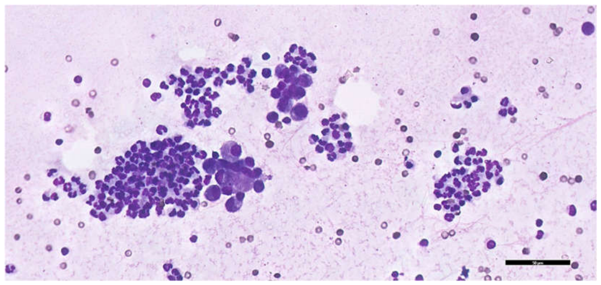
Figure 2.
Sysmex XN-1000V scattergrams from first exudate analysed in whole-blood mode. The left panel shows the white blood cell differential (WDF) channel, from which TNCC-WDF was obtained, whereas the right panel shows the white cell nucleated (WNR) channel, from which TNCC-WNR was obtained. D (dark blue), debris; LMNE (light blue), leukocyte populations other than basophils; MN, mononuclear cells, including macrophages, monocytes and lymphocytes; PMN, polymorphonuclear cells, predominantly neutrophils. In this exudate, the marked discrepancy of nucleated-cell events between the WDF and WNR channels resulted in a high inter-channel ratio (ΔTNC = 60.58). TNCC, total nucleated cell count; ΔTNC, delta total nucleated cells.
Figure 2.
Sysmex XN-1000V scattergrams from first exudate analysed in whole-blood mode. The left panel shows the white blood cell differential (WDF) channel, from which TNCC-WDF was obtained, whereas the right panel shows the white cell nucleated (WNR) channel, from which TNCC-WNR was obtained. D (dark blue), debris; LMNE (light blue), leukocyte populations other than basophils; MN, mononuclear cells, including macrophages, monocytes and lymphocytes; PMN, polymorphonuclear cells, predominantly neutrophils. In this exudate, the marked discrepancy of nucleated-cell events between the WDF and WNR channels resulted in a high inter-channel ratio (ΔTNC = 60.58). TNCC, total nucleated cell count; ΔTNC, delta total nucleated cells.
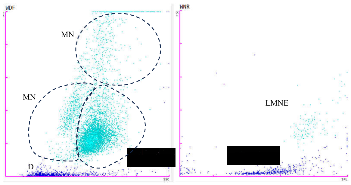
Figure 3.
Schematic representation of the cell tube block technique. (A) Filling of the capillary tube with the fluid sample. (B) Sealing of the lower end of the capillary tube with clay. (C) Centrifugation of the filled capillary tube to concentrate cells at the buffy coat interface. (D) Cutting the capillary tube at both ends of the buffy coat to recover the concentrated cellular material. (E) Transfer of the recovered cellular material into 10% neutral buffered formalin for fixation.
Figure 3.
Schematic representation of the cell tube block technique. (A) Filling of the capillary tube with the fluid sample. (B) Sealing of the lower end of the capillary tube with clay. (C) Centrifugation of the filled capillary tube to concentrate cells at the buffy coat interface. (D) Cutting the capillary tube at both ends of the buffy coat to recover the concentrated cellular material. (E) Transfer of the recovered cellular material into 10% neutral buffered formalin for fixation.
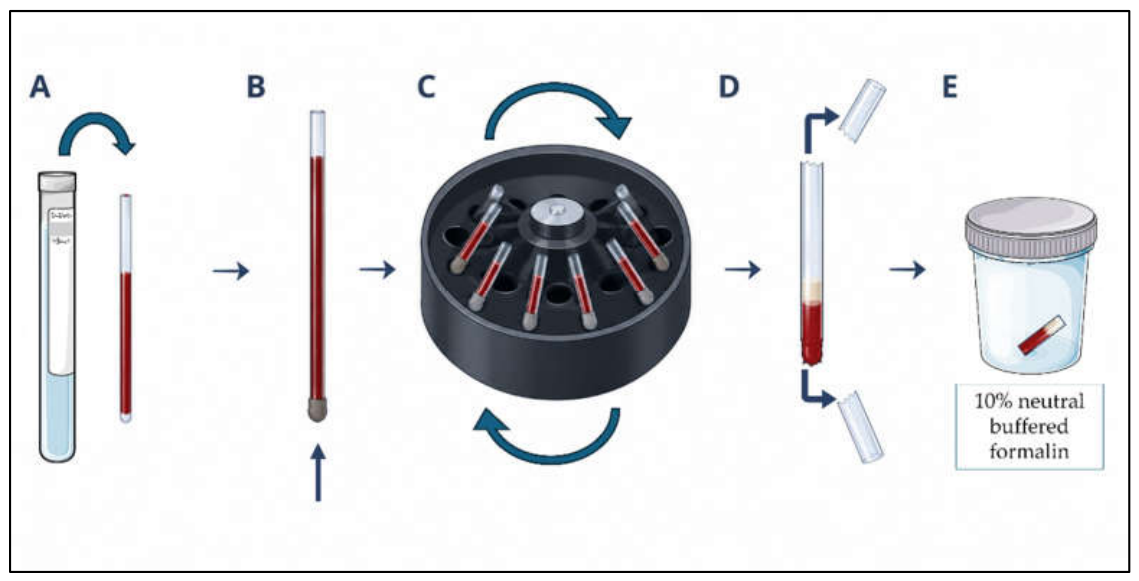
Figure 4.
Abdominal effusion cell tube block). Upper left: haematoxylin and eosin-stained section showing macrophage-rich inflammation with non-degenerate neutrophils. (Upper right): FCoV immunohistochemistry with monoclonal antibody clone FIPV3-70, demonstrating cytoplasmic immunoreactivity in macrophages. (Lower panel): higher-magnification view of immunoreactive macrophages (black arrows). Immunostaining was visualised using 3,3′-diaminobenzidine with haematoxylin counterstaining. Digital images acquired using Philips IntelliSite Pathology Solution 3.2. Scale bars = 100 μm (upper panels) and 50 μm (lower panel).
Figure 4.
Abdominal effusion cell tube block). Upper left: haematoxylin and eosin-stained section showing macrophage-rich inflammation with non-degenerate neutrophils. (Upper right): FCoV immunohistochemistry with monoclonal antibody clone FIPV3-70, demonstrating cytoplasmic immunoreactivity in macrophages. (Lower panel): higher-magnification view of immunoreactive macrophages (black arrows). Immunostaining was visualised using 3,3′-diaminobenzidine with haematoxylin counterstaining. Digital images acquired using Philips IntelliSite Pathology Solution 3.2. Scale bars = 100 μm (upper panels) and 50 μm (lower panel).
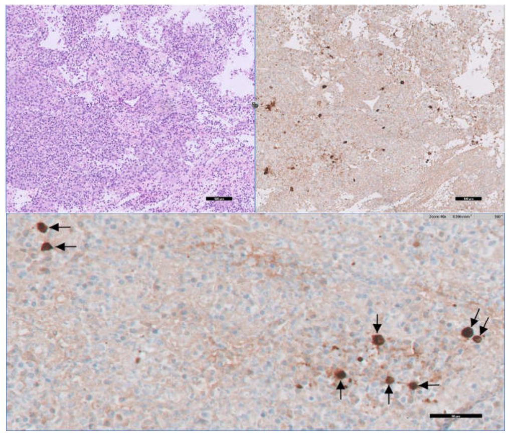

Table 1.
Clinical timeline of the effusive episode, treatment and follow-up.
| Date | Clinical milestone |
| October 2023 | Dyspnoea and pleural effusion with thyroid and cardiac disease. Fluid is described as a non-septic exudate. |
| 2–19 December 2023 | Initial abdominal effusion investigation. FCoV testing reported on 6 December. FIP recorded on 15 December, drainage on 17 December and hospitalisation on 19 December. |
| 20 December 2023 | Subcutaneous GS-441524 was initiated at 10 mg/kg once daily, with sublingual buprenorphine (0.02 mg/kg) administered approximately 2 h before each injection to improve tolerance. |
| January 2024 | Clinical improvement by 8 January. Haematocrit within the reference interval on 20 January. No free abdominal fluid detected at the assessment on 25 January. |
| 24 February–22 March 2024 | Temporary change to oral GS-441524 treatment, then return to subcutaneous GS-441524 injections because of difficulty administering oral medication. Antiviral treatment was completed on 22 March. |
| April 2024–2025 | Serum albumin-to-globulin ratio was 0.93 in April 2024, while FCoV antibody seropositivity persisted. Despite progressive weight loss noted in October 2024, α1-acid glycoprotein remained within the reference interval, and no recurrent effusion or other clinical findings supporting FIP relapse were documented. The cat therefore remained in clinical remission from FIP, while cardiac and thyroid disease continued to require management. |
| 16 January 2026 | Death during acute cardiopulmonary decompensation. No necropsy or confirmed FIP recurrence. |
Calendar dates refer to the clinical history and laboratory reports.
Table 2.
Serial effusion findings in two samples collected 9 days apart.
| Investigation | Initial sample Day 0 | Repeat sample Day 9 |
| Total protein (g/dL) | 5.46 | 8.14 |
| Albumin (g/dL) | 1.78 | 1.90 |
| Calculated globulins (g/dL) | 3.68 | 6.24 |
| Effusion A/G ratio | 0.48 | 0.30 |
| Rivalta test | Positive | Negative |
| Cellular count (cells/µL) | 7,267 | 9,117 |
| TNCC-WDF (× 103/µL) | 7,267 | 9,117 |
| TNCC-WNR (× 103/µL) | 0,120 | 4,240 |
| Reported ΔTNC | 60.58 | 2.15 |
| FIP Effusion Index | 126.2 | 7.17 |
All results refer to effusion. Day 0 denotes the first effusion sample, and day 9 denotes the repeat sample. ΔTNC and the FIP Effusion Index are dimensionless. Recalculation using albumin/globulins before rounding gives 125.2 and 7.06, respectively. These arithmetic differences do not represent additional measurements. The follow-up calculation assumes the published WDF/WNR convention. A/G, albumin-to-globulin ratio; FIP, feline infectious peritonitis; TNCC, total nucleated cell count; WDF, white blood cell differential channel; WNR, white cell nucleated channel.
Disclaimer/Publisher’s Note: The statements, opinions and data contained in all publications are solely those of the individual author(s) and contributor(s) and not of MDPI and/or the editor(s). MDPI and/or the editor(s) disclaim responsibility for any injury to people or property resulting from any ideas, methods, instructions or products referred to in the content. |
© 2026 by the authors. Licensee MDPI, Basel, Switzerland. This article is an open access article distributed under the terms and conditions of the Creative Commons Attribution (CC BY) license (http://creativecommons.org/licenses/by/4.0/).
Copyright: This open access article is published under a Creative Commons CC BY 4.0 license, which permit the free download, distribution, and reuse, provided that the author and preprint are cited in any reuse.