
Submitted:
01 October 2026
Posted:
06 October 2026
You are already at the latest version
Abstract
The dissemination of antibiotic resistance genes (ARGs) via conjugative plasmids is a major driver of the antimicrobial resistance crisis. The optrA gene, which confers cross-resistance to linezolid and florfenicol, is frequently detected in Enterococcus isolates and disseminated via plasmids. This study investigated the inhibitory effect of carvacrol on horizontal transfer of an optrA-carrying plasmid in porcine Enterococcus faecalis. In vitro conjugation assays showed that subinhibitory carvacrol (1/32 MIC, 16 µg/mL) significantly reduced conjugation efficiency in a dose- and time-dependent manner. In vivo (mouse model) treatment with 32 μg/mL carvacrol decreased transconjugant recovery by 62.8%. Mechanistically, carvacrol disrupted membrane integrity, depleted intracellular ATP, sup-pressed ROS production, reduced biofilm formation, and downregulated conjugation-associated genes (type VI secretion protein, conjugal transfer protein, prgB, asa1) as shown by transcriptomics and RT-qPCR. Collectively, carvacrol inhibits optrA plasmid transfer via multi-target mechanisms involving membrane damage, energy metabolism interference, and suppression of conjugation gene expression. This study highlights carvacrol as a natural agent to control the transfer of ARGs.
Keywords:
optrA
; Enterococcus faecalis
; carvacrol
; conjugation
; pheromone-responsive plasmid
1. Introduction
Antibiotic resistance has emerged as a significant danger to human health. Infectious diseases driven by resistant bacteria already claim some 700,000 lives each year worldwide, with projections suggesting more than 10 million annual deaths by 2050[1]. Horizontal gene transfer (HGT) among bacterial, occurring both within and across species, accelerates the spread of ARGs at an alarming rate. There are three classical HGT mechanisms have been described, including conjugation, transduction, and transformation[2]. Among these, conjugation (the direct transfer of genetic material between bacterial cells via physical contact) is recognized as the primary and most efficient mechanism driving the dissemination of ARGs[3,4].
In recent years, inhibiting plasmid-mediated conjugation has emerged as a strategy to control the spread of antibiotic resistance genes. Research has focused on natural products (e.g., cinnamic acid, flavonoids), T4SS-targeting small molecules, and biological tools such as CRISPR-Cas systems. However, most of these candidates remain at the laboratory stage, requiring lengthy safety evaluations and regulatory approvals before practical application. Approved as a feed additive in China (for broilers) and the EU (for multiple species), carvacrol promotes growth and gut health through antimicrobial, anti-inflammatory, and antioxidant mechanisms, serving as an effective alternative to antibiotic growth promoters[5]. Carvacrol is a phenolic monoterpenoid found abundantly in essential oils of Origanum and Thymus species, and could exert bactericidal effects through complex mechanisms, including disruption of cell membrane integrity, dissipation of the proton motive force, depletion of intracellular ATP, induction of reactive oxygen species (ROS), and inhibition of efflux pumps[6]. Additionally, subinhibitory concentrations of carvacrol have been shown to impair bacterial motility, biofilm formation, and quorum sensing[7,8]. However, the potential of carvacrol to inhibit conjugative plasmid transfer, particularly in Gram-positive bacteria such as E. faecalis, has not been comprehensively investigated.
Of particular concern is the emergence of resistance to oxazolidinones, exemplified by linezolid—a last-resort antibiotic for treating infections caused by vancomycin-resistant E. faecium (VRE) and methicillin-resistant Staphylococcus aureus (MRSA). The optrA gene, which encodes an ATP-binding cassette (ABC) transporter protein, confers transferable resistance to oxazolidinones and phenicols such as linezolid and florfenicol[9]. Recent surveillance studies have documented the widespread prevalence of optrA in Enterococcus isolates derived from livestock, food products, and clinical settings across 29 countries/regions of six continents [10,11,12]. Notably, optrA genes are frequently carried on pheromone-responsive conjugative plasmids, enabling efficient intercellular transfer with frequencies up to 10⁻² per donor cell[13]. This high-efficiency dissemination mechanism accelerates the spread of resistance genes, rendering the containment of plasmid-borne optrA a critical priority in combatting antimicrobial resistance.
This study aimed to evaluate the inhibitory effect of carvacrol on the horizontal transfer of an optrA-bearing plasmid in porcine E. faecalis and to elucidate the underlying molecular mechanisms through a combination of phenotypic, biochemical, and transcriptomic approaches. Our findings provide novel insights into the anti-conjugative properties of carvacrol and its potential applications in mitigating the dissemination of plasmid-mediated antibiotic resistance.
2. Materials and Methods
2.1. Bacterial Strains and Growth Conditions
The donor strain used in this study was E. faecalis E211, an isolate from swine that harbors the pheromone-responsive conjugative plasmid pE211 carrying the optrA gene [13]. The recipient strain was E. faecalis JH2-2, a plasmid-free, rifampicin-resistant and fusidic acid-resistant laboratory strain. Both strains were stored in our laboratory and routinely cultured in Brain Heart Infusion (BHI) broth or on BHI agar at 37 °C. Antibiotics were supplemented where appropriate as follows: florfenicol (10 μg/mL), rifampicin (32 μg/mL), and fusidic acid (32 μg/mL).
2.2. Minimum Inhibitory Concentration (MIC) Determination
The MIC of carvacrol for both donor and recipient strains was determined by the standard broth microdilution method according to CLSI guidelines. Briefly, overnight cultures were adjusted to 0.5 McFarland turbidity (~1 × 10⁸ CFU/mL) and diluted 100-fold in BHI broth. Serial two-fold dilutions of carvacrol (final concentration range of 8–512 μg/mL) were prepared in 96-well plates. Following inoculation with bacterial suspension, plates were incubated at 37 °C for 16–18 h, and the MIC was defined as the lowest concentration of carvacrol that inhibited visible bacterial growth.
2.3. Growth Curve Analysis
To evaluate the effect of subinhibitory concentrations of carvacrol on bacterial growth, overnight cultures of donor and recipient strains were diluted to OD₆₀₀ = 0.05 in BHI broth containing carvacrol at concentrations of 0 (control), 16, 32, 64 and 128μg/mL. Bacterial growth was monitored by measuring OD₆₀₀ at 0, 1, 2, 4, 6, 8, 12, and 24 h using a microplate reader. All experiments were performed in triplicate.
2.4. In Vitro Conjugation Assay
Quantitative conjugation assays were performed as previously described[14]. Overnight cultures of donor (E211) and recipient (JH2-2) strains were adjusted to 1 × 10⁸ CFU/mL and mixed at a donor-to-recipient ratio of 1:4. The mixtures were co-cultured in BHI broth containing carvacrol at concentrations of 0, 16, 32, and 64 μg/mL (corresponding to 0, 1/32, 1/16, and 1/8 MIC, respectively) at 37 °C. At 6 h post-co-culture, serial dilutions of the conjugation mixtures were plated onto BHI agar supplemented with florfenicol (10 μg/mL) and rifampicin (32 μg/mL) to select for transconjugants. Conjugation frequency was calculated as the number of transconjugants per donor cell. All assays were performed in triplicate. Transconjugant colonies were screened for the presence of the optrA gene and the donor-specific bcrA gene, as well as the recipient-specific int5801 gene, via PCR using previously described primers.
2.5. Murine In Vivo Conjugation Model
Six-week-old female ICR mice (n = 25) were obtained from Henan Skebes Biotechnology Co., Ltd. (Henan, China). All animal experiments were approved by the Animal Welfare and Ethical Review Committee of Anhui Agricultural University. Prior to inoculation, mice were acclimatized for 5 days and provided with sterile water and antibiotic-free feed. To establish intestinal colonization, mice were orally gavaged with 200 μL of a bacterial suspension containing E211 and JH2-2 (1 × 10⁹ CFU total) with or without carvacrol at concentrations of 32, 64, and 128 μg/mL. Fecal samples were collected 24 h post-gavage, homogenized in PBS, serially diluted, and plated onto BHI agar supplemented with florfenicol (10 μg/mL), rifampicin (32 μg/mL), and fusidic acid (32 μg/mL) to enumerate transconjugants. Putative transconjugants were confirmed by PCR detection of the optrA gene and the recipient-specific int5801 gene.
2.6. Measurement of Intracellular ROS Levels
ROS levels were measured using the fluorescent probe 2′,7′-dichlorodihydrofluorescein diacetate (DCFH-DA) (Beyotime Biotechnology, Shanghai, China). Bacterial suspensions (OD₆₀₀ = 0.5) were incubated with carvacrol at concentrations of 0, 16, 32, 64, and 128 μg/mL for 1 h at 37 °C. DCFH-DA was added at a final concentration of 10 μmol/L, and the mixture was incubated for an additional 30 min. Fluorescence intensity was measured using a microplate reader at an excitation wavelength of 488 nm and an emission wavelength of 525 nm.
2.7. Measurement of ATP Levels
Intracellular and extracellular ATP concentrations were measured using a bioluminescence-based ATP Assay Kit (Beyotime Biotechnology, Shanghai, China). Bacteria were treated with carvacrol at concentrations ranging from 16 to 512 μg/mL for 1 h at 37 °C. After centrifugation, the supernatant (extracellular fraction) was collected, and the bacterial pellet was lysed to release intracellular ATP. ATP concentrations were determined following the manufacturer’s protocol using a luminometer.
2.8. Membrane Permeability Assay
Membrane integrity was assessed by measuring the uptake of propidium iodide (PI) (Beyotime, Shanghai, China), a membrane-impermeable fluorescent dye that enters cells with compromised membranes. Bacteria were treated with carvacrol as described above, followed by incubation with PI at a final concentration of 10 μmol/L for 30 min at 37 °C. Fluorescence intensity was measured using a microplate reader at an excitation wavelength of 535 nm and an emission wavelength of 615 nm.
2.9. Scanning Electron Microscopy (SEM)
Bacterial morphology was visualized by SEM. Donor, recipient, and conjugation mixture cultures were treated with carvacrol (32 μg/mL) for 2 h at 37 °C. Bacterial pellets were fixed with 2.5% glutaraldehyde at 4 °C overnight, dehydrated through an ethanol gradient and acetone, subjected to critical point drying, sputter-coated with gold–palladium, and imaged using a field-emission scanning electron microscope (S-4800, Hitachi, Japan).
2.10. Biofilm Formation Assay
Biofilm formation was assessed using a crystal violet staining method in 24-well plates. Bacterial cultures (OD₆₀₀ = 0.05) were incubated with carvacrol at concentrations of 32, 64, and 128 μg/mL for 24 h at 37 °C. After incubation, the supernatant was removed, plates were washed twice with PBS, and adherent biofilms were fixed with methanol for 15 min, stained with 0.1% crystal violet for 15 min, destained with 33% glacial acetic acid, and the optical density at 570 nm was measured.
2.11. Transcriptomic Analysis
Donor strain E211 and recipient strain JH2-2 were grown to mid-log phase in BHI broth with or without carvacrol (32 μg/mL) for 8 h. Total RNA was extracted using TRIzol reagent, and ribosomal RNA was removed via Ribo-Zero treatment. Strand-specific RNA-seq libraries were prepared using an ALFA-SEQ RNA Library Prep Kit and sequenced on an Illumina platform, yielding 150 bp paired-end reads. Raw reads were trimmed and quality-filtered, and gene expression levels were quantified and normalized using transcripts per million (TPM). Principal component analysis (PCA) was conducted base on gene expression levels, and sample clustering was evaluated according to their distribution along the first two principal component (PCA1 and PCA2). Differentially expressed genes (DEGs) were identified using DESeq2 with criteria of |log₂ fold change| >2 and adjusted P-value < 0.05. GO enrichment and KEGG pathway analyses were performed using clusterProfiler.
2.12. RNA Extraction and RT-qPCR Validation
Total RNA was extracted from the donor strain E211 treated with or without carvacrol (32 μg/mL) using the Bacterial RNA Extraction Kit (Vazyme, Nanjing, China). Reverse transcription was performed using a HiScript III 1st Strand cDNA Synthesis Kit, and quantitative PCR was performed using SYBR Green Master Mix on a CFX96 real-time PCR system (Bio-Rad, Hercules, CA, USA). The primers used are listed in Supplementary Table S1. Relative expression levels were calculated using the 2⁻ΔΔCT method with 16S rRNA as the endogenous control.
2.13. Statistical Analysis
All experiments were performed in triplicate, and data are expressed as mean ± standard deviation (SD). Statistical significance was determined using one-way ANOVA followed by Tukey’s post-hoc test for multiple comparisons. A P-value < 0.05 was considered statistically significant.
3. Results
3.1. Carvacrol Suppresses optrA Plasmid Transfer In Vitro and In Vivo
The MIC of carvacrol against both donor (E. faecalis E211) and recipient (E. faecalis JH2-2) strains were found to be 512 μg/mL. Growth curve analysis demonstrated that carvacrol at low concentrations (≤ 64 μg/mL for E211 and ≤ 32 μg/mL for JH2-2) did not significantly inhibit bacterial growth (Figure 1A,B). Consistent with the in vitro observations, in vivo administration of carvacrol significantly reduced transconjugant recovery from mouse fecal samples. Therefore, sub-MIC concentrations (32, 64, and 128 μg/mL) were selected for vivo and 16, 32, and 64 μg/mL for vitro conjugation assays, respectively. In vivo, no transconjugants were recovered from fecal samples of the PBS-treated group, indicating the absence of endogenous conjugation between the donor strain E211 and the recipient strain JH2-2 in the murine gut. In contrast, mice inoculated with both donor and recipient strains exhibited the highest transconjugant counts. Carvacrol treatment (32–128 µg/mL) resulted in a marked reduction in transconjugant recovery compared with the bacteria-only control. Specifically, carvacrol at 32, 64, and 128 µg/mL reduced transconjugant numbers by 62.8%, 65.7%, and 80.4% (Figure 1C). In vitro, after 6 h post-co-culture with 16μg/mL carvacrol, conjugation efficiency was decreased by 58.39%, 32 μg/mL decreased by 70.15%, and 64μg/mL decreased by 82.06% relative to the control, respectively. Which significantly reduced the transfer frequency of the optrA-carrying plasmid from E211 to JH2-2 in a dose-dependent manner. (Figure 1D). These findings demonstrate that carvacrol effectively suppresses the conjugative transfer of the optrA-carrying plasmid in vivo and vitro.
3.2. Carvacrol Alters Redox Homeostasis and Energy Metabolism
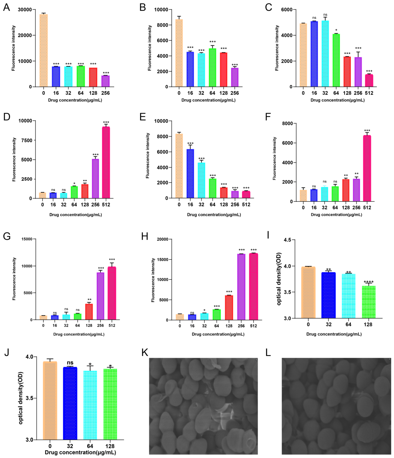
Treatment with carvacrol significantly altered intracellular ROS levels. Contrary to the pro-oxidant effects often described in certain bacteria, carvacrol (32–128 μg/mL) significantly reduced ROS levels in both donor and recipient E. faecalis strains, indicating an alteration of redox homeostasis rather than classical oxidative burst induction (Figure 2A,B). Carvacrol treatment also markedly affected ATP metabolism. Intracellular ATP levels in both donor and recipient strains decreased progressively with increasing carvacrol concentrations (16–512 μg/mL). Conversely, extracellular ATP levels increased in a dose-dependent manner (Figure 2C–F), suggesting that carvacrol compromises cell membrane integrity, leading to ATP leakage. This energy depletion is likely to impair bacterial physiological functions, including conjugation-related processes.
3.3. Carvacrol Compromises Membrane Integrity and Reduces Biofilm Formation
Membrane permeability, assessed via PI uptake, increased significantly at carvacrol concentrations ≥ 64 μg/mL in both strains (Figure 2G,H). Notably, the recipient strain JH2-2 was more sensitive to carvacrol-induced membrane damage than the donor strain, with detectable permeability changes occurring at 64 μg/mL (P < 0.001), whereas E211 required 128 μg/mL to exhibit significant membrane disruption (P < 0.001). SEM imaging provided direct visualization of membrane damage. Treatment with 32 μg/mL carvacrol induced marked morphological alterations in the conjugation mixture, including bacterial aggregation, surface indentation, and overt membrane rupture, while control bacteria appeared smooth and intact (Figure 2K,L). Interestingly, carvacrol at the same concentration caused no obvious damage to donor or recipient bacteria when cultured separately (Supplementary Figure S1), suggesting that the conjugation process as determined by cell–cell contact may itself sensitize bacteria to carvacrol. Biofilm formation was assessed by crystal violet staining. Carvacrol significantly inhibited biofilm formation in both donor and recipient strains in a concentration-dependent manner (Figure 2I,J). For E211, biofilm formation was significantly reduced at 32 μg/mL (P < 0.01) and further suppressed at 64 μg/mL (P < 0.001) and 128 μg/mL (P < 0.001). For JH2-2, significant inhibition was observed only at 64 μg/mL (P < 0.01), highlighting strain-dependent susceptibility.
3.4. Transcriptomic Analysis Reveals Dysregulation of Metabolic Pathways and Conjugation Genes
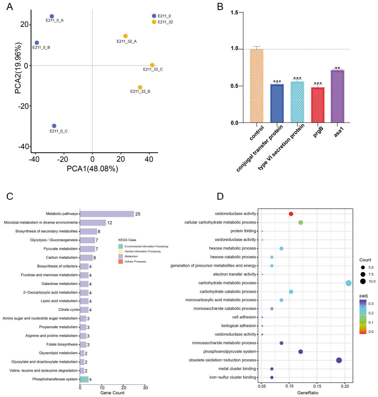
To gain insights into the molecular mechanisms underlying carvacrol-mediated conjugation inhibition, RNA-seq was performed on donor E211 and recipient JH2-2 cells treated with 32 μg/mL carvacrol. Principal component analysis (PCA) showed clear separation between carvacrol-treated and control groups in both E211 and JH2-2, indicating that carvacrol significantly alters their overall profiles (Figure 3A, Supplementary Figure S2).
For donor strain E211, KEGG pathway enrichment of upregulated DEGs showed enrichment in metabolic pathways, fructose and mannose metabolism, amino acid biosynthesis, ABC transporters, and the two-component system (Supplementary Figure S3A). Downregulated DEGs were enriched in metabolic pathways, amino acid biosynthesis, glycolysis/gluconeogenesis, and pyruvate metabolism (Figure 3C). GO enrichment analysis of downregulated genes revealed significant representation in carbohydrate metabolic processes (cellular carbohydrate metabolic process, hexose metabolic process), oxidoreductase activity (acting on the aldehyde or oxo group of donors), and protein folding (Figure 4D). These results suggest that carvacrol disrupts energy metabolism, redox balance, and protein homeostasis in donor cells.
For recipient strain JH2-2, upregulated DEGs were enriched in metabolic pathways, microbial metabolism in diverse environments, and the two-component system (Supplementary Figure S3B). Downregulated DEGs were enriched in phosphotransferase system (PTS)-mediated sugar transport, membrane-associated processes, and transporter activity (Supplementary Figure S3C). GO analysis of downregulated genes prominently featured the phosphoenolpyruvate-dependent sugar phosphotransferase system, protein-N(PI)-phosphohistidine-sugar phosphotransferase activity, and transmembrane transporter activity (Supplementary Figure S4B). Strikingly, membrane-related GO terms, including integral component of plasma membrane and integral component of membrane, were also downregulated, indicating that carvacrol impairs membrane structural integrity in recipient cells.
Figure 3.
(A) PCA of E. faecalis E211 following treatment with 32μg/mL carvacrol compared with untreated control. (B) Effects of carvacrol on the gene expression level of donor E. faecalis E211. (C) KEGG pathway enrichment analysis of downregulated expressed genes in E. faecalis E211 after treatment with 32μg/mL carvacrol. (D) GO enrichment analysis diagram of differentially downregulated expressed genes in the E. faecalis E211 after treatment with 32μg/mL carvacrol.
Figure 3.
(A) PCA of E. faecalis E211 following treatment with 32μg/mL carvacrol compared with untreated control. (B) Effects of carvacrol on the gene expression level of donor E. faecalis E211. (C) KEGG pathway enrichment analysis of downregulated expressed genes in E. faecalis E211 after treatment with 32μg/mL carvacrol. (D) GO enrichment analysis diagram of differentially downregulated expressed genes in the E. faecalis E211 after treatment with 32μg/mL carvacrol.
3.5. Carvacrol Suppresses the Expression of Conjugation-Associated Genes
To validate the RNA-seq findings, the expression levels of key conjugation-related genes were quantified by RT-qPCR in donor strain E211 after exposure to 32 μg/mL carvacrol. As shown in Figure 3B, carvacrol significantly suppressed the expression of the type VI secretion protein gene (P < 0.001), the conjugal transfer protein gene (P < 0.001), prgB (P < 0.001), and asa1 (P < 0.001). Conversely, the suppression of prgB, which encodes a surface adhesin essential for mating pair formation, and asa1, involved in aggregation substance synthesis, is particularly relevan given the role of these genes in pheromone-responsive plasmid transfer. These results collectively indicate that carvacrol inhibits conjugation by downregulating critical genes required for efficient plasmid transfer.
4. Discussion
The global dissemination of antibiotic resistance genes via horizontal transfer is a defining challenge of the antimicrobial resistance crisis[15]. Among the various mobile genetic elements, pheromone-responsive conjugative plasmids in E. faecalis are of particular concern because of their capacity to efficiently disseminate resistance genes, including the critical cross-resistance gene optrA, among bacterial populations[13,16]. In this study, we demonstrated that subinhibitory concentrations of the natural phenolic compound carvacrol effectively reduce optrA plasmid transfer in porcine E. faecalis both in vitro and in vivo. Our integrated approach, combining biochemical assays, transcriptomics, and RT-qPCR, revealed that carvacrol inhibits conjugation through a multi-faceted mechanism involving membrane damage, energy depletion, and suppression of key conjugation-related genes.
4.1. Carvacrol Inhibits Conjugation via Membrane Disruption
Conjugation requires physical contact between donor and recipient cells, a process mediated by surface adhesins such as aggregation substance encoded by asa1 and the mating channel-forming protein PrgB[17,18]. Our SEM observations directly demonstrated that 32 μg/mL carvacrol induced morphological abnormalities and overt membrane rupture specifically in the conjugation mixture, while donor or recipient cells cultured alone remained intact. This observation is consistent with the interpretation that actively conjugating cells undergo physiological changes, increased metabolic activity, membrane fluidity, or cell-surface contact that render them more vulnerable to carvacrol’s membrane-inserting activity. Once inserted into the phospholipid bilayer, carvacrol disrupts membrane order and increases permeability, leading to the leakage of essential intracellular components[19]. This interpretation is supported by our membrane permeability data, which showed increased PI uptake at carvacrol concentrations ≥ 64 μg/mL in both strains, and ATP leakage results, which demonstrated dose-dependent depletion of intracellular ATP and accumulation of extracellular ATP. The concentrations required to observe detectable membrane damage (64–128 μg/mL) were higher than those that sufficed to significantly reduce conjugation efficiency (16 μg/mL). This discrepancy indicates that while membrane disruption certainly contributes to the observed phenotype, additional, more sensitive mechanisms are likely operational at lower concentrations, as discussed below.
4.2. Energy Metabolism and Redox Homeostasis as Critical Targets
Conjugative DNA processing and active translocation are energy-demanding processes requiring ATP[20]. Our data show that carvacrol dose-dependently depletes intracellular ATP pools, a finding consistent with previous studies documenting ATP depletion as a primary antimicrobial effect of carvacrol[21,22] . The increase in extracellular ATP further supports the notion that carvacral increases membrane permeability. This energetic collapse would be expected to negatively impact all ATP-dependent steps of conjugation, including relaxosome assembly, unwinding of the plasmid origin of transfer, and active pumping of the single-stranded DNA through the type IV secretion system (T4SS).
Transcriptomic analysis of the donor strain E211 shed further light on this energy crisis. KEGG pathway analysis revealed downregulation of glycolysis/gluconeogenesis, pyruvate metabolism, and carbohydrate catabolic processes, while GO analysis of downregulated genes enriched the terms “cellular carbohydrate metabolic process”, “hexose catabolic process”, “generation of precursor metabolites and energy”, and “oxidoreductase activity” (acting on aldehyde or oxo donors with NAD/NADP as acceptors). The downregulation of oxidoreductase genes is particularly noteworthy because these enzymes form an integral part of the electron transport chain, and their suppression would impair proton motive force generation and ATP synthesis. Indeed, the observed reduction in intracellular ROS levels (32 μg/mL carvacrol) is somewhat unexpected given that carvacrol has been reported to induce oxidative stress in E. coli [23]. The likely explanation lies in the lower sub-MIC carvacrol concentrations used in our study (1/16 to 1/2 MIC), in contrast to the bactericidal concentrations used in many ROS studies. At these sub-MIC levels, general metabolic suppression—including that of respiratory chain activity—may reduce ROS generation. Additionally, genome analysis of E211 reveals the presence of several putative stress response regulators, suggesting potential strain-specific differences in the response to carvacrol.
4.3. Suppression of Conjugation-Associated Gene Expression
The RT-qPCR results provide a molecular rationale for the conjugation inhibition observed. Suppression of prgB and asa1 expression is particularly relevant because these genes are central to pheromone-responsive plasmid transfer in E. faecalis. PrgB encodes an aggregation substance that mediates donor–recipient cell contact, while asa1 is directly involved in the synthesis of the aggregation substance extracellular protein[17]. Both are under the control of the pheromone-sensing two-component regulatory system that activates plasmid transfer. The suppression of these genes at the transcriptional level would directly impair the formation of mating aggregates and subsequent DNA transfer. Furthermore, downregulation of type VI secretion protein and conjugal transfer protein genes likely impacts the assembly or function of the T4SS, the macromolecular channel that transports the single-stranded DNA substrate into the recipient cell. The significance of our findings is supported by another study also showing that carvacrol directly binds to and inhibits SarA activity, a global regulator controlling virulence gene expression in S. aureus[24]. A similar regulatory interference may occur in E. faecalis, potentially involving homologs of the pheromone-responsive two-component system (TraA/TraB). The upregulation of traA (P < 0.001) and the lack of change in traE may represent a compensatory transcriptional response, but the net effect—reduced conjugation efficiency—indicates that the downregulation of functionally dominant genes (e.g., prgB, asa1, T4SS components) overrides any potential compensatory mechanisms.
4.4. In Vivo Relevance and Implications
The in vivo mouse model demonstrated that oral administration of carvacrol significantly reduced optrA plasmid transfer in the intestinal environment, with a 62.8% reduction in transconjugants at 32 μg/mL carvacrol. This finding is of translational relevance because the gut is a major reservoir for antibiotic resistance genes and a primary site for conjugative dissemination among commensal and pathogenic bacteria[25]. The effective concentration (32 μg/mL) was well below the MIC for both donor and recipient strains (512 μg/mL), a finding that aligns with a growing number of studies showing that sub-MIC concentrations of natural compounds can interfere with horizontal gene transfer without exerting selective pressure for resistance.
The potential advantages of using conjugation inhibitors as adjunctive strategies are threefold: they reduce resistance propagation, do not exert direct growth-inhibitory pressure (lowering the risk of resistance selection), and target a process required for spread rather than growth per se. However, carvacrol is hydrophobic and exhibits low water solubility, which could limit its bioavailability. Encapsulation formulations, such as nanoemulsions or liposomal delivery systems, have been successfully used to enhance the stability and delivery of essential oil components and warrant investigation for in vivo applications of carvacrol as an anti-conjugation agent[26].
4.5. Study Limitations and Future Directions
This study has several limitations. First, we used only one donor–recipient pair in the conjugation assays, which included a single pheromone-responsive plasmid type (pE211). The generalizability of our findings to other plasmid families (e.g., pMG1-like, Inc18 family) and other Gram-positive or Gram-negative species remains to be determined. Second, the concentrations of carvacrol used in vivo (32–128 μg/mL) were higher than those typically achieved through dietary intake or essential oil supplementation. Future work should include pharmacokinetic studies to determine achievable gut concentrations and to optimize dosing regimens. Additionally, although our RNA-seq and RT-qPCR data identified candidate genes affected by carvacrol, direct evidence of protein-level effects or physical interactions between carvacrol and regulatory proteins is lacking. Future studies employing surface plasmon resonance or bacterial two-hybrid assays to evaluate direct binding of carvacrol to TraA/TraB two-component system regulators would be valuable.
5. Conclusions
In summary, this study demonstrates that subinhibitory concentrations of carvacrol effectively inhibit the conjugative transfer of the optrA plasmid in E. faecalis in both in vitro and in vivo settings. The anti-conjugative activity of carvacrol involves a multi-target mechanism encompassing membrane disruption, ATP depletion, suppression of carbohydrate and energy metabolism, and transcriptional downregulation of key conjugation-associated genes, including prgB, asa1, type VI secretion protein, and conjugal transfer protein. These findings suggest that carvacrol, as a naturally derived compound, could be developed as an adjunctive agent to limit the horizontal spread of plasmid-borne antibiotic resistance genes, especially within the gut environment.:
Supplementary Materials
The following supporting information can be downloaded at the website of this paper posted on Preprints.org.
Funding
This research was funded by the National Key Research and Development Program of China (NO. 2024YFE0106300), the Key Project of Natural Science Foundation of Anhui Provincial Department of Education (2024AH050468), the National Natural Science Foundation of China (32573427) and the UK Research and Innovation (MR/Y015223/1).
Institutional Review Board Statement
The animal study protocol was approved by the Institutional Animal Care and Use Committee of Anhui Agricultural University (IACUC-20240620, 2024-06-20).
Informed Consent Statement
Not applicable.
Conflicts of Interest
The authors declare no conflicts of interest.
Ethics declarations
All animal experiments in this study were approved by Institutional Animal Care and Use Committee (IACUC) of Anhui Agricultural University under permit number IACUC-20240620.
Consent of publication
Not applicable.
References
- Huemer, M.; Mairpady Shambat, S.; Brugger, S. D.; Zinkernagel, A. S. Antibiotic resistance and persistence-Implications for human health and treatment perspectives. EMBO Rep. 2020, 21(12), e51034. [Google Scholar] [CrossRef] [PubMed]
- Arnold, B. J.; Huang, I. T.; Hanage, W. P. Horizontal gene transfer and adaptive evolution in bacteria. Nat. Rev. Microbiol. 2022, 20(4), 206–18. [Google Scholar] [CrossRef] [PubMed]
- Wang, X.; Zhang, H.; Yu, S.; Li, D.; Gillings, M. R.; Ren, H.; Mao, D.; Guo, J.; Luo, Y. Inter-plasmid transfer of antibiotic resistance genes accelerates antibiotic resistance in bacterial pathogens. ISME J. 2024, 18(1), wrad032. [Google Scholar] [CrossRef] [PubMed]
- Castañeda-Barba, S.; Top, E. M.; Stalder, T. Plasmids, a molecular cornerstone of antimicrobial resistance in the One Health era. Nat. Rev. Microbiol. 2024, 22(1), 18–32. [Google Scholar] [CrossRef] [PubMed]
- Liu, X.; Li, X.; Chen, R.; Liu, J.; Liu, R.; Zhao, R.; Luo, A.; Zhao, J.; Hao, J.; Yang, S.; Chen, A. Effects of dietary supplementation with a thymol-carvacrol blend on growth performance and intestinal health of poultry. Front. Vet. Sci. 2025, 12, 1739666. [Google Scholar] [CrossRef] [PubMed]
- Friedman, M. Chemistry and multibeneficial bioactivities of carvacrol (4-isopropyl-2-methylphenol), a component of essential oils produced by aromatic plants and spices. J. Agric. Food Chem. 2014, 62(31), 7652–70. [Google Scholar] [CrossRef] [PubMed]
- Burt, S. A.; Ojo-Fakunle, V. T.; Woertman, J.; Veldhuizen, E. J. The natural antimicrobial carvacrol inhibits quorum sensing in Chromobacterium violaceum and reduces bacterial biofilm formation at sub-lethal concentrations. PLoS ONE 2014, 9(4), e93414. [Google Scholar] [CrossRef] [PubMed]
- Li, J. G.; Chen, X. F.; Lu, T. Y.; Zhang, J.; Dai, S. H.; Sun, J.; Liu, Y. H.; Liao, X. P.; Zhou, Y. F. Increased activity of β-Lactam antibiotics in combination with carvacrol against MRSA bacteremia and catheter-associated biofilm infections. ACS Infect. Dis. 2023, 9(12), 2482–93. [Google Scholar] [CrossRef] [PubMed]
- Wang, Y.; Lv, Y.; Cai, J.; Schwarz, S.; Cui, L.; Hu, Z.; Zhang, R.; Li, J.; Zhao, Q.; He, T.; Wang, D.; Wang, Z.; Shen, Y.; Li, Y.; Feßler, A. T.; Wu, C.; Yu, H.; Deng, X.; Xia, X.; Shen, J. A novel gene, optrA, that confers transferable resistance to oxazolidinones and phenicols and its presence in Enterococcus faecalis and Enterococcus faecium of human and animal origin. J. Antimicrob. Chemother. 2015, 70(8), 2182–90. [Google Scholar] [CrossRef] [PubMed]
- Chen, W.; Wang, Q.; Wu, H.; Xia, P.; Tian, R.; Li, R.; Xia, L. Molecular epidemiology, phenotypic and genomic characterization of antibiotic-resistant enterococcal isolates from diverse farm animals in Xinjiang, China. Sci. Total Environ. 2024, 912, 168683. [Google Scholar] [CrossRef] [PubMed]
- Xuan, H.; Xia, L.; Schwarz, S.; Jia, H.; Yao, X.; Wang, S.; Li, R.; Wei, J.; Li, Z.; Shao, D.; Liu, K.; Qiu, Y.; Ma, Z.; Li, B. Various mobile genetic elements carrying optrA in Enterococcus faecium and Enterococcus faecalis isolates from swine within the same farm. J. Antimicrob. Chemother. 2023, 78(2), 504–11. [Google Scholar] [CrossRef] [PubMed]
- Wang, Y.; Li, X.; Fu, Y.; Chen, Y.; Wang, Y.; Ye, D.; Wang, C.; Hu, X.; Zhou, L.; Du, J.; Shen, J.; Xia, X. Association of florfenicol residues with the abundance of oxazolidinone resistance genes in livestock manures. J. Hazard. Mater. 2020, 399, 123059. [Google Scholar] [CrossRef] [PubMed]
- Shang, Y.; Li, D.; Shan, X.; Schwarz, S.; Zhang, S. M.; Chen, Y. X.; Ouyang, W.; Du, X. D. Analysis of two pheromone-responsive conjugative multiresistance plasmids carrying the novel mobile optrA locus from Enterococcus faecalis. Infect. Drug Resist. 2019, 12, 2355–62. [Google Scholar] [CrossRef] [PubMed]
- Shan, X.; Li, C.; Zhang, L.; Zou, C.; Yu, R.; Schwarz, S.; Shang, Y.; Li, D.; Brenciani, A.; Du, X. D. poxtA amplification and mutations in 23S rRNA confer enhanced linezolid resistance in Enterococcus faecalis. J. Antimicrob. Chemother. 2024, 79(12), 3199–203. [Google Scholar] [CrossRef] [PubMed]
- Starikova, I.; Al-Haroni, M.; Werner, G.; Roberts, A. P.; Sørum, V.; Nielsen, K. M.; Johnsen, P. J. Fitness costs of various mobile genetic elements in Enterococcus faecium and Enterococcus faecalis. J. Antimicrob. Chemother. 2013, 68(12), 2755–65. [Google Scholar] [CrossRef] [PubMed]
- Zou, J.; Tang, Z.; Yan, J.; Liu, H.; Chen, Y.; Zhang, D.; Zhao, J.; Tang, Y.; Zhang, J.; Xia, Y. Dissemination of linezolid resistance through sex pheromone plasmid transfer in Enterococcus faecalis. Front. Microbiol. 2020, 11, 1185. [Google Scholar] [CrossRef] [PubMed]
- Sterling, A. J.; Snelling, W. J.; Naughton, P. J.; Ternan, N. G.; Dooley, J. S. G. Competent but complex communication: The phenomena of pheromone-responsive plasmids. PLoS Pathog. 2020, 16(4), e1008310. [Google Scholar] [CrossRef] [PubMed]
- Wagner, T. M.; Pöntinen, A. K.; Fenzel, C. K.; Engi, D.; Janice, J.; Almeida-Santos, A. C.; Tedim, A. P.; Freitas, A. R.; Peixe, L.; van Schaik, W.; Johannessen, M.; Hegstad, K. Interactions between commensal Enterococcus faecium and Enterococcus lactis and clinical isolates of Enterococcus faecium. FEMS Microbes 2024, 5, xtae009. [Google Scholar] [CrossRef] [PubMed]
- Veldhuizen, E. J.; Tjeerdsma-van Bokhoven, J. L.; Zweijtzer, C.; Burt, S. A.; Haagsman, H. P. Structural requirements for the antimicrobial activity of carvacrol. J. Agric. Food Chem. 2006, 54(5), 1874–9. [Google Scholar] [CrossRef] [PubMed]
- Valenzuela-Gómez, F.; Arechaga, I.; Cabezón, E. Nanopore sensing reveals a preferential pathway for the co-translocational unfolding of a conjugative relaxase-DNA complex. Nucleic Acids Res. 2023, 51(13), 6857–69. [Google Scholar] [CrossRef] [PubMed]
- Monzote, L.; Stamberg, W.; Staniek, K.; Gille, L. Toxic effects of carvacrol, caryophyllene oxide, and ascaridole from essential oil of Chenopodium ambrosioides on mitochondria. Toxicol. Appl. Pharmacol. 2009, 240(3), 337–47. [Google Scholar] [CrossRef] [PubMed]
- Niu, C.; Wang, C.; Yang, Y.; Chen, R.; Zhang, J.; Chen, H.; Zhuge, Y.; Li, J.; Cheng, J.; Xu, K.; Chu, M.; Ren, C.; Zhang, C.; Jia, C. Carvacrol induces Candida albicans apoptosis associated with Ca(2+)/Calcineurin pathway. Front. Cell. Infect. Microbiol. 2020, 10, 192. [Google Scholar] [CrossRef] [PubMed]
- Di Pasqua, R.; Betts, G.; Hoskins, N.; Edwards, M.; Ercolini, D.; Mauriello, G. Membrane toxicity of antimicrobial compounds from essential oils. J. Agric. Food Chem. 2007, 55(12), 4863–70. [Google Scholar] [CrossRef] [PubMed]
- Selvaraj, A.; Valliammai, A.; Muthuramalingam, P.; Priya, A.; Suba, M.; Ramesh, M.; Karutha Pandian, S. Carvacrol targets SarA and CrtM of methicillin-resistant Staphylococcus aureus to mitigate biofilm formation and staphyloxanthin synthesis: An in vitro and in vivo approach. ACS Omega 2020, 5(48), 31100–114. [Google Scholar] [CrossRef] [PubMed]
- Klümper, U.; Riber, L.; Dechesne, A.; Sannazzarro, A.; Hansen, L. H.; Sørensen, S. J.; Smets, B. F. Broad host range plasmids can invade an unexpectedly diverse fraction of a soil bacterial community. ISME J. 2015, 9(4), 934–45. [Google Scholar] [CrossRef] [PubMed]
- Mechmechani, S.; Gharsallaoui, A.; Fadel, A.; El Omari, K.; Khelissa, S.; Hamze, M.; Chihib, N. E. Microencapsulation of carvacrol as an efficient tool to fight Pseudomonas aeruginosa and Enterococcus faecalis biofilms. PLoS ONE 2022, 17(7), e0270200. [Google Scholar] [CrossRef] [PubMed]
Figure 1.
| Effects of different concentrations of concentrations of carvacrol on E. faecalis. (A) Growth curves of donor E. faecalis E211 under different concentrations of carvacrol. (B) Growth curves of recipient E. faecalis JH2-2 under different concentrations of carvacrol. (C) Statistics of the number of conjugant colonies on resistance plates in fecal samples of mice in each group. (D) Conjugation efficiency after 6 hours of treatment with different concentrations of carvacrol.
Figure 1.
| Effects of different concentrations of concentrations of carvacrol on E. faecalis. (A) Growth curves of donor E. faecalis E211 under different concentrations of carvacrol. (B) Growth curves of recipient E. faecalis JH2-2 under different concentrations of carvacrol. (C) Statistics of the number of conjugant colonies on resistance plates in fecal samples of mice in each group. (D) Conjugation efficiency after 6 hours of treatment with different concentrations of carvacrol.
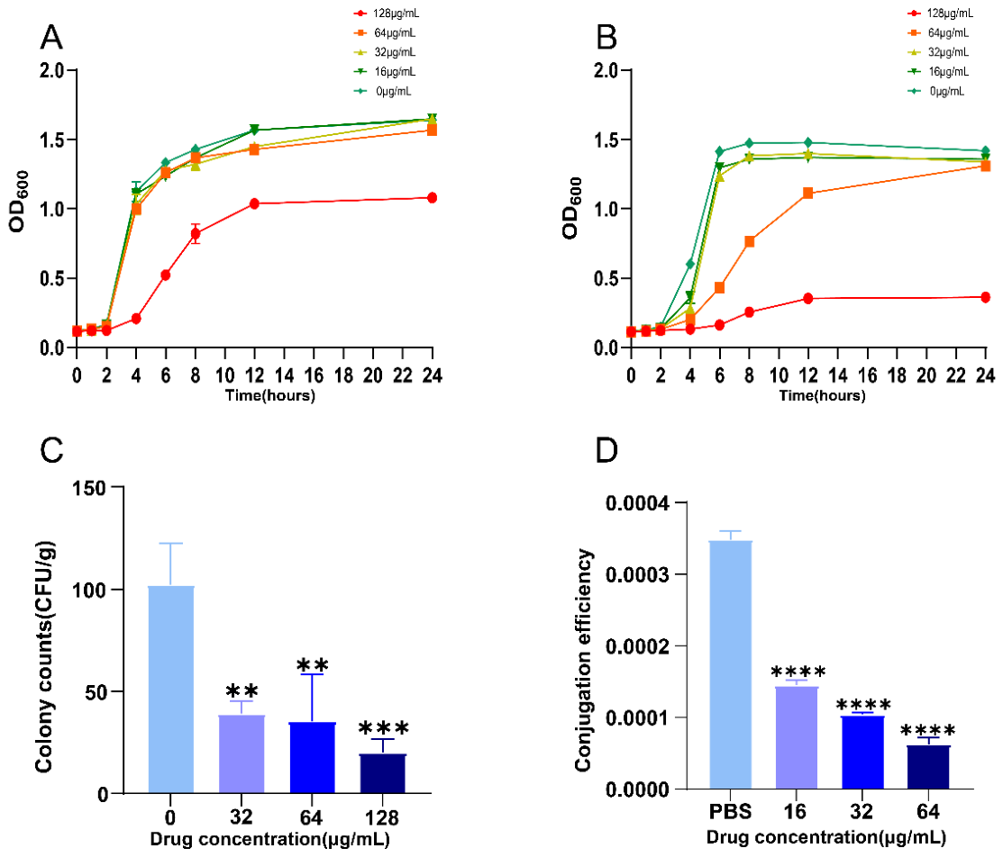
Figure 2.
Biochemical parameter analysis of Enterococcus under carvacrol treatment | (A) Effect of different concentrations of carvacrol on reactive oxygen species (ROS) levels in donor E. faecalis E211. (B) Effect of different concentrations of carvacrol on reactive oxygen species (ROS) levels in recipient E. faecalis JH2-2. (C) Effect of different concentrations of carvacrol on intracellular ATP levels in donor E. faecalis E211. (D) Effects of different concentrations of carvacrol on extracellular ATP levels in donor E. faecalis E211. (E) Effect of different concentrations of carvacrol on intracellular ATP levels in recipient E. faecalis JH2-2. (F) Effect of different concentrations of carvacrol on extracellular ATP levels in recipient E. faecalis JH2-2. (G) Effect of different concentrations of carvacrol on membrane integrity in donor E. faecalis E211. (H) Effect of different concentrations of carvacrol on membrane integrity in recipient E. faecalis JH2-2. (I) Biofilm formation of E. faecalis E211 under different concentrations of carvacrol. (J) Biofilm formation of E. faecalis JH2-2 under different concentrations of carvacrol. (K) Scanning electron micrographs of the conjugation system under treatment with 32μg/mL carvacrol. (L) Scanning electron micrographs of the conjugation system in the untreated control group.
Figure 2.
Biochemical parameter analysis of Enterococcus under carvacrol treatment | (A) Effect of different concentrations of carvacrol on reactive oxygen species (ROS) levels in donor E. faecalis E211. (B) Effect of different concentrations of carvacrol on reactive oxygen species (ROS) levels in recipient E. faecalis JH2-2. (C) Effect of different concentrations of carvacrol on intracellular ATP levels in donor E. faecalis E211. (D) Effects of different concentrations of carvacrol on extracellular ATP levels in donor E. faecalis E211. (E) Effect of different concentrations of carvacrol on intracellular ATP levels in recipient E. faecalis JH2-2. (F) Effect of different concentrations of carvacrol on extracellular ATP levels in recipient E. faecalis JH2-2. (G) Effect of different concentrations of carvacrol on membrane integrity in donor E. faecalis E211. (H) Effect of different concentrations of carvacrol on membrane integrity in recipient E. faecalis JH2-2. (I) Biofilm formation of E. faecalis E211 under different concentrations of carvacrol. (J) Biofilm formation of E. faecalis JH2-2 under different concentrations of carvacrol. (K) Scanning electron micrographs of the conjugation system under treatment with 32μg/mL carvacrol. (L) Scanning electron micrographs of the conjugation system in the untreated control group.
Disclaimer/Publisher’s Note: The statements, opinions and data contained in all publications are solely those of the individual author(s) and contributor(s) and not of MDPI and/or the editor(s). MDPI and/or the editor(s) disclaim responsibility for any injury to people or property resulting from any ideas, methods, instructions or products referred to in the content. |
© 2026 by the authors. Licensee MDPI, Basel, Switzerland. This article is an open access article distributed under the terms and conditions of the Creative Commons Attribution (CC BY) license (http://creativecommons.org/licenses/by/4.0/).
Copyright: This open access article is published under a Creative Commons CC BY 4.0 license, which permit the free download, distribution, and reuse, provided that the author and preprint are cited in any reuse.