

Submitted:
26 September 2026
Posted:
30 September 2026
You are already at the latest version
Abstract
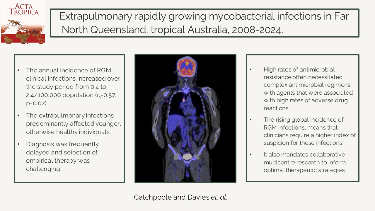
Background: The global incidence of rapidly growing mycobacterial (RGM) infections is rising. We examined cases of extrapulmonary RGM infection in Far North Queensland (FNQ), tropical Australia, to characterise their presentation and the challenges in their management. Methods: All RGM isolated from extrapulmonary sites in FNQ residents between January 2008 and December 2024 were identified; the isolates’ microbiological characteristics and the patients’ clinical course were reviewed. Results: Mycobacterium fortuitum group (51/144, 35%), Mycobacterium abscessus complex (45/144, 31%) and Mycobacterium chelonae (30/144, 21%) were the most frequently isolated RGM. Clinical infection was confirmed in 103/144 (72%) isolates. The annual incidence of RGM clinical infections increased over the study period from 0.4 to 2.4/100,000 population (rs=0.57, p=0.02). Patients had a median (interquartile range (IQR)) age of 52 (33-66) years; 10/103 (10%) were immunosuppressed while only 6/103 (6%) had other significant comorbidity. Preceding environmental trauma was documented in 39/103 (38%), 38/103 (37%) followed surgery and 23/103 (22%) infections involved prosthetic material. The median (IQR) duration of symptoms at diagnosis was 5 (2-12) weeks. Antimicrobial susceptibility testing demonstrated high rates of resistance; 78 patients were treated with antibiotics for a median (IQR) duration of 18 (16-24) weeks with 47/78 (60%) experiencing an adverse drug reaction. Surgical management was necessary in 64/103 (62%). There were 6 microbiologically-confirmed relapses but no deaths directly attributable to the infection or its treatment. Conclusion: Extrapulmonary RGM infections are increasing in frequency in tropical Australia and predominantly affect younger, healthy individuals. Delayed diagnosis and adverse events from antimicrobial therapy are common.
Keywords:
non-tuberculous mycobacteria
; rapidly growing mycobacteria
; clinical management
; antimicrobial resistance
; adverse drug reactions
; tropical medicine
Introduction
Rapidly growing mycobacteria (RGM) are environmental organisms with a worldwide distribution. They are termed rapidly growing as, unlike most other mycobacteria, they typically grow in subculture within one week. Significant changes in the nomenclature of the RGM have occurred in the past decade, but common clinical isolates can be broadly categorised into the Mycobacterium abscessus complex, the Mycobacterium fortuitum group and Mycobacterium chelonae.
RGM have the potential to cause a wide spectrum of disease; pulmonary infection usually occurs in people with underlying structural lung disease or immunocompromise, but extrapulmonary disease can occur in both immunocompetent and immunocompromised hosts [1]. The most common presentation of extrapulmonary disease is skin and soft tissue infection or surgical wound infection, although lymphadenitis, keratitis, prosthesis-related and catheter-related infections also occur [2]. Disseminated infection is rare and is usually seen in immunocompromised hosts. The diagnosis of extrapulmonary RGM infection is challenging and is frequently delayed, with the median reported symptom duration at diagnosis as long as 10 months [3]. There are currently no prospective randomised controlled trials evaluating the management of extrapulmonary disease and recommendations are often extrapolated from pulmonary nontuberculous mycobacteria (NTM) guidelines and expert opinion [4]. The choice of antibiotic therapy is guided by antimicrobial susceptibility testing (AST), however data correlating in vitro susceptibility and real-world clinical outcomes for pulmonary disease is limited, and for extrapulmonary disease it is non-existent [5]. The high rates of reported resistance, the absence of susceptibility breakpoints for many commonly used agents and the limited number of oral options, particularly for M. abscessus infections, means that clinicians must use their best judgment when choosing complex and often poorly tolerated antimicrobial regimens [6]. Management frequently requires a prolonged course of antibiotic therapy in combination with surgical management; both of which may be associated with significant morbidity [7,8].
The incidence of NTM infections is increasing globally [9,10]. The explanation for this increase is unknown but may include an older population [11], expanded used of immunosuppressive and immunomodulatory therapies, greater use of prosthetic material in surgical procedures [3,12] and decreasing Bacillus Calmette-Guerin (BCG) vaccination [11]. Improvements in diagnostic capacity, increased clinician awareness and their status as a notifiable condition in many jurisdictions are also likely to be contributing factors [13]. The incidence of NTM infections is greater in tropical regions, an observation which has been linked to warmer temperatures and greater rainfall in these locations [14,15,16,17].
Far North Queensland (FNQ), is a region of 380,000 km2 situated in Australia’s tropical north. The incidence of all NTM infections in FNQ has increased to 27.5/100,000 people in the urban Cairns region, and to 69.4/100,000 people in the remote Cape York and Torres Strait Islands region (Figure 1), the highest in the state of Queensland [18]. RGM cause 10-20% of NTM infections in Queensland and more than half of these RGM infections are extrapulmonary [18]. Anecdotally, resistance to usual first line therapies is common and management is often complicated. This study aimed to examine the characteristics and clinical course of FNQ patients with RGM infections and to describe the efficacy and safety of current treatment approaches. It was hoped that these data might inform future management strategies locally and other regions where serious RGM infections are being increasingly encountered.
Methods
Study Setting
This retrospective study was performed at Cairns Hospital, the referral hospital for the FNQ region. Cairns Hospital serves a population of approximately 290,000 people, approximately 17% of whom identify as Aboriginal and Torres Strait Islander Australians, hereafter referred to respectfully collectively as First Nations Australians [19]. FNQ has a tropical climate and is generally hot and humid with a distinct wet and dry season. The region experiences high rainfall (annual average rainfall 1085mm) particularly in the December–March wet season, when severe weather events including tropical cyclones and flooding are also common [20]. This tropical climate has a significant impact on the incidence of local prevalent infectious diseases [21].
Identification of Cases
RGM isolates were identified using the Queensland Health electronic laboratory information system (AUSLAB). Isolates were included in the study if they had culture-confirmed Mycobacterium abscessus complex, M. fortuitum group, M. chelonae, or another RGM, isolated from extrapulmonary sites between January 1, 2008, and December 31, 2024. This study period was chosen as it coincided with the introduction of electronic discharge summaries in the Queensland public health system. Additional clinical cases of RGM infection, which were diagnosed in external private laboratories but managed by Cairns Hospital Department of Infectious Diseases during the same period were identified in the department’s prospectively edited database.
Microbiological Characteristics
The Queensland Mycobacterium Reference Laboratory (QMRL) performed identification and susceptibility testing for all isolates; with the exception of three isolates managed by external laboratories. Identification and susceptibility testing methods evolved over the period from the Hain Lifescience GenoType Mycobacterium CM Line probe assay (Bruker Hain Lifescience, Nehren, Germany) to HSP65/16s based Sanger sequencing and bioMérieux Vitek MS MALDI-TOF (bioMérieux, Marcy-l'Étoile, France) identification. Where reported, M. abscessus subspeciation was done by real-time polymerase chain reaction (RT-PCR) targeting the truncated erm41 gene allowing differentiation of subspecies massiliense from the other two subspecies [22]. Only isolates which underwent susceptibility testing using Clinical and Laboratory Standards Institute (CLSI) M24, CLSI M62, or CLSI M24S [23,24,25] microbroth dilution procedure were included in the analysis of isolate susceptibilities. Sensititre Myco RAPMYCOI AST plate (Thermo Fisher Scientific [Waltham, USA]; previously TREK Diagnostics) was used for all microdilutions. Isolates collected prior to 2011 underwent testing with only disc diffusion methods and were not included in the microbiological analysis.
Clinical Course
Medical records were reviewed to define patient demographics and comorbidities. The presence of immunosuppression was specifically sought and was defined as a history of a solid organ or haematological stem cell transplant or current treatment with immunosuppressive agents, including corticosteroids, chemotherapy, or immunomodulatory therapies.
Comorbidity was quantified using the Charlson Comorbidity Index; individuals were said to have significant comorbidity of they had a Charlson Comorbidity Index ≥ 5 [26]. Infection was defined as isolation of an organism from a skin site with a consistent lesion or isolation from a sterile site, confirmed as clinically significant by an attending Infectious Diseases specialist physician. As the focus of the report was on extrapulmonary disease, all pulmonary and pleural isolates were excluded. The duration of symptoms at diagnosis, any preceding trauma or surgery and the presence of any prosthetic material in situ were recorded. Antibiotic therapy, surgery, associated adverse events, and outcome of the infection were recorded when these were available. Adverse events were graded using Common Terminology Criteria for Adverse Events (CTCAE) v5.0 [27]. Relapse was defined as clinical and/or microbiological recurrent evidence of disease following cessation of treatment.
Statistical Analysis
Data were entered into an electronic database (Microsoft Excel) and analysed statistical software (Stata version 18.5). Groups were compared using the chi squared, Fisher’s exact test or logistic regression, where appropriate. Trends over time were determined using Spearman's rank correlation coefficient.
Ethical Statement
The Far North Queensland Human Research Ethics Committee provided approval for the study (Reference EX/2025/QCH/121496 (Nov ver 3) – 1966 QA). As the data were retrospective and de-identified, the committee waived the requirement for patient consent.
Results
There were 144 extrapulmonary RGM isolates identified in the FNQ region during the study period. Isolates from the M. fortuitum group (51/144 (35%)), the M. abscessus complex (45/144, 31%) and M. chelonae (30/144, 21%) were the most common; 16/144 (11%) isolates were not from these 3 groups (Supplementary Table 1). An additional two strains were initially identified as M. chelonae however on review had minimum inhibitory concentrations (MICs) to tobramycin of 8-16ug/ml. This is discordant with expected results for the species and, given this uncertainty, these two cases were subsequently removed from analysis of clinical isolates. Of the 144 isolates, 103 (72%) were deemed to be clinically significant and had adequate clinical data for review (Figure 2).
The annual incidence of extrapulmonary RGM infections in the FNQ region increased from 0.4/100,000 population in 2008 to 2.4/100,000 population in 2024 (rs=0.57, p=0.02) (Figure 3).
Demographics and Risk Factors
The median (interquartile range (IQR)) age of the 103 patients with clinical evidence of infection was of 52 (33-66) years; 51 (50%) were female (Table 1). Most patients (71/103, 69%) were aged <65 years; 6/103 (6%) identified as First Nations Australians. 10/103 (10%) infections occurred in the setting of immunosuppression. An additional patient – who presented with M. abscessus blood stream infection – was subsequently diagnosed with an acquired interferon gamma (IFNγ) antibody syndrome. There were 14/103 (14%) with type 2 diabetes mellitus and three had peripheral vascular disease. It was possible to determine the Charlson Comorbidity Index in 102 cases: 69/102 (68%) had a score of 0 and only 6/102 (6%) had a score of ≥ 5.
Clinical Presentation and Site of Infection
The median (IQR) duration of symptoms at diagnosis was 5 (2-12) weeks in the 83/103 (81%) who had this recorded. Details of selected epidemiological associations and the clinical presentations are presented in Table 1. Antecedent environmental trauma included a tick bite, a crocodile bite, penetrating injuries and contaminated crush injuries. The M. fortuitum group infection was more likely to be associated with antecedent environmental trauma than was infection with other organisms (24/41 (59%) versus 15/62 (24%), odds ratio (95% confidence interval): 4.4 (1.9-10.4), p=0.001).
Osteomyelitis was present in 16/103 (16%). Environmental trauma (including lacerations, penetrating and crush injuries) preceded 13/16 (81%) episodes of osteomyelitis; prosthetic material (including fixation material and prosthetic joints) was present in 9/16 (56%) cases.
Prosthetic material was present in 14 additional infections, including six Tenckhoff catheters, two abdominal mesh infections, two breast prostheses, a subcutaneous line, a Hickman line, a suprapubic catheter and a gastric band. Four infections were associated with breast augmentations; M. flavescens was isolated from a patient with post-operative infection following breast augmentation performed in Thailand and M. duvalli was isolated from a patient with post-operative infection following breast augmentation locally in FNQ. Infections of surgical sites without prosthetic material present represented 17/103 (17%) of the cases.
One of the bloodstream infections occurred in a 53-year-old man with acquired IFNγ deficiency who developed disseminated M. abscessus infection with multiple relapses. Another in a 70-year-old woman with polymicrobial M. abscessus and M. fortuitum complex bloodstream infection that complicated treatment for metastatic cervical squamous cell carcinoma via a peripherally inserted central catheter (PICC). The third was a 54-year-old female with an infection associated with a gastric band (implanted 20 years prior) with a secondary bloodstream infection. There were no clusters of infection associated with specific procedures or RGM species.
Antimicrobial Susceptibility
Detailed AST was available for 93 of the 103 clinically significant isolates, the remaining 10 were collected prior to 2011 when only disc diffusion methods were employed. The three most common organisms accounted for 84/93 of these isolates (Table 2); the other 9 clinical isolates were from the less common RGM subspecies.
Within the M. abscessus complex, a high proportion 18/35 (51%) were clarithromycin susceptible at 14-day incubation, 20 of these isolates underwent subspeciation. Of the 12 confirmed M. abscessus subsp. abscessus/bolletii, 3 (25%) were clarithromycin susceptible, while all 8 M. abscessus subsp. massiliense isolates were clarithromycin susceptible, as anticipated. Prior to 2013 M. abscessus complex isolates did not undergo 14-day clarithromycin incubation, clarithromycin susceptibility from those isolates has been excluded from the analysis. Linezolid susceptibility was low (21%). Within this group, resistance to most oral agents was high including fluoroquinolones, trimethoprim/sulfamethoxazole (TMP/SMX) and doxycycline.
Tigecycline testing was performed on 91 isolates; 87 (96%) had a minimum inhibitory concentration (MIC) of between 0.03 and 0.5, noting that no established breakpoints exist for interpretation.
Treatment
Detailed treatment information was available for 91 cases (Table 3); the remaining cases were either lost to follow-up or transferred to another facility for ongoing care (Supplementary Figure 2).
Adverse drug reactions (ADRs) occurred in 47/78 (60%) of the patients receiving antibiotics, 18 (38%) of those were severe (grade 3-4). There were 29 patients who had minor (grade 1-2) ADRs; including dysgeusia, gastrointestinal intolerance, mild acute kidney injury, QTc prolongation, myelosuppression, photosensitivity and uncomplicated drug rash or eosinophilia (Supplementary Table 2, Table 3 and Table 4). Debilitating nausea was the most frequent treatment limiting ADR across all species.
Of the 64 patients undergoing surgical management, 15 (23%) required more than one procedure. All patients with a prosthetic infection had the prosthesis removed.
Treatment of M. abscessus Complex
26 unique first line combinations were used in the 33 infections treated with antibiotics (with or without surgery); the most common being amikacin, cefoxitin and clofazimine (4/33 (12%)) (see Supplementary Table 2). Clarithromycin was the most commonly used antibiotic (in 22/33 cases). A combination of ≥3 agents were used in first line regimens for 24/33 (73%) of cases and monotherapy was used in only two cases of skin and soft tissue infections where no additional oral antibiotics had been reported as susceptible.
Of those treated with antibiotics, 12/33 (36%) patients experienced a grade 3-4 ADR. This was most commonly attributable to amikacin with four cases of ototoxicity (two with moderate-severe permanent hearing impairment) and two cases of vestibulotoxicity (one associated with permanent disabling symptoms) and a severe acute kidney injury (AKI) which required medical management but not renal replacement therapy. A case of Drug Reaction with Eosinophilia and Systemic Symptoms (DRESS) was also seen in a patient who received cefoxitin although they did not require intensive care unit admission. Grade 1-2 reactions were also common with 35 events in total. A greater proportion of the Grade 3-4 reactions occurred in patients with M. abscessus complex infection than patients infected with the other RGMs (12/33 (36%) versus 6/45 (13%), odds ratio (95% confidence interval): 3.7 (1.2-11.3), p=0.02).
Surgical management was required in 22/38 (58%) M. abscessus infections. Three bone and joint infections were managed with amputation; one with a forearm amputation in the setting of immunosuppression for Crohn’s disease in a patient in whom amikacin-attributed hearing loss limited antibiotic therapy. Another patient required an above knee amputation for osseointegrated tibial prosthetic infection without prior targeted antibiotics. A third patient required a Girdlestone’s procedure for polymicrobial prosthetic hip joint infection.
Treatment of the M. fortuitum Group
32/37 patient were treated with antibiotics (with or without surgery); this included 18 unique first line antibiotic combinations (Supplementary Table 3). ≥3 agents were used in first line regimens for 18/32 (56%) of M. fortuitum group cases. Grade 3-4 ADRs were less common than in the M. abscessus complex and included cases of amikacin causing an AKI (without requirement for renal replacement therapy), grade 3 pancreatitis attributed to tigecycline, a PICC-associated DVT and a TMP/SXT-induced episode of DRESS requiring hospitalisation without intensive care support. TMP/SXT was the most frequently used antibiotic (24/32 (75%) cases) and was associated with high rates of grade 1-2 ADRs (9/24 (38%)), including mild AKI, hypersensitivity reactions and myelosuppression and one grade 3 reaction (DRESS). Surgical intervention with or without antibiotics was required in 27/37 (73%) cases.
Treatment of M. chelonae
M. chelonae was managed with antibiotics – with or without surgery – in 6/8 (5 of which were managed with exclusively oral agents) and 6 cases required surgical management. Monotherapy was used in one skin and soft tissue infection where no additional oral antibiotics that had been reported as susceptible. ADRs were relatively rare in these patients (Supplementary Table 4).
Outcomes
Many patients were lost to follow-up but of the remaining a clinical cure was documented in 75 cases. Six microbiologically confirmed relapses occurred, all of whom had at least one treatment related adverse event, which had required revision to their antibiotic regimen prior to their relapse. There were no deaths attributable to infection or treatment, although three patients died with unresolved infection with their deaths due to another cause. The first was a 58-year-old patient with IFNγ antibody syndrome who died of complications from decompensated cirrhosis (attributed to metabolic dysfunction-associated fatty liver disease and chronic hepatitis B) which developed in his fifth consecutive year of antimicrobial therapy for relapsed disseminated M. abscessus subsp. abscessus infection. The second, an 85-year-old man, died of methicillin-resistant Staphylococcus aureus bacteraemia from a polymicrobial (including M. abscessus subsp. massiliense and Pseudomonas aeruginosa) hip prosthetic joint infection. The third was a 61-year-old female with end-stage renal failure who died following withdrawal of dialysis after having experienced significant drug toxicities during the treatment of M. abscessus subsp. abscessus peritoneal-dialysis peritonitis treatment in the preceding 12 months.
Discussion
There was a greater than fivefold increase in the annual incidence of extrapulmonary RGM infections in this region of tropical Australia over the 17 years of the study period. However, although RGM infection is classically reported in immunocompromised individuals or in those with underling health conditions, it was notable that less than 10% of the cohort had any immunocompromise and over two thirds had no comorbidity. The patients’ clinical presentations were heterogenous, and the diagnosis of RGM infection was frequently delayed. Though it is difficult to know exactly how these delays changed outcome, many patients experienced months of symptoms before treatment was commenced. Patient management was further complicated by high rates of antimicrobial resistance which made selection of empiric antibiotic therapy difficult. The resulting complex regimens were frequently poorly tolerated: almost 40% of the patients who received antibiotics reported a severe adverse drug reaction. In this context, surgery was frequently required to achieve cure, adding to the complexity of patient care. Despite these many challenges, generally excellent clinical outcomes were possible with the comprehensive multidisciplinary care available in Australia’s well-resourced health system.
The growing burden of these infections – and the complexity of their management– is not unique to FNQ. Amongst other host and environmental factors, rising incidence of NTM worldwide has been attributed to improved diagnostics, clinician awareness and climate change [16]. Although the incidence of RGM infection is lower in temperate locations it also appears to be increasing [28]. A nationwide study in Denmark reported an increase in the incidence of extrapulmonary NTM disease from 0.3 to 0.6 per 100,000 people between 2000-2017 [29].
However, the incidence of NTM infections is even higher in tropical settings [14]. In the Northern Territory, a region of Australia with a similar climate and demography to FNQ, a recent study demonstrated an increased incidence of skin and soft tissue NTM cases (from 6.1 per 100,000 people to 15.2 per 100,000 people over the study period). Interestingly, as in our study, they found comparatively low rates of infections in First Nations Australians, which is notable as this population experiences a disproportionately higher burden of other infectious diseases in tropical Australia [15,30,31,32,33].
Many patients developed infection following environmental trauma or surgical procedures, both having been long recognised as a risk factor for RGM skin, soft tissue and bone and joint infections [10,34]. Whether inoculation is occurring during the procedure or whether these injuries are providing a portal of entry of environmental organisms in the post-operative period is uncertain. Our finding of a predilection for lower limb infections is consistent with previous work and supports the mechanism of opportunistic environmental exposure to the organism via a breach to the skin barrier [35]. Although peripheral vascular disease and type 2 diabetes mellitus are common risk factors for bacterial lower limb infection in the region [36], they were not common in our cohort. Additionally, while spatial clustering of M. abscessus complex infections has previously been described in other parts of Australia, this was also not seen in our cohort [37].
In other studies populations, infection has been associated with older [9], more comorbid [38] and more immunosuppressed [39] patient groups. However, the high burden of extrapulmonary RGM in this region of tropical Australia is not explained by increasing comorbidity in an aging population; the median age of patients in this cohort was 52 and over 90% were immunocompetent. Similar trends were seen in the recent Northern Territory study, with only 6% of patients being over 69 years old and only 37% had a predisposing medical comorbidity (many of whom also had skin trauma) [15]. In addition, our study highlights the frequent delay in diagnosis that is experienced by patients with RGM infections and reinforces the role for early consideration of less common pathogens in patients living in tropical regions with non-resolving infection, particularly where trauma or injury may have occurred [40].
Antimicrobial resistance to tested agents was common, particularly among isolates from the M. abscessus complex. The susceptibility to linezolid was only 21%, compared to previous reported rates of 33 - 40% in Australia [41]. There is marked geographic variability in the susceptibility of M. abscessus complex isolates to linezolid with Korean, Chinese, Taiwanese, Venezuelan and United Kingdom groups reporting susceptibility rates between 5 and 96% [42,43,44,45]. High rates of reported resistance to other oral agents were demonstrated in the M. abscessus complex, in line with another Australian study [41]. The explanation for this finding is uncertain and may be partly explained by well-established laboratory challenges in AST for NTM which limit MIC interpretation [46]. Regardless of the accuracy of these results, it clearly influenced the choice of therapy in this cohort, and the resulting high rates of aminoglycoside use and related AKIs and vestibular and ototoxicity.
Consensus guidelines for the management of pulmonary NTM including RGM offer evidence-based approaches for agent selection and treatment duration [5], however, the applicability of these strategies (without existing clinical trial data) to extrapulmonary infection has not been established. A recent review of extrapulmonary NTM outlines treatment recommendations for mild and severe infection whilst highlighting the paucity of high-quality data to inform these recommendations [4]. The variability of antimicrobial agents that are used reflect a need to balance the risk of toxicity, variable antimicrobial susceptibility patterns and limited data to inform optimal treatment regimens. The median treatment duration in this study was 18 weeks and frequently required use of antimicrobials with well-established risk of toxicity. Indeed, ADRs were common in this cohort and were frequently treatment limiting or, indeed, resulted in permanent disability in some cases. For clinicians, using prolonged courses of toxic parenteral antibiotics to treat a soft tissue infection with an organism of low virulence appears to conflict with a patient-centred approach, particularly given recent trends toward shorter parenteral duration for many bacterial infections [47]. Newer treatment strategies using dual beta-lactam combinations, [48] bedaquiline (used in 5 cases in this study), [49,50] and/or omadacycline against M. abscessus complex may decrease treatment associated toxicity [51].
The limited speciation conducted for most of these isolates and the changes in speciation techniques over time complicates the interpretation of the relationship between organism and treatment response. Real-time PCR M. abscessus subspeciation expands the opportunity to examine treatment responses by organism subspecies and has the potential to inform treatment choices and potentially improve outcomes. In studies of extrapulmonary infection and in our study, treatment outcomes are worse in the M. abscessus complex infection than for other RGM [6]. This was particularly the case for M. abscessus subsp. abscessus when compared with M. abscessus subsp. massiliense where this data was available for review [52]. Overall outcomes in extrapulmonary infection are better than those of pulmonary infection and it is notable that there were no deaths directly related to infection or its treatment in this cohort [7].
While this is one of the larger series of extrapulmonary RGM, the small number of infections, the heterogeneity of patient presentations and limitations of retrospective data collection, increase the risk of type 2 errors. This limits our ability to draw conclusions about risk factors for individual infections and the relative risks and benefits of different treatment strategies. The limited identification in the earlier part of the study period and small numbers only allow analysis at a group rather than species level. Microbiological or clinical associations of particular subspecies within a grouping may therefore not be evident in our analysis. The study only enrolled patients managed in the public hospital system with clinical information unavailable for patients managed entirely in the community or within the private health system, potentially biasing the complexity and demographic mix of the cohort. This may also have led to reduced case ascertainment impacting on our reported local incidence. Acknowledging this fact, very few patients with complex infections are managed outside of the public health system in the FNQ region.
With species specific identification now more readily available, future studies correlating clinical risk factors for infection and species related treatment response may be able to better guide future management. With the increasing incidence of RGM infections, multi-centre trials may be able to resolve questions around the apparent variation in susceptibility and treatment response observed across the range of small retrospective analyses currently available. It may be possible to devise optimal treatment approaches tailored to the clinical presentation and the infecting organism. Advances in direct molecular diagnosis may expedite species identification in the future, which will assist with timely selection of optimal empirical therapy and inform prognostication and the use of adjunctive treatments.
Conclusion
Extrapulmonary infections due to RGM are increasing in incidence in tropical Australia and have a significant impact on health system resources and the patients’ quality of life. In contrast to the published literature, extrapulmonary RGM infections in tropical Australia predominantly affect younger, healthy individuals. Isolates are frequently highly resistant – and this fact and the limited literature exploring the optimal therapy of RGM infections – makes the choice of empirical therapy challenging. The resulting selected antimicrobial regimens are usually complex, often poorly tolerated and frequently cause significant morbidity. The burden of RGM infection is increasing globally, and clinicians need to understand far more about the microbiology of these organisms – and the pathophysiology of their infections – to provide optimal care for their patients––.
Supplementary Materials
The following supporting information can be downloaded at the website of this paper posted on Preprints.org.
References
- Brown-Elliott, B.A.; Philley, J.V. Rapidly Growing Mycobacteria. Microbiol. Spectr. 2017, 5(1). [Google Scholar] [PubMed]
- Griffith, D.E.; Aksamit, T.; Brown-Elliott, B.A.; Catanzaro, A.; Daley, C.; Gordin, F.; et al. An official ATS/IDSA statement: diagnosis, treatment, and prevention of nontuberculous mycobacterial diseases. Am. J. Respir. Crit. Care Med. 2007, 175(4), 367–416. [Google Scholar] [PubMed]
- Lopez, M.; Croley, J.; Murphy, K.D. Atypical Mycobacterial Infections of the Upper Extremity: Becoming More Atypical? Hand (N Y). 2017, 12(2), 188–92. [Google Scholar] [PubMed]
- Rath, S.; Firdaus, S.; Nayak, G.; Mohanty, M. Extrapulmonary Nontuberculous Mycobacteria Infection: The New-Age Neglected Infectious Disease. Cureus 2025, 17(3), e81017. [Google Scholar] [PubMed]
- Daley, C.L.; Iaccarino, J.M.; Lange, C.; Cambau, E.; Wallace, R.J.; Andrejak, C.; et al. Treatment of Nontuberculous Mycobacterial Pulmonary Disease: An Official ATS/ERS/ESCMID/IDSA Clinical Practice Guideline. Clin. Infect. Dis. 2020, 71(4), 905–13. [Google Scholar] [PubMed]
- Wi, Y.M. Treatment of Extrapulmonary Nontuberculous Mycobacterial Diseases. Infect. Chemother. 2019, 51(3), 245–55. [Google Scholar] [PubMed]
- Holt, M.R.; Kasperbauer, S. Management of Extrapulmonary Nontuberculous Mycobacterial Infections. Semin Respir. Crit. Care Med. 2018, 39(3), 399–410. [Google Scholar] [PubMed]
- Ding, L.W.; Lai, C.C.; Lee, L.N.; Huang, L.M.; Hsueh, P.R. Lymphadenitis caused by non-tuberculous mycobacteria in a university hospital in Taiwan: predominance of rapidly growing mycobacteria and high recurrence rate. J. Formos. Med. Assoc. 2005, 104(12), 897–904. [Google Scholar] [PubMed]
- Sander, M.A.; Isaac-Renton, J.L.; Tyrrell, G.J. Cutaneous Nontuberculous Mycobacterial Infections in Alberta, Canada: An Epidemiologic Study and Review. J. Cutan. Med. Surg. 2018, 22(5), 479–83. [Google Scholar] [PubMed]
- Wentworth, A.B.; Drage, L.A.; Wengenack, N.L.; Wilson, J.W.; Lohse, C.M. Increased incidence of cutaneous nontuberculous mycobacterial infection, 1980 to 2009: a population-based study. Mayo Clin. Proc. 2013, 88(1), 38–45. [Google Scholar] [PubMed]
- Marras, T.K.; Daley, C.L. Epidemiology of human pulmonary infection with nontuberculous mycobacteria. Clin. Chest Med. 2002, 23(3), 553–67. [Google Scholar] [CrossRef] [PubMed]
- Smidt, K.P.; Stern, P.J.; Kiefhaber, T.R. Atypical Mycobacterial Infections of the Upper Extremity. Orthopedics 2018, 41(3), e383–e8. [Google Scholar] [PubMed]
- Zhou, J.; Jia, Q.; Liu, L.; Liang, L.; Zhang, H.; He, C.; et al. Epidemiology and clinical outcomes in skin and soft tissue nontuberculous mycobacteria infections: A retrospective study. J. Infect. Public Health 2025, 18(3), 102655. [Google Scholar] [PubMed]
- Ratnatunga, C.N.; Lutzky, V.P.; Kupz, A.; Doolan, D.L.; Reid, D.W.; Field, M.; et al. The Rise of Non-Tuberculosis Mycobacterial Lung Disease. Front Immunol. 2020, 11, 303. [Google Scholar] [PubMed]
- Nohrenberg, M.; Wright, A.; Krause, V. Non-tuberculous mycobacterial skin and soft tissue infections in the Northern Territory, Australia, 1989-2021. Int. J. Infect. Dis. 2023, 135, 125–31. [Google Scholar] [PubMed]
- Thomson, R.M.; Furuya-Kanamori, L.; Coffey, C.; Bell, S.C.; Knibbs, L.D.; Lau, C.L. Influence of climate variables on the rising incidence of nontuberculous mycobacterial (NTM) infections in Queensland, Australia 2001-2016. Sci. Total Environ. 2020, 740, 139796. [Google Scholar] [PubMed]
- Thomson, R.; Donnan, E.; Konstantinos, A. Notification of Nontuberculous Mycobacteria: An Australian Perspective. Ann. Am. Thorac. Soc. 2017, 14(3), 318–23. [Google Scholar] [PubMed]
- Nontuberculous mycobacteria in Queensland 2016. In Queensland Health; Queensland Government, 2016.
- Australian census; Australian Bureau of Statistics: Canberra, 2021; Available online: https://www.abs.gov.au/census.
- Climate change in the Far North Queensland region; Department of Environment and Science. Queensland Government: Brisbane, 2019.
- Fairhead, L.J.; Smith, S.; Sim, B.Z.; Stewart, A.G.A.; Stewart, J.D.; Binotto, E.; et al. The seasonality of infections in tropical Far North Queensland, Australia: A 21-year retrospective evaluation of the seasonal patterns of six endemic pathogens. PLoS Glob. Public Health 2022, 2(5), e0000506. [Google Scholar] [PubMed]
- Syrmis, M.W.; Pandey, S.; Tolson, C.; Carter, R.; Congdon, J.; Sloots, T.; et al. Identification of Mycobacterium abscessus complex and M. abscessus subsp. massiliense culture isolates by real-time assays. J. Med. Microbiol. 2015, 64(7), 790–4. [Google Scholar] [PubMed]
- CLSI. Performance Standards for Susceptibility Testing of Mycobacteria, Nocardia spp. and Other Aerobic Actinomycetes. Second Edition. CLSI supplement M24S. Clinical and Laboratory Standards Institute, 2023.
- CLSI. CLSI document M24-A2.; Susceptibility Testing of Mycobacteria, Nocardiae, and Other Aerobic Actinomycetes; Approved Standard—Second Edition. Clinical and Laboratory Standards Institute: Wayne, PA, 2011.
- CLSI. Performance Standards for Susceptibility Testing of Mycobacteria, Nocardia spp. and Other Aerobic Actinomycetes. First Edition. In CLSI supplement M62; Clinical and Laboratory Standards Institute: Wayne, PA, 2018. [Google Scholar]
- Charlson, M.E.; Pompei, P.; Ales, K.L.; MacKenzie, C.R. A new method of classifying prognostic comorbidity in longitudinal studies: development and validation. J. Chronic Dis. 1987, 40(5), 373–83. [Google Scholar] [CrossRef] [PubMed]
- Freites-Martinez, A.; Santana, N.; Arias-Santiago, S.; Viera, A. Using the Common Terminology Criteria for Adverse Events (CTCAE - Version 5.0) to Evaluate the Severity of Adverse Events of Anticancer Therapies. Actas Dermosifiliogr. (Engl Ed) 2021, 112(1), 90–2. [Google Scholar] [PubMed]
- Winthrop, K.L.; Marras, T.K.; Adjemian, J.; Zhang, H.; Wang, P.; Zhang, Q. Incidence and Prevalence of Nontuberculous Mycobacterial Lung Disease in a Large U.S. Managed Care Health Plan, 2008-2015. Ann. Am. Thorac. Soc. 2020, 17(2), 178–85. [Google Scholar] [PubMed]
- Pedersen, A.A.; Løkke, A.; Fløe, A.; Ibsen, R.; Johansen, I.S.; Hilberg, O. Nationwide Increasing Incidence of Nontuberculous Mycobacterial Diseases Among Adults in Denmark: Eighteen Years of Follow-Up. Chest 2024, 166(2), 271–80. [Google Scholar] [PubMed]
- Currie, B.J.; Mayo, M.; Ward, L.M.; Kaestli, M.; Meumann, E.M.; Webb, J.R.; et al. The Darwin Prospective Melioidosis Study: a 30-year prospective, observational investigation. Lancet Infect. Dis. 2021, 21(12), 1737–46. [Google Scholar] [PubMed]
- Hanson, J.; Smith, S.; Stewart, J.; Horne, P.; Ramsamy, N. Melioidosis – a disease of socioeconomic disadvantage. PLoS Negl. Trop. Dis. 2021, 15(6), e0009544. [Google Scholar] [PubMed]
- Hanson, J.; Smith, S.; Brooks, J.; Groch, T.; Sivalingam, S.; Curnow, V.; et al. The applicability of commonly used predictive scoring systems in Indigenous Australians with sepsis: An observational study. PLoS ONE 2020, 15(7), e0236339. [Google Scholar] [PubMed]
- Davis, J.S.; Cheng, A.C.; McMillan, M.; Humphrey, A.B.; Stephens, D.P.; Anstey, N.M. Sepsis in the tropical Top End of Australia's Northern Territory: disease burden and impact on Indigenous Australians. Med. J. Aust. 2011, 194(10), 519–24. [Google Scholar] [PubMed]
- Coda, R.; Waller, S.; Vopat, B. Nontuberculosis Mycobacterium Infections in Orthopaedic Surgery: Review of the Epidemiology, Pathogenesis, Diagnosis, and Treatment Guidelines. J. Am. Acad. Orthop. Surg. Glob. Res. Rev. 2025, 9(5). [Google Scholar] [PubMed]
- Diaz, M.A.A.; Huff, T.N.; Libertin, C.R. Nontuberculous mycobacterial infections of the lower extremities: A 15-year experience. J. Clin. Tuberc. Other Mycobact. Dis. 2019, 15, 100091. [Google Scholar] [PubMed]
- Dadwal, P.; Bonner, B.; Fraser, D.; Loveridge, J.; Withey, G.; Puri, A.; et al. Bone and joint infections due to melioidosis; diagnostic and management strategies to optimise outcomes. PLoS Negl. Trop. Dis. 2024, 18(7), e0012317. [Google Scholar] [PubMed]
- Chou, M.P.; Clements, A.C.; Thomson, R.M. A spatial epidemiological analysis of nontuberculous mycobacterial infections in Queensland, Australia. BMC Infect. Dis. 2014, 14, 279. [Google Scholar] [PubMed]
- Bodle, E.E.; Cunningham, J.A.; Della-Latta, P.; Schluger, N.W.; Saiman, L. Epidemiology of nontuberculous mycobacteria in patients without HIV infection, New York City. Emerg. Infect. Dis. 2008, 14(3), 390–6. [Google Scholar] [PubMed]
- Uslan, D.Z.; Kowalski, T.J.; Wengenack, N.L.; Virk, A.; Wilson, J.W. Skin and soft tissue infections due to rapidly growing mycobacteria: comparison of clinical features, treatment, and susceptibility. Arch. Dermatol. 2006, 142(10), 1287–92. [Google Scholar] [PubMed]
- Smith, S.; Marquardt, T.; Jennison, A.V.; D'Addona, A.; Stewart, J.; Yarwood, T.; et al. Clinical Manifestations and Genomic Evaluation of Melioidosis Outbreak among Children after Sporting Event, Australia. Emerg. Infect. Dis. 2023, 29(11), 2218–28. [Google Scholar] [PubMed]
- Chua, K.Y.; Bustamante, A.; Jelfs, P.; Chen, S.C.; Sintchenko, V. Antibiotic susceptibility of diverse Mycobacterium abscessus complex strains in New South Wales, Australia. Pathology 2015, 47(7), 678–82. [Google Scholar] [PubMed]
- Guo, Y.; Cao, X.; Yu, J.; Zhan, Q.; Yang, J.; Wu, X.; et al. Antimicrobial Susceptibility of Mycobacterium abscessus Complex Clinical Isolates from a Chinese Tertiary Hospital. Infect. Drug Resist. 2020, 13, 2001–10. [Google Scholar] [PubMed]
- Cho, E.H.; Huh, H.J.; Song, D.J.; Lee, S.H.; Kim, C.K.; Shin, S.Y.; et al. Drug susceptibility patterns of Mycobacterium abscessus and Mycobacterium massiliense isolated from respiratory specimens. Diagn. Microbiol. Infect. Dis. 2019, 93(2), 107–11. [Google Scholar] [PubMed]
- Da Mata-Jardín, O.; Angulo, A.; Rodríguez, M.; Fernández-Figueiras, S.; de Waard, J.H. Drug susceptibility patterns of rapidly growing mycobacteria isolated from skin and soft tissue infections in Venezuela. Eur. J. Clin. Microbiol. Infect. Dis. 2020, 39(3), 433–41. [Google Scholar] [PubMed]
- Cowman, S.; Burns, K.; Benson, S.; Wilson, R.; Loebinger, M.R. The antimicrobial susceptibility of non-tuberculous mycobacteria. J. Infect. 2016, 72(3), 324–31. [Google Scholar] [PubMed]
- Brown-Elliott, B.A.; Woods, G.L. Antimycobacterial Susceptibility Testing of Nontuberculous Mycobacteria. J. Clin. Microbiol. 2019, 57(10). [Google Scholar] [PubMed]
- Spellberg, B. The New Antibiotic Mantra-"Shorter Is Better. JAMA Intern Med. 2016, 176(9), 1254–5. [Google Scholar] [PubMed]
- Pandey, R.; Chen, L.; Manca, C.; Jenkins, S.; Glaser, L.; Vinnard, C.; et al. Dual beta-Lactam Combinations Highly Active against Mycobacterium abscessus Complex In Vitro. MBio 2019, 10(1). [Google Scholar] [PubMed]
- Brown-Elliott, B.A.; Wallace, R.J., Jr. In Vitro Susceptibility Testing of Bedaquiline against Mycobacterium abscessus Complex. Antimicrob. Agents Chemother. 2019, 63(2). [Google Scholar] [PubMed]
- Le Moigne, V.; Raynaud, C.; Moreau, F.; Dupont, C.; Nigou, J.; Neyrolles, O.; et al. Efficacy of Bedaquiline, Alone or in Combination with Imipenem, against Mycobacterium abscessus in C3HeB/FeJ Mice. Antimicrob. Agents Chemother. 2020, 64(6). [Google Scholar] [PubMed]
- Mingora, C.M.; Bullington, W.; Faasuamalie, P.E.; Levin, A.; Porter, G.; Stadnick, R.; et al. Long-term Safety and Tolerability of Omadacycline for the Treatment of Mycobacterium abscessus Infections. Open Forum Infect. Dis. 2023, 10(7), ofad335. [Google Scholar] [PubMed]
- Jeong, S.H.; Kim, S.Y.; Huh, H.J.; Ki, C.S.; Lee, N.Y.; Kang, C.I.; et al. Mycobacteriological characteristics and treatment outcomes in extrapulmonary Mycobacterium abscessus complex infections. Int. J. Infect. Dis. 2017, 60, 49–56. [Google Scholar] [PubMed]
Figure 1.
The study region of Far North Queensland.
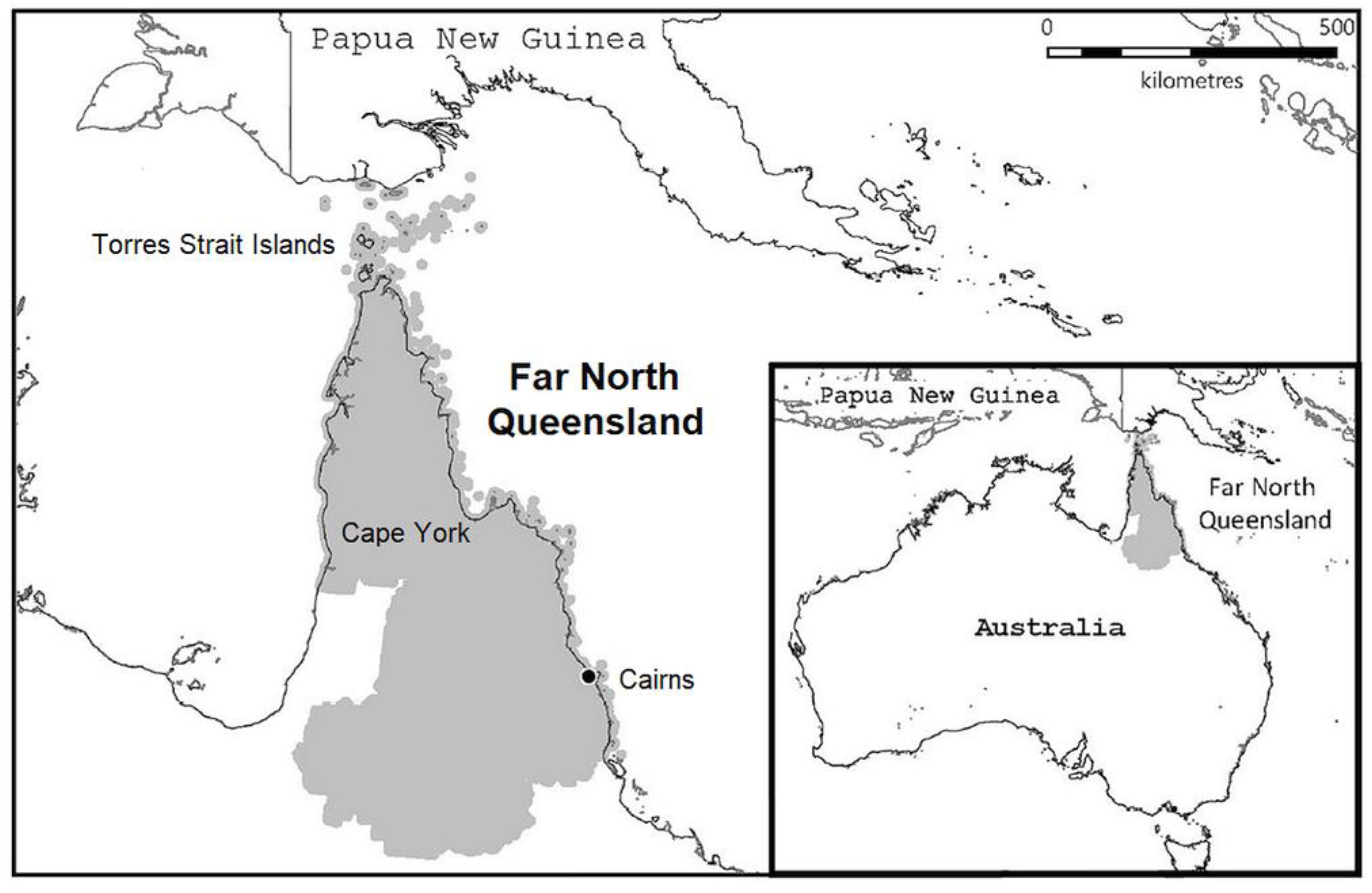
Figure 2.
Extrapulmonary RGM isolates. a As determined by attending Infectious Diseases specialist. b Other species included: M. mageritense, M. flavescens, M. duvalii, M. latzerense, M. smegmatis, M. saopaulense and M. immunogenum.
Figure 2.
Extrapulmonary RGM isolates. a As determined by attending Infectious Diseases specialist. b Other species included: M. mageritense, M. flavescens, M. duvalii, M. latzerense, M. smegmatis, M. saopaulense and M. immunogenum.
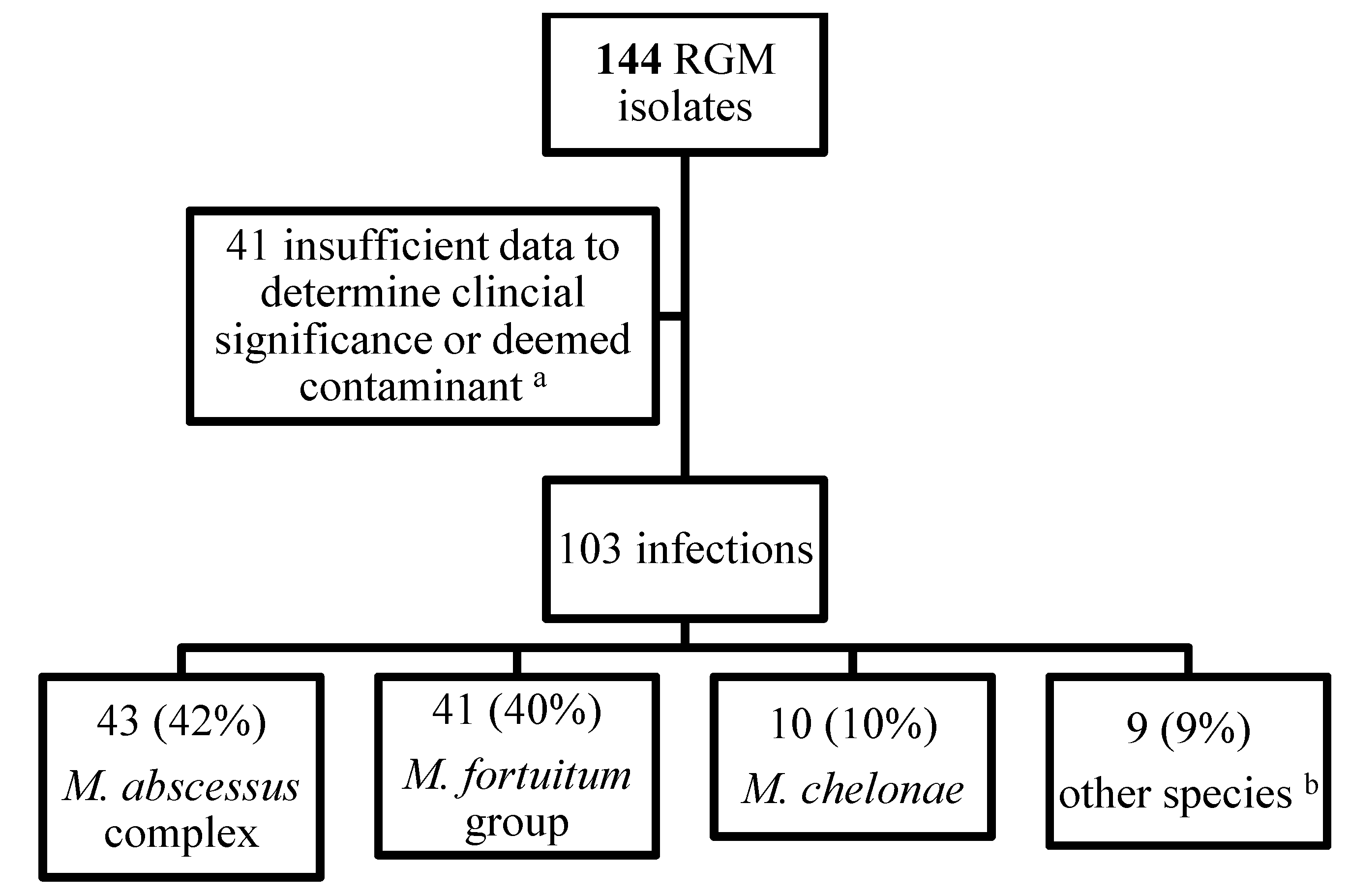
Figure 3.
Incidence of extrapulmonary RGM infections in Far North Queensland, Australia between 2008-2024, with line of best fit.
Figure 3.
Incidence of extrapulmonary RGM infections in Far North Queensland, Australia between 2008-2024, with line of best fit.
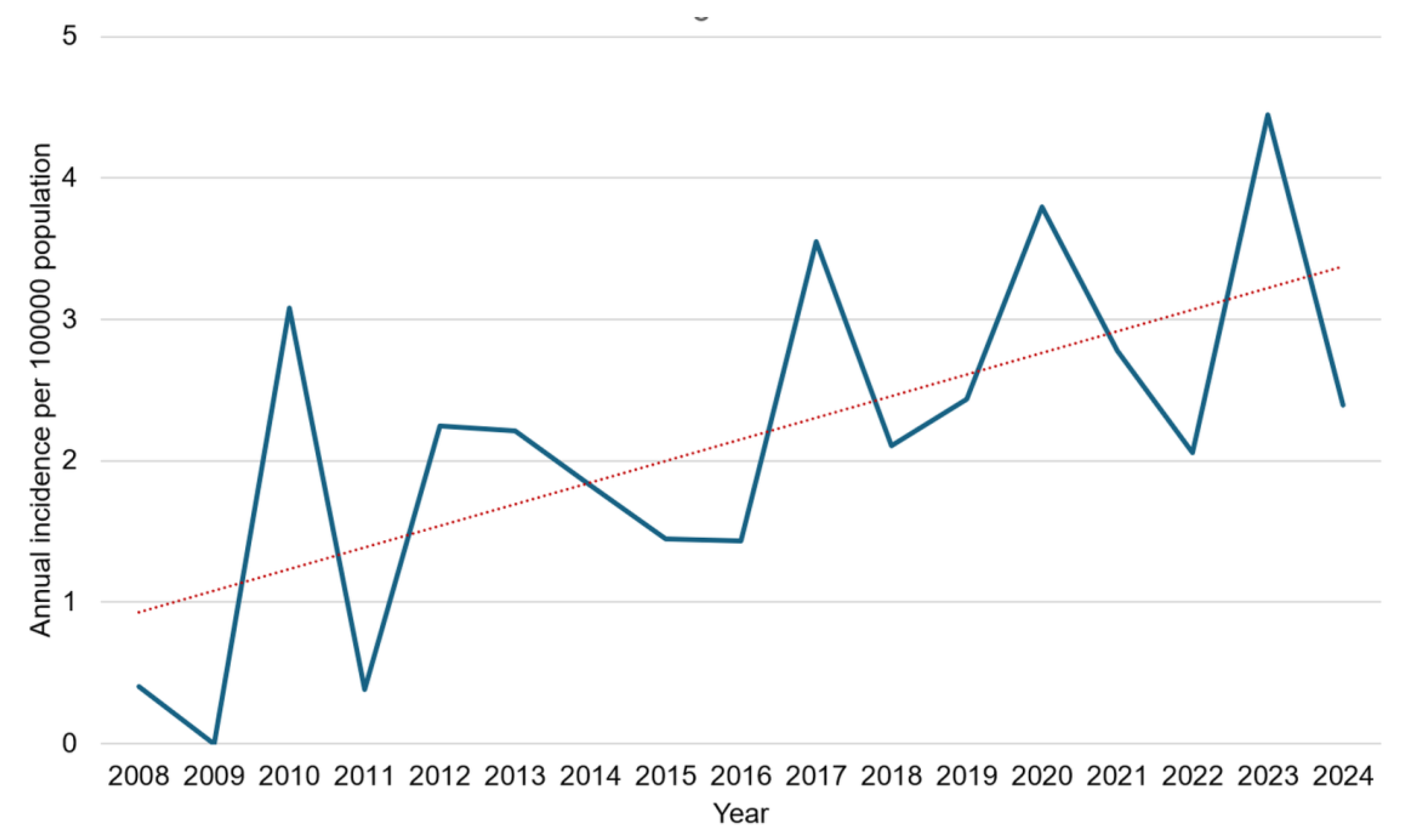

Table 1.
Demographic information and clinical presentation of patients with extrapulmonary RGM infection in FNQ 2008-2024.
Table 1.
Demographic information and clinical presentation of patients with extrapulmonary RGM infection in FNQ 2008-2024.
| Clinical information |
Total n=103 |
M. abscessuscomplex n=43 |
M. fortuitumgroup n=41 |
M. chelonae n=10 |
Other RGM n=9 |
||
| Age ≥ 65 years | 32 (31) | 10 (23) | 15 (37) | 4 (40) | 3 (33) | ||
| Infection site | |||||||
| Skin and soft tissue | 71 (69) | 26 (62) | 29 (71) | 8 (80) | 8 (89) | ||
| Lower limb/foot | 34 | 15 | 12 | 6 | 1 | ||
| Upper limb/hand | 14 | 6 | 5 | 1 | 2 | ||
| Chest/Abdomen | 14 | 3 | 9 | 1 | 1 | ||
| Breast | 7 | 1 | 2 | 0 | 4 | ||
| Head/neck | 2 | 1 | 1 | 0 | 0 | ||
| Bone and joint | 16 (16) | 4 | 9 | 2 | 1 | ||
| Catheter-associated | 9 (9) | 7 | 2 | 0 | 0 | ||
| Blood stream | 3 (3) | 2 | 1 | 0 | 0 | ||
| Lymph node | 2 (2) | 2 | 0 | 0 | 0 | ||
| Urinary tract | 1 (1) | 1 | 0 | 0 | 0 | ||
| Eye | 1 (1) | 1 | 0 | 0 | 0 | ||
| Immunosuppression | 10 (10) | 4 (10) | 3 (7) | 3 (30) | 0 | ||
| Charlson Comorbidity Index score ≥5 | 6 (6) | 3 (7) | 2 (49) | 1 (10) | 0 | ||
| Post traumatic | 39 (38) | 10 (23) | 24 (59) | 3 (30) | 2 (22) | ||
| Post-surgical a | 38 (37) | 14 (33) | 17 (41) | 2 (20) | 5 (56) | ||
| Prosthetic material involved | 23 (22) | 10 (23) | 9 (22) | 1 (10) | 3 (33) | ||
Absolute number and percentage are presented. RGM: rapidly growing mycobacteria. a Infection following trauma with primary surgical management are not included in the post-surgical infection group.
Table 2.
Antimicrobial susceptibility testing of 84 clinical extrapulmonary RGM isolates from FNQ 2011-2024.a.
Table 2.
Antimicrobial susceptibility testing of 84 clinical extrapulmonary RGM isolates from FNQ 2011-2024.a.
| M. abscessuscomplex n = 40 | M. fortuitumgroup n = 37 | M. chelonaen = 7 | |||||||
| Antimicrobial |
S n (%) |
I n (%) |
R n (%) |
S n (%) |
I n (%) |
R n (%) |
S n (%) |
I n (%) |
R n (%) |
| Cefoxitin | 1 (3) | 34 (85) | 5 (13) | 5 (14) | 29 (78) | 4 (11) | 0 (0) | 0 (0) | 7 (100) |
| TMP/SMX | 0 (0) | 0 (0) | 40 (100) | 27 (73) | 0 (0) | 10 (27) | 0 (0) | 0 (0) | 7 (100) |
| Moxifloxacin | 0 (0) | 0 (0) | 40 (100) | 37 (100) | 0 (0) | 0 (0) | 0 (0) | 1 (14) | 6 (86) |
| Doxycycline | 0 (0) | 0 (0) | 40 (100) | 14 (38) | 0 (0) | 23 (62) | 3 (43) | 0 (0) | 4 (57) |
| Imipenem | 3 (8) | 32 (80) | 5 (13) | 14 (38) | 20 (54) | 2 (5) | 0 (0) | 4 (57) | 3 (43) |
| Linezolid | 8 (21) | 20 (51) | 12 (30) | 26 (70) | 7 (19) | 4 (11) | 3 (43) | 2 (29) | 2 (29) |
| Amikacin | 39 (98) | 1 (3) | 0 (0) | 35 (95) | 2 (5) | 0 (0) | 5 (71) | 2 (29) | 0 (0) |
| Ciprofloxacin | 0 (0) | 2 (5) | 38 (95) | 35 (95) | 2 (5) | 0 (0) | 0 (0) | 1 (10) | 6 (86) |
| Tobramycin | 0 (0) | 3 (8) | 37 (93) | 3 (8) | 9 (24) | 25 (68) | 7 (100) | 0 (0) | 0 (0) |
| Clarithromycin | 18 (51) | 0 (0) | 17 (49) | 19 (51) | 5 (14) | 13 (35) | 7 (100) | 0 (0) | 0 (0) |
a Susceptibility testing only presented for the most common organisms: M. abscessus complex, M. fortuitum group and M. chelonae. Susceptibility testing by microbroth dilution M. abscessus complex, M. fortuitum group and M. chelonae isolates. Clarithromycin susceptibility testing for M. abscessus complex isolates interpreted following 14-day incubation, 5 M. abscessus complex isolates did not have clarithromycin 14-day incubation results available. All other results were interpreted at 3-5 days of incubation. CLSI methods and interpretive criteria were used; CLSI M42-A2ref (2011-2018), CLSI M62-Ed1ref (2018-2023), CLSI M24s-Ed2ref (2023-2024) [23,24,25], 14-day clarithromycin incubation for M. abscessus complex isolates was routinely undertaken from 2013. Abbreviations: S, susceptible; I, intermediate; R, resistant; TMP/SMX, Trimethoprim/sulfamethoxazole.
Table 3.
Treatment and clinical outcomes.
| Total clinical cases | n=103 | % or IQR |
| Treatment (clinical information available) | 91 | 88% |
|
2 | 3% |
|
25 | 27% |
|
11 | 12% |
|
53 | 58% |
|
70 | 77% |
|
18 | 16-24 |
| Clinical outcomes | ||
|
75 | 82% |
|
18 | 17% |
|
12 | 11% |
|
6 | 6% |
|
0 | |
|
3 | 3% |
NTM: non-tuberculous mycobacteria. IQR: interquartile range. a Included case with SPC removal and a case of wound management with vacuum-assisted closure (VAC) therapy. b Surgical and/or medical. c Lost to follow-up excludes patients whose charts contain documentation of ongoing follow-up without clinical/microbiological evidence of recurrence despite not formally being reviewed in Infectious Diseases clinic.
Disclaimer/Publisher’s Note: The statements, opinions and data contained in all publications are solely those of the individual author(s) and contributor(s) and not of MDPI and/or the editor(s). MDPI and/or the editor(s) disclaim responsibility for any injury to people or property resulting from any ideas, methods, instructions or products referred to in the content. |
© 2026 by the authors. Licensee MDPI, Basel, Switzerland. This article is an open access article distributed under the terms and conditions of the Creative Commons Attribution (CC BY) license (http://creativecommons.org/licenses/by/4.0/).
Copyright: This open access article is published under a Creative Commons CC BY 4.0 license, which permit the free download, distribution, and reuse, provided that the author and preprint are cited in any reuse.