

Submitted:
29 September 2026
Posted:
29 September 2026
You are already at the latest version
Abstract
Myocarditis is a dynamic disease and myocardial oedema, injury, ventricular dysfunction, and fibrosis may evolve differently over time. Cardiovascular magnetic resonance (CMR) has become central to its non-invasive assessment, providing complementary information on myocardial tissue through T2-sensitive imaging, native T1, extracellular volume (ECV), and late gadolinium enhancement (LGE). The meaning of these findings, however, depends on when imaging is performed. Follow-up CMR may therefore provide a better understanding of myocardial recovery, helping to distinguish resolving disease from persistent abnormalities or healed injury with residual scar. Based on the temporal evolution of CMR findings, we propose four conceptual patterns of myocardial recovery: near-complete imaging recovery, resolving inflammatory injury, persistent inflammatory imaging abnormalities, and healed injury with residual LGE/scar-like substrate. These patterns are intended as a framework for interpreting serial CMR rather than as formally validated imaging phenotypes. In selected patients, integration with echocardiography, molecular imaging, genetics, and endomyocardial biopsy may provide further insight into the underlying disease process. Whether this longitudinal approach can improve risk stratification and ultimately guide therapeutic decisions remains an important question for future studies.
Keywords:
myocarditis
; cardiac magnetic resonance
; myocardial inflammation
; tissue characterization
; parametric mapping
; longitudinal imaging
1. Introduction
Myocarditis is an inflammatory disease of the myocardium that may result from infectious agents, exposure to toxic substances, or immune-mediated mechanisms. Its clinical course is highly variable: while most cases resolve spontaneously, myocarditis may occasionally present with severe myocardial injury or sudden cardiac death and, in a subset of patients, persistent inflammation and myocardial scarring may lead to adverse ventricular remodelling and eventually to dilated or hypokinetic non-dilated cardiomyopathy. Acute myocarditis generally refers to disease presenting within one month of symptom onset, whereas chronic inflammatory cardiomyopathy is characterized by persistent myocardial inflammation in the setting of an established cardiomyopathic phenotype [1].
The 2025 ESC Guidelines introduced a revised conceptual framework for myocarditis and pericarditis, recognizing these conditions as part of a broader spectrum termed inflammatory myopericardial syndrome (IMPS). IMPS is proposed as an umbrella term during the initial diagnostic work-up, encompassing isolated myocarditis and pericarditis as well as overlapping forms, including myopericarditis and perimyocarditis, until a more specific diagnosis is established [3]. This approach reflects the shared inflammatory nature, overlapping clinical presentations, and frequent coexistence of myocardial and pericardial involvement, moving beyond the traditional view of these conditions as separate entities. Importantly, the new framework also emphasizes a clinically driven diagnostic pathway in which multimodality cardiovascular imaging, particularly cardiovascular magnetic resonance (CMR), plays a central role in the non-invasive identification of myocardial inflammation and in defining the predominant pattern of cardiac involvement. Within this framework, imaging is not merely supportive of the diagnosis but contributes to phenotypic characterization by defining the presence, distribution, and extent of myocardial and pericardial involvement. However, CMR findings should not be considered diagnostic of myocarditis in isolation, as tissue abnormalities such as myocardial oedema, increased native T1 or T2, and non-ischaemic late gadolinium enhancement (LGE) are not pathognomonic and may occur in other myocardial diseases. Accordingly, the diagnosis of myocarditis remains an integrated clinical diagnosis, requiring CMR findings to be interpreted in conjunction with the clinical presentation, biomarkers, ECG and, in selected cases, endomyocardial biopsy and other diagnostic investigations. The IMPS concept therefore reinforces the need to interpret imaging findings as part of a continuous and potentially evolving inflammatory spectrum rather than within rigid diagnostic categories. This paradigm is particularly relevant to longitudinal imaging, as the relative contribution of myocardial inflammation, pericardial involvement, ventricular dysfunction, and residual structural abnormalities may change throughout the course of the disease.
Historically, much of the diagnostic framework for myocarditis has been organized around a binary question on the presence or absence of myocardial inflammation. This approach is understandable because establishing the diagnosis remains challenging. Endomyocardial biopsy (EMB) provides unique histological, immunohistochemical, and potentially etiological information but is not required in every patient and remains particularly important in high-risk presentations or when the identification of a specific histological subtype may have a therapeutic impact [1,2,3]. CMR, in contrast, has emerged as the principal non-invasive technique for myocardial tissue characterization and plays a central role in the assessment of clinically suspected myocarditis. Accordingly, the 2025 ESC Guidelines recommend CMR in patients with clinically suspected myocarditis for the non-invasive diagnosis of myocardial inflammation using the updated Lake Louise Criteria (Class I, Level of Evidence B); within this framework, CMR-proven myocardial involvement in an appropriate clinical presentation can establish a definite clinical diagnosis of myocarditis, while tissue characterization and the spatial distribution of myocardial injury contribute to distinguishing inflammatory injury from alternative causes of acute myocardial damage [3].
The Lake Louise Criteria (LLC), first proposed in 2009, established a CMR-based approach to the diagnosis of myocarditis using three main features of myocardial inflammation: oedema, hyperaemia and increased capillary permeability, and non-ischaemic myocardial injury identified by LGE. The criteria were subsequently revised in 2018 to incorporate quantitative tissue characterization with parametric mapping, improving the ability of CMR to detect myocardial inflammatory changes. According to the updated LLC, CMR findings support acute myocardial inflammation when at least one T2-based criterion, reflecting increased myocardial water content, is combined with at least one T1-based criterion indicating myocardial injury. This approach therefore integrates complementary information on the two major components of acute myocarditis: inflammatory oedema and tissue damage [3,4]. An improved ability of CMR to characterize acute myocarditis and a reinforced role in contemporary diagnostic algorithms were both achieved.
Besides CMR, other imaging modalities may provide useful information in patients with suspected myocarditis. Transthoracic echocardiography is generally the first imaging examination performed, although its diagnostic specificity is limited. Findings range from a completely normal examination to regional wall motion abnormalities, global left ventricular dysfunction, increased wall thickness related to myocardial oedema, right ventricular involvement, and pericardial effusion. Echocardiography also has an important role in the assessment of haemodynamic status and in monitoring changes in ventricular function during the acute phase. Speckle-tracking echocardiography may provide additional information, as impaired global longitudinal strain can be detected even when left ventricular ejection fraction is preserved [5].
Cardiac computed tomography (CT) is particularly useful in patients presenting with chest pain and elevated troponin levels, in whom coronary artery disease needs to be excluded. In appropriately selected patients, coronary CT angiography provides a non-invasive alternative to invasive coronary angiography and may help exclude obstructive coronary artery disease in selected patients with an infarct-like presentation. Cardiac CT can also provide information on left ventricular function, including the presence of global or regional wall motion abnormalities. More recently, advances in CT technology have expanded its potential beyond coronary and functional assessment. Late iodine enhancement and CT-derived extracellular volume quantification allow myocardial tissue characterization and may provide additional information on myocardial injury. However, despite promising initial results, the clinical role of these techniques in patients with suspected myocarditis remains to be fully established [6,7,8].
18F-fluorodeoxyglucose positron emission tomography (FDG-PET) provides an alternative approach for the assessment of myocardial inflammation by detecting the increased glucose metabolism of activated inflammatory cells. Although FDG-PET is well established in the evaluation of cardiac sarcoidosis, its role in the routine diagnostic work-up of myocarditis remains more limited and even challenging, especially in patients in whom irregular heart rhythms may affect image quality and is highly dependent on patient preparation. The presence of an implantable cardioverter-defibrillator (ICD) can generate susceptibility artefacts that interfere with myocardial tissue characterization [9,10,11,12].
The complementary roles, strengths, and limitations of cardiovascular imaging modalities in myocarditis are summarized in Table 1.
However, the diagnostic phase represents only the beginning of the disease trajectory.
Myocarditis is an intrinsically dynamic disease, in which inflammatory infiltration, myocardial oedema, myocyte injury, necrosis, and subsequent repair and fibrosis evolve over time and at different rates. Importantly, clinical and biochemical recovery do not necessarily parallel resolution of myocardial abnormalities, as CMR signs of myocardial injury may persist despite normalization of symptoms and cardiac biomarkers. Likewise, recovery of left ventricular systolic function does not invariably imply complete tissue healing, and residual LGE may remain detectable after resolution of myocardial oedema. Conversely, both oedema and LGE observed during the acute phase may substantially decrease over the following weeks or months, highlighting the importance of the timing of imaging assessment and, when clinically indicated, serial CMR evaluation. Consequently, CMR findings should be interpreted in relation to the timing of imaging, as they reflect a disease process that evolves over time. [13,14,15].
These observations support a longitudinal approach to cardiovascular imaging in myocarditis, as the significance of imaging findings changes throughout the course of the disease. At presentation, the main goal is to identify active myocardial inflammation, whereas follow-up imaging may help assess its resolution or persistence and, at later stages, characterize the residual myocardial substrate.
In this review, we examine myocarditis from this longitudinal imaging perspective, focusing on CMR as the key imaging modality and on the transition from active inflammation to resolving injury, persistent abnormalities, and residual scar. We also discuss the implications of these findings for prognosis, arrhythmic risk stratification, follow-up, return to exercise, and imaging-guided management.
2. What Are We Imaging in Myocarditis with CMR?
CMR provides a multiparametric assessment of suspected myocarditis by combining cardiac morphology and function with complementary markers of myocardial tissue composition. A comprehensive examination should therefore be considered as a sequence of acquisitions addressing different components of myocardial injury rather than as a single diagnostic test. Cine imaging defines the functional phenotype, T2-sensitive imaging and parametric mapping assess myocardial water content and tissue composition, ECV quantifies extracellular expansion, whereas LGE primarily depicts focal differences in gadolinium distribution. The integration of these findings, rather than any individual CMR abnormality, forms the basis for the non-invasive characterization of myocardial inflammation [4,23].
Importantly, the diagnostic performance of CMR is strongly influenced by the timing of imaging. Diagnostic accuracy is highest when CMR is performed early in the disease course, ideally within the first 2 weeks after symptom onset, when myocardial oedema and other inflammation-related tissue abnormalities are most likely to be detectable; as these abnormalities progressively resolve, the sensitivity of CMR for identifying the acute inflammatory phenotype may decrease [3,70].
As illustrated in Figure 1, multiparametric CMR interrogates complementary components of myocardial injury, including myocardial water content and tissue composition, extracellular compartment expansion, and focal myocardial injury or residual scar, whose biological significance varies according to the timing of imaging and should therefore be interpreted in an integrated manner.
1.1. Cine Imaging: Morphology and Ventricular Function
A CMR examination generally begins with cine imaging, which provides a comprehensive assessment of cardiac morphology, ventricular volumes, and global and regional biventricular systolic function. In patients with myocarditis, ventricular function may range from completely preserved to severe global or regional dysfunction, depending on the extent and clinical phenotype of myocardial involvement. When present, regional wall motion abnormalities may show a non-coronary distribution, reflecting the heterogeneous and patchy nature of myocardial involvement rather than conforming to a single epicardial coronary territory. Cine imaging also allows assessment of myocardial wall thickness, which may transiently increase in the presence of substantial myocardial oedema, as well as associated findings such as pericardial effusion [3]. More specifically, acute myocarditis may result in an apparent hypertrophic or “pseudo-hypertrophic” phenotype. Histopathological studies have demonstrated that this phenomenon predominantly reflects interstitial myocardial oedema rather than true cardiomyocyte hypertrophy, with wall thickness decreasing during convalescence despite no significant change in cardiomyocyte diameter [73]. Consistently, serial CMR studies have demonstrated a reduction in LV mass during recovery from acute myocarditis in parallel with the resolution of myocardial oedema [74]. Recognition of this transient morphological phenotype is clinically relevant, as marked oedema-related wall thickening may mimic a hypertrophic phenotype during the acute phase; regression of wall thickness and myocardial mass together with resolution of oedema at follow-up supports its transient nature.
Importantly, however, preserved left ventricular ejection fraction does not exclude significant myocardial injury, particularly in patients with an infarct-like presentation [66]. This apparent dissociation can partly be explained by the complex transmural architecture of myocardial contraction. The subendocardial layer, despite accounting for less than approximately 20% of myocardial wall thickness, contains predominantly longitudinally oriented fibres and plays a major role in longitudinal left ventricular mechanics, whereas mid-wall and subepicardial fibres contribute importantly to circumferential shortening, wall thickening, and rotational mechanics [67]. Consequently, early or spatially limited impairment of one component of myocardial deformation may be partially compensated by preserved or enhanced mechanics in other myocardial layers, allowing global LVEF and visually assessed wall motion to remain apparently normal despite underlying myocardial injury [66,67]. Moreover, conventional cine imaging primarily evaluates changes in ventricular volumes, wall thickening, and macroscopic wall motion and does not directly characterize the complex three-dimensional and transmural deformation of myocardial fibres [66,68].
More sensitive measures of myocardial mechanics, including strain assessment by CMR feature tracking, tagging, or other deformation techniques, may therefore reveal subtle functional abnormalities that are not apparent from LVEF alone [66,67,68,69]. Functional assessment should consequently be considered complementary to, rather than a surrogate for, myocardial tissue characterization, since preserved global systolic function neither excludes myocardial injury nor necessarily implies complete myocardial recovery.
1.2. STIR Sequences and T2-Weighted Imaging: Acute Myocardial Injury
Once cardiac morphology and function have been evaluated, conventional T2-weighted imaging represents the first step in myocardial tissue characterization and is primarily aimed at detecting increased myocardial water content [4,26]. Acute myocardial oedema (AMO), defined as an increase in myocardial water content, is an early response to inflammatory activation and increased vascular permeability in acute myocardial injury. It initially reflects intracellular water accumulation (cytotoxic oedema), followed by expansion of the interstitial compartment (vasogenic oedema) [16,17,18]. Myocardial oedema prolongs T2 relaxation and consequently results in increased signal intensity on T2-weighted images, providing an imaging marker of increased myocardial water content [4,16,26].
Short-tau inversion recovery (STIR) sequences, typically acquired using a black-blood triple-inversion recovery technique, suppress the signal from flowing blood and fat while preserving T2-weighted contrast, thereby allowing areas of myocardial oedema to appear hyperintense relative to unaffected myocardium [26]. In acute myocarditis, increased T2-weighted signal may be regional or diffuse and commonly shows a non-ischaemic distribution. Typically, acute myocardial oedema predominantly involves the mid-myocardial or subepicardial layers and may affect different myocardial regions, with frequent involvement of the inferolateral wall [19]. Conventional T2-weighted imaging therefore provides a qualitative or semi-quantitative marker of myocardial oedema and represented the principal T2-based approach in the original Lake Louise Criteria [26].
Historically, myocardial oedema on T2-weighted imaging has been assessed either visually or semi-quantitatively using the T2 signal-intensity ratio, calculated by comparing myocardial signal intensity with that of skeletal muscle within the same image. A myocardial-to-skeletal-muscle signal-intensity ratio ≥2.0 was proposed as evidence of myocardial oedema in the original Lake Louise Criteria [26]. However, this approach has several important technical limitations. Image quality may be affected by respiratory and cardiac motion, incomplete blood suppression, surface-coil inhomogeneity, and low signal-to-noise ratio. Furthermore, skeletal muscle is not always an appropriate internal reference, particularly in systemic inflammatory disorders in which skeletal muscle may itself be involved. These limitations reduce the sensitivity of conventional T2-weighted imaging, particularly when myocardial involvement is mild or diffuse rather than focal [4,26].
These limitations have progressively favoured the use of quantitative T2 mapping, which provides direct pixel-wise measurements of myocardial T2 relaxation time rather than relying on relative signal intensity and is discussed in detail below [4,23]. Conventional T2-weighted imaging nevertheless remains useful as a qualitative assessment of the presence and spatial distribution of myocardial oedema.
1.3. Native T1 Mapping
Native T1 mapping provides a quantitative, contrast-free assessment of myocardial tissue composition. T1 represents the longitudinal relaxation time of myocardial tissue and is influenced by both intracellular and extracellular components. In normal myocardium, native T1 values reflect the composite signal arising from cardiomyocytes and the interstitial space, whereas pathological changes that increase tissue water content or expand the extracellular compartment generally result in prolongation of myocardial T1 relaxation times [4,23,58]. Native T1 values are field-strength-, sequence-, vendor-, and site-dependent; therefore, absolute measurements should always be interpreted against locally validated reference ranges rather than using universal cut-off values [23,58].
In acute myocarditis, native T1 is typically increased as a consequence of myocardial oedema, inflammation-related tissue injury, and expansion of the interstitial space. Unlike T2, which more closely reflects changes in myocardial water content, native T1 is sensitive to a broader spectrum of tissue abnormalities. Increased native T1 may therefore identify both focal and diffuse myocardial involvement and can remain abnormal even when conventional T2-weighted imaging is inconclusive [4,23,37]. Because native T1 mapping does not require administration of gadolinium-based contrast agents, it also provides tissue-characterization information when contrast administration is contraindicated or undesirable [4,23].
A major advantage of native T1 mapping is its quantitative and pixel-wise nature. Conventional LGE relies on differences in signal intensity between abnormal and relatively normal myocardium and may therefore be less sensitive to homogeneous or diffuse myocardial abnormalities. Native T1 mapping, by assigning an absolute T1 value to each voxel, can detect diffuse alterations in myocardial composition even in the absence of a visually apparent regional reference. This feature is particularly relevant in inflammatory myocardial disease, in which myocardial involvement may extend beyond areas showing visually detectable LGE [4,23,37].
However, the high sensitivity of native T1 is accompanied by limited biological specificity. An elevated native T1 value cannot, by itself, distinguish active inflammation from other processes that alter myocardial composition. T1 prolongation may result from increased free water during acute oedema, extracellular expansion, necrosis, or fibrosis, whereas other tissue alterations, such as lipid or iron accumulation, may decrease native T1 [4,23,58]. Consequently, native T1 should not be regarded as a specific marker of active myocardial inflammation, nor can an elevated T1 value alone establish a diagnosis of myocarditis. Its interpretation requires integration with T2-based markers, LGE, ventricular function, clinical presentation, and other diagnostic findings [3,4].
This distinction becomes particularly important when T1 and T2 are considered together. Concomitant elevation of native T1 and T2 in the acute clinical setting is consistent with myocardial injury occurring in the presence of increased myocardial water content, whereas persistent native T1 elevation after normalization of T2 may reflect residual extracellular expansion or structural remodelling rather than ongoing oedema. Nevertheless, these combinations represent imaging patterns rather than direct histological characterization, and CMR cannot determine from T1 values alone whether the underlying substrate represents inflammation, fibrosis, or a combination of both [4,37,43].
The temporal behaviour of native T1 may therefore provide additional information during follow-up. Native T1 values generally decrease as acute myocardial injury and oedema resolve, although their evolution may not parallel that of T2 because the two parameters reflect partially different tissue properties [37,56]. Persistent native T1 abnormalities after normalization of T2 may identify residual myocardial alterations and should be interpreted together with ECV and LGE to help characterize whether the imaging phenotype is evolving from an oedema-dominant acute injury toward a more stable pattern of structural remodelling [37,43,56]. Accordingly, the greatest value of native T1 in myocarditis lies not in its interpretation as an isolated biomarker, but in its integration within a multiparametric and longitudinal CMR assessment.
1.4. T2 Mapping
Unlike conventional T2-weighted imaging, which relies on relative differences in signal intensity between myocardium and reference tissues, T2 mapping generates a pixel-wise parametric map in which each voxel is assigned an absolute T2 relaxation time, expressed in milliseconds [4,23]. Different acquisition strategies are available, most based on T2-prepared balanced steady-state free-precession sequences acquired with different T2 preparation times, from which the myocardial T2 relaxation curve is derived [23].
The biological basis of T2 mapping relies on the close relationship between T2 relaxation and tissue water content. In acute myocardial injury, intracellular and interstitial water accumulation prolongs T2 relaxation, resulting in increased myocardial T2 values. In acute myocarditis, elevated T2 therefore provides quantitative evidence of increased myocardial water content and represents a T2-based criterion within the updated Lake Louise Criteria [4]. Compared with native T1, T2 is more closely related to tissue water content and is less influenced by chronic extracellular expansion and fibrosis, making it particularly useful for identifying the oedematous component of acute myocardial injury [4,23,37].
An important advantage of T2 mapping over conventional T2-weighted imaging is its ability to detect diffuse myocardial abnormalities. When myocardial oedema is widespread, conventional T2-weighted techniques may fail to demonstrate a clear regional difference because apparently normal myocardium may no longer provide an adequate internal reference. Similarly, calculation of the myocardial-to-skeletal-muscle signal-intensity ratio may be unreliable when skeletal muscle is affected by systemic inflammatory disease. By providing direct quantitative measurements of myocardial T2 without requiring comparison with skeletal muscle, T2 mapping partly overcomes these limitations and improves the detection of diffuse or subtle increases in myocardial water content [4,23].
Nevertheless, T2 mapping should not be regarded as an absolute or technique-independent measurement. Normal T2 values depend on magnetic field strength, pulse sequence, acquisition parameters, scanner platform, and post-processing methodology, and substantial differences may exist between centres. Consequently, T2 values should be interpreted using sequence- and site-specific reference ranges rather than universal numerical cut-offs [23]. Technical factors including motion, partial-volume effects, inadequate breath-holding, susceptibility artefacts and contamination from blood or adjacent tissues may also influence measured T2 values, particularly in the subendocardial and subepicardial regions [23].
As with conventional T2-weighted imaging, elevated myocardial T2 is not specific for myocarditis. T2 mapping detects increased tissue water content rather than the underlying aetiology and may therefore be abnormal in other conditions characterized by acute myocardial oedema, including myocardial infarction and stress cardiomyopathy [4,23]. Accordingly, an elevated T2 value should not be interpreted as direct evidence of inflammatory cell infiltration or as sufficient to establish a diagnosis of myocarditis. Its diagnostic value derives from integration with T1-based markers of myocardial injury, the distribution of abnormalities, and the overall clinical presentation [3,4].
The temporal evolution of T2 abnormalities is particularly relevant in myocarditis. Owing to its quantitative nature, T2 mapping allows changes in myocardial water content to be assessed serially over time. In uncomplicated acute myocarditis, myocardial T2 values generally decrease during follow-up, broadly paralleling resolution of myocardial oedema [14,15,56]. A progressive decline toward the institutional reference range is therefore consistent with resolution of the oedematous component of myocardial injury, whereas persistently or recurrently elevated T2 may indicate delayed resolution of myocardial oedema and, in the appropriate clinical context, raise suspicion of ongoing inflammatory activity [14,15,56]. However, the timing of T2 normalization is variable, and no universally validated T2 threshold can distinguish active from healed myocarditis. Persistent T2 elevation should therefore not be considered equivalent to histologically proven active inflammation.
The relationship between T2 and the other tissue markers becomes particularly informative during follow-up. Concomitant elevation of T2 and native T1 in the appropriate clinical setting supports an oedema-rich acute myocardial injury phenotype, whereas normalization of T2 with persistence of T1 abnormalities or LGE suggests a transition away from predominantly oedematous injury toward residual extracellular or structural abnormalities [37,43,56]. Thus, rather than providing a binary marker of active versus inactive myocarditis, T2 mapping contributes to a multiparametric characterization of the myocardial phenotype and, particularly when assessed serially, may help define the transition from acute injury toward myocardial healing.
The prognostic significance of myocardial oedema is less straightforward. Rather than representing an adverse prognostic marker per se, acute myocardial oedema reflects an active and potentially reversible component of myocardial injury [20,21]. However, neither the presence nor the extent of myocardial oedema independently predicted long-term outcome, for which impaired baseline LVEF remained the main independent CMR predictor [21]. Greater global myocardial inflammation has similarly been associated with a higher likelihood of subsequent recovery of LVEF [22]. These observations suggest that the prognostic meaning of an oedema-dominant acute phenotype may differ from that of persistent structural myocardial injury, although the mechanisms underlying these associations remain incompletely understood [20,21,22]. Accordingly, myocardial oedema should not be considered as an isolated prognostic marker but interpreted together with ventricular function, LGE burden and distribution, timing of CMR, arrhythmic presentation, and the overall clinical phenotype [3,31,33].
1.5. Contrast Agent Administration and Extracellular Volume (ECV)
Following administration of a gadolinium-based extracellular contrast agent, post-contrast T1 mapping can be combined with native T1 measurements and blood haematocrit to calculate the myocardial ECV. ECV represents the proportion of myocardial tissue occupied by the extracellular compartment and is derived from the relative change in myocardial and blood T1 relaxation rates before and after contrast administration, corrected for the patient’s haematocrit [23,58]. Because gadolinium distributes within the extracellular space but does not cross intact cell membranes, ECV provides a quantitative estimate of extracellular space expansion and, unlike LGE, does not rely primarily on the presence of a relatively normal myocardial reference region. Post-contrast T1 mapping is generally performed after sufficient contrast equilibration, typically approximately 10–30 minutes after contrast administration, although acquisition protocols should follow sequence- and site-specific recommendations [23].
In acute myocarditis, ECV may increase because of interstitial oedema, tissue injury and extracellular matrix expansion [23,43]. Importantly, therefore, increased ECV during the acute phase should not be interpreted as synonymous with myocardial fibrosis. The measured extracellular space may be expanded by both potentially reversible inflammatory oedema and irreversible structural changes, and these processes may coexist within the same myocardium. Accordingly, ECV provides quantitative information on the extent of extracellular expansion but has limited ability to determine its underlying biological substrate when considered in isolation [23,43].
This distinction is particularly important when ECV is interpreted together with native T1 and T2 mapping. Whereas T2 is more closely related to myocardial water content, native T1 is influenced by changes in both intracellular and extracellular tissue composition, while ECV more specifically quantifies expansion of the extracellular compartment. In the acute setting, concomitant increases in T2, native T1, and ECV may therefore characterize an oedema-rich inflammatory injury phenotype. Conversely, normalization of T2 accompanied by persistent ECV expansion may suggest that the extracellular abnormality is no longer predominantly driven by oedema and may increasingly reflect residual structural remodelling [37,43]. Nevertheless, such combinations remain imaging phenotypes rather than direct histological measurements, and neither ECV nor its temporal evolution can independently distinguish inflammation from fibrosis.
The longitudinal interpretation of ECV is therefore attractive but complex. In principle, a reduction in ECV occurring in parallel with normalization of T2 may reflect resolution of inflammatory oedema and contraction of the previously expanded extracellular compartment, whereas persistently increased ECV after resolution of oedema-sensitive abnormalities may be consistent with residual extracellular matrix expansion and fibrotic remodelling [24,43]. This temporal framework may be particularly useful in myocarditis because the biological meaning of an elevated ECV is likely to change throughout the disease course: extracellular expansion during the acute phase may contain a substantial reversible component, while persistent abnormalities at later follow-up are more likely to represent structural myocardial alteration.
However, evidence supporting the prognostic significance of serial ECV changes in myocarditis remains limited, and validated thresholds distinguishing reversible inflammatory extracellular expansion from irreversible fibrosis are currently lacking. ECV should therefore not be interpreted as a stand-alone marker of either active inflammation or myocardial scar. Rather, its greatest value lies in its integration with T2 mapping, native T1, LGE, ventricular function, and the clinical course, particularly when serial CMR examinations are available. In this context, ECV may contribute to characterizing the transition from acute inflammatory injury to healing or persistent structural remodelling, while avoiding the oversimplification that extracellular expansion necessarily represents fibrosis.
1.6. LGE: Myocardial Injury and Scar Like Substrate
LGE imaging is generally performed approximately 10–15 minutes after administration of a gadolinium-based contrast agent, according to protocol, using inversion-recovery sequences in which the signal of normal myocardium is nulled. Regions with relatively greater gadolinium retention appear hyperintense, allowing the detection and spatial characterization of focal myocardial injury [23,26]. Unlike ECV, which provides a quantitative estimate of extracellular space expansion, LGE primarily provides spatial information on the presence, distribution, and extent of regional myocardial abnormalities, making the anatomical pattern of enhancement an integral component of its interpretation.
In acute myocarditis, LGE characteristically follows a non-ischaemic distribution, most commonly involving the subepicardial or mid-myocardial layers, with a frequent predilection for the inferolateral wall [4,26,27]. This pattern differs from the subendocardial or transmural enhancement conforming to a coronary vascular territory that characterizes ischaemic myocardial injury. Nevertheless, considerable heterogeneity exists, and LGE may involve other myocardial regions according to the underlying aetiology and clinical phenotype. In particular, mid-wall septal involvement has been repeatedly described and appears to carry important prognostic implications [29,31,32,33].
Importantly, LGE should not be regarded as synonymous with myocardial fibrosis, particularly when CMR is performed during the acute phase. Acute inflammatory injury may produce enhancement that subsequently decreases or disappears as the acute process resolves. Serial CMR studies have demonstrated substantial reductions in LGE burden during follow-up, indicating that at least part of the enhancement observed at presentation does not represent irreversible replacement fibrosis [14,15]. Consequently, the biological meaning of LGE is inherently dependent on the timing of imaging.
This temporal dependence becomes particularly relevant after resolution of myocardial oedema. Persistent LGE in the presence of normalized T2 is more likely to represent residual structural myocardial damage and replacement fibrosis than enhancement observed during the acute inflammatory phase [14,15]. This distinction, however, remains inferential, as CMR cannot directly determine tissue histology. Accordingly, persistent LGE should be considered an imaging finding consistent with residual post-inflammatory scar rather than direct proof of mature fibrosis. Conversely, reduction or disappearance of LGE during follow-up supports the concept that acute enhancement may contain a reversible component and highlights the limitations of using a single acute CMR examination to define the definitive scar burden.
Beyond its diagnostic role, LGE provides important prognostic and arrhythmic information. Its prognostic significance appears to depend not only on its presence or extent but also on its anatomical distribution [29,30,31,32,33]. Septal, particularly mid-wall septal, LGE has been associated with a higher risk of adverse cardiovascular events and incomplete recovery, including among patients with preserved LVEF [29,31,32]. Meta-analytic evidence further supports an association between LGE and adverse outcomes in myocarditis, although differences in patient populations, timing of CMR, and definitions of clinical endpoints should be considered when interpreting these data [33,35].
The prognostic significance of LGE may itself evolve over time. Acute LGE burden may be less informative than residual LGE assessed after myocardial oedema has resolved, when enhancement is more likely to reflect a stable structural substrate [14,15]. In this setting, persistent scar may provide an anatomical substrate for ventricular arrhythmias even after normalization of ventricular function and resolution of other markers of acute inflammation. Thus, the clinically relevant post-myocarditis LGE phenotype should be considered multidimensional and time-dependent, incorporating presence, extent, anatomical distribution, relationship with oedema, and temporal evolution, rather than simply classifying LGE as present or absent.
1.7. Pattern Matters
Beyond its presence and extent, the anatomical distribution of LGE is a fundamental component of CMR interpretation. In myocarditis, LGE typically follows a non-ischemic pattern, most commonly involving the subepicardial or mid-myocardial layers, with a predilection for the basal-to-mid inferolateral wall [4,26,27]. Conversely, myocardial infarction characteristically produces LGE beginning in the subendocardium, reflecting the greater vulnerability of this layer to ischemia, with variable extension toward the epicardium according to the severity and duration of coronary occlusion. Thus, an ischemic pattern is defined not simply by the transmural extent of enhancement, but by involvement of the subendocardium in a distribution corresponding to the perfusion territory of an epicardial coronary artery [4,26].
Accordingly, the distinction between ischemic and non-ischemic injury is generally straightforward when LGE is confined to the subepicardial or mid-myocardial layers, as these patterns strongly favor a non-ischemic mechanism. However, myocarditis is not invariably confined to these layers. In more extensive inflammatory injury, enhancement may progressively involve a greater proportion of myocardial wall thickness and may occasionally become transmural, including the subendocardium [4]. Transmural LGE should therefore not automatically be considered synonymous with myocardial infarction. In this setting, the overall spatial distribution of the lesion becomes critical: transmural enhancement conforming to a recognizable coronary vascular territory strongly favors an ischemic mechanism, whereas patchy or multifocal transmural enhancement extending beyond a single coronary territory may still be compatible with inflammatory myocardial injury.
Importantly, transmurality should not be considered synonymous with an ischemic origin. Although myocarditis typically produces subepicardial or mid-wall LGE while ischemic injury characteristically involves the subendocardium and follows a coronary distribution, inflammatory lesions may occasionally show transmural extension. Indeed, transmural extension of otherwise non-ischemic LGE has been documented in patients with infarct-like myocarditis [50]. Once the full myocardial thickness is involved, however, transmurality alone becomes less informative regarding the underlying mechanism of injury, as completed myocardial infarction may also produce transmural LGE. Accordingly, differentiation between inflammatory and ischemic injury should rely primarily on the overall spatial distribution of enhancement and its relationship to coronary territories, together with the remaining CMR findings and clinical presentation, rather than on transmurality per se [26,50,51].
This distinction is particularly relevant in patients with an infarct-like presentation, in whom chest pain, troponin elevation and electrocardiographic abnormalities may occur in both myocarditis and acute myocardial infarction. Co-localization of transmural LGE with myocardial oedema does not by itself resolve this differential diagnosis. A subendocardial-to-transmural lesion following a coronary territory should raise strong suspicion of myocardial infarction, whereas a patchy, multifocal or non-coronary distribution, particularly when associated with additional subepicardial or mid-wall lesions elsewhere, supports a non-ischemic inflammatory process [4,26]. Coronary anatomy and the overall clinical context therefore remain essential when the LGE pattern is atypical or fully transmural.
Finally, pattern also carries prognostic information within myocarditis itself. The typical inferolateral subepicardial pattern should be distinguished from mid-wall anteroseptal involvement, which has been associated with a less favorable outcome even in patients with preserved LVEF. In the ITAMY study, anteroseptal mid-wall LGE was independently associated with adverse outcomes compared with other LGE patterns, emphasizing that the clinical significance of LGE depends not only on how much enhancement is present, but also on where within the myocardium it is located [31].
3. Myocarditis as an Imaging Trajectory
1.1. From a Diagnostic Event to a Dynamic Disease
Myocarditis should not be regarded as a single acute event, as the different biological components of the disease may evolve and recover at different rates. In most patients, the initial inflammatory response is followed by myocardial injury and a gradual resolution of inflammation, eventually leading to tissue resolution after the acute phase, with or without residual fibrosis. In some cases, however, inflammation may persist or recur, resulting in ongoing myocardial damage and, ultimately, progression towards an inflammatory cardiomyopathy. These phases are not clearly separated and may overlap considerably between individual patients [1,34].
Clinical, biochemical, functional, and tissue recovery do not necessarily occur in parallel. Symptoms and cardiac biomarkers may normalize before complete recovery of ventricular function or resolution of myocardial abnormalities. Similarly, normalization of left ventricular ejection fraction does not necessarily indicate complete myocardial healing. Thus, patients who are asymptomatic and have normal troponin levels and preserved ventricular function may still show evidence of myocardial edema or residual LGE [3].
This temporal dissociation is particularly relevant when interpreting imaging findings during follow-up. Cardiovascular imaging, and CMR in particular, provides information that cannot be inferred from clinical or biochemical recovery alone, allowing assessment of residual myocardial inflammation and structural damage throughout the course of the disease.
1.2. CMR as the Cornerstone of Non-Invasive Tissue Characterization
CMR has become central to the evaluation of suspected myocarditis because it simultaneously provides ventricular morphology, biventricular function, myocardial tissue characterization, and assessment of associated pericardial involvement [2].
The updated Lake Louise Criteria introduced a multiparametric CMR approach for the assessment of myocardial inflammation. CMR evidence of myocardial inflammation is supported by the coexistence of at least one T2-based marker of myocardial edema and one T1-based marker of myocardial injury. T2 abnormalities include increased myocardial signal intensity on T2-weighted imaging or prolonged T2 relaxation times, whereas T1-based markers comprise increased native T1, extracellular volume expansion, and LGE in a non-ischemic pattern. Additional findings, such as pericardial involvement and regional or global ventricular dysfunction, may further support the diagnosis [3,4]. These criteria remain the reference standard for CMR assessment in the acute phase. However, they were developed to identify active myocardial inflammation rather than to characterize abnormalities persisting during follow-up. In this setting, distinguishing ongoing inflammation from residual post-inflammatory scar remains a key challenge. Follow-up CMR therefore serves a different purpose, helping to distinguish persistent inflammatory alterations from residual structural damage and to better define the post-inflammatory myocardial substrate.
The diagnostic strength of CMR in myocarditis lies in the integration of complementary imaging findings rather than in any single abnormal parameter. Individual CMR markers interrogate different components of myocardial injury and may show limited diagnostic performance when considered in isolation; conversely, the concordance of multiple abnormalities increases the confidence that the observed imaging phenotype reflects clinically relevant myocardial injury. In a biopsy-referenced cohort of 132 patients with suspected myocarditis, Lurz et al. demonstrated that a multiparametric approach based on the conventional Lake Louise Criteria provided better diagnostic performance than individual CMR parameters, particularly in acute presentations. Using the presence of at least two of three conventional criteria, CMR showed a sensitivity of 76%, specificity of 54%, and diagnostic accuracy of 68% in the overall population, increasing to 81%, 71%, and 79%, respectively, in patients with acute myocarditis [70]. Importantly, however, diagnostic performance was substantially lower in patients with chronic presentations, emphasizing that the value of multiparametric CMR is strongly dependent on disease stage and timing of imaging.
The influence of the clinical phenotype was further demonstrated by Francone et al. in patients with biopsy-proven acute myocarditis. CMR sensitivity varied according to clinical presentation and the extent and type of myocardial cell injury, highlighting that the probability of detecting characteristic CMR abnormalities is not uniform across the spectrum of myocarditis [71]. Thus, even a comprehensive CMR examination may be less sensitive in phenotypes in which myocardial injury is less extensive or produces less conspicuous tissue abnormalities, reinforcing the need to interpret negative or equivocal CMR findings according to the clinical presentation and timing of the examination.
The introduction of quantitative tissue characterization has further expanded this multiparametric approach. Radunski et al. demonstrated that quantitative T1, T2, and ECV measurements provide additional diagnostic information in patients with severe myocarditis, with selected combinations of quantitative and conventional CMR parameters improving diagnostic accuracy compared with conventional criteria alone [72].
Accordingly, the diagnostic contribution of CMR should not be viewed as simple accumulation of abnormal findings, but rather as the concordant integration of complementary markers of myocardial water content, tissue composition, extracellular expansion, and focal injury. The greater the concordance of these findings within an appropriate clinical setting, the stronger the imaging evidence supporting myocarditis; nevertheless, CMR does not provide direct histological or aetiological characterization, and its diagnostic performance remains influenced by clinical phenotype, disease severity, and – critically - the timing of imaging.
4. Serial CMR and the Transition from Inflammation to Post-Acute Phase
Longitudinal CMR studies provide some of the most compelling evidence that the imaging phenotype of myocarditis changes substantially after the acute presentation.
1.1. The Six-Month Perspective
Aquaro et al. evaluated 187 patients with acute myocarditis who underwent CMR at presentation and at 6-month follow-up. Myocardial edema resolved in most patients, whereas LGE showed a more variable course, persisting in the majority but decreasing or completely disappearing in a substantial proportion of cases. These findings indicate that LGE detected during the acute phase does not necessarily represent irreversible myocardial fibrosis, as part of the initial enhancement may regress with resolution of edema and acute myocardial injury. Importantly, persistent LGE after edema resolution was associated with a worse prognosis, particularly when a mid-wall septal pattern was present [15].
Accordingly, the progressive reduction in LGE burden observed during follow-up may reflect resolution of the reversible components contributing to acute enhancement and/or progressive remodelling of the residual myocardial injury, a phenomenon commonly referred to as “scar shrinkage”. Importantly, changes in LGE extent should not be interpreted as direct evidence of histological regression of fibrosis, as CMR provides an imaging surrogate of tissue injury and direct longitudinal validation against histopathology is lacking. Accordingly, even complete disappearance of visually detectable LGE does not necessarily imply complete histological restitution, since a small residual scar may theoretically shrink below the spatial resolution of CMR. Conversely, LGE persisting after resolution of oedema is more likely to reflect the residual post-inflammatory structural substrate and may therefore provide a more representative estimate of definitive scar burden [15].
These findings emphasize the importance of considering the timing of CMR when interpreting LGE in myocarditis. While acute-phase LGE reflects myocardial injury in the setting of active inflammation, its persistence after oedema resolution more likely identifies established myocardial damage. Thus, the prognostic meaning of LGE may become clearer once the inflammatory edema has resolved.
1.2. Is Three Months Enough?
More recent data have addressed whether the definitive post-myocarditis imaging phenotype may emerge earlier.
Pieroni and colleagues performed CMR at presentation, three months, and twelve months in patients with uncomplicated acute myocarditis. Between baseline and three months, edema markedly decreased, LGE burden declined, LVEF improved, and left ventricular mass decreased. Edema persisted at three months in a minority of patients and subsequently resolved. Importantly, among patients in whom edema had resolved, relatively little additional change in LGE was observed between three and twelve months. Over long-term follow-up, the extent of LGE measured at three and twelve months was associated with persistent ventricular arrhythmias, whereas acute LGE burden showed a weaker relationship [14].
Although this was a relatively small study and its findings should not be generalized to all forms of myocarditis, it raises an important hypothesis: CMR performed after edema resolution may characterize the residual arrhythmogenic substrate more accurately than acute CMR. This distinction may have implications for both prognosis and timing of follow-up [36,37].
1.3. Evolution of Arrhythmic Risk
One of the most clinically important consequences of myocarditis is the development of ventricular arrhythmias.
The mechanisms of arrhythmogenesis are likely to change across the disease trajectory. During the acute phase, myocardial edema, inflammation, and myocyte injury may promote electrical instability by affecting ion-channel function, calcium handling, and myocardial conduction, often resulting in polymorphic ventricular arrhythmias. As inflammation subsides, residual inflammatory activity may coexist with early reparative changes and developing fibrosis, creating a heterogeneous and potentially arrhythmogenic substrate. In the post-acute phase, replacement fibrosis becomes increasingly relevant, providing areas of slow conduction that may facilitate scar-related re-entry and sustained monomorphic ventricular tachycardia. Thus, arrhythmic risk may progressively shift from an inflammation-driven mechanism in the acute phase to a predominantly scar-related substrate after myocardial healing [2,3,38,39].
A wide spectrum of arrhythmias may occur throughout the course of myocarditis. However, the arrhythmic presentation remains less well characterized than other clinical phenotypes of myocarditis. Arrhythmias may develop during active myocardial inflammation or persist after the acute phase, with considerable heterogeneity in their mechanisms and clinical manifestations. The arrhythmic phenotype also appears to vary according to disease stage, with polymorphic and irregular ventricular arrhythmias occurring more frequently during active inflammation, whereas monomorphic and regular ventricular arrhythmias are more commonly observed after the acute phase. Moreover, myocarditis itself may remain unrecognized in patients presenting primarily with arrhythmias, making available data difficult to interpret. For these reasons, arrhythmic myocarditis represents a distinct clinical scenario that requires specific considerations in terms of diagnosis, risk stratification, and management [38,39].
Advances in cardiac imaging, particularly CMR, have improved the detection and characterization of myocardial inflammation and fibrosis, while providing valuable information for arrhythmic risk stratification [4,31,40]. Indeed, it provides imaging correlates of the transition from active inflammation to inflammatory electrical instability, heterogenous healing, residual scar and re-entrant arrhythmogenic substrate. Repeat CMR after the acute phase may be considered in selected patients, particularly in those presenting with arrhythmias, persistent symptoms, impaired ventricular function, or extensive LGE at baseline. However, no established quantitative LGE threshold currently guides ICD implantation. LGE findings should therefore be interpreted in the broader context of ventricular function, arrhythmic phenotype, clinical presentation, and genetic background [1,2,3,41].
Future risk stratification may move beyond the simple assessment of LGE presence or extent, towards a more detailed characterization of scar architecture. Features such as scar heterogeneity, border-zone characteristics, spatial distribution, and transmural involvement may provide additional information on the arrhythmogenic substrate. Integration of these imaging findings with electro-anatomical mapping may further improve substrate characterization, strengthening the role of combined CMR and electrophysiological assessment in patients with previous myocarditis.
1.4. When Should Imaging Be Repeated?
The value of repeat imaging after myocarditis is increasingly recognized, although the timing of follow-up should likely be tailored to the clinical course rather than based on a fixed schedule. The 2025 ESC Guidelines recommend follow-up in all patients, combining clinical assessment with ECG, biomarkers, rhythm monitoring, echocardiography, and CMR according to disease severity and subsequent evolution [3].
Accordingly, the 2025 ESC Guidelines recommend structured follow-up in all patients with myocarditis, including CMR performed at least within the first 6 months after the index event (Class I, Level of Evidence C). Follow-up CMR contributes to the assessment of disease remission or persistence, characterization of residual myocardial abnormalities, and longitudinal risk stratification, thereby supporting individualized clinical management. Importantly, integration of CMR findings with symptoms, biomarkers, ventricular function, and rhythm assessment also contributes to determining the appropriate timing for a personalized return to work and physical activity [3]. CMR performed during the first months after the index event can provide a more complete picture of myocardial recovery by assessing whether oedema has resolved and ventricular function has recovered, while also documenting changes in LGE and the extent of residual scar. Persistent abnormalities may raise concern for ongoing myocardial injury and, when interpreted alongside the clinical course, may help determine whether further investigation is warranted and whether progression towards normal physical activity can be considered [1,15].
Current evidence supports the clinical relevance of reassessment within approximately three to six months.
Aquaro et al., in a multicentre study including patients from the ITAMY cohort, demonstrated that CMR findings continue to evolve after the acute phase, with substantial changes in both myocardial oedema and LGE over the first six months. During the acute phase, LGE should not necessarily be interpreted as established fibrosis, as it may partly reflect reversible myocardial injury and can decrease or even disappear during follow-up. In contrast, LGE that persists at 6 months after myocardial oedema has resolved is more likely to represent residual fibrosis and has been associated with a less favourable prognosis, particularly when involving the mid-wall septal region. Persistent oedema, on the other hand, may indicate that the healing process is still ongoing and that further recovery remains possible. [15].
Pieroni et al. suggested that in uncomplicated acute myocarditis, much of the edema resolution and LGE stabilization may already occur by three months [14].
These findings are not necessarily conflicting, but rather suggest that the most appropriate timing for repeat CMR should be guided by the specific clinical question it is intended to address.
Earlier repeat CMR may be considered when the clinical course remains uncertain or recovery is incomplete. Persistent or recurrent symptoms, ongoing biomarker elevation, ventricular dysfunction, or clinically relevant arrhythmias may raise concern for unresolved myocardial injury and justify reassessment before the usual follow-up interval. An earlier examination may also be useful when uncertainty about persistent inflammatory activity could influence subsequent diagnostic or management decisions [2,3].
This time point may therefore help distinguish ongoing recovery from a more stable, scar-dominant pattern and, together with clinical and rhythm assessment, contribute to decisions regarding return to more intensive physical activity [14,15,64,65].
Further CMR beyond this period may be appropriate when myocardial abnormalities have not resolved or when the subsequent clinical course raises new concerns. Persistent oedema, progressive ventricular remodelling, or substantial residual scar, particularly when accompanied by ventricular arrhythmias, may justify continued imaging surveillance and consideration of an underlying inflammatory cardiomyopathy. In these patients, the timing of additional CMR should be individualized according to the evolution of both imaging and clinical findings [3,14].
Ultimately, rather than relying on a fixed follow-up interval, the timing of repeat CMR should be tailored to the individual clinical course and to the specific information sought from the examination.
5. Longitudinal CMR: From Active Inflammation to Post-Acute Phase
1.1. Temporal Changes in Tissue Markers
Because T2 is sensitive to myocardial water content, serial T2 mapping may provide a quantitative representation of edema resolution, beyond that obtained from a single follow-up measurement. A progressive decline toward the institutional reference range is consistent with resolution of myocardial edema, whereas persistently or recurrently elevated T2 values may suggest delayed resolution or ongoing inflammatory activity [1,3,14]. Thus, the temporal trajectory of T2 may be more informative than an isolated abnormal value, although the clinical and prognostic significance of different T2 trajectories remains to be established.
Native T1 is highly sensitive to myocardial abnormalities, although its specificity is limited. In the acute phase of myocarditis, T1 elevation may reflect both myocardial edema and tissue injury, whereas persistent T1 abnormalities after edema resolution may be related to residual extracellular expansion or fibrosis. Native T1 should therefore be interpreted together with T2 and LGE rather than in isolation [4]. For instance, concomitant elevation of T1 and T2 may be consistent with active inflammatory injury, while increased T1 in the presence of normal T2 and persistent LGE may more likely reflect residual structural remodelling. Although these patterns have not been formally validated as distinct CMR phenotypes, they can illustrate the potential value of a multiparametric approach in characterizing the underlying myocardial changes over time.
According to Lurz et al, ECV can provide an indirect marker of diffuse extracellular matrix expansion and may correlate with diffuse fibrosis when significant inflammation is absent. When inflammation and fibrosis coexist, as may occur in myocarditis, ECV reflects the combined contribution of both processes and therefore cannot be interpreted as a specific measure of diffuse fibrosis alone [43].
During acute myocardial inflammation, LGE may overestimate the amount of definitive fibrosis. The interpretation changes when LGE persists after normalization of edema-sensitive markers: at this stage, the probability that enhancement represents mature replacement fibrosis is more likely. This distinction is not absolute, because CMR cannot directly determine tissue histology. Nevertheless, it is consistent with the observed temporal evolution of CMR abnormalities and provides a useful clinical framework [14,15]. However, the clinically meaningful post-myocarditis scar phenotype should be considered likely multidimensional rather than binary, focusing on presence, extent and anatomical distribution of LGE [14,31].
Taken together, these observations highlight the value of serial CMR in myocarditis, as changes in T2, T1, ECV, and LGE over time may reflect different stages of myocardial injury and healing. Their significance should therefore be interpreted according to the timing of imaging and integrated with the clinical context, rather than relying on individual CMR findings in isolation.
1.2. A “Mismatch” Between Imaging and Biology
Serial CMR may reveal a “mismatch” between imaging findings and the biological course of myocarditis, as clinical recovery, resolution of myocardial oedema, normalization of ventricular function, and regression of LGE do not necessarily occur in parallel. Moreover, the significance of individual CMR abnormalities may change over time. CMR findings should therefore be interpreted according to their temporal evolution and in the context of the overall clinical course [3,13,14,15]. Based on the temporal evolution of CMR abnormalities, different patterns of myocardial recovery may be recognized during follow-up. Although these patterns have not been formally validated as distinct imaging “phenotypes”, they may provide a useful conceptual framework for interpreting serial CMR findings, ranging from near-complete imaging recovery to resolving or persistent inflammatory abnormalities and healed myocardial injury with residual scar.
The proposed longitudinal CMR patterns, as a conceptual framework, and their key imaging characteristics are summarized in Figure 2.
1.2.1. Near-Complete Imaging Recovery
In some patients, follow-up CMR shows near-complete resolution of the abnormalities observed during the acute phase. Oedema-sensitive markers return to normal, T1 abnormalities resolve or substantially decrease, ventricular function recovers, and LGE is absent or limited to small residual areas. This pattern represents the closest CMR correlate of apparent myocardial healing [15,44]. Nevertheless, normalization of CMR findings cannot establish complete histological resolution, as CMR provides an indirect assessment of myocardial inflammation and tissue injury rather than direct histological characterization.
1.2.2. Resolving Inflammatory Injury
A different pattern is observed when CMR abnormalities improve over time without completely normalizing. Oedema-sensitive markers may remain mildly elevated, while T1 abnormalities and LGE progressively decrease. This may be compatible with resolving inflammatory injury, depending on timing and clinical context. [37]. In this setting, the evolution of imaging findings may be more informative than whether an individual measurement remains outside the local reference range. A progressive reduction in T2 and T1 values, myocardial mass, and LGE burden may therefore support ongoing myocardial healing despite incomplete imaging normalization [37].
1.2.3. Persistent Inflammatory Imaging Abnormalities
Persistent abnormalities on follow-up CMR raise greater concern for ongoing myocardial inflammation, particularly when elevated T2 is accompanied by persistent symptoms, ventricular dysfunction, biomarker elevation, arrhythmias, or other CMR abnormalities such as increased native T1. These findings, however, require cautious interpretation. CMR identifies tissue changes associated with inflammation but does not directly demonstrate inflammatory cell infiltration or establish its underlying cause. Persistent CMR abnormalities alone are therefore insufficient to diagnose chronic active myocarditis or to guide decisions regarding immunosuppressive therapy. When clinically relevant, they should be interpreted within the overall clinical picture and may warrant further investigation, including endomyocardial biopsy when histological or aetiological clarification is expected to influence management [3,45,46].
1.2.4. Post-Acute Phase Injury with Residual Scar
Another clinically relevant pattern is characterized by resolution of myocardial oedema and recovery of ventricular function despite persistent LGE. Once oedema-sensitive abnormalities have normalized, residual LGE is more likely to represent residual post-inflammatory scar/fibrotic substrate rather than ongoing inflammatory activity. This phenotype should therefore not necessarily be interpreted as evidence of active myocarditis, but rather as the structural consequence of a previous inflammatory injury. Recognizing this transition has important clinical implications, as the focus may shift from assessing persistent inflammation to evaluating the potential consequences of residual scar, particularly ventricular arrhythmias and adverse ventricular remodelling [38,39,40,41].
6. Beyond Diagnosis: The Evolving Role of CMR
Longitudinal imaging may identify disease trajectories that are not typical of self-limited myocarditis. Persistent or recurrent inflammatory abnormalities, progressive ventricular dysfunction, or an evolving arrhythmic or structural phenotype should raise suspicion of an underlying disease process and may prompt further diagnostic evaluation, including genetic testing or EMB when clinically appropriate [2,4]. This is particularly relevant for genetically mediated cardiomyopathies that may present with myocarditis-like inflammatory episodes, highlighting the increasing integration of imaging, genetics, and pathology in the evaluation of patients with recurrent or persistent myocardial inflammation [53,54].
In patients already receiving immunomodulatory therapy, serial imaging may provide complementary information on treatment response. Changes in CMR markers of myocardial inflammation, particularly T2, together with recovery of ventricular function and, when available, reduction in metabolic activity on FDG-PET, may help assess the evolution of disease activity during treatment. However, no imaging parameter has yet been validated as a specific surrogate of therapeutic response in myocarditis, and imaging findings should therefore be interpreted together with the clinical course, biomarkers, and, when indicated, histological findings [56].
A particularly promising future role of CMR in myocarditis is therapeutic phenotyping. Together with molecular imaging, it may help distinguish persistent inflammatory activity from resolving injury or established scar, potentially identifying patients more likely to benefit from targeted anti-inflammatory or immunomodulatory treatment. At present, however, this concept remains largely investigational and requires prospective validation in phenotype-guided therapeutic trials [2,3,4,55].
7. Future Directions and Knowledge Gaps
An important emerging question is whether longitudinal changes in CMR tissue markers can move beyond diagnosis toward a more comprehensive assessment of disease activity and evolution.
Standardization of parametric mapping, faster and potentially contrast-free protocols, and improved characterization of the transition from active inflammation to healing and residual scar may enhance longitudinal assessment [24,56].
Integration of CMR with other imaging modalities, biomarkers, genetics, and pathology could further refine disease phenotyping, particularly in patients with persistent or recurrent myocardial injury. In parallel, radiomics and artificial intelligence may allow extraction of quantitative imaging features beyond conventional visual assessment [60,61].
Moreover, one of the most promising applications of CMR may be its contribution to therapeutic phenotyping, with imaging used not only to characterize disease but also to identify patients more likely to benefit from specific treatments and to monitor their response. However, these applications remain largely investigational and will require standardized protocols and prospective validation in imaging-guided therapeutic trials [3].
Despite major advances, important gaps remain in the longitudinal interpretation of CMR findings in myocarditis. The natural evolution of T1 and T2 abnormalities, the significance of persistent T2 elevation, and the transition from acute LGE to established scar are not yet fully defined [3]. Although observational studies support the prognostic value of follow-up imaging, it remains uncertain whether serial CMR provides incremental information sufficient to guide therapeutic, arrhythmic, or return-to-exercise decisions [62]. Further studies are also needed to establish whether multimodality imaging can reliably distinguish persistent inflammation from inactive scar and self-limited myocarditis from inflammatory presentations of genetic cardiomyopathy [53]. Ultimately, prospective studies should determine whether imaging-guided management translates into improved clinical outcomes.
8. Conclusions
Cardiovascular imaging has substantially changed the diagnosis and longitudinal assessment of myocarditis. Yet myocarditis is not a static condition. Oedema, myocardial injury, ventricular dysfunction, and subsequent repair or fibrosis may evolve at different rates, and improvement in symptoms, biomarkers, and ventricular function does not necessarily coincide with normalization of myocardial tissue abnormalities.
CMR is particularly well suited to capture these changes. T2-sensitive imaging, native T1, ECV, and LGE provide complementary information, but their significance depends on when imaging is performed. This is especially relevant for LGE, which in the acute phase may reflect different components of myocardial injury and may regress during recovery. In contrast, LGE persisting after resolution of oedema is more likely to reflect a residual post-inflammatory scar/fibrotic substrate. Follow-up CMR should therefore be interpreted in terms of how findings have changed from the acute phase, rather than simply according to whether abnormalities are still present [56,57,58].
Serial imaging may help recognize different patterns of recovery, from resolution of inflammatory injury to persistent abnormalities or residual scar. Although these patterns still require prospective validation, they may provide a useful framework for integrating CMR with the clinical course, biomarkers, ventricular function, and rhythm assessment. This may be particularly relevant when recovery is incomplete or atypical and further investigation, including molecular imaging, genetic testing, or EMB, is being considered.
The value of imaging in myocarditis may therefore increasingly lie in its ability to follow the disease over time. A better understanding of how myocardial abnormalities evolve could refine follow-up, risk assessment, and return-to-exercise decisions and, in the future, may help identify patients who could benefit from more targeted diagnostic or therapeutic strategies.
As summarized in the Central Illustration (Figure 3), myocarditis should ultimately be viewed as a dynamic disease trajectory rather than a static imaging diagnosis, in which myocardial inflammation, tissue injury, ventricular function, and residual structural damage evolve along partially independent temporal trajectories. Integrating these complementary dimensions over time may help distinguish active inflammation, resolving injury, persistent inflammatory activity, and healed disease with or without a residual scar substrate, thereby shifting the role of cardiovascular imaging from diagnosis at a single time point towards longitudinal disease characterization.
In this evolving framework, the value of cardiovascular imaging lies not only in identifying myocardial involvement, but increasingly in defining where each patient lies along the disease trajectory and what biological substrate remains over time.
Author Contributions
CR: Data curation, Investigation, Writing – original draft, Writing – review & editing. AP: Supervision, Validation, Writing – review. RS, GA, GB, LP, MM, AG, FR: visualization.
Funding
This research received no external funding.
Informed Consent Statement
Informed consent was obtained from all subjects involved in the manuscript (CMR images).
Conflicts of Interest
The authors declare no conflicts of interest.
Abbreviations
The following abbreviations are used in this manuscript:
| AMO Acute myocardial oedema |
| CMR Cardiovascular magnetic resonance |
| CT Computed tomography |
| ECG Electrocardiogram |
| ECV Extracellular volume |
| EMB Endomyocardial biopsy |
| ESC European Society of Cardiology |
| FDG Fluorodeoxyglucose |
| FDG-PET 18F-fluorodeoxyglucose positron emission tomography |
| ICD Implantable cardioverter-defibrillator |
| LGE Late gadolinium enhancement |
| LLC Lake Louise Criteria |
| LV Left ventricular / left ventricle |
| LV-GLS Left ventricular global longitudinal strain |
| LVEF Left ventricular ejection fraction |
| PET Positron emission tomography |
| PET-CMR Positron emission tomography–cardiovascular magnetic resonance |
| STIR Short-tau inversion recovery |
| TTE Transthoracic echocardiography |
References
- Ammirati, E.; Frigerio, M.; Adler, E.D.; Basso, C.; Birnie, D.H.; Brambatti, M.; Friedrich, M.G.; Klingel, K.; Lehtonen, J.; Moslehi, J.J.; Pedrotti, P.; Rimoldi, O.E.; Schultheiss, H.P.; Tschöpe, C.; Cooper, L.T., Jr.; Camici, P.G. Management of Acute Myocarditis and Chronic Inflammatory Cardiomyopathy: An Expert Consensus Document. Circ. Heart Fail 2020, 13(11), e007405. [Google Scholar] [CrossRef]
- Writing Committee; Drazner, M.H.; Bozkurt, B.; Cooper, L.T.; Aggarwal, N.R.; Basso, C.; Bhave, N.M.; Caforio, A.L.P.; Ferreira, V.M.; Heidecker, B.; Kontorovich, A.R.; Martín, P.; Roth, G.A.; Van Eyk, J.E. 2024 ACC Expert Consensus Decision Pathway on Strategies and Criteria for the Diagnosis and Management of Myocarditis: A Report of the American College of Cardiology Solution Set Oversight Committee. J. Am. Coll. Cardiol. 2025, 85(4), 391–431. [Google Scholar] [CrossRef]
- Schulz-Menger, J.; Collini, V.; Gröschel, J.; Adler, Y.; Brucato, A.; Christian, V.; Ferreira, V.M.; Gandjbakhch, E.; Heidecker, B.; Kerneis, M.; Klein, A.L.; Klingel, K.; Lazaros, G.; Lorusso, R.; Nesukay, E.G.; Rahimi, K.; Ristić, A.D.; Rucinski, M.; Sade, L.E.; Schaubroeck, H.; Semb, A.G.; Sinagra, G.; Thune, J.J.; Imazio, M.; ESC Scientific Document Group. 2025 ESC Guidelines for the management of myocarditis and pericarditis. Eur. Heart J. 2025, 46(40), 3952–4041. [Google Scholar] [CrossRef]
- Ferreira, V.M.; Schulz-Menger, J.; Holmvang, G.; Kramer, C.M.; Carbone, I.; Sechtem, U.; Kindermann, I.; Gutberlet, M.; Cooper, L.T.; Liu, P.; Friedrich, M.G. Cardiovascular Magnetic Resonance in Nonischemic Myocardial Inflammation: Expert Recommendations. J. Am. Coll. Cardiol. 2018, 72(24), 3158–3176. [Google Scholar] [CrossRef]
- Muscogiuri, G.; Guaricci, A.I.; Cau, R.; Saba, L.; Senatieri, A.; Chierchia, G.; Pontone, G.; Volpato, V.; Palmisano, A.; Esposito, A.; Basile, P.; Marra, P.; D'angelo, T.; Booz, C.; Rabbat, M.; Sironi, S. Multimodality imaging in acute myocarditis. J. Clin. Ultrasound 2022, 50(8), 1097–1109. [Google Scholar] [CrossRef]
- Adeboye, A.; Alkhatib, D.; Butt, A.; Yedlapati, N.; Garg, N. A Review of the Role of Imaging Modalities in the Evaluation of Viral Myocarditis with a Special Focus on COVID-19-Related Myocarditis. Diagnostics 2022, 12(2), 549. [Google Scholar] [CrossRef]
- Budoff, M.J.; Dowe, D.; Jollis, J.G.; Gitter, M.; Sutherland, J.; Halamert, E.; Scherer, M.; Bellinger, R.; Martin, A.; Benton, R.; et al. Diagnostic performance of 64-multidetector row coronary computed tomographic angiography for evaluation of coronary artery stenosis in individuals without known coronary artery disease: Results from the prospective multicenter ACCURACY (Assessment by Coronary Computed Tomographic Angiography of Individuals Undergoing Invasive Coronary Angiography) trial. J. Am. Coll. Cardiol. 2008, 52, 1724–1732. [Google Scholar] [CrossRef]
- 8Steffani, S.; Piscione, M.; Gaudio, D.; Meghnagi, G.; Montella, V.; Fiorini, F.; Micillo, A.; Tagliati, C.; Asmundo, L.; Manenti, G.; et al. Diagnostic Value and Operational Recommendations for Late Iodine Enhancement and ECV Quantification in Single-Energy Computed Tomography: A Narrative Review. Diagnostics 2026, 16, 2547. [Google Scholar] [CrossRef]
- Peretto, G.; Busnardo, E.; Ferro, P.; Palmisano, A.; Vignale, D.; Esposito, A.; De Luca, G.; Campochiaro, C.; Sartorelli, S.; De Gaspari, M.; Rizzo, S.; Dagna, L.; Basso, C.; Gianolli, L.; Della Bella, P.; Sala, S. Clinical Applications of FDG-PET Scan in Arrhythmic Myocarditis. JACC Cardiovasc Imaging 2022, 15(10), 1771–1780. [Google Scholar] [CrossRef]
- Caforio, A.L.; Pankuweit, S.; Arbustini, E.; Basso, C.; Gimeno-Blanes, J.; Felix, S.B.; Fu, M.; Heliö, T.; Heymans, S.; Jahns, R.; Klingel, K.; Linhart, A.; Maisch, B.; McKenna, W.; Mogensen, J.; Pinto, Y.M.; Ristic, A.; Schultheiss, H.P.; Seggewiss, H.; Tavazzi, L.; Thiene, G.; Yilmaz, A.; Charron, P.; Elliott, P.M.; European Society of Cardiology Working Group on Myocardial and Pericardial Diseases. Current state of knowledge on aetiology, diagnosis, management, and therapy of myocarditis: a position statement of the European Society of Cardiology Working Group on Myocardial and Pericardial Diseases. Eur. Heart J. 2013, 34(33), 2636-48, 2648a-2648d. [Google Scholar] [CrossRef]
- Nensa, F.; Kloth, J.; Tezgah, E.; Poeppel, T.D.; Heusch, P.; Goebel, J.; Nassenstein, K.; Schlosser, T. Feasibility of FDG-PET in myocarditis: Comparison to CMR using integrated PET/MRI. J. Nucl. Cardiol. 2018, 25(3), 785–794. [Google Scholar] [CrossRef]
- Peretto, G.; Sala, S.; Basso, C.; Rizzo, S.; Radinovic, A.; Frontera, A.; Limite, L.R.; Paglino, G.; Bisceglia, C.; De Luca, G.; Campochiaro, C.; Sartorelli, S.; Palmisano, A.; Esposito, A.; Busnardo, E.; Villatore, A.; Baratto, F.; Cireddu, M.; Marzi, A.; D'Angelo, G.; Gulletta, S.; Vergara, P.; De Cobelli, F.; Dagna, L.; Mazzone, P.; Della Bella, P. Inflammation as a Predictor of Recurrent Ventricular Tachycardia After Ablation in Patients With Myocarditis. J. Am. Coll. Cardiol. 2020, 76(14), 1644–1656. [Google Scholar] [CrossRef]
- Berg, J.; Kottwitz, J.; Baltensperger, N.; Kissel, C.K.; Lovrinovic, M.; Mehra, T.; Scherff, F.; Schmied, C.; Templin, C.; Lüscher, T.F.; Heidecker, B.; Manka, R. Cardiac Magnetic Resonance Imaging in Myocarditis Reveals Persistent Disease Activity Despite Normalization of Cardiac Enzymes and Inflammatory Parameters at 3-Month Follow-Up. Circ. Heart Fail 2017, 10(11), e004262. [Google Scholar] [CrossRef]
- Pieroni, M.; Ciabatti, M.; Zocchi, C.; Tavanti, V.; Camporeale, A.; Saletti, E.; Fumagalli, C.; Venezia, D.; Lombardi, M.; Olivotto, I.; Bolognese, L. Optimal timing of follow-up cardiac magnetic resonance in patients with uncomplicated acute myocarditis. Int. J. Cardiol. 2024, 397, 131603. [Google Scholar] [CrossRef]
- Aquaro, G.D.; Ghebru Habtemicael, Y.; Camastra, G.; Monti, L.; Dellegrottaglie, S.; Moro, C.; Lanzillo, C.; Scatteia, A.; Di Roma, M.; Pontone, G.; Perazzolo Marra, M.; Barison, A.; Di Bella, G. “Cardiac Magnetic Resonance” Working Group of the Italian Society of Cardiology. Prognostic Value of Repeating Cardiac Magnetic Resonance in Patients With Acute Myocarditis. J. Am. Coll. Cardiol. 2019, 74(20), 2439–2448. [Google Scholar] [CrossRef]
- Sinigiani, G.; De Michieli, L.; De Conti, G.; Ricci, F.; De Lazzari, M.; Migliore, F.; Perazzolo Marra, M.; Zorzi, A.; Corrado, D.; Cipriani, A. Cardiac Magnetic Resonance-Detected Acute Myocardial Edema as Predictor of Favourable Prognosis: A Comprehensive Review. J. Cardiovasc Dev. Dis. 2023, 10(8), 319. [Google Scholar] [CrossRef]
- Eitel, I.; Friedrich, M.G. T2-weighted cardiovascular magnetic resonance in acute cardiac disease. J. Cardiovasc Magn. Reson. 2011, 13(1), 13. [Google Scholar] [CrossRef]
- Scallan, J.; Huxley, V.H.; Korthuis, R.J. Capillary Fluid Exchange: Regulation, Functions, and Pathology; Morgan & Claypool Life Sciences: San Rafael, CA, USA, 2010. [Google Scholar] [PubMed]
- Expert Panel on Cardiac Imaging; Rajiah, P.; Kirsch, J.; Bolen, M.A.; Batlle, J.C.; Brown, R.K.J.; Francois, C.J.; Galizia, M.S.; Hanneman, K.; Inacio, J.R.; Johri, A.M.; Lee, D.C.; Singh, S.P.; Villines, T.C.; Wann, S.; Zimmerman, S.L.; Abbara, S. ACR Appropriateness Criteria® Nonischemic Myocardial Disease with Clinical Manifestations (Ischemic Cardiomyopathy Already Excluded). J. Am. Coll. Radiol. 2021, 18(5S), S83–S105. [Google Scholar] [CrossRef]
- De Lazzari, M.; Zorzi, A.; Baritussio, A.; Siciliano, M.; Migliore, F.; Susana, A.; Giorgi, B.; Lacognata, C.; Iliceto, S.; Perazzolo Marra, M.; Corrado, D. Relationship between T-wave inversion and transmural myocardial edema as evidenced by cardiac magnetic resonance in patients with clinically suspected acute myocarditis: clinical and prognostic implications. J. Electrocardiol. 2016, 49(4), 587–95. [Google Scholar] [CrossRef]
- Bohbot, Y.; Garot, J.; Hovasse, T.; Unterseeh, T.; Di Lena, C.; Boukefoussa, W.; Tawa, C.; Renard, C.; Limouzineau, I.; Duhamel, S.; Garot, P.; Tribouilloy, C.; Sanguineti, F. Clinical and Cardiovascular Magnetic Resonance Predictors of Early and Long-Term Clinical Outcome in Acute Myocarditis. Front Cardiovasc Med. 2022, 9, 886607. [Google Scholar] [CrossRef]
- McLellan, A.J.; McKenzie, S.C.; Taylor, A.J. Cardiac magnetic resonance imaging predicts recovery of left ventricular function in acute onset cardiomyopathy. Heart Lung Circ. 2012, 21(1), 30–5. [Google Scholar] [CrossRef]
- Messroghli, D.R.; Moon, J.C.; Ferreira, V.M.; Grosse-Wortmann, L.; He, T.; Kellman, P.; Mascherbauer, J.; Nezafat, R.; Salerno, M.; Schelbert, E.B.; Taylor, A.J.; Thompson, R.; Ugander, M.; van Heeswijk, R.B.; Friedrich, M.G. Clinical recommendations for cardiovascular magnetic resonance mapping of T1, T2, T2* and extracellular volume: A consensus statement by the Society for Cardiovascular Magnetic Resonance (SCMR) endorsed by the European Association for Cardiovascular Imaging (EACVI). J. Cardiovasc Magn. Reson. Erratum in: J Cardiovasc Magn Reson. 2018 Feb 7;20(1):9. doi: 10.1186/s12968-017-0408-9. 2017, 19(1), 75. [Google Scholar] [CrossRef]
- Luetkens, J.A.; Homsi, R.; Dabir, D.; Kuetting, D.L.; Marx, C.; Doerner, J.; Schlesinger-Irsch, U.; Andrié, R.; Sprinkart, A.M.; Schmeel, F.C.; Stehning, C.; Fimmers, R.; Gieseke, J.; Naehle, C.P.; Schild, H.H.; Thomas, D.K. Comprehensive Cardiac Magnetic Resonance for Short-Term Follow-Up in Acute Myocarditis. J. Am. Heart Assoc. 2016, 5(7), e003603. [Google Scholar] [CrossRef]
- Aquaro, G.D.; De Gori, C.; Faggioni, L.; Parisella, M.L.; Cioni, D.; Lencioni, R.; Neri, E. Diagnostic and prognostic role of late gadolinium enhancement in cardiomyopathies. Eur. Heart J. Suppl. 2023, 25 (Suppl C), C130–C136. [Google Scholar] [CrossRef]
- Friedrich, M.G.; Sechtem, U.; Schulz-Menger, J.; Holmvang, G.; Alakija, P.; Cooper, L.T.; White, J.A.; Abdel-Aty, H.; Gutberlet, M.; Prasad, S.; Aletras, A.; Laissy, J.P.; Paterson, I.; Filipchuk, N.G.; Kumar, A.; Pauschinger, M.; Liu, P. International Consensus Group on Cardiovascular Magnetic Resonance in Myocarditis. Cardiovascular magnetic resonance in myocarditis: A JACC White Paper. J. Am. Coll. Cardiol. 2009, 53(17), 1475–87. [Google Scholar] [CrossRef]
- Mahrholdt, H.; Goedecke, C.; Wagner, A.; Meinhardt, G.; Athanasiadis, A.; Vogelsberg, H.; Fritz, P.; Klingel, K.; Kandolf, R.; Sechtem, U. Cardiovascular magnetic resonance assessment of human myocarditis: a comparison to histology and molecular pathology. Circulation 2004, 109(10), 1250–8. [Google Scholar] [CrossRef]
- Felker, G.M.; Hu, W.; Hare, J.M.; Hruban, R.H.; Baughman, K.L.; Kasper, E.K. The spectrum of dilated cardiomyopathy. The Johns Hopkins experience with 1,278 patients. Medicine 1999, 78(4), 270–83. [Google Scholar] [CrossRef]
- Grün, S.; Schumm, J.; Greulich, S.; Wagner, A.; Schneider, S.; Bruder, O.; Kispert, E.M.; Hill, S.; Ong, P.; Klingel, K.; Kandolf, R.; Sechtem, U.; Mahrholdt, H. Long-term follow-up of biopsy-proven viral myocarditis: predictors of mortality and incomplete recovery. J. Am. Coll. Cardiol. 2012, 59(18), 1604–15. [Google Scholar] [CrossRef]
- Sanguineti, F.; Garot, P.; Mana, M.; O'h-Ici, D.; Hovasse, T.; Unterseeh, T.; Louvard, Y.; Troussier, X.; Morice, M.C.; Garot, J. Cardiovascular magnetic resonance predictors of clinical outcome in patients with suspected acute myocarditis. J. Cardiovasc Magn. Reson. 2015, 17(1), 78. [Google Scholar] [CrossRef]
- Aquaro, G.D.; Perfetti, M.; Camastra, G.; Monti, L.; Dellegrottaglie, S.; Moro, C.; Pepe, A.; Todiere, G.; Lanzillo, C.; Scatteia, A.; Di Roma, M.; Pontone, G.; Perazzolo Marra, M.; Barison, A.; Di Bella, G. Cardiac Magnetic Resonance Working Group of the Italian Society of Cardiology. Cardiac MR With Late Gadolinium Enhancement in Acute Myocarditis With Preserved Systolic Function: ITAMY Study. J. Am. Coll. Cardiol. 2017, 70(16), 1977–1987. [Google Scholar] [CrossRef]
- Imazio, M.; Angelico, G.; Andriani, M.; Lobetti-Bodoni, L.; Davini, O.; Giustetto, C.; Rinaldi, M. Prevalence and Prognostic Impact of Septal Late Gadolinium Enhancement in Acute Myocarditis With or Without Preserved Left Ventricular Function. Am. J. Cardiol. 2018, 122(11), 1955–1958. [Google Scholar] [CrossRef]
- Georgiopoulos, G.; Figliozzi, S.; Sanguineti, F.; Aquaro, G.D.; di Bella, G.; Stamatelopoulos, K.; Chiribiri, A.; Garot, J.; Masci, P.G.; Ismail, T.F. Prognostic Impact of Late Gadolinium Enhancement by Cardiovascular Magnetic Resonance in Myocarditis: A Systematic Review and Meta-Analysis. Circ. Cardiovasc Imaging 2021, 14(1), e011492. [Google Scholar] [CrossRef]
- Tschöpe, C.; Ammirati, E.; Bozkurt, B.; Caforio, A.L.P.; Cooper, L.T.; Felix, S.B.; Hare, J.M.; Heidecker, B.; Heymans, S.; Hübner, N.; Kelle, S.; Klingel, K.; Maatz, H.; Parwani, A.S.; Spillmann, F.; Starling, R.C.; Tsutsui, H.; Seferovic, P.; Van Linthout, S. Myocarditis and inflammatory cardiomyopathy: current evidence and future directions. Nat. Rev. Cardiol. Epub. 2021, 18(3), 169–193. [Google Scholar] [CrossRef]
- Yang, F.; Wang, J.; Li, W.; Xu, Y.; Wan, K.; Zeng, R.; Chen, Y. The prognostic value of late gadolinium enhancement in myocarditis and clinically suspected myocarditis: systematic review and meta-analysis. Eur. Radiol. 2020, 30(5), 2616–2626. [Google Scholar] [CrossRef]
- Priori, S.G.; Blomström-Lundqvist, C.; Mazzanti, A.; Blom, N.; Borggrefe, M.; Camm, J.; Elliott, P.M.; Fitzsimons, D.; Hatala, R.; Hindricks, G.; Kirchhof, P.; Kjeldsen, K.; Kuck, K.H.; Hernandez-Madrid, A.; Nikolaou, N.; Norekvål, T.M.; Spaulding, C.; Van Veldhuisen, D.J.; ESC Scientific Document Group. 2015 ESC Guidelines for the management of patients with ventricular arrhythmias and the prevention of sudden cardiac death: The Task Force for the Management of Patients with Ventricular Arrhythmias and the Prevention of Sudden Cardiac Death of the European Society of Cardiology (ESC). Endorsed by: Association for European Paediatric and Congenital Cardiology (AEPC). Eur. Heart J. 2015, 36(41), 2793–2867. [Google Scholar] [CrossRef]
- Eichhorn, C.; Greulich, S.; Bucciarelli-Ducci, C.; Sznitman, R.; Kwong, R.Y.; Gräni, C. Multiparametric Cardiovascular Magnetic Resonance Approach in Diagnosing, Monitoring, and Prognostication of Myocarditis. JACC Cardiovasc Imaging 2022, 15(7), 1325–1338. [Google Scholar] [CrossRef]
- Peretto, G.; Sala, S.; Rizzo, S.; De Luca, G.; Campochiaro, C.; Sartorelli, S.; Benedetti, G.; Palmisano, A.; Esposito, A.; Tresoldi, M.; Thiene, G.; Basso, C.; Della Bella, P. Arrhythmias in myocarditis: State of the art. Heart Rhythm 2019, 16(5), 793–801. [Google Scholar] [CrossRef]
- Peretto, G.; Sala, S.; Rizzo, S.; Palmisano, A.; Esposito, A.; De Cobelli, F.; Campochiaro, C.; De Luca, G.; Foppoli, L.; Dagna, L.; Thiene, G.; Basso, C.; Della Bella, P. Ventricular Arrhythmias in Myocarditis: Characterization and Relationships With Myocardial Inflammation. J. Am. Coll. Cardiol. 2020, 75(9), 1046–1057. [Google Scholar] [CrossRef]
- Narducci, M.L.; Ballacci, F.; Giordano, F.; Collini, V.; Imazio, M. Sudden cardiac death after acute myocarditis with arrhythmic presentation: hunting for risk predictors - a systematic review and meta-analysis. Open Heart 2024, 11(2), e002985. [Google Scholar] [CrossRef]
- Imazio, M.; Collini, V.; Tomat, M.; Venturelli, F.; Spollero, G.; Gröschel, J.; Lazarou, E.; Schulz-Menger, J.; Lazaros, G. Arrhythmic myocarditis and prevention of sudden cardiac death: current evidence, risk stratification, and management. Hell. J. Cardiol. 2026, S1109-9666(26)00128-4. [Google Scholar] [CrossRef]
- Cau, R.; Falconi, G.; Suri, J.S.; Saba, L. T2 mapping in acute myocarditis: advancing quantitative cardiovascular magnetic resonance (CMR) imaging for precision medicine. Clin. Radiol. 2025, 88, 107007. [Google Scholar] [CrossRef]
- Lurz, J.A.; Luecke, C.; Lang, D.; Besler, C.; Rommel, K.P.; Klingel, K.; Kandolf, R.; Adams, V.; Schöne, K.; Hindricks, G.; Schuler, G.; Linke, A.; Thiele, H.; Gutberlet, M.; Lurz, P. CMR-Derived Extracellular Volume Fraction as a Marker for Myocardial Fibrosis: The Importance of Coexisting Myocardial Inflammation. JACC Cardiovasc Imaging 2018, 11(1), 38–45. [Google Scholar] [CrossRef]
- Baritussio, A.; Cheng, C.Y.; Simeti, G.; Ocagli, H.; Lorenzoni, G.; Giordani, A.S.; Basso, C.; Rizzo, S.; De Gaspari, M.; Motta, R.; De Conti, G.; Perazzolo Marra, M.; Tarantini, G.; Iliceto, S.; Gregori, D.; Marcolongo, R.; Caforio, A.L.P. CMR Predictors of Favorable Outcome in Myocarditis: A Single-Center Experience. J. Clin. Med. 2024, 13(5), 1229. [Google Scholar] [CrossRef]
- Krumm, P.; Brendel, J.M.; Klingel, K.; Müller, K.A.L.; Kübler, J.; Gräni, C.; Gawaz, M.; Nikolaou, K.; Greulich, S. Using Multiparametric Cardiac Magnetic Resonance to Phenotype and Differentiate Biopsy-Proven Chronic from Healed Myocarditis and Dilated Cardiomyopathy. J. Clin. Med. 2022, 11(17), 5047. [Google Scholar] [CrossRef]
- Alam, W.; Hamrouni, H.; Choneva, I.; Moini, C. Multiparametric CMR in Myocarditis: A Comprehensive Review of Diagnostic Advances, Prognostic Value, and the Challenge of Genetic Mimics. Biomedicines 2026, 14(3), 588. [Google Scholar] [CrossRef]
- Khanna, S.; Li, C.; Amarasekera, A.T.; Bhat, A.; Chen, H.H.L.; Gan, G.C.H.; Tan, T.C. Echocardiographic parameters of cardiac structure and function in the diagnosis of acute myocarditis in adult patients: A systematic review and meta-analysis. Echocardiography 2024, 41(2), e15760. [Google Scholar] [CrossRef]
- Trachtenberg, B.H.; Hare, J.M. Inflammatory cardiomyopathic syndromes. Circ. Res. 2017, 121, 803–18. [Google Scholar] [CrossRef]
- Kandels, J.; Richter, S.; Hagendorff, A.; Kragholm, K.; Tayal, B.; Laufs, U.; Denecke, T.; Stöbe, S. Comparison of left ventricular deformation abnormalities by echocardiography with cardiac magnetic resonance imaging in patients with acute myocarditis and preserved left ventricular ejection fraction. Front Cardiovasc Med. 2024, 10, 1322145. [Google Scholar] [CrossRef]
- Isaak, A.; Wirtz, J.; Kravchenko, D.; Mesropyan, N.; Bischoff, L.M.; Bienert, S.; Weinhold, L.; Pieper, C.C.; Attenberger, U.; Öztürk, C.; Zimmer, S.; Kuetting, D.; Luetkens, J.A. Cardiac MRI in infarct-like myocarditis: transmural extension of late gadolinium enhancement is associated with worse outcomes. Insights Imaging 2024, 15(1), 246. [Google Scholar] [CrossRef]
- Lintingre, P.F.; Nivet, H.; Clément-Guinaudeau, S.; Camaioni, C.; Sridi, S.; Corneloup, O.; Gerbaud, E.; Coste, P.; Dournes, G.; Latrabe, V.; Laurent, F.; Montaudon, M.; Cochet, H. High-Resolution Late Gadolinium Enhancement Magnetic Resonance for the Diagnosis of Myocardial Infarction With Nonobstructed Coronary Arteries. JACC Cardiovasc Imaging 2020, 13(5), 1135–1148. [Google Scholar] [CrossRef]
- Shi, T.; Miller, E.J. Novel Radiotracers for Molecular Imaging of Myocardial Inflammation: an Update Focused on Clinical Translation of Non-18F-FDG Radiotracers. Curr. Cardiovasc Imaging Rep. 2023, 16(1), 1–9. [Google Scholar] [CrossRef]
- Bariani, R.; Rigato, I.; Cipriani, A.; Bueno Marinas, M.; Celeghin, R.; Basso, C.; Corrado, D.; Pilichou, K.; Bauce, B. Myocarditis-like Episodes in Patients with Arrhythmogenic Cardiomyopathy: A Systematic Review on the So-Called Hot-Phase of the Disease. Biomolecules 2022, 12(9), 1324. [Google Scholar] [CrossRef]
- Brandão, M.; Bariani, R.; Rigato, I.; Bauce, B. Desmoplakin Cardiomyopathy: Comprehensive Review of an Increasingly Recognized Entity. J. Clin. Med. 2023, 12(7), 2660. [Google Scholar] [CrossRef]
- Debs, D.; Rushworth, P.; Liu, J.; Lee, C.; Moslehi, J.; Salerno, M. Multimodality Imaging in Myocarditis: Integrating Etiology, Diagnosis, and Risk Stratification. Curr. Cardiol. Rep. 2026, 28(1), 54. [Google Scholar] [CrossRef]
- Bohnen, S.; Radunski, U.K.; Lund, G.K.; Ojeda, F.; Looft, Y.; Senel, M.; Radziwolek, L.; Avanesov, M.; Tahir, E.; Stehning, C.; Schnackenburg, B.; Adam, G.; Blankenberg, S.; Muellerleile, K. Tissue characterization by T1 and T2 mapping cardiovascular magnetic resonance imaging to monitor myocardial inflammation in healing myocarditis. Eur. Heart J. Cardiovasc Imaging 2017, 18(7), 744–751. [Google Scholar] [CrossRef]
- Haaf, P.; Buser, P.T. Map to the future of cardiac magnetic resonance in myocarditis. Eur. Heart J. Cardiovasc Imaging 2017, 18(7), 752–753. [Google Scholar] [CrossRef]
- Moon, J.C.; Messroghli, D.R.; Kellman, P.; Piechnik, S.K.; Robson, M.D.; Ugander, M.; Gatehouse, P.D.; Arai, A.E.; Friedrich, M.G.; Neubauer, S.; Schulz-Menger, J.; Schelbert, E.B.; Society for Cardiovascular Magnetic Resonance Imaging; Cardiovascular Magnetic Resonance Working Group of the European Society of Cardiology. Myocardial T1 mapping and extracellular volume quantification: a Society for Cardiovascular Magnetic Resonance (SCMR) and CMR Working Group of the European Society of Cardiology consensus statement. J. Cardiovasc Magn. Reson. 2013, 15(1), 92. [Google Scholar] [CrossRef]
- Shyam-Sundar, V.; Slabaugh, G.; Petersen, S.E.; Aung, N.; Mohiddin, S.A.; Khanji, M.Y. Myocarditis in 2025: ESC guidelines advance the field and highlight the opportunity for evidence to follow. Eur. Heart J. Qual. Care Clin. Outcomes 2026, 12(3), 431–432. [Google Scholar] [CrossRef]
- Caobelli, F.; Cabrero, J.B.; Galea, N.; Haaf, P.; Loewe, C.; Luetkens, J.A.; Muscogiuri, G.; Francone, M. Cardiovascular magnetic resonance (CMR) and positron emission tomography (PET) imaging in the diagnosis and follow-up of patients with acute myocarditis and chronic inflammatory cardiomyopathy: A review paper with practical recommendations on behalf of the European Society of Cardiovascular Radiology (ESCR). Int. J. Cardiovasc Imaging 2023, 39(11), 2221–2235. [Google Scholar] [CrossRef]
- Łajczak, P.M.; Jóźwik, K. Artificial intelligence and myocarditis-a systematic review of current applications. Heart Fail Rev. 2024, 29(6), 1217–1234. [Google Scholar] [CrossRef]
- Peretto, G.; Basso, C.; Berruezo, A.; Bollano, E.; Caforio, A.L.P.; Casella, M.; De Chillou, C.; Ebert, M.; Gati, S.; Imazio, M.; Kanaoka, K.; Kuchynka, P.; Asensio-Lafuente, E.; Merino, J.L.; Álvarez Ortiz, A.; Pantazis, A.; Pillarisetti, J.; Rosano, G.; Seferović, P.M.; Tfelt-Hansen, J.; Tung, R.; Zeppenfeld, K.; Tschöpe, C. Workup and management of rhythm disorders in myocarditis and inflammatory cardiomyopathy: a clinical consensus statement of the European Heart Rhythm Association and the Heart Failure Association of the ESC, the ESC Working Group on Myocardial & Pericardial Diseases, the European Association of Preventive Cardiology of the ESC, the Heart Rhythm Society, the Asian Pacific Heart Rhythm Society, and the Latin American Heart Rhythm Society. Eur. J. Heart Fail 2026, xuag271. [Google Scholar] [CrossRef]
- Bartoli, A.; Gnasso, C.; Palmisano, A.; Bettinelli, A.; Vignale, D.; Esposito, A. Myocardial Characterization on CT: Late Iodine Enhancement and Extracellular Volume. Echocardiography 2025, 42(2), e70108. [Google Scholar] [CrossRef]
- Bryde, R.E.; Cooper, L.T., Jr.; Fairweather, D.; Di Florio, D.N.; Martinez, M.W. Exercise After Acute Myocarditis: When and How to Return to Sports. Cardiol. Clin. 2023, 41(1), 107–115. [Google Scholar] [CrossRef]
- Gallinoro, C.M.; Scatteia, A.; Catapano, D.; Pascale, C.E.; Russo, G.; Di Meglio, F.; Dellegrottaglie, S. 2020 ESC Guidelines on Sports Cardiology: Impact of CMR Criteria on Return-to-Play Clearance After Acute Myocarditis. J. Cardiovasc Dev. Dis. 2025, 12(12), 469. [Google Scholar] [CrossRef]
- Weber, L.; Sokolska, J.M.; Nadarevic, T.; Karolyi, M.; Baessler, B.; Fischer, X.; Sokolski, M.; von Spiczak, J.; Polacin, M.; Matziris, I.; Alkadhi, H.; Robert, M. Impact of myocardial injury on regional left ventricular function in the course of acute myocarditis with preserved ejection fraction: insights from segmental feature tracking strain analysis using cine cardiac MRI. Int. J. Cardiovasc Imaging 2022, 38(8), 1851–1861. [Google Scholar] [CrossRef]
- Scatteia, A.; Baritussio, A.; Bucciarelli-Ducci, C. Strain imaging using cardiac magnetic resonance. Heart Fail Rev. 2017, 22(4), 465–476. [Google Scholar] [CrossRef]
- Isaak, A.; Kravchenko, D.; Mesropyan, N.; Endler, C.; Bischoff, L.M.; Vollbrecht, T.; Thomas, D.; Dabir, D.; Zimmer, S.; Attenberger, U.; Kuetting, D.; Luetkens, J.A. Layer-specific Strain Analysis with Cardiac MRI Feature Tracking in Acute Myocarditis. Radiol. Cardiothorac. Imaging 2022, 4(3), e210318. [Google Scholar] [CrossRef]
- Lagan, J.; Schmitt, M.; Miller, C.A. Clinical applications of multi-parametric CMR in myocarditis and systemic inflammatory diseases. Int. J. Cardiovasc Imaging 2018, 34(1), 35–54. [Google Scholar] [CrossRef]
- Lurz, P.; Eitel, I.; Adam, J.; Steiner, J.; Grothoff, M.; Desch, S.; Fuernau, G.; de Waha, S.; Sareban, M.; Luecke, C.; Klingel, K.; Kandolf, R.; Schuler, G.; Gutberlet, M.; Thiele, H. Diagnostic performance of CMR imaging compared with EMB in patients with suspected myocarditis. JACC Cardiovasc Imaging 2012, 5(5), 513–24. [Google Scholar] [CrossRef]
- Francone, M.; Chimenti, C.; Galea, N.; Scopelliti, F.; Verardo, R.; Galea, R.; Carbone, I.; Catalano, C.; Fedele, F.; Frustaci, A. CMR sensitivity varies with clinical presentation and extent of cell necrosis in biopsy-proven acute myocarditis. JACC Cardiovasc Imaging 2014, 7(3), 254–63. [Google Scholar] [CrossRef]
- Radunski, U.K.; Lund, G.K.; Stehning, C.; Schnackenburg, B.; Bohnen, S.; Adam, G.; Blankenberg, S.; Muellerleile, K. CMR in patients with severe myocarditis: diagnostic value of quantitative tissue markers including extracellular volume imaging. JACC Cardiovasc Imaging 2014, 7(7), 667–75. [Google Scholar] [CrossRef]
- Hiramitsu, S.; Morimoto, S.; Kato, S.; Uemura, A.; Kubo, N.; Kimura, K.; Sugiura, A.; Itoh, T.; Hishida, H. Transient ventricular wall thickening in acute myocarditis: a serial echocardiographic and histopathologic study. Jpn. Circ. J. 2001, 65(10), 863–6. [Google Scholar] [CrossRef]
- Zagrosek, A.; Wassmuth, R.; Abdel-Aty, H.; Rudolph, A.; Dietz, R.; Schulz-Menger, J. Relation between myocardial edema and myocardial mass during the acute and convalescent phase of myocarditis--a CMR study. J. Cardiovasc Magn. Reson. 2008, 10(1), 19. [Google Scholar] [CrossRef]
Figure 1.
Complementary information provided by CMR tissue markers in myocarditis: T2-weighted imaging, native T1 mapping, extracellular volume, and late gadolinium enhancement reflect different components of myocardial injury, and their interpretation may change across the disease course. Abbreviations: AMO, acute myocardial oedema; ECV extracellular volume; LGE, late gadolinium enhancement.
Figure 1.
Complementary information provided by CMR tissue markers in myocarditis: T2-weighted imaging, native T1 mapping, extracellular volume, and late gadolinium enhancement reflect different components of myocardial injury, and their interpretation may change across the disease course. Abbreviations: AMO, acute myocardial oedema; ECV extracellular volume; LGE, late gadolinium enhancement.
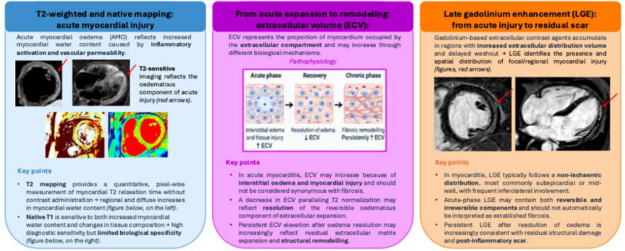
Figure 2.
The proposed longitudinal CMR patterns, as a conceptual framework, and their key imaging characteristics. Note: These patterns represent a conceptual framework based on the longitudinal evolution of CMR abnormalities and should not be considered formally validated imaging phenotypes. Individual findings should be interpreted in the context of clinical presentation, biomarkers, ventricular function, and rhythm assessment. Abbreviations: LGE, late gadolinium enhancement; LV function, left ventricular function.
Figure 2.
The proposed longitudinal CMR patterns, as a conceptual framework, and their key imaging characteristics. Note: These patterns represent a conceptual framework based on the longitudinal evolution of CMR abnormalities and should not be considered formally validated imaging phenotypes. Individual findings should be interpreted in the context of clinical presentation, biomarkers, ventricular function, and rhythm assessment. Abbreviations: LGE, late gadolinium enhancement; LV function, left ventricular function.
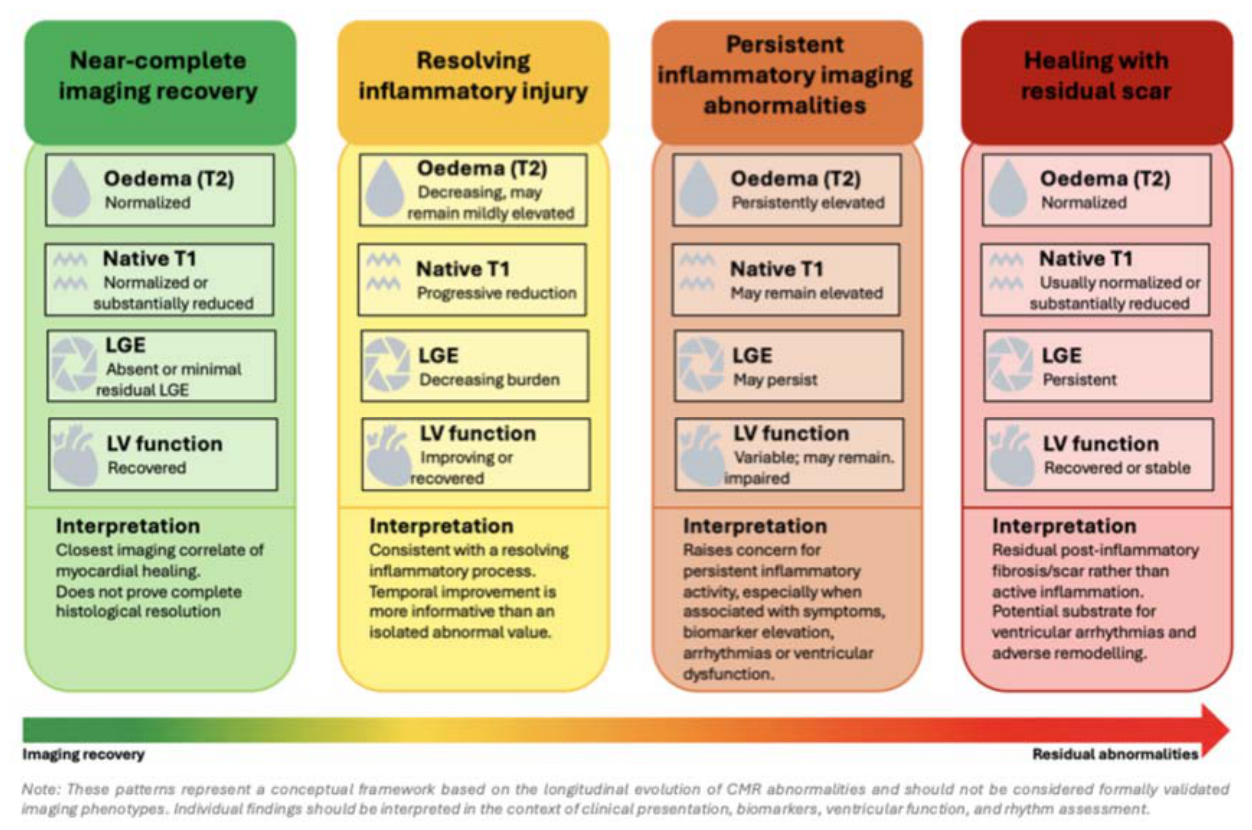
Figure 3.
Central Illustration explaining the longitudinal imaging trajectory of myocarditis. The figure illustrates the temporal evolution of the main biological, CMR, echocardiographic, and molecular imaging findings across different stages of myocarditis, from acute inflammatory injury to resolving or persistent inflammation and, ultimately, near-complete recovery or healed injury with residual scar. On the right, the clinical-biological components of recovery are illustrated: they do not necessarily evolve in parallel, and their timing and degree of resolution may vary considerably between patients. The second panel on the right highlights the key clinical questions that may guide imaging interpretation across the disease trajectory. The lower part of the figure summarizes the potential clinical applications of longitudinal CMR, from diagnosis and risk stratification to disease monitoring, return-to-exercise assessment, and future imaging-guided management. The proposed trajectories are conceptual and should not be interpreted as fixed or formally validated phenotypes. Abbreviations: CMR, cardiovascular magnetic resonance; ECV, extracellular volume; LGE, late gadolinium enhancement; LV function, left ventricular function; GLS, global longitudinal strain; 18F-FDG-PET, 18F-fluorodeoxyglucose positron emission tomography; RWMA, regional wall motion abnormalities; EMB, endomyocardial biopsy; PET, positron emission tomography.
Figure 3.
Central Illustration explaining the longitudinal imaging trajectory of myocarditis. The figure illustrates the temporal evolution of the main biological, CMR, echocardiographic, and molecular imaging findings across different stages of myocarditis, from acute inflammatory injury to resolving or persistent inflammation and, ultimately, near-complete recovery or healed injury with residual scar. On the right, the clinical-biological components of recovery are illustrated: they do not necessarily evolve in parallel, and their timing and degree of resolution may vary considerably between patients. The second panel on the right highlights the key clinical questions that may guide imaging interpretation across the disease trajectory. The lower part of the figure summarizes the potential clinical applications of longitudinal CMR, from diagnosis and risk stratification to disease monitoring, return-to-exercise assessment, and future imaging-guided management. The proposed trajectories are conceptual and should not be interpreted as fixed or formally validated phenotypes. Abbreviations: CMR, cardiovascular magnetic resonance; ECV, extracellular volume; LGE, late gadolinium enhancement; LV function, left ventricular function; GLS, global longitudinal strain; 18F-FDG-PET, 18F-fluorodeoxyglucose positron emission tomography; RWMA, regional wall motion abnormalities; EMB, endomyocardial biopsy; PET, positron emission tomography.
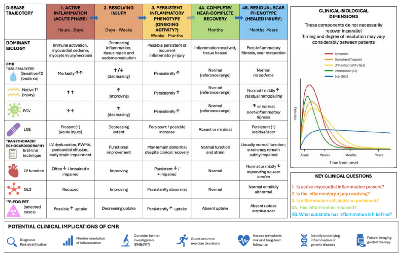

Table 1.
Complementary roles, strengths, and limitations of cardiovascular imaging modalities in myocarditis.
Table 1.
Complementary roles, strengths, and limitations of cardiovascular imaging modalities in myocarditis.
| Imaging modality | Main target | Strengths | Limitations |
| Transthoracic echocardiography (TTE) [3,49] | First-line assessment of biventricular function, regional wall motion, haemodynamic, pericardial effusion, and complications. Speckle-tracking echocardiography (STE) allows the assessment of global and regional myocardial deformation and detection of subtle functional abnormalities. |
Widely available, rapid, bedside assessment, no ionizing radiation, well suited to serial functional evaluation. May identify subclinical dysfunction despite preserved LVEF, useful for regional and longitudinal functional assessment. |
Limited specificity for myocarditis, may be normal in mild or infarct-like presentations, limited tissue characterization. Dependent on image quality and analysis software, limited disease specificity, diagnostic thresholds are not standardized. |
| Cardiac magnetic resonance (CMR) [3,4,24,43,60] | Comprehensive assessment of ventricular function and myocardial tissue, including oedema, injury, extracellular expansion, and fibrosis/scar. T1/T2 mapping and ECV allows quantitative assessment of myocardial water content and diffuse tissue/extracellular abnormalities. |
Reference non-invasive technique for tissue characterization, multiparametric assessment, no ionizing radiation, central role in diagnosis and follow-up. Detects diffuse abnormalities, quantitative, useful for longitudinal assessment, native T1 and T2 can be acquired without contrast. |
Limited availability, longer examination, motion/arrhythmia artefacts, gadolinium may be unsuitable in selected patients, findings are not histologically specific. Reference ranges vary with scanner, sequence, and centre; limited biological specificity; ECV requires contrast and haematocrit; thresholds for persistent disease activity remain uncertain. |
| Cardiac CT/CCTA [1,63] | Exclusion of coronary artery disease, assessment of cardiac anatomy and function, emerging tissue characterization with late iodine enhancement and CT-derived ECV. | Rapid, high spatial resolution, useful when acute coronary syndrome is part of the differential diagnosis, option when CMR is not feasible. | Ionizing radiation, iodinated contrast, less established tissue characterization than CMR, limited evidence for routine myocarditis assessment. |
| 18F-FDG PET [3,9,60] | Assessment of myocardial metabolic inflammatory activity, particularly in selected or diagnostically challenging cases. | Provides complementary information on inflammatory metabolism, may be useful when CMR is limited or discordant, potential role in treatment monitoring. | Limited evidence for routine uncomplicated myocarditis, demanding preparation, physiological myocardial uptake may impair interpretation, radiation exposure, limited standardization. |
| Hybrid PET/CMR [60] | Combined structural, tissue, functional, and metabolic characterization. | Potential simultaneous integration of CMR-defined injury/scar with metabolic inflammatory activity. | Limited availability, high cost and technical complexity, evidence in myocarditis remains limited, clinical role is not established. |
Abbreviations: CCTA, coronary computed tomography angiography; CMR, cardiovascular magnetic resonance; ECV, extracellular volume; FDG, fluorodeoxyglucose; LVEF, left ventricular ejection fraction; PET, positron emission tomography; STE, speckle-tracking echocardiography; TTE, transthoracic echocardiography.
Disclaimer/Publisher’s Note: The statements, opinions and data contained in all publications are solely those of the individual author(s) and contributor(s) and not of MDPI and/or the editor(s). MDPI and/or the editor(s) disclaim responsibility for any injury to people or property resulting from any ideas, methods, instructions or products referred to in the content. |
© 2026 by the authors. Licensee MDPI, Basel, Switzerland. This article is an open access article distributed under the terms and conditions of the Creative Commons Attribution (CC BY) license.
Copyright: This open access article is published under a Creative Commons CC BY 4.0 license, which permit the free download, distribution, and reuse, provided that the author and preprint are cited in any reuse.