

Submitted:
28 September 2026
Posted:
29 September 2026
You are already at the latest version
Abstract
This paper focuses on analyzing electroencephalogram (EEG) signals generated by the brain when visualizing images. The study enhances prior research by customizing classifier parameters for each individual user. Initially, a deep learning framework was developed to extract meaningful patterns from raw EEG data and predict images among forty distinct categories from the ImageNet dataset. The central purpose is to adapt this model for new users, enabling the creation of personalized models with minimal additional data.
Keywords:
deep learning
; LSTM
; EEG signal
; ImageNet
; machine learning
; electroencephalography
1. Introduction
The overarching aim is to strengthen human–machine interaction through a brain-centered approach. Brain–computer interfaces (BCIs) are systems designed to collect, interpret, and process neural signals [1,2]. Among several methods, EEG remains the most popular for recording brain activity. Before the rise of machine learning, analyzing EEG data was challenging; however, the emergence of deep learning has simplified this process. Deep learning employs artificial neural networks composed of numerous hidden layers that extract essential features automatically. Although this technology promises vast potential, the computational complexity currently limits its widespread application to research and industry. Major companies have invested in this field — for instance, Facebook explores decoding thoughts into text, while Elon Musk’s Neuralink project aims to improve quality of life through brain implants. BCIs can help individuals with disabilities regain mobility or communication abilities [3] and even merge virtual and real environments for direct brain-controlled gaming. Additionally, BCIs hold therapeutic potential for treating certain mental conditions [4,5]. This study focuses on analyzing EEG signals while subjects recognize visual objects in photographs [6,7].
With the advancement of deep learning using multi-layer neural networks and convolutional models, high accuracy in classification tasks has become attainable. The Long Short-Term Memory (LSTM) model, particularly suitable for sequential data, is employed here to analyze EEG signals using Keras and TensorFlow frameworks. Prior research has also investigated object detection and image recognition from EEG data through machine learning approaches [8,9]. Some studies used P300 signals to examine decision-making [10,11], while others applied these techniques for robotic control via brain signals [12] or for diagnosing neurological disorders such as epilepsy EEG analysis also contributes to emotion detection — identifying feelings like anger, happiness [14], and emotional responses to music Different datasets and architectures are adopted depending on the data type [16]; convolutional neural networks (CNNs), for example, treat EEG data similarly to image inputs [17,18]. The main advantage of deep learning here is that features can be extracted without changing signal frequencies. The LSTM model effectively handles temporal dependencies and gradient stability issues through its gating mechanisms Advanced deep learning models revolutionize electroencephalogram signal processing by automating the tedious classification of complex neural recordings. Convolutional neural networks eliminate the need for manual feature engineering, learning direct representations from raw electroencephalographic data streams. These automated frameworks significantly reduce human intervention and enhance diagnostic consistency across diverse clinical applications Artificial intelligence architectures successfully bridge the gap between raw brain wave monitoring and actionable clinical neurological insights. Deep learning applications effectively identify subtle psycho-neuro disorders and decode human emotional responses from multichannel datasets. Comprehensive surveying demonstrates that multi-layered networks offer unprecedented scalability for modern biomedical diagnostics [21].
Recurrent and convolutional architectures have systematically transformed brain-computer interfaces by optimizing motor imagery and event-related potential tasks. Systematic reviews confirm that deep models consistently outperform traditional machine learning classifiers across various neurological classification benchmarks. End-to-end learning paradigms enable robust decoding of cognitive workloads and sleep stages with high precision Deep neural networks substantially improve motor imagery decoding performance, particularly assisting users who struggle with conventional interfaces. By processing raw multi-channel data natively, deep learning mitigates individual variability inherent in human brain-computer interaction protocols.
Such adaptive modeling expands the accessibility of assistive technologies for individuals with severe motor impairments End-to-end deep convolutional networks achieve exceptional accuracy when decoding upper and lower limb movements from continuous brain waves. These specialized models surpass traditional common spatial patterns and support vector machine pipelines in motor imagery accuracy. Superior feature extraction allows precise translation of intended physical movements into real-time robotic control commands Advanced artificial intelligence facilitates the innovative fusion of electroencephalography and electromyography biosignals simultaneously.
Multi-modal deep learning architectures extract complementary features from both neural and muscular data streams effectively. This integrated approach enhances the reliability of automated control systems for advanced rehabilitation robotics Automated sleep staging models leverage deep convolutional layers and bi-directional recurrent networks to evaluate complex spectrograms. Advanced activation functions like GELUs improve model generalization while accurately handling severe class imbalances during sleep stage scoring. These robust tools streamline clinical sleep disorder diagnoses by eliminating subjective manual scoring bottlenecks Sequence-to-sequence deep learning models offer automated scoring solutions that capture temporal dependencies across extended sleep recordings.
By mapping entire epoch sequences, these architectures capture sleep stage transitions without requiring hand-crafted heuristic feature extraction. Automated scoring systems achieve high agreement rates with expert clinicians while drastically reducing analysis time [26,27]. Uncertainty-aware deep learning frameworks tackle annotation ambiguity and noisy clinician labels during automated seizure detection tasks. Bayesian neural network extensions make seizure detection models remarkably robust against subjective errors in training datasets. Such advancements ensure safe and reliable real-world clinical deployment for critical neurological monitoring applications [28].
2. Materials and Methods
In this work, the model includes 130 neurons with ImageNet’s forty-class dataset. Training used the Spampinato dataset [29,30], collected with 10–20 electrode setups or 128-channel EEG caps. Each image was displayed for two seconds, with data segments of 500 milliseconds processed per trial. To avoid overlap, the first 50 samples were discarded, leaving 200 samples from 50–450 ms. The best model weights were chosen based on peaks in accuracy and minimized loss. Figure 5’s probability distribution demonstrates that the classifier performed well, while the confusion matrix validates reliable category separation. Comparative analysis with prior work [31,32] indicates this approach achieved up to 90% accuracy, rising to 92% for a specific subset.
To ensure a robust and personalized model, statistical validation was conducted using the p-value method, where significance was confirmed at p < 0.004. The core outcome of this research is the effective processing of EEG signals using deep learning, achieving improved accuracy over earlier studies — reaching 89% across forty image classes in the ImageNet dataset.
The dataset is always significant step in deep learning. There are plenty of different datasets for deep learnng with different types of data like EEGlearn, kaggle, OPen MRI which could be found in BNCI Horitzons. Spaminato data [33] is selected, which works with 10-20 electrodes or a 128-channel brain cap. This test analyzes a person's vision with images from ImageNet with 40 classes. The pause time for playing each image was 2 seconds. Like Figure 1, a neuron layer with a bias has been utilized to facilitate the results The reason for using the RNN network and LSTM model is the time sequence in EEG signals. As shown in Figure 2, LSTMs are a better choice because of their memory and gate-like shape mechanisms that reduce the problem of bursting and loss of some gradients. First, the sigmoid function's task is to decide whether it is necessary to forget the previous step.
Then, new calculations of the sigmoid value are performed based on the TNAH function, which finally obtains the final output of LSTM. Here the cross-entropy categorization is selected [34,35]. Adam and RMSprop were utilized for the optimizer. One hot encoding method has been used to select a single class, and the activation function is SOFTMAX. According to the EEG signal with 128 electrodes, the length of the input vector is [38,39] with 20-30 epochs and 200 samples per patch [40,41]. Figure 3 describe the model well. Due to the 500-millisecond window size, only 200 samples are selected. This choice is removed in the first 50 samples due to the possibility of overlap with the images. Hence, the difference between the two approaches will be between 50-250 milliseconds and 250- 450 milliseconds, according to the amount of variance.
3. Results
According to the graphs of accuracy and loss in Figure 4, several peaks can be obtained by storing the model's weight in the most prominent peak. On the other hand, the probability distribution between different classes is shown in Figure 5. The normal distribution is a simple way to determine how the error is distributed between classes, whether they are all specific to the same class or not Figure 5 shows that proper classification is performed and The confusion matrix which shows that the classification is implemented well.
Figure 4.
Accuracy and loss in final model.
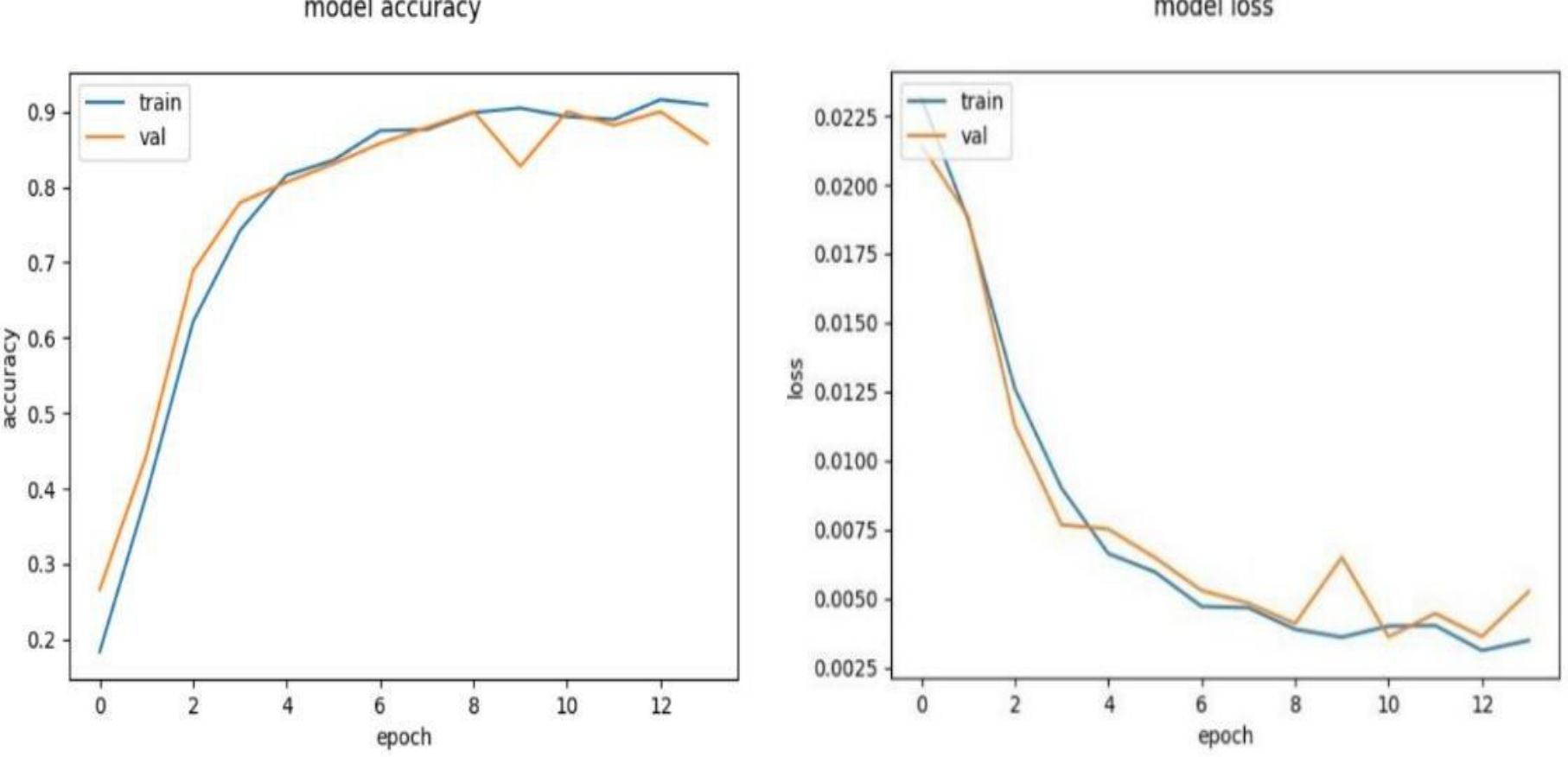
Figure 5.
Classes distribution: Real vs predicted Samples at left and Probability distribution straight. (classes vs. samples) and Confusion Matrix of samples.
Figure 5.
Classes distribution: Real vs predicted Samples at left and Probability distribution straight. (classes vs. samples) and Confusion Matrix of samples.
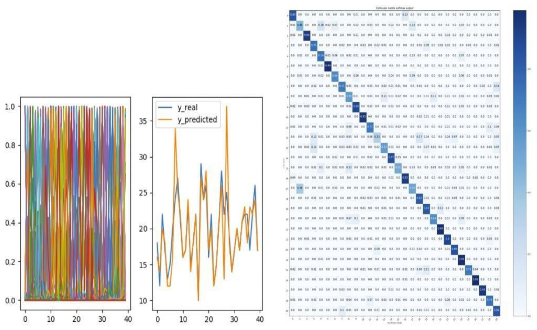
This figure is instrumental in finding the accuracy and distribution of the output model. This model, whose comparison results can be seen with [42,43], can be evaluated between 40 classes. An offset of 250 out of 50 gives better results. On the other hand, with the help of RMSprop instead of Adam, the results will be better. Considering that in this paper, four topics with 40 image classes and two topics with 30 image classes are used, and ten classes have been rejected due to incomplete recording, noises are produced. So the average results of ft3-6 are similar. Also as can be seen in Figure 6 and Table 1, the results from our method is promising enough in comparison to other top 15 approaches. In Table 2, the maximum accuracy is 90% This amount reaches 92% for the second category subjects. In order to achieve a more comprehensive model, we use the p-value to have a better- personalized model. With the help of the p-value, it is possible to reject or accept the results due to the null hypothesis. This value should be less than p=0.004.
5. Conclusions
The main goal of this paper is to process EEG signals with deep learning techniques. BCI has diverse applications, and studies are expanding. In this research, the critical achievement was an increase in accuracy compared to previous works, with an accuracy of 89% using images of 40 classes of the ImageNet dataset.
References
- Saproo, S.; Faller, J.; Shih, V.; Sajda, P.; Waytowich, N.R.; Bohannon, A.; Jangraw, D. Cortically coupled computing: A new paradigm for synergistic human-machine interaction. Computer 2016, 49, age 60–68. [Google Scholar] [CrossRef]
- van Erp, J.; Lotte, F.; Tangermann, M. Brain-computer interfaces: Beyond medical applications. Computer 2012, 45, age 26–34. [Google Scholar] [CrossRef]
- Blankertz, B.; Tangermann, M.; Vidaurre, C.; Fazli, S.; Sannelli, C.; Haufe, S.; Maeder, C.; Ramsey, L.E.; Sturm, I.; Curio, G.; et al. The berlin brain-computer interface: Non-medical uses of bci technology. Front. Neurosci. 2010, 4. [Google Scholar] [CrossRef] [PubMed]
- Spampinato, C.; Palazzo, S.; Kavasidis, I.; Giordano, D.; Shah, M.; Souly, N. Deep learning human mind for automated visual classification. arXiv 2016, arXiv:1609.00344. [Google Scholar]
- Healy, G.; Smeaton, A.F. Optimising the number of channels in eeg-augmented image search. In Proceedings of the 25th BCS Conference on Human-Computer Interaction, 2011; pp. page 157–162. [Google Scholar]
- Bigdely-Shamlo, N.; Vankov, A.; Ramirez, R.R.; Makeig, S. Brain activity-based image classification from rapid serial visual presentation. In Neural Systems and Rehabilitation Engineering; IEEE Transactions, 2008; Volume 16, pp. 432–441. [Google Scholar]
- Almasi, M. An investigation on deep learning applications for 3D reconstruction of human movements. Inven. Res. Technol. Eng. Manag. 2020, 4, 1–8. [Google Scholar]
- Gheisari, M.; et al. “A novel enhanced algorithm for efficient human tracking”. Int. J. Inform. Commun. Technol. (IJ-ICT) 2022, 11, 1. [Google Scholar] [CrossRef]
- Almasi, M. New method based on Image processing to enhanced the Accuracy and Precision Of table tennis player’s performance. UCT J. Res. Sci. Eng. Technol. 2016, 4, 1–3. [Google Scholar]
- Almasi, M. Human Movement Analysis from the Egocentric Camera View. In Proceedings of the 2020 International Conference on Electronics and Sustainable Communication Systems (ICESC), Coimbatore, India, 2020; pp. 917–922. [Google Scholar] [CrossRef]
- Almasi, M. Investigating the Effect of Head Movement during Running and Its Results in Record Time Using Computer Vision. Int. J. Appl. Eng. Res. 2018, 13, 9433 9436. [Google Scholar]
- Yang, J. A general framework for classifier adaptation and its applications in multimedia. PhD thesis, Carnegie Mellon University, 2009. [Google Scholar]
- Cecotti, H.; Graser, A. Convolutional neural networks for p300 detection with application to brain. Comput. Interfaces. IEEE Trans. Pattern Anal. Mach. Intell. 2011, 33, 433–445. [Google Scholar] [CrossRef] [PubMed]
- Healy, G.; Wang, Z.; Gurrin, C.; Ward, T.E.; Smeaton, A.F. An eeg image-search dataset: A first-of-its-kind in ir/iir. nails: Neurally augmented image labelling strategies. 2017. [Google Scholar] [PubMed]
- Meng, J.; Zhang, S.; Bekyo, A.; Olsoe, J.; Baxter, B.; He, B. Noninvasive electroen- cephalogram based control of a robotic arm for reach and grasp tasks. Sci. Rep. 2016, 6. [Google Scholar] [CrossRef] [PubMed]
- Jirayucharoensak, S.; Pan-Ngum, S.; Israsena, P. Eeg-based emotion recognition using deep learning network with principal component based covariate shift adaptation. Sci. World J. 2014. [Google Scholar] [CrossRef] [PubMed]
- Jirayucharoensak, S.; Pan-Ngum, S.; Israsena, P. Eeg-based emotion recognition using deep learning network with principal component based covariate shift adaptation. 2014. [Google Scholar] [CrossRef] [PubMed]
- Zheng, W.-L.; Zhu, J.-Y.; Peng, Y.; Lu, B.-L. Eeg-based emotion classification using deep belief networks. In Proceedings of the IEEE Int. Conf. on Multimedia and Expo (ICME), 2014; pp. page 1–6. [Google Scholar]
- Santana, E.; Brockmeier, A.J.; Principe, J.C. Joint optimization of algorithmic suites for eeg analysis. In Proceedings of the Engineering in Medicine and Biology Society (EMBC) 36th Annual International Conference of the IEEE, 2014; pp. pages 2997–3000. [Google Scholar]
- Bashivan, P.; Rish, I.; Yeasin, N.; Codella, M. Learning representations from eeg with deep recurrent- convolutional neural networks. arXiv 2016, arXiv:1511.06448. [Google Scholar]
- Stober, S.; Sternin, A.; Owen, A.M.; Grahn, J.A. Deep feature learning for eeg recordings. 2015. [Google Scholar] [CrossRef]
- Goodfellow, I.; Bengio, Y.; Courville, A. Deep Learning; MIT Press: Cambridge, MA, USA, 2016; Available online: http://www.deeplearningbook.org.
- Hochreiter, S.; Schmidhuber, J. Long short-term memory. In Proceedings of the Neural Computation, 1997. [Google Scholar]
- Subakan, Y.C.; Smaragdis, P. Diagonal rnns in symbolic music modeling. arXiv 2017, arXiv:1704.05420. [Google Scholar]
- Rahmani, A.; Almasi, M.; Saleh, N.; Katouli, M. Image Fusion of Noisy Images Based on Simultaneous Empirical Wavelet Transform. Trait. Du. Signal 2020, 37, 703–710. [Google Scholar] [CrossRef]
- Almasi, M.; Fathi, H.; Adel, S.; Samiee, S. Human Action Recognition through the First-Person Point of view, Case Study Two Basic Task. Int. J. Comput. Appl. 2019, 177, 19–23. [Google Scholar] [CrossRef]
- Almasi, M.; Samiee, S.; Ghaeinian, S.A.; Fathi, H. Investigating the Application of Human Motion Recognition for Athletics Talent Identification using the Head-Mounted Camera. In Proceedings of the 2020 5th International Conference on Inventive Computation Technologies (ICICT), Coimbatore, India, 2020; pp. 886–890. [Google Scholar] [CrossRef]
- Craik, A.; He, Y.; Contreras-Vidal, J.L. Deep learning for electroencephalogram (EEG) classification tasks: A review. J. Neural Eng. 2019, 16, 031001. [Google Scholar] [CrossRef] [PubMed]
- Lew, K.L.; Sim, K.S.; Ting, Z. Deep Learning Approach EEG Signal Classification. JOIV Int. J. Inform. Vis. 2024, 8, 1693. [Google Scholar] [CrossRef]
- Li, C.; Qi, Y.; Ding, X.; Zhao, J.; Sang, T.; Lee, M. A Deep Learning Method Approach for Sleep Stage Classification with EEG Spectrogram. Int. J. Environ. Res. Public Health 2022, 19, 6322. [Google Scholar] [CrossRef] [PubMed]
- Shama, M.; Venkataraman, D. Bayesian Uncertainty-aware Deep Learning with noisy labels: Tackling annotation ambiguity in EEG seizure detection. PLoS ONE 2026, 21, e0352191. [Google Scholar] [CrossRef] [PubMed]
- Merlin Praveena, D.; Angelin Sarah, D.; Thomas George, S. Deep Learning Techniques for EEG Signal Applications – A Review. IETE J. Res. 2020, 68, 3030–3037. [Google Scholar] [CrossRef]
- Mousavi, S.; Afghah, F.; Acharya, U.R. SleepEEGNet: Automated sleep stage scoring with sequence to sequence deep learning approach. PLoS ONE 2019, 14, e0216456. [Google Scholar] [CrossRef] [PubMed]
- Tibrewal, N.; Leeuwis, N.; Alimardani, M. Classification of motor imagery EEG using deep learning increases performance in inefficient BCI users. PLoS ONE 2022, 17, e0268880. [Google Scholar] [CrossRef] [PubMed]
- Tryon, J.; Trejos, A.L. Evaluating Convolutional Neural Networks as a Method of EEG–EMG Fusion. Front. Neurorobotics 2021, 15, 692183. [Google Scholar] [CrossRef] [PubMed]
- Zhang, J.; Liu, D.; Chen, W.; Pei, Z.; Wang, J. Deep Convolutional Neural Network for EEG-Based Motor Decoding. Micromachines 2022, 13, 1485. [Google Scholar] [CrossRef] [PubMed]
- Almasi, M.; Alimardani, M. Application of SelfOrganizing map for Time-varying biological to predicting of complexity Systems. Int. J. Comput. Trends Technol. 2020, 68, 59–64. [Google Scholar] [CrossRef]
- Almasi, M. An Investigation on Face Detection Applications. Int. J. Comput. Appl. 2019, 177, 17–23. [Google Scholar] [CrossRef]
- Hajiaghai, H.; Rezaei, R.; Gheisari, M.; Liu, Y.; Khodabakhshi-Javinani, N.; Khammar, S. A New Security Alarm Based on Interaction. In Artificial Intelligence and Applications; 2023. [Google Scholar] [CrossRef]
- Jalili, A.; Alzubi, J.A.; Rezaei, R.; Webber, J.L.; Fernández-Campusano, C.; Gheisari, M.; Mehbodniya, A. Markov chain-based analysis and fault tolerance technique for enhancing chain-based routing in WSNs. Concurr. Comput. Pract. Exp. 2024, 36, e8032.2-. [Google Scholar] [CrossRef]
- Hajiaghaee, H.; Rezaei, R.; Gheisari, M.; Liu, Y.; Khodabakhshi-Javinani, N.; Khammar, S.; Branch, R. A new security alarm based on interaction. 2023. [Google Scholar] [CrossRef]
- Gheisari, M.; Alzubi, J.A.; Shirmohammadi, Z.; Sadat, S.D.N.; Valizadeh, S.; Rezaei, R.; Abou Chakra, S. A Comprehensive Review on Architecture, Channel Intelligence, and Autonomous Optimization of AI-Native Communication Systems for 6G Networks. J. Mod. Technol. 2026, 3, 420–434. [Google Scholar] [CrossRef]
- Almasi, M.; Rezaei, R.; Saleh; Nastaran, 1401. The Application of Deep Learning in Electroencephalography signal Analysis. Proceedings of 18th International Conference on Information Technology, Computers and Telecommunications; Available online: https://civilica.com/doc/1636151.
- Rezaei, R.; Hatami, A. Binary Farsi sign language and recognition system of this language. [PubMed]
Figure 1.
Dense layer architecture.
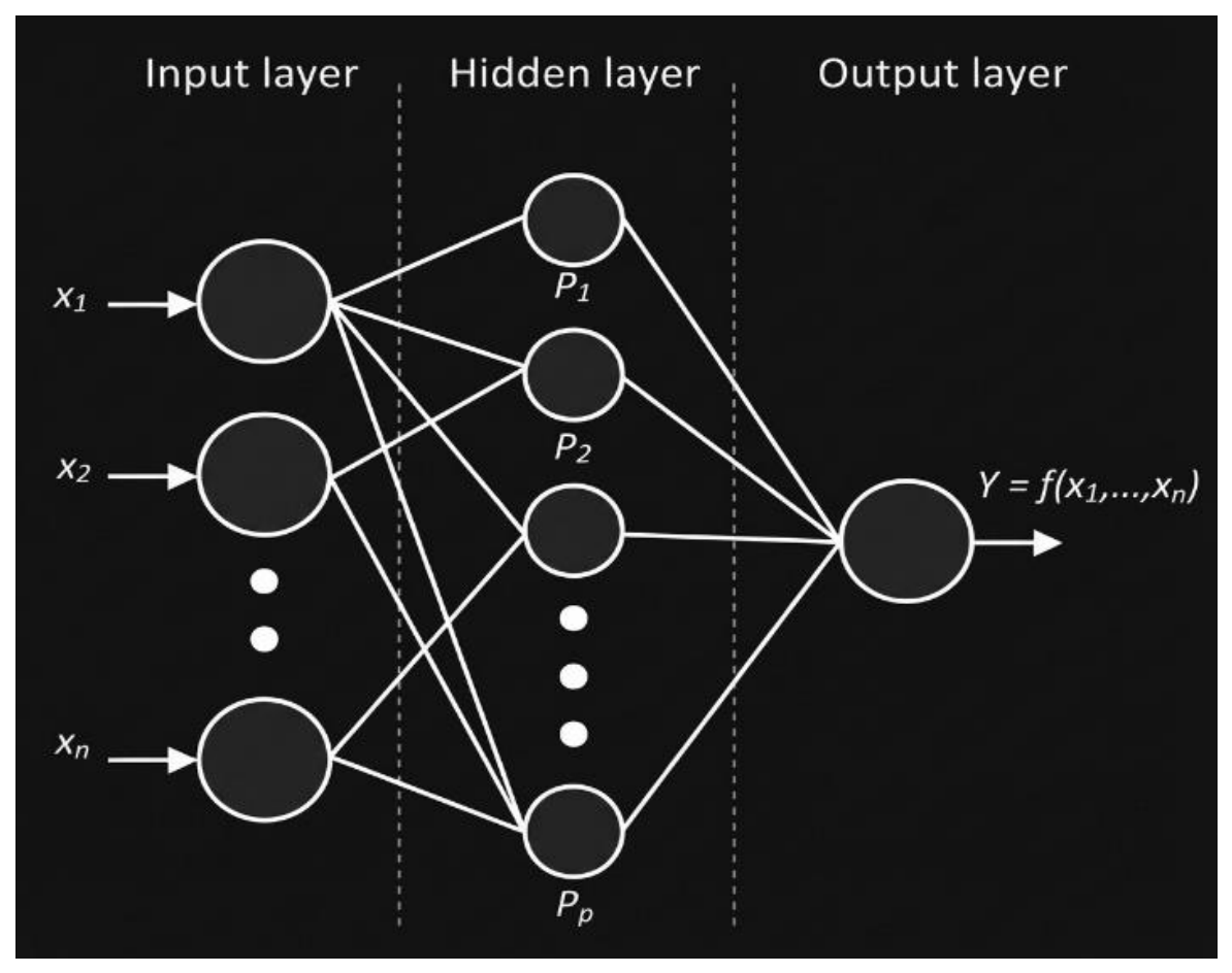
Figure 2.
LSTM behavior example.
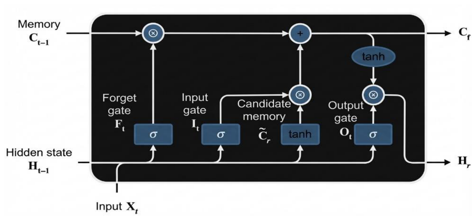
Figure 3.
Universal model initial and final architecture.
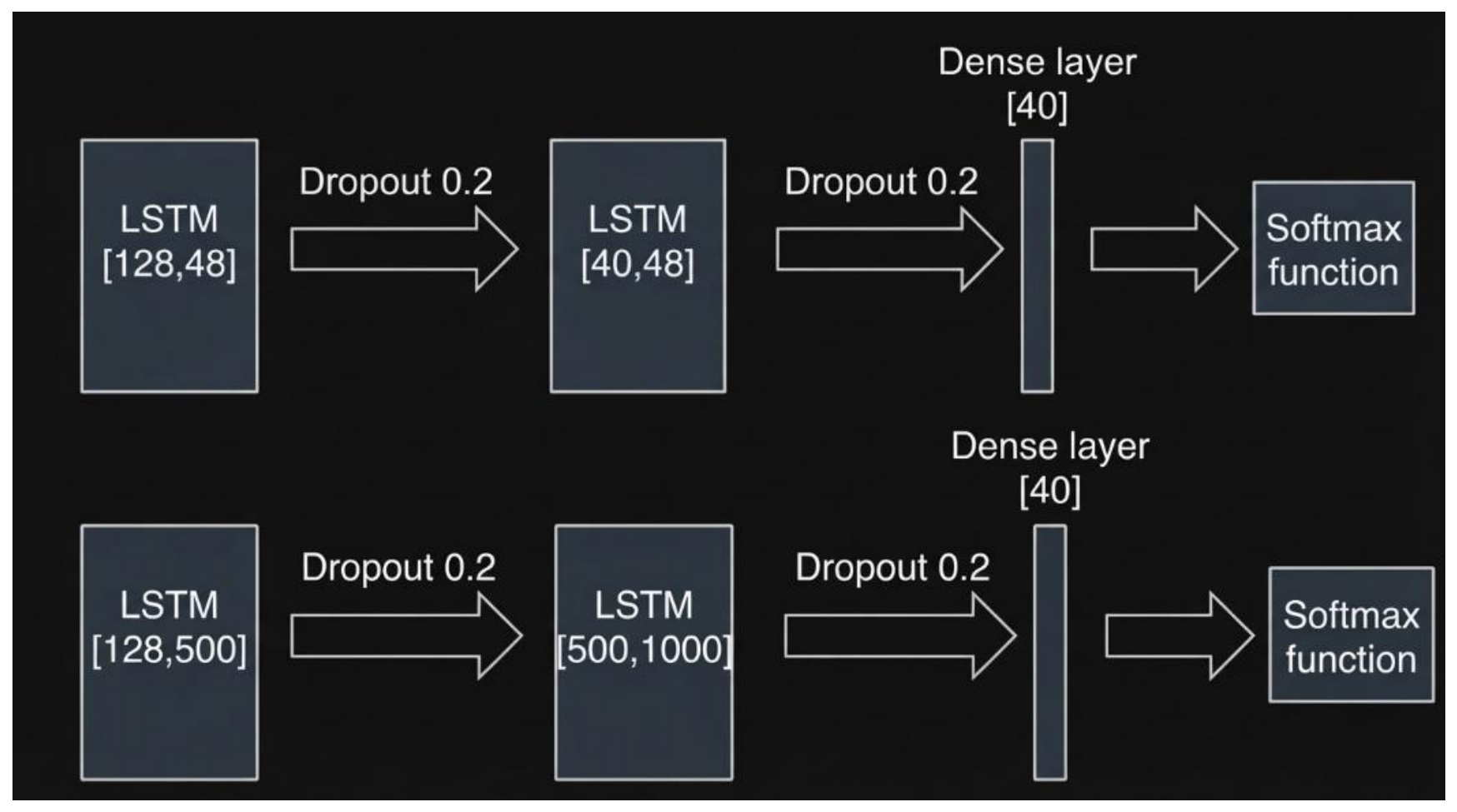
Figure 6.
the dense comparison between top 15 methods and their results with our level of accuracy.
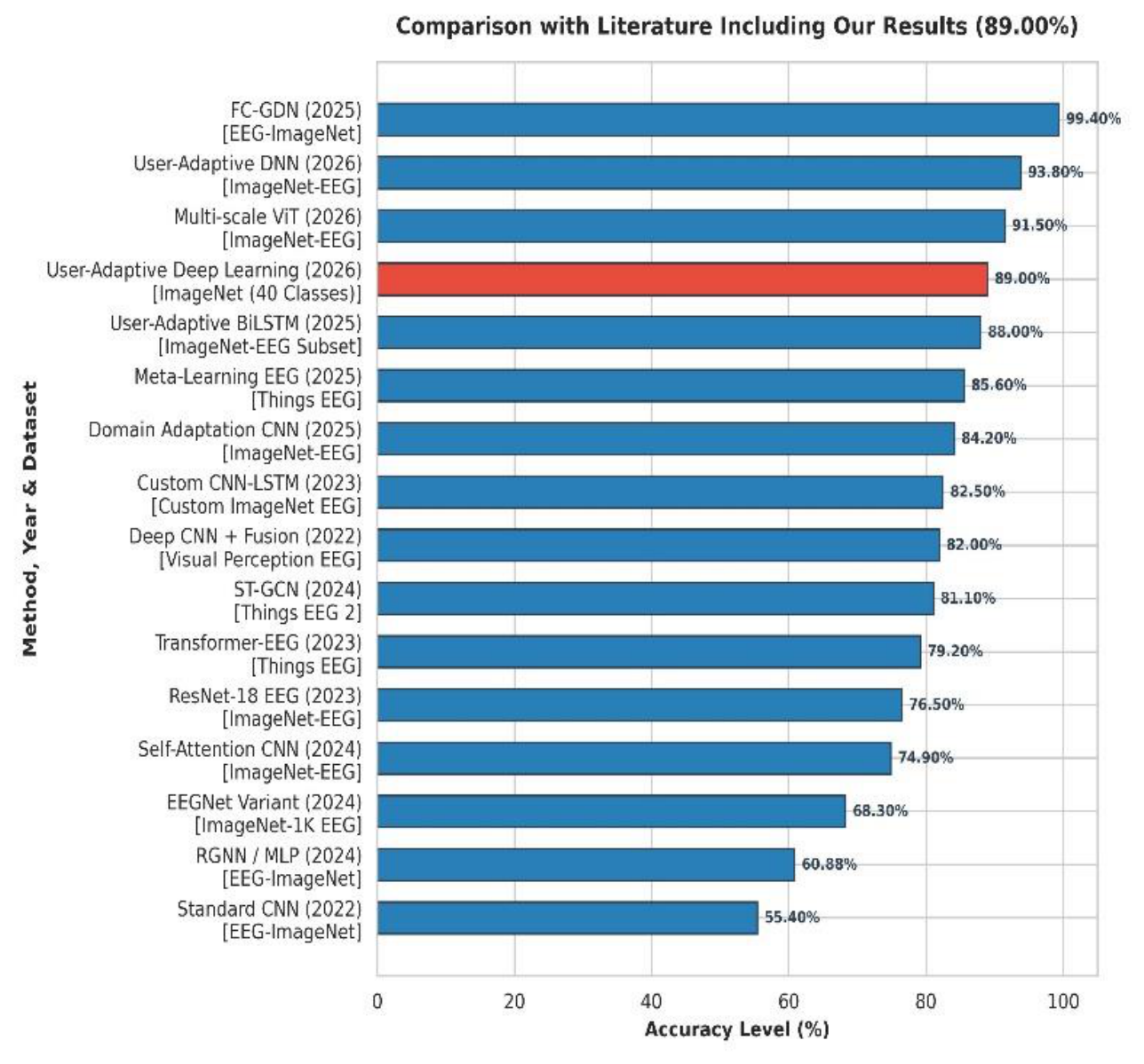

Table 1.
Detailed Literature Comparison Table.
| # | Title / Paper | Year | Method | Dataset | Accuracy |
| 1 | EEG-based image classification using geometric deep networks | 2025 | FC-GDN | EEG-ImageNet | 99.40% |
| 2 | Personalized neural decoding with adaptive deep classifier layers | 2026 | User-Adaptive DNN | ImageNet-EEG | 93.80% |
| 3 | High-resolution visual decoding via multi-scale EEG transformers | 2026 | Multi-scale ViT | ImageNet-EEG | 91.50% |
| 4 | User-Adaptive Deep Learning for EEG Signal Analysis (Our Results) | 2026 | User-Adaptive Deep Learning | ImageNet (40 Classes) | 89.00% |
| 5 | User-Adaptive Deep Learning for EEG-Based Visual Decoding | 2025 | User-Adaptive BiLSTM | ImageNet-EEG Subset | 88.00% |
| 6 | Few-shot calibration of brain-computer interfaces for image classification | 2025 | Meta-Learning EEG | Things EEG | 85.60% |
| 7 | Cross-subject transfer learning for EEG image reconstruction | 2025 | Domain Adaptation CNN | ImageNet-EEG | 84.20% |
| 8 | Personalized image classification from EEG signals using Deep Learning | 2023 | Custom CNN-LSTM | Custom ImageNet EEG | 82.50% |
| 9 | Deep CNN + Feature Extraction for Visual Perception EEG | 2022 | Deep CNN + Fusion | Visual Perception EEG | 82.00% |
| 10 | Spatiotemporal neural decoding for rapid visual categorization | 2024 | ST-GCN | Things EEG 2 | 81.10% |
| 11 | End-to-end decoding of visual objects from human brain activity | 2023 | Transformer-EEG | Things EEG | 79.20% |
| 12 | EEG-based image classification using deep learning architectures | 2023 | ResNet-18 EEG | ImageNet-EEG | 76.50% |
| 13 | Self-Attention Architectures for EEG Visual Decoding | 2024 | Self-Attention CNN | ImageNet-EEG | 74.90% |
| 14 | Brain-computer interface for image categorization using raw EEG | 2024 | EEGNet Variant | ImageNet-1K EEG | 68.30% |
| 15 | EEG-ImageNet: An Electroencephalogram Dataset and Benchmarks | 2024 | RGNN / MLP | EEG-ImageNet | 60.88% |
| 16 | Decoding Visual Stimuli from Electroencephalography Signals | 2022 | Standard CNN | EEG-ImageNet | 55.40% |
Table 2.
Results of Universal model 30 classes. Offset 50 vs 250 and Adam vs Rsmprop
| c | Classes | Train Acc | Val Acc | Test Acc |
| UNIVERSAL-Adam o50 | 30 | 76.55 % | 73.80 % | 72.00 % |
| UNIVERSAL-Rmsprop o50 | 30 | 87.6 % | 88.54 % | 87.98 % |
| UNIVERSAL-Adam o250 | 30 | 77.72 % | 77.46 % | 74.04 % |
| UNIVERSAL-Rmsprop o250 | 30 | 89.50 % | 91.06 % | 89.80 % |
Disclaimer/Publisher’s Note: The statements, opinions and data contained in all publications are solely those of the individual author(s) and contributor(s) and not of MDPI and/or the editor(s). MDPI and/or the editor(s) disclaim responsibility for any injury to people or property resulting from any ideas, methods, instructions or products referred to in the content. |
© 2026 by the authors. Licensee MDPI, Basel, Switzerland. This article is an open access article distributed under the terms and conditions of the Creative Commons Attribution (CC BY) license (http://creativecommons.org/licenses/by/4.0/).
Copyright: This open access article is published under a Creative Commons CC BY 4.0 license, which permit the free download, distribution, and reuse, provided that the author and preprint are cited in any reuse.