

Submitted:
28 September 2026
Posted:
29 September 2026
You are already at the latest version
Abstract
Connective tissue grafts (CTGs) remain a vital component in periodontal and pe-ri-implant plastic surgery; however, their harvesting creates a second surgical wound at the palatal donor site. This narrative review critically synthesizes the evidence on com-plications, technical pitfalls, and strategies for reducing donor-site morbidity following the harvesting of subepithelial connective tissue grafts and de-epithelialized gingival grafts. Subepithelial harvesting techniques support primary wound closure but require adequate residual flap thickness and careful dissection. In contrast, harvesting de-epithelialized gingival graft creates an open wound that demands effective hemostasis and protection; however, this approach provides greater control over graft dimensions and composition. No single harvesting technique or adjunctive intervention is universally superior. In addition to the harvesting technique and the resulting wound configuration, donor-site morbidity is determined by interactions among patient-related medical factors, palatal anatomy, graft dimensions and composition, and performance of the surgery. Pain and bleeding are the most frequently assessed early outcomes, whereas wound dehiscence, necrosis, infection, persistent sensory disturbances, and symptomatic scarring remain insufficiently quantified. The risk of postoperative morbidity and complications may be reduced through careful medical and anatomical assessment, the use of the minimum effective graft dimensions, atraumatic tissue handling, preservation of adequately vascularized residual tissues, effective hemostasis with appropriate wound protection, and easy-to-follow postoperative instructions for patients. Standardized endpoints, outcome measures, assessment intervals, and CTG-specific risk-assessment tools are needed to improve the comparability of evidence and guide clinical decision-making.
Keywords:
connective tissue graft
; palatal donor site
; postoperative complications
; postoperative morbidity
1. Introduction
Connective tissue grafts (CTGs) remain among the most established treatment modalities in periodontal and peri-implant plastic surgery. They are widely used for root coverage, gingival phenotype modification, and soft tissue augmentation around teeth and implants, particularly when optimal aesthetic outcomes, increased soft tissue thickness, and long-term stability of the clinical results are desired [1,2,3,4]. However, their principal biological disadvantage is the creation of a second surgical wound at the palatal donor site.
For the purpose this review, CTGs are defined as connective tissue grafts harvested from the palate using one of two different approaches: split-flap preparation to obtain a subepithelial connective tissue graft (SCTG) or de-epithelialization to obtain a de-epithelialized gingival graft - DGG) (Figure 1).
These techniques, which have the strongest evidence base, differ not only in terms of surgical access and graft preparation but also in the extend of wound exposure, control of graft thickness, and, particularly subsequent healing patterns. Consequently, SCTG and DDG harvesting should not be regarded as equivalent methods with respect to donor-site complications and morbidity [5,6].
The present review focuses on complications associated with graft harvesting from the anterior and posterior lateral palate, which are the most frequently used donor regions. Within these regions, anatomical variability, differences in mucosal thickness and tissue composition, the location of the greater palatine foramen, and the course of the greater palatine artery define the practical limits of safe palatal tissue harvesting [7,8,9,10].
For the purpose of this review, donor-site morbidity includes the local postoperative burden associated with the palatal wound, comprises pain, bleeding, functional discomfort, sensory disturbances, and other patient-reported impairments. Mild pain, transient discomfort, minor bleeding, and temporary impairment of eating and speaking may represent expected consequences of palatal surgery. By contrast, clinically relevant complications are unforeseen events that are more appropriately defined by their intensity, persistence and progression, or the need for additional clinical intervention [6,11].
Complications following palatal graft harvesting should not automatically be attributed to technical errors. Some adverse events may occur despite appropriate surgical planning, consideration of anatomical factors, and careful performance of the procedure, particularly in patients with anatomical variability, thin palatal mucosa, poor plaque control, or systemic conditions, as well as in those who smoke or use medications that affect the bleeding risk or wound healing process [8,12,13,14].
Nevertheless, technical errors are clinically important because they represent potentially modifiable contributors to morbidity. Excessive dissection depth, inadequate thickness of the remaining tissues, and ineffective hemostasis may increase the risk or severity of postoperative complications [6,15].
Although systematic reviews have addressed selected aspects of palatal donor-site morbidity, they generally evaluate narrowly defined interventions or outcomes [16,17,18,19,20]. Consequently, limited guidance is available for interpreting complications as clinical events resulting from interactions among anatomy, surgical technique, wound exposure, patient-related factors, and postoperative care.
A narrative review is therefore justified because it allows these elements to be integrated into a clinically oriented framework rather than evaluated as isolated variables. Accordingly, this review aims to identify modifiable risk factors, clarify the mechanisms underlying donor-site complications, and outline principles for preventing, reducing and managing morbidity following palatal CTG harvesting.
2. Objective and Review Approach
The objective of this narrative review is to critically synthesize the current evidence and clinically relevant concepts concerning complications, technical errors, and strategies for reducing door-site morbidity following palatal connective tissue graft harvesting. The review focuses on donor-site complications associated SCTG and DGG. Studies on free gingival grafts were included only when their findings were relevant and applicable to palatal donor-site morbidity, wound healing, hemostasis, or donor-site protection.
A literature search for full-text publications in English was conducted in three electronic databases—PubMed, Scopus, and Web of Science—and supplemented by a search of Google Scholar. The search covered the period from 1974, when subepithelial connective tissue graft harvesting was first described in the literature. The final database search was performed at the end of May 2026. The reference lists of key publications were also screened manually.
Search terms included combination of “connective tissue graft”, “subepithelial connective tissue graft”, “de-epithelialized graft”, “palatal donor site”, “donor-site morbidity”, “palatal wound healing”, “postoperative pain”, “bleeding”, hemostasis”, “palatal stent”. Publications were selected in two stages: title and abstract screening followed by full-text assessment performer independently by two authors (AD and DC). Disagreements were resolved through discussion and consensus. When consensus could not be reached, the final decision was made by a third author (AK).
The evidence base included umbrella, systematic and narrative reviews; randomized clinical trials; prospective clinical studies; and anatomical, histological, and imaging-based investigations. The key studies included in this narrative review are presented in Table 1.
Clinical interpretation was based primarily on human studies. Cadaveric studies were included when they provided essential information on palatal anatomical relationships. The literature was organized according to anatomical factors, harvesting techniques, type of complication, preventive interventions, and management strategies.
Given the narrative nature of this review, no formal risk-of-bias assessment or meta-analysis was performed. Systematic reviews and randomized clinical trials were prioritized when evaluating palatal neuro-vascular anatomy, patient-reported outcomes, palatal wound healing and preventive interventions. Observational studies were considered particularly relevant to surgical safety and the composition and thickness of palatal tissues.
Because donor-site morbidity is often evaluated using heterogeneous patient-reported and wound-healing outcomes, numerical results were interpreted within their specific clinical context rather than directly compared across studies. When the available evidence was limited or conflicting, conclusions were presented as clinical interpretations rather than definitive recommendations. Accordingly, this article should be regarded as a narrative synthesis that integrates the available evidence on palatal connective tissue graft harvesting and considers its implications for everyday clinical practice.
3. Palatal Anatomy and Histological Characteristics Relevant to CTG Harvesting
The anatomical characteristics of the hard palate are critical to understanding the technical errors and complications associated with connective tissue graft harvesting. Current evidence suggests that donor-site morbidity is determined not only by the type of harvesting technique and how it is performed, but also by the complex interplay among palatal mucosal thickness, graft dimensions, and the course of the greater palatine neurovascular bundle (GPB) [7,24]. Both graft quality and donor-site morbidity are substantially influenced by the layered architecture of the palatal tissues and regional variations in palatal mucosal thickness [7]. The palate should not be considered a homogeneous donor site.
3.1. Histological Architecture of the Palatal Mucosa
Histologically, the palatal masticatory mucosa consists of keratinized stratified squamous epithelium, lamina propria, and submucosa. The orthokeratinized epithelium, which is 0.24-0.41 mm thick, forms the outer protective layer. The lamina propria beneath it comprises two distinct layers: a papillary layer with a greater cellular density and a reticular layer containing thicker collagen bundles. These differences may be clinically relevant and graft orientation during the procedure may be important [31,32]. The thickness of the lamina propia is relatively consistent throughout the palate, except in the maxillary tuberosity, where it is greater. The underlying submucosa contains blood vessels, nerves, adipose tissue, and glandular elements. When the epithelium is excluded, the lamina propria and submucosa account for approximately 37% and 63% of the tissue volume, respectively It has been suggested that graft composition may affect graft integration and tissue response [7,24,31,32].
From a graft-harvesting perspective, the lamina propia is the most relevant component. Excessively superficial harvesting may produce a thin or epithelial-rich graft, whereas excessively deep harvesting increases donor-site trauma and may incorporate a greater proportion of submucosal tissue. The relative proportions of fibrous connective tissue and adipose or glandular components within CTG specimens depend on both the donor region and the harvesting technique [7,33,34].
3.2. Palatal Mucosal Thickness and Residual Tissue Dimensions
Palatal mucosal thickness exhibits substantial interindividual and site-specific variability. Reported mean values range from approximately 1.8 to 3.9 mm, depending on the anatomical region and measurement method, as discussed in the Section 5, Preoperative issues. In general, tissue thickness tends to increase from the gingival margin toward the palatal midline and from the canine toward the molar region, although localized thinning may occur near the palatal root prominence of the first molar [35].
Importantly, greater tissue thickness does not necessarily indicate a more favorable donor site, because graft harvested from thicker palatal mucosa may contain a larger proportion of adipose or glandular tissue rather than the desired dense lamina propia [7]. Palatal tissues thickness influences postoperative morbidity by determining the amount of tissue remaining after graft harvesting. With the DGG technique, this corresponds to the thickness of the tissue remaining over the underlying bone, whereas with the SCTG technique, the thickness of the residual palatal flap appears to be more important. The residual flap should ideally be maintained at a thickness of ≥1 mm to minimize the risk of flap sloughing and necrosis, which may consequently increase postoperative discomfort [5,25].
3.3. Greater Palatine Neurovascular Anatomy
The greater palatine neurovascular bundle represents the principal anatomical limitation during palatal graft harvesting. The greater palatine artery (GPA) and nerve emerge through the greater palatine foramen, which is most commonly located at the midpalatal aspect of the maxillary third molar (57-70% of cases), approximately 8 mm from the margin of the alveolar process. After emerging from the foramen, the greater palatine artery courses anteriorly toward the incisive foramen. Beyond the bony prominence, it typically gives rise to medial and canine branches. The greater palatine nerve runs medial to the artery. The distance between the GPA and the cement-enamel junction varies along the palate. It is generally greater in the posterior region, particularly around the second molar (13.9 ± 1.0 mm), and decreases towards the canine region (9.9 ± 2.9 mm), where the vertical safety margin is reduced [8,18,36].
However, considerable anatomical variation in the branching pattern of the GPA must be considered. A clinically challenging configuration occurs when a vessel courses close to the cement-enamel junction of the posterior teeth. In this configuration, the medial branch arises from the lateral branch before the latter reaches the bony prominence, whereas the canine branch emerges immediately beyond the prominence. This unfavorable clinical pattern has been identified in approximately 25% of cases [37]. Palatal vault morphology further influences this anatomical relationship, because a shallow vault may position the neurovascular bundle closer to the alveolar crest [38].
3.4. Clinical Definition of the Palatal Safe Zone
Taken together, the anatomical evidence supports a comprehensive and individualized definition of the palatal safe zone. This zone should be determined not only by the course of the GPA, including its distance from the CEJ and anatomical variations, but also by soft-tissue thickness and the morphological of donor site. Consequently, a universally defined harvesting area may not adequately account for anatomical variability [8,9,10].
4. Techniques for Connective Tissue Graft Harvesting
4.1. Classification and Clinical Implications
Palatal connective tissue graft harvesting techniques differ primarily in incision design, the extent of flap elevation, surgical visibility, control of graft thickness, and the possibility of primary wound closure. From a donor-site perspective, currently used techniques can be classified as subepithelial or open-wound approaches. Subepithelial techniques preserve epithelial coverage, permit primary closure, and promote healing by primary intention. By contrast, open-wound technique leaves the palatal wound exposed to heal by secondary intention, which generally requires more time and may be associated with a greater risk of infection or scarring [29]. This distinction is clinically relevant because it reflects the need to balance graft accessibility, donor-site preservation, and graft composition to minimize donor-site morbidity and complications.
4.2. Histological Characteristics of SCTGs and DGGs
Histological studies indicate that subepithelial connective tissue grafts (SCTG) are less dense and contains a smaller proportion of lamina propia than de-epithelialized gingival grafts (DGGs). They greater adipose and glandular tissue content, may make SCTGs less dimensionally stable and may act as a barrier to vascularization during the initial phase of healing. By contrast DGGs appear firmer, less susceptible to postoperative shrinkage, and easier to handle [5,7]. However, their denser structure may make them more susceptible to necrosis than SCTGs [24]. Additionally, epithelial remnants have been reported [34]. The amount of tissue available and tissue structure characteristic of grafts depend on the donor site. Consequently, the selection of the donor region should be guided by the intended clinical application [24].
Several techniques have been proposed for harvesting CTGs from the palate. Because the available evidence does not support the superiority of any single approach, the following section outlines the historical development of selected procedures and discusses their current scientific and clinical rationale.
4.3. Subepithelial Connective Tissue Graft Techniques
In 1974, Edel described a palatal connective tissue harvesting approach involving one horizontal incision and two vertical releasing incisions, subsequently referred to as the trap-door technique (TD) [39]. Although the procedure was originally proposed to increase the width of the keratinized gingival, it represented an important early step in the development of subepithelial connective tissue graft procedures and, subsequently, bilaminar techniques for the treatment of gingival recession.
The main advantage of the TD approach is that it provides direct access to the underlying connective tissue while preserving an epithelialized flap over the donor site, thereby facilitating primary closure and potentially reducing postoperative discomfort. The three-incision design also provides good visualization of the donor bed and relatively predictable control of graft dimensions during harvesting. However, these benefits require elevation and subsequent repositioning of the palatal flap, making the procedure more technically demanding and potentially increasing the complexity of donor-site management.
In 1982, Langer and Calagna were the first to use connective tissue grafts for root coverage, thereby providing a bilaminar blood supply to the graft [40]. Since then, numerous techniques for harvesting donor tissues have been described.
In 1994, Bruno modified the TD technique by eliminating the vertical releasing incisions. Access to the underlying connective tissue was obtained through two parallel horizontal incisions with limited flap elevation[41]. Although it remained a two-incision approach, this modification represented an important step toward less invasive palatal harvesting. It also preceded the development of the single-incision techniques introduced by Hürzeler and Weng in 1999 [42].
The single-incision technique (SIT) was designed to minimize surgical trauma at the donor site while allowing predictable harvesting of a SCTG. A single horizontal incision is made in the palatal mucosa, through which the connective tissue graft is carefully dissected and harvested without requiring the need for vertical releasing incisions [42]. This approach reduces surgical manipulation at the donor site while allowing primary wound closure. However, limited surgical visibility makes control of graft thickness more operator-dependent than with the trap-door approach.
4.4. De-Epithelialized Gingival Graft Technique
The de-epithelialized gingival graft represents a distinct surgical concept. Although Zucchelli et al. briefly mentioned this approach in 2003 as an alternative in the presence of unfavorable palatal anatomical conditions, the first clinical evaluation of DGGs as an alternative to conventionally harvested SCTGs was conducted by Zucchelli and colleagues in 2010 [5,43].
In this technique, an epithelialized gingival graft is initially harvested and subsequently de-epithelialized extraorally. This approach permits precise control of graft dimensions and thickness and produces a graft composed predominantly of lamina propria. De-epithelialization may also be performed intraorally using a diamond bur or diode laser [26,44].
In root-coverage procedures, a DGG combined with a coronally advanced flap (CAF) has been reported to provide greater mean root coverage, recession reduction, keratinized tissue gain, probing-depth reduction, and clinical attachment level gain than a CAF combined with a SCTG [45]. Similarly, when used in conjunction with the tunnel technique, a DGG resulted in greater mean and complete root coverage than a SCTG. However, in this study, the treatment modality was not identified as a significant predictor of these outcomes [26].
A principal limitation of the DGG technique is the creation of an intentionally open palatal wound that heals by secondary intention, potentially resulting in greater postoperative morbidity.
4.5. Selection of the Harvesting Technique
The key distinction among harvesting techniques therefore lies not merely in the number of incisions but in the balance between surgical access and control of graft characteristics, on the one hand, and donor-site protection, on the other. Subepithelial approaches are generally preferred when primary closure is a priority, whereas de-epithelialized grafts may be advantageous when a large volume of tissue is required or precise control of graft dimensions and composition is desired.
5. Preoperative Issues
Preoperative errors in palatal connective tissue graft harvesting arise when clinically relevant risk factors are not identified and incorporated into the surgical plan. In practical terms, these pitfalls involve four principal domains: medical risk assessment; anatomical evaluation and technique selection; planning of graft dimensions; and informed consent, patient preparation, and strategies to minimize morbidity.
5.1. Preoperative Risk Assessment of Systemic Conditions and Smoking
The most critical preoperative oversights are those that increase the risk of uncontrolled bleeding or impaired wound healing. Anticoagulant and antiplatelet therapy should not be regarded as an automatic contraindication to palatal connective tissue graft harvesting. The relevant consideration is the balance between thromboembolic risk and local bleeding risk, taking into account the type of medication, indication for therapy, stability of the underlying condition, and feasibility of achieving local hemostasis. Therefore, the error lies not necessarily in performing surgery on a patient receiving these medications, but in failing to establish a hemostatic strategy or, when indicated, to consult the treating physician [46].
The same principle applies to systemic conditions that may impair wound healing, including diabetes mellitus, immunosuppression, hematologic disease, liver disease, vascular disorders, uncontrolled hypertension, and disorders affecting collagen turnover. However, risk thresholds specific to connective tissue graft harvesting remain poorly defined [12,14,46].
Smoking should also be documented as a modifiable risk factor because of its potential adverse effects on wound healing. Robust evidence on the effects of smoking on palatal donor-site healing remains limited. Nevertheless, smokers have been reported to exhibit an approximately 30% lower blood vessel density in subepithelial connective tissue grafts than nonsmokers. This reduced vascularity may delay graft revascularization and compromise healing [14].
5.2. Preoperative Assessment of Palatal Anatomy
A second source of error is failure to assess palatal anatomy. The palate should not be considered a uniform donor site because mucosal thickness, tissue composition, and the course of the greater palatine neurovascular bundle vary among patients and across different regions of the same palate [8]. Preoperative planning should therefore include identification of the intended donor area, estimation of tissue thickness, and selection of an appropriate harvesting technique with consideration of anatomical limitations.
Transmucosal bone-sounding with a periodontal probe or an endodontic instrument equipped with a silicone stopper (Figure 2(a)), performed under minimal local anesthesia, remains a commonly used chair side method. Ultrasonography (Figure 2(b)) or cone-beam computed tomography (Figure (2c)) may be considered in selected cases, such as when tissue thickness is uncertain, palatal vault anatomy is unfavorable, or extensive graft harvesting is planned [9,10]. In challenging cases, ultrasound-guided graft harvesting appears to be a promising approach [47].
5.3. Planning of Graft Dimensions
Incorrect planning of graft dimensions represents another avoidable preoperative error. The objective is not to harvest the largest possible graft but rather the smallest graft that predictably fulfills the requirements of the recipient-site. Templates or measured patterns may facilitate the transfer of recipient-site dimensions to the palate. Graft harvesting may be considered excessive when the graft size or thickness exceeds clinical requirements, unnecessarily enlarges the donor-site wound, or necessitates extension beyond the anatomical safe zone [1,24].
5.4. Informed Consent and Morbidity-Oriented Patient Preparation
Informed consent should be distinguished from morbidity-oriented patient preparation. Informed consent documents the patient’s acceptance of the procedure and its associated risks, whereas patient education prepares the individual for the expected postoperative course, appropriate self-care, and timely recognition of abnormal bleeding, delayed healing, necrosis, or sensory disturbances. Written and verbal instructions should therefore be regarded as components of complication prevention rather than merely administrative requirements [12,48].
6. Intraoperative Technical Errors
Intraoperative errors during palatal connective tissue graft harvesting should be distinguished from complications and adverse events that may occur despite technically correct surgery. In this review, a technical error is defined as a modifiable deviation in incision design, dissection depth, tissue handling, hemostasis, or wound stabilization. By contrast, complications such as bleeding, delayed epithelialization, pain, necrosis, and sensory disturbances, discussed in the following sections, are clinical outcomes that may result from technical errors but may also arise from anatomical variability or patient-related susceptibility [6,41]. This distinction is important because most available studies report donor-site morbidity rather than directly examining its association with specific intraoperative errors.
6.1. Inappropriate Incision Placement
One of the principal intraoperative errors is inappropriate incision placement. The incision should be planned according to the selected harvesting technique, the required graft dimensions, and relevant aspects of palatal anatomy, including mucosal thickness and the course of the greater GPA. A coronal incision placed too close to the gingival margin may compromise the remaining coronal tissue bridge and impair flap vascularity. Additional care is therefore required in patients with periodontal disease.
Conversely, an excessively apical incision may increase the risk of injury to the GPA or its branches, especially in patients with a shallow palatal vault, at posterior donor sites, or when a graft with a greater apicocoronal dimension is required. [8,9,10]. Incision placement should therefore not be based on a fixed harvesting map but should be adapted to patient’s anatomy and the surgical requirements.
6.2. Inadequate Control of Dissection Depth
A second clinically relevant, technique-dependent error is inadequate control of the dissection depth, resulting in either excessively deep or excessively superficial harvesting. From the recipient-site perspective, adequate graft volume and a favorable connective tissue composition are required. However, unnecessary extension of the dissection into adipose, glandular, or highly vascular submucosal tissues may compromise graft quality, increase intraoperative bleeding, and expose deeper anatomical structures, thereby potentially delaying palatal donor-site healing [33,34,49].
According to Wei et al. [49], maintaining a residual tissue thickness of ≥2 mm is essential for optimal long-term palatal wound healing. In subepithelial harvesting techniques, inadequate control of the dissection depth—particularly during the operator’s learning curve—may result in excessive thinning of the overlying flap. This may compromise its vascular supply and interfere with primary wound closure, thereby increasing the risk of flap sloughing and necrosis [5,25].
6.3. Intraoperative Failure to Adhere to the Planned Graft Dimensions
Inappropriate graft dimension may result from inaccurate transfer of the preoperatively determined measurements to the donor site. The objective is to harvest a graft that corresponds to the recipient-site requirements, based on direct measurements or a surgical template. Harvesting a graft that is larger or thicker than required unnecessarily enlarges the donor-site wound, increases tissue trauma, and may necessitate extension of the harvesting area into anatomically less favorable regions [1,24].
6.4. Excessive Tissue Trauma and Prolonged Operative Time
Excessive tissue trauma represents another modifiable intraoperative factor. Repeated instrumentation, crushing of the wound margins, excessive traction, and prolonged tissue manipulation may compromise tissue vitality, exacerbate the inflammatory response, and ultimately delay wound healing. Magnification aids, including operating microscopes and surgical loupes, may enhance visualization and precision in periodontal plastic surgery, thereby facilitating atraumatic tissue handling and accurate wound-edge approximation [50].
Although several studies have reported an association between operative time and postoperative morbidity, direct evidence establishing surgical duration as an independent determinant of palatal donor-site morbidity remains limited [12,30,51]. Nevertheless, a large cohort study comparing postoperative morbidity across various surgical procedures identified operative time—including the time required for graft harvesting—as the strongest predictor of postoperative complications. Each additional minute of surgery was associated with a 4% increase in the odds of moderate-to-severe pain and a 3% increase in the odds of moderate-to-severe swelling [12].
However, the association between longer operative times and postoperative morbidity should be interpreted cautiously, as prolonged procedures may reflect challenging anatomy, larger graft dimensions, intraoperative bleeding, or limited operator experience rather than represent an independent causal factor [11,52].
6.5. Inadequate Hemostasis and Wound Stabilization
Hemostasis-related errors include failure to identify the source of bleeding, inadequate compression, and premature wound closure in the presence of active bleeding. Persistent pulsatile bleeding, rapid reaccumulation of blood after compression, or inability to achieve stable clot formation should raise concern for clinically significant vascular injury and prompt appropriate management before wound closure. The procedure should not be completed until active bleeding has been controlled and a stable clot has formed [30,52].
7. Intraoperative Complications
Intraoperative complications associated with palatal connective tissue graft harvesting are defined as undesirable events that occur during the surgical procedure. Available evidence indicates that these complications are clinically relevant; however, their true incidence remains insufficiently quantified. Most studies available in the literature focus primarily on postoperative pain, wound healing, wound-dressing materials, or patient-reported morbidity rather than on standardized intraoperative outcomes [17,20,21]. Similarly, studies comparing harvesting techniques or hemostatic approaches have not reported objective rates of severe intraoperative bleeding, flap perforation, or neurovascular injury [23,25,53].
7.1. Intraoperative Bleeding
Bleeding is one of the most clinically significant and potentially hazardous intraoperative complications associated with palatal graft harvesting. It should be assessed according to its source and severity. Diffuse oozing from small vessels within the palatal mucosa is common during dissection and can generally be controlled with local hemostatic measures. These measure include external pressure with moistened gauze for approximately 5 minutes, followed by suturing when indicated [52,54]. Bleeding that persists despite these measures (Figure 3), should be classified as immediate bleeding, and considered an intraoperative complication [21].
A more clinically significant, although probably uncommon, event is direct injury to the main trunk of the greater palatine artery (GPA) or one of its branches [21,30,52]. As previously noted, the actual prevalence of such injuries remains unknown. Excessive or prolonged intraoperative bleeding resulting from vascular injury in the palatal region may substantially impair visualization of the surgical field, disrupt the procedure, and prolong operative time. Bleeding from the GPA may be difficult to control and may result in significant blood loss and palatal tissue necrosis. Intraoperative bleeding may also increase patient anxiety and adversely affect perception of treatment. [52].
7.2. Flap Thinning and Perforation
Flap perforation and excessive flap thinning represent additional major intraoperative complications associated with closed subepithelial harvesting techniques. Although these complications may occur despite careful surgical execution, they more commonly result from technique-sensitive errors, including inappropriate incision depth, uncontrolled dissection, or excessive traction [6,55].
From a clinical perspective, excessive flap thinning and perforation should be viewed as interrelated events. As excessively thinned flap may perforate during dissection, whereas perforation may convert a donor site intended for primary closure into a partially exposed wound. This may increase bleeding and contribute to palatal tissue sloughing, although the magnitude of these effects has not been precisely quantified [6,56].
8. Early Postoperative Morbidity and Complications
For the purposes of this review, early postoperative complications are defined as adverse events occurring within the first 7–14 days after palatal connective tissue graft harvesting. This interval corresponds to the period of clot stabilization, early epithelial repair, and increased mechanical vulnerability of the palatal wound. It is also consistent with the early follow-up periods commonly used in clinical studies [12,23,27,57]. Pain, bleeding, swelling, and transient functional limitation occurring during this period are considered indicators of early donor-site morbidity [21]. Postoperative morbidity following connective tissue graft harvesting is relatively common but is generally mild and manageable [16]. Persistent sensory symptoms, scarring and sequelae of delayed wound healing are discussed separately as late donor-site complications in Section 10.
Importantly, the assessment of early postoperative complications should include both clinician-assessed outcomes and patient-reported outcomes (PROs). Combining PROs with objective clinical assessments provides a more comprehensive evaluation of postoperative morbidity. This approach is particularly relevant to palatal graft harvesting because the clinical assessed severity of a complication may not correspond directly to the burden perceived by the patient. PROs therefore represent clinically meaningful endpoints in both clinical practice and research [58].
8.1. Postoperative Pain
Postoperative pain is the most frequently assessed early postoperative outcome and a major determinant of patient-perceived morbidity following palatal graft harvesting. It is most commonly evaluated using a visual analogue scale (VAS), with analgesic consumption serving as an additional indirect measure. Pain is an expected consequence of surgery, typically peaks between postoperative days 1 and 3, and subsequently decreases, usually resolving within approximately 2 weeks [16,17,21,25].
Harvesting techniques that preserve epithelial coverage and permit primary wound closure have been associated with less postoperative pain and discomfort [12,22,23,27,28,29,57]. However, when flap necrosis occurs, pain intensify and appears to correlate with the extent of necrosis [5,59].
Postoperative pain and discomfort following de-epithelialized gingival graft (DGG) harvesting may be reduced by limiting graft dimensions and thickness and providing appropriate wound protection. Smaller graft dimensions—particularly an apicocoronal height of up to 4 mm and a thickness of <2 mm—combined with the preservation of a residual palatal tissue thickness of >2 mm have been associated with reduced postoperative morbidity [5,24,49,60]. Zucchelli et al. [5] reported no significant difference in patient-reported palatal pain between the traditional trap-door technique and DGG harvesting when the open donor-site wound was protected with an absorbable hemostatic material.
The effectiveness of adjunctive strategies for reducing postoperative pain and discomfort and promoting wound healing has been extensively evaluated in the literature [17,19,20]. These approaches are discussed in Section 11, Risk reduction strategies for donor-site morbidity and complications.
Current evidence does not support a universal ranking of graft-harvesting techniques according to postoperative pain, despite the potential relevance of this outcome to clinical decision-making. Interpretation of the available evidence is further limited by the failure of several studies to distinguish between donor- and recipient-site outcomes, making it difficult to attribute reported pain and discomfort specifically to the palatal donor site [22,26,57]. An additional limitation is the heterogeneity among studies in term of graft dimensions, residual tissue thickness, flap thickness, suturing techniques, donor-site protection, postoperative analgesic protocols and other aspects of postoperative care.
8.2. Delayed Postoperative Bleeding
Delayed postoperative bleeding, defined as bleeding that develops or recurs after completion of the surgical procedure, should be assessed according to its clinical severity. Minor oozing that resolves spontaneously or can be controlled with local pressure is relatively common and should be distinguished from clinically significant hemorrhage requiring professional intervention [22,46].
The risk and clinical presentation of palatal donor-site bleeding vary according to the harvesting technique and mode of wound healing. Donor sites left to heal by secondary intention after DGG or FGG harvesting may be more susceptible to postoperative bleeding [12,61]. The absence of epithelial coverage may increase the risk of mechanical clot disruption during eating, speaking, tongue movement, or oral hygiene. Bleeding episodes following these procedures typically occur between postoperative day 3 and 7, when the healing wound remains mechanically vulnerable [11,12]. Consequently, effective hemostasis and local wound protection, as discussed in Section 11, are especially important in open-wound procedures.
By contrast, subepithelial techniques generally permit repositioning and primary closure of the palatal flap, providing mechanical coverage and protection of the underlying clot [24,29,56]. Delayed bleeding appears to be uncommon following these procedures [26,27,53,62]. When it occurs, it may result from inadequate intraoperative hemostasis, disruption of the clot beneath the flap, flap instability, or suture loosening. Because the donor wound is covered by the repositioned palatal flap, bleeding may initially remain concealed beneath the tissue. Excessive bleeding may nevertheless result in hematoma formation [11,63].
8.3. Swelling and Nonspecific Discomfort
Swelling, nonspecific discomfort, and changes in eating habits or speech are relevant patient-reported outcomes, although they are assessed less consistently than pain and bleeding. These symptoms may reflect the extent of tissue manipulation, operative time, concomitant recipient-site surgery, and the degree of mechanical protection provided to the donor-site wound [11].
Patients undergoing open-wound harvesting procedures appear more likely to experience postoperative swelling than those undergoing subepithelial graft harvesting. Smoking has also been associated with an approximately threefold increase in the likelihood of swelling [12]. In the study by Yen et al., swelling occurred in all participants and was the most frequently reported postoperative complication following SCTG harvesting [64].
8.4. Changes in Eating Habits and Masticatory Function
Changes in eating habits may also occur during the early postoperative period [17,27]. These changes may involve food consistency—ranging from liquid to soft or solid foods—and food temperature, as well as difficulties with chewing. These outcomes are generally assessed using a 10-cm VAS [21].
Greater impairment in eating was reported on the third postoperative day after DGG harvesting than after SCTG harvesting [22]. However, this finding should be interpreted cautiously, as temporary dietary modification is routinely recommended as part of postoperative care to protect the donor site and minimize mechanical trauma.
9. Complications Related to Palatal Wound Healing
9.1. Clinical Monitoring of Wound Healing
Wound healing monitoring is an important component of postoperative care, as it facilitates the early identification of potential complications. The donor site should be monitored until complete restitutio ad integrum is achieved [11]. Various methods have been used to assess wound healing clinically, including the modified Early Wound Healing Index (EHI), the hydrogen peroxide test for epithelialization, VAS-based assessment of changes in color, contour, and, measurement of the wound closure, and visual or computer-assisted analysis of clinical photographs [21].
Complications related to palatal wound healing should be distinguished from transient symptoms experienced during the early postoperative period. Accordingly, this section does not address pain, bleeding, or discomfort occurring as isolated events during the first postoperative week, but rather focuses on their persistence or progression as indicators of impaired healing. Clinically relevant wound-healing complications include delayed epithelialization, wound dehiscence, tissue necrosis, and infection. In the available literature, epithelialization and overall wound healing have been assessed more frequently than dehiscence, necrosis, or infection as separate outcomes, limiting direct comparisons across studies [11,20,21].
9.2. Delayed Wound Healing and Epithelialization
Delayed healing is commonly discussed in the context of palatal donor-site morbidity; however, no uniform definition has been establish for connective tissue graft donor sites. Studies use different outcome measures, assessment time points, and clinical scales [21,25,62].
Nevertheless, current evidence suggests that healing is influenced primarily by wound characteristics, the method of donor-site closure, and local wound stabilization. Clinical trials evaluating different methods of wound stabilization and protection—such as suturing technique, collagen sponge, cyanoacrylate tissue adhesives, and platelet-rich fibrin—have generally assessed healing alongside pain and other patient-reported outcomes rather than evaluating impaired healing as an isolated outcome [53,65,66].
9.3. Wound Dehiscence, Flap Sloughing and Necrosis
Wound dehiscence, flap sloughing, and flap necrosis (Figure 4) are clinically relevant complications associated with subepithelial graft-harvesting approaches [6,23,56]. Although palatal wound dehiscence is rarely reported, it may occur along sutured incision lines because of premature suture loosening or breakage, marginal tissue laceration caused by excessive wound tension, sutures placed too close to the incision line, or traumatic flap handling [5,11]. Clinically, dehiscence increases wound exposure and may convert a donor site intended to heal by primary intention into one that heals partially by secondary intention.
Flap sloughing and necrosis are potential ischemic complications associated with excessive thinning of the residual flap or disruption of its vascular supply. Residual flap thickness plays a critical role in maintaining adequate perfusion and preventing necrosis. Ideally, the residual flap should be maintained at a thickness of ≥1 mm and should gradually increase in thickness from the incision line toward the deeper portion of the palate [25].
Clinically, flap sloughing or necrosis may manifest as persistent or increasing pain, grayish tissue discoloration, tissue breakdown, malodor, or delayed epithelialization [1]. The reported frequency of partial or complete sloughing or necrosis of the residual flap following SCTG harvesting ranges from 20% to 92% and appears to be influenced by the harvesting approach, with a greater prevalence associated with the trapdoor technique than with the single-incision technique [5,6,25].
The severity of wound breakdown may be assessed using the modified EHI, with score of 4 and 5 indicating severe healing impairment. Complete flap necrosis with bone exposure was reported in 1 of 36 patients in the study by Frikl et al., and occurred exclusively in the trap-door group [23]. In the study by Maino et al. evaluating the single-incision technique, partial or complete necrosis of the residual palatal flap, corresponding to EHI scores of 4 or 5, was observed in 25% of participants at 1 week [25].
9.4. Infection
Infection appears to be uncommon following palatal CTG harvesting [12,59,64]. Increasing rather than decreasing pain, suppuration, malodor, fever, progressive swelling or delayed epithelialization should prompt clinical reassessment [11]. In a practice- based study involving 500 patients, infection was reported in only 0.8% of cases [59]. This low incidence may partly reflect the requirement for excellent preoperative oral hygiene and the use of antiseptic agents as part of the postoperative care regimen.
9.5. Herpetic Reactivation
10. Late Donor-Site Complications
Late donor-site complications should be defined as symptoms or sequelae that persist beyond the expected healing period of the palatal donor site. Despite their potential clinical relevance, persistent sensory disturbances, symptomatic scarring, and surface irregularities remain poorly characterized and underreported.
10.1. Sensory Disturbances
Postoperative sensory disturbances may include sensory loss, numbness, burning sensations, altered thermal perception, and mechanical hypersensitivity [16,17,67,68]. The dense sensory innervation of the palate, combined with interindividual anatomical variation in the course of the greater palatine nerve, provides a biologically plausible explanation for these disturbances following connective tissue graft harvesting [27,38,69,70,71].
Postoperative localized somatosensory changes are usually transient, with normal sensation typically returning within 4–8 weeks [11,27]. These changes generally do not impair essential oral functions [68].
An electric shock-like sensation affecting the donor site has also been reported, although it occurred in only 1.3% of participants [68]. Similarly, a study involving 500 patients reported a low prevalence of sensory disturbances. Transient paresthesia at donor site following SCTG harvesting was observed in two patients (0.4%), whereas persistent paresthesia lasting more than 3 months was reported in one patient (0.2%) [59].
The course of sensory recovery may be influenced by the graft-harvesting technique. In a study comparing three approaches—the trap-door (TD), free gingival graft (FGG), and single-incision (SIT) techniques—sensory recovery was faster in the SIT group. At 2 weeks, normal sensitivity was restored in 25% of participants in the SI group, compared with 8% in the TD group and 0% in the FGG group [27]. However, these between-group differences did not reach statistical significance.
In a recent study using objective sensory testing, only thermal sensitivity changed significantly over time, with hypersensitivity observed at 3 months and subsequent hyposensitivity at 6 months. Nonpainful sensory disturbances, particularly numbness, were reported at 18.4% of donor sites [68].
10.2. Scarring and Palatal Surface Irregularities
Late scar-related complications have rarely been documented, as most clinical studies focus on early wound healing and patient-reported morbidity rather than long-term scar morphology or changes in palatal contour [17,25,29].
Scarring and palatal surface irregularities (Figure 5) are clinically relevant primarily when they cause symptoms rather than being merely visible. They may manifest as tongue discomfort, food retention, or intolerance of removable dental appliances. Such consequences are clinically plausible, particularly when healing involves extensive tissue loss or occurs by secondary intention. However , their incidence and severity have not been reliably quantified in studies of connective tissue graft harvesting [20].
Overall, late donor-site complications following palatal CTG harvesting remain poorly characterized and underreported. Current understanding of these complications is based primarily on biological plausibility, anatomical evidence, and expert clinical interpretation rather than on robust long-term quantitative data [17,68].
11. Risk-Reduction Strategies for Donor-Site Morbidity and Complications
Given the lack of evidence supporting a single protocol capable of eliminating donor-site morbidity, the term “risk reduction” is a more appropriate than “prevention” in the context of palatal connective tissue graft harvesting. Accordingly, the available recommendations should be regarded as a pragmatic, evidence-informed framework based on palatal anatomy, findings from studies of donor-site morbidity, principles of periodontal plastic surgery, and the limited body of CTG-specific clinical evidence. The issue is farther complicated by the fact that, although patient-reported outcomes are frequently evaluated, severe complications—including major bleeding, necrosis, infection, and persistent sensory disturbances—remain insufficiently quantified [17,20].
11.1. Preoperative Risk Stratification
Risk reduction begins with a comprehensive preoperative risk assessment. The patient’s medical history should be reviewed to identify factors that may increase the risk of bleeding or impair wound healing, including a history of bleeding episodes, coagulation disorders, antiplatelet or anticoagulant therapy, combined antithrombotic therapy, relevant systemic disease, smoking, and the use of herbal supplements that may affect hemostasis [13,14,46].
However, CTG-specific protocols addressing the perioperative management of anticoagulant therapy, international normalized ratio (INR) thresholds, and indicators for mandatory medical consultation are lacking. Clinical decisions should therefore be individualized according to the patient’s systemic risk, the anticipated extent of the donor-site wound, and the feasibility of achieving adequate local hemostasis.
11.2. Anatomy-Guided Planning
Anatomy-guided planning is essential for donor-site safety. Palatal mucosal thickness and the course of the greater palatine artery determine the dimensions of the surgical safety zone and should guide the selection of the harvesting technique, incision placement, and dissection depth [8,9]. Risk-reduction strategies should be tailored to the individual patient based on anatomical findings and should include adherence to evidence-based recommendations and careful palpation to locate the greater palatine foramen [6].
In patients with thin palatal mucosa or an increased risk of bleeding, as well as in case requiring a large graft, the surgical plan may need to be modified. Possible modifications include reducing the graft dimensions, selecting an alternative harvesting technique, staging the treatment, or using a soft-tissue substitute. Technique selection should remain individualized, as the available evidence does not support a universal algorithm for choosing between subepithelial and de-epithelialized graft-harvesting approaches [1,5,7,23,24,27].
Harvesting a more superficial DGG may reduce the risk of injury to deeper palatal vessels, whereas SCTG harvesting may carry a greater risk of vascular injury and subsequent bleeding [72]. Because SCTG harvesting requires adequate palatal mucosal thickness, limiting the harvesting area to region anterior to the first molar has been proposed. By contrast, harvesting a free mucosal graft for subsequent de-epithelialization involves shallower incisions and may be extended posterior to the first molar, with a lower risk of GBA injury [7].
11.3. Strategies for Postoperative Pain Control
The graft-harvesting technique may also influence the risk of severe postoperative pain. Healing by primary intention at an SCTG donor site may reduce this risk, provided that adequate residual flap thickness is maintained and flap sloughing or necrosis is avoided. However, Zucchelli et al. [5] reported that when the open palatal wound resulting from DGG harvesting was adequately managed and protected, postoperative discomfort was comparable to that observed following SCTG harvesting with the trap-door technique [5].
Local therapeutic interventions, including platelet-rich fibrin, hyaluronic acid, and enamel matrix derivatives, may reduce postoperative pain and discomfort by promoting wound healing and epithelialization following open-wound graft harvesting [73,74,75].
A network meta-analysis ranked leucocyte- and platelet-rich fibrin (L-PRF) as the most effective of the evaluated intervention for reducing postoperative pain. On postoperative day 1, the interventions were ranked as follows: L-PRF, ozone therapy, laser therapy, suturing, and hemostatic sponge. On day 3, L-PRF remained the highest-ranked interventions, following by suturing, laser therapy, ozone therapy, and hemostatic sponge. On day 7, L-PRF continued to rank highest for postoperative pain control [76]. Table 2 summarizes systematic reviews evaluating therapeutic approaches that may reduce pain by promoting wound healing following palatal graft harvesting.
11.4. Intraoperative Tissue Management
Intraoperative risk reduction depends on minimizing avoidable tissue trauma, achieving effective and sustained hemostasis, and ensuring wound stability. Graft dimensions should be limited to the minimum necessary to achieve the treatment objectives at the recipient site. Controlled dissection depth, preservation of adequate residual flap thickness, atraumatic tissue handling, and stable wound closure are particularly important during palatal graft harvesting [25].
11.5. Hemostasis and Local Wound Stabilization
Adequate hemostasis and wound stability should be achieved before completion of the surgical procedure. The routine initial approach involves applying external pressure with moistened gauze for approximately 5 minutes, followed by suturing when indicated [52,54]. The risk of intraoperative bleeding may also be reduced by placing a greater palatine compression suture before graft harvesting [30]. Bleeding that persists despite these measures should be considered an intraoperative complication and classified as immediate bleeding [21].
Adjunctive interventions have been proposed to achieve hemostasis at the donor site, particularly in wounds healing by secondary intention. These interventions include collagen or gelatin sponges, oxidized regenerated cellulose, plant-derived extract, and cyanoacrylate tissue adhesives [51,53,54,65,90,91,92]. As with postoperative pain, a network meta-analysis ranked L-PRF as the most effective of the evaluated interventions for reducing postoperative bleeding following open-wound graft harvesting [76].
The risk of delayed bleeding, particularly during the early healing phase of an open palatal wound, may also be reduced by protecting the donor site with periodontal dressing, flowable composite resin, or a palatal stent. However, the use of palatal stents has generally been associated with greater analgesic consumption [17,20].
Although these adjunctive measures may improve clot stability, wound protection, and patient comfort, the available evidence is heterogeneous and is frequently derived from studies of FGG or DGG donor sites, limiting its direct extrapolation to SCTG donor sites [17,20,76]. Local hemostatic and therapeutic interventions should therefore be selected according to wound morphology, bleeding characteristics, and patient-related risk factors [53,54,61,65,89,91,93].
11.6. Postoperative Instructions and Patient Adherence
Postoperative instructions are an important component of risk reduction, although evidence regarding their optimal content and duration following SCTG harvesting remains limited. Patients should receive both written and verbal instructions concerning wound protection, dietary modifications, oral hygiene restrictions, the use of a palatal stent when indicated, the management of postoperative pain and bleeding, and warning signs requiring clinical reassessment.
Overall, the risk of donor-site morbidity and complications may be reduced through a combination of thorough preoperative risk assessment, anatomy-guided planning of the harvesting procedure, appropriate technique selection, atraumatic tissue handling, effective hemostasis, wound stabilization and protection, and patient adherence to postoperative recommendations.
12. Management of Postoperative Complications
The management strategies described in this section constitute a pragmatic clinical framework rather than a validated treatment algorithm. This distinction reflects the fragmented nature of the available evidence, as not all data are derived directly from studies of subepithelial CTG donor sites and de-epithelialized grafts. Some recommendations are extrapolated from studies of FGG donor sites, palatal anatomy, and general principles of periodontal and oral surgery [5,8,11,18,29,46,57,76]. Moreover, severe complications—such as arterial injury, extensive necrosis, pseudoaneurysm, and persistent sensory disturbances—are uncommon and consequently underrepresented in randomized clinical trials.
A practical management sequence should include identification of the complication, assessment of its severity, and identification of local causative or contributing factors. A decision should then be made regarding continued observation versus escalation of care, followed by appropriate follow-up planning. This structured approach is particularly important because the same clinical finding—such as pain or incomplete epithelialization—may reflect normal healing, mechanical irritation, clot instability, or infection, depending on its timing, severity, and associated clinical features.
12.1. Management of Bleeding
Delayed bleeding should be managed according to its source, severity, and timing. Minor oozing should be distinguished from focal vascular bleeding, clot disruption, stent-related trauma, or an expanding hematoma. Minor bleeding can generally be controlled by applying firm direct pressure with moistened gauze for 10–20 minutes.
Persistent, recurrent, or pulsatile bleeding requires prompt clinical intervention. Management should begin with identification of the bleeding source and may subsequently include renewed compression, administration of a local anesthetic containing a vasoconstrictor when not contraindicated, placement of compression sutures, carefully targeted electrocautery, or application of local hemostatic materials [22,46,52,61].
Hemostatic methods should not be regarded as interchangeable, as they serve different functions. Sutures stabilize the tissues, collagen or gelatin sponges support clot formation, cyanoacrylate tissue adhesives provide a superficial protective barrier, and palatal stents provide mechanical protection and compression. In patients receiving antiplatelet or anticoagulant therapy, local hemostasis remains central; however, any modification of medication should be individualized and coordinated with the prescribing physician when necessary.
12.2. Management of Postoperative Pain
Pain should be interpreted in relation to the expected postoperative course. Decreasing pain that is consistent with clinical appearance of the wound can generally be managed symptomatically. Conversely, pain that increases after an initial improvement, becomes throbbing, appears disproportionate to the clinical findings, or is associated with malodor, swelling, suppuration, bone exposure, or necrosis should prompt clinical reassessment, as discussed below.
The available literature does not support a universal CTG-specific protocol for postoperative analgesia [17]. Analgesic regimens following surgical procedures involving palatal graft harvesting most commonly include non-opioid analgesics, such as paracetamol (acetaminophen), and nonsteroidal anti-inflammatory drugs (NSAIDs), particularly ibuprofen [5,22,26]. Topically applied flurbiprofen has also been proposed [74]. The use of NSAIDs should be individualized according to the patient’s bleeding risk and gastrointestinal, renal, and cardiovascular status. Opioid analgesics are rarely required and should be reserved for selected cases of severe pain that is inadequately controlled with non-opioid therapy [94].
12.3. Delayed Healing and Suspected Infection
Delayed healing should be suspected when the degree of epithelialization, wound contraction, or flap integrity is clearly inconsistent with the expected healing course for the harvesting technique used. Initial management should address potential local contributing factors, including plaque accumulation, smoking, mechanical irritation, clot instability, excessive pressure from a palatal stent, and premature loss of the wound dressing [6].
Slow but otherwise uncomplicated epithelialization does not justify routine antibiotic therapy. By contrast, suppuration, increasing erythema, swelling, malodor, or fever should raise suspicion of infection. Management may include local debridement, saline irrigation, the use of topical antiseptic, and—only when clinically indicated—systemic antibiotic therapy [11,95].
12.4. Wound Dehiscence and Necrosis
Wound dehiscence and necrosis should be assessed according to their extent and progression. Limited superficial defects may be managed conservatively by eliminating local trauma, maintaining adequate plaque control with adjunctive chlorhexidine rinses, and providing close clinical monitoring.
12.5. Persistent Sensory Disturbances
Persistent sensory disturbances should be documented by recording their location, extent, and duration. Clinical monitoring for spontaneous resolution is appropriate in most cases, whereas persistent or progressive symptoms may warrant referral to a specialist for further evaluation [68].
13. Discussion
The principal conclusion of this narrative review is that palatal donor-site morbidity is determined not by the graft-harvesting technique alone, but by the interplay of multiple factors. The most important of these include patient susceptibility, palatal anatomy, graft requirements, wound configuration, surgical execution, and postoperative care. A mechanism-based interpretation helps distinguish expected postoperative symptoms from true complications and the inherent biological burden of surgery from modifiable technical determinants. Mild pain or minor oozing may occur despite technically appropriate surgery, whereas technical pitfalls—such as excessive dissection depth, inadequate residual flap thickness, insufficient hemostasis, or inadequate wound protection—may increase the risk of adverse events [5,15,24,49,60]. Pain and bleeding are the most frequently assessed outcomes, whereas wound dehiscence, necrosis, infection, sensory disturbances, and symptomatic scar-related changes are evaluated less consistently [12,16,17,21].
The available evidence therefore supports an anatomy-guided and wound-specific, rather than exclusively technique-driven, approach to donor-site safety. Palatal mucosal thickness, site-specific tissue composition, required graft dimensions, and the course of the greater palatine neurovascular bundle determine the dimensions of the usable surgical field and its safety margins [7,8,9,10,24]. However, interindividual and intraindividual anatomical variability means that neither a fixed “safe zone” nor a universally appropriate graft thickness can be assumed. The smallest graft capable of achieving the treatment objective at the recipient site should be harvested, with incision placement, dissection depth and harvesting technique adapted to the available tissue volume and vascular anatomy [1,24,96]. A thicker palate does not necessarily constitute a more favorable donor site, as the increased thickness may reflect a greater proportion of adipose or glandular tissue rather than dense connective tissue [7].
Palatal graft-harvesting technique should also be considered in relation to the wound configuration they create. Subepithelial approaches preserve epithelial coverage and are intended to achieve primary wound closure, whereas DGG harvesting creates an open wound that heals by secondary intention [5]. Differences in postoperative pain, bleeding, and epithelialization may therefore reflect differences in wound biology. Closed-wound approaches may reduce wound exposure but require adequate control of graft thickness and preservation of a viable residual flap. Open techniques precise control over graft dimensions but require meticulous hemostasis, appropriate wound protection, and close monitoring of the healing [5,16,25,53].
As with all surgery procedures, complete elimination of donor-site morbidity following graft harvesting is not possible. However, several measures may reduce its likelihood and severity. These include identification of systemic and anatomical risk factors, determination of the minimum effective graft dimensions, selection of the harvesting technique according to tissue availability and the anticipated wound configuration, preservation of adequately vascularized residual tissue, achievement of secure hemostasis before the patient discharge, and provision of wound-specific postoperative instructions, including information on warning signs requiring urgent clinical reassessment. Adjunctive interventions—such as compression sutures, hemostatic sponges, cyanoacrylate tissue adhesives, platelet-rich fibrin, and hyaluronic acid—may support clot stability, wound healing, and patient comfort but cannot compensate for inadequate surgical planning or traumatic tissue handling [21,53,61,65,76,89,91].
The evidence base remains heterogeneous with respect to graft-harvesting techniques, graft dimensions, wound-closure methods, pharmacological regimens, and follow-up duration [17,20,21]. Severe complications and long-term neurosensory outcomes are rarely evaluated as primary endpoints. Furthermore, many risk-reduction principles are based on anatomical evidence and established surgical concepts rather than on clinical trials specifically designed to determine the contribution of individual technical factors to complications associated with particular harvesting techniques.
This review is limited by its narrative design, restriction to full-text English-language publications, and lack of a formal risk-of-bias assessment, and meta-analysis. Evidence from FGG donor sites was included when DGG-specific data were unavailable; however, such evidence may not be directly applicable to SCTG donor site healing by primary intention. These limitations preclude a quantitative ranking of graft-harvesting techniques or adjunctive interventions.
14. Conclusion
Nevertheless, the most consistently supported clinical conclusion is that donor-site morbidity following palatal connective tissue graft harvesting may be reduced but not completely eliminated. The most consistent determinants interplay, among them are graft requirements, individual palatal anatomy, and modifiable technical and operator-dependent factors. Their clinical effects are influenced by surgical planning, technical execution, and operator experience.
Pain and bleeding are the predominant early outcomes, whereas wound infection, necrosis, persistent sensory disturbance, and symptomatic scar changes are clinically important but remain insufficiently quantified.
No single harvesting technique or adjunctive material is universally better. Technique selection should balance recipient-site requirements against the biological consequences of primary wound closure or open-wound healing. The selected approach should aim to reduce morbidity, promote patient acceptance, provide an adequate volume of tissue, and minimize pain and the risk of postoperative complications.
Clinically, the risk of postoperative morbidity and complications is best reduced through a structured management pathway comprising medical and anatomical assessment, determination of minimum effective graft dimensions, controlled tissue handling, secure hemostasis, technique-specific wound protection, clear postoperative instructions, and timely reassessment of abnormal symptoms.
Because the available evidence is heterogeneous and focuses largely on short-term patient-reported outcomes, these principles should be applied as evidence-informed guidance in everyday clinical practice.
15. Future Direction
Future research on connective tissue graft donor-site harvesting should prioritize standardization and clinical comparability rather than merely increasing the number of interventional trials. A core outcome set is needed to distinguish expected postoperative symptoms from clinically relevant complications and to define bleeding severity, delayed epithelialization, necrosis, infection, functional impairment, and neurosensory disturbance using reproducible criteria, and standardized endpoints
Prospective studies should evaluated modifiable surgical variables and clot stability in relation to anatomical characteristics, systemic conditions, and a pharmacological therapies and interventions evaluations should be based on their classification according to their mechanisms of action.
The development of validated CTG-specific risk-stratification tools, practical algorithms for harvesting-technique selection, and using standardized outcomes and assessment time points may be clinically valuable.
Finally, future research should evaluate standardized preoperative checklists, morbidity-focused informed-consent procedures, standardized patient-education protocols with pathways for early clinical reassessment. This would support the development of clinically applicable, evidence-based algorithms for reducing donor-site morbidity and the risk of complications, as well as for managing complications when they occur.
Author Contributions
Conceptualization, A.D., and A.K.; writing—original draft preparation, A.D., and D.C; writing—review and editing, M.L and A.K.; visualization, A.D and DC.; supervision, A.D., M.L. and A.K.; All authors have read and agreed to the published version of the manuscript.
Funding
This research received no external funding.
Institutional Review Board Statement
Not applicable.
Informed Consent Statement
Informed consent for the publication of the clinical photographs was obtained from all patients.
Data Availability Statement
No new data were created in this study. Data sharing is not applicable.
Acknowledgments
During the preparation of this manuscript, the authors used ChatGPT, based on the GPT-5 model to improve readability and linguistic quality of the text. All AI-generated suggestions were critically verified by the authors, who assume full responsibility for the final content of the manuscript.
Conflicts of Interest
The authors declare no conflicts of interest.
Abbreviations
The following abbreviations are used in this manuscript:
| CTG | Connective tissue graft |
| SCTG | Subepithelial connective tissue graft |
| DGG | De-epithelialized gingival graft |
| FGG | Free gingival graft |
| GPF | Greater palatine foramen |
| GPB | Greater palatine bundle |
| GPA | Greater palatine artery |
| GPCS | Greater palatine compression suture |
| CAF | Coronally advanced flap |
| TUN | Tunnel technique |
| TD | Trap-door technique |
| SIT | Single incision technique |
| VAS | Visual analogue scale |
| EHI | Early wound healing index |
| PROs | Patient-reported outcomes |
| LLLT | Low-level laser therapy |
| PBM | Photobiomodulation |
| PRF | Platelet rich fibrin |
| L-PRF | Leucocyte and platelet rich fibrin |
| OT | Ozone therapy |
| CTAs | Cyanoacrylate tissue adhesives |
| HA | Hyaluronic acid |
| NSAIDs | Nonsteroidal anti-inflammatory drugs |
References
- Zuhr, O.; Bäumer, D.; Hürzeler, M. The addition of soft tissue replacement grafts in plastic periodontal and implant surgery: critical elements in design and execution. J. Clin. Periodontol. 2014, 41 Suppl 15, S123–42. [Google Scholar] [CrossRef] [PubMed]
- Zucchelli, G.; Mounssif, I. Periodontal plastic surgery. Periodontol. 2000 2015, 68, 333–68. [Google Scholar] [CrossRef] [PubMed]
- Barootchi, S.; Tavelli, L.; Zucchelli, G.; Giannobile, W.V.; Wang, H.L. Gingival phenotype modification therapies on natural teeth: A network meta-analysis. J. Periodontol. 2020, 91, 1386–1399. [Google Scholar] [CrossRef] [PubMed]
- Zucchelli, G.; Tavelli, L.; McGuire, M.K.; Rasperini, G.; Feinberg, S.E.; Wang, H.L.; Giannobile, W.V. Autogenous soft tissue grafting for periodontal and peri-implant plastic surgical reconstruction. J. Periodontol. 2020, 91, 9–16. [Google Scholar] [CrossRef] [PubMed]
- Zucchelli, G.; Mele, M.; Stefanini, M.; Mazzotti, C.; Marzadori, M.; Montebugnoli, L.; de Sanctis, M. Patient morbidity and root coverage outcome after subepithelial connective tissue and de-epithelialized grafts: a comparative randomized-controlled clinical trial. J. Clin. Periodontol. 2010, 37, 728–38. [Google Scholar] [CrossRef] [PubMed]
- Tavelli, L.; Barootchi, S.; Stefanini, M.; Zucchelli, G.; Giannobile, W.V.; Wang, H.L. Wound healing dynamics, morbidity, and complications of palatal soft-tissue harvesting. Periodontol. 2000 2023, 92, 90–119. [Google Scholar] [CrossRef] [PubMed]
- Bertl, K.; Pifl, M.; Hirtler, L.; Rendl, B.; Nürnberger, S.; Stavropoulos, A.; Ulm, C. Relative Composition of Fibrous Connective and Fatty/Glandular Tissue in Connective Tissue Grafts Depends on the Harvesting Technique but not the Donor Site of the Hard Palate. J. Periodontol. 2015, 86, 1331–9. [Google Scholar] [CrossRef] [PubMed]
- Tavelli, L.; Barootchi, S.; Ravidà, A.; Oh, T.J.; Wang, H.L. What Is the Safety Zone for Palatal Soft Tissue Graft Harvesting Based on the Locations of the Greater Palatine Artery and Foramen? A Systematic Review. J. Oral Maxillofac. Surg. 2019, 77, 271.e1–271.e9. [Google Scholar] [CrossRef] [PubMed]
- Aldhanhani, H.; Kukreja, B.J.; Reddy, S.; D'souza, J.; Abdelmagyd, H. Determination of Palatal Soft Tissue Thickness and Safe Zone for Palatal Soft Tissue Harvest Using CBCT: A Retrospective Study. Int. J. Dent. 2023, 2023, 8417073. [Google Scholar] [CrossRef] [PubMed]
- Oduncuoğlu, B.F.; Karslioğlu, H.; Karasu, I.N.; Nisanci Yilmaz, M.N.; Inonu, E. Assessment of palatal mucosal thickness and location of the greater palatine foramen using cone-beam computed tomography: a retrospective study. Oral Radiol. 2023, 39, 784–791. [Google Scholar] [CrossRef] [PubMed]
- Pippi, R. Post-Surgical Clinical Monitoring of Soft Tissue Wound Healing in Periodontal and Implant Surgery. Int. J. Med. Sci. 2017, 14, 721–728. [Google Scholar] [CrossRef] [PubMed]
- Griffin, T.J.; Cheung, W.S.; Zavras, A.I.; Damoulis, P.D. Postoperative complications following gingival augmentation procedures. J. Periodontol. 2006, 77, 2070–9. [Google Scholar] [CrossRef] [PubMed]
- Abebe, W. Review of herbal medications with the potential to cause bleeding: dental implications, and risk prediction and prevention avenues. EPMA J. 2019, 10, 51–64. [Google Scholar] [CrossRef] [PubMed]
- Souza, S.L.; Macedo, G.O.; Tunes, R.S.; Silveira e Souza, A.M.; Novaes, A.B., Jr.; Grisi, M.F.; Taba, M., Jr.; Palioto, D.B.; Correa, V.M. Subepithelial connective tissue graft for root coverage in smokers and non-smokers: a clinical and histologic controlled study in humans. J. Periodontol. 2008, 79, 1014–21. [Google Scholar] [CrossRef] [PubMed]
- Mazzotti, C.; Mounssif, I.; Rendón, A.; Mele, M.; Sangiorgi, M.; Stefanini, M.; Zucchelli, G. Complications and treatment errors in root coverage procedures. Periodontol. 2000 2023, 92, 62–89. [Google Scholar] [CrossRef] [PubMed]
- Figueras-Alvarez, O.; López, N.Q.; Montero, J.; Hernández, N.L.; Flores-Fraile, J. Postoperative complications at the palatal donor site following autologous soft tissue grafting: a systematic review. BMC Oral Health 2026, 26, 716. [Google Scholar] [CrossRef] [PubMed]
- Chong, W.J.; Razali, M.; Baharin, B.; Nik Azis, N.M. Postoperative pain and morbidity at the donor site after palatal soft tissue harvesting: a systematic review. BMC Oral Health 2025, 25, 1960. [Google Scholar] [CrossRef] [PubMed]
- Tomaszewska, I.M.; Tomaszewski, K.A.; Kmiotek, E.K.; Pena, I.Z.; Urbanik, A.; Nowakowski, M.; Walocha, J.A. Anatomical landmarks for the localization of the greater palatine foramen--a study of 1200 head CTs, 150 dry skulls, systematic review of literature and meta-analysis. J. Anat. 2014, 225, 419–35. [Google Scholar] [CrossRef] [PubMed]
- de Almeida, M.C.L.; Rocha, R.G.G.; Magno, M.B.; Lima, R.R.; Saito, M.T. Performance of multiple therapeutic approaches for palatal wound healing after soft tissue graft removal - an overview of systematic reviews. Clin. Oral Investig. 2024, 28, 347. [Google Scholar] [CrossRef] [PubMed]
- Leite, G.G.; Viana, K.S.S.; Cota, L.O.M.; Abreu, L.G.; Esteves Lima, R.P.; Costa, F.O. Efficacy of different interventions on the morbidity of the palatal donor area after free gingival graft and connective tissue graft: A systematic review. Jpn. Dent. Sci. Rev. 2025, 61, 31–40. [Google Scholar] [CrossRef] [PubMed]
- Malpartida-Carrillo, V.; Tinedo-Lopez, P.L.; Guerrero, M.E.; Huamani-Echaccaya, J.L.; Özcan, M.; Rösing, C.K. Outcome measurements following palatal soft tissue graft harvesting: A review. J. Clin. Exp. Dent. 2021, 13, e527–e535. [Google Scholar] [CrossRef] [PubMed]
- Mashaly, M.; Ghallab, N.A.; Elbattawy, W.; Elarab, A.E. Soft Tissue Augmentation Using De-Epithelialized Free Gingival Graft Compared to Single-line Incision Subepithelial Connective Tissue Graft in the Management of Miller Class I and II Gingival Recession: A Randomized Controlled Clinical Trial. Contemp. Clin. Dent. 2022, 13, 227–235. [Google Scholar] [CrossRef] [PubMed]
- Fickl, S.; Fischer, K.R.; Jockel-Schneider, Y.; Stappert, C.F.; Schlagenhauf, U.; Kebschull, M. Early wound healing and patient morbidity after single-incision vs. trap-door graft harvesting from the palate--a clinical study. Clin. Oral Investig. 2014, 18, 2213–9. [Google Scholar] [CrossRef] [PubMed]
- Zucchelli, G.; Mounssif, I.; Mazzotti, C.; Montebugnoli, L.; Sangiorgi, M.; Mele, M.; Stefanini, M. Does the dimension of the graft influence patient morbidity and root coverage outcomes? A randomized controlled clinical trial. J. Clin. Periodontol. 2014, 41, 708–16. [Google Scholar] [CrossRef] [PubMed]
- Maino, G.N.E.; Valles, C.; Santos, A.; Pascual, A.; Esquinas, C.; Nart, J. Influence of suturing technique on wound healing and patient morbidity after connective tissue harvesting. A randomized clinical trial. J. Clin. Periodontol. 2018, 45, 977–985. [Google Scholar] [CrossRef] [PubMed]
- Bakhishov, H.; Isler, S.C.; Bozyel, B.; Yıldırım, B.; Tekindal, M.A.; Ozdemir, B. De-epithelialized gingival graft versus subepithelial connective tissue graft in the treatment of multiple adjacent gingival recessions using the tunnel technique: 1-year results of a randomized clinical trial. J. Clin. Periodontol. 2021, 48, 970–983. [Google Scholar] [CrossRef] [PubMed]
- Del Pizzo, M.; Modica, F.; Bethaz, N.; Priotto, P.; Romagnoli, R. The connective tissue graft: a comparative clinical evaluation of wound healing at the palatal donor site. A preliminary study. J. Clin. Periodontol. 2002, 29, 848–54. [Google Scholar] [CrossRef] [PubMed]
- Wessel, J.R.; Tatakis, D.N. Patient outcomes following subepithelial connective tissue graft and free gingival graft procedures. J. Periodontol. 2008, 79, 425–30. [Google Scholar] [CrossRef] [PubMed]
- Lessa, E.M.; Fernandes, G.V.O.; Fernandes, J.C.H.; Joly, J.C. Clinical evaluation of two techniques of soft tissue graft removal (free gingival graft/de-epithelialized and linear/subepithelial technique) from the palate: A prospective cohort study. J. Adv. Periodontol. Implant Dent. 2025, 17, 196–204. [Google Scholar] [CrossRef] [PubMed]
- Ziyaettin, M.; Canpolat, N.G.K.; Karaduman, B. Palatal pre-suturing effects on hemostasis and morbidity in connective tissue grafting: A randomized controlled trial. J. Periodontol. 2026, 97, 422–434. [Google Scholar] [CrossRef] [PubMed]
- Dellavia, C.; Ricci, G.; Pettinari, L.; Allievi, C.; Grizzi, F.; Gagliano, N. Human palatal and tuberosity mucosa as donor sites for ridge augmentation. Int. J. Periodontics Restor. Dent. 2014, 34, 179–86. [Google Scholar] [CrossRef] [PubMed]
- García-Caballero, L.; Gándara, M.; Cepeda-Emiliani, A.; Gallego, R.; Gude, F.; Suárez-Quintanilla, J.; Ramos-Barbosa, I.; Blanco-Carrión, J. Histological and histomorphometric study of human palatal mucosa: Implications for connective tissue graft harvesting. J. Clin. Periodontol. 2023, 50, 784–795. [Google Scholar] [CrossRef] [PubMed]
- Azar, E.L.; Rojas, M.A.; Mandalunis, P.; Gualtieri, A.; Carranza, N. Histological evaluation of subepithelial connective tissue grafts harvested by two different techniques: Preliminary study in humans. Acta Odontol. Latinoam. 2019, 32, 10–16. [Google Scholar] [PubMed]
- Azar, E.L.; Rojas, M.A.; Patricia, M.; Carranza, N. Histologic and Histomorphometric Analyses of De-epithelialized Free Gingival Graft in Humans. Int. J. Periodontics Restor. Dent. 2019, 39, 221–226. [Google Scholar] [CrossRef] [PubMed]
- Said, K.N.; Abu Khalid, A.S.; Farook, F.F. Anatomic factors influencing dimensions of soft tissue graft from the hard palate. A clinical study. Clin. Exp. Dent. Res. 2020, 6, 462–469. [Google Scholar] [CrossRef] [PubMed]
- Ikuta, C.R.; Cardoso, C.L.; Ferreira-Júnior, O.; Lauris, J.R.; Souza, P.H.; Rubira-Bullen, I.R. Position of the greater palatine foramen: an anatomical study through cone beam computed tomography images. Surg. Radiol. Anat. 2013, 35, 837–42. [Google Scholar] [CrossRef] [PubMed]
- Yu, S.K.; Lee, M.H.; Park, B.S.; Jeon, Y.H.; Chung, Y.Y.; Kim, H.J. Topographical relationship of the greater palatine artery and the palatal spine. Significance for periodontal surgery. J. Clin. Periodontol. 2014, 41, 908–13. [Google Scholar] [CrossRef] [PubMed]
- Reiser, G.M.; Bruno, J.F.; Mahan, P.E.; Larkin, L.H. The subepithelial connective tissue graft palatal donor site: anatomic considerations for surgeons. Int. J. Periodontics Restor. Dent. 1996, 16, 130–7. [Google Scholar]
- Edel, A. Clinical evaluation of free connective tissue grafts used to increase the width of keratinised gingiva. J. Clin. Periodontol. 1974, 1, 185–96. [Google Scholar] [CrossRef] [PubMed]
- Langer, B.; Langer, L. Subepithelial connective tissue graft technique for root coverage. J. Periodontol. 1985, 56, 715–20. [Google Scholar] [CrossRef] [PubMed]
- Bruno, J.F. Connective tissue graft technique assuring wide root coverage. Int. J. Periodontics Restor. Dent. 1994, 14, 126–37. [Google Scholar]
- Hürzeler, M.B.; Weng, D. A single-incision technique to harvest subepithelial connective tissue grafts from the palate. Int. J. Periodontics Restor. Dent. 1999, 19, 279–87. [Google Scholar]
- Zucchelli, G.; Amore, C.; Sforza, N.M.; Montebugnoli, L.; De Sanctis, M. Bilaminar techniques for the treatment of recession-type defects. A comparative clinical study. J. Clin. Periodontol. 2003, 30, 862–70. [Google Scholar] [CrossRef] [PubMed]
- Ozcelik, O.; Seydaoglu, G.; Haytac, C.M. Diode laser for harvesting de-epithelialized palatal graft in the treatment of gingival recession defects: a randomized clinical trial. J. Clin. Periodontol. 2016, 43, 63–71. [Google Scholar] [CrossRef] [PubMed]
- Tavelli, L.; Ravidà, A.; Lin, G.H.; Del Amo, F.S.; Tattan, M.; Wang, H.L. Comparison between Subepithelial Connective Tissue Graft and De-epithelialized Gingival Graft: A systematic review and a meta-analysis. J. Int. Acad. Periodontol. 2019, 21, 82–96. [Google Scholar] [PubMed]
- Kamoh, A.; Swantek, J. Hemostasis in oral surgery. Dent. Clin. North Am. 2012, 56, 17–23, vii. [Google Scholar] [CrossRef] [PubMed]
- Mancini, L.; Tavelli, L.; Barootchi, S.; Jung, R.E.; Thoma, D.S. Echo-guided soft tissue harvesting: A novel approach identifying tissue thickness, density, vascularization and a safe harvesting zone in the palatal region. Int. J. Oral Implantol. (Berl) 2025, 18, 169–179. [Google Scholar] [PubMed]
- Brenner, L.H.; Brenner, A.T.; Horowitz, D. Beyond informed consent: educating the patient. Clin. Orthop. Relat. Res. 2009, 467, 348–51. [Google Scholar] [CrossRef]
- Wei, N.; Han, Z.; Hu, W.; Wei, Y.; Zhang, H. Graft Dimensions and Residual Tissue Thickness Impact on Donor Site Healing After FGG Procedure: A 3D Comparative Study. Int. Dent. J. 2025, 75, 103858. [Google Scholar] [CrossRef] [PubMed]
- Yadav, V.S.; Salaria, S.K.; Bhatia, A.; Yadav, R. Periodontal microsurgery: Reaching new heights of precision. J. Indian Soc. Periodontol. 2018, 22, 5–11. [Google Scholar] [CrossRef] [PubMed]
- Castro-Gaspar, C.; Olmedo-Gaya, M.V.; Romero-Olid, M.N.; Lisbona-Gonzalez, M.J.; Vallecillo-Rivas, M.; Reyes-Botella, C. Comparison between Tissue Adhesive Cyanoacrylate and Suture for Palatal Fibromucosa Healing: A Randomized Controlled Study. Materials 2021, 14, 7009. [Google Scholar] [CrossRef]
- Kulkarni, M.R.; Shettar, L.G.; Bakshi, P.V.; Nikhil, K. Palatal pre-suturing for perioperative hemostasis at free gingival graft donor sites: A randomized, controlled clinical trial. J. Periodontol. 2021, 92, 1441–1447. [Google Scholar] [CrossRef] [PubMed]
- Schinini, G.; Sales, D.; Gómez, M.V.; Romanelli, H.J.; Chambrone, L. Healing of donor sites of connective tissue grafts harvested by the single incision technique: A randomized clinical trial evaluating the use of collagen hemostatic sponge with or without sutures. J. Periodontol. 2021, 92, 629–636. [Google Scholar] [CrossRef]
- Palombo, D.; Dobos, A.; Duran, M.L.; Esporrin, J.S.; Sanz, M. Harvest of epithelialized gingival grafts without application of hemostatic sutures: A randomized clinical trial using laser speckle contrast imaging. J. Periodontol. 2024, 95, 1160–1170. [Google Scholar] [CrossRef]
- Bhatavadekar, N.B.; Gharpure, A.S. Controlled Palatal Harvest Technique for Harvesting a Palatal Subepithelial Connective Tissue Graft. Compend Contin. Educ. Dent. 2018, 39, e9–e12. [Google Scholar] [PubMed]
- Lorenzana, E.R.; Allen, E.P. The single-incision palatal harvest technique: a strategy for esthetics and patient comfort. Int. J. Periodontics Restor. Dent. 2000, 20, 297–305. [Google Scholar]
- Beymouri, A.; Yaghobee, S.; Khorsand, A.; Safi, Y. Comparison of morbidity at the donor site and clinical efficacy at the recipient site between two different connective tissue graft harvesting techniques from the palate: A randomized clinical trial. J. Adv. Periodontol. Implant Dent. 2023, 15, 108–116. [Google Scholar] [CrossRef] [PubMed]
- McGuire, M.K.; Scheyer, E.T.; Gwaltney, C. Commentary: incorporating patient-reported outcomes in periodontal clinical trials. J. Periodontol. 2014, 85, 1313–9. [Google Scholar] [CrossRef] [PubMed]
- Harris, R.J. A comparison of two techniques for obtaining a connective tissue graft from the palate. Int. J. Periodontics Restor. Dent. 1997, 17, 260–71. [Google Scholar]
- Burkhardt, R.; Hämmerle, C.H.; Lang, N.P. Research Group on Oral Soft Tissue Biology & Wound Healing. Self-reported pain perception of patients after mucosal graft harvesting in the palatal area. J. Clin. Periodontol. 2015, 42, 281–7. [Google Scholar] [CrossRef] [PubMed]
- Rossmann, J.A.; Rees, T.D. A comparative evaluation of hemostatic agents in the management of soft tissue graft donor site bleeding. J. Periodontol. 1999, 70, 1369–75. [Google Scholar] [CrossRef] [PubMed]
- Lektemur Alpan, A.; Torumtay Cin, G. PRF improves wound healing and postoperative discomfort after harvesting subepithelial connective tissue graft from palate: a randomized controlled trial. Clin. Oral Investig. 2020, 24, 425–436. [Google Scholar] [CrossRef] [PubMed]
- Segal, J.; Patel, M.; Woo, H.; Pruitt, R. Pseudoaneurysm of the Greater Palatine Vessel Following Subepithelial Connective Tissue Graft. J. Oral Implantol. 2019, 45, 483–485. [Google Scholar] [CrossRef] [PubMed]
- Yen, C.A.; Griffin, T.J.; Cheung, W.S.; Chen, J. Effects of platelet concentrate on palatal wound healing after connective tissue graft harvesting. J. Periodontol. 2007, 78, 601–10. [Google Scholar] [CrossRef] [PubMed]
- Stavropoulou, C.; Atout, R.N.; Brownlee, M.; Schroth, R.J.; Kelekis-Cholakis, A. A randomized clinical trial of cyanoacrylate tissue adhesives in donor site of connective tissue grafts. J. Periodontol. 2019, 90, 608–615. [Google Scholar] [CrossRef] [PubMed]
- Bozkurt, E.; Özdemir, E.Ç. The efficacy of injectable platelet-rich fibrin versus photobiomodulation therapy on palatal wound healing: a randomized, controlled, clinical trial. Clin. Oral Investig. 2025, 29, 512. [Google Scholar] [CrossRef] [PubMed]
- Buff, L.R.; Bürklin, T.; Eickholz, P.; Mönting, J.S.; Ratka-Krüger, P. Does harvesting connective tissue grafts from the palate cause persistent sensory dysfunction? A pilot study. Quintessence Int. 2009, 40, 479–89. [Google Scholar] [PubMed]
- do Nascimento, E.B.; Jurkevicz, T.S.; Bonjardim, L.R.; Sant' Ana, A.C.P.; Damante, C.A.; Zangrando, M.S.R. Qualitative somatosensory evaluation of recipient and donor sites of subepithelial connective tissue grafts: a preliminary study. Clin. Oral Investig. 2024, 28, 555. [Google Scholar] [CrossRef] [PubMed]
- Halata, Z.; Cooper, B.Y.; Baumann, K.I.; Schwegmann, C.; Friedman, R.M. Sensory nerve endings in the hard palate and papilla incisiva of the goat. Exp. Brain Res. 1999, 129, 218–28. [Google Scholar] [CrossRef] [PubMed]
- Cho, K.H.; Yu, S.K.; Lee, M.H.; Lee, D.S.; Kim, H.J. Histological assessment of the palatal mucosa and greater palatine artery with reference to subepithelial connective tissue grafting. Anat. Cell Biol. 2013, 46, 171–6. [Google Scholar] [CrossRef] [PubMed]
- Klosek, S.K.; Rungruang, T. Anatomical study of the greater palatine artery and related structures of the palatal vault: considerations for palate as the subepithelial connective tissue graft donor site. Surg. Radiol. Anat. 2009, 31, 245–50. [Google Scholar] [CrossRef] [PubMed]
- Tavelli, L.; Barootchi, S.; Namazi, S.S.; Chan, H.L.; Brzezinski, D.; Danciu, T.; Wang, H.L. The influence of palatal harvesting technique on the donor site vascular injury: A split-mouth comparative cadaver study. J. Periodontol. 2020, 91, 83–92. [Google Scholar] [CrossRef] [PubMed]
- Kızıltoprak, M.; Uslu, M.Ö. Comparison of the effects of injectable platelet-rich fibrin and autologous fibrin glue applications on palatal wound healing: a randomized controlled clinical trial. Clin. Oral Investig. 2020, 24, 4549–4561. [Google Scholar] [CrossRef] [PubMed]
- Alpan, A.L.; Cin, G.T. Comparison of hyaluronic acid, hypochlorous acid, and flurbiprofen on postoperative morbidity in palatal donor area: a randomized controlled clinical trial. Clin. Oral Invest 2023, 27, 2735–46. [Google Scholar] [CrossRef] [PubMed]
- Miguel, M.M.V.; Mathias-Santamaria, I.F.; Rossato, A.; Ferraz, L.F.F.; Rangel, T.P.; Casarin, R.C.V.; Tatakis, D.N.; Santamaria, M.P. Enamel matrix derivative effects on palatal mucosa wound healing: Randomized clinical trial. J. Periodontal Res. 2021, 56, 1213–1222. [Google Scholar] [CrossRef] [PubMed]
- Oliveira, J.A.; da Silveira, M.I.; Soares, L.F.F.; Alves, R.O.; Carrera, T.M.I.; Azevedo, M.R.; Oliveira, G.J.P./L.; Pigossi, S.C. Wound-healing agents for palatal donor area: A network meta-analysis. Clin. Oral Implant. Res. 2024, 35, 359–376. [Google Scholar] [CrossRef] [PubMed]
- Al-Shibani, N. Low-intensity laser for harvesting palatal graft for the treatment of gingival recession: A systematic review. J. Investig. Clin. Dent. 2019, 10, e12368. [Google Scholar] [CrossRef] [PubMed]
- Ebrahimi, P.; Hadilou, M.; Naserneysari, F.; Dolatabadi, A.; Tarzemany, R.; Vahed, N.; Nikniaz, L.; Fekrazad, R.; Gholami, L. Effect of photobiomodulation in secondary intention gingival wound healing-a systematic review and meta-analysis. BMC Oral Health 2021, 21, 258. [Google Scholar] [CrossRef] [PubMed]
- Zhao, H.; Hu, J.; Zhao, L. The effect of low-level laser therapy as an adjunct to periodontal surgery in the management of postoperative pain and wound healing: a systematic review and meta-analysis. Lasers Med. Sci. 2021, 36, 175–187. [Google Scholar] [CrossRef] [PubMed]
- Fonseca, M.A.; Lima, G.C.; Araújo, S.L.; Barcelos, G.L.C.; Brasil-Neto, O.B.; Macedo, T.T.; Souza, J.G.S.; Shibli, J.A. Photobiomodulation-assisted surgical wound healing at palatal donor site area: a systematic review. Quintessence Int. 2026, 57, 230–241. [Google Scholar] [CrossRef] [PubMed]
- Gusman, D.J.; Matheus, H.R.; Alves, B.E.; de Oliveira, A.M.; Britto, A.S.; Novaes, V.C.; Nagata, M.J.; Batista, V.S.; de Almeida, J.M. Platelet-rich fibrin for wound healing of palatal donor sites of free gingival grafts: Systematic review and meta-analysis. J. Clin. Exp. Dent. 2021, 13, e190–e200. [Google Scholar] [CrossRef] [PubMed]
- Martins, A. A.; Viana, J. C.; da Cunha, R. I.; Avelino, L. B.; Galvão, M. H.; de Araújo, C. M.; Tavejra, K.V.M.; Vieira, G. H. Platelet-rich fibrin as palatal bandage in gingival graft harvest: A systematic review and meta-analysis. J. Int. Acad. Periodontol. 2022, 24, 85–102. [Google Scholar]
- Meza-Mauricio, J.; Furquim, C.P.; Geldres, A.; Mendoza-Azpur, G.; Retamal-Valdes, B.; Moraschini, V.; Faveri, M. Is the use of platelet-rich fibrin effective in the healing, control of pain, and postoperative bleeding in the palatal area after free gingival graft harvesting? A systematic review of randomized clinical studies. Clin. Oral Investig. 2021, 25, 4239–4249. [Google Scholar] [CrossRef] [PubMed]
- Miron, R. J.; Fujioka-Kobayashi, M.; Bishara, M.; Zhang, Y.; Hernandez, M.; Choukroun, J. Platelet-rich fibrin and soft tissue wound healing: a systematic review. Tissue Eng. Part B Rev. 2017, 23, 83–99. [Google Scholar] [CrossRef] [PubMed]
- Estrin, N.E.; Tran, T.B.; Ahmad, P.; Farshidfar, N.; Romanos, G.E.; Sculean, A.; Miron, R.J. Analgesic effects of platelet-rich fibrin (PRF): A systematic review. Periodontol. 2000 2025, 99, 132–184. [Google Scholar] [CrossRef] [PubMed]
- Palma, L.F.; Joia, C.; Chambrone, L. Effects of ozone therapy on periodontal and peri-implant surgical wound healing: a systematic review. Quintessence Int. 2023, 54, 100–110. [Google Scholar] [CrossRef] [PubMed]
- Escobar, M.; Pauletto, P.; Benfatti, C.A.M.; Cruz, A.C.C.; Flores-Mir, C.; Henriques, B.A.P.C. Effect of cyanoacrylate tissue adhesive in postoperative palatal pain management: a systematic review. Clin. Oral Investig. 2021, 25, 3609–3622. [Google Scholar] [CrossRef] [PubMed]
- Veríssimo, A.H.; Ribeiro, A.K.C.; Martins, A.R.L.A.; Gurgel, B.C.V.; Lins, R.D.A.U. Comparative analysis of the hemostatic, analgesic and healing effects of cyanoacrylate on free gingival graft surgical wounds in donor and recipient areas: a systematic review. J. Mater. Sci. Mater. Med. 2021, 32, 98. [Google Scholar] [CrossRef] [PubMed]
- Joshi, V.M.; Kandaswamy, E.; Germain, J.S.; Schiavo, J.H.; Fm, H.S. Effect of hyaluronic acid on palatal wound healing: A systematic review. Clin. Oral Investig. 2024, 28, 565. [Google Scholar] [CrossRef] [PubMed]
- Tavelli, L.; Asa'ad, F.; Acunzo, R.; Pagni, G.; Consonni, D.; Rasperini, G. Minimizing Patient Morbidity Following Palatal Gingival Harvesting: A Randomized Controlled Clinical Study. Int. J. Periodontics Restor. Dent. 2018, 38, e127–e134. [Google Scholar] [CrossRef] [PubMed]
- Chiu, T.S.; Chou, H.C.; Kuo, P.J.; Liang, J.Y.; Chiu, H.C. A novel design of palatal stent to reduce donor site morbidity in periodontal plastic surgery. J. Dent. Sci. 2020, 15, 136–140. [Google Scholar] [CrossRef] [PubMed]
- Keceli, H.G.; Aylikci, B.U.; Koseoglu, S.; Dolgun, A. Evaluation of palatal donor site haemostasis and wound healing after free gingival graft surgery. J. Clin. Periodontol. 2015, 42, 582–9. [Google Scholar] [CrossRef] [PubMed]
- Jankowski, T.; Jankowska, A.; Palczewska-Komsa, M.; Jedliński, M.; Kazimierczak, W.; Janiszewska-Olszowska, J. Patient Experience and Wound Healing Outcomes Using Different Palatal Protection Methods After Free Gingival Grafts: A Systematic Review. J. Funct. Biomater. 2024, 15, 360. [Google Scholar] [CrossRef] [PubMed]
- Al-Bayati, O.; Font, K.; Soldatos, N.; Carlson, E.; Parsons, J.; Powell, C.A. Evaluation of the need to prescribe opioid medication to control post-surgical pain of different periodontal/oral surgeries. J. Periodontol. 2021, 92, 1030–1035. [Google Scholar] [CrossRef] [PubMed]
- National Institute for Health and Care Excellence. 2019 Surgical site infections: prevention and treatment (NICE guideline NG125). Available online: https://www.nice.org.uk/guidance/ng125.
- Monnet-Corti, V.; Santini, A.; Glise, J.M.; Fouque-Deruelle, C.; Dillier, F.L.; Liébart, M.F.; Borghetti, A. Connective tissue graft for gingival recession treatment: assessment of the maximum graft dimensions at the palatal vault as a donor site. J. Periodontol. 2006, 77, 899–902. [Google Scholar] [CrossRef] [PubMed]
Figure 1.
Schematic illustration of the harvesting of a subepithelial connective tissue graft and a de-epithelialized gingival graft from the palate. Clinical photographs taken and illustrations created by A.D.
Figure 1.
Schematic illustration of the harvesting of a subepithelial connective tissue graft and a de-epithelialized gingival graft from the palate. Clinical photographs taken and illustrations created by A.D.
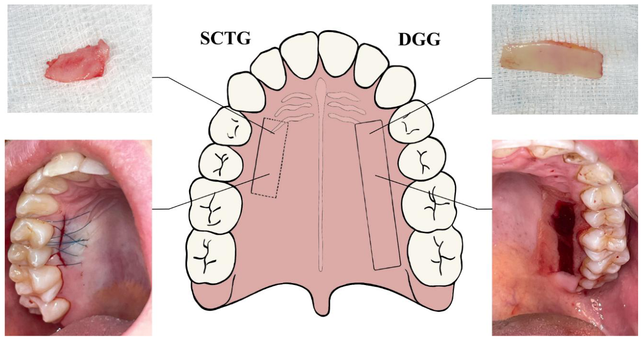
Figure 2.
Preoperative measurement of palatal mucosal thickness using (a) an endodontic instrument, (b) an ultrasound device, and (c) cone-beam computed tomography. Clinical photographs taken by A.D.
Figure 2.
Preoperative measurement of palatal mucosal thickness using (a) an endodontic instrument, (b) an ultrasound device, and (c) cone-beam computed tomography. Clinical photographs taken by A.D.
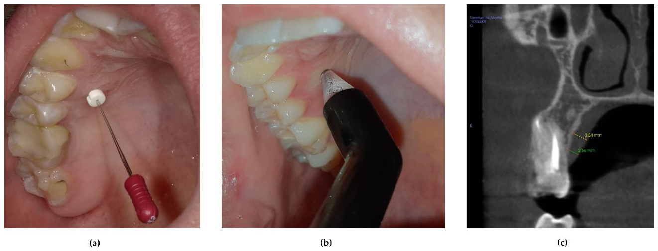
Figure 3.
Immediate bleeding as an intraoperative complications. Clinical photography taken by A.D.
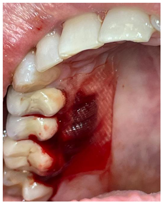
Figure 4.
Postoperative complications related to donor-site wound healing: (a) flap dehiscence; (b) flap sloughing; and (c) flap necrosis. Clinical photographs taken by A.D.
Figure 4.
Postoperative complications related to donor-site wound healing: (a) flap dehiscence; (b) flap sloughing; and (c) flap necrosis. Clinical photographs taken by A.D.
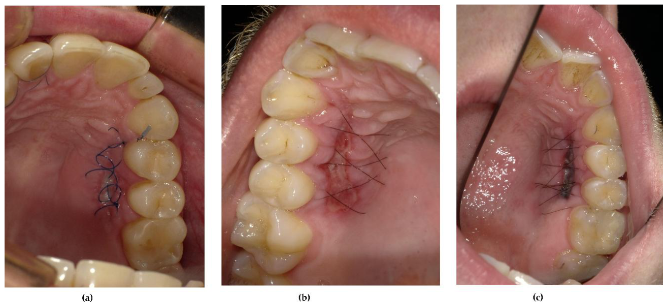
Figure 5.
Late donor-site complications: (a) palatal surface irregularities and (b) scarring. Clinical photographs taken by A.D.
Figure 5.
Late donor-site complications: (a) palatal surface irregularities and (b) scarring. Clinical photographs taken by A.D.
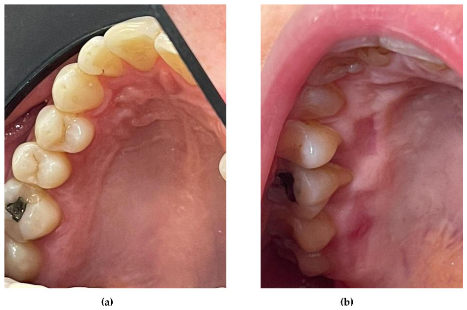

Table 1.
Characteristics of the key studies included in the narrative literature review.
| Author and year of publication | Study design | Objective | Number of subjects or studies | Graft type, harvesting technique or studies included | Outcomes evaluation |
Main donor-site findings |
|
|---|---|---|---|---|---|---|---|
| Palatal anatomy | |||||||
| Tomaszewska et al., 2014 [18] | Systematic review and meta-analysis | To localize the greater palatine foramen (GPF) in relation to multiple anatomical landmarks, systematically review the literature, and to perform a meta-analysis of the relation between the GPF and maxillary molars. | 27 studies included in the systematic review; 23 studies included in the meta-analyses 6927 GPFs. |
50 adult, human skulls and 1,200 computed tomography scans of the head. |
Location of the GPF. | The GPF was frequently located opposite the third molar (63,6%). The maxillary molars were identified as the most reliable landmarks for locating the GPF. | |
| Tavelli et al., 2019a [8] | Systematic review |
To provide guidelines for defining a safety zone for palatal graft harvesting. | 26 studies; 5,768 hemipalates. |
Cadaveric, computed tomography or cone beam computed tomography studies. | Location of the GPF and the course of the greater palatine artery in relation to the maxillary teeth. |
Based on the anatomic finding, a safety zone for palatal harvesting was proposed. |
|
| Complications and post-operative morbidity | |||||||
| Malpartida-Carrillo et al. 2014 [21] |
Systematic review |
To present comprehensive information on outcome measure related to postoperative palatal wound healing and postoperative patient-reported morbidity. | 31 studies. | Free gingival grafts (FGGs) and connective tissue grafts (CTGs). | Palatal wound healing; patient-reported outcomes, including pain, analgesic, consumption, discomfort and quality of life, burning sensation, and changes in eating habits. | The most commonly used outcome measures for postoperative palatal wound healing were the hydrogen peroxide test, tissue color match, visual inspection, and bleeding assessment. The most frequently assess patient-reported morbidity outcomes were pain, analgesic consumption, discomfort, burning sensation, and changes in eating habits. |
|
| Chong et al., 2025 [17] | Systematic review | To evaluate the nature of postoperative pain and donor site morbidity following palatal soft tissue harvesting procedures, involving various donor site wound dressings. | 69 studies. | Free gingival graft (FGG) and de-epithelialized gingival graft (DGG) techniques. | Patient-reported outcomes including quality of life bleeding, burning sensations, altered sensory perception, and changes in eating behavior. | Postoperative pain was most pronounced during the first three days and progressively decreased over time. Wound dressings or physical barriers could reduce pain, although outcomes varied according to the intervention used. | |
| Figueras-Alvarez et al., 2026 [16] | Systematic review | To analyze postoperative complications at the palatal donor site following the harvesting of autologous soft tissue grafts. |
16 studies. | Autologous graft. | Healing, postoperative pain; discomfort; bleeding; and sensory disturbances. |
Postoperative pain was the most common complication and was generally mild, transient, and manageable. Adjunctive strategies reduced discomfort and supported healing. Bleeding and sensory disturbances were less frequent and transient. | |
| Mashaly et al., 2002 [22] |
Randomized controlled clinical trial | To compare patient morbidity, and root-coverage outcomes following coronally advanced flap (CAF) combined with either subepithelial connective tissue graft (SCTG) or de-epithelialized gingival graft (DGG) for the treatment of Miller Class I and II gingival recessions. | 28 Egyptian participants: Test group, CAF+DGG (n=14); control group, CAF+SCTG (n=14). |
SCTG harvested using single-incision technique; extraorally de-epithelialized graft (DGG). |
Patient-reported outcomes: pain without differentiation by surgical site (VAS), stress VAS, bleeding (yes/no), inability to chew (VAS), and satisfaction (yes/no). Outcomes were assessed at 3,7, and 14 days postoperatively. |
At 2 weeks, patients treated with CAF + DGG reported significantly greater stress and inability to chew at 2 weeks than those in the SCTG group. | |
| Zucchelli et al. 2010 [5] | Two-center, double-blind, parallel-group, randomized controlled clinical trial | To compare the patient morbidity and root coverage outcomes following coronally advanced flap (CAF) combined with either a subepithelial connective tissue (SCTG) or de-epithelialized gingival (DGG) grafts. | 50 Italian participants: test group, CAF + DGG (n=25), control group, CAF + SCTG (n= 25). | SCTG harvested using trap door, DGG. | Palatal tissue thickness; graft thickness; analgesics consumption (mg); post-operative discomfort; bleeding; stress and inability to chew, assessed using VAS. Outcome were assessed 1week postoperatively. |
No statistically significant between-group differences were observed in analgesic consumption, postoperative discomfort or bleeding. The SCTG group reported lower stress and a better ability to chew. Analgesic consumption increased with increasing graft height and in the presence of dehiscence or necrosis of the primary flap. Pain was negatively correlated with the residual thickness of the soft tissue covering the palatal bone. | |
| Fickl et al., 2014 [23] |
Randomized, single-blinded controlled trial |
To compare wound healing and patient-reported pain following the use of the single-incision (SIT), modified single-incision (mSIT) and trap-door (TD) surgical techniques for harvesting subepithelial connective tissue graft. | 36 German participants: SIT group (n=12); mSIT group (n=12);: TD group (n=12). | SCTG harvested using single-incision, modified single-incision, or trap door technique. |
Early-Wound Healing Index (EHI); postoperative pain assessed using VAS; and timing, amount and duration of analgesic use. Outcome were assessed 1 week postoperatively. |
The SIT and mSIT groups demonstrated improved early wound healing. Secondary healing as well as the cumulative dosage and duration of analgesic use, was significantly reduced in SIT and mSIT groups. |
|
| Zucchelli et al., 2014 [24] | Randomized, double-blind, parallel-group controlled trial | To determine whether patient morbidity could be reduced by decreasing the graft thickness and height | 60 Italian participants: test group, 30 defects, thickness < 2mm and height 4mm: control group, 30 defect, thickness ≥ 2mm and height corresponding to the bone dehiscence. | Extraorally de-epithelialized gingival graft (DGG). |
Postoperative pain assessed according to analgesic consumption (mg); discomfort (VAS); bleeding (VAS); and inability to chew (VAS). Outcomes were assessed 1 week postoperatively. |
Reducing graft thickness and height decreased patient morbidity without compromising the root-coverage outcomes. | |
| Maino et al., 2018 [25] |
Randomized, single-blind clinical trial | To investigate the influence of suturing technique and preoperative palatal mucosal thickness and postoperative residual flap thickness on the early palatal wound healing of the palate after CTG harvesting. | 36 Spanish participants: continuous interlocking suture group (n=18); interrupted criss-cross suture group (n=18). | SCTG harvested using single incision technique. |
Preoperative palatal mucosal thickness; postoperative residual palatal flap thickness; Early Wound Healing Index (EHI) assessed at 7 days postoperatively, and postoperative pain assessed using VAS at 1, 2, 3, 7, 14, 21 and 28 days postoperatively. | A significant relationship was found between postoperative residual flap thickness and the EHI. The number of days with discomfort was negatively correlated with residual flap thickness. No statistically significant association was found between suturing technique and wound healing. |
|
| Bakhishov et al., 2021 [26] |
Randomized, single-blind, parallel-group controlled trial | To compare the clinical efficacy and postoperative morbidity of a de-epithelialized gingival graft (DGG) and a subepithelial connective tissue graft (SCTG), both combined with tunnel technique (TUN) for the treatment of multiple gingival recessions. | 27 Turkish participants: DGG + TUN group (n=13), SCTG + TUN group (n=14). |
Intraorally de-epithelialized gingival graft (DGG); SCTG harvested using single incision technique. | Postoperative pain; discomfort; sensitivity; and bleeding at the palatal donor site, assessed using VAS or dichotomous responses (yes/no). Outcomes were assessed at 1, 2, 3, 7, 14 and 28 days postoperatively. |
No statistically significant differences were observed between the DGG + TUN and the SCTG + TUN in postoperative pain, discomfort, sensitivity or systemic analgesic consumption. | |
| Del Pizzo et al., 2002 [27] |
Comparative preliminary clinical study | To evaluate early palatal wound healing following three different connective tissue graft procedures: the single-incision (SI), the free gingival graft (FGG), and the trapdoor (TD) techniques | 36 Italian participants: SI group (n= 12); FGG group (n=12), TD group (=12). |
SCTG harvested using single incision technique or trap door technique; FGG. |
Immediate and delayed bleeding; sensibility; complete epithelialization; discomfort; and variation of eating habits. Outcomes were assessed at 1, 2, 3, 4, 6, and 8 weeks postoperatively. | Complete epithelialization occurred significantly earlier following SCTG harvesting than following FGG. Discomfort was significantly lower in the SI and TD groups than in the FGG group. |
|
| Griffin et al., 2006 [12] |
Prospective observational clinical study | To compare the frequency of pain, swelling, and bleeding following free gingival graft (FSTG) and subepithelial connective tissue graft (SCTG) procedures and to identify potential predictors of these complications. |
228 American participants undergoing 242 procedures: FSTG (n=70 procedure); SCTG (n=172 procedures). | Full thickness SCTG harvested using Bruno technique; FSTG |
Postoperative pain using numerical scale 0 to 10; swelling; and bleeding. Outcomes were assessed at 1 week postoperatively. |
Longer surgical duration was strongly correlated with postoperative pain or swelling. Smokers were three times more likely to experience postoperative swelling. FSTG procedures were associated with threefold increase in the probability or moderate-to-severe pain and bleeding. |
|
| Wessel and Tatakis, 2008 [28] |
Prospective observational parallel-group study |
To compare patient-reported outcomes following connective tissue graft (CTG) and free gingival graft (FGG). | 23 American participants: CTG group (n=12); FGG group (n=11). |
CTG harvested using Harris or Hurzeler and Weng technique; FGG with the use of palatal stent. | Postoperative pain assessed using VAS; number of analgesic tablets taken; and duration of analgesic use in days. Outcomes were assessed at 3 days and 3 weeks postoperatively. | FGG harvesting was associated with a higher incidence of donor site pain than CTG harvesting during the early postoperative period. |
|
| Lessa et al., 2025 [29] |
Prospective cohort study | To compare the postoperative donor-site morbidity following two graft-harvesting techniques: de-epithelialized gingival graft (DGG) and the linear technique (SCTG). | 16 Brazilian participants: DGG group (n=8); SCTG group (n=8). | DGG; SCTG harvested using single incision technique. |
Postoperative pain assessed using VAS; number of anti-inflammatory medications taken; and visual assessment of healing. Outcomes were assessed at 1, 2, and 3 weeks postoperatively. |
The DGG technique was associated with greater postoperative pain and a higher requirements for analgesic and anti-inflammatory medication than the SIT. |
|
| Post-surgical management of donor site | |||||||
| de Almeida et al., 2024 [19] | Umbrella review | To assess the effects of different therapies on wound healing and postoperative discomfort following autogenous palatal graft harvesting. | 10 systematic reviews comprising 25 randomized clinical trials. | DGG and SCTG. | Systematic reviews that evaluating: wound healing, postoperative pain, bleeding, and analgesic consumption. | Low-level laser therapy (LLLT) , platelet-rich fibrin (PRF), cyanoacrylate tissue adhesives (CTAs), and ozone therapy improved in wound healing. LLLT, PRF, and CTAs reduced pain and analgesic consumption, whereas PRF and CTAs reduced bleeding. Regarding methodological quality, two systematic reviews were rated as critically low quality, five as low quality, two as moderate quality, and one as high quality. | |
| Leite et al., 2025 [20] | Systematic review | To evaluate the efficacy of different approaches to pain management following free gingival graft (FGG) and de-epithelialized connective tissue graft (SCTG) procedures and their effects on oral health-related quality of life (OHRQoL). | 54 studies evaluating 43 different interventions. | FGG; DGG. | Studies evaluating: postoperative pain and its impact on the OHRQOL |
Overall, the interventions were associated with a reduction in postoperative pain over time. However, it was not possible to determine which intervention was superior, because many studies combined interventions with different mechanisms of action. | |
| Ziyaettin et al., 2026 [30] | Prospective randomized, double-blind, controlled trial | To assess the effects of palatal pre-suturing using a greater palatine compression suture (GPCS) on bleeding, postoperative morbidity, wound healing, and quality of life following subepithelial connective tissue graft harvesting. | 32 Turkish participants: GPCS before graft harvesting (n=16); control without GPCS (n=16). | SCTG harvested using single incision technique | Intraoperative bleeding; surgical duration; postoperative bleeding, pain, analgesic use; Landry wound healing index, and the Turkish version of the 14-item Oral Health Impact Profile (OHIP-14-TR). Outcomes were assessed at 3 days, 1, 2 weeks, 1, 3 months postoperatively. |
Pre-harvesting GPCS reduced intraoperative bleeding, shortened surgical duration, and had beneficial effects on postoperative morbidity and wound healing. | |
Table 2.
Systematic reviews of therapeutic approaches to enhance wound healing following palatal graft harvesting.
Table 2.
Systematic reviews of therapeutic approaches to enhance wound healing following palatal graft harvesting.
| Author and year of publication | Main findings | |
|---|---|---|
| Low-level laser therapy (LLLT) / photobiomodulation (PBM) |
Al-Shibani et al., 2019 [77] Ebrahimi et al.., 2021 [78] Zhao et al.., 2021 [79] Fonseca et al. 2026 [80] |
LLT/PBM was associated with statistically significant improvements in early wound healing and a short time to complete wound epithelialization compared with spontaneous healing. However, findings regarding postoperative pain reduction and analgesic consumption were heterogeneous across studies. |
| Platelet-rich fibrin (PRF) | Gusman et al., 2021 [81] Martins et al., 2022 [82] Meza-Mauricio et al., 2021 [83] Miron et al., 2017 [84] Estrin et al., 2025 [85] |
PRF was associated with faster wound healing, reduced postoperative pain, lower analgesic consumption, and a marked reduction in immediate postoperative bleeding. |
| Ozone therapy (OT) | Palma et al., 2023 [86] | OT was associated with improved wound healing. |
| Cyanoacrylate tissue adhesives (CTAs) | Escobar et al., 2021 [87] Veríssimo et al., 2021 [88] |
CTA were associated with faster wound healing, improve control of immediate bleeding, reduced postoperative pain, and lower analgesic consumption. |
| Hyaluronic acid (HA) |
Joshi et al., 2024 [89] | HA was associated with improved wound epithelialization and color matching, as well as reduced patient-reported post-operative pain. |
Disclaimer/Publisher’s Note: The statements, opinions and data contained in all publications are solely those of the individual author(s) and contributor(s) and not of MDPI and/or the editor(s). MDPI and/or the editor(s) disclaim responsibility for any injury to people or property resulting from any ideas, methods, instructions or products referred to in the content. |
© 2026 by the authors. Licensee MDPI, Basel, Switzerland. This article is an open access article distributed under the terms and conditions of the Creative Commons Attribution (CC BY) license (http://creativecommons.org/licenses/by/4.0/).
Copyright: This open access article is published under a Creative Commons CC BY 4.0 license, which permit the free download, distribution, and reuse, provided that the author and preprint are cited in any reuse.