

Submitted:
26 September 2026
Posted:
29 September 2026
You are already at the latest version
Abstract
Background: Secondary caries or caries around restorations or sealants is a common reason for replacement of restorations. However, distinguishing true secondary caries from restoration-related defects remains clinically difficult and may lead to overtreatment and unnecessary removal of healthy tooth structure. Objectives: This scoping review aimed to map how diagnostic strategies for secondary caries are used and how these findings are translated into treatment decisions, while identifying diagnostic thresholds and informing an evidence-based decision framework. Methods: Searches were performed in seven electronic databases, gray literature sources, and guideline repositories without restrictions on language or publication year. Evidence addressing diagnostic strategies for secondary caries, as well as relevant guidelines and consensus documents, were included. Data were organized descriptively, and the methodological quality of primary studies was critically evaluated. Results: Forty-eight documents were included (34 original studies and 14 guidelines or consensus statements). Visual–tactile examination, commonly combined with bitewings, was the most frequently reported detection approach. Adjunctive methods, including fluorescence-based devices, optical imaging, and artificial intelligence, were investigated as complementary detection tools, with heterogeneous performance across study designs. Treatment decisions were primarily driven by lesion activity and suspected dentine involvement rather than marginal appearance alone. Explicit diagnostic thresholds were rarely predefined, and substantial variability was observed in how diagnostic findings informed treatment decisions. Conclusions: The evidence supports visual–tactile assessment complemented by bitewings as first-line approaches within a diagnostic strategy for secondary caries evaluation. However, heterogeneity in diagnostic thresholds and treatment decision criteria limits comparability across studies and may contribute to inconsistent clinical management. Greater standardization in reporting diagnostic strategies and decision frameworks is needed to support evidence-based and conservative care. Clinical significance: This review provides an evidence-informed clinical framework based on visual–tactile and radiographic assessment for secondary caries around restorations. By integrating detection methods, lesion interpretation, and commonly reported clinical thresholds, the framework illustrates how findings from routine assessment are translated into treatment strategies. The findings indicate that treatment decisions are more consistently guided by lesion activity and dentine involvement than by restoration defects alone, supporting a more conservative approach under diagnostic uncertainty and highlighting the need for greater standardization in clinical criteria.
Keywords:
caries detection
; diagnostic thresholds
; clinical decision-making
; restorative dentistry
; practice guidelines
1. Introduction
Secondary caries, also known as Caries Around Restorations or Sealants (CARS), is frequently cited as a cause of restoration failure and replacement in clinical practice [1,2,3]. When it comes to CARS, replacement of restorations is usually done to control the lesion; however, observational and clinical studies have raised concerns that secondary caries may be overdiagnosed, which could lead to premature intervention and the unnecessary removal of healthy dental tissue [2,3,4,5]. Repeated replacement procedures increase treatment complexity and costs over time, contributing to a restorative cycle [6,7].
Secondary caries presents distinct diagnostic challenges compared with primary caries. The presence of restorations limits direct visual and tactile access to the tooth surface and introduces confounding factors that complicate clinical interpretation [3,8]. Findings such as marginal staining, ditching, or small gaps are frequently observed during restoration assessment, yet their relationship with biologically active secondary caries remains uncertain when considered in isolation [9,10]. Consequently, clinicians are often confronted with a diagnostic grey zone, in which distinguishing lesion progression from aging-related restoration changes relies heavily on individual interpretation [8,11]. This diagnostic uncertainty is closely related to the structural complexity of the tooth–restoration interface, where restoration margins, gaps, staining, residual carious tissue, and subsurface lesion progression may produce overlapping clinical and radiographic appearances (Figure 1).
Visual–tactile examination is widely regarded as the primary detection method for assessing restorations suspected of secondary caries and is consistently recommended in clinical studies and guidelines [12,13,14,15]. Bitewing radiography is commonly used as an adjunct, particularly in proximal or submarginal areas where direct inspection is limited [9,16,17]. Conventional diagnostic approaches generally demonstrate high specificity but limited sensitivity for lesions beneath apparently intact restoration margins, contributing to diagnostic uncertainty depending on clinical context [16,18]. The frequent discrepancies between clinical and radiographic findings emphasize that radiographic data should serve as a complementary tool within a diagnostic strategy, rather than a standalone criterion for operative intervention [19,20,21].
To address the limitations of conventional assessment, several adjunctive diagnostic methods have been proposed, including fluorescence-based devices, optical imaging techniques and intraoral photography, as well as artificial intelligence (AI)–based systems designed to support the interpretation of diagnostic images [22,23,24,25]. These approaches have mainly been investigated as complementary tools to enhance lesion detection. However, their reported performance varies according to threshold definition, lesion location, and restoration-related confounders; moreover, concerns regarding false-positive findings have been raised [26,27]. Furthermore, the extent to which identifying additional suspicious findings translates into improved clinical management decision-making remains insufficiently explored [7,8].
Beyond identifying the lesion, a major clinical challenge is translating diagnostic findings into treatment decisions. The concept of diagnostic strategies can be understood as an overarching framework integrating detection (acquisition of clinical findings), diagnostic interpretation (assessment of lesion presence and activity), and management decision-making (selection of management approaches) [8,12,13,15]. Most research focuses on accuracy metrics, such as sensitivity and specificity, but often fails to explain how these results guide the choice between monitoring, repairing, or replacing a restoration [1,2,28]. Consequently, the clinical thresholds for operative intervention remain poorly defined and inconsistent. This lack of standardization explains why different clinicians may choose widely different treatments for the same clinical finding, frequently leading to unpredictable management and potential overtreatment [5,28,29].
Given the heterogeneity of study designs, diagnostic methods, reference standards, and outcome measures in this field, a comprehensive mapping of the available clinical evidence is warranted. Therefore, the aim of this scoping review was to map how diagnostic strategies are applied in the assessment of secondary caries lesions, from the identification of clinical findings to their interpretation and translation into management decisions and, based on this synthesis, to inform an evidence-based clinical decision framework.
2. Methods
This scoping review was conducted and reported in accordance with the PRISMA-ScR (Preferred Reporting Items for Systematic Reviews and Meta-Analyses extension for Scoping Reviews) [30] and followed the methodological guidance of the Joanna Briggs Institute (JBI) for scoping reviews [31]. The review protocol was prospectively registered in the Open Science Framework (OSF; https://osf.io/jhzwu/?view_only=78efa71806624a68a695fa9727f27f61).
The review addresses the following research questions:
- Which detection or diagnostic methods are reported for the assessment of secondary caries lesions around restorations or sealants?
- Which diagnostic thresholds or criteria are used to support clinical decision-making for secondary caries lesion management?
- What recommendations can be made for future clinical studies investigating the detection and management decision-making for secondary caries?
2.1. Eligibility Criteria
Primary clinical studies of any design (randomized clinical trials, non-randomized controlled trials, and observational studies including cohort and cross-sectional designs) were eligible if they investigated detection or diagnostic methods and/or management decision-making thresholds related to secondary caries in restored primary or permanent teeth. All restorative materials and techniques were considered eligible, including direct and indirect restorations made of composite resin, amalgam, glass ionomer cement (GIC), gold, ceramic, or metal.
Studies were excluded if they were narrative or systematic literature reviews, in vitro or in situ using extracted teeth or artificially induced caries models, non-human studies, and purely technical or laboratory validation studies.
Clinical guidelines, consensus statements, or recommendation documents were included as a distinct evidence category when they explicitly addressed secondary caries (or equivalent terms) within detection, diagnostic, management decision-making thresholds, or management frameworks. For these documents, a report of clinical data was not required.
2.2. Information Sources and Search Strategy
Search strategies were developed with the assistance of an experienced health sciences librarian. No restrictions were applied for language or publication year. Full search strategies are provided in Supplementary Material A.
Electronic searches were conducted in PubMed, Web of Science, Scopus, LIVIVO, Cochrane, Embase, and Google Scholar (for gray literature, first 100 results sorted by relevance). Additional searches were performed in guideline repositories, including the National Institute for Health and Care Excellence (NICE), Guidelines International Network (GIN), and Trip Database. These searches were complemented by targeted searches of professional dental organizations and manual screening of reference lists from included titles. The initial search was conducted on March 3, 2024, and updated on July 13, 2026.
2.3. PCC Acronym and Selection Process
The review was guided by the Population-Concept-Context (PCC) framework, recommended for scoping reviews [31]:
- Population: Children and adults with restored primary or permanent teeth;
- Concept: Clinical detection methods, diagnostic interpretation, and management decision-making thresholds for secondary caries lesions;
- Context: Clinical assessment and management of restorations in dental care settings.
After duplicate removal using Mendeley, titles and abstracts were screened independently by two reviewers (E.T.C. and G.S.L.) using Rayyan [32]. Full text of potentially eligible records was retrieved and assessed independently using the same eligibility criteria. Disagreements were resolved through online discussion rounds, with arbitration by senior researchers in cariology (F.M.M. and M.S.C.).
2.4. Data Collection Process
When full texts or key information were unavailable, corresponding authors were contacted by e-mail and, when applicable, via professional platforms (e.g., ResearchGate). Studies were excluded if no response was obtained after three contact attempts spaced over one week apart. Data extraction used a standardized Excel spreadsheet, pilot-tested on five studies. Discrepancies were resolved by consensus between the reviewers and arbitrators.
2.5. Data Items
For primary clinical studies, extracted data included: study identification (authors, year), country and setting (university, private, public, or mixed), study design, dentition type (primary/permanent), follow-up period (when applicable), sample characteristics (number of patients, teeth, restorations, or images), restoration type and material, index diagnostic test(s), reference standard (when applicable), reported diagnostic or treatment decision thresholds (predefined or author-defined), diagnostic performance outcomes (e.g., sensitivity, specificity, AUC, kappa, ICC), and secondary caries prevalence when calculable.
For clinical guidelines and consensus documents, extracted data included document scope, terminology used for secondary caries, diagnostic approaches, management decision-making thresholds, management recommendations, and the availability of practical decision-support tools. For research protocols, extracted data focused on study objectives, targeted diagnostic methods or thresholds, planned outcomes, and anticipated contributions to clinical decision-making. The complete data extraction dataset is available at the OSF repository.
2.6. Data Synthesis
The extracted data were collected and summarized descriptively to map the existing evidence on secondary caries detection and management decision-making. Results were grouped according to the type of evidence: (i) primary clinical studies; subdivided by diagnostic method (e.g., visual–tactile, radiographic, adjunctive technologies) and by the decision thresholds that guided management (e.g., visual criteria, radiographic depth, surface texture, presence of soft tissue, or restoration integrity); (ii) recommendations and concepts from guidelines were synthesized into flowcharts and the critical appraisal was performed using Joanna Briggs Institute (JBI) tools [33,34,35]. The results were presented in tables and used to discuss the quality of the evidence but this did not influence the inclusion of studies.
3. Results
3.1. Sample Selection
The combined search yielded 11,479 records. After duplicate removal and two-stage screening, 48 documents met the PCC and eligibility criteria, comprising 34 primary studies [7,9,19,20,22,23,24,25,27,28,36,37,38,39,40,41,42,43,44,45,46,47,48,49,50,51,52,53,54,55,56,57,58,59] and 14 guidelines/consensus statements [8,10,12,13,14,15,60,61,62,63,64,65,66,67] (Figure 2; Supplementary Material B). Primary clinical studies constituted the main body of evidence and were included for data extraction and methodological appraisal. Guidelines and consensus documents were analyzed separately to map how secondary caries detection, management decision-making, and management are framed in authoritative clinical recommendations. In addition, three published research protocols were identified [68,69,70], these were linked alongside the primary study that generated the data included in this review, for methodological context in the data collection phase (Table 1).
3.2. Studies Characteristics
Among the 34 primary clinical studies, twenty-one (21/34) primarily evaluated diagnostic performance, although analytical strategies varied across investigations. Nine studies quantified diagnostic performance using accuracy-based metrics, including sensitivity, specificity, AUC, or related measures, typically supported by an explicit reference standard (e.g., clinical inspection or expert consensus) [24,25,27,36,45,46,49,51,57]. Five studies assessed diagnostic performance through examiner agreement, reporting inter- or intra-observer reliability metrics such as kappa or intraclass correlation coefficients, particularly when evaluated methods relied on human interpretation of visual–tactile or radiographic findings [22,28,39,47,48]. Another eight investigations compared diagnostic methods based on the proportion of detected versus non-detected lesions and/or their resulting treatment decisions. These studies reported descriptive comparisons across methods, criteria or time points [19,23,37,40,41,43,58,59].
In addition, a subset of observational studies (7/34) was primarily designed to compare alternative diagnostic threshold frameworks for secondary caries while maintaining similar underlying detection methods [9,20,50,53,56,58,59]. These investigations evaluated how different visual criteria or grading systems, such as FDI-based criteria, CARS, modified USPHS, or combined clinical–radiographic frameworks, translated into treatment decisions (monitoring, repair, or replacement). One study adopted a health-economic perspective (1/34), evaluating secondary caries management strategies through cost-effectiveness modelling [7]. One randomized clinical trial (1/34) compared different visual criteria for secondary caries detection and their impact on treatment decisions [52].
Five non-randomized prospective clinical studies (5/34) evaluated secondary caries as a longitudinal clinical outcome within comparisons of restorative materials or restorative strategies. In these investigations, visual–tactile and radiographic criteria were applied pragmatically during follow-up, while the analytical emphasis was placed on caries occurrence, restoration failure, or replacement over time, rather than on diagnostic performance or decision thresholds [38,42,44,54,55]. Figure 3 summarizes the distribution of study designs, settings and origins, sample characteristics, and reference standards across the included studies, highlighting the predominance of diagnostic-oriented investigations (21/34).
3.3. Methodological Quality of the Evidence and Studies Limitations
A critical appraisal of the included primary studies was conducted using the appropriate Joanna Briggs Institute (JBI) checklists according to study design. Diagnostic accuracy studies showed moderate methodological heterogeneity. Most studies presented low risk of bias for outcome measurement and timing, as index tests and reference standards were generally applied within comparable periods [7,9,17,19,20,22,23,24,25,27,36,37,39,40,41,43,45,46,47,48,49,50,51,53,56,57,58,59].
However, important limitations were identified. Sampling methods were frequently unclear or at high risk of bias, with several studies relying on convenience or enriched samples, limiting representativeness [17,24,25,36,39,40,41,45,48,51,57,58]. In addition, threshold pre-specification was rarely reported [22,23,24,25,27,36,41,45,46,47,59], and blinding between index tests and reference standards was often unclear, increasing the risk of biased interpretation of diagnostic findings [24,25,43,45,46,47,51,57].
Regarding the reference standard, restoration removal was commonly used but typically restricted to teeth with clear clinical indication, while early or uncertain lesions were assessed using alternative comparators [22,40,41,43,57,59]. This selective verification may have influenced both diagnostic accuracy estimates and their translation into clinical management decision-making.
Quasi-experimental studies showed acceptable methodological structure but were limited by unclear group comparability, incomplete follow-up, and insufficient reporting of outcome assessment [38,42,44,54,55]. Only one randomized controlled trial was identified [52], presenting low risk of bias across all domains, but the limited number of RCTs restricts the strength of the available evidence. Overall, these limitations contribute to variability in how diagnostic information is interpreted and applied in clinical management decision-making. The overall risk of bias across study designs is summarized in Supplementary Material C.
3.4. Clinical Guidelines and Recommendations for Secondary Caries Management
Fourteen clinical guidelines, consensus statements, or recommendation documents addressing secondary caries were identified (Table 2). These documents were published between 2010 and 2024 and issued by international scientific societies and professional dental organizations, including the European Organization for Caries Research (ORCA), the European Federation of Conservative Dentistry (EFCD), the German Society of Conservative Dentistry (DGZ), the FDI World Dental Federation, the International Caries Classification and Management System (ICCMS™) and CariesCare International groups, and the American Academy of Pediatric Dentistry (AAPD).
Two documents (2/14) were specifically focused on secondary caries detection and management decision-making. The EFCD–ORCA–DGZ Delphi consensus on proximal and secondary caries [15] and the ORCA/EFCD consensus on secondary caries detection, management decision-making, and management [8] explicitly addressed diagnostic approaches and clinical decision criteria for secondary caries. Restoration-evaluation frameworks were represented by the original FDI criteria [12] and its revised version [13] (2/14), which incorporated secondary caries within standardized visual–tactile scoring systems linked to management actions.
Several documents addressed secondary caries within broader caries management systems (4/14), including the ICCMS™ Guide for Practitioners and Educators [64], the CariesCare practice guide [65], and the transition from ICDAS to CariesCare International frameworks [14]. Terminology-focused guidance was provided by the ORCA/IADR consensus on caries terminology [66], which defined secondary caries conceptually without proposing diagnostic thresholds or management algorithms.
Pediatric-oriented guidance documents were issued by the American Academy of Pediatric Dentistry, including the Guideline on Restorative Dentistry [60], Pediatric Restorative Dentistry: Best Practices [61], Policy on Medically-Necessary Care [62], and the Fluoride Therapy: Best Practices [63]. Additional recommendations relevant to lesion assessment and progression included the ORCA/EFCD clinical recommendations for caries diagnosis and activity assessment [10] and guidance on the evidence base for professional and self-care prevention [67].
Across documents, visual–tactile examination, commonly supplemented by bitewing radiographs, was consistently described as the primary approach for detecting secondary caries. Explicit numeric diagnostic thresholds were uncommon; instead, most guidelines operationalized management decision-making through categorical or score-based criteria linked to lesion severity, cavitation, dentine involvement, or lesion activity. Extracted data from clinical guidelines and consensus documents were systematically charted, and reported detection/diagnostic criteria and management pathways were organized into decision frameworks to visually summarize the findings.
3.5. Which Detection or Diagnostic Methods are Reported for Assessment of Secondary Caries Lesions Around Restorations or Sealants?
Visual–tactile (VT) examination, frequently operationalized through structured criteria (e.g., USPHS/Ryge, FDI, CARS, ICDAS-based approaches), was the core detection method for secondary caries around restorations or sealants, either as the primary index method or as the clinical comparator [9,20,22,23,27,28,36,38,39,40,41,42,43,44,48,49,52,53,55,56,58,59].
This VT-centered approach is consistent with guidance documents, which consistently position standardized visual–tactile assessment (often under clean, dry, and illuminated conditions) as the first-line method for secondary caries evaluation [8,10,12,13,14,15,60,61,62,63,64,65,66,67]. Specificity is generally high (≈ 0.85), but sensitivity is lower (< 0.60) for lesions hidden beneath apparently intact margins [36,48].
Radiographic assessment, particularly bitewing radiographs (BW), was the most frequently reported adjunct method and was used to support detection in areas where VT examination is limited (e.g., proximal or submarginal regions) and to support diagnostic interpretation, particularly regarding lesion depth staging [7,9,19,20,25,28,36,37,39,42,43,45,48,49,50,51,52,56]. Alternative radiographic strategies, including panoramic and periapical imaging, were occasionally reported, particularly in AI-assisted workflows [46,47]. In guidance documents, BW radiographs are consistently framed as an adjunct to VT examination, particularly when clinical inspection is limited [8,10,15,57,58,64,65]. Bitewings performed better than panoramic or periapical images for detecting proximal and submarginal caries, offering improved visualization of the cemento-enamel junction and reduced anatomical overlap [19,43,57].
A smaller group of studies evaluated light-based or optical adjuncts intended to complement VT and radiographic assessment. Laser fluorescence devices (e.g., DIAGNOdent) and fluorescence-based cameras (e.g., VistaCam) were assessed in clinical cohorts, typically alongside VT and/or BW, with reported performance varying according to threshold definition and the presence of confounders such as marginal staining or defects [7,22,27,36,49]. Fibre-optic transillumination (FOTI) was explored in earlier clinical comparisons [37,49], and optical coherence tomography (OCT) was evaluated as an adjunct imaging modality [37]. While these approaches were generally positioned as sensitivity-enhancing tools, their clinical interpretation remained dependent on threshold definition and verification strategy.
Intraoral and digital photography was reported either to support clinical documentation or as an input for automated analysis. Studies using photographic assessment included comparisons against clinical validation frameworks and expert-based interpretation [23,24], with additional use in multimodal workflows [27]. These approaches were consistently framed as complementary and required careful interpretation to distinguish discoloration and marginal defects from true lesions adjacent to restorations.
Finally, artificial intelligence (AI) approaches represented the most recent diagnostic strand, using radiographs and/or photographs as inputs to convolutional neural network-based or object-detection models. AI systems were evaluated on bitewings for secondary caries identification [25,45,46,51,57] and within multi-source imaging pipelines integrating panoramic and bitewing data [47]. AI applied to photographic datasets was also reported [24]. Across these studies, AI was generally positioned as a second-reader or decision-support tool, with acknowledged limitations related to single-centre datasets and limited external validation [25,45,47,51].
3.6. What Thresholds Guide the Decision-Making for the Treatment of Secondary Caries?
Standalone decision thresholds were rarely predefined in the included evidence. These thresholds do not operate in isolation but emerge from the integration of detection and diagnostic interpretation processes, ultimately guiding management decision-making. Instead, treatment decisions for secondary caries were guided by the concurrent use of clinical, radiographic, and index-based criteria, which together defined implicit operative thresholds.
Indicators of biological activity represented the most consistent trigger for operative and non-operative management. Soft or leathery dentine on probing, plaque stagnation adjacent to restoration margins, chalky or opaque enamel, and signs of rapid lesion progression were repeatedly associated with decisions toward active monitoring, non-operative, or operative approaches [9,20,22,27,38,39,40,41,48,50,52,53,54,56,58,59]. These criteria align with guideline frameworks that position lesion activity and cavitation as primary decision nodes preceding operative intervention [8,12,13,15,64].
Radiographic assessment was used as an auxiliary tool for lesion staging, particularly when restorations limited direct visualization and probing [19,36,41,43,48,49]. Radiographs contributed mainly by estimating dentine involvement beneath restorations, supporting decisions regarding monitoring, non-operative or operative approaches [52,56]. Within structured evaluation systems, radiographic findings were interpreted alongside clinical findings rather than as independent diagnostic thresholds [12,13,14,64,65].
Other modifiers, including patient symptoms, lesion depth toward the inner dentine third, and overall caries risk, consistently shifted decisions toward earlier intervention in higher-risk scenarios [12,16,17,38,40,64]. Marginal gaps or isolated staining were frequently associated with restoration replacement; however, most studies and guidelines agreed that these findings, when considered in isolation, should not be interpreted as evidence of secondary caries [9,13,20,48,50,52,53,54,56,58,59,65]. A decision-analytic model indicated that intervening before radiographic evidence of dentine involvement increases treatment costs and accelerates the restorative cycle, supporting higher operative thresholds in low-risk patients [7,14]. An integrated synthesis of the evidence identified in this review is presented in Figure 4, combining detection methods, diagnostic thresholds, and their translation into treatment decisions, along with current guidance and identified gaps.
3.7. What Recommendations can be Made for Future Clinical Studies Investigating the Detection and Decision-Making for Secondary Caries?
A consistent trend toward conservative, risk-oriented management of secondary caries lesions was observed in the literature. Monitoring, refurbishment, or repair were commonly associated with acceptable outcomes when lesions were inactive, uncertain, or limited in extent, whereas full restoration replacement was more frequently reported in association with lesions showing clear activity or progression [7,9,12,20,53].
An important methodological gap concerns how management decisions are operationalized and reported. Many studies described treatment outcomes without clearly specifying how lesion activity, accessibility, radiographic staging, and patient-level factors were integrated into management decision-making. Structured frameworks, such as the FDI criteria, ICCMS™, and CariesCare, offer shared terminology and decision structures that may enhance transparency compared with unstructured clinical judgment, although their application ultimately depends on professional interpretation and clinical experience [13,14,64,65].
Heterogeneity was also observed in the interpretation of restoration-related changes, including marginal staining, ditching, or gap formation. Several clinical cohorts indicated that these findings, when considered in isolation, were weak indicators of biologically active secondary caries and should not be equated with disease presence [9,48,50,52,53,54]. Distinguishing restoration aging or defects from true caries outcomes is therefore essential, as treating these entities as equivalent may inflate secondary caries prevalence and obscure disease-related events [7,10,12].
Overall, future investigations would benefit from explicitly situating management decisions within defined interpretative frameworks while acknowledging the inherent subjectivity of secondary caries assessment and the role of clinical experience. Clear reporting of how restorative defects, lesion activity, and progression are distinguished may improve consistency across studies and support more meaningful comparisons of management strategies.
3.8. Is it Possible to Create an Evidence-Based Framework from Detection to Decision-Making for Secondary Caries?
The synthesis of the included clinical studies and guidelines supports an evidence-informed framework linking secondary caries detection, diagnostic interpretation, and management decision-making. Rather than defining a single operative threshold, the available evidence suggests a sequential approach in which visual-tactile findings, radiographic information, lesion activity, and patient-level factors are interpreted together.
Figure 5 summarizes the detection and diagnostic interpretation pathway. Visual-tactile assessment remains the first-line approach, particularly for accessible occlusal and free surfaces, whereas bitewings provide complementary information when direct inspection is limited, especially in proximal or submarginal areas. Within this pathway, radiographic findings mainly support lesion depth estimation and suspected dentine involvement. However, enamel-limited or uncertain radiographic findings should be interpreted cautiously and not automatically translated into operative treatment.
Diagnostic categories were translated into management decision-making and presented in Figure 6. Findings consistent with sound restorations, inactive lesions, or uncertain/non-cavitated lesions generally support monitoring, preventive care, refurbishment, sealing, or repair. In contrast, operative intervention is more consistently supported when there is evidence of lesion activity, cavitation, soft dentine, progression, or likely dentine involvement. Restoration-related findings such as marginal staining, ditching, gaps, or discoloration may contribute to diagnostic uncertainty but should not be considered sufficient evidence of secondary caries when assessed in isolation.
Although this framework is consistent with the available evidence, its application remains limited by heterogeneous diagnostic thresholds, variable reference standards, and the scarcity of controlled studies evaluating management outcomes. Future studies should explicitly report how diagnostic findings are translated into management decisions and should evaluate whether these decisions improve clinically relevant outcomes beyond lesion detection alone.
4. Discussion
This scoping review mapped the available clinical literature on detection methods and management decision-making thresholds for secondary caries. The findings reinforce that secondary caries detection, diagnostic interpretation and management decision-making differ from primary caries, as existing restorations inherently limit direct lesion access and introduce diagnostic uncertainty at both early and advanced stages of disease [8,66], highlighting the structural features of the tooth–restoration interface that may complicate lesion detection and interpretation.
Visual–tactile examination combined with bitewings is the most commonly used approach for the assessment of secondary caries; however, important limitations persist, particularly in situations where restorations obscure lesion access. While this strategy performs reliably in clearly sound or cavitated conditions, its ability to resolve ambiguous findings (e.g., hidden lesions, minor restorative margin defects or suspected subsurface lesions) is limited. In these scenarios, conventional methods tend to show high specificity but only moderate sensitivity beneath apparently intact margins, increasing diagnostic uncertainty and variability in interpretation [9,36,48]. These observations are consistent with previous systematic reviews addressing CARS detection [16,17,18,26]. As a result, the clinical challenge extends beyond lesion detection to include diagnostic interpretation under uncertainty, particularly in avoiding both over- and under-interpretation of findings that may lead to inappropriate operative intervention [7,8].
Beyond lesion detection, the evidence supports the relevance of lesion activity–based criteria in guiding conservative and targeted management strategies. Longitudinal clinical cohorts showed that management decision-making grounded in lesion activity, progression, and biological behavior, rather than restorative marginal defects alone, was associated with lower restoration replacement rates without compromising clinical outcomes over time [9,20,52]. These findings align with contemporary caries management concepts that prioritize biological processes and patient risk over purely morphological or aesthetic features.
Radiographic assessment played a complementary role in this process, rather than being used in isolation to support diagnostic interpretation and, in turn, guide management decisions in uncertain clinical scenarios. Bitewings were mainly used to identify lesions not directly accessible to clinical inspection and to estimate dentine involvement beneath existing restorations, thereby estimating the expected invasiveness of management [9,20,41,48]. This perspective is further supported by clinical investigations in primary teeth, suggesting that the benefits of adjunctive methods, such as bitewings, might be limited depending on the clinical context [21]. While combining detection strategies often enhances sensitivity, it may concurrently lead to a reduction in specificity without necessarily improving the overall clinical utility [21,71]. Therefore, in scenarios with a lower prevalence of advanced lesions, a more cautious integration of adjunctive information is recommended to avoid shifts toward more invasive treatment decisions that do not translate into clear outcomes for the patient [12,13].
Other adjunctive diagnostic technologies, including fluorescence-based devices, high-resolution intraoral photography, and artificial intelligence–based systems, have been explored as complementary tools to conventional assessment of caries around restorations [8,26,57]. While several studies reported context-dependent gains in sensitivity, these often occur at the expense of specificity, which is a critical concern in populations where the prevalence of active secondary lesions is low. In such scenarios, even a marginal decrease in specificity disproportionately increases the rate of false positives, potentially driving a cycle of unnecessary re-treatments. This phenomenon reinforces spectrum bias often found in diagnostic studies, where technologies are tested against advanced lesions that do not represent the subtle diagnostic ‘grey zone’ encountered in high-functioning clinical practices [26,71,72]. In this context, methodological heterogeneity, inconsistent reference standards, and limited external validation restrict the current applicability of these technologies in routine management of secondary caries [8,26].
Importantly, adjunctive methods did not consistently improve management decision-making in clinically unequivocal situations. Their potential value emerged mainly in scenarios where visual–tactile assessment was constrained by restorative overlap or subsurface lesion progression, representing the diagnostic grey zone of secondary caries [22,27,37,49]. In such contexts, the clinical relevance of adjunctive tools depends less on maximizing sensitivity and more on supporting calibrated interpretation under conditions of uncertainty [22,27,37,49].
Across clinical studies and guideline frameworks, management decision-making thresholds consistently prioritized lesion activity and dentine involvement over marginal appearance alone [38,40,48,53]. The application of lesion-centred criteria, such as CARS-based thresholds, has been associated with reduced restoration replacement without compromising outcomes in observational and clinical studies [9,20,50,52,56]. These findings are coherent with guideline-based frameworks that position cavitation and lesion activity as primary decision nodes preceding operative intervention [8,12,13,64].
From a broader perspective, the findings suggest that advances in secondary caries detection should be judged by their ability to inform decision-making in clinically ambiguous situations rather than by gains in sensitivity alone [7,8]. Methods capable of resolving uncertainty without disproportionately increasing false-positive findings are more likely to contribute meaningfully to patient-centred care [23,53].
A previous appraisal of clinical practice guidelines using the AGREE II instrument identified substantial variability in methodological rigor and limited guidance on implementation, particularly regarding economic considerations and stakeholder involvement [73]. These observations are consistent with the present review, in which recommendations for secondary caries detection and management were conceptually aligned but inconsistently operationalized across documents.
Despite growing evidence, secondary caries remains insufficiently defined as a standardized outcome, with heterogeneous reporting of diagnostic criteria, lesion staging, and decision thresholds across studies. This lack of standardization limits comparability and complicates translating diagnostic performance into consistent clinical management decision-making. Preventive and risk-based strategies aimed at maintaining patients at low caries risk are therefore likely to reduce secondary caries incidence and progression, particularly in the early stages. However, limited evidence supporting minimally invasive interventions for initial and moderate secondary caries lesions represents a critical gap that may inadvertently contribute to unnecessary restoration replacement.
This review is subject to limitations, including the possibility of missed studies, publication bias, and the inherent subjectivity of study selection and evidence synthesis. Critical appraisal was conducted using JBI tools focused on diagnostic performance principles, even though not all included studies were designed as diagnostic accuracy investigations. Substantial heterogeneity in study design, diagnostic methods, reference standards, and outcome definitions also limited deeper comparative analyses. Furthermore, since management decision-making thresholds were frequently reported as secondary observations within restorative survival studies rather than as primary outcomes, the identification of specific intervention criteria often relied on interpretative extraction of implicit clinical protocols. Finally, consistent with the scoping review methodology, a quantitative synthesis of diagnostic accuracy or treatment effects was not performed; thus, the comparative effectiveness of the identified strategies is presented descriptively rather than statistically.
5. Conclusion
Clinical evidence and guidance documents converge on visual–tactile assessment complemented by bitewings as first-line strategies for CARS evaluation. Adjunct detection tools showed context-dependent performance, particularly in proximal or visually restricted areas, but still lack robust external validation. Management decision-making is predominantly driven by activity-related signs and suspected dentine involvement, supporting conservative management when diagnostic uncertainty is high. Standardized reporting of thresholds and decision frameworks is a key priority for future clinical research, alongside studies evaluating patient-reported outcomes such as sensitivity, satisfaction, and oral health-related quality of life.
Declaration of generative AI
During the preparation of this report, the authors used text generative AI tools to assist in improving language clarity and readability. All outputs were reviewed, edited, and validated by the authors, who take full responsibility for the final content of the manuscript.
- ETC and GSL are supported by Fundação de Amparo à Pesquisa do Estado do Rio Grande do Sul (FAPERGS, Grant Number: 25/2551-0002840-0 and Conselho Nacional de Desenvolvimento Científico e Tecnológico, Grant Number: 444911/2024-3) (Brazil);
- ETC, VHDR, and GSL are supported by the Coordenação de Aperfeiçoamento de Pessoal de Nível Superior – Brasil (CAPES Print) – Finance Code 001 (Brazil);
- MCH, and VHDR are supported by Radboudumc (Nijmegen, Netherlands);
- FMM is supported by Fundação de Amparo à Pesquisa do Estado de São Paulo (FAPESP, Grant Number: 2012/17888-1; Conselho Nacional de Desenvolvimento Científico e Tecnológico);
- MSC is supported by Radboudumc (Nijmegen, Netherlands) and by the Conselho Nacional de Desenvolvimento Científico e Tecnológico (Brazil);
- The Department of Dentistry, Radboud University Medical Center, is the operational sponsor of the project as the holder of human and facility resources and all related data for the project. The sponsor will manage the conduct, analysis, reporting, and data from the study.
Author Contributions
ETC: Conceptualization, Writing – original draft, Writing – review & editing, Project administration; GSL: Conceptualization, Data curation, Writing – review & editing, Supervision; VHRD: Writing – review & editing; FMM: Supervision, Writing – review & editing; MSC: Conceptualization, Writing – original draft, Writing – review & editing, Project administration; MCH: Conceptualization, Supervision, Writing – review & editing.
Data Availability Statement
The data supporting the findings of this study are publicly available in the Open Science Framework repository at (https://osf.io/jhzwu/?view_only=78efa71806624a68a695fa9727f27f61).
Acknowledgments
The authors are grateful to Marloes Ijff (RadboudUMC, NL) for her assistance in developing the search strategies and Maria Eduarda Lima do Nascimento Marinho (UFPel, BR) for her contribution in figure development and design consultation.
Conflicts of Interest
The authors declare no conflicts .
References
- Mjör, I.A. Clinical diagnosis of recurrent caries. J. Am. Dent. Assoc. 2005, 136(10), 1426–33. [Google Scholar] [CrossRef] [PubMed]
- Opdam, N.J.; van de Sande, F.H.; Bronkhorst, E.; Cenci, M.S.; Bottenberg, P.; Pallesen, U.; et al. Longevity of posterior composite restorations: a systematic review and meta-analysis. J. Dent. Res. 2014, 93(10), 943–9. [Google Scholar] [CrossRef] [PubMed]
- Nedeljkovic, I.; De Munck, J.; Vanloy, A.; Declerck, D.; Lambrechts, P.; Peumans, M.; et al. Secondary caries: prevalence, characteristics, and approach. Clin. Oral Investig. 2020, 24(2), 683–91. [Google Scholar] [CrossRef] [PubMed]
- Alenezi, A.; Alkhudhayri, O.; Altowaijri, F.; Aloufi, L.; Alharbi, F.; Alrasheed, M.; Almutairi, H.; Alanazi, A.; Yehya, M.; Al Asmari, D. Secondary caries in fixed dental prostheses: long-term clinical evaluation. Clin. Exp. Dent. Res. 2023, 9(1), 249–57. [Google Scholar] [CrossRef] [PubMed]
- Hatipoğlu, Ö.; Martins, J.F.B.; Karobari, M.I.; Taha, N.; Aldhelai, T.A.; Ayyad, D.M.; et al. Clinical decision-making of repair vs. replacement of defective direct dental restorations: a multinational cross-sectional study with meta-analysis. J. Esthet. Restor. Dent. 2024. [Google Scholar] [CrossRef] [PubMed]
- Mjör, I.A. The reasons for replacement and the age of failed restorations in general dental practice. Acta Odontol. Scand. 1997, 55(1), 58–63. [Google Scholar] [CrossRef] [PubMed]
- Schwendicke, F.; Brouwer, F.; Paris, S.; Stolpe, M. Detecting proximal secondary caries lesions: a cost-effectiveness analysis. J. Dent. Res. 2016, 95(2), 152–9. [Google Scholar] [CrossRef] [PubMed]
- Askar, H.; Krois, J.; Göstemeyer, G.; Bottenberg, P.; Zero, D.; Banerjee, A.; et al. Secondary caries: what is it, and how it can be controlled, detected, and managed? Clin. Oral Investig. 2020, 24(5), 1869–76. [Google Scholar] [CrossRef] [PubMed]
- Moro, B.L.P.; Freitas, R.D.; Pontes, L.R.A.; Pássaro, A.L.; Lenzi, T.L.; Tedesco, T.K.; et al. Influence of different clinical criteria on the decision to replace restorations in primary teeth. J. Dent. 2020, 101, 103421. [Google Scholar] [CrossRef] [PubMed]
- Neuhaus, K.W.; Kühnisch, J.; Banerjee, A.; Martignon, S.; Ricketts, D.; Schwendicke, F.; et al. Organization for Caries Research–European Federation of Conservative Dentistry consensus report on clinical recommendations for caries diagnosis. Paper II: caries lesion activity and progression assessment. Caries Res. 2024, 58(5), 511–20. [Google Scholar] [CrossRef] [PubMed]
- Baelum, V.; Fejerskov, O. How big is the problem? Epidemiological. Dent. Caries Dis. Its Clin. Manag. 2015, 21. [Google Scholar]
- Hickel, R.; Peschke, A.; Tyas, M.; et al. FDI World Dental Federation: clinical criteria for the evaluation of direct and indirect restorations—update and clinical examples. Clin. Oral Investig. 2010, 14, 349–66. [Google Scholar] [CrossRef] [PubMed]
- Hickel, R.; Mesinger, S.; Opdam, N.; et al. Revised FDI criteria for evaluating direct and indirect dental restorations—recommendations for its clinical use, interpretation, and reporting. Clin. Oral Investig. 2023, 27, 2573–92. [Google Scholar] [CrossRef] [PubMed]
- Pitts, N.B.; Banerjee, A.; Mazevet, M.E.; Goffin, G.; Martignon, S. From “ICDAS” to “CariesCare International”. Br. Dent. J. 2021, 231(12), 769–74. [Google Scholar] [CrossRef] [PubMed]
- Schwendicke, F.; Splieth, C.H.; Bottenberg, P.; et al. How to intervene in the caries process in adults: proximal and secondary caries? Clin. Oral Investig. 2020, 24(9), 3315–21. [Google Scholar] [CrossRef] [PubMed]
- Brouwer, F.; Askar, H.; Paris, S.; Schwendicke, F. Detecting secondary caries lesions: a systematic review and meta-analysis. J. Dent. Res. 2016, 95(2), 143–51. [Google Scholar] [CrossRef] [PubMed]
- Signori, C.; Gimenez, T.; Mendes, F.M.; Huysmans, M.D.N.J.M.; Opdam, N.J.M.; Cenci, M.S. Clinical relevance of studies on the visual and radiographic methods for detecting secondary caries lesions. J. Dent. 2018, 75, 22–33. [Google Scholar] [CrossRef] [PubMed]
- Ku, J.C.K.; Lam, W.Y.H.; Li, K.Y.; Hsung, R.T.C.; Chu, C.H.; Yu, O.Y. Accuracy of detection methods for secondary caries around direct restorations: a systematic review and meta-analysis. J. Dent. 2025, 153, 105541. [Google Scholar] [CrossRef] [PubMed]
- Zoellner, A.; Heuermann, M.; Weber, H.P.; Gaengler, P. Secondary caries in crowned teeth: correlation of clinical and radiographic findings. J. Prosthet. Dent. 2002, 88(3), 314–9. [Google Scholar] [CrossRef] [PubMed]
- Uehara, J.L.U.; Signori, C.; Romero, V.H.D.; Mendes, F.M.; Cenci, M.S. Accuracy of two visual criteria for the assessment of caries around restorations. Caries Res. 2023, 57(1), 12–20. [Google Scholar] [CrossRef] [PubMed]
- Mendes, F.M.; Novaes, T.F.; Matos, R.; Bittar, D.G.; Piovesan, C.; Gimenez, T.; et al. Radiographic and laser fluorescence methods have no benefits for detecting caries in primary teeth. Caries Res. 2012, 46(6), 536–43. [Google Scholar] [CrossRef] [PubMed]
- Hamishaki, K.S.; Chiniforush, N.; Monzavi, A.; Khazarazifard, M.J. An in vivo comparison of two diagnostic methods in secondary caries detection. J. Dent. 2014, 11(1), 17–21. [Google Scholar]
- Moncada, G.; Silva, F.; Angel, P.; et al. Evaluation of dental restorations: a comparative study between clinical and digital photographic assessments. Oper. Dent. 2014, 39(2), E45-56. [Google Scholar] [CrossRef] [PubMed]
- Ding, B.; Zhang, Z.; Liang, Y.; et al. Detection of dental caries in oral photographs taken by mobile phones based on the YOLOv3 algorithm. Ann. Transl. Med. 2021, 9(21), 1622. [Google Scholar] [CrossRef] [PubMed]
- Chaves, E.T.; Vinayahalingam, S.; van Nistelrooij, N.; Xi, T.; Romero, V.H.D.; Flügge, T.; et al. Detection of caries around restorations on bitewings using deep learning. J. Dent. 2024, 143, 104886. [Google Scholar] [CrossRef] [PubMed]
- Spagopoulos, D.; Michou, S.; Gizani, S.; Pappa, E.; Rahiotis, C. Fluorescence and near-infrared light for detection of secondary caries: a systematic review. Dent. J. 2023, 11(12), 271. [Google Scholar] [CrossRef] [PubMed]
- Adly, A.M.; Ibrahim, S.H.; El-Zoghbi, A.F. Clinical validity of fluorescence-based devices versus visual-tactile method in detection of secondary caries around resin composite restorations: diagnostic accuracy study. BDJ Open 2025, 11(1), 2. [Google Scholar] [CrossRef] [PubMed]
- Signori, C.; Laske, M.; Bronkhorst, E.M.; Huysmans, M.D.N.J.M.; Cenci, M.S.; Opdam, N.J.M. Impact of individual-risk factors on caries treatment performed by general dental practitioners. J. Dent. 2019, 81, 85–90. [Google Scholar] [CrossRef] [PubMed]
- Bader, J.D.; Shugars, D.A. Understanding dentists’ restorative treatment decisions. J. Public Health Dent. 1992, 52(2), 102–10. [Google Scholar] [CrossRef] [PubMed]
- Tricco, A.C.; Lillie, E.; Zarin, W.; O’Brien, K.K.; Colquhoun, H.; Levac, D.; et al. PRISMA extension for scoping reviews (PRISMA-ScR): checklist and explanation. Ann. Intern Med. 2018, 169(7), 467–73. [Google Scholar] [CrossRef] [PubMed]
- Peters, M.D.J.; Godfrey, C.M.; Khalil, H.; McInerney, P.; Parker, D.; Soares, C.B. Guidance for conducting systematic scoping reviews. Int. J. Evid. Based Healthc. 2015, 13(3), 141–6. [Google Scholar] [CrossRef] [PubMed]
- Ouzzani, M.; Hammady, H.; Fedorowicz, Z.; Elmagarmid, A. Rayyan—a web and mobile app for systematic reviews. Syst. Rev. 2016, 5(1), 210. [Google Scholar] [CrossRef] [PubMed]
- Campbell, J.M.; Klugar, M.; Ding, S.; Carmody, D.P.; Hakonsen, S.J.; Jadotte, Y.T.; et al. Diagnostic test accuracy: methods for systematic review and meta-analysis. Int. J. Evid. Based Healthc. 2015, 13(3), 154–62. [Google Scholar] [CrossRef] [PubMed]
- Barker, T.H.; Habibi, N.; Aromataris, E.; Stone, J.C.; Leonardi-Bee, J.; Sears, K.; et al. The revised JBI critical appraisal tool for the assessment of risk of bias quasi-experimental studies. JBI Evid. Synth. 2024, 22(3), 378–88. [Google Scholar] [CrossRef] [PubMed]
- Barker, T.H.; Stone, J.C.; Sears, K.; Klugar, M.; Tufanaru, C.; Leonardi-Bee, J.; et al. The revised JBI critical appraisal tool for the assessment of risk of bias for randomized controlled trials. JBI Evid. Synth. 2023, 21(3), 494–506. [Google Scholar] [CrossRef] [PubMed]
- Bamzahim, M.; Aljehani, A.; Shi, X.Q. Clinical performance of DIAGNOdent in the detection of secondary carious lesions. Acta Odontol. Scand. 2005, 63(1), 26–30. [Google Scholar] [CrossRef] [PubMed]
- Choksi, S.K.; Brady, J.M.; Dang, D.H.; Rao, M.S. Detecting approximal dental caries with transillumination: a clinical evaluation. J. Am. Dent. Assoc. 1994, 125(8), 1098–102. [Google Scholar] [CrossRef] [PubMed]
- Donly, K.J.; Segura, A.; Kanellis, M.; Erickson, R.L. Clinical performance and caries inhibition of resin-modified glass ionomer cement and amalgam restorations. J. Am. Dent. Assoc. 1999, 130(10), 1459–66. [Google Scholar] [CrossRef] [PubMed]
- Ermiş, R.B.; Aydin, U. Examiner agreement in the replacement decision of Class I amalgam restorations. J. Contemp. Dent. Pract. 2004, 5(2), 81–92. [Google Scholar] [CrossRef]
- Foster, L.V. Validity of clinical judgements for the presence of secondary caries associated with defective amalgam restorations. Br. Dent. J. 1994, 177(3), 89–93. [Google Scholar] [CrossRef] [PubMed]
- Gratt, B.M.; White, S.C.; Bauer, J.G. A clinical comparison between xeroradiography and film radiography for the detection of recurrent caries. Oral Surg. Oral Med. Oral Pathol. 1988, 65(4), 483–9. [Google Scholar] [CrossRef] [PubMed]
- Haveman, C.W.; Summitt, J.B.; Burgess, J.O.; Carlson, K. Three restorative materials and topical fluoride gel used in xerostomic patients: a clinical comparison. J. Am. Dent. Assoc. 2003, 134(2), 177–84. [Google Scholar] [CrossRef] [PubMed]
- Hewlett, E.R.; Atchison, K.A.; White, S.C.; Flack, V. Radiographic secondary caries prevalence in teeth with clinically defective restorations. J. Dent. Res. 1993, 72(12), 1604–8. [Google Scholar] [CrossRef] [PubMed]
- Hollanders, A.C.C.; Kuper, N.K.; Bronkhorst, E.M.; Laske, M.; Huysmans, M.C.D.N.J.M. Effectiveness of adhesive containing MDPB: a practice-based clinical trial. Dent. Mater. 2023, 39(8), 756. [Google Scholar] [CrossRef] [PubMed]
- Karakuş, R.; Öziç, M.Ü.; Tassoker, M. AI-assisted detection of interproximal, occlusal, and secondary caries on bite-wing radiographs: a single-shot deep learning approach. J. Imaging Inf. Med. 2024, 37, 3146–59. [Google Scholar] [CrossRef] [PubMed]
- Köse, E.; Hoşgör, S. Assessment of posterior interproximal surfaces on bitewing and panoramic radiographs by dental interns and an artificial intelligence application. Oral Radiol. 2025. [Google Scholar] [CrossRef] [PubMed]
- Kwiatek, J.; Leśna, M.; Piskórz, W.; Kaczewiak, J. Comparison of the diagnostic accuracy of an AI-based system for dental caries detection and clinical evaluation conducted by dentists. J. Clin. Med. 2025, 14(5), 1566. [Google Scholar] [CrossRef] [PubMed]
- Lino, J.R.; Ramos-Jorge, J.; Coelho, V.S.; et al. Association and comparison between visual inspection and bitewing radiography for the detection of recurrent dental caries under restorations. Int. Dent. J. 2015, 65(4), 178–81. [Google Scholar] [CrossRef] [PubMed]
- Mansour, S.; Ajdaharian, J.; Nabelsi, T.; Chan, G.; Wilder-Smith, P. Comparison of caries diagnostic modalities: a clinical study in 40 subjects. Lasers Surg. Med. 2016, 48(10), 924–8. [Google Scholar] [CrossRef] [PubMed]
- Maydana, G.; Romero, V.H.D.; Signori, C.; Uehara, J.L.S.; van de Sande, F.H.; Cenci, M.S. Impact of using bitewing radiographs alone or in combination with clinical information on treatment decisions. Braz. Dent. J. 2024, 35, e246005. [Google Scholar] [CrossRef] [PubMed]
- van Nistelrooij, N.; Chaves, E.T.; Cenci, M.S.; Cao, L.; Loomans, B.A.C.; Xi, T.; et al. Deep learning-based algorithm for staging secondary caries in bitewings. Caries Res. 2025, 59(3), 163–73. [Google Scholar] [CrossRef] [PubMed]
- Romero, V.H.D.; Signori, C.; Uehara, J.L.S.; Montagner, A.F.; van de Sande, F.H.; Maydana, G.; et al. Diagnostic strategies for restorations management: a 70-month randomized clinical trial. J. Dent. Res. 2024, 103(7), 697–704. [Google Scholar] [CrossRef] [PubMed]
- Signori, C.; CaCIA collaborative group; Uehara, J.L.S.; Romero, V.H.D.; Moro, B.L.P.; Braga, M.M.; et al. Comparison of two clinical approaches based on visual criteria for secondary caries assessments and treatment decisions in permanent posterior teeth. BMC Oral Health 2022, 22(1), 77. [Google Scholar] [CrossRef] [PubMed]
- Skartveit, L.; Riordan, P.J.; Aldallal, E. Effect of fluoride in amalgam on secondary caries incidence. Community Dent. Oral Epidemiol. 1994, 22(2), 122–5. [Google Scholar] [CrossRef] [PubMed]
- Sonbul, H.M. Influence of bioactive restorative materials on secondary caries prevention: a clinical and in vitro study. J. Pharm. Bioallied Sci. 2025, 17 (Suppl 2), S1787–S1789. [Google Scholar] [CrossRef] [PubMed]
- Waheed, A.; Khokhar, S.A.; Minhal; Asif, S. Comparison of two clinical approaches FDI vs CARS visual criteria for secondary caries evaluation in permanent posterior teeth. Ann. King Edw. Med. Univ. 2024, 30(2), 210–6. [Google Scholar] [CrossRef]
- Bayırlı, A.B.; Kesgin, B.; Uytun, M.; Kuran, A.; Çitir, M.; Yavuz, M.B.; et al. Segmentation-based multi-class detection and radiographic charting of periodontal and restorative conditions on bitewing radiographs using deep learning. Diagnostics 2026, 16(2), 322. [Google Scholar] [CrossRef] [PubMed]
- Silva, C.L.; Cavalheiro, C.P.; Toazza, J.C.; Leite, A.C.; Casagrande, L.; Lenzi, T.L. Impact of visual criteria for caries detection around restorations on reproducibility and treatment decision. Pesqui. Bras. Odontopediatria Clin. Integr. 2026, 26, e250125. [Google Scholar] [CrossRef]
- Shivhare, S.; Khanna, R.; Singh, R.K.; Ansari, A.A.; Shakya, V.K. A comparison of different clinical diagnostic criteria for adhesive restorations in children aged 5 to 10 years: a cross-sectional observational study. Quintessence Int. 2026, 57(1), 36–50. [Google Scholar] [CrossRef] [PubMed]
- American Academy of Pediatric Dentistry. Guideline on restorative dentistry; AAPD: Chicago, 2016. [Google Scholar]
- American Academy of Pediatric Dentistry. Pediatric restorative dentistry; AAPD: Chicago, 2019. [Google Scholar]
- American Academy of Pediatric Dentistry. Policy on medically necessary care; AAPD: Chicago, 2019. [Google Scholar]
- American Academy of Pediatric Dentistry. Fluoride therapy; AAPD: Chicago, 2020. [Google Scholar]
- Pitts, N.B.; Ismail, A.I.; Martignon, S.; Ekstrand, K.R.; Douglas, G.V.A.; Longbottom, C.; et al. ICCMS™ guide for practitioners and educators. 2014. [Google Scholar] [CrossRef]
- Martignon, S.; Pitts, N.B.; Goffin, G.; Mazevet, M.; Douglas, G.V.A.; Newton, J.T.; et al. CariesCare practice guide: consensus on evidence into practice. Br. Dent. J. 2019, 227(5), 353–62. [Google Scholar] [CrossRef] [PubMed]
- Machiulskiene, V.; Campus, G.; Carvalho, J.C.; Dige, I.; Ekstrand, K.R.; Jablonski-Momeni, A.; et al. Terminology of dental caries and dental caries management: consensus report. Caries Res. 2020, 54(1), 7–14. [Google Scholar] [CrossRef] [PubMed]
- Twetman, S. The evidence base for professional and self-care prevention—caries, erosion and sensitivity. BMC Oral Health 2015, 15 (Suppl 1), S1. [Google Scholar] [CrossRef] [PubMed]
- Freitas, R.D.; Moro, B.L.P.; Pontes, L.R.A.; Maia, H.C.M.; Passaro, A.L.; Oliveira, R.C.; et al. The economic impact of two diagnostic strategies in the management of restorations in primary teeth: a health economic analysis plan. Trials 2021, 22(1), 794. [Google Scholar] [CrossRef] [PubMed]
- Moro, B.L.P.; Signori, C.; Freitas, R.D.; Pontes, L.R.A.; Lenzi, T.L.; Tedesco, T.K.; et al. The effect of two clinical criteria in the assessment of caries lesions around restorations in children (CARDEC-03): study protocol for a diagnostic randomized clinical trial. F1000Res 2020, 9, 650. [Google Scholar] [CrossRef]
- Signori, C.; Moro, B.L.P.; Uehara, J.L.S.; Romero, V.H.D.; de Oliveira, E.F.; Braga, M.M.; et al. Study protocol for a diagnostic randomized clinical trial to evaluate the effect of two clinical criteria in the assessment of caries lesions around restorations in adults (CaCIA trial). BMC Oral Health 2020, 20(1), 317. [Google Scholar] [CrossRef] [PubMed]
- Gimenez, T.; Tedesco, T.K.; Janoian, F.; Braga, M.M.; Raggio, D.P.; Deery, C.; et al. What is the most accurate method for detecting caries lesions? A systematic review. Community Dent. Oral Epidemiol. 2021, 49(3), 216–24. [Google Scholar] [CrossRef] [PubMed]
- Meléndez Rojas, P.; Rodríguez Luengo, M.; Durán Anrique, M.; Niklander, S.; Villalobos Dellafiori, M.F.; et al. Artificial intelligence tools for dental caries detection: a scoping review. Oral 2025, 5(4), 102. [Google Scholar] [CrossRef]
- Lamloum, D.; Arghittu, A.; Ferrara, P.; Castiglia, P.; Dettori, M.; Gaeta, M.; et al. A systematic review of clinical practice guidelines for caries prevention following the AGREE II checklist. Healthcare 2023, 11(13), 1895. [Google Scholar] [CrossRef] [PubMed]
Figure 1.
Conceptual representation of secondary caries lesion development and progression.
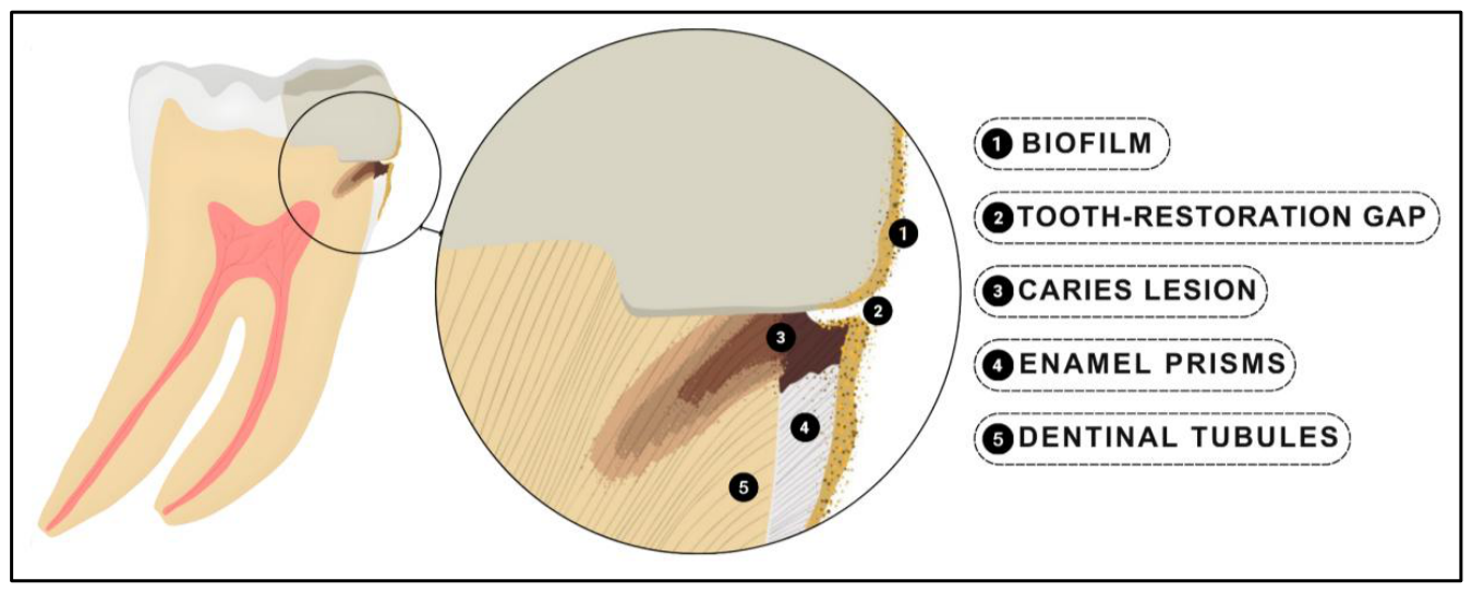
Figure 2.
Flowchart of search results and selection process.
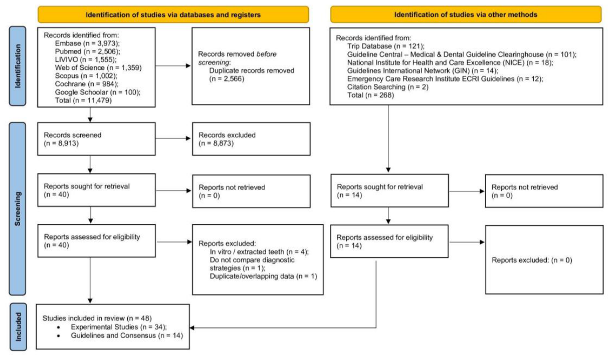
Figure 3.
Overview of the characteristics of the 34 included studies in this scoping review. RCT = Randomized Controlled Trial; NRCT = Non-Randomized Controlled Trial; GIC = Glass Ionomer Cement.
Figure 3.
Overview of the characteristics of the 34 included studies in this scoping review. RCT = Randomized Controlled Trial; NRCT = Non-Randomized Controlled Trial; GIC = Glass Ionomer Cement.
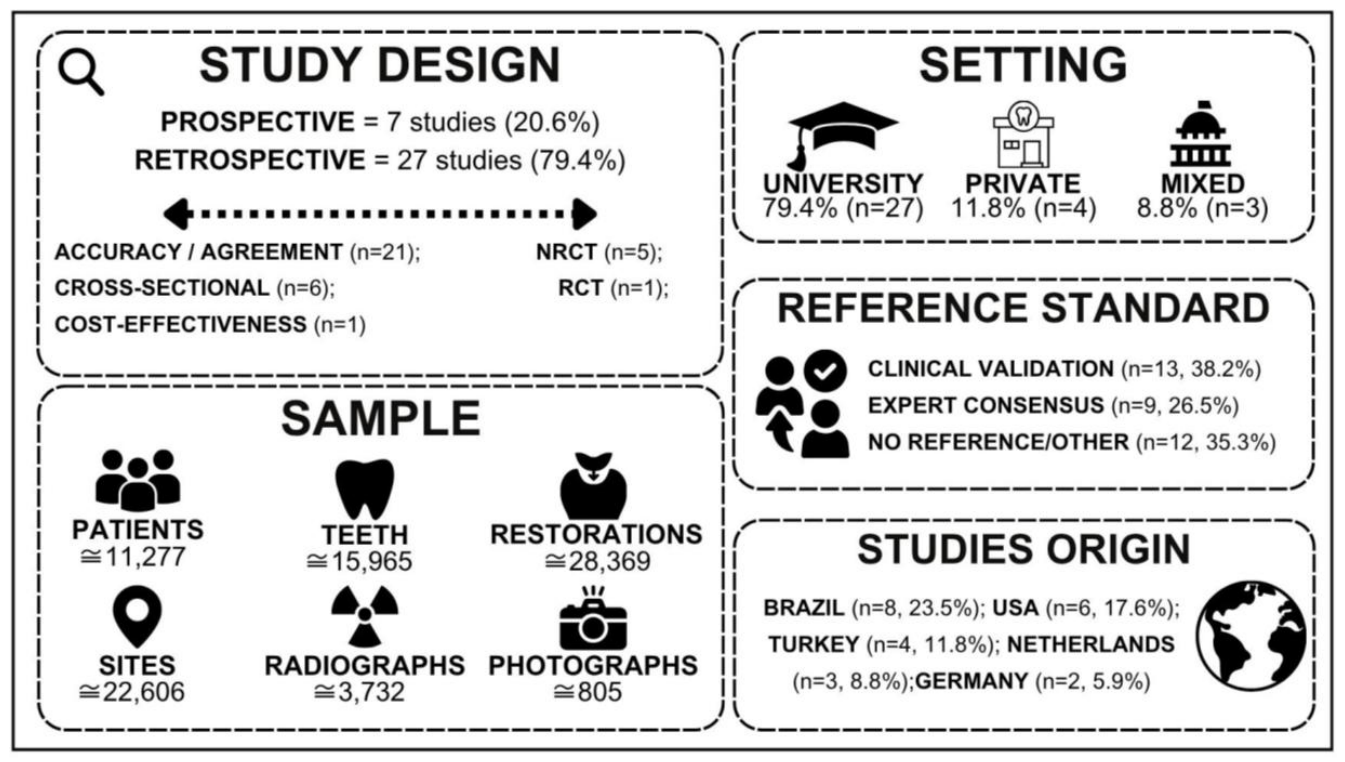
Figure 4.
Integrated framework of detection methods, decision thresholds, and clinical guidance for secondary caries (CARS). Values (n/N) represent the number of studies reporting each method out of the total included clinical studies (N = 34).
Figure 4.
Integrated framework of detection methods, decision thresholds, and clinical guidance for secondary caries (CARS). Values (n/N) represent the number of studies reporting each method out of the total included clinical studies (N = 34).
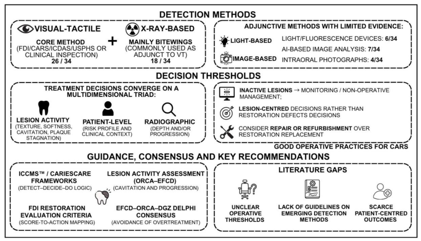
Figure 5.
Decision tree for the visual–tactile and radiographic detection of secondary caries lesions in occlusal and proximal surfaces.
Figure 5.
Decision tree for the visual–tactile and radiographic detection of secondary caries lesions in occlusal and proximal surfaces.
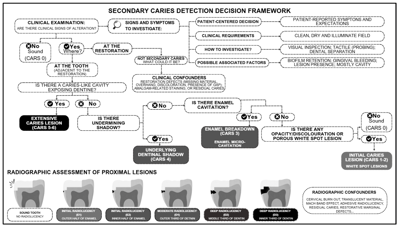
Figure 6.
Decision-making framework for the management of secondary caries lesions.
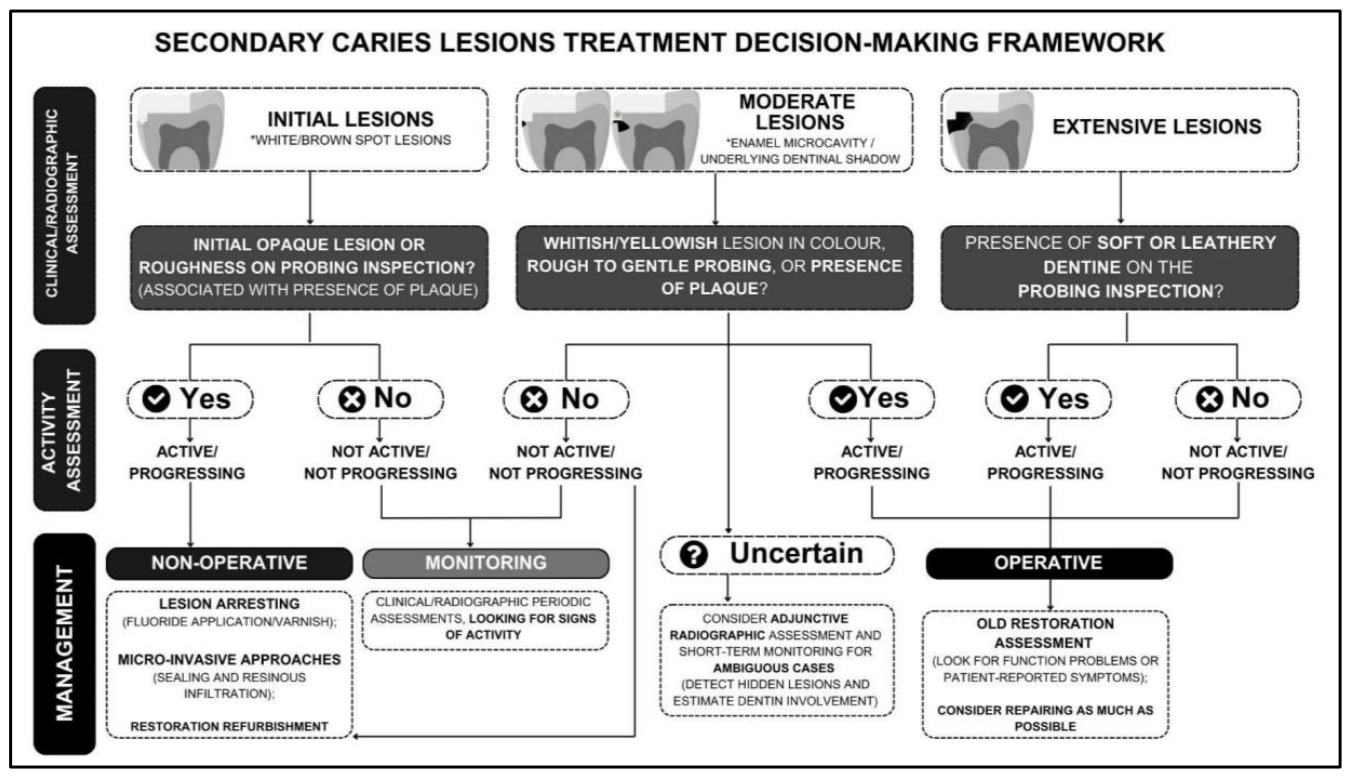

Table 1.
Overview of the identified diagnostic studies in relation to the method used and characteristics of the setup with stepwise included studies.
Table 1.
Overview of the identified diagnostic studies in relation to the method used and characteristics of the setup with stepwise included studies.
| Author, year | Design | Sample (unit + size) | Index groups | Reference standard / Comparator | Caries thresholds and cut-offs |
|---|---|---|---|---|---|
| Bayirli et al., 2026 [57] | Observational | Bitewings (1,197); Surfaces (7,860) | XR (BW); AI (segmentation-based CNN) | Expert consensus |
|
| Shivhare et al., 2026 [58] | Observational | Children (80); Teeth (267); Restored surfaces (475) | VT (FDI; CARS; modified USPHS) | No Reference Standard |
|
| Silva et al., 2026 [59] | Observational | Photographs (25); Specialists (5) | VT (FDI; CARS) | Expert consensus |
|
| Adly et al., 2025 [27] | Observational |
Patients (20); Teeth (30); Margin sites (204, non-proximal) | VT (FDI); LB (DIAGNOdent); LB/DI (VistaCam) | Restoration removal (± follow-up) |
|
| Kosé et al., 2025 [46] | Observational | Patients (11); Interproximal surfaces (352); Restored surfaces (35) | XR (BW; OPG); AI (CranioCatch) | Expert Consensus |
|
| Kwiatek et al., 2025 [47] | Observational | Patients (127); Teeth (3,622) | VT; XR (OPG); AI (Diagnocat) | Expert Consensus |
|
| Sonbul et al., 2025 [55] | Clinical Trial | Patients (60); Class II restorations (60) | VT (CARS); | Restoration removal (± follow-up) |
|
| Chaves et al., 2024 [25] | Observational | Patients (383); Bitewings (425); Restorations (2,550); Surfaces (12,750) | XR (BW); AI (CNN) | Expert Consensus |
|
| Karakus et al., 2024 [45] | Observational | Patients (624); Bitewings (860); Secondary lesions (112, test set) | XR (BW); AI (YOLOv8) | Expert Consensus |
|
| Maydana et al., 2024 [50] | Observational | Cases (212 bitewings/restorations) | XR (BW; BW+CARS) | Expert Consensus |
|
| Nistelrooij et al., 2024 [51] | Observational | Patients (383); Bitewings (413); Restorations (2,612) | XR (BW); AI (CNN) | Expert Consensus |
|
| Romero et al., 2024 [52] | Clinical Trial | Patients (185); Restorations (727) | VT (FDI; CARS) | Restoration removal (± follow-up) |
|
| Waheed et al., 2024 [56] | Observational | Patients (160); Posterior teeth (650) | VT (FDI; CARS) | No Reference Standard |
|
| Hollanders et al., 2023 [44] | Clinical Trial | Patients (5,102); Composite restorations (10,151) | Adhesive systems (MDPB-containing; conventional) | Clinical Validation (Comparator VT) |
|
| Uehara et al., 2023 [20] | Observational | Patients (96); Posterior restorations (305) (171 comp; 134 amalgam; 1-5 surfaces) | VT (FDI; CARS) | Restoration removal (± follow-up) |
|
| Moro et al., 2022 [9] | Observational | Primary tooth restorations (480) (1-3 surfaces) | VT (FDI; CARS) | Restoration removal (± follow-up) |
|
| Signori et al., 2022 [53] | Observational | Patients (185); Posterior restorations (718) | VT (FDI; CARS) | No Reference Standard |
|
| Ding et al., 2021 [24] | Observational | Patients (640); Intraoral photographs (640; 570 train / 70 test) | DI (intraoral photographs); AI (CNN) | Expert Consensus |
|
| Signori et al., 2019 [28] | Observational | Dentists (11 GDPs); Proximal restored surfaces (770) | XR (BW) | Expert consensus |
|
| Zoellner et al., 2017 [19] | Observational | Patients (100); Restorations (820); Interproximal surfaces (2,664); Sound control teeth (512) | VT; XR (bitewings) | No Reference Standard |
|
| Mansour et al., 2016 [49] | Observational | Patients (40); Teeth (932) (426 untreated; 506 restored/sealed) | VT; XR; LB (DIAGNOdent); LB (OCT) | Clinical Validation (VT examination + XR) |
|
| Lino et al., 2015 [48] | Observational | Patients (18); Restored posterior teeth (85) | VT (ICDAS + activity); XR (BW) | Radiographic reference |
|
| Schwendicke et al., 2015 [7] | Cost-effectiveness model | Simulated restorations (1 occluso-proximal molar per mouth) | VT; XR (BW); LB (Laser fluorescence) | No Reference Standard | NA |
| Hamishaki et al., 2014 [22] | Observational | Posterior teeth (60, amalgam restorations) | VT (Ekstrand); LB (DIAGNOdent) | No Reference Standard (Comparator: VT + XR) |
|
| Moncada et al., 2014 [23] | Observational | Patients (36); Restorations (89 - 32 comp; 57 amalgam); Images (356) | VT (USPHS/Ryge); DI (intraoral photographs) | Clinical Validation (VT examination) |
|
| Bamzahim et al., 2005 [36] | Observational | Patients (21); Teeth (51 posterior); Marginal sites (51, amalgam) | VT; XR (BW); LB (DIAGNOdent) | Restoration removal |
|
| Ermis et al., 2004 [39] | Observational | Students (167); Posterior teeth (112) | VT; XR (BW) | No Reference Standard (Comparator: VT) |
|
| Haveman et al., 2003 [42] | Clinical Trial | Patients (9, xerostomic); Restorations (95) | Restorative materials (GIC; RMGIC; Amalgam) | Clinical Validation (Comparator VT) |
|
| Choksi et al., 1994 [37] | Observational | Patients (297); Teeth (1,782 maxillary); Approximal surfaces (3,564) | VT; XR (BW, periapical); LB (FOTI) | No Reference Standard (Comparator: VT + XR) |
|
| Foster et al., 1994 [40] | Observational | Restorations (80) | VT | Restoration removal |
|
| Donly et al., 1999 [38] | Clinical Trial | Children (40); Primary molars (80); Proximal lesions monitored (44 subset) | Restorative materials (RMGIC; Amalgam) | No Reference Standard (Comparator: VT + XR) |
|
| Skartveit et al., 1994 [54] | Clinical Trial | Children (415); Molars (704; 352 pairs) Occlusal amalgam restorations | Restorative materials (fluoride-containing vs conventional amalgam) | Clinical Validation (VT examination + XR) |
|
| Hewlett et al., 1993 [43] | Observational | Patients (490); Restored teeth (6,285); Defective margins (822) | VT; XR (BW; periapical) | Radiographic reference |
|
| Gratt et al., 1988 [41] | Observational | Posterior teeth (200) | VT; XR (BW, periapical) | Restoration removal |
|
| Abbreviations: VT: visual/tactile examination; XR: radiographic examination; BW: bitewing radiographs; OPG: panoramic radiographs; LB: light-based method; FOTI: fiber-optic transillumination; OCT: optical coherence tomography; AI: artificial intelligence; CNN: convolutional neural network; YOLO: You Only Look Once object detection algorithm; IoU: Intersection over Union; DI: digital/intraoral photography; FDI: Fédération Dentaire Internationale evaluation criteria; USPHS: United States Public Health Service criteria; ICDAS: International Caries Detection and Assessment System; CARS: Caries Associated with Restorations or Sealants; ADA: American Dental Association radiographic caries classification; CDA: Council on Dental Affairs (ADA-based clinical evaluation criteria); GDP: general dental practitioner. NR indicates not reported. | |||||
Table 2.
Overview of the identified clinical practice guidelines and consensus statements regarding recommendations on secondary caries detection and management.
Table 2.
Overview of the identified clinical practice guidelines and consensus statements regarding recommendations on secondary caries detection and management.
| Guideline / consensus | Issuing body / Year | Target population | Detection approach (tree entry points) | Decision criteria / thresholds | Management direction |
|---|---|---|---|---|---|
| Clinical recommendations for caries diagnosis: lesion activity and progression assessment | ORCA / EFCD, 2024 [10] | Clinicians and researchers | Visual–tactile assessment is recommended. Tactile examination particularly useful at less accessible cervical restoration margins. Repeated clinical and radiographic assessments may help identify progression. | CARS activity should be assessed similarly to primary lesions, considering plaque, roughness, cleansability, saliva, and fluoride exposure. A marginal gap alone does not indicate active lesion. | Activity-based management: active lesions require non-operative and/or operative care according to severity, whereas inactive lesions are generally monitored. |
| Fluoride Therapy (Best Practices) | AAPD, 2023 [63] | Pediatric and special-care populations | No CARS-specific detection procedure; contributes after lesion identification through risk-based assessment. | Fluoride use is based on caries risk and lesion status; SDF is recommended for cavitated lesions generally. | Non-operative arresting branch through fluoride varnish, topical fluorides, and SDF. |
| Revised FDI criteria for evaluating restorations | FDI, 2023 [13] | General dental practice | Examination after cleaning and drying, with careful probing. The lesion must be directly associated with the restoration margin; staining without demineralization should not be classified as CARS. Radiographs may complement clinically indicated examinations. | Five severity scores distinguish no CARS, initial, established non-cavitated lesions or microcavitation, localized dentinal cavities suitable for repair, and extensive cavities unsuitable for repair. | 5R pathway: review/monitor → refurbish → reseal → repair → replace, according to lesion extent, restoration condition, and caries activity. |
| Pediatric Restorative Dentistry (Best Practice) | AAPD, 2023 [58] | Children and adolescents | Visual–tactile assessment combined with radiographic monitoring to detect cavitation or progression. Marginal staining should not be mistaken for CARS. | Restorative thresholds based on cavitation, shadowing, radiographic progression over time, and patient-level risk. | Risk-based surveillance with preventive care for non-progressing lesions and operative care when clinical or radiographic criteria are met. |
| From ICDAS to CariesCare International | ICCMS / CariesCare International groups, 2021 [14] | General dental practice | Conceptual presentation of the CariesCare 4D cycle. Detection framed within integrated visual–tactile and radiographic caries assessment systems. | Reinforces integration of severity, activity, risk, and shared decision-making within a broader framework. | Detect–Decide–Do and Determine–Detect–Decide–Do organization and the move toward preventive and minimally invasive care. |
| Delphi consensus on proximal and secondary caries | EFCD / ORCA / DGZ, 2020 [15] | Adults | Visual-tactile examination combined with bitewings under clean, dry and illuminate conditions. Detection should reflect individual risk and suspicious lesions should be monitored. | Intervention thresholds linked primarily to cavitation and dentin involvement; secondary caries thresholds considered context-dependent. | Review, refurbishment, resealing, or repair should be preferred over complete replacement whenever possible. |
| Secondary caries: what is it, and how can it be controlled, detected, and managed? | ORCA / EFCD [8] | General dental practice | Critical appraisal of available detection methods. Emphasizes confounders, avoidance of false positives, and possible sequential use of sensitive and specific methods. | Highlights trade-off between sensitivity and overtreatment; stresses role of clinical confounders (e.g., marginal defects). | Repair preferred over replacement; decisions guided by lesion severity and patient/operator risk factors. |
| Policy on Medically-Necessary Care | AAPD, 2019 [62] | Children and adolescents | Supports the patient-centered entry point through consideration of risk, symptoms, function, individual circumstances, and access to care. | Treatment needs are based on disease burden, symptoms, risk, feasibility, and consequences of delaying care. | Preventive care and durable restorative treatment when necessary, including periodic evaluation and maintenance of sealants. |
| Terminology of dental caries and caries management (Consensus report) | ORCA / IADR [66] | Clinicians and researchers | Not a diagnostic guide; provides standardized conceptual definitions. | Standardizes the concepts of detection, severity, activity, progression, and monitoring. | Terminology for the non-operative, operative, and monitoring branches. |
| CariesCare practice guide: consensus on evidence into practice | CariesCare International, 2019 [65] | General dental practice | Visual examination of clean teeth and radiographs when indicated. CARS uses the same stages as primary caries, followed by activity assessment; restoration status is evaluated separately. | Severity, activity, radiographic depth, risk, and patient priorities guide decisions. Moderate lesions represent the principal area of uncertainty. | Determine–Detect–Decide–Do cycle, prioritizing non-operative control and monitoring, with adaptation, repair, or replacement according to restoration conditions. |
| Guideline on Restorative Dentistry | AAPD, 2016 [60] | Children and adolescents | Visual–tactile assessment and radiographic monitoring to identify cavitation, shadowing, or lesion progression. | Decision to restore based on cavitation, enamel shadowing, or radiographic progression over time. | Risk-based management with surveillance and preventive care; restorative intervention when criteria are met, favoring minimally invasive strategies |
| The evidence base for professional and self-care prevention | Prevention in Practice meeting proceedings, 2015 [67] | General dental practice | Detection not focused on restorations. Early identification of initial/non-cavitated lesions within routine clinical examination. | Emphasizes lesion stage and caries risk rather than restoration-adjacent criteria. | Preventive and monitoring branches through fluoride, SDF, and sealing of suitable early lesions. |
| ICCMS™ Guide for Practitioners and Educators | ICDAS Foundation, 2014 [64] | Practitioners and educators | Visual–tactile examination of clean surfaces, complemented by radiographs when indicated. CARS is classified using the same scores as primary caries. | Decision-making based on lesion severity and activity status rather than isolated findings. | Non-operative care for initial active lesions and conservative operative care. Defective or carious margins should be sealed or repaired whenever possible. |
| FDI criteria for evaluation of restorations | FDI, 2010 [12] | General dental practice | Standardized clinical evaluation of restorations and assesses marginal staining separately from CARS. Radiographs may provide complementary information. | Five scores distinguish absence of caries, localized or extensive demineralization, localized cavitation suitable for repair, and extensive disease requiring replacement. | Explicit ladder linking scores to actions: monitor → prevention or refurbishment → repair → replacement. |
Abbreviations: AAPD: American Academy of Pediatric Dentistry; CARS: caries adjacent to restorations or sealants; DGZ: German Society of Conservative Dentistry; EFCD: European Federation of Conservative Dentistry; FDI: World Dental Federation; IADR: International Association for Dental Research; ICDAS: International Caries Detection and Assessment System; ICCMS: International Caries Classification and Management System; ORCA: European Organization for Caries Research; QLF: quantitative light-induced fluorescence; SDF: silver diamine fluoride. Note: ICCMS™, CariesCare International, and From ICDAS to CariesCare International represent successive and complementary developments of the same caries-management framework. The AAPD documents similarly provide complementary guidance on pediatric restorative care, medically necessary care, and fluoride therapy.
Disclaimer/Publisher’s Note: The statements, opinions and data contained in all publications are solely those of the individual author(s) and contributor(s) and not of MDPI and/or the editor(s). MDPI and/or the editor(s) disclaim responsibility for any injury to people or property resulting from any ideas, methods, instructions or products referred to in the content. |
© 2026 by the authors. Licensee MDPI, Basel, Switzerland. This article is an open access article distributed under the terms and conditions of the Creative Commons Attribution (CC BY) license (http://creativecommons.org/licenses/by/4.0/).
Copyright: This open access article is published under a Creative Commons CC BY 4.0 license, which permit the free download, distribution, and reuse, provided that the author and preprint are cited in any reuse.