
Submitted:
26 September 2026
Posted:
28 September 2026
You are already at the latest version
Abstract
Interferon (IFN)-induced protein 6 (IFI6) is an important member of the IFN-stimulated gene (ISG) family, mainly enriched in the inner mitochondrial membrane and endoplasmic reticulum (ER). It plays significant roles in anti-apoptosis, immune regulation, maintenance of mitochondrial homeostasis, antiviral defense, and promotion of tumor progression. At the molecular mechanism level, IFI6 inhibits mitochondrial membrane depolarization, cytochrome c release, and caspase activation. It also regulates the B-cell lymphoma 2 (Bcl-2)/Bcl-2 interacting mediator of cell death (Bim) expression balance, activates the janus kinase (JAK)/signal transducer and activator of transcription (STAT) and phosphatidylinositol 3-kinase-protein kinase B (PI3K-AKT) pathways, and inhibits the degradation of ferroptosis-related proteins solute carrier family 7 member 11 (SLC7A11) and glutathione peroxidase 4 (GPX4), thereby enhancing cell survival and proliferation through multiple pathways. In terms of immune regulation, IFI6 also affects immune cells (such as T/B cells, etc.) and the tumor microenvironment. IFI6 regulates mitochondrial reactive oxygen species (mtROS) and mitochondrial calcium homeostasis, influencing respiratory chain assembly and metabolism. In terms of virus regulation, IFI6 may inhibit the replication of multiple viruses (such as hepatitis B virus (HBV), hepatitis C virus (HCV), SARS-CoV-2, etc.) by inhibiting flavivirus-induced ER invagination, interfering with retinoic acid-inducible gene I (RIG-I)-like receptor signaling pathways, and inhibiting epidermal growth factor receptor (EGFR) kinase activity. In addition, IFI6 is highly expressed in autoimmune diseases and malignant tumors and is associated with proliferation, metastasis, and poor prognosis. In this review, we aim to highlight IFI6 as a potential key target in disease diagnosis and treatment.
Keywords:
IFI6
; interferon-stimulated gene (ISG)
; tumor progression and immune regulation
Introduction
The interferon (IFN) system is a core component of the body’s innate immune defense and plays an irreplaceable role in resisting viral infections and regulating anti-tumor immunity. When pathogens invade, pattern recognition receptors (PRRs) on the surface of host cells recognize pathogen-associated molecular patterns (PAMPs), activating downstream signaling cascades and inducing the production and secretion of type I IFNs. After binding to their cell surface receptors, type I IFNs initiate the transcription of IFN-stimulated genes (ISGs). The protein products encoded by these ISGs directly or indirectly perform various biological functions such as antiviral defense, immune regulation, anti-proliferation, and pro-apoptosis [1]. As one of the ISGs, IFN-induced protein 6 (IFI6) has achieved significant progress in functional research, and the complexity of its biological functions is gradually being revealed. However, its precise mechanisms of action under different pathological conditions, its interactive regulatory networks with other signaling pathways, and the feasibility and safety of targeting it as a therapeutic target still require in-depth investigation. Although relevant basic research exists, the clinical value of IFI6 in autoimmune diseases, tumors, and viral infections remains to be validated. This study systematically reviews the structure, biological functions, and research progress of IFI6 in various human diseases, providing a theoretical reference for subsequent basic mechanistic research and clinical translation.
Structure of IFI6
IFI6, also known as IFI6-16 and G1P3 (ISG 6-16), belongs to the FAM14 family and is one of the ISGs. Located on the short arm of chromosome 1 at 1p35, IFI6 encodes a 130-amino-acid mitochondria-related protein that is induced and upregulated by type I IFN, including IFN-α and IFN-β. It plays important roles in various biological functions such as regulating apoptosis and immunity. In recent years, multiple studies have gradually revealed its important roles in various diseases, such as autoimmune diseases, tumors, and viral infectious diseases [2].
The protein structure of IFI6 consists sequentially of an N-terminal signal peptide, followed by hydrophilic region 1 (HL-1), transmembrane region 1 (TM-1), linker region (L), transmembrane region 2 (TM-2), and hydrophilic region 2 (HL-2). This dual transmembrane-dual hydrophilic structural pattern is highly conserved in the FAM14 family [3]. Studies using the surface probability (Emini) method to predict the probability of a region being located on the protein surface revealed that the signal peptide and HL-1 and HL-2 regions are more easily exposed to solvent, suggesting that these regions may be key sites for protein-protein interactions. The polar HL-1 may form an extended random coil, while HL-2 may form an amphipathic helix. Since two transmembrane domains separate HL-1 and HL-2, they may not form well-folded domains but instead protrude as unfolded rope-like structures for binding by other proteins [3]. Early studies found that IFI6 is mainly localized to the inner mitochondrial membrane and co-localizes with cytochrome c. In recent years, IFI6 has been shown to be essentially an endoplasmic reticulum (ER)-resident integral membrane protein. IFI6 targets the secretory pathway through its N-terminal signal peptide and ultimately anchors to the ER membrane, with its transmembrane region fixing the protein within the membrane and its hydrophilic regions exposed to the cytoplasm or ER lumen [4]. This seemingly contradictory localization result may reflect the dynamic subcellular distribution of IFI6 under different cell types, stimulation conditions, or time points, and this dynamic subcellular localization may be closely related to its multiple biological functions.
Biological Functions of IFI6
Although the biological functions of IFI6 are currently limitedly understood, IFI6, as a multifunctional IFN-stimulated gene, primarily exerts its biological functions through three interrelated dimensions: anti-apoptosis, immune regulation, and mitochondrial function regulation (Figure 1). In terms of anti-apoptosis, IFI6 promotes cell survival through multiple mechanisms including inhibiting mitochondrial membrane depolarization to block cytochrome c release and caspase activation, activating the janus kinase (JAK)/signal transducer and activator of transcription (STAT) and phosphatidylinositol 3-kinase-protein kinase B (PI3K-AKT) pathways, and inhibiting ferroptosis. In terms of immune regulation, IFI6 acts as a negative feedback factor of the retinoic acid-inducible gene I (RIG-I) signaling pathway to inhibit excessive IFN responses, and is associated with the functional regulation of Tregs, CD4+ T cells, and B cells. In terms of mitochondrial function, IFI6 affects respiratory chain complex assembly and cell metabolism by regulating mitochondrial reactive oxygen species (mtROS) production and mitochondrial calcium homeostasis. Through these functions, IFI6 plays important pathological regulatory roles in tumors, autoimmune diseases, and viral infections [5,6].
Anti-Apoptosis
Inhibition of the mitochondrial apoptotic pathway. IFI6 is mainly enriched in the inner mitochondrial membrane and co-localizes with cytochrome c. Under apoptotic signal stimulation, mitochondrial outer membrane permeability increases, cytochrome c is released into the cytoplasm, binds with apoptotic protease activating factor-1 (Apaf-1), causing it to form polymers, and promotes caspase-9 binding to form apoptosomes, thereby activating caspase-3 and leading to cell apoptosis. IFI6 maintains the integrity of the mitochondrial outer membrane by inhibiting mitochondrial membrane depolarization, thereby blocking cytochrome c release, inhibiting caspase-3 activity, or delaying apoptotic signals initiated by the tumor necrosis factor (TNF)-related apoptosis-inducing ligand (TRAIL)/caspase-8 pathway, thus inhibiting cell apoptosis [7]. Studies have found that IFI6 can regulate the balance between Bcl-2 and Bim expression, stabilize mitochondria, and thereby resist cell apoptosis. Meanwhile, studies have revealed that mitochondrial Ca²⁺ overload may induce tumor cell apoptosis through IFI6 downregulation [8]. IFI6 knockdown increases reactive oxygen species accumulation, thereby inhibiting cancer cell proliferation and inducing apoptosis; similarly, high IFI6 expression can inhibit apoptosis [2]. In esophageal squamous cell carcinoma (ESCC), IFI6 silencing significantly promoted mtROS generation, and the reactive oxygen species inhibitor N-acetylcysteine reversed IFI6 silencing-induced apoptosis, strongly demonstrating the role of IFI6 silencing in reducing cell survival induced by reactive oxygen species [9]. This section focuses on how IFI6 inhibits mitochondria as an apoptotic signaling hub from transmitting death instructions to cells, while the physiological function regulation of IFI6 on mitochondria as energy metabolic organelles will be detailed in the mitochondrial function regulation section.
Inhibition of caspase activation. IFI6 can directly inhibit caspase activation. In TRAIL-induced apoptosis models, IFI6 overexpression significantly reduced caspase-8 and caspase-3 activation cleavage. In dengue virus-infected vascular endothelial cells, IFI6 overexpression inhibited caspase-3 and caspase-9 activity. Notably, the inhibitory effect of IFI6 on caspases is relatively limited, while its inhibitory effect on caspase-9 is more significant, revealing that IFI6 mainly targets the mitochondria-mediated intrinsic apoptotic pathway rather than the death receptor-mediated extrinsic apoptotic pathway [7,10].
Activation of JAK/STAT and PI3K-AKT signaling pathways. IFI6 can activate the JAK/STAT signaling pathway, promoting STAT1 and STAT3 phosphorylation. Phosphorylated STAT3 translocates to the nucleus and upregulates the transcription of anti-apoptotic genes (such as Bcl-2, Bcl-xL, etc.). In gastric cancer cells, IFI6 upregulates Bcl-2 expression through the JAK/STAT3 pathway, inhibiting mitochondria-mediated apoptosis. Studies have shown that both IFI6 knockdown and overexpression alter estrogen synthesis, cell proliferation, and apoptosis in granulosa cells. IFI6 can activate the PI3K-AKT signaling pathway, upregulate activator protein 1, promote estrogen synthesis and cell proliferation, and simultaneously inhibit apoptosis [11]. In addition, IFI6 is positively correlated with the nuclear factor (NF-κB) pathway, and NF-κB activation slows the cell cycle, induces anti-apoptotic proteins and cell stemness, and enhances tumorigenicity and chemotherapy resistance [6].
Inhibition of ferroptosis. Ferroptosis is an iron-dependent lipid peroxidation-driven form of programmed cell death. IFI6 overexpression leads to elevated levels of ferroptosis-inhibitory proteins solute carrier family 7 member 11 (SLC7A11) and glutathione peroxidase 4 (GPX4), thereby enhancing cellular antioxidant capacity, reducing lipid peroxidation levels, and inhibiting ferroptosis [12,13].
Regulation of ER stress. The localization of IFI6 on the ER membrane suggests that it may participate in the regulation of ER stress. In esophageal squamous cell carcinoma, IFI6 knockdown leads to elevated expression of ER stress markers glucose-regulated protein 78, C/EBP homologous protein, and X-box binding protein 1 (XBP-1), spliced form XBP-1s, accompanied by a significant increase in intracellular calcium levels. ER calcium release to mitochondria leads to mitochondrial calcium overload, which in turn induces the opening of the mitochondrial permeability transition pore and cell apoptosis. This mechanism reveals the important role of IFI6 in ER-mitochondrial calcium signaling crosstalk and explains why IFI6 knockdown simultaneously activates both ER stress and mitochondrial apoptosis pro-apoptotic pathways [9].
High IFI6 expression levels upregulate gene sets related to cell proliferation and negatively regulate apoptosis [9]. Studies have shown that IFI6 knockdown reduces cell survival and colony formation while inducing apoptosis through upregulation of the p53/p21 signaling pathway [14]. IFI6 can regulate the radiation sensitivity of human skin cells through complex mechanisms. IFI6 is highly expressed in irradiated human and monkey skin tissues and can provide radiation protection for skin cells against radiation-induced apoptosis and oxidative stress damage. This process appears to be mediated by IFI6 participating in the regulation of heat shock transcription factor 1-mediated processes to enhance skin cell radiation protection. Similarly, IFI6 confers radioresistance in cancer cells [15]. IFI6 downregulation leads to spontaneous apoptosis of keratinocytes, revealing the anti-apoptotic properties of IFI6 in non-malignant cells [16].
Regulation of the Immune System
IFI6 can regulate the activation of RIG-I. RIG-I is an important sensor for viral infection and can recognize 5′-triphosphorylated single-stranded RNA and short double-stranded RNA. After ligand recognition, RIG-I interacts with mitochondrial antiviral signaling protein (MAVS) on mitochondria through its N-terminal caspase recruitment domain, initiating downstream signaling cascades and inducing the production of IFN and inflammatory proteins. In addition, the RIG-I-like receptor signaling pathway responsible for detecting viral infections and activating immune responses is highly correlated with IFI6[17]. IFI6 overexpression reduces the ubiquitination level of RIG-I, thereby inhibiting RIG-I activation and downstream IFN-β production. Conversely, IFI6 knockdown or knockout enhances RIG-I ubiquitination, IFN-β transcription, and ISG expression. Therefore, IFI6 acts as a negative feedback regulator of the RIG-I signaling pathway [18].
IFI6 inhibition may reduce immunosuppressive signals and reshape macrophage or T cell populations by affecting mitochondrial reactive enzymes and IFN signaling pathways [19]. Studies have shown that IFI6 expression is positively correlated with regulatory T cells (Tregs), a population critical for maintaining immune tolerance and dampening anti-tumor immunity. The abundant infiltration of Tregs in tumor tissues is frequently associated with poor prognosis in cancer patients, which may partly explain why elevated IFI6 expression correlates with unfavorable clinical outcomes. IFI6 has a significant correlation with CD4+ T cells, which play important functions in coordinating the immune system. In addition, IFI6 also shows significant correlation with CD8A and CD8B cells, suggesting that it may affect the cytotoxic function of CD8+ T cells. Moreover, IFI6 expression is negatively correlated with naive B cells and plasma cells, implying that high IFI6 levels may impair the recruitment and activity of B cell subsets in the tumor microenvironment, thereby facilitating tumor progression and contributing to an immunosuppressive milieu [19]. High IFI6 expression can also affect the function of T cell receptors (TCR) and B cell receptors (BCR). IFI6 may regulate the activation threshold of lymphocytes by inhibiting the strength of TCR and BCR signaling pathways, thereby affecting the body’s immune response and tumor cell infiltration and immune surveillance [17,19].
Regulation of Mitochondrial Function
As detailed above, IFI6 exerts anti-apoptotic effects by inhibiting the mitochondrial apoptotic pathway. This section discusses the physiological function regulation of IFI6 on mitochondria as the cellular energy metabolism center. Studies have demonstrated that IFI6 downregulation leads to mitochondrial Ca2+ uniporter-mediated mitochondrial calcium overload, inhibiting mitochondrial supercomplex assembly and respiratory phosphorylation efficiency, and increasing mtROS generation [20]. IFI6 can both inhibit and promote oxygen species (ROS) production, with specific effects depending on cell type and experimental conditions. In addition, IFI6 regulates the assembly and function of mitochondrial respiratory chain complexes. In IFI6-knockdown cells, the activities of mitochondrial respiratory chain complexes I, III, and IV are reduced, mitochondrial oxygen consumption rate is decreased, and cells rely more on glycolysis for ATP production [21].
The Multifaceted Roles of IFI6 in Diseases and Infections
In the previous section, the physiological functions of IFI6 were described. At the same time, abnormal expression of IFI6 is associated with various diseases such as neurodegeneration, autoimmunity, tumors, and viral infections, and is related to disease activity, severity, drug resistance, and outcomes. The following section will discuss the possible roles or confirmed clinical or relevant evidence that IFI6 may play or has been proven in various types of diseases and viral infections (Table 1).
Neurological Diseases
Aicardi goutières syndrome (AGS) is a group of rare diseases predominantly involving the nervous system and skin, caused by mutations in multiple genes (such as three-prime repair exonuclease 1, adenosine deaminase acting on RNA 1, IFN induced with helicase C domain 1), leading to the accumulation of abnormal DNA and RNA metabolites, which in turn activates the type I IFN pathway and causes persistent inflammatory responses. The IFN signaling gene expression score has potential as a biomarker in AGS, and a 4-gene approach was the best-performing model, which includes IFI6[22].
Major depressive disorder (MDD) is a common psychiatric disorder characterized by high prevalence, high recurrence rate, and limited response to existing treatments. In a study of hippocampal dentate gyrus tissue biopsy samples from MDD patients, significantly downregulated IFI6 expression was detected, accompanied by changes in the expression of other inflammation-related genes. IFI6 downregulation may be related to the weakening of its anti-apoptotic function. In the hippocampus of MDD patients, increased apoptosis may lead to neuronal loss and shrinkage of hippocampal volume, the latter being one of the most typical neuroimaging changes in MDD, indicating that IFI6 may play a key role in MDD [23].
Autoimmune Diseases
The hallmark of autoimmune diseases is immune system dysregulation, leading to immune responses against self-antigens, causing tissue damage and organ dysfunction. As an important downstream effector molecule of the type I IFN pathway, IFI6 shows differential expression in various autoimmune diseases and is associated with disease activity and severity.
Systemic lupus erythematosus (SLE) is a common autoimmune disease characterized by multi-organ involvement, autoantibody production, and persistent activation of the type I IFN pathway [64]. IFI6 is one of the differentially expressed genes between SLE and healthy samples, and in CD14⁺ monocytes derived from SLE patients, IFI6 expression levels correlate with lupus activity severity, suggesting that IFI6 may be a potential biomarker for detecting SLE disease activity [25,65]. Notably, a study showed that the combination of IFI6 with other genes (such as interferon induced transmembrane protein 1 (IFITM1), interferon induced protein 44 like (IFI44L), MX dynamin-like GTPase 1 (MX1), and interferon alpha inducible protein 27 (IFI27)) exhibited excellent performance in distinguishing SLE from healthy controls and other autoimmune diseases. The IFN score is an important indicator for evaluating SLE disease activity and treatment response, and IFI6 is one of the core components of this score [26,66]. Severe SLE patients can develop lupus nephritis (LN), and IFI6 is one of the 23 hub genes of LN, potentially becoming a biomarker and new therapeutic target for LN diagnosis and treatment [27].
Rheumatoid arthritis (RA) is a chronic autoimmune disease characterized by symmetric polyarticular synovitis, with arthritis and joint damage as its features. The type I IFN pathway also plays an important role in the pathogenesis of RA. IFI6 shows differential expression in RA patients, and untreated RA patients with high IFN scores at diagnosis are associated with poor clinical outcomes, suggesting that it may serve as a biomarker for RA [67]. IFI6 expression in RA synovial tissue has also been reported. In RA synovial fibroblasts, IFI6 expression levels are elevated compared to osteoarthritis patients and correlate with the degree of synovial inflammation. IFI6 may participate in the pathological process of RA joint destruction by inhibiting synovial fibroblast apoptosis, promoting synovial hyperplasia and pannus formation [68].
Multiple sclerosis is an autoimmune disease mainly characterized by inflammatory demyelinating lesions in the white matter of the central nervous system. IFI6 may become a key detection marker for multiple sclerosis. It is highly expressed in platelets of multiple sclerosis patients, and in a gene expression comparison study of 8 pairs of monozygotic twins, the twin with multiple sclerosis showed significant upregulation of IFI6 compared to their healthy sibling, suggesting that environmental and epigenetic factors beyond genetic background play important roles in the regulation of IFI6 expression [3,28].
Psoriasis is a common chronic inflammatory skin disease characterized by excessive keratinocyte proliferation and abnormal differentiation, accompanied by significant immune cell infiltration and inflammatory factor release. In psoriasis patients, IFI6 is upregulated approximately 9-fold in non-lesional epidermis and up to 400-fold in lesional epidermis, and its expression level is positively correlated with the degree of inflammation. It may participate in the development of psoriasis by inhibiting keratinocyte apoptosis. In normal epidermis, keratinocyte differentiation is accompanied by programmed cell death, which is an important mechanism for maintaining epidermal homeostasis, and IFI6 overexpression may disrupt this balance, leading to abnormal survival and excessive proliferation of keratinocytes. Mechanistically, IFI6 inhibits mitochondrial membrane depolarization and cytochrome c release, blocking the caspase activation pathway [16,30]. It is worth noting that IFI6 has the highest expression in proliferating keratinocytes, and its expression gradually decreases as the cells differentiate. This is consistent with the pathological feature of excessive proliferation of keratinocytes and blocked differentiation in the epidermis of psoriasis. In psoriatic lesions, the abnormal activation of the type I IFN signaling pathway is one of the key events in the initiation of the disease, and IFI6 is an important effector molecule downstream of this pathway. A meta-analysis of the common tissue transcriptome characteristics of psoriasis and ulcerative colitis showed that IFI6, along with chemokines such as C-X-C motif chemokine ligand 9 (CXCL9), CXCL10, and CXCL11, jointly drives the abnormal activation of the IFN signaling pathway. These chemokines can recruit Th1 cells to the inflammatory site, amplifying the local inflammatory response, suggesting that IFI6 may drive the disease process in psoriasis by regulating T cell activity [16,26].
Polymyositis (PM) and dermatomyositis (DM) are common autoimmune diseases characterized by skeletal muscle damage and weakness. The type I IFN pathway plays an important role in the pathogenesis of DM and PM. In a bioinformatics analysis, IFI6 was identified as one of the five hub genes of PM/DM [29]. IFI6 expression is elevated in various immune cells of patients with melanoma differentiation-associated gene 5+ dermatomyositis (MDA5+ DM). Single-cell sequencing results showed that IFI6 is upregulated in CD14+ monocytes, CD16+ monocytes, dendritic cells, and multiple T cell subsets of patients with MDA5+ DM complicated by rapidly progressive interstitial lung disease, and is associated with disease progression [31,34].
Primary sjögren’s syndrome(pSS)is a chronic autoimmune disease characterized by lymphocytic infiltration of exocrine glands such as salivary and lacrimal glands, presenting with symptoms of dry mouth and dry eyes. The type I IFN pathway plays a central role in the pathogenesis of pSS. IFI6 is overexpressed in pSS patients and is one of the six specific differentially expressed genes in pSS. Notably, IFI6 expression levels are positively correlated with serum IgG levels, anti-SSA/SSB antibody titers, and disease activity in pSS patients. IFI6 is also associated with the infiltration levels of various immune cells in the peripheral blood of pSS patients, especially closely related to the activation status of dendritic cells and B cells [32].
In systemic sclerosis (SSc), IFI6 is highly expressed in non-microvascular endothelial cells of patients with SSc-associated interstitial lung disease and participates in the progression of SSc vasculopathy [69]. IFI6 expression is upregulated in Antiphospholipid Syndrome (APS) patients and is associated with early disease onset and poor outcomes [34]. In type 1 diabetes, IFI6 expression is elevated in insulitis and may participate in immune damage of pancreatic β cells [35]. In the screening of comorbidity biomarkers for psoriasis and inflammatory bowel disease (IBD), IFI6 was also identified as one of the 23 biomarkers of IBD [70].
Cancers
Tumor Microenvironment
IFI6 is mainly expressed in epithelial tumor cells and is associated with changes in immune cell composition, suggesting that it may play a role in shaping the immune microenvironment [19]. IFI6 expression is positively correlated with Tregs, and massive infiltration of Tregs in the tumor microenvironment is usually associated with poor prognosis in patients. IFI6 expression is negatively correlated with naive B cells and plasma cells, suggesting that high IFI6 expression may be associated with reduced recruitment and activity of B cell subsets in the tumor microenvironment, which is beneficial for tumor immune evasion [19]. In addition, IFI6 participates in the regulation of mtROS, a process that may link IFN signaling, mitochondrial metabolism, and immune regulation in the tumor microenvironment. Therefore, IFI6 may indirectly affect the expression of immunosuppressive cytokines or regulate the composition and activity of immune cells to shape the tumor microenvironment through mtROS regulation [17,19,71].
IFI6 plays a complex role in the tumor microenvironment. It can increase the anti-tumor response through immune stimulatory factors, while on the other hand, IFI6 may inhibit immune suppressive molecules, various pathways, and immune cells, leading to tumor progression [19] . The increase of IFI6 is associated with the induction of type 2/17 helper T cells, resulting in the production of immunosuppressive type 2 and 17-related cytokines, indicating that IFI6 has an inhibitory effect on the tumor microenvironment in esophageal squamous cell carcinoma (ESCC) [37]. Exosomes derived from tumor-associated macrophages (TAMs) can activate IFI6 transcription in TAMs through IRF9, and the exosomes secreted by TAMs deliver IFI6 to breast cancer cells, thereby promoting their proliferation, migration, and invasion, while inhibiting apoptosis and ROS accumulation, and driving tumor progression [72]. In adult B-cell acute lymphoblastic leukemia (B-ALL), bone marrow mesenchymal stem cells as key cells in the tumor microenvironment, the overexpression of IFI6 in mesenchymal stem cells seems to promote the proliferation of B-ALL cells, and this process may be initiated by the stromal cell-derived factor 1/C-X-C motif chemokine receptor 4 (SDF-1/CXCR4) axis of IFI6 activating the extracellular signal-regulated kinase (ERK) signaling pathway, thereby promoting the proliferation of leukemia cells [8]. In lung adenocarcinoma, a study based on single-cell spatial transcriptomics technology indicates that an epithelial cell subpopulation (E3-IFI6) marked by IFI6 and IFITM1 and other IFN response genes is significantly enriched in the high-expression group of molecular programs related to immune therapy response. The spatial distribution analysis shows that E3-IFI6 cells have significant spatial proximity to the Ma-MHC macrophage subpopulation, and they may mediate cell-to-cell interactions through the amyloid precursor protein-CD74 axis, and this signal is significantly enhanced in patients with high potential for immune therapy response. After co-culturing lung adenocarcinoma cells with IFI6-overexpressing cells with M0 macrophages, pro-inflammatory gene expression is significantly upregulated while anti-inflammatory gene expression is downregulated, indicating that IFI6-overexpressing tumor cells can drive macrophages to polarize towards a pro-inflammatory phenotype and maintain anti-tumor immune activity [41].
Tumor Cell Proliferation, Survival, and Migration
IFI6 can promote tumor cell proliferation and survival through its classical anti-apoptotic function. In gastric cancer, IFI6 is mainly enriched in the inner mitochondrial membrane of tumor cells, inhibiting the activity of caspase-3 by depolarization of the mitochondrial membrane and release of cytochrome c, thereby inhibiting apoptosis of cancer cells. Studies have shown that IFI6 is significantly highly expressed in tumor tissues of ESCC patients and in ESCC cell lines in vitro. Knockdown of IFI6 can inhibit the proliferation of ESCC and induce apoptosis by increasing ROS accumulation. Moreover, high expression of IFI6 can inhibit the apoptosis of tumor cells by suppressing the characteristics of IFN-1, activate the redox signal induced by immune endocrine, and then weaken the toxicity of the apoptosis pathway induced by high mtROS, thereby interfering with mtROS-mediated apoptosis [9]. In ESCC, inhibition of IFI6 can enhance the uptake of calcium by mitochondria, accumulate ROS, and induce apoptosis [73] . On the contrary, in tongue squamous cell carcinoma (TSCC), when IFI6 is ectopically expressed, the mitochondrial membrane potential is disrupted, which can inhibit caspase-mediated apoptosis. The high expression of IFI6 can counteract the inhibitory effect of overexpression of activating transcription factor 3 (ATF3) on tumor cell proliferation and migration. At the same time, the expression of IFI6 can counteract the changes in AKT or ERK activity caused by the absence or overexpression of ATF3[42] . In breast cancer cells, silencing the expression of IFI6 can trigger apoptosis in BT-549 cells and inhibit the growth of ER-positive breast cancer cell line MCF-7[19] . Moreover, studies have shown that IFI6 is a key downstream effector molecule of ubiquitin conjugating enzyme E2 T (UBE2T), and UBE2T inhibition leads to the downregulation of IFI6 expression, which in turn induces DNA replication stress, cell cycle arrest, and apoptosis. In breast cancer cells with UBE2T knockdown, supplementing IFI6 can partially restore cell growth [74]. In triple-negative breast cancer, the mitochondrial aspartate/glutamate carrier solute carrier family 25 member 13 enhances complex I-dependent oxidative phosphorylation and limits mtROS, promoting STAT3 activation and nuclear translocation, and nuclear STAT3 directly induces IFI6 transcription, while IFI6 maintains mitochondrial function, limits lipid peroxidation and Fe²⁺ accumulation to confer iron death resistance to tumor cells [75]. Candidate genes with epigenetic upregulation, IFI6, have been confirmed to be upregulated in cutaneous squamous cell carcinoma (cSCC) and are believed to play an important role in promoting cell proliferation, tumor cell invasion and migration [43]. In pancreatic cancer, upregulation of IFI6 can inhibit its apoptosis and promote cell proliferation [17,44] . In human multiple myeloma cell lines, IFI6 can temporarily antagonize TRAIL-induced apoptosis by maintaining mitochondrial integrity, prevent the release of pro-apoptotic factors (such as cytochrome c, Smac/DIABLO, etc.), and inhibit TRAIL-induced caspase cleavage through the death signal complex. IFI6 can also regulate the balance of Bcl-2/Bim expression to stabilize mitochondria and resist apoptosis. Knockdown of IFI6 reduces the survival rate of colorectal cancer cells and induces apoptosis by upregulating p53, p21 and BCL2-associated X [14]. IFI6 is upregulated in melanoma and its subgroup neuroblastoma rat sarcoma (NRAS)-mutated melanoma, and its expression is related to the NRAS mutation status. Knockdown of IFI6 can lead to DNA replication stress through E2F transcription factor 2 regulation of DNA replication, thereby inhibiting melanoma growth through senescence or induction of apoptosis [5].
IFI6 can promote tumor cell migration and invasion by regulating mtROS and actin cytoskeleton and reformation. In breast cancer, IFI6 can regulate the production of mtROS, triggering the action of multiple pathways, which further promote the rearrangement of F-actin cytoskeleton and the formation of migration structures, enhancing the metastasis and invasion of cells [39,71]. Studies using lentiviral barcodes combined with single-cell RNA sequencing technology further revealed that metastatic breast cancer originates from a rare promoting metastasis clone in the primary tumor, characterized by extracellular matrix remodeling and the activation of the dsRNA-IFN1 signaling pathway, and silencing IFI6 can significantly inhibit in vitro migration and in vivo metastasis [76]. In renal cell carcinoma (RCC), STX17 divergent transcript (STX17-DT) promotes tumor cell migration and invasion by upregulating the expression of IFI6[13].
Drug Resistance and Prognosis
IFI6 is closely related to the resistance of various chemotherapeutic drugs and targeted drugs, and is an important molecular in tumor resistance research. At the same time, high expression of IFI6 is significantly associated with poor prognosis in various tumors. IFI6 shows high expression in gastric cancer and esophageal cancer, and is associated with tumor invasiveness and poor prognosis. In esophageal cancer, a high expression level of IFI6 is related to a lower tumor stage and an increased risk of lymph node metastasis, and it may be associated with the TP53 mutation status [17] . IFI6 overexpresses under the influence of the activator of heat shock 90 kDa protein ATPase activity 1 (AHSA1)-heat shock protein 90 alpha family class A member 1(HSP90AA1) complex, enhancing the drug resistance of epidermal growth factor receptor (EGFR)-mutated lung adenocarcinoma cells. This is related to IFI6 stabilizing mitochondrial function, reducing cell apoptosis and promoting the survival of lung adenocarcinoma cells. AHSA1 and HSP90AA1 prolong the half-life of IFI6 by reducing its ubiquitination degradation, and IFI6 promotes cell survival by inhibiting the activation of caspase-3/7 and maintaining mitochondrial membrane potential [40]. At the same time, IFI6 is associated with the drug resistance of various tumors, such as with oxaliplatin resistance in colorectal cancer, docetaxel resistance in prostate cancer, and tamoxifen resistance in breast cancer. The specific mechanisms of drug resistance need further study [12]. High expression of IFI6 is associated with poor prognosis in breast cancer patients with ER-positive, PR-positive, HER2-positive and lymph node-positive subtypes of breast cancer [19]. In hematological tumor research, the abnormally expressed IFI6 in multiple myeloma is an important factor leading to the chemical resistance of myeloma cells [8]. In colorectal cancer, the downregulation of IFI6 has been reported to activate the ROS-induced p38 mitogen-activated protein kinase (MAPK) pathway, affecting its resistance to oxaliplatin [39]. In glioma, IFI6 enhances the resistance of tumor cells to temozolomide by inhibiting ferroptosis. The overexpression of IFI6 leads to an increase in the levels of ferroptosis inhibitor proteins SLC7A11 and GPX4, reducing lipid peroxidation levels and decreasing cell apoptosis, while BCL11 transcription factor A, as the transcriptional inhibitor of IFI6, can promote ferroptosis by downregulating IFI6 and restoring the sensitivity to temozolomide [12]. In glioblastoma, microglia promote the resistance of tumor cells to temozolomide by inducing the expression of IFI6 and other ISGs [46]. Studies have shown that IFI6 is positively correlated with the NF-κB pathway. In ovarian cancer, IFI6 may affect cancer cell proliferation, invasiveness, and drug resistance through the NF-κB pathway [6]. In RCC, STX17-DT regulates the stability of IFI6 mRNA by recruiting and binding hnRNPA1, thereby reducing mtROS accumulation and attenuating ferroptosis, while IFI6 overexpression inhibits STX17-DT knockdown-induced ferroptosis, enhancing RCC resistance to axitinib [13]. IFI6 affects the radiosensitivity of tumors by regulating mtROS levels. In irradiated skin cells, the increase in IFI6 levels provides radiation protection for the cells, but the mechanism needs further clarification [15].
Viral Infection
Flaviviruses, such as yellow fever virus (YFV), west nile virus (WNV), dengue virus (DENV), zika virus (ZIKV), and hepatitis C virus (HCV), induce the formation of characteristic single-membrane invagination structures, namely replication organelles, on the ER membrane after infecting host cells. These structures provide physical scaffolds and shielding environments for viral RNA replication, protecting viral RNA from degradation by host nucleases and recognition by innate immune sensors [4,52]. IFI6 is located in the ER and has previously been shown to inhibit flavivirus amplification and infection by impeding the formation of replication organelles [54]. In IFI6-overexpressing cells, viral RNA replication levels and viral protein expression are significantly reduced after infection, while viral entry and primary translation are unaffected. Meanwhile, studies have found that virus-induced ER membrane invagination is significantly reduced in IFI6-overexpressing cells, suggesting that IFI6 plays a role in the formation and initiation stages of viral replication organelles. This mechanism gives IFI6 broad-spectrum anti-flavivirus activity and makes it difficult to produce viral escape mutations. Due to the unique viral specificity of IFI6, its mechanism is likely specifically targeted at the ER single-membrane invagination induced by flaviviruses, but it cannot act on the ER-derived double-membrane vesicles formed during the replication of hepatitis viruses and coronaviruses [4]. The interaction of IFI6 with binding immunoglobulin protein (BiP) also plays an important role in its antiviral function. After BiP knockout, IFI6 protein levels significantly decrease, and IFI6 antiviral activity is lost. ATPase-deficient BiP mutants cannot restore IFI6 expression and antiviral function [4]. In addition, IFI6 overexpression can also inhibit DENV-induced vascular endothelial cell (VECs) apoptosis and ZIKV infection-related cell death through caspase-dependent pathways [10,51,52].
IFI6 has an inhibitory effect on hepatitis B virus (HBV). IFI6 can delay cytochrome c release, block HBV-specific CD8⁺ T cells from transmitting apoptotic signals through Bim, and after detaching from antigen-induced apoptosis, CD8⁺ T cells proliferate and differentiate into activated CD8⁺ T cells to clear HBV infection [77]. Moreover, in vitro and in vivo experiments have shown that IFI6 can inhibit HBV replication by specifically binding to the EnhII/Cp region [53]. There is some controversy regarding the effect of IFI6 on HCV. IFI6 can inhibit the entry and replication of HCV by inhibiting the function of EGFR kinase and the interaction between CD81 and Cludin1 (CLDN1), thereby exerting inhibitory effects on HCV [50]. However, a study reported that overexpression of IFI6 actually promoted HCV replication in cell culture and weakened the antiviral activity of IFN-α against HCV. Additionally, another study found that the p7 ion channel protein of HCV can co-precipitate with IFI6, and the overexpression of p7, as a key immune escape protein, can inhibit the antiviral function of IFI6 by depolarizing mitochondrial potential [78,79].
Studies have shown that after knocking down IFI6 expression, reduced production of infectious Influenza A virus (IAV) and SARS-CoV-2 was observed [17] . In a scRNAseq study, six genes (IFI44L, MX1, MX dynamin-like GTPase 2, IFN regulatory factor 7, IFI6, and ISG15) constituted the core of the anti-IAV transcriptomic tissue response within the early window of IAV infection [55]. However, reduced production of infectious IAV was observed after IFI6 knockdown, suggesting that IFI6 may also play an unexpected promoting role in the IAV life cycle. Studies have found that IFI6 is a negative regulator of innate immune responses. IFI6 reduces IFN and ISGs production by inhibiting RIG-I activation. Therefore, IFI6 knockdown relieves this negative feedback inhibition, enhancing antiviral immune responses and thereby reducing viral replication. This finding explains some seemingly contradictory results: IFI6 inhibits the replication of certain viruses when overexpressed, but also inhibits the replication of other viruses when knocked down [18]. In SARS-CoV-2 cases, IFI6 expression is associated with disease outcomes. In hospitalized patients, IFI6 expression levels are negatively correlated with disease severity; moderate cases have higher IFI6 expression than severe cases, and IFI6 shows a protective effect against SARS-CoV-2 infection [54]. In human lung tissue explant infection models, IFI6 is one of the most significantly upregulated ISGs after SARS-CoV-2 infection. However, knocking down IFI6 also reduces the production of infectious SARS-CoV-2 virions, similar to IAV. IFI6 may promote SARS-CoV-2 replication by inhibiting innate immune responses. Therefore, these findings suggest that the expression polymorphism of IFI6 should not be ignored [49].
IFI6 can participate in the antiretroviral activity of IFN-γ and inhibit the infection of vectors based on human immunodeficiency virus type 1 (HIV-1), but in another study, it was shown that IFI6 has no effect on HIV-1 infection in jurkat T cells, suggesting that further research is needed to prove whether the antiviral activity of IFI6 depends on the selected cell line [57]. In chicken embryo fibroblasts, overexpression of IFI6 significantly inhibits the replication of avian reovirus (ARV), and overexpression upregulates the expression of MDA5, MAVS, and stimulator of interferon genes (STING), and downregulates the expression of interferon regulatory factor 1 (IRF1) and IFN-β, suggesting that IFI6 may affect the replication of avian reovirus by regulating the innate immune signaling pathway [2]. IFI6 inhibits ebola virus (EBOV), and it has been identified as one of the ISGs that can significantly reduce the titer of EBOV. IFI6 inhibits the infection of EBOV from the aspects of virus entry, replication, transcription, and virus particle formation, which may be due to the direct or indirect interaction of IFI6 with the viral nuclear protein NP, but the specific mechanism needs to be further clarified [56]. IFI6 may inhibit the replication of human bocavirus 1 (HBoV1) by participating in the regulation of the balance between viral-induced cell pyroptosis and apoptosis [58]. In peripheral blood mononuclear cells of secondary syphilis patients, the expression level of IFI6 is upregulated and can be used as a diagnostic criterion [59].
Other Diseases
Differential expression of IFI6 is associated with obese asthma. In CD4⁺ T cells of obese asthma patients with low type 2 inflammation, the expression of IFI6 and other IFN signaling pathway-related genes is significantly different from normal controls. IFI6 may participate in the pathogenesis of obesity-related asthma by affecting T cell metabolic reprogramming, but the specific role requires further research [47]. In the cellular heterogeneity of peripheral blood from patients with moyamoya disease, IFI6 was identified as one of the potential key genes [60]. IFI6 has also been identified as a key driver gene of kikuchi-fujimoto disease (KFD), a rare self-limiting histiocytic necrotizing lymphadenitis, suggesting that abnormal activation of the type I IFN pathway plays a central role in the pathogenesis of KFD [61]. Gaucher disease (GD) is a common lysosomal storage disorder. GD patients have glucocerebroside accumulation in macrophages due to glucocerebrosidase gene mutations, thereby activating immune and inflammatory responses. IFI6 upregulation may reflect activation of the type I IFN pathway, suggesting that IFI6 may serve as a monitoring indicator for GD disease activity [62]. IFI6 is closely related to the type I IFN signaling pathway, and type I IFN is an important mediator in the pathogenesis of atherosclerosis (AS). IFI6 may participate in the pathogenesis of AS through the type I IFN signaling pathway. In addition, IFI6 is also associated with mitochondrial dysfunction, which plays an important role in the pathogenesis of AS [33]. Hypertensive nephropathy (HTN) is associated with high IFI6 expression. IFI6 is upregulated in glomerular endothelial cells and may be related to glomerular hypertension and endothelial injury caused by hypertension. IFI6 may participate in endothelial cell survival and function maintenance in HTN [63].
Conclusions
Increasing evidence indicates that IFI6 is a multifunctional IFN-stimulated gene that is widely involved in various physiological processes and disease states. Its unique inner mitochondrial membrane and ER localization, tissue-specific expression patterns, and interactions with other signaling molecules reflect the complexity of its functions. Recent studies suggest that IFI6 plays important roles in affecting cell apoptosis, immune regulation, mitochondrial metabolism, and oxidative stress. Although the mechanisms by which IFI6 participates in various diseases are not fully understood, many current studies have explored its differential expression in various neurological diseases, autoimmune diseases, tumors, and viral infections, and many studies have deeply analyzed its involvement in disease occurrence and development. However, the current understanding of the mechanisms by which IFI6 mediates disease progression is still limited. Elucidating its functions under various pathological conditions will open new avenues for disease intervention and treatment based on IFI6 regulation.
Funding
This study was supported by the National Natural Science Foundation of China (82572070), Sichuan Science and Technology Program (2025ZNSFSC0638, 2024YFFK0062).
Institutional Review Board Statement
Not applicable.
Informed Consent Statement
Not applicable.
Data Availability Statement
No new data were created or analyzed in this study. Data sharing is not applicable to this article.
Conflicts of Interest
The authors declare no conflicts of interest.
References
- Interferon-stimulated genes: A complex web of host defenses—PubMed. Available online: https://pubmed.ncbi.nlm.nih.gov/24555472/ (accessed on 21 July 2026).
- Wan, L.; Wang, S.; Xie, Z.; et al. Chicken IFI6 inhibits avian reovirus replication and affects related innate immune signaling pathways. Front Microbiol. 2023, 14, 1237438. [Google Scholar] [CrossRef] [PubMed]
- Cheriyath, V.; Leaman, D.W.; Borden, E.C. Emerging roles of FAM14 family members (G1P3/ISG 6–16 and ISG12/IFI27) in innate immunity and cancer. J. Interferon Cytokine Res. 2011, 31, 173–181. [Google Scholar] [CrossRef] [PubMed]
- Richardson, R.B.; Ohlson, M.B.; Eitson, J.L.; et al. A CRISPR screen identifies IFI6 as an ER-resident interferon effector that blocks flavivirus replication. Nat. Microbiol. 2018, 3, 1214–1223. [Google Scholar] [CrossRef] [PubMed]
- Gupta, R.; Forloni, M.; Bisserier, M.; et al. Interferon alpha-inducible protein 6 regulates NRASQ61K-induced melanomagenesis and growth. eLife 5, e16432. [CrossRef] [PubMed]
- Zhao, H.; Li, Z.; Gao, Y.; et al. Single-cell RNA-sequencing portraying functional diversity and clinical implications of IFI6 in ovarian cancer. Front Cell Dev. Biol. 2021, 9, 677697. [Google Scholar] [CrossRef] [PubMed]
- Tahara, E.; Tahara, H.; Kanno, M.; et al. G1P3, an interferon inducible gene 6-16, is expressed in gastric cancers and inhibits mitochondrial-mediated apoptosis in gastric cancer cell line TMK-1 cell. Cancer Immunol. Immunother. 2005, 54, 729–740. [Google Scholar] [CrossRef] [PubMed]
- Pan, C.; Hu, T.; Liu, P.; et al. BM-MSCs display altered gene expression profiles in B-cell acute lymphoblastic leukemia niches and exert pro-proliferative effects via overexpression of IFI6. J. Transl. Med. 2023, 21, 593. [Google Scholar] [CrossRef] [PubMed]
- Gao, Y.; Dai, J.; Xu, X.P.; et al. Role of interferon alpha-inducible protein 6 in modulating the proliferation, apoptosis and senescence of oesophageal squamous cell carcinoma cells. J. Physiol. Pharmacol. 2023, 74. [Google Scholar] [CrossRef] [PubMed]
- Limonta, D.; Capó, V.; Torres, G.; et al. Apoptosis in tissues from fatal dengue shock syndrome. J. Clin. Virol. 2007, 40, 50–54. [Google Scholar] [CrossRef] [PubMed]
- Yuan, H.; Guo, X.; Li, Y.; et al. IFI6 regulation of physiological functions in granulosa cells. Cell. Signal. 2026, 140, 112348. [Google Scholar] [CrossRef] [PubMed]
- Xu, H.; Lu, J.; Wu, L.; et al. BCL11A-triggered IFI6 inhibition reduces glioma cell resistance to temozolomide by promoting ferroptosis. Neurol. Res. 2025, 0, 1–13. [Google Scholar] [CrossRef] [PubMed]
- Pan, Y.; Liu, S.; Shu, G.; et al. STX17-DT facilitates axitinib resistance in renal cell carcinoma by inhibiting mitochondrial ROS accumulation and ferroptosis. Cell Death Dis. 2025, 16, 125. [Google Scholar] [CrossRef] [PubMed]
- Lv, C.; Li, M. IFI6 predicts prognosis and promotes cell growth of human colorectal cancer. Trop. J. Pharm. Res. 2023, 22, 245–251. [Google Scholar] [CrossRef]
- Interferon-α inducible protein 6 (IFI6) confers protection against ionizing radiation in skin cells. J. Dermatol. Sci. 2020, 100, 139–147. [CrossRef] [PubMed]
- Szegedi, K.; Sonkoly, E.; Nagy, N.; et al. The anti-apoptotic protein G1P3 is overexpressed in psoriasis and regulated by the non-coding RNA, PRINS. Exp. Dermatol. 2010, 19, 269–278. [Google Scholar] [CrossRef] [PubMed]
- Viet-Nhi, N.-K.; Minh Quan, T.; Cong Truc, V.; et al. Multi-omics analysis reveals the IFI6 gene as a prognostic indicator and therapeutic target in esophageal cancer. Int. J. Mol. Sci. 2024, 25, 2691. [Google Scholar] [CrossRef] [PubMed]
- Villamayor, L.; Rivero, V.; López-García, D.; et al. Interferon alpha inducible protein 6 is a negative regulator of innate immune responses by modulating RIG-I activation. Front Immunol. 2023, 14, 1105309. [Google Scholar] [CrossRef] [PubMed]
- Jiang, L.; Xing, C.; Li, M.; Zhao, Z. Bioinformatics analysis of IFI6 as a novel prognostic biomarker and its correlation with immune infiltration in breast cancer. Sci. Rep. 2025, 15, 36446. [Google Scholar] [CrossRef] [PubMed]
- Lenaz, G.; Tioli, G.; Falasca, A.I.; Genova, M.L. Complex I function in mitochondrial supercomplexes. Biochim Biophys. Acta 2016, 1857, 991–1000. [Google Scholar] [CrossRef] [PubMed]
- Booth, D.M.; Enyedi, B.; Geiszt, M.; et al. Redox nanodomains are induced by and control calcium signaling at the ER-mitochondrial interface. Mol. Cell 2016, 63, 240–248. [Google Scholar] [CrossRef] [PubMed]
- Adang, L.A.; D’Aiello, R.; Takanohashi, A.; et al. IFN-signaling gene expression as a diagnostic biomarker for monogenic interferonopathies. JCI Insight 9, e178456. [CrossRef] [PubMed]
- Mahajan, G.J.; Vallender, E.J.; Garrett, M.R.; et al. Altered neuro-inflammatory gene expression in hippocampus in major depressive disorder. Prog. Neuropsychopharmacol. Biol. Psychiatry 2018, 82, 177–186. [Google Scholar] [CrossRef] [PubMed]
- Rodríguez-Carrio, J.; López, P.; Alperi-López, M.; et al. IRF4 and IRGs delineate clinically relevant gene expression signatures in systemic lupus erythematosus and rheumatoid arthritis. Front Immunol. 2019, 9, 3085. [Google Scholar] [CrossRef] [PubMed]
- Wang, H.; Jiang, L.; Xiao, Y.; et al. HERC5, IFI6, IFIT3, and OASL as potential diagnostic biomarkers for systemic lupus erythematosus: An integrated bioinformatics, machine learning and clinical validation. Clin. Rheumatol. 2026, 45, 875–889. [Google Scholar] [CrossRef] [PubMed]
- Cheng, M.; Pei, Y.; Li, B.; et al. Transcriptome integration analysis of shared biomarkers and common immune mechanisms in SLE and PSO. Infect. Genet. Evol. 2026, 138, 105886. [Google Scholar] [CrossRef] [PubMed]
- Qing, J.; Song, W.; Tian, L.; et al. Potential small molecules for therapy of lupus nephritis based on genetic effect and immune infiltration. BioMed Res. Int. 2022, 2022, 2259164. [Google Scholar] [CrossRef] [PubMed]
- Sol, N.; Leurs, C.E.; Veld, SGI ’t; et al. Blood platelet RNA enables the detection of multiple sclerosis. Mult. Scler. J. Exp. Transl. Clin. 2020, 6, 2055217320946784. [Google Scholar] [CrossRef] [PubMed]
- Chen, S.; Li, H.; Zhan, H.; et al. Identification of hub biomarkers and immune cell infiltration in polymyositis and dermatomyositis. Aging 2022, 14, 4530–4555. [Google Scholar] [CrossRef] [PubMed]
- Szlavicz, E.; Olah, P.; Szabo, K.; et al. Analysis of psoriasis-relevant gene expression and exon usage alterations after silencing of SR-rich splicing regulators. Exp. Dermatol. 2018, 27, 656–662. [Google Scholar] [CrossRef] [PubMed]
- Weng, C.; Liu, E.; Zheng, H.; et al. Single-cell sequencing reveals distinct peripheral immune responses in anti-MDA5 antibody positive dermatomyositis with rapidly progressive interstitial lung disease. Arthritis Res. Ther. 2025, 27, 189. [Google Scholar] [CrossRef] [PubMed]
- Wei, B.; Yue, Q.; Ka, Y.; et al. Identification and validation of IFI44 as a novel biomarker for primary sjögren’s syndrome. J. Inflamm. Res. 2024, 17, 5723–5740. [Google Scholar] [CrossRef] [PubMed]
- Wang, F.; Lyu, X.Y.; Qin, Y.M.; Xie, M.J. Relationships between systemic sclerosis and atherosclerosis: Screening for mitochondria-related biomarkers. Front Genet 2024, 15, 1375331. [Google Scholar] [CrossRef] [PubMed]
- Enhanced type I interferon gene signature in primary antiphospholipid syndrome: Association with earlier disease onset and preeclampsia. Autoimmun. Rev. 2019, 18, 393–398. [CrossRef] [PubMed]
- Kallionpää, H.; Elo, L.L.; Laajala, E.; et al. Innate immune activity is detected prior to seroconversion in children with HLA-conferred type 1 diabetes susceptibility. Diabetes 2014, 63, 2402–2414. [Google Scholar] [CrossRef] [PubMed]
- Ding, R.; Zheng, Y.; Bu, J. Exploration of the biomarkers of comorbidity of psoriasis with inflammatory bowel disease and their association with immune infiltration. Skin. Res. Technol. 2023, 29, e13536. [Google Scholar] [CrossRef] [PubMed]
- Liu, J.; Chen, H.; Qiao, G.; et al. PLEK2 and IFI6, representing mesenchymal and immune-suppressive microenvironment, predicts resistance to neoadjuvant immunotherapy in esophageal squamous cell carcinoma. Cancer Immunol. Immunother. 2022, 72, 881–893. [Google Scholar] [CrossRef] [PubMed]
- Cheriyath, V.; Kuhns, M.A.; Jacobs, B.S.; et al. G1P3, an interferon- and estrogen-induced survival protein contributes to hyperplasia, tamoxifen resistance and poor outcomes in breast cancer. Oncogene 2012, 31, 2222–2236. [Google Scholar] [CrossRef] [PubMed]
- Huang, C.; Zhou, T.; Ma, L.; Zhao, S. IFI6 downregulation reverses oxaliplatin resistance of colorectal cancer cells by activating the ROS-induced p38MAPK signaling pathway. Biol. Pharm. Bull. 2023, 46, 26–34. [Google Scholar] [CrossRef] [PubMed]
- Sui, Y.; Shen, Z.; Pan, R.; et al. AHSA1-HSP90AA1 complex stabilized IFI6 and TGFB1 promotes mitochondrial stability and EMT in EGFR-mutated lung adenocarcinoma under osimertinib pressure. Cell Death Dis. 2025, 16, 298. [Google Scholar] [CrossRef] [PubMed]
- Ma, J.; Li, X.; Qian, N.; et al. Single-cell spatial analysis stratifies lung adenocarcinoma with rare actionable mutations and reveals immune-modulatory cellular crosstalk. Cell Rep. Med. 2026, 7, 102760. [Google Scholar] [CrossRef] [PubMed]
- Xu, L.; Zu, T.; Li, T.; et al. ATF3 downmodulates its new targets IFI6 and IFI27 to suppress the growth and migration of tongue squamous cell carcinoma cells. PLoS Genet 2021, 17, e1009283. [Google Scholar] [CrossRef] [PubMed]
- Zou, D.-D.; Sun, Y.-Z.; Li, X.-J.; et al. Single-cell sequencing highlights heterogeneity and malignant progression in actinic keratosis and cutaneous squamous cell carcinoma. eLife 12, e85270. [CrossRef] [PubMed]
- Yin, X.; Yang, J.; Chen, J.; et al. LncRNA CTD-3252C9.4 modulates pancreatic cancer cell survival and apoptosis through regulating IFI6 transcription. Cancer Cell Int. 2021, 21, 433. [Google Scholar] [CrossRef] [PubMed]
- Leichner, G.S.; Schweitzer, I.; Dror, S.; et al. Primary melanoma miRNA trafficking induces lymphangiogenesis. J. Investig. Dermatol. 2023, 143, 1788–1798.e7. [Google Scholar] [CrossRef] [PubMed]
- Sørensen, M.D.; Olsen, R.F.S.; Burton, M.; et al. Microglia induce an interferon-stimulated gene expression profile in glioblastoma and increase glioblastoma resistance to temozolomide. Neuropathol. Appl. Neurobiol. 2024, 50, e13016. [Google Scholar] [CrossRef] [PubMed]
- Peng, Y.; Dong, S.; Yang, Z.; et al. Identification of docetaxel-related biomarkers for prostate cancer. Andrologia 2021, 53, e14079. [Google Scholar] [CrossRef] [PubMed]
- Kohsaka, S.; Hirata, M.; Ikegami, M.; et al. Comprehensive molecular and clinicopathological profiling of desmoid tumours. Eur. J. Cancer 2021, 145, 109–120. [Google Scholar] [CrossRef] [PubMed]
- Gajate-Arenas, M.; Fricke-Galindo, I.; García-Pérez, O.; et al. The immune response of OAS1, IRF9, and IFI6 genes in the pathogenesis of COVID-19. Int. J. Mol. Sci. 2024, 25, 4632. [Google Scholar] [CrossRef] [PubMed]
- Meyer, K.; Kwon, Y.-C.; Liu, S.; et al. Interferon-α inducible protein 6 impairs EGFR activation by CD81 and inhibits hepatitis C virus infection. Sci. Rep. 2015, 5, 9012. [Google Scholar] [CrossRef] [PubMed]
- Berthoux, L. The restrictome of flaviviruses. Virol. Sin. 2020, 35, 363–377. [Google Scholar] [CrossRef] [PubMed]
- Dukhovny, A.; Lamkiewicz, K.; Chen, Q.; et al. A CRISPR activation screen identifies genes that protect against zika virus infection. J. Virol. 2019, 93, e00211-19. [Google Scholar] [CrossRef] [PubMed]
- Sajid, M.; Ullah, H.; Yan, K.; et al. The functional and antiviral activity of interferon alpha-inducible IFI6 against hepatitis B virus replication and gene expression. Front Immunol. 2021, 12, 634937. [Google Scholar] [CrossRef] [PubMed]
- Le Pen, J.; Rice, C.M. The antiviral state of the cell: Lessons from SARS-CoV-2. Curr. Opin. Immunol. 2024, 87, 102426. [Google Scholar] [CrossRef] [PubMed]
- Sohail, A.; Waqas, F.H.; Braubach, P.; et al. Differential transcriptomic host responses in the early phase of viral and bacterial infections in human lung tissue explants ex vivo. Respir. Res. 2024, 25, 369. [Google Scholar] [CrossRef] [PubMed]
- Kuroda, M.; Halfmann, P.J.; Hill-Batorski, L.; et al. Identification of interferon-stimulated genes that attenuate ebola virus infection. Nat. Commun. 2020, 11, 2953. [Google Scholar] [CrossRef] [PubMed]
- Kubo, Y.; Yasui, K.; Izumida, M.; et al. IDO1, FAT10, IFI6, and GILT are involved in the antiretroviral activity of γ-interferon and IDO1 restricts retrovirus infection by autophagy enhancement. Cells 2022, 11, 2240. [Google Scholar] [CrossRef] [PubMed]
- Deng, X.; Zou, W.; Xiong, M.; et al. Human parvovirus infection of human airway epithelia induces pyroptotic cell death by inhibiting apoptosis. J. Virol. 2017, 91, e01533-17. [Google Scholar] [CrossRef] [PubMed]
- Zhao, Y.; Liu, D. Expressions of interferon-stimulated genes in peripheral blood mononuclear cells from patients with secondary syphilis. Infect. Genet. Evol. 2021, 96, 105137. [Google Scholar] [CrossRef] [PubMed]
- Tang, Q.; Li, W.; Huang, J.; et al. Single-cell sequencing analysis of peripheral blood in patients with moyamoya disease. Orphanet J. Rare Dis. 2023, 18, 174. [Google Scholar] [CrossRef] [PubMed]
- Li, E.Y.; Xu, J.; Nelson, N.D.; et al. Kikuchi-fujimoto disease is mediated by an aberrant type I interferon response. Mod. Pathol. 2022, 35, 462–469. [Google Scholar] [CrossRef] [PubMed]
- Ługowska, A.; Hetmańczyk-Sawicka, K.; Iwanicka-Nowicka, R.; et al. Gene expression profile in patients with gaucher disease indicates activation of inflammatory processes. Sci. Rep. 2019, 9, 6060. [Google Scholar] [CrossRef] [PubMed]
- Tang, R.; Lin, W.; Shen, C.; et al. Single-cell transcriptomics uncover hub genes and cell-cell crosstalk in patients with hypertensive nephropathy. Int. Immunopharmacol. 2023, 125, 111104. [Google Scholar] [CrossRef] [PubMed]
- Tsokos, G.C.; Lo, M.S.; Costa Reis, P.; Sullivan, K.E. New insights into the immunopathogenesis of systemic lupus erythematosus. Nat. Rev. Rheumatol. 2016, 12, 716–730. [Google Scholar] [CrossRef] [PubMed]
- Deng, Y.J.; Huang, Z.X.; Zhou, C.J.; et al. Gene profiling involved in immature CD4+ T lymphocyte responsible for systemic lupus erythematosus. Mol. Immunol. 2006, 43, 1497–1507. [Google Scholar] [CrossRef] [PubMed]
- Tao, Y.; Hua, G.; Min, S.; et al. Verification of biological markers of subacute cutaneous lupus erythematosus via TMT labelling proteomics combined with transcriptome data. Ann. Med. 2025, 57, 2500696. [Google Scholar] [CrossRef] [PubMed]
- Rodríguez-Carrio, J.; Alperi-López, M.; López, P.; et al. Heterogeneity of the type I interferon signature in rheumatoid arthritis: A potential limitation for its use as a clinical biomarker. Front Immunol. 2018, 8, 2007. [Google Scholar] [CrossRef] [PubMed]
- Smiljanovic, B.; Grün, J.R.; Biesen, R.; et al. The multifaceted balance of TNF-α and type I/II interferon responses in SLE and RA: How monocytes manage the impact of cytokines. J. Mol. Med. (Berl) 2012, 90, 1295–1309. [Google Scholar] [CrossRef] [PubMed]
- Piera-Velazquez, S.; Mendoza, F.A.; Addya, S.; et al. Increased expression of interferon regulated and antiviral response genes in CD31+/CD102+ lung microvascular endothelial cells from systemic sclerosis patients with end-stage interstitial lung disease. Clin. Exp. Rheumatol. 2021, 39, 1298–1306. [Google Scholar] [CrossRef] [PubMed]
- Xi, L.; Garcet, S.; Ye, Z.; et al. A shared tissue transcriptome signature and pathways in psoriasis and ulcerative colitis. Sci. Rep. 2022, 12, 19740. [Google Scholar] [CrossRef] [PubMed]
- Cheriyath, V.; Kaur, J.; Davenport, A.; et al. G1P3 (IFI6), a mitochondrial localised antiapoptotic protein, promotes metastatic potential of breast cancer cells through mtROS. Br. J. Cancer 2018, 119, 52–64. [Google Scholar] [CrossRef] [PubMed]
- Zhao, X.; Wang, Y.; Yang, X.; Guo, H. TAMs-derived exosomes promote breast cancer progression by regulating the IRF9/IFI6 axis. Breast Cancer 2026, 33, 164–177. [Google Scholar] [CrossRef] [PubMed]
- Butt, D.Q.; Harun, M.H.; Che Jalil, N.A.; et al. Protumorigenic interferon-stimulated genes in cancer: A comprehensive review. Cureus 16, e63216. [CrossRef] [PubMed]
- Dutta, R.; Guruvaiah, P.; Reddi, K.K.; et al. UBE2T promotes breast cancer tumor growth by suppressing DNA replication stress. NAR Cancer 2022, 4, zcac035. [Google Scholar] [CrossRef] [PubMed]
- Zhu, Y.; Tang, Y.; Chen, Y.; et al. Mitochondrial carrier SLC25A13 drives ferroptosis resistance and immune evasion via a STAT3-IFI6 circuit in breast cancer. Adv. Sci. (Weinh) 2026, e75818. [Google Scholar] [CrossRef] [PubMed]
- Roda, N.; Cossa, A.; Hillje, R.; et al. A rare subset of primary tumor cells with concomitant hyperactivation of extracellular matrix remodeling and dsRNA-IFN1 signaling metastasizes in breast cancer. Cancer Res. 2023, 83, 2155–2170. [Google Scholar] [CrossRef] [PubMed]
- Park, G.-H.; Kim, K.-Y.; Cho, S.W.; et al. Association between interferon-inducible protein 6 (IFI6) polymorphisms and hepatitis B virus clearance. Genom. Inf. 2013, 11, 15–23. [Google Scholar] [CrossRef] [PubMed]
- Chen, S.; Li, S.; Chen, L. Interferon-inducible protein 6-16 (IFI-6-16, ISG16) promotes hepatitis C virus replication in vitro. J. Med. Virol. 2016, 88, 109–114. [Google Scholar] [CrossRef] [PubMed]
- Qi, H.; Chu, V.; Wu, N.C.; et al. Systematic identification of anti-interferon function on hepatitis C virus genome reveals p7 as an immune evasion protein. Proc. Natl. Acad. Sci. U S A 2017, 114, 2018–2023. [Google Scholar] [CrossRef] [PubMed]
Figure 1.
The biological function of IFI6. Bcl-2: B-cell lymphoma 2, Bim: Bcl-2 interacting mediator of cell death, ROS: reactive oxygen, JAK: janus kinase, STAT: signal transducer and activator of transcription, AP-1: activator protein 1, PI3K: phosphoinositide 3-kinase, AKT: protein kinase B, SLC7A11: solute carrier family 7 member 11, GPX4: glutathione peroxidase 4, RIG-I: retinoic acid-inducible gene I, MAVS: mitochondrial antiviral signaling protein, IFN: interferon, ISGs: interferon-stimulated genes, TCR: T cell receptor, BCR: B cell receptor, MCU: mitochondrial Ca2+ uniporter, mtROS: mitochondrial reactive oxygen species.
Figure 1.
The biological function of IFI6. Bcl-2: B-cell lymphoma 2, Bim: Bcl-2 interacting mediator of cell death, ROS: reactive oxygen, JAK: janus kinase, STAT: signal transducer and activator of transcription, AP-1: activator protein 1, PI3K: phosphoinositide 3-kinase, AKT: protein kinase B, SLC7A11: solute carrier family 7 member 11, GPX4: glutathione peroxidase 4, RIG-I: retinoic acid-inducible gene I, MAVS: mitochondrial antiviral signaling protein, IFN: interferon, ISGs: interferon-stimulated genes, TCR: T cell receptor, BCR: B cell receptor, MCU: mitochondrial Ca2+ uniporter, mtROS: mitochondrial reactive oxygen species.
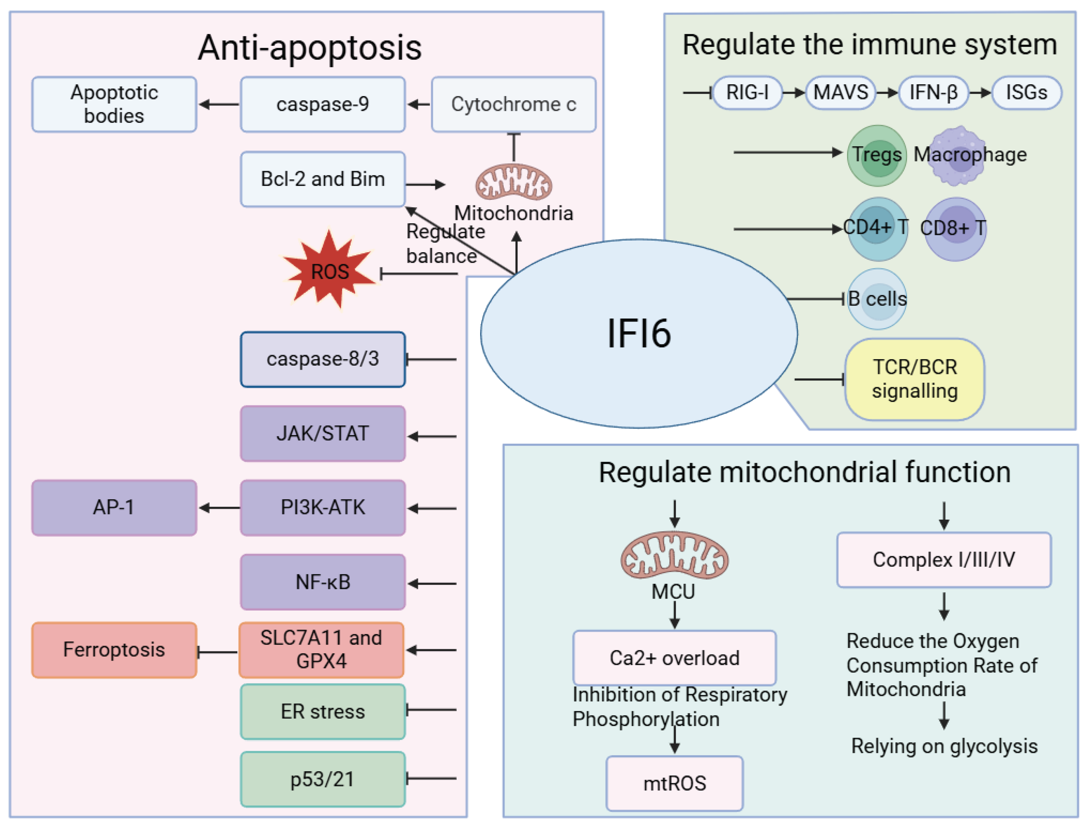

Table 1.
Clinical or relevant evidence of IFI6 associated with diseases.
| Disease | Clinical or Related Evidence |
| Neurological diseases | |
| AGS | IFI6 has potential as a diagnostic biomarker in AGS [22] |
| MDD | IFI6 expression levels are downregulated in the hippocampus of MDD patients [23] |
| Autoimmune diseases | |
| SLE | IFI6 expression levels are elevated in SLE patients and correlate with disease severity; it is a core component of the IFN score and a potential diagnostic biomarker [24,25,26] |
| LN | IFI6 may become a biomarker and new drug target for the diagnosis and treatment of LN [27] |
| RA | IFI6 is highly expressed in RA patients and correlates with the degree of synovial inflammation [24] |
| Multiple sclerosis | IFI6 is highly expressed in platelets of multiple sclerosis patients [3,28] |
| Psoriasis | IFI6 is a major IFN-inducible gene in psoriasis [29]. IFI6 is highly expressed in psoriasis, and its expression level is positively correlated with the degree of inflammation. It may participate in the pathogenesis of psoriasis by inhibiting spontaneous keratinocyte apoptosis [16,30] |
| PM/DM | IFI6 is one of the important hub genes of PM/DM [29]. Elevated IFI6 expression is associated with the progression of rapidly progressive interstitial lung disease in MDA5+ DM [29,31] |
| pSS | IFI6 is overexpressed in pSS, and its expression level is positively correlated with serum IgG levels, anti-SSA/SSB antibody titers, and disease activity in pSS patients [32] |
| SSc | IFI6 may participate in the progression of SSc vasculopathy [33] |
| APS | IFI6 expression is upregulated in APS patients and is associated with early disease onset and poor outcomes [34] |
| Type 1 diabetes | IFI6 is highly expressed and may be related to immune damage of pancreatic β cells [35] |
| IBD | IFI6 has been identified as a biomarker of IBD [36] |
| Cancers | |
| ESCC | IFI6 inhibits the tumor microenvironment of ESCC [37]. IFI6 is highly expressed in both ESCC patients and cultured ESCC cells [2,9,13] |
| Breast cancer | IFI6 is associated with the tumor microenvironment in breast cancer, enhances breast cancer cell invasion and migration, and is associated with drug resistance [12,38,39] |
| B-ALL | Overexpression of IFI6 in bone marrow mesenchymal stem cells may be a key factor promoting B-ALL cell proliferation [8] |
| Lung adenocarcinoma | High expression of IFI6 can maintain the tumor immune activity and enhance the survival and drug resistance of lung adenocarcinoma cells [40,41] |
| Gastric cancer | IFI6 is mainly enriched in the inner mitochondrial membrane of gastric cancer cells, inhibiting mitochondrial membrane depolarization and cytochrome c release to inhibit caspase-3 activity, thereby inhibiting cancer cell apoptosis [2,38] |
| TSCC | IFI6 expression in TSCC cells is higher than in normal tissues [42] |
| cSCC | IFI6 has been confirmed to be upregulated in cSCC [43] |
| Multiple myeloma | IFI6 is abnormally expressed in multiple myeloma and is associated with chemoresistance; it antagonizes TRAIL-induced apoptosis by preserving mitochondrial integrity [8,38] |
| Pancreatic cancer | Upregulation of IFI6 expression can inhibit pancreatic cancer cell apoptosis and promote cell proliferation [17,44] |
| Colorectal cancer | IFI6 is highly expressed in colorectal cancer cells and is associated with poor prognosis [14,39] |
| Melanoma | IFI6 is overexpressed in patient melanoma samples [45] |
| NRAS mutant melanoma | Knockdown of IFI6 inhibits cell transformation and melanoma growth [5] |
| Glioma | IFI6 overexpression inhibits ferroptosis in glioma cells, increasing drug resistance and survival rate [12] |
| Glioblastoma | IFI6 shows highly significant and differential expression in glioblastoma cells [46] |
| Prostate cancer | IFI6 is associated with immune infiltration and drug resistance in prostate cancer [12,47] |
| Ovarian cancer | IFI6 is positively correlated with the NF-κB pathway, affecting ovarian cancer cell proliferation, invasiveness, and drug resistance [6] |
| RCC | IFI6 overexpression restores axitinib resistance in long non-coding RNA STX17-DT-knockdown RCC cells [13] |
| Liver cancer | IFI6 is associated with drug resistance and prognosis in liver cancer cells [2] |
| DT | Upregulation of IFI6 may contribute to DT cell survival and invasion and is associated with the prognosis of stratified DT [48] |
| Viral infections | |
| YFV | High IFI6 expression inhibits YFV-induced ER invagination, thereby inhibiting YFV [49,50] |
| WNV | IFI6 has an inhibitory effect on WNV [4] |
| DENV | IFI6 inhibits DENV-induced VEC apoptosis through a caspase-dependent pathway [10,51] |
| ZIKV | IFI6 overexpression may inhibit ZIKV infection-related cell death [52] |
| HBV | High IFI6 expression can inhibit HBV replication and gene expression [2,53] |
| HCV | IFI6 can inhibit HCV entry and replication [49,50] |
| SARS-CoV-2 | In hospitalized patients, IFI6 expression levels are higher in moderate cases than in severe cases, and IFI6 expression is associated with lower mortality risk and IMV [49,54] |
| IAV | IFI6 has been identified as one of the core transcriptomic response mRNAs to IAV infection [55] |
| ARV | IFI6 overexpression inhibits ARV replication [2] |
| EBOV | IFI6 has an inhibitory effect on EBOV transcription and replication [56] |
| HIV-1 | IFI6 can inhibit HIV-1-based vector infection [57] |
| HBoV1 | IFI6 inhibition significantly reduces HBoV1 replication [58] |
| Secondary syphilis | IFI6 is upregulated in peripheral blood mononuclear cells of patients with secondary syphilis and can serve as a diagnostic criterion [59] |
| Other diseases | |
| Obese asthma | Differential expression of IFI6 is associated with obese asthma [47] |
| Moyamoya disease |
IFI6 is one of the potential key genes for the heterogeneity of peripheral blood cells in Moyamoya disease [60] |
| KFD | IFI6 has been identified as a key seed gene for KFD [61] |
| GD | IFI6 is significantly highly expressed in GD [62] |
| AS | IFI6 can aggravate AS through the type I IFN signaling pathway [33] |
| HTN | IFI6 is upregulated in the endothelial cells of HTN [63] |
AGS: aicardi goutières syndrome, MDD: major depressive disorder, SLE: systemic lupus erythematosus, LN: lupus nephritis, RA: rheumatoid arthritis, PM/DM: polymyositis/dermatomyositis, MDA5: Melanoma differentiation-associated gene 5, pSS: primary sjögren’s syndrome, SSc: systemic sclerosis, APS: antiphospholipid syndrome, ESCC: esophageal squamous cell carcinoma, B-ALL: adult B-cell acute lymphoblastic leukemia, TSCC: tongue squamous cell carcinoma, cSCC: cutaneous squamous cell carcinoma, NRAS: neuroblastoma rat sarcoma, RCC: renal cell carcinoma, STX17-DT: STX17 divergent transcript, DT: Desmoid tumor, YFV: yellow fever virus, ER: endoplasmic reticulum, WNV: west nile virus, DENV: dengue virus, VECs: vascular endothelial cell, ZIKV: zika virus, HBV: hepatitis B virus, HCV: hepatitis C virus, IMV: invasive mechanical ventilation, IAV: influenza A virus, ARV: avian reovirus, EBOV: zaire ebola virus, HIV: human immunodeficiency virus, HBoV1: human bocavirus 1, KFD: kikuchi-fujimoto disease, IBD: inflammatory bowel disease, GD: gaucher disease, AS: atherosclerosis, HTN: hypertensive nephropathy.
Disclaimer/Publisher’s Note: The statements, opinions and data contained in all publications are solely those of the individual author(s) and contributor(s) and not of MDPI and/or the editor(s). MDPI and/or the editor(s) disclaim responsibility for any injury to people or property resulting from any ideas, methods, instructions or products referred to in the content. |
© 2026 by the authors. Licensee MDPI, Basel, Switzerland. This article is an open access article distributed under the terms and conditions of the Creative Commons Attribution (CC BY) license.
Copyright: This open access article is published under a Creative Commons CC BY 4.0 license, which permit the free download, distribution, and reuse, provided that the author and preprint are cited in any reuse.